Review

Current Sample Preparation Methodologies for Determination of Catecholamines and Their Metabolites

1
Physics Diagnostic Division, The First Affiliated Hospital of Zhengzhou University, Zhengzhou 450052, China
2
Key Laboratory of Targeting Therapy and Diagnosis for Critical Diseases of Henan Province, School of Pharmaceutical Sciences, Zhengzhou University, Zhengzhou 450001, China
3
College of Public Health, Zhengzhou University, Zhengzhou 450001, China
4
HEJ Research Institute of Chemistry, International Center for Chemical and Biological Sciences, University of Karachi, Karachi 75270, Pakistan
*
Authors to whom correspondence should be addressed.
Molecules 2022, 27(9), 2702; https://doi.org/10.3390/molecules27092702
Submission received: 6 March 2022 / Revised: 17 April 2022 / Accepted: 19 April 2022 / Published: 22 April 2022
(This article belongs to the Special Issue Recent Advances and Future Trends in Sample Preparation)

Abstract

:
Catecholamines (CAs) and their metabolites play significant roles in many physiological processes. Changes in CAs concentration in vivo can serve as potential indicators for the diagnosis of several diseases such as pheochromocytoma and paraganglioma. Thus, the accurate quantification of CAs and their metabolites in biological samples is quite important and has attracted great research interest. However, due to their extremely low concentrations and numerous co-existing biological interferences, direct analysis of these endogenous compounds often suffers from severe difficulties. Employing suitable sample preparation techniques before instrument detection to enrich the target analytes and remove the interferences is a practicable and straightforward approach. To date, many sample preparation techniques such as solid-phase extraction (SPE), and liquid-liquid extraction (LLE) have been utilized to extract CAs and their metabolites from various biological samples. More recently, several modern techniques such as solid-phase microextraction (SPME), liquid–liquid microextraction (LLME), dispersive solid-phase extraction (DSPE), and chemical derivatizations have also been used with certain advanced features of automation and miniaturization. There are no review articles with the emphasis on sample preparations for the determination of catecholamine neurotransmitters in biological samples. Thus, this review aims to summarize recent progress and advances from 2015 to 2021, with emphasis on the sample preparation techniques combined with separation-based detection methods such capillary electrophoresis (CE) or liquid chromatography (LC) with various detectors. The current review manuscript would be helpful for the researchers with their research interests in diagnostic analysis and biological systems to choose suitable sample pretreatment and detection methods.

1. Introduction

Catecholamines (CAs), a class of neurotransmitters including dopamine (DA), norepinephrine (NE), and epinephrine (E), are circulating hormones synthesized by the phenylalanine-tyrosine metabolic pathway [1,2]. The levels of CAs along with their metabolites have significant correlations with the occurrence and development of many types of diseases, such as obesity [3], depression [4], Alzheimer’s disease [5], Parkinson’s disease [6,7], and phaeochromocytoma [8]. Different metabolites are associated with these catecholamines. 3,4-Dihydroxyphenylacetaldehyde (DOPAL) and catecholaldehyde, which is detoxified by aldehyde dehydrogenase (ALDH), are associated with dopamine. Similarly, metanephrines include normetanephrine and metanephrine, homovanillic acid (3-methoxy-4-hydroxyphenylacetic acid) and vanillylmandelic acid (3-methoxy-4-hydroxymandelic acid) are some other examples of metabolites produced from epinephrine, norepinephrine, and dopamine [9]. Therefore, the accurate quantification of CAs and their metabolites in biological samples is of great significance, especially in early clinical diagnosis, in-depth understanding of pathology, and contribution to developing better treatments.
Up to date, various detection techniques including sensing methods [10,11,12], spectroscopic methods [13,14], and chromatographic methods [15,16,17,18] have been utilized for the detection of endogenous CAs and their metabolites. Among these methods, separation-based detection techniques such as liquid chromatography (LC) and capillary electrophoresis (CE) have become the primary and most widely used techniques due to their unique characteristics of separating CAs from numerous co-existing interferences, exhibiting excellent specificity, precision, and reproducibility. LC or CE coupled with various detectors including ultraviolet (UV) [8], fluorescence (FLD) [19], electrochemical detection (ECD) [20,21], and mass spectrometry (MS) [22,23] all have been used for detecting CAs and their metabolites (Figure 1).
Although separation-based detection techniques such as LC or CE can separate target analytes from biological interferences based on the property differences to some extent, some practical problems still exist in the direct analysis of complex biological samples. Due to the extremely low concentrations of endogenous CAs, the sensitivity of direct detection is usually insufficient to meet the requirement of measurement [24]. The complex biological matrix may also result in the co-elution of interferences together with the target analytes, thus decreasing the detection specificity and precision. Besides, the sample morphology may not be compatible with the used detection techniques. Therefore, suitable sample preparations before instrument detection are always needed.
For the present, various sample preparation techniques have been developed. Although many determination methods for catecholamine neurotransmitters have been concluded in some reviews in recent years [11,13,14,25,26], a comprehensive summary focused with the emphasis on sample preparations for the determination of catecholamine neurotransmitters in biological samples is missing. Thus, here we present a comprehensive overview of the progress and advances of sample preparation methods for pretreatment of samples for CAs detection from 2015 to 2021, and detailed analytical information on these successful methods is also provided.

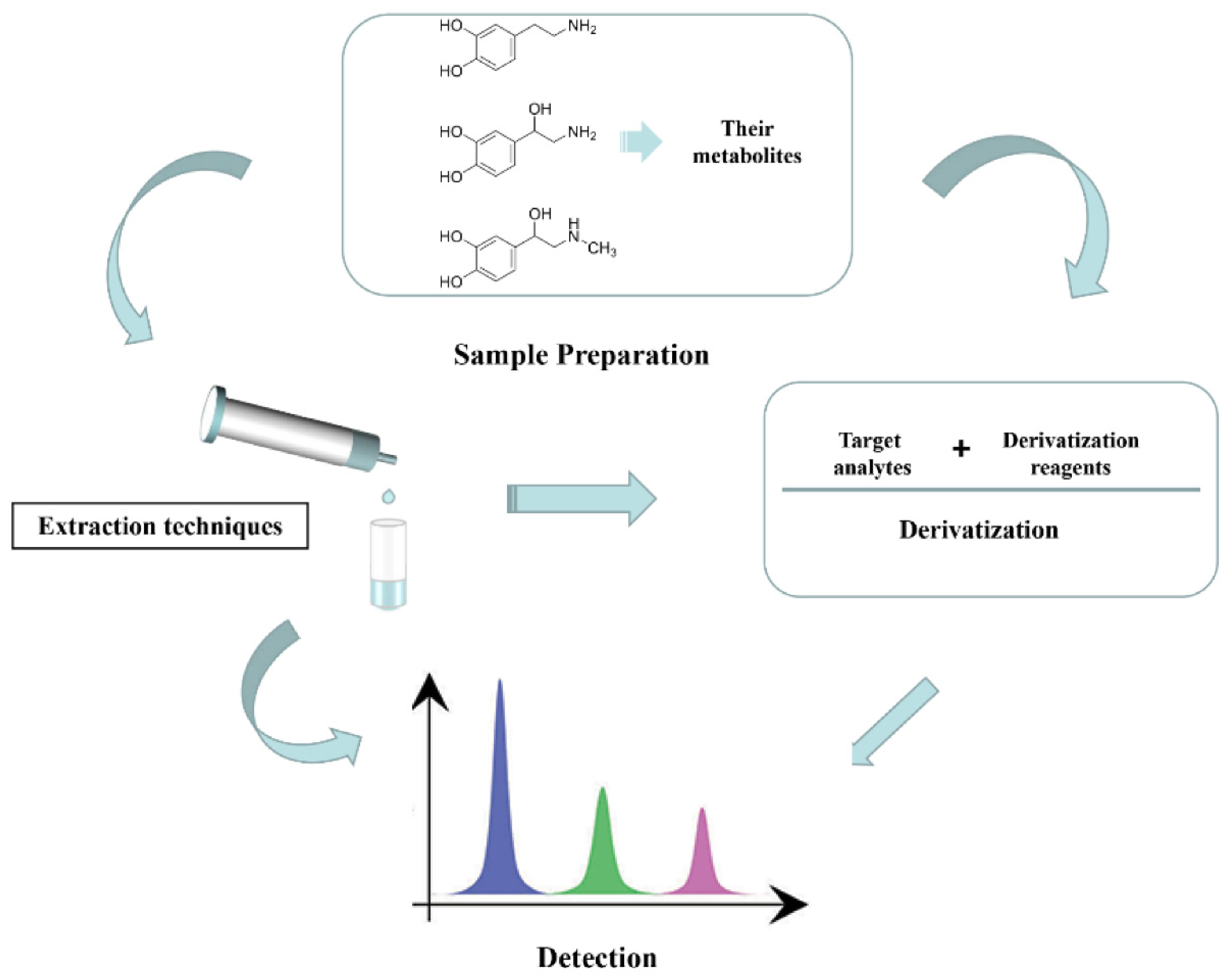
2. Sample Preparation

Normally, conventional liquid–liquid extraction (LLE), solid-phase extraction (SPE) technique, and chemical derivatization are regularly employed in bio-sample preparation. To decrease the sample volume required, the cost and time consumption, the trend in current sample preparation is moving toward environmental friendliness, miniaturization, simplicity, and automation. Hence, many novel sample preparation techniques have been developed, including solid-phase microextraction (SPME), liquid–liquid microextraction (LLME), dispersive solid-phase extraction (DSPE), and chemical derivatizations based on newly synthetic derivatization reagents. A brief description of these sample preparation techniques is given in the following sections, with relevant applications in which they are used for the detection of CAs and their metabolites in different matrices.

2.1. Liquid–Liquid Extraction-Based Approaches

Liquid–liquid extraction (LLE) is one of the oldest sample preparation methods, and it is based on solubility differences to implement a transfer of target analytes from the aqueous sample into another immiscible liquid phase [27]. The pH may also affect the distribution of the solute in both phases because the CAs containing phenol hydroxyl and amino are amphoteric substances. So solvent polarity and pH of the aqueous phases are the major factors to be considered for the analysis of CAs and their metabolites. Although the use of LLE alone provides a good purification effect as a non-specific method, the application of LLE has become limited due to the complexity of the CAs and their metabolites. Other sample preparation procedures are often combined with LLE in the detection of CAs and their metabolites. A few newly developed methods applying the LLE technique in combination with derivatization have been published on the determination of CAs and their metabolites [28]. Zhao et al. [29] designed and synthesized derivatization reagent 4′-carbonyl chloride rosaline (CCR) for simultaneous labeling of amino and hydroxyl groups in CAs and their metabolites. LLE was employed to extract the CCR-derivatives from the aqueous samples into the 4-bromoanisole, which could easily remove complex biological matrices and excess derivatization reagent.
Nevertheless, conventional LLE exhibits some shortcomings, such as the use of large sample volumes and toxic organic solvents (ethyl acetate, dichloromethane, chloroform, and carbon tetrachloride); these drawbacks make LLE time-consuming, laborious, and environmentally unfriendly. To overcome the disadvantage of LLE, liquid-phase microextraction (LPME), a new extract technique based on LLE is developed in 1996 [30]. LPME possesses fairly obvious advantages compared with liquid–liquid extraction like miniaturization, simplicity, rapidity, and environmental friendliness. So far, LPME has evolved into various forms such as dispersive liquid–liquid microextraction (DLLME) [29,31], single-drop microextraction (SDME) [32], ultrasound-assisted liquid–liquid microextraction (UA-LLME), hollow fiber liquid-phase microextraction (HF-LPME) [33], and headspace liquid-phase microextraction (HS-LPME) [34]. Recently, these various types of LPME methods have been successfully used for the separation and preconcentration of CAs and their metabolites in biological samples. Konieczna et al. [35] combined dispersive liquid–liquid microextraction (DLLME) with liquid chromatography coupled to mass spectrometry (LC-MS) to develop a simple and sensitive method for the determination of neurotransmitters in human urine samples. A mixture of ethanol (dispersion solvent) and dichloromethane (extraction solvent) was quickly injected into a human urine sample to form a turbid solution in an Eppendorf tube. After centrifugation, a simple clean-up process was realized and this method also exhibits good linearity and sensitivity. For aqueous biological samples, nonpolar or less polar water-immiscible organic solvents organic solvent is usually used as extraction solvent. It is difficult to extract CAs and their metabolites from the aqueous solution due to the low distribution coefficient of hydrophilic analytes in the organic solvent. Therefore, some novel strategies have been proposed to solve this predicament. Jiang et al. [36] attempted to add various carriers into the organic phase for improving the extraction efficiency of analytes, and analytes were effectively purified using negligible volumes of extracting solvent followed by centrifuging and collecting the precipitated phase by solidifying the aqueous stripping phase.

2.2. Solid-Phase Extraction (SPE)

Solid-phase extraction (SPE) is the most commonly employed method for the separation and concentration of target analytes. The principle of SPE is through retaining compounds of interest on solid-phase sorbents, and the purpose of purification of analytes is subsequently carried out by selective elution. In comparison to LLE, there are several distinct advantages, such as higher quantitative recovery, use of less organic solvents, greater flexibility due to the use of different sorbents, and greater possibility of automation [37]. Applications of SPE techniques for preconcentration of CAs and their metabolites in biological samples are summarized in Table 1.
Among a variety of sorbents, weak cation exchange (WCX)-based sorbents are preferred due to their improved extraction efficiency and excellent repeatability for the extraction of CAs and their metabolites [38,40]. In addition, reversed-phase hydrophobic- hydrophilic balanced (HLB) based sorbents have been also widely used including Strata-X-CW, Bond-Elut Plexa [39,43,44,46]. These sorbents based on WCX and HLB are all in mixed-mode SPE for the isolation of CAs and their metabolites, which has good retention for polar and non-polar compounds in water and can stay stable in wide pH ranges. Therefore, these developed methods using sorbents based on WCX and HLB, and easily obtained satisfactory linearity and recovery. Along with the increasing requirement for CAs detection, the trend in recent years is miniaturization, simplification, and automation. In SPE, many unusual changes also take place. Various novel product forms are created, including well-plates [41], and discs that differed from conventional column cartridges. Li et al. used a 96-well HLB microplate to accomplish a simple extraction of monoamine neurotransmitters from complex urine [47]. The extraction process has the significant characteristic of miniaturization and high throughput. The sorbent is the key factor affecting the SPE performance, some highly selective sorbents are designed and synthesized for preconcentration of CAs and their metabolites. Hou et al. [8] utilized the affinity between cis-diol-containing CAs and boric acid to prepare boronate-modified hollow dummy template imprinted polymers (B-hDIPs) for isolating CAs from urine samples, and extremely high extraction efficiency is obtained due to covalent bonding. The extraction of CAs in human urine was optimized using electrospun composite fibers functionalized with 4-carboxybenzo-18-crown-6 ether modified XAD resin and polystyrene by Chen et al. [45], and they packed this composite fiber into 96-well columns and achieved a miniaturization analysis of CAs. Moreover, several attempts have also been carried out about automated analysis of CAs and their metabolites [42]. This SPE mode has the advantage of using less sample and solvents volume, avoiding intensive labor, and is also the development trend of sample analysis in the future.
Metal-organic frameworks (MOFs) are a fascinating class of organic–inorganic hybrids with unique properties including high surface area, regular three-dimensional structure, high porosity, and tunable surface chemistry. MOFs are being used as solid-phase extraction sorbents for the preconcentration and enrichment of biomolecules from the different complex samples [51,52]. Xing et al. [48] used polytannic acid (PTA) modified borate-functionalized metal-organic framework for the extraction and preconcentration of CAs and their metabolites from human urine samples. MOFs were prepared by metal-ligand-fragment co-assembly strategy and further modified with magnetic iron oxide nanoparticles to form Fe3O4@PTA@MIL-100(Fe)-B and applied as magnetic solid-phase extraction (MSPE) sorbents (Figure 2). For screening, the variable for extraction efficiency of the modified MOFs was investigated by Plackett–Burman design. Optimal extraction conditions were selected by Box–Behnken design. HPLC coupled with fluorescence detection was used for the analysis of catecholamines. Although the proposed method showed promising efficiency, the construction of such a highly ordered network is challenging. The stability of MOFs in harsh experimental conditions also hinders their practical applications.
In a similar approach, amino-functionalized polyhedral oligomeric silsesquioxanes (POSS-8NH2) were constructed by Wei et al. and further attached to polydopamine-coated magnetized graphene oxide via covalent modification [49]. The prepared sorbent (magGO@POSS-BA) contained cubic boronic acid and showed significantly higher selectivity for CAs due to the presence of cis-diol selective boronate affinity. The cage-like structure of POSS enhanced the surface area for better adsorption and acted as a building block for the composite formation. Although the material showed an extraordinarily high capacity for CAs in urine samples, the cost of synthesis is very high. Moreover, the adsorbent showed better selectivity for ortho-phenols.
A packed fiber solid-phase extraction approach is also reported recently for the detection of CAs. Electrospun poly crown ether composite was applied to selectively isolate CAs from urine samples with high reproducibility and precision [50]. The proposed online PFSPE-HPLC approach minimized the sample loss, decreased the operational errors, and improved the sensitivity of the analysis. The proposed approach is simple and rapid, one limitation is the selectivity in complex biological samples.

2.3. Dispersive Solid Phase Extraction/Microextraction (DSPE/DSPME)

Dispersive solid-phase extraction (DSPE) is a rapidly developing extraction technique since it was introduced in 2003 by Anastassiades and coworkers [53]. It isolates analytes from the liquid matrix by using a solid sorbent, which is directly dispersed into the sample solution. The adsorption process can be completed quickly by assisting by means of vortex, ultrasounds, etc. Then, the sorbent particles adsorbed analytes are separated by centrifugation, filtration, or magnetic attraction [54]. Finally, it needs to choose a suitable elution solution and elute the analytes. The elution solution can be directly injected into the detection instrument, or evaporated and reconstituted into a minor volume to achieve preconcentration and purification of analytes. DSPE as a highly developed form of SPE has attracted a lot of interest from analysts for the clean-up of CAs and their metabolites because of its unique superiority compared to SPE, such as being rapid, cheap, effective, and convenient [55]. Applications of DPSE techniques for preconcentration of CAs and their metabolites in different biological samples are summarized in Table 2.
Magnetic nanoparticles with surface modification are a promising adsorbent material for the separation of CAs and their metabolites [19,57]. Its nanoscale size can provide a relatively large specific surface area to obtain more active sites. Furthermore, there have also been studies using hollow-structured nanoparticles to make adsorbents larger [60]. Its magnetism makes it easy to separate it from the sample solution with the assistance of magnets. Meanwhile, the researchers also attempt to make the adsorbents possess more excellent selectivity by modifying various groups on the surface of magnetic nanoparticles [56,58]. For these above reasons, the DSPE procedure of the CAs detection has been greatly upgraded. In addition to this, the application of carbon nanotube for the detection of CAs and their metabolites has been reported by Ma et al. and Murtada et al. [55,59].
Biogenic monoamines are a class of catecholamines commonly used for the diagnosis of catecholamine-producing tumors. So, it is clinically significant to design novel methods for the detection of biogenic monoamines from biological samples. For this purpose, Pawliszyn et al. [61] proposed a strategy based on two different pretreatment methods: packed fibers solid phase extraction (PFSPE) and thin-film solid-phase microextraction (TF-SPME). The main goal of the study was to accurately quantify the metabolites of biogenic monoamines (epinephrine, norepinephrine, 3-methoxytyramine, dopamine, serotonin, normetanephrine, histamine, and metanephrine) in urine samples. Extraction was carried out via the thin-film blade format SPME method using a hydrophilic–lipophilic balance (HLB) coating. For PFSPE polycrown ether (PCE) composite nanofiber was used for comparison, and an extraction recovery of 35.7–74.8% was obtained under optimal conditions. TF-SPME method showed better performance than the PFSPE method, for the simultaneous determination of eight metabolites (Figure 3).
Molecularly imprinted polymers (MIPs) are used as a dispersive solid-phase extraction sorbent before UHPLC/MS/MS for simultaneous quantitative analysis of CAs and their metabolites. MIPs are highly selective for specific molecules, so they are commonly used in biological analysis. Podjava et al. [62] proposed a combined non-covalent and semi-covalent imprinting approach using different types of polymers and crosslinkers. The imprinted polymers showed good imprinting factors ranging from 3.1 to 5.6, indicating high selectivity of the analysis. In addition, the proposed method efficiently distinguished between methylated CAs and non-methylated CAs. Although MIPs-based extraction strategies are highly efficient and selective for particular types of analytes, the availability of monomers and cross-linkers for imprinting is a huge challenge. In addition, the cost of imprinting polymers is usually very high, limiting their practical applications.
In a recent approach, borated zirconia is used for the extraction of plasma catecholamines, followed by LC-MS/MS detection. Borated zirconia has an inherent affinity toward cis-diol compounds and it forms a five or six-membered reversible cyclic ester in basic conditions, capturing the cis-diol compounds and then releasing them in acidic conditions. The working principle of this method is simple and well-known and the sorbent efficiently captured dopamine (DA), epinephrine (E), and norepinephrine (NE) from the plasma samples of healthy volunteers [24].

2.4. Solid-Phase Microextraction (SPME)

SPME was first proposed in 1989 as an alternative to conventional extraction techniques and has been progressively favored by analysts in recent years [63]. It involves equilibrium between analytes and the fiber coating and followed by desorption using the suitable elution solution. The SPME procedure has a distinct features compared to other extraction techniques, including the simplicity of operation, use of small sample volumes, solvent-free extraction, and easily achieved automation. The main arrangements of SPME can be performed into fiber SPME and in-tube SPME. With the development of microextraction technology, other new forms of SPME have also been proposed: thin-film, in-tip, and in-needle microextraction, rotating disk sorptive extraction (RDSE), and capillary microextraction. SPME can overcome the defects of traditional solid-phase extraction techniques and exhibit high performance, and it has been successfully introduced in many fields of food, medicine and hygiene, biochemistry, toxicology, and forensic medicine.
For analysis of CAs and their metabolites, methods using coated fiber have been described, and many researchers put a significant effort into the development and optimization of these fibrous materials over the years. Recently, the fiber SPME has been innovated by Monteleone et al. [64]. The performance of five SPME fibers was evaluated, and the optimal one is finally determined to be the polyacrylate fiber. The fiber coated with the suitable organic polymer can extract and enrich derivatives of CAs and their metabolites from complex urine matrices by immersing them into the sample vial. Using such an easy procedure, an accurate assay of analytes can be completed. However, fiber SPME exhibits some restrictions, especially in bio-analytical laboratories, involving fragile properties or its life reduced at high salt concentrations. Hence, in-tube SPME gets more preference by degrees and become a universal extraction technique, since the open tubular fused silica capillary used for in-tube SPME can tune the column length and the thickness of extractant coating, and the analytes can be eluted by appropriate desorption solution and directly injected into CE or HPLC system after repeated extraction through an internally coated capillary. The in-tube SPME methods employing long-chain ionic liquids based on imidazole [65] and phenylboronic acid [66,67] for adsorbing CAs and their metabolites have been described. Initial in-tube SPME involves mainly offline applications [68,69], but some attempts for detection of CAs and their metabolites have been also taken to accomplish the convenient automation of the extraction process with online applications increasingly possible. Automated in-tube SPME not only reduces human errors caused by manual manipulation (Figure 4) but also provides significant gains in sensitivity and accuracy compared to manual off-line techniques [70,71]. Meanwhile, solvent-free operation and high throughput are also distinctive traits of automated in-tube SPME. However, these methods involved the application of automated in-tube SPME to date and have been combined with laboratory-made devices only, which makes the popularization of automated in-tube SPME limited in routine bio-analysis.
Microextraction by packed sorbent (MEPS) is a miniaturized form of solid-phase microextraction and it is commonly used these days for the extraction of a variety of compounds including drugs and metabolites. MEPS involves directly integrated packing into the syringe, and sample introduction does not require a separate robot. Another advantage of MEPS is that more than 100 plasma or urine samples could be analyzed by a single MEPS bed [72]. Xin et al. [73] designed a simple, rapid, and cost-effective method for the quantitative determination of CAs and their metabolites from urine samples. The designed method was semi-automated microextraction involving a digitally controlled syringe, before LC-MS/MS analysis. Different types of extraction sorbents including a polar enhanced polymer (PEP), cation-exchange (CX) and C18, and porous graphitized carbon (PGC) were tested. The whole analysis was completed within 4 min. The precision of the analysis was ≤12.8%, recoveries were 89–109.5%, and linear ranges were 0.167–686 ng/mL. The designed method showed comparable efficiency for PPGLs with commercial biochemical tests.
MXenes are a recent fascination in material science research, exhibiting some exciting properties. Magnetic boronic acid-modified Ti3C2Tx composites are reported for the extraction of CAs with high selectivity and specificity, as shown in Figure 5 [74]. Due to the unique 2D layered structures of the sorbent, 319.6 μmol/g of dopamine is absorbed within 2 min, which facilitated the molecular transport and shortened the diffusion path. Fast adsorption is also attributed to the van der Waals forces, π–π stacking, hydrogen bonding, multilayer adsorption, and the synergetic interactions of borate affinity. The method showed superior performance to several other materials and was applied for the detection of CAs quantitative detection from urine samples of Alzheimer’s disease patients.

2.5. Derivatization

Derivatization is an indispensable part of sample preparation in many cases, and it has been widely applied to bio-analysis [75]. In the process of trace analysis, some analytes cannot or are difficult to separate and detect directly. For example, the components to be measured have no or weak absorption in the UV-visible region; derivatization needs to be adopted at this moment. Derivatization can quantitatively transform the target compound into another compound that is easy to be analyzed and detected by using a derivatization reagent having a special functional group, and it can allow an obvious gain of the sensitivity of detection and separations of analytes, especially in complex biological samples. Moreover, derivatization can also be used to protect compounds that are unstable during the analysis process.
Catecholamine (CAs) and their metabolites are usually easy to be oxidized due to cis-diol structure and present at a low concentration level in bio-sample; sample pretreatment often applies the derivatization of the CAs and their metabolites to solve this predicament. Derivatization reagents employing acylation were frequently used in different methods. The derivatives of CAs and their metabolites possessed better chemical stability and were easier to be identified by a detector. Dansyl chloride (Dns-Cl) as a classic derivatization reagent, has been recently reported for the determination of neurotransmitters and their metabolites in plasma samples by Zhao et al. [76]. This kind of derivatization reagent has the advantages of simply derivatization operation, good derivative stability, and quantifiable reaction process; other similar reagents have also been developed like benzoyl chloride [23], 10-methyl-acridone-2-sulfonyl chloride [77], etc. However, these derivatization reagents have more or fewer limitations in their applications, some commercial derivatization reagents were also innovatively developed and applied for the derivatization of CAs and their metabolites. Ellis et al. reported a demethylation labeling method for urinary CAs and their metabolites [78]. Recently, Azaryan et al. [79] proposed a responsible approach for the determination of catecholamine in human urine using 9-fluorenyl-methoxycarbonyl chloride (FMOC-Cl). The application of FMOC-Cl can allow a complete quantitative reaction in a short time at room temperature, and this proposed procedure was successfully validated on real samples. Lissamine rhodamine B sulfonyl chloride (LRSC) was employed for the first time as a derivatization reagent for multiple NTs in LID rat brain microdialysates by Wei et al. [80]. In addition, the analyst has also designed and synthesized several derivatization reagents for CAs such as 1,3,5,7-tetramethyl-8-(N-hydroxysuccinimidyl butyricester)-difluoroboradiaza-s-indacene (TMBB-Su) [81], 4-carbonyl chloride rosamine (CCR) [29], 10-ethyl-acridone-3-sulfonyl chloride (EASC) [82], and all of them exert an excellent effect.
Our group has recently reported a novel strategy based on a combination of extraction and derivatization, coupled with LC-MS/MS analysis [83]. Zirconium oxide (ZrO2) nanoparticles were used for the extraction of cis-diol compounds in neutral pH conditions, and phenyl isothiocyanate (PITC) was employed as the derivatizing agent. The integration of extraction and derivatization techniques minimized the sample loss, increases the detection accuracy, and simplified the analysis. Finally, the proposed strategy was applied for the detection of CAs in healthy people and pheochromocytoma patients. Applications of derivatization for improving the sensitivity of CAs and their metabolites in biological samples are summarized in Table 3.

3. Analytical Techniques

The measurements of CAs and their metabolites have long been an area of interest for researchers because of their important role in physiology and pathology. For decades, various detection methods have been proposed for determining the levels of CAs and their metabolites, and these analysis techniques mainly consist of high-performance liquid chromatography (HPLC) and capillary electrophoresis (CE), which are often coupled with high-performance detectors such as ultraviolet (UV) detection, fluorescence detection (FLD), laser-induced fluorescence detector (LIFD) [71], and mass spectrometry (MS). Initially, the UV detector was often introduced for the determination of CAs and their metabolites because it exhibits good stability and linearity of analysis under varying separation conditions (Figure 6A) [8,60,81,85,86]. However, by an overview of the methods for the measurements of CAs and their metabolites from 2015 to 2021 concerning CE and HPLC methods, fluorescence detection (FLD) [87] and mass spectrometry (MS) [38,88,89,90] have been more popular in comparison to UV detector. FLD is suitable for analysis of low abundance catecholamines since it has higher sensitivity and better selectivity than UV detector (Figure 6B), but due to the low fluorescence intensity of CAs and their metabolites, it needs to be introduced with the groups emitting strong fluorescence before using FLD. Later, the MS detector has risen to the forefront with the emergence of MS, increasingly taking center stage as the detection tool of choice for CAs and their metabolites. MS detection provides a lower limit of detection (LOD) and better specificity due to the identification of analytes by both retention times and molecular masses, and it can eliminate matrix interference and obtain significant gain for the determination of CAs and their metabolites, especially in complex samples. As an example, Kovac et al., [23] proposed LC-MS/MS method to identify and quantify CAs and their metabolites in cerebrospinal fluid (CSF) from the rat model for human tauopathy. Using this approach, they accurately detected changes in the concentration of CAs and their metabolites in CSF depending on the high-mass accuracy provided by the MS detector (Figure 6C). Zheng et al. [77] reported a highly sensitive strategy to detect monoamine neurotransmitters (MANTs) and their precursors and metabolites at trace levels in rat brain microdialysates. The LOQs provided by the MS detector are much lower than those provided by UV, and the combined use of various sample preparation techniques and the MS detector improves the sensitivity that can be achieved allowing the determination of CAs and their metabolites at concentrations down to 0.01 ng/mL [57].
Catecholamines and 3-methoxytyramine (3-MT) are clinically significant biomolecules and are useful for the diagnosis of phaeochromocytomas and paragangliomas (PPGLs) CAs and 3-MT are highly polar and unstable, so their determination is often limited by conventional methods. In addition, these molecules require a longer time for elution in LC-MS analysis, restricting their clinical applications [91]. Therefore, the isotope labeling strategy is very useful for detecting such compounds at the clinical level. Meiling et al. [92] developed an interesting strategy for the detection of CAs and their metabolites from plasma samples. Waters Oasis WCX μElution solid-phase extraction (SPE) was used for the extraction of metabolites, before UPLC MS/MS analysis. The whole analysis was completed in 3 min and 7 PPGLs and 408 non-PPGLs patient plasma samples were tested using the designed method. The proposed method showed high recoveries for targeted analytes ranging from 95–115% with a carryover effect of less than 10%. In addition, the ranges were 0.10–30.00 nmol/L for NE and DA, 1.00–300.00 pg/mL for 3-MT, and 05–30.00 nmol/L for E, MN, and NMN.
Chemiluminescence is another versatile technique, used for the detection of a variety of analytes from biological samples. Li et al. [93] proposed a novel, sensitive, and reliable method based on Ag (III) complex chemiluminescence detection for the analysis of monoamine neurotransmitters and metabolites including 5-hydroxypentylacetic acid (5-HIAA), levodopa (l-DOPA), epinephrine (E), 3-methoxy-4-hydroxyphenylglycol (MHPG), dopamine (DA), 3,4-dihydroxyphenylacetic acid (DOPAC), and serotonin (5-HT). From free-radical capture experiments and CL spectra, the inhibition mechanisms for luminol-[Ag(HIO6)2]5−-analytes CL system was investigated. Apart from several advantages for the detection of CAs and their metabolites, this method has considerably higher LODs, so trace level detection of these compounds is practically difficult. Moreover, the selectivity of the method is limited and chemiluminescence technology does not apply to all such compounds.
The in-sample ion-pairing chromatography (IPC) LC-MS/MS method is also very effective in the detection of catecholamines and their metabolites from plasma samples. In such methods, special care is needed to divert the ion-pairing reagents to waste, without entering the mass spectrometer. Marianne et al. [94] used the ion-pairing technique to quantitatively detect epinephrine (E) and norepinephrine (NE) from plasma samples with LOQ values of 0.20 and 0.02 nmol/L, respectively. Ion-pairing reagents can contaminate the ion source of the mass spectrometer, so it is mandatory to remove those before reaching the MS. In addition, ion pairs do not apply to CAs, limiting their applications in this field.
Extraction methods, coupled with electrophoretic detection, are also employed for the detection of CAa and their metabolites from biological fluids. Polikarpova et al. [95] reported a new approach based on a nano-sized polystyrene-based cation exchanger (NSCE), coated inside the capillary for the extraction and preconcentration of CAs and amino acids. The presence of surface sulfo groups efficiently separated and enriched the target compounds and their electrophoretic mobility was optimized by varying the pH. The coupling of NSCE capillaries and field-amplified sample stacking improved the detection sensitivity of the method. Designing such systems is complicated and time-consuming, and the sensitivity of the electrophoretic method is also low, compared with mass spectrometric methods.
In recent research, CAs are also detected by a spectrophotometric method employing label-free silver triangular nanoplates [96]. The mechanism of detection is elaborated in Figure 7. The proposed method is simple and novel, based on the change in morphology of nanoplates, as a result, its shift to the short-wavelength region or change in local surface plasmon resonance (LSPR) band intensity is monitored. The method is rapid, selective, and sensitive to catecholamines with different functional groups.

4. Conclusions and Perspective

CAs and their metabolites play crucial functions in the regulation of physiological activities and the diagnosis of diseases. The current review presents an overview of recently reported sample preparation methods involved in separation-based detection techniques like LC and CE for the determination of CAs and their metabolites in biological samples. Among the reported sample preparation techniques, liquid-phase extraction has been utilized due to its simple operation and speed; however, this technique has obvious limits for the determination of CAs and their metabolites in an aqueous matrix. Luckily, SPE as an alternative has been extensively used due to its attractive advantages (compared to LLE), including the low-consuming of toxic solvents, stable recovery, and higher enrichment factors. Cation exchange, alumina, and phenylboronic acid are commonly developed for complexing CAs and their metabolites. Up until now, the traditional SPE has gradually developed into SPME, thereby, achieving higher sensitivity by using only a very small sample size. Moreover, an online extraction mode can liberate the analyst from the tedious sample preparation process, and greatly reduce the time consumed by sample preparation. At the same time, derivatization is an effective means to improve the analytical sensitivity of CAs and their metabolites. After sample preparation processes, the appropriate detection tool needs to be selected. Although UV and FLD detector has been reported, the MS detector is by far the preferred option for the detection of CAs and their metabolites. There are numerous interfering compounds in the biological sample, therefore, using an MS detector, accurate identification and quantification can be easily achieved. Moreover, with LC-MS/MS, high-throughput analysis of CAs and their metabolites is also no longer a problem.
Focusing on the determination of CAs and their metabolites, recent developments and trends for the detection of CAs and their metabolites are recognized. Conventional sample preparation techniques like PP, LLE, and SPE are still the most extensively employed for the analysis of CAs and their metabolites; however, their development needs to proceed further. Nevertheless, the automation and online connection with HPLC or CE method of these extraction techniques have not yet been fully exploited, and only a few attempts have been made to couple SPE/SPME online. It seems clear that research on the high-throughput analysis of CAs and their metabolites using online extraction coupled with MS detector will continue to be a hot topic in future.

Author Contributions

Conceptualization, N.S.; writing—original draft preparation, N.S., X.B., M.Z., B.W. and X.X. (Xinli Xu); writing—review and editing, X.S., D.H., X.X. (Xia Xu) and D.C. project administration, D.C.; funding acquisition, X.X. (Xia Xu) and D.C. All authors have read and agreed to the published version of the manuscript.

Funding

This research was funded by the “China Postdoctoral Science Foundation, grant number 2021M702937” and “the Natural Science Foundation of Henan Province of China, grant number 202300410407, 202300410455”.

Institutional Review Board Statement

Not applicable.

Informed Consent Statement

Not applicable.

Data Availability Statement

Not applicable.

Conflicts of Interest

The authors declare no conflict of interest.

References

  1. Sanchez-Lopez, E.; Marcos, A.; Ambrosio, E.; Marina, M.L.; Crego, A.L. Enantioseparation of the constituents involved in the phenylalanine-tyrosine metabolic pathway by capillary electrophoresis tandem mass spectrometry. J. Chromatogr. A 2016, 1467, 372–382. [Google Scholar] [CrossRef] [PubMed]
  2. Mercimek-Mahmutoglu, S.; Sidky, S.; Patel, J.; Kyriakopoulou, L.; Hyland, K. Retrospective review of cerebrospinal fluid catecholamine and serotonin metabolites for indications and diagnosis of neurotransmitter disorders. Mol. Genet. Metab. 2014, 111, 279–280. [Google Scholar]
  3. Jiang, C.; Carillion, A.; Na, N.; Jong, A.D.; Amour, J. Modification of the β-adrenoceptor stimulation pathway in zucker obese and obese diabetic rat myocardium. Crit. Care Med. 2015, 43, e241–e249. [Google Scholar] [CrossRef] [PubMed] [Green Version]
  4. Huang, F.; Li, J.; Shi, H.L.; Wang, T.T.; Muhtar, W.; Du, M.; Zhang, B.B.; Wu, H.; Yang, L.; Hu, Z.B.; et al. Simultaneous quantification of seven hippocampal neurotransmitters in depression mice by LC-MS/MS. J. Neurosci. Methods 2014, 229, 8–14. [Google Scholar] [CrossRef] [PubMed]
  5. Lv, C.X.; Li, Q.; Liu, X.J.; He, B.S.; Sui, Z.Y.; Xu, H.R.; Yin, Y.D.; Liu, R.; Bi, K.S. Determination of catecholamines and their metabolites in rat urine by ultra-performance liquid chromatography-tandem mass spectrometry for the study of identifying potential markers for Alzheimer’s disease. J. Mass Spectrom. 2015, 50, 354–363. [Google Scholar] [CrossRef] [PubMed]
  6. LeWitt, P. Recent advances in CSF biomarkers for Parkinson’s disease. Parkinsonism Relat. Disord. 2012, 18, S49–S51. [Google Scholar] [CrossRef]
  7. Brichta, L.; Greengard, P.; Flajolet, M. Advances in the pharmacological treatment of Parkinson’s disease: Targeting neurotransmitter systems. Trends Neurosci. 2013, 36, 543–554. [Google Scholar] [CrossRef]
  8. Hou, X.Y.; Huang, W.; Tong, Y.K.; Tian, M.M. Hollow dummy template imprinted boronate-modified polymers for extraction of norepinephrine, epinephrine and dopamine prior to quantitation by HPLC. Microchim. Acta 2019, 186, 9. [Google Scholar] [CrossRef]
  9. Finkielstain, G.; Jha, S.; Merke, D. Chapter 9—Adrenal disorders. In Biochemical and Molecular Basis of Pediatric Disease, 5th ed.; Dietzen, D., Bennett, M., Wong, E., Haymond, S., Eds.; Academic Press: Cambridge, MA, USA, 2021; pp. 267–296. [Google Scholar]
  10. Mazloum-Ardakani, M.; Khoshroo, A. High performance electrochemical sensor based on fullerene-functionalized carbon nanotubes/ionic liquid: Determination of some catecholamines. Electrochem. Commun. 2014, 42, 9–12. [Google Scholar] [CrossRef]
  11. Ribeiro, J.A.; Fernandes, P.M.V.; Pereira, C.M.; Silva, F. Electrochemical sensors and biosensors for determination of catecholamine neurotransmitters: A review. Talanta 2016, 160, 653–679. [Google Scholar] [CrossRef]
  12. Si, B.; Song, E. Recent Advances in the Detection of Neurotransmitters. Chemosensors 2018, 6, 1. [Google Scholar] [CrossRef] [Green Version]
  13. Gorbunova, M.V.; Gutorova, S.V.; Berseneva, D.A.; Apyari, V.V.; Zaitsev, V.D.; Dmitrienko, S.G.; Zolotov, Y.A. Spectroscopic methods for determination of catecholamines: A mini-review. Appl. Spectrosc. Rev. 2019, 54, 631–652. [Google Scholar] [CrossRef]
  14. Moslehipour, A. Recent Advances in Fluorescence Detection of Catecholamines. J. Chem. Rev. 2020, 2, 130–147. [Google Scholar]
  15. Bicker, J.; Fortuna, A.; Alves, G.; Falcão, A. Liquid chromatographic methods for the quantification of catecholamines and their metabolites in several biological samples—A review. Anal. Chim. Acta 2013, 768, 12–34. [Google Scholar] [CrossRef] [PubMed]
  16. Weaver, C.L.; Li, H.; Luo, X.; Cui, X.T. A graphene oxide/conducting polymer nanocomposite for electrochemical dopamine detection: Origin of improved sensitivity and specificity. J. Mater. Chem. B. 2014, 2, 5209–5219. [Google Scholar] [CrossRef] [PubMed]
  17. Liu, C.-H.; Yu, C.-J.; Tseng, W.-L. Fluorescence assay of catecholamines based on the inhibition of peroxidase-like activity of magnetite nanoparticles. Anal. Chim. Acta 2012, 745, 143–148. [Google Scholar] [CrossRef] [PubMed]
  18. Alam, A.-M.; Kamruzzaman, M.; Lee, S.H.; Kim, Y.H.; Kim, S.Y.; Kim, G.M.; Jo, H.J.; Kim, S.H. Determination of catecholamines based on the measurement of the metal nanoparticle-enhanced fluorescence of their terbium complexes. Microchim. Acta 2012, 176, 153–161. [Google Scholar] [CrossRef]
  19. He, M.F.; Wang, C.Z.; Wei, Y.M. Selective enrichment and determination of monoamine neurotransmitters by CU(II) immobilized magnetic solid phase extraction coupled with high-performance liquid chromatography-fluorescence detection. Talanta 2016, 147, 437–444. [Google Scholar] [CrossRef]
  20. Ferry, B.; Gifu, E.P.; Sandu, I.; Denoroy, L.; Parrot, S. Analysis of microdialysate monoamines, including noradrenaline, dopamine and serotonin, using capillary ultra-high performance liquid chromatography and electrochemical detection. J. Chromatogr. B 2014, 951, 52–57. [Google Scholar] [CrossRef]
  21. Van Schoors, J.; Lens, C.; Maes, K.; Michotte, Y.; Smolders, I.; Van Eeckhaut, A. Reassessment of the antioxidative mixture for the challenging electrochemical determination of dopamine, noradrenaline and serotonin in microdialysis samples. J. Chromatogr. B 2015, 998, 63–71. [Google Scholar] [CrossRef]
  22. Zhou, W.B.; Zhu, B.J.; Liu, F.; Lyu, C.M.; Zhang, S.; Yan, C.; Cheng, Y.; Wei, H. A rapid and simple method for the simultaneous determination of four endogenous monoamine neurotransmitters in rat brain using hydrophilic interaction liquid chromatography coupled with atmospheric-pressure chemical ionization tandem mass spectrometry. J. Chromatogr. B 2015, 1002, 379–386. [Google Scholar] [CrossRef] [PubMed]
  23. Kovac, A.; Somikova, Z.; Zilka, N.; Novak, M. Liquid chromatography-tandem mass spectrometry method for determination of panel of neurotransmitters in cerebrospinal fluid from the rat model for tauopathy. Talanta 2014, 119, 284–290. [Google Scholar] [CrossRef] [PubMed]
  24. Le, J.; Sun, T.; Peng, R.; Yuan, T.-F.; Feng, Y.-Q.; Wang, S.-T.; Li, Y. LC-MS/MS determination of plasma catecholamines after selective extraction by borated zirconia. Microchim. Acta 2020, 187, 165. [Google Scholar] [CrossRef] [PubMed]
  25. Beitollahi, H.; Safaei, M.; Tajik, S. Different Electrochemical Sensors for Determination of Dopamine as Neurotransmitter in Mixed and Clinical Samples: A Review. Anal. Bioanal. Chem. Res. 2019, 6, 81–96. [Google Scholar]
  26. Därr, R.; Kuhn, M.; Bode, C.; Bornstein, S.R.; Pacak, K.; Lenders, J.W.M.; Eisenhofer, G. Accuracy of recommended sampling and assay methods for the determination of plasma-free and urinary fractionated metanephrines in the diagnosis of pheochromocytoma and paraganglioma: A systematic review. Endocrine 2017, 56, 495–503. [Google Scholar] [CrossRef]
  27. Wu, N.; Kubo, T.; Hall, A.O.; Zurcher, D.M.; Phadke, S.; Wallace, R.L.; McNeil, A.J. Adapting Meaningful Learning Strategies to Teach Liquid–Liquid Extractions. J. Chem. Educ. 2019, 97, 80–86. [Google Scholar] [CrossRef]
  28. Jung-Klawitter, S.; Kuseyri Hübschmann, O. Analysis of Catecholamines and Pterins in Inborn Errors of Monoamine Neurotransmitter Metabolism—From Past to Future. Cells 2019, 8, 867. [Google Scholar] [CrossRef] [Green Version]
  29. Zhao, X.E.; He, Y.R.; Yan, P.; Wei, N.; Wang, R.J.; Sun, J.; Zheng, L.F.; Zhu, S.Y.; You, J.M. Sensitive and accurate determination of neurotransmitters from in vivo rat brain microdialysate of Parkinson’s disease using in situ ultrasound-assisted derivatization dispersive liquid-liquid microextraction by UHPLC-MS/MS. RSC Adv. 2016, 6, 108635–108644. [Google Scholar] [CrossRef]
  30. Liu, H.; Dasgupta, P.K. Analytical Chemistry in a Drop. Solvent Extraction in a Microdrop. Anal. Chem. 1996, 68, 1817–1821. [Google Scholar] [CrossRef]
  31. Rezaee, M.; Assadi, Y.; Hosseinia, M.R.M.; Aghaee, E.; Ahmadi, F.; Berijani, S. Determination of organic compounds in water using dispersive liquid-liquid microextraction. J. Chromatogr. A 2006, 1116, 1–9. [Google Scholar] [CrossRef]
  32. He, Y.; Lee, H.K. Liquid-phase microextraction in a single drop of organic solvent by using a conventional microsyringe. Anal. Chem. 1997, 69, 4634–4640. [Google Scholar] [CrossRef]
  33. Pedersen-Bjergaard, S.; Rasmussen, K.E. Liquid−Liquid−Liquid Microextraction for Sample Preparation of Biological Fluids Prior to Capillary Electrophoresis. Anal. Chem. 1999, 71, 2650–2656. [Google Scholar] [CrossRef] [PubMed]
  34. Shen, G.; Lee, H.K. Headspace liquid-phase microextraction of chlorobenzenes in soil with gas chromatography-electron capture detection. Anal. Chem. 2003, 75, 98–103. [Google Scholar] [CrossRef] [PubMed]
  35. Konieczna, L.; Roszkowska, A.; Niedzwiecki, M.; Baczek, T. Hydrophilic interaction chromatography combined with dispersive liquid-liquid microextraction as a preconcentration tool for the simultaneous determination of the panel of underivatized neurotransmitters in human urine samples. J. Chromatogr. A 2016, 1431, 111–121. [Google Scholar] [CrossRef]
  36. Jiang, L.W.; Chen, Y.B.; Chen, Y.J.; Ma, M.; Tan, Y.M.; Tang, H.; Chen, B. Determination of monoamine neurotransmitters in human urine by carrier-mediated liquid-phase microextraction based on solidification of stripping phase. Talanta 2015, 144, 356–362. [Google Scholar] [CrossRef]
  37. Zwir-Ferenc, A.; Biziuk, M. Solid phase extraction technique - Trends, opportunities and applications. Pol. J. Environ. Stud. 2006, 15, 677–690. [Google Scholar]
  38. Adaway, J.E.; Peitzsch, M.; Keevil, B.G. A novel method for the measurement of plasma metanephrines using online solid phase extraction-liquid chromatography tandem mass spectrometry. Ann. Clin. Biochem. 2015, 52, 361–369. [Google Scholar] [CrossRef] [Green Version]
  39. Woo, H.I.; Yang, J.S.; Oh, H.J.; Cho, Y.Y.; Kim, J.H.; Park, H.D.; Lee, S.Y. A simple and rapid analytical method based on solid-phase extraction and liquid chromatography-tandem mass spectrometry for the simultaneous determination of free catecholamines and metanephrines in urine and its application to routine clinical analysis. Clin. Biochem. 2016, 49, 573–579. [Google Scholar] [CrossRef]
  40. Shen, Y.; Luo, X.; Li, H.J.; Guan, Q.; Cheng, L.M. A simple and robust liquid chromatography tandem mass spectrometry assay for determination of plasma free metanephrines and its application to routine clinical testing for diagnosis of pheochromocytoma. Biomed. Chromatogr. 2019, 33, 8. [Google Scholar] [CrossRef]
  41. Li, X.G.; Li, S.; Kellermann, G. An integrated liquid chromatography-tandem mass spectrometry approach for the ultra-sensitive determination of catecholamines in human peripheral blood mononuclear cells to assess neural-immune communication. J. Chromatogr. A 2016, 1449, 54–61. [Google Scholar] [CrossRef]
  42. Kanamori, T.; Funatsu, T.; Tsunoda, M. Determination of catecholamines and related compounds in mouse urine using column-switching HPLC. Analyst 2016, 141, 2568–2573. [Google Scholar] [CrossRef] [PubMed] [Green Version]
  43. Shen, Y.; Cheng, L.M.; Guan, Q.; Li, H.J.; Lu, J.; Wang, X. Development and validation of a liquid chromatography tandem mass spectrometry method for the measurement of urinary catecholamines in diagnosis of pheochromocytoma. Biomed. Chromatogr. 2017, 31, 7. [Google Scholar] [CrossRef] [PubMed]
  44. Roiffé, R.R.; Ribeiro, W.D.; Sardela, V.F.; de la Cruz, M.N.S.; de Souza, K.R.; Pereira, H.M.G.; Aquino Neto, F.R. Development of a sensitive and fast method for detection of catecholamines and metabolites by HRMS. Microchem. J. 2019, 150, 104173. [Google Scholar] [CrossRef]
  45. Chen, L.Q.; Wang, H.; Xu, Z.; Zhang, Q.Y.; Liu, J.; Shen, J.; Zhang, W.Q. High-throughput and selective solid-phase extraction of urinary catecholamines by crown ether-modified resin composite fiber. J. Chromatogr. A 2018, 1561, 48–55. [Google Scholar] [CrossRef] [PubMed]
  46. Gabler, J.; Wang, S. Quantifcation of metanephrine and normetanephrine in urine using liquid chromatography-tandem mass spectrometry. In Clinical Applications of Mass Spectrometry in Biomolecular Analysis: Methods and Protocols; Garg, U., Ed.; Springer: New York, NY, USA, 2016; pp. 149–157. [Google Scholar]
  47. Li, X.G.; Li, S.; Kellermann, G. Simultaneous extraction and determination of monoamine neurotransmitters in human urine for clinical routine testing based on a dual functional solid phase extraction assisted by phenylboronic acid coupled with liquid chromatography-tandem mass spectrometry. Anal. Bioanal. Chem. 2017, 409, 2859–2871. [Google Scholar]
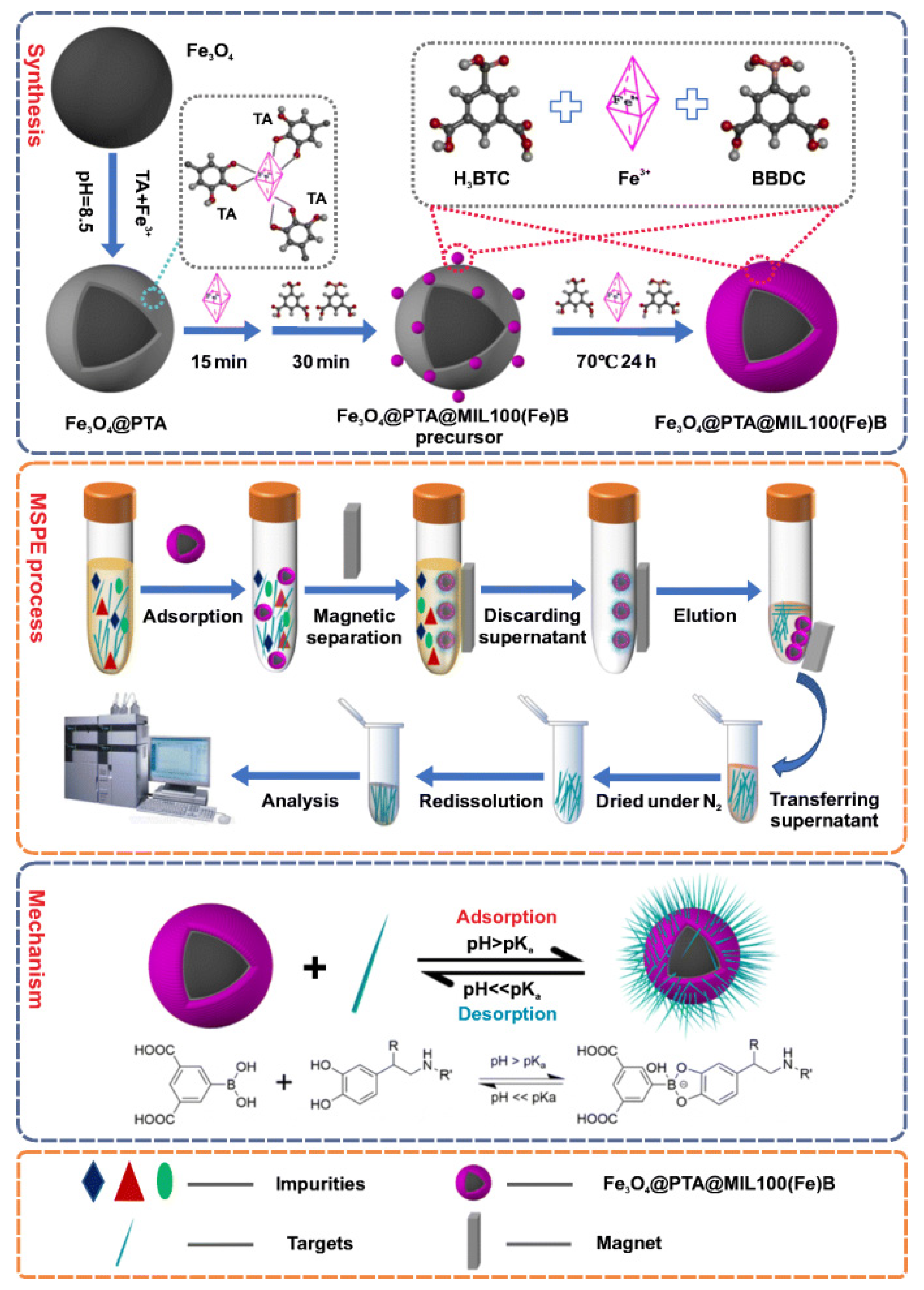
  48. Xing, Y.; Li, J.; Chen, M.; Wang, X.; Hou, X. Tannic acid-directed synthesis of magnetic and boronic acid-functionalized metal-organic frameworks for selective extraction and quantification of catecholamines in human urine. Microchim. Acta 2021, 188, 225. [Google Scholar] [CrossRef]
  49. Zhang, S.; Tang, Y.; Chen, Y.; Zhang, J.; Wei, Y. Boronic acid-modified polyhedral oligomeric silsesquioxanes on polydopamine-coated magnetized graphene oxide for selective and high-capacity extraction of the catecholamines epinephrine, dopamine and isoprenaline. Microchim. Acta 2020, 187, 77. [Google Scholar] [CrossRef]
  50. Chen, L.; Tang, Y.; Xu, B.; Xu, Z.; Shen, J.; Zhang, W. Automated on-line packed fiber solid phase extraction for determination of urinary catecholamines. J. Chromatogr. B 2020, 1139, 121983. [Google Scholar] [CrossRef]
  51. Mohyuddin, A.; Hussain, D.; Fatima, B.; Athar, M.; Ashiq, M.N.; Najam-ul-Haq, M. Gallic acid functionalized UiO-66 for the recovery of ribosylated metabolites from human urine samples. Talanta 2019, 201, 23–32. [Google Scholar] [CrossRef]
  52. Saeed, A.; Hussain, D.; Saleem, S.; Mehdi, S.; Javeed, R.; Jabeen, F.; Najam-ul-Haq, M. Metal–organic framework-based affinity materials in proteomics. Anal. Bioanal. Chem. 2019, 411, 1745–1759. [Google Scholar] [CrossRef]
  53. Anastassiades, M.; Lehotay, S.J.; Stajnbaher, D.; Schenck, F.J. Fast and easy multiresidue method employing acetonitrile extraction/partitioning and “dispersive solid-phase extraction” for the determination of pesticide residues in produce. J. AOAC Int. 2003, 86, 412–431. [Google Scholar] [CrossRef] [PubMed] [Green Version]
  54. Kumar, A.; Malik, A.K.; Pico, Y. Sample preparation methods for the determination of pesticides in foods using CE-UV/MS. Electrophoresis 2010, 31, 2115–2125. [Google Scholar] [CrossRef] [PubMed]
  55. Ma, J.B.; Qiu, H.W.; Rui, Q.H.; Liao, Y.F.; Chen, Y.M.; Xu, J.; Zhan, P.P.; Zhao, Y.G. Fast determination of catecholamines in human plasma using carboxyl-functionalized magnetic-carbon nanotube molecularly imprinted polymer followed by liquid chromatography-tandem quadrupole mass spectrometry. J. Chromatogr. A 2016, 1429, 86–96. [Google Scholar] [CrossRef] [PubMed]
  56. Jiang, L.W.; Chen, Y.B.; Luo, Y.M.; Tan, Y.M.; Ma, M.; Chen, B.; Xie, Q.J.; Luo, X.B. Determination of catecholamines in urine using aminophenylboronic acid functionalized magnetic nanoparticles extraction followed by high-performance liquid chromatography and electrochemical detection. J. Sep. Sci. 2015, 38, 460–467. [Google Scholar] [CrossRef]
  57. He, X.Y.; Yu, Y.; Li, Y. Facile synthesis of boronic acid-functionalized magnetic metal-organic frameworks for selective extraction and quantification of catecholamines in rat plasma. RSC Adv. 2018, 8, 41976–41985. [Google Scholar] [CrossRef] [Green Version]
  58. Qin, Q.; Li, H.; Shi, X.Z.; Xu, G.W. Facile synthesis of Fe3O4@polyethyleneimine modified with 4-formylphenylboronic acid for the highly selective extraction of major catecholamines from human urine. J. Sep. Sci. 2015, 38, 2857–2864. [Google Scholar] [CrossRef]
  59. Murtada, K.; de Andrés, F.; Galván, I.; Ríos, Á.; Zougagh, M. LC-MS determination of catecholamines and related metabolites in red deer urine and hair extracted using magnetic multi-walled carbon nanotube poly(styrene-co-divinylbenzene) composite. J. Chromatogr. B 2020, 1136, 121878. [Google Scholar] [CrossRef]
  60. He, H.B.; Zhou, Z.Q.; Dong, C.; Wang, X.; Yu, Q.W.; Lei, Y.Y.; Luo, L.Q.; Feng, Y.Q. Facile synthesis of a boronate affinity sorbent from mesoporous nanomagnetic polyhedral oligomeric silsesquioxanes composite and its application for enrichment of catecholamines in human urine. Anal. Chim. Acta 2016, 944, 1–13. [Google Scholar] [CrossRef] [Green Version]
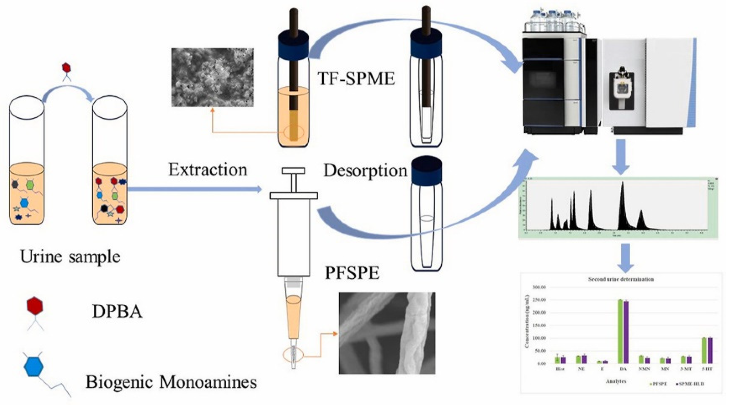
  61. Chen, L.; Singh, V.; Rickert, D.; Khaled, A.; Pawliszyn, J. High throughput determination of free biogenic monoamines and their metabolites in urine using thin-film solid phase microextraction. Talanta 2021, 232, 122438. [Google Scholar] [CrossRef]
  62. Podjava, A.; Šilaks, A. Synthesis and sorptive properties of molecularly imprinted polymer for simultaneous isolation of catecholamines and their metabolites from biological fluids. J. Liq. Chromatogr. 2021, 44, 181–188. [Google Scholar] [CrossRef]
  63. Liu, S.; Huang, Y.; Qian, C.; Xiang, Z.; Ouyang, G. Physical assistive technologies of solid-phase microextraction: Recent trends and future perspectives. Trends Anal. Chem. 2020, 115916. [Google Scholar] [CrossRef]
  64. Monteleone, M.; Naccarato, A.; Sindona, G.; Tagarelli, A. A reliable and simple method for the assay of neuroendocrine tumor markers in human urine by solid-phase microextraction-gas chromatography-triple quadrupole mass spectrometry. Anal. Chim. Acta 2013, 759, 66–73. [Google Scholar] [CrossRef] [PubMed]
  65. Bessonova, E.; Kartsova, L.; Gallyamova, V. Ionic liquids based on imidazole for online concentration of catecholamines in capillary electrophoresis. J. Sep. Sci. 2017, 40, 2304–2311. [Google Scholar] [CrossRef] [PubMed]
  66. Espina-Benitez, M.B.; Randon, J.; Demesmay, C.; Dugas, V. Development and application of a new in-line coupling of a miniaturized boronate affinity monolithic column with capillary zone electrophoresis for the selective enrichment and analysis of cis-diol-containing compounds. J. Chromatogr. A 2017, 1494, 65–76. [Google Scholar] [CrossRef] [PubMed]
  67. Fang, W.L.; Xia, L.J.; Huang, X.; Guo, X.F.; Ding, J.; Wang, H.; Feng, Y.Q. Highly Sensitive Determination for Catecholamines Using Boronate Affinity Polymer Monolith Microextraction with In-Situ Derivatization and HPLC Fluorescence Detection. Chromatographia 2018, 81, 1381–1389. [Google Scholar] [CrossRef]
  68. Cudjoe, E.; Pawliszyn, J. Optimization of solid phase microextraction coatings for liquid chromatography mass spectrometry determination of neurotransmitters. J. Chromatogr. A 2014, 1341, 1–7. [Google Scholar] [CrossRef]
  69. Zhou, X.G.; Zhu, A.W.; Shi, G.Y. Selective extraction and analysis of catecholamines in rat blood microdialysate by polymeric ionic liquid-diphenylboric acid-packed capillary column and fast separation in high-performance liquid chromatography-electrochemical detector. J. Chromatogr. A 2015, 1409, 125–131. [Google Scholar] [CrossRef]
  70. Yang, X.T.; Hu, Y.F.; Li, G.K. Online micro-solid-phase extraction based on boronate affinity monolithic column coupled with high-performance liquid chromatography for the determination of monoamine neurotransmitters in human urine. J. Chromatogr. A 2014, 1342, 37–43. [Google Scholar] [CrossRef]
  71. Zhang, Q.Y.; Gong, M.J. On-line preconcentration of fluorescent derivatives of catecholamines in cerebrospinal fluid using flow-gated capillary electrophoresis. J. Chromatogr. A 2016, 1450, 112–120. [Google Scholar] [CrossRef]
  72. Saracino, M.A.; Santarcangelo, L.; Raggi, M.A.; Mercolini, L. Microextraction by packed sorbent (MEPS) to analyze catecholamines in innovative biological samples. J. Pharm. Biomed. 2015, 104, 122–129. [Google Scholar] [CrossRef]
  73. Xiong, X.; Zhang, Y. Simple, rapid, and cost-effective microextraction by the packed sorbent method for quantifying of urinary free catecholamines and metanephrines using liquid chromatography-tandem mass spectrometry and its application in clinical analysis. Anal. Bioanal. Chem. 2020, 412, 2763–2775. [Google Scholar] [CrossRef] [PubMed]
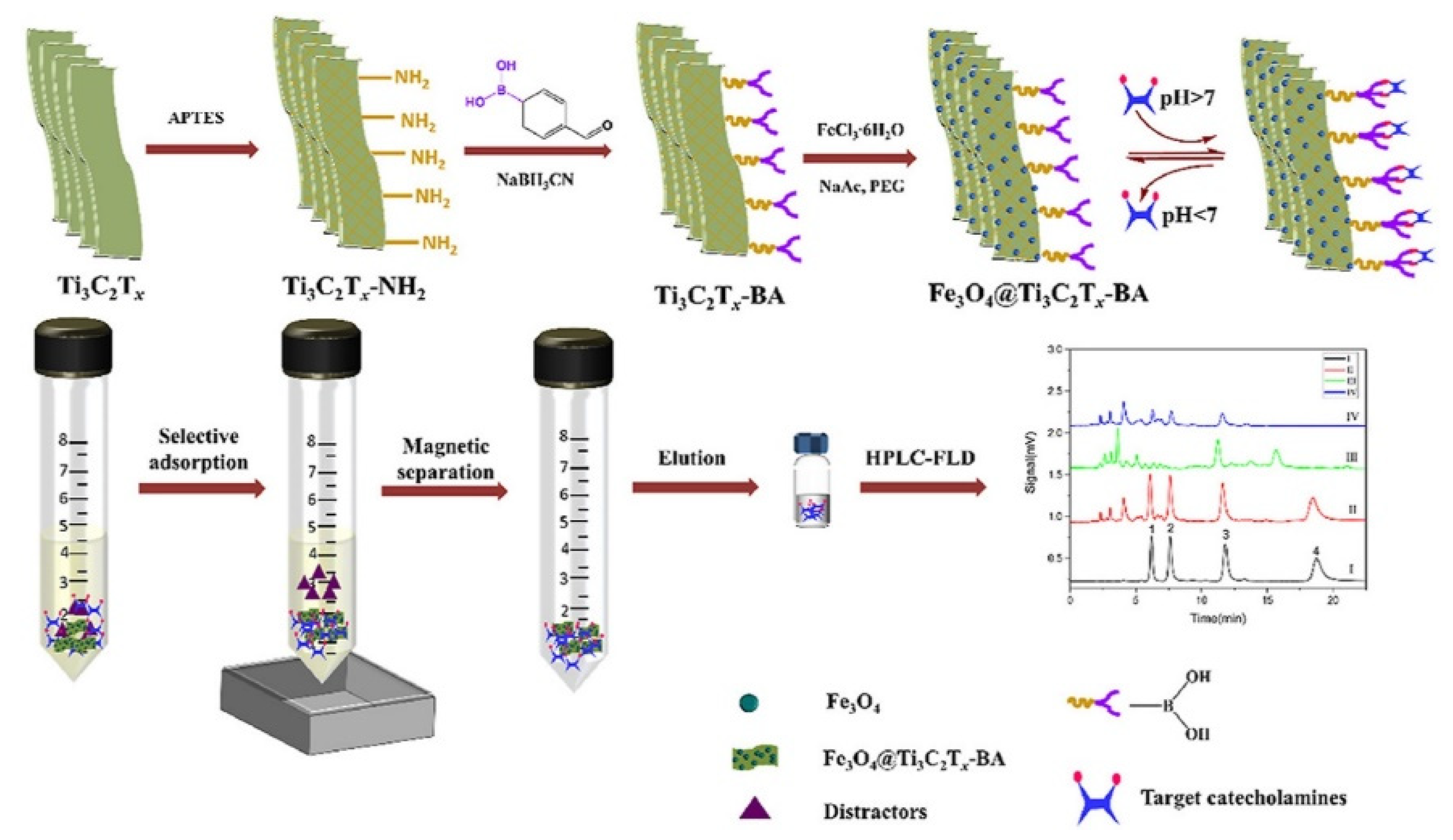
  74. Hu, K.; Pang, T.; Shi, Y.; Han, P.; Zhao, Y.; Zhao, W.; Zeng, H.; Zhang, S.; Zhang, Z. Magnetic borate-modified Mxene: A highly affinity material for the extraction of catecholamines. Anal. Chim. Acta 2021, 1176, 338769. [Google Scholar] [CrossRef] [PubMed]
  75. Zhang, T.Y.; Li, S.; Zhu, Q.F.; Wang, Q.; Hussain, D.; Feng, Y.Q. Derivatization for liquid chromatography-electrospray ionization-mass spectrometry analysis of small-molecular weight compounds. Trends Anal. Chem. 2019, 119, 115608. [Google Scholar] [CrossRef]
  76. Zhao, L.S.; Zheng, S.N.; Su, G.Y.; Lu, X.M.; Yang, J.Y.; Xiong, Z.L.; Wu, C.F. In vivo study on the neurotransmitters and their metabolites change in depressive disorder rat plasma by ultra high performance liquid chromatography coupled to tandem mass spectrometry. J. Chromatogr. B 2015, 988, 59–65. [Google Scholar] [CrossRef] [PubMed]
  77. Zheng, L.F.; Zhao, X.E.; Zhu, S.Y.; Tao, Y.D.; Ji, W.H.; Geng, Y.L.; Wang, X.A.; Chen, G.A.; You, J.M. A new combined method of stable isotope-labeling derivatization-ultrasound-assisted dispersive liquid-liquid microextraction for the determination of neurotransmitters in rat brain microdialysates by ultra high performance liquid chromatography tandem mass spectrometry. J. Chromatogr. B 2017, 1054, 64–72. [Google Scholar]
  78. Ellis, A.G.; Zeglinski, P.T.; Coleman, K.E.; Whiting, M.J. Dilute, derivatise and shoot: Measurement of urinary free metanephrines and catecholamines as ethyl derivatives by LC-MSMS. Clin. Mass Spectrom. 2017, 34–41. [Google Scholar] [CrossRef]
  79. Azaryan, A.; Ligor, T.; Buszewski, B.; Temerdashev, A.; Dmitrieva, E.; Gashimova, E. LC-MS/MS Determination of Catecholamines in Urine Using FMOC-Cl Derivatization on Solid-Phase Extraction Cartridge. Chromatographia 2018, 81, 1487–1494. [Google Scholar] [CrossRef]
  80. Xiang, D.; Yin, L.; Wang, C.; Zhang, L. High electrochemical performance of RuO2–Fe2O3 nanoparticles embedded ordered mesoporous carbon as a supercapacitor electrode material. Energy 2016, 106, 103–111. [Google Scholar] [CrossRef]
  81. Huang, X.; Guo, X.F.; Wang, H.; Zhang, H.S. Analysis of catecholamines and related compounds in one whole metabolic pathway with high performance liquid chromatography based on derivatization. Arab. J. Chem. 2019, 12, 1159–1167. [Google Scholar] [CrossRef] [Green Version]
  82. Zhao, X.E.; Zhu, S.Y.; Yang, H.M.; You, J.M.; Song, F.R.; Liu, Z.Q.; Liu, S.Y. Simultaneous determination of amino acid and monoamine neurotransmitters in PC12 cells and rats models of Parkinson’s disease using a sensitizing derivatization reagent by UHPLC-MS/MS. J. Chromatogr. B 2015, 995, 15–23. [Google Scholar] [CrossRef]
  83. Chen, D.; Zhang, J.X.; Cui, W.Q.; Zhang, J.W.; Wu, D.Q.; Yu, X.R.; Luo, Y.B.; Jiang, X.Y.; Zhu, F.P.; Hussain, D.; et al. A simultaneous extraction/derivatization strategy coupled with liquid chromatography–tandem mass spectrometry for the determination of free catecholamines in biological fluids. J. Chromatogr. A 2021, 1654, 462474. [Google Scholar] [CrossRef] [PubMed]
  84. Lee, W.; Park, N.H.; Ahn, T.B.; Chung, B.C.; Hong, J. Profiling of a wide range of neurochemicals in human urine by very-high-performance liquid chromatography-tandem mass spectrometry combined with in situ selective derivatization. J. Chromatogr. A 2017, 1526, 47–57. [Google Scholar] [CrossRef] [PubMed]
  85. Liu, G.S.; Chen, J.N.; Ma, Y.F. Simultaneous determination of catecholamines and polyamines in PC-12 cell extracts by micellar electrokinetic capillary chromatography with ultraviolet absorbance detection. J. Chromatogr. B 2004, 805, 281–288. [Google Scholar] [CrossRef] [PubMed]
  86. Lin, T.H.; Lu, C.Y.; Tseng, W.L. Selective enrichment of catecholamines using iron oxide nanoparticles followed by CE with UV detection. Electrophoresis 2013, 34, 297–303. [Google Scholar] [CrossRef] [PubMed]
  87. Li, X.G.; Li, S.; Kellermann, G. Pre-analytical and analytical validations and clinical applications of a miniaturized, simple and cost-effective solid phase extraction combined with LC-MS/MS for the simultaneous determination of catecholamines and metanephrines in spot urine samples. Talanta 2016, 159, 238–247. [Google Scholar] [CrossRef]
  88. Peaston, R.T.; Graham, K.S.; Chambers, E.; van der Molen, J.C.; Ball, S. Performance of plasma free metanephrines measured by liquid chromatography-tandem mass spectrometry in the diagnosis of pheochromocytoma. Clin. Chim. Acta 2010, 411, 546–552. [Google Scholar] [CrossRef]
  89. Heideloff, C.; Payto, D.; Wang, S. Quantitation of Free Metanephrines in Plasma by Liquid Chromatography-Tandem Mass Spectrometry. Methods Mol. Biol. 2016, 1378, 139–147. [Google Scholar]
  90. Naccarato, A.; Gionfriddo, E.; Sindona, G.; Tagarelli, A. Development of a simple and rapid solid phase microextraction-gas chromatography-triple quadrupole mass spectrometry method for the analysis of dopamine, serotonin and norepinephrine in human urine. Anal. Chim. Acta 2014, 810, 17–24. [Google Scholar] [CrossRef]
  91. Lagerstedt, S.A.; O’Kane, D.J.; Singh, R.J. Measurement of Plasma Free Metanephrine and Normetanephrine by Liquid Chromatography–Tandem Mass Spectrometry for Diagnosis of Pheochromocytoma. Clin. Chem. 2004, 50, 603–611. [Google Scholar] [CrossRef] [Green Version]
  92. Jian, M.; Huang, H.; Li, K.; Chuan, L.; Li, L.; Jiang, L. A 3-min UPLC-MS/MS method for the simultaneous determination of plasma catecholamines and their metabolites: Method verification and diagnostic efficiency. Clin. Biochem. 2021, 87, 67–73. [Google Scholar] [CrossRef]
  93. Ma, L.; Zhao, T.; Zhang, P.; Liu, M.; Shi, H.; Kang, W. Determination of monoamine neurotransmitters and metabolites by high-performance liquid chromatography based on Ag(III) complex chemiluminescence detection. Anal. Biochem. 2020, 593, 113594. [Google Scholar] [CrossRef] [PubMed]
  94. Bergmann, M.L.; Schmedes, A. Highly sensitive LC-MS/MS analysis of catecholamines in plasma. Clin. Biochem. 2020, 82, 51–57. [Google Scholar] [CrossRef] [PubMed]
  95. Polikarpova, D.; Makeeva, D.; Kolotilina, N.; Dolgonosov, A.; Peshkova, M.; Kartsova, L. Nanosized cation exchanger for the electrophoretic separation and preconcentration of catecholamines and amino acids. Electrophoresis 2020, 41, 1031–1038. [Google Scholar] [CrossRef] [PubMed]
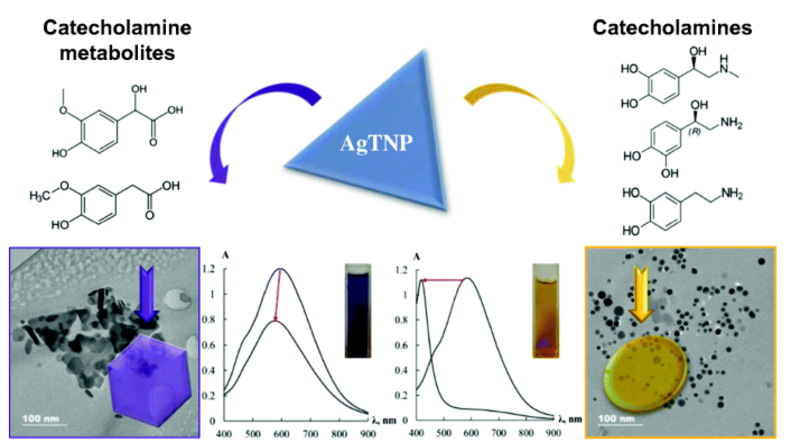
  96. Zaytsev, V.D.; Furletov, A.A.; Apyari, V.V.; Garshev, A.V.; Dmitrienko, S.G.; Zolotov, Y.A. Label-free silver triangular nanoplates for spectrophotometric determination of catecholamines and their metabolites. Microchim. Acta 2020, 187, 610. [Google Scholar] [CrossRef]
Figure 1. The analysis process for the determination of CAs and their metabolites.
Molecules 27 02702 g001
Figure 2. Synthesis mechanism and extraction of CAs by Fe3O4@PTA@MIL-100(Fe)-B [48].
Molecules 27 02702 g002
Figure 3. Determination of free biogenic monoamines and their metabolites in urine using thin-film solid-phase microextraction [61].
Molecules 27 02702 g003
Figure 4. Schematic diagram of the online micro-solid-phase extraction system, reprinted from [70,71]. (A) The in-situ polymerization of monolithic column (a), schematic diagram of the online micro-solid-phase extraction system coupled to HPLC (b). (B) Schematic procedures showing on-line preconcentration and separation. (a) Sample injection; (b) focusing; and (c) separation.
Molecules 27 02702 g004
Figure 5. Magnetic borate-modified MXene composite for the extraction of CAs [74].
Molecules 27 02702 g005
Figure 6. (A) HPLC-UV chromatograms (a), elution from B-hDIPs-SPE spiked with 0.5 μg mL−1 (b), spiked with 1.0 μg mL−1 (c), spiked with 2.0 μg mL−1 (d). Peaks: (1) norepinephrine, (2) epinephrine, (3) dopamine. HPLC conditions were: a Thermo scientific C18 column (250 mm × 4.6 mm, 5 mm) and UV detector at 280 nm, mobile phase, MeOH-NaH2PO4 (20 mmol L−1, pH 4.0) (5:95, v/v) with flow rate at 0.5 mL min−1 with injection volume 10 μL. Reproduced with permission from Reference [8]. (B) Typical chromatogram of derivatives of the six selected catecholamines and related compounds. Mobile phase: methanol and 20 mM pH 3.5 H3Cit–Na2HPO4 buffer. Detection: fluorescence (490/510 nm). Flow rate: 0.7 mL/min. Injection volume: 20 μL. Concentration: 0.05 μM each (L-DOPA concentration: 0.1 μM). Peaks: (1) L-DOPA; (2) Tyr; (3) NE; (4) E; (5) DA; (6) MN and (a) TMBB-Su hydrolyzate. Reproduced with permission from Reference [81]. (C) Extracted ion chromatogram of ten targeted analytes. Selected reaction monitoring (SRM) was used to detect targeted analytes. Chromatographic conditions: column, Acquity UPLC BEH C18 column (2.1 mm × 150 mm, 1.7 μm particle size) with VanGuard pre-column; flow rate = 0.3 mL/min; column temperature 30 °C. peaks: Glu—glutamic acid, GABA—γ-aminobutyric acid, HIS—histamine, 5-HIAA—5-hydroxyindoleacetic acid, HVA—homovanilic acid, SER—5-hydroxytryptamine, DOPAC—3,4-dihydroxyphenylacetic acid, NADR—noradrenaline, ADR—adrenaline, DA—dopamine. Reproduced with permission from Reference [23].
Molecules 27 02702 g006
Figure 7. Label-free silver triangular nanoplates for spectrophotometric determination of catecholamines and their metabolites [96].
Molecules 27 02702 g007
Table 1. Solid-phase extraction techniques used for extracting CAs and their metabolites in various biological samples.
SorbentsAnalytesMatrixLOQsDetectionRef.
Oasis weak cation exchange (WCX)normetanephrine (NMN),
3-methoxytyramine (3-MT),
metanephrine (MN)
human plasmaNMN: 75.0 pmol/L
3-MT: 37.5 pmol/L
MN: 37.5 pmol/L
LC-MS/MS[38]
Strata-X-CWnorepinephrine (NE),
dopamine (DA),
epinephrine (E)
human plasmaNE: 7.4 ng/mL
DA: 5.4 ng/mL
E: 3.8 ng/mL
LC-MS/MS[39]
Evolute® WCXMN, NMNhuman plasmaMN: 0.07 nmol/L
NMN: 0.06 nmol/L
LC-MS/MS[40]
96-well hydrophilic-lipophilic-balanced (HLB) Elution plateE, NE, DAhuman peripheral blood mononuclear cells (PBMC)E: 1 pg/mL
NE: 5 pg/mL
DA: 5 pg/mL
LC-MS/MS[41]
precolumn modified with phenylboronic acidE, NE, DAmouse urineNE: 163 fmol/L
DA: 127 fmol/L
E: 196 fmol/L
LC-FLD[42]
HLB solid-phase cartridgesE, NE, DAhuman urineNE: 0.4 ng/mL
DA: 0.3 ng/mL
E: 0.2 ng/mL
LC-MS/MS[43]
Strata-X-CWE, NE, DA, MN, NMNurineNE: 5.0 ng/mL
DA: 5.0 ng/mL
E: 5.0 ng/mL
MN: 5.0 ng/mL
NMN: 5.0 ng/mL
LC-MS/MS[44]
electrospun composite fibersE, NE, DAhuman urineNE: 0.2 ng/mL
DA: 0.5 ng/mL
E: 0.2 ng/mL
LC-FLD[45]
boronate-modified hollow dummy template imprinted polymers (B-hDIPs)E, NE, DAhuman urineNE: 157.0 ng/mL
DA: 141.0 ng/mL
E: 51.0 ng/mL
LC-UV[8]
Bond-Elut PlexaMN, NMNurineMN: 0.2 μmol/L
NMN: 0.3 μmol/L
LC-MS/MS[46]
96-well HLB microplateE, NE, DA, serotonine
(5-HT)
human urineNE: 2.0 ng/mL
DA: 4.0 ng/mL
E: 1.0 ng/mL
5-HT: 4.0 ng/mL
LC-MS/MS[47]
Fe3O4@PTA@MIL-100(Fe)-Bnorepinephrine, epinephrine, and dopamineHuman UrineNE: 0.050 ng/mL
E: 0.11 ng/mL
DA: 0.20 ng/mL
LC-FLD[48]
magGO@POSS-BAepinephrine, dopamine, and isoprenalineHuman urine0.54–2.3 ng·mL−1LC-FLD[49]
Polycrown ether composite nanofiberCatecholaminesHuman urine1 ng/mLLC-FLD[50]
Table 2. Dispersive solid-phase extraction/microextraction (DSPE/DSPME) techniques used for extracting CAs and their metabolites in various biological samples.
SorbentsAnalytesMatrixLOQsDetectionRef.
aminophenylboronic acid functionalized magnetic nanoparticlesNE, DA, Ehuman urineNE: 26.0 ng/mL
DA: 23.6 ng/mL
E: 6.7 ng/mL
HPLC-ECD[56]
MG@MIL-100-B composites (boronic acid functionalized MIL-100)NE, DA, Erat plasmaNE: 0.10 ng/mL
DA: 0.01 ng/mL
E: 0.01 ng/mL
HPLC-MS/MS[57]
Fe3O4@PEI-FPBANE, DA, Ehuman urineNE: 0.20 ng/mL
DA: 0.03 ng/mL
E: 0.08 ng/mL
LC-MS[58]
CF@m-CNTs-MIPNE, DA, Ehuman plasmaNE: 0.076 ng/mL
DA: 0.010 ng/mL
E: 0.018 ng/mL
UFLC-MS/MS[55]
magnetic MWCNT poly(STY-DVB) compositeNE, DA, E, DL-3,4-dihydroxymandelic acid (DHMA), DL-3,4-dihydroxyphenyl glycol (DOPEG)red deer urineNE: 248 ng/mL
DA: 205 ng/mL
E: 188 ng/mL
DHMA: 146 ng/mL
DOPEG: 232 ng/mL
LC-MS[59]
IDA-Cu(II) functionalized Fe3O4@SiO2 (Fe3O4@SiO2 @IDA-Cu) magnetic
nanoparticles (MNPs)
NE, DA, E, 5-HT, isoprenaline(IP), tyramine (TA)rabbit plasmaNE: 0.43 ng/mL
DA: 0.20 ng/mL
E: 0.33 ng/mL
5-HT: 0.16 ng/mL
IP: 0.31 ng/mL
TA: 0.27 ng/mL
HPLC-FLD[19]
Fe3O4@POSS-AAPBANE, isoprenaline hydrochloride (IE), Ehuman urineNE: 2.70 ng/mL
IE: 4.40 ng/mL
E: 2.96 ng/mL
HPLC-UV[60]
Polycrown ether (PCE) composite nanofiberbiogenic monoaminesHuman urine0.25–500 ng/mLUPLC-MS/MS[61]
Borated zirconiaepinephrine (E), norepinephrine (NE), and dopamine (DA),PlasmaE: 0.008 ng/mL
NE: 0.020 ng/mL
DA 0.004 ng/mL
LC-MS/MS[24]
Table 3. Derivatization used for extracting CAs and their metabolites in various biological samples.
Derivatization
Reagents
AnalytesMatrixDerivatization ConditionsLOQsLODsDetectionRef.
T (°C)pHTime (min)
10-methyl-acridone-2-sulfonyl chlorideL-DOPA, DA, NE, E, Trp, 5-HTP, 5-HTrat brain microdialysates3710.530.015–0.040 nmol/L0.002–0.010 nmol/LUHPLC-MS/MS[77]
TMBB-SuTyr, L-DOPA,
DA, NE, E, and MN
mice liver and brain257.640--0.10–0.40 nmol/LHPLC-FLD[81]
acetaldehydeNE, DA, E, NMN, MN, 3-MThuman urine365301–14 nmol/L--LC-MS/MS[78]
Benzoyl chlorideNE, DA, E, 5-HT, 5-hydroxyindoleacetic acid (5-HIAA), homovanilic acid (HVA), glutamic acid (Glu), γ-aminobutyric acid (GABA)rat cerebrospinal fluid (CSF)257.4--0.15–5.00 ng/mL0.02–2.00 ng/mLUHPLC-MS[23]
FMOC-ClE, DA, octopaminehuman urine259.5205–50 ng/mL2.5–25 ng/mLHPLC-MS/MS[79]
methanol20 neurochemicalshuman urine90<4900.3–12.0 ng/mL0.1–3.6 ng/mLUPLC-MS/MS[84]
4-carbonyl chloride rosamine (CCR)21 neurotransmitters (NTs)rat brain and blood259.51--1 × 10⁻⁴–3 × 10⁻³ nmol/LUPLC-MS/MS[29]
10-ethyl-acridone-3-sulfonyl chloride (EASC)Glu, Asp, Gly, GABA, taurine (Tau), DA, 5-HTPC12 cells3710.530.004–3.8 nmol/L0.014–13.1 nmol/LUPLC-MS/MS[82]
Lissamine rhodamine B sulfonylchloride (LRSC)DA, 5-HT and their biosynthesis precursors and metabolitesrat brain microdialysates3710.530.002–0.008 nmol/L0.015–0.040 nmol/LUHPLC-MS/MS[80]
dansyl chlorideNE, DA, 5-HT, HVA, HIAA, GABA, Glurat plasma6511.020--0.991–5030 fmol/LHPLC-MS/MS[76]
ZrO2/phenyl isothiocyanate (PITC)norepinephrine (NE), epinephrine (E) and dopamine (DA)Human Urine2510100.035 ng/mL0.100 ng/mLUHPLC-MS/MS[83]
Publisher’s Note: MDPI stays neutral with regard to jurisdictional claims in published maps and institutional affiliations.

