Abstract
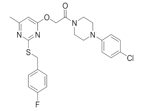
A number of uracil amides cleave poly (ADP-ribose) polymerase and therefore novel thiouracil amide compounds were synthesized and screened for the loss of cell viability in a human-estrogen-receptor-positive breast cancer cell line. The synthesized compounds exhibited moderate to significant efficacy against human breast cancer cells, where the compound 5e IC50 value was found to be 18 μM. Thouracil amide compounds 5a and 5e inhibited the catalytical activity of PARP1, enhanced cleavage of PARP1, enhanced phosphorylation of H2AX, and increased CASPASE 3/7 activity. Finally, in silico analysis demonstrated that compound 5e interacted with PARP1. Hence, specific thiouracil amides may serve as new drug-seeds for the development of PARP inhibitors for use in oncology.
1. Introduction
Poly (ADP-ribose) polymerase (PARP) is an enzyme which catalyzes poly ADP-ribosylation of specific target proteins. The PARP family contains 17 enzymes, among which PARP1 is the most abundant and widely studied [1,2,3]. It is involved in a number of cellular functions, such as DNA repair, recombination, and gene transcription [4]. More than 70 PARP inhibitors are undergoing clinical trials, some of which are approved by the U.S. Food and Drug Administration, for instance, Niraparib, Olaparib, Talazoparib, and Rucaparib [5,6,7,8]. Evidence suggests that PARP plays an important role in cancer cell signaling, chromatin regulation, and metastatic processes that involve cell survival, migration, and invasion [9]. For example, in estrogen-receptor-positive (ER+) breast cancer, PARP regulates the transcriptional activity of ERα, and is reported to be downstream of ER-dependent transcriptional responses [10].
A number of PARP1 inhibitors (PARPi) have been reported; however, the commonly used example of a PARPi, 3-substituted benzamide (3-AB) was a nicotinamide analog [11]. Generally, benzamides inhibit PARP due to their structural analogy to NAD and therefore compete with NAD at the catalytic domain of PARPs [12]. Despite their ability to elucidate the function of PARP, benzamides fail to show PARP inhibitory activity in nanomolar ranges and exhibited off-target effects that precluded their clinical evaluation. Additionally, indazole amides have been shown to be PARP1 inhibitors [13].
Conversely, substituted uracil derivatives have been reported as potent inhibitors of PARP by inhibiting thymidylate synthesis as well as basal excision repair (BER) mechanisms [14,15]. Indeed, thiouracil- or uracil-based compounds are known to act on cancer cells via an anti-folate mechanism, where its misincorporation is genotoxic and eventually contributes to DNA double-strand break and ultimately cell death [16].
The synthesis, characterization, and efficacy of new thiouracil amides were therefore investigated herein and found a lead compound 5e that inhibits both PARP1 catalytic activity and breast cancer cell viability.
2. Results and Discussion
2.1. Chemical Synthesis of Novel Thiouracil Amides
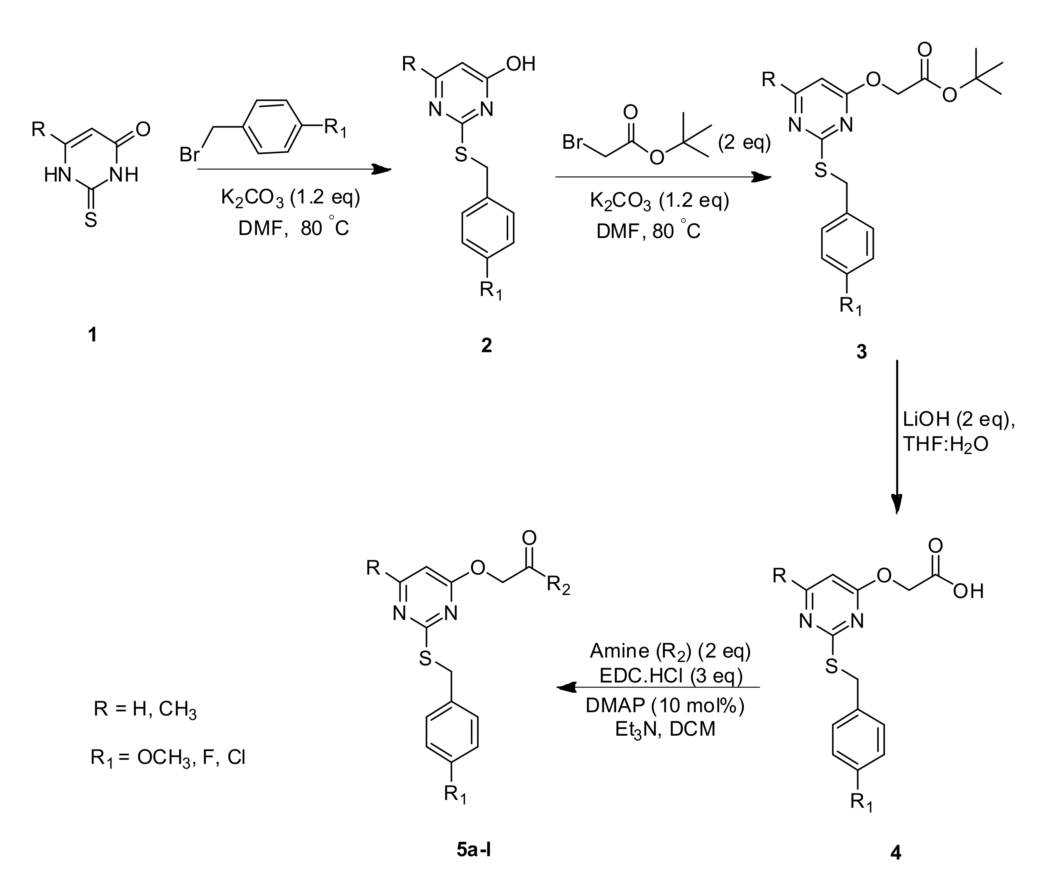
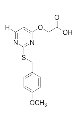
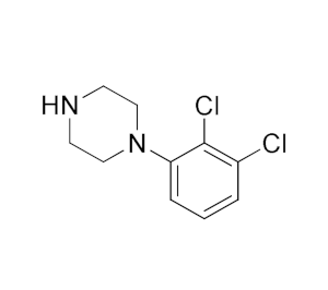
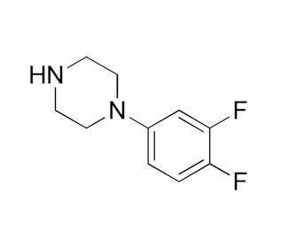
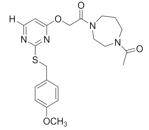
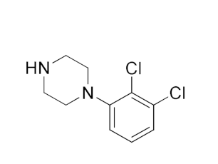
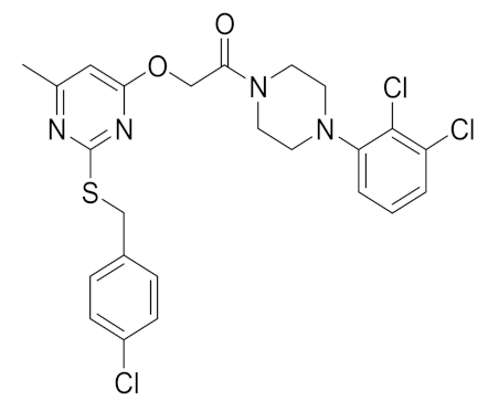
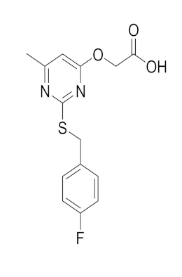
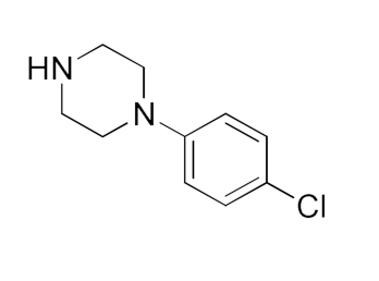
The synthesis of novel thiouracil derivatives and their characterization is herein reported. In the present investigation, regioisomeric 2-thiopyrimidine acetic acids were utilized as a template to prepare a library of novel amides with various amines, as described in Scheme 1.
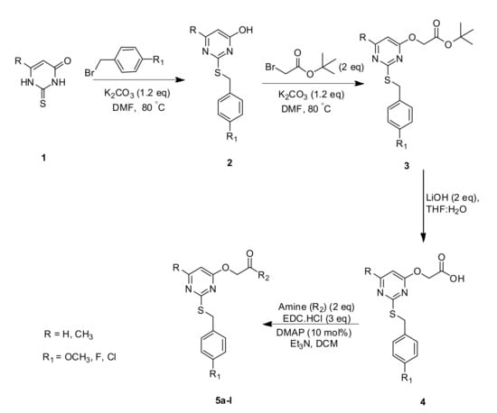
Scheme 1.
Synthesis of thiouracil containing amide derivatives.
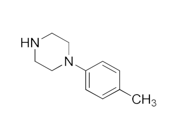
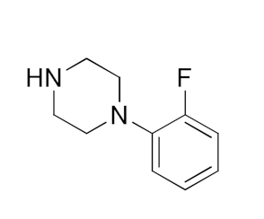
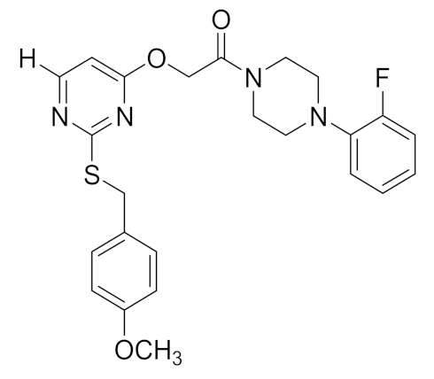
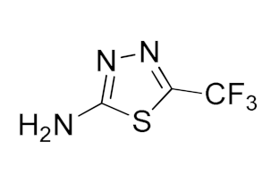
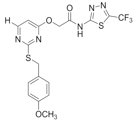
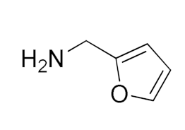
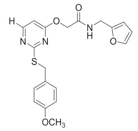
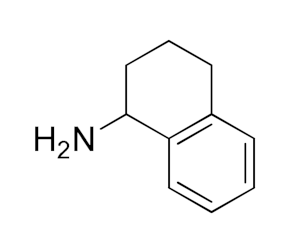
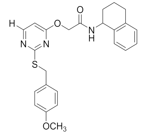
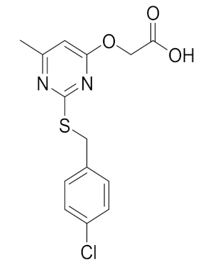
The synthetic sequence commenced by reacting 2-thiouracil with substituted benzyl derivative to afford S-benzylated products. The phenolic hydroxyl group in S-benzylated products was treated with tert-butyl acetate to afford tert-butyl esters which were subjected to hydrolysis to afford 2-thiopyrimidine acetic acid. The acid so obtained was coupled with various amines to afford amides 5a–5l. The structures of these newly synthesized amide derivatives were confirmed by 1H NMR, 13CNMR, and LC/MS analysis.
2.2. Novel Thiouracil Derivatives Produce Loss of Breast Cancer Cell Viability
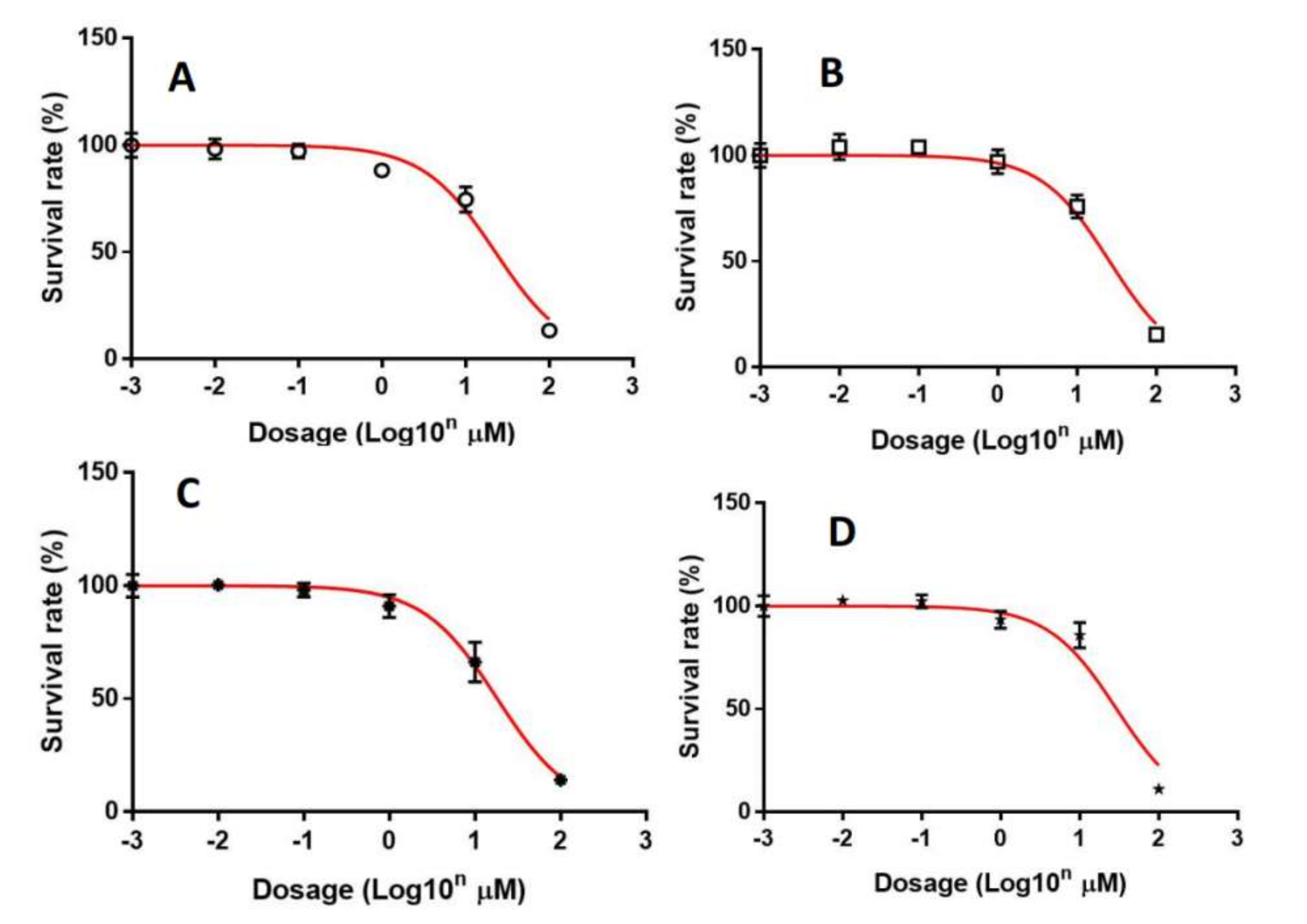
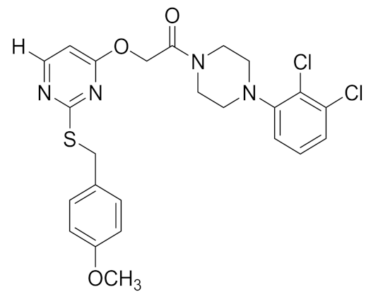
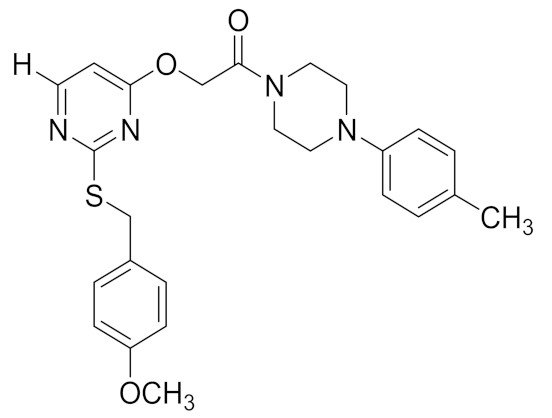
The synthesized compounds were screened for loss of cell viability using the ER+ breast cancer cell line by Alamar Blue cell viability assays [17,18,19,20,21,22]. The compounds were dissolved in DMSO and cells were treated in a dose-dependent manner for 72 h. Amongst the tested amides, compounds 5a, 5b, 5e, and 5f showed significant loss of cell viability with IC50 values of 22.68, 25.71, 18.23, and 29.34 µM, respectively, as shown in Figure 1 and Table 1. The other compounds tested from the series exhibited less efficacy. The thiouracil amides possessing tolyl or halophenyl groups displayed significant loss of breast cancer cell viability, according to studies on the structure–activity relationship of this library of compounds. Furthermore, the loss of cell viability produced by the lead compounds (5a, 5e) was compared with Olaparib using immortalized but otherwise normal human mammary epithelial cells (MCF-10A) as an indicator of effect on normal cells. It was observed that these compounds produced loss of cell viability of MCF-10A cells with IC50 values of 46.9 and 52.3 µM for compounds 5a and 5e, respectively, which were comparable to Olaparib, where IC50 was observed to be 57.3 µM (Supplementary Materials).
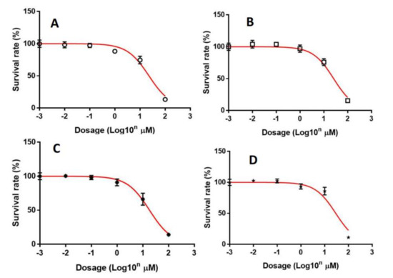
Figure 1.
Loss of MCF-7 cell viability produced by compounds 5a, 5b, 5e, and 5f. MCF-7 were exposed to 5a (A), 5b (B), 5e (C), and 5f (D) for 72 h and the viability of cells was analyzed by Alamar Blue assays. The results are presented as mean ± S.E.M. of triplicate determinations.

Table 1.
Thiouracil amides structure and their cytotoxicity results tabulated.
2.3. Compounds 5a and 5e Inhibit PARP1 Activity
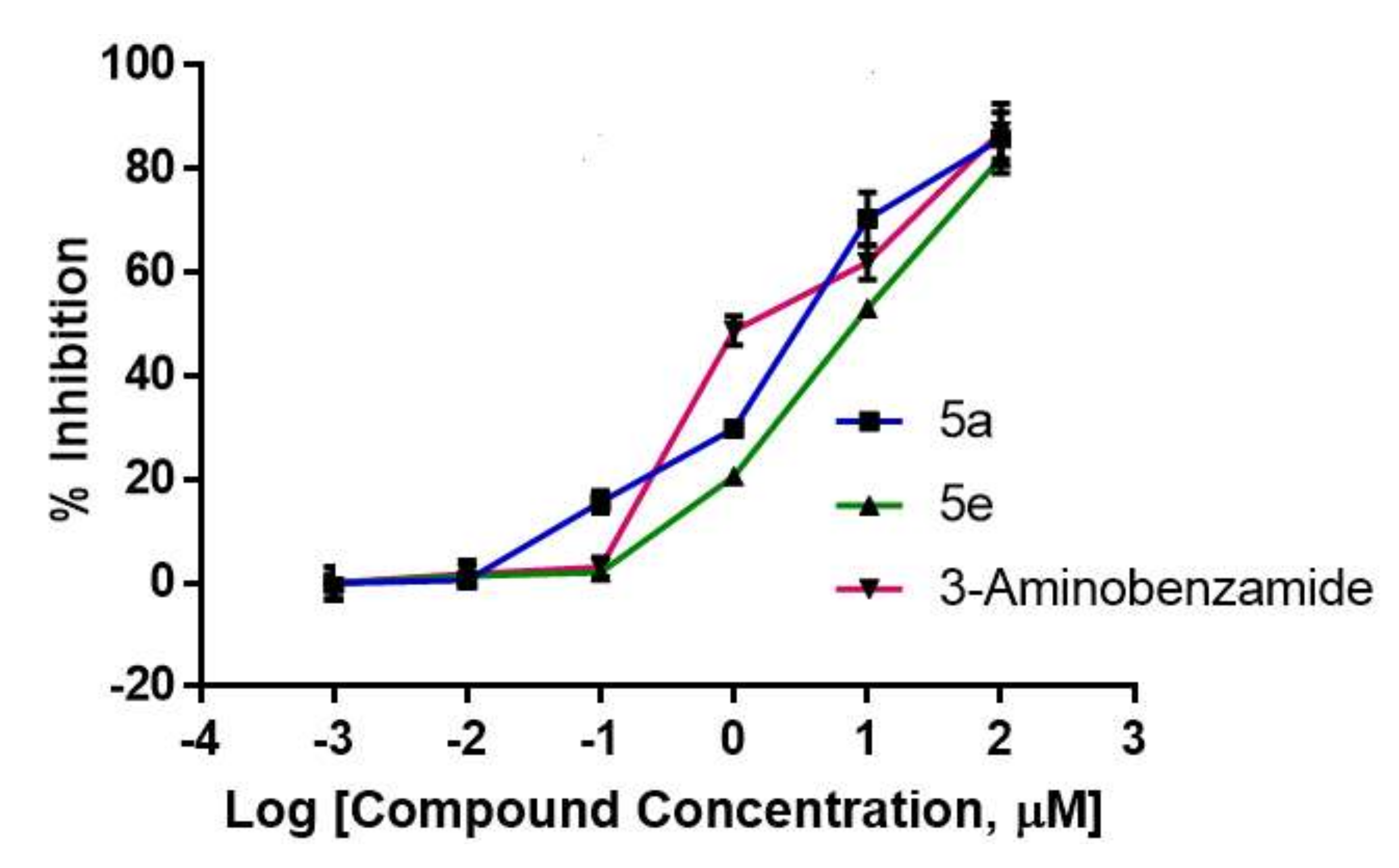
Compounds 5a and 5e were tested for their capacity to inhibit PARP1 catalytic activity [23]. Compounds 5a and 5e were used at concentrations of 0.01, 0.1, 1, 10, and 100 µM. Compound 5a was found to inhibit the catalytical activity of PARP1 0.5, 15.6, 29.8, 70.3, and 85.8% at the tested concentrations, whereas compound 5e inhibited PARP1 activity at 1.3, 2.0, 20.6, 53, and 82.1%. 3-aminobenzamide inhibited PARP1 activity by 1.7, 3, 48.7, 61.9, and 87.2% at the tested concentrations, respectively (Figure 2).
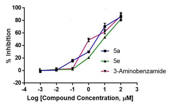
Figure 2.
Compounds 5a, 5e, and 3-aminobenzamide inhibited the PARP1 catalytic activity. Graphical representation of the inhibition of the catalytic activity of PARP1 by indicated compounds (n = 3). Data are expressed as mean ± SEM.
2.4. Lead Compounds Increased PARP1 Cleavage, Phospho-H2AX Levels, and Caspase-3/7 Activation in MCF-7 Cells
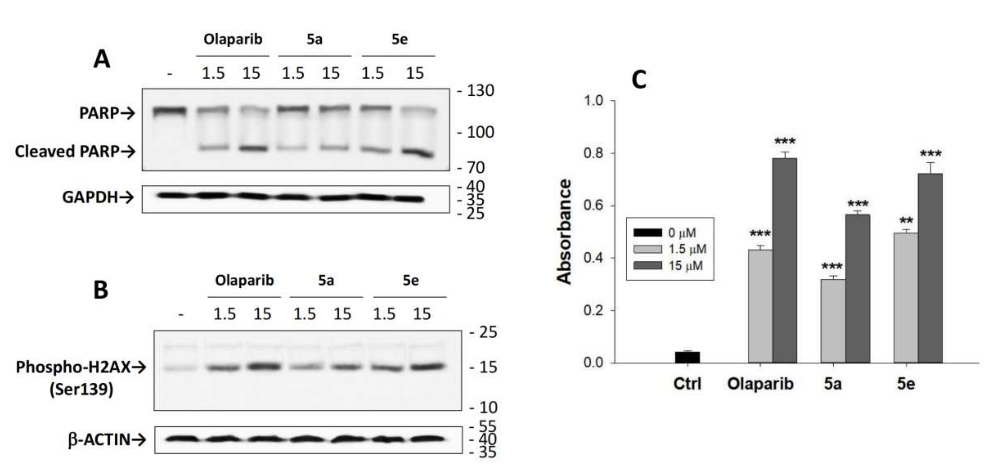
The effects of compounds 5a, 5e, and Olaparib on the cleavage of PARP1 in MCF-7 cells were performed using the previously reported protocol [24,25,26,27]. MCF-7 cells were incubated with 5a, 5e, or Olaparib at the indicated concentrations. As observed in Figure 3A, treatment of breast cancer cells with either compound 5a or 5e produce PARP cleavage comparable to that observed with Olaparib.
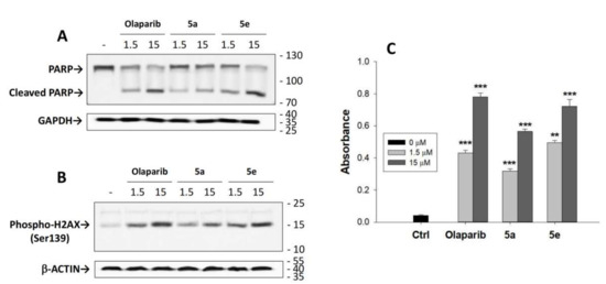
Figure 3.
Compounds 5a, 5e, and Olaparib cleave PARP1 (A) and increase phosphorylation of H2A-X (B) and CASPASE-3/7 activity (C) in MCF-7 cells. MCF-7 cells were treated with or without compounds 5a, 5e, or Olaparib at the indicated concentrations. The respective protein bands were detected by Western blot analysis. GAPDH or β-ACTIN were used as input controls. Data are expressed as mean ± SEM. ** p ≤ 0.01; *** p ≤ 0.001.
Phosphorylation of Ser139 in H2AX is an indicator of cellular DNA damage which occurs upon PARP inhibition [28]. For example, Olaparib has been reported to enhance H2AX phosphorylation in various cancer cell lines [29]. Therefore, the effect of compounds 5a or 5e or Olaparib on the level of p-H2AX in MCF-7 cells was determined (Figure 3B). It was observed that compounds 5a and 5e increased phosphorylation of H2AX in MCF-7 cells comparable to that observed with Olaparib.
The effect of compounds 5a or 5e and Olaparib on CASPASE 3/7 activity was also examined as an indicator of apoptosis [30,31,32]. Hence, CASPASE 3/7 activity was determined after treatment of breast cancer cells with compounds 5a or 5e or Olaparib. Both compounds 5a and 5e increased CASPASE 3/7 activity in a dose-dependent manner and were roughly comparable to that observed with Olaparib (Figure 3C).
2.5. Density Functional Theory (DFT) Calculations
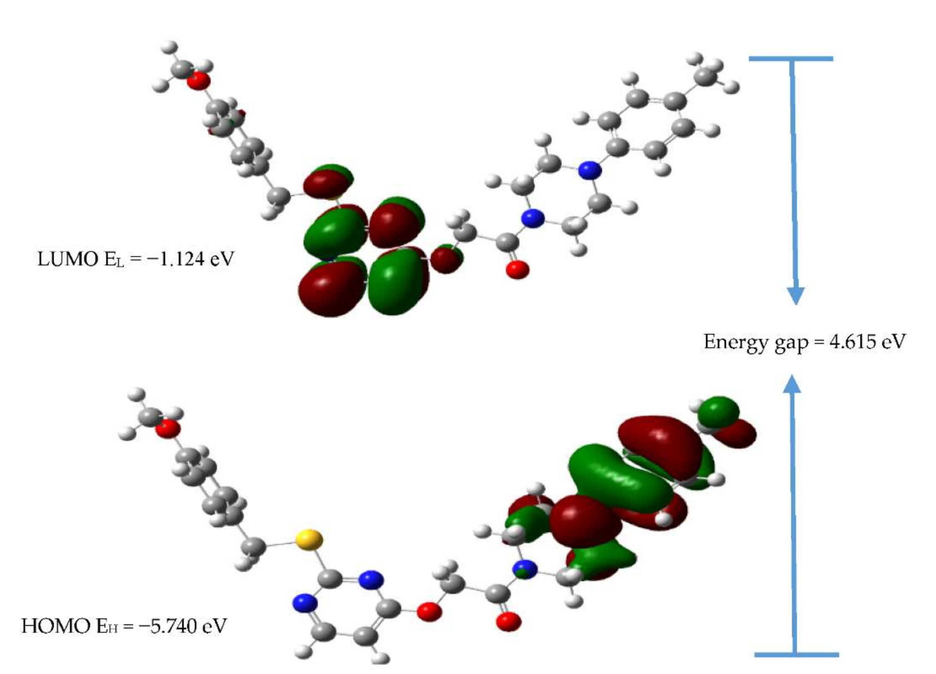
DFT calculations were performed to understand the stability of compound 5e in the catalytical grove of PARP1 [33,34,35]. According to the frontier molecular orbital theory, the highest occupied molecular orbitals (HOMO) and lowest unoccupied molecular orbitals (LUMO) are the most effective factors in bioactivity. HOMO has the priority to provide electrons, while LUMO can accept electrons. Thus, studies on the frontier orbital energy may provide useful information of the biological mechanism [36]. The B3LYP level of theory was used to calculate the energy of HOMO, LUMO, and the Egap value, which were used to study the chemical stability, molecular polarizability, and chemical softness or hardness of compound 5e. The distribution and energy levels of the HOMO and LUMO for 5e are shown in Figure 4. The positive phase of the molecule is represented in red color, whereas the negative phase is in green color. It is clear from the plots that the HOMO is localized on the 1-(4-methylphenyl)piperazine ring. In contrast, LUMO is delocalized on the methoxypyrimidine ring and sulfur atom. The HOMO and LUMO values of conformer 5e are −5.740 eV and −1.124 eV, respectively, and the energy separation between HOMO and LUMO is 4.615 eV. The stabilization and interactions of the compounds with the catalytic site of the enzyme may be explained by global reactivity descriptors and are represented in Table 2. The ionization energy can be expressed as I = −EHOMO = 5.740 eV, and the electron affinity can be expressed as A = −ELUMO = 1.124 eV. Hardness represents the resistance of a chemical system against the deformation of the electron cloud under the slight interference encountered in the chemical process and can be expressed as η = (ELUMO − EHOMO)/2. The hardness and softness values of the 5e molecule are 2.307 eV and 0.216 e−1 V−1, respectively. With the smaller value of softness, the molecule is chemically stable.
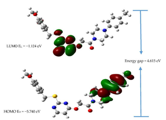
Figure 4.
The HOMO and LOMO of molecule 5e obtained by the DFT/B3LYP/6-31+G (d,p) method.

Table 2.
The calculated FMOs and global chemical reactivity descriptors.
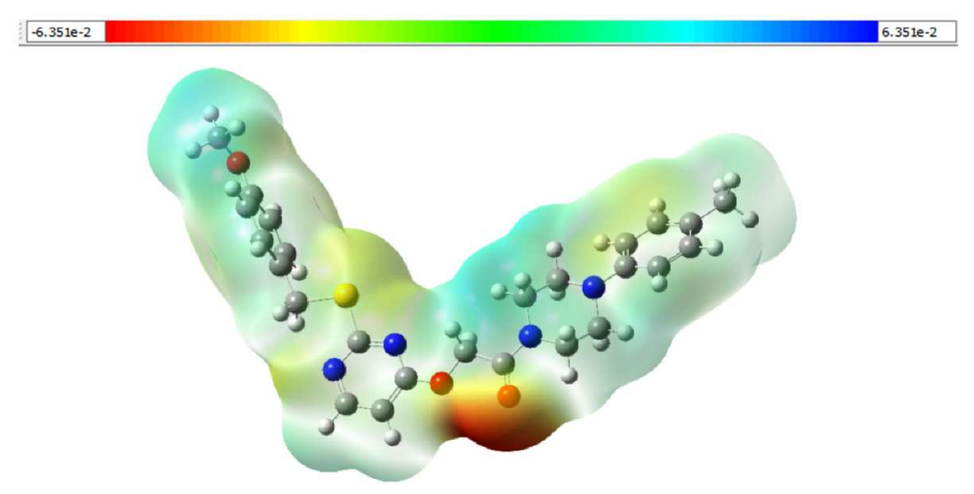
MEPs are fundamental measures of the interaction strength of the nearby charges, nuclei, and electrons at a particular position, thus enabling visualization of the charge distribution and charge-related characteristics of molecules. To facilitate interpretation of the electrostatic potential data, a visual representation with a chromatogram is used. The B3LYP/6-31+G (d,p) level of theory was used to study the molecular electrostatic potential in the intermolecular interaction region of conformer 5e. The resulting MEP map with a range of −6.351 × 10⁻²–6.351 × 10⁻² depicts its potential distribution (see Figure 5). The O atom of the C=O bond of molecule 5e is surrounded by a positive charge, indicating a possible nucleophilic attack site. Furthermore, a negatively charged region is located on the hydrogen atoms of the piperazine ring, indicating a probable electrophilic attack site.
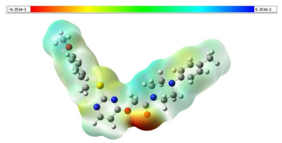
Figure 5.
Molecular electrostatic potential surfaces of compound 5e.
To determine the conformational stability of compounds at the PARP1 binding site, MD simulations are employed to assess structural and dynamic changes throughout the protein–ligand interactions [37]. The complex structures are solved using orthorhombic simple point charge (SPC) water models. On periodic boundary conditions, the system was determined to have a cut-off for Lennard–Jones interactions of 10 Å. In the integration steps, a time step of 2.0 fs was used. The system temperature and pressure were set to 300 K and 1.01325 bar, respectively, using the Nose–Hoover thermostat and Martyna–Tobias–Klein protocols. Before the production run, the system was minimized, equilibrated, and 100 ns MD simulations were then performed [38].
2.6. Bioinformatic Analysis of the Binding of Compound 5e towards the Catalytical Grove of PARP1
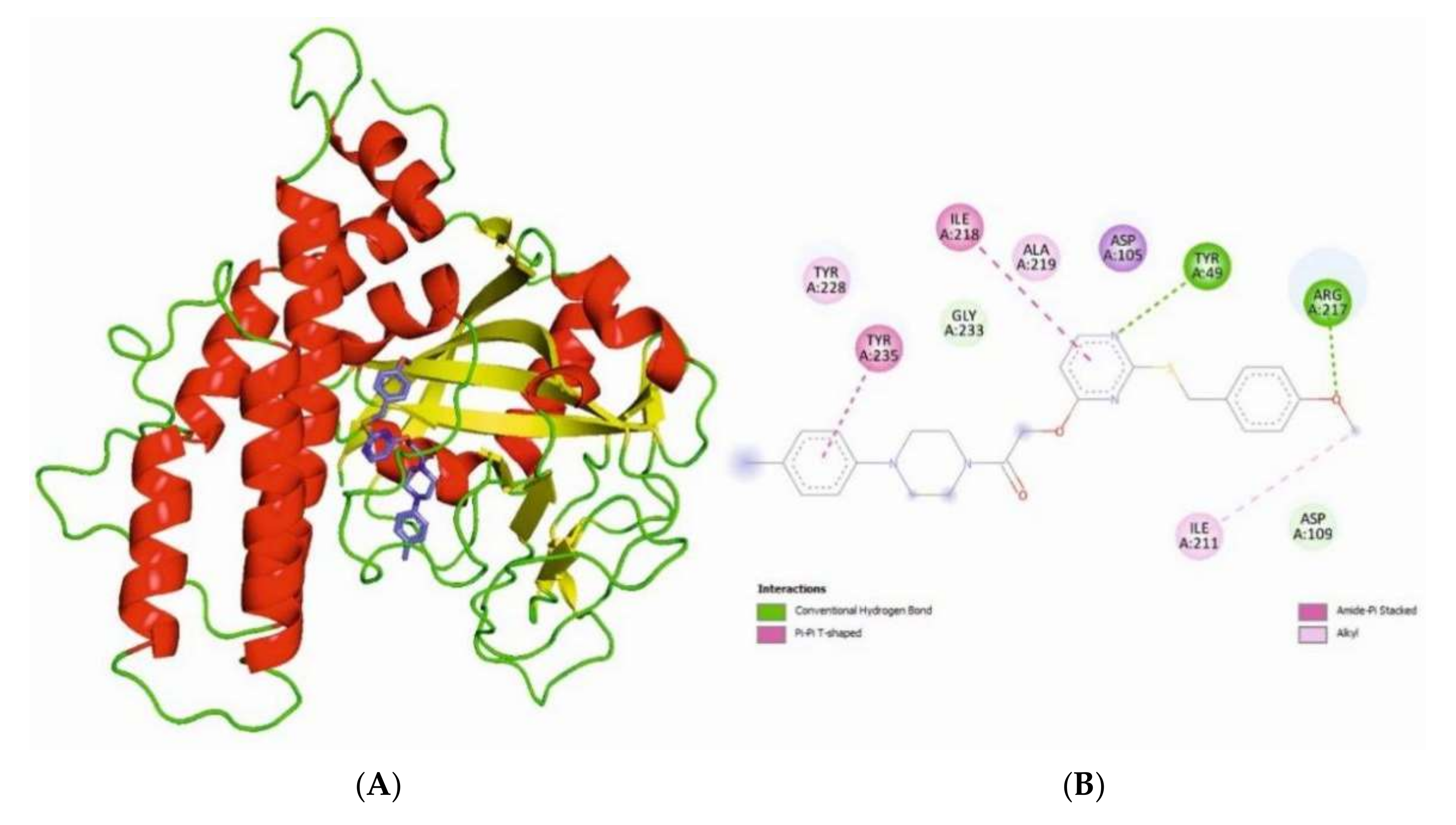
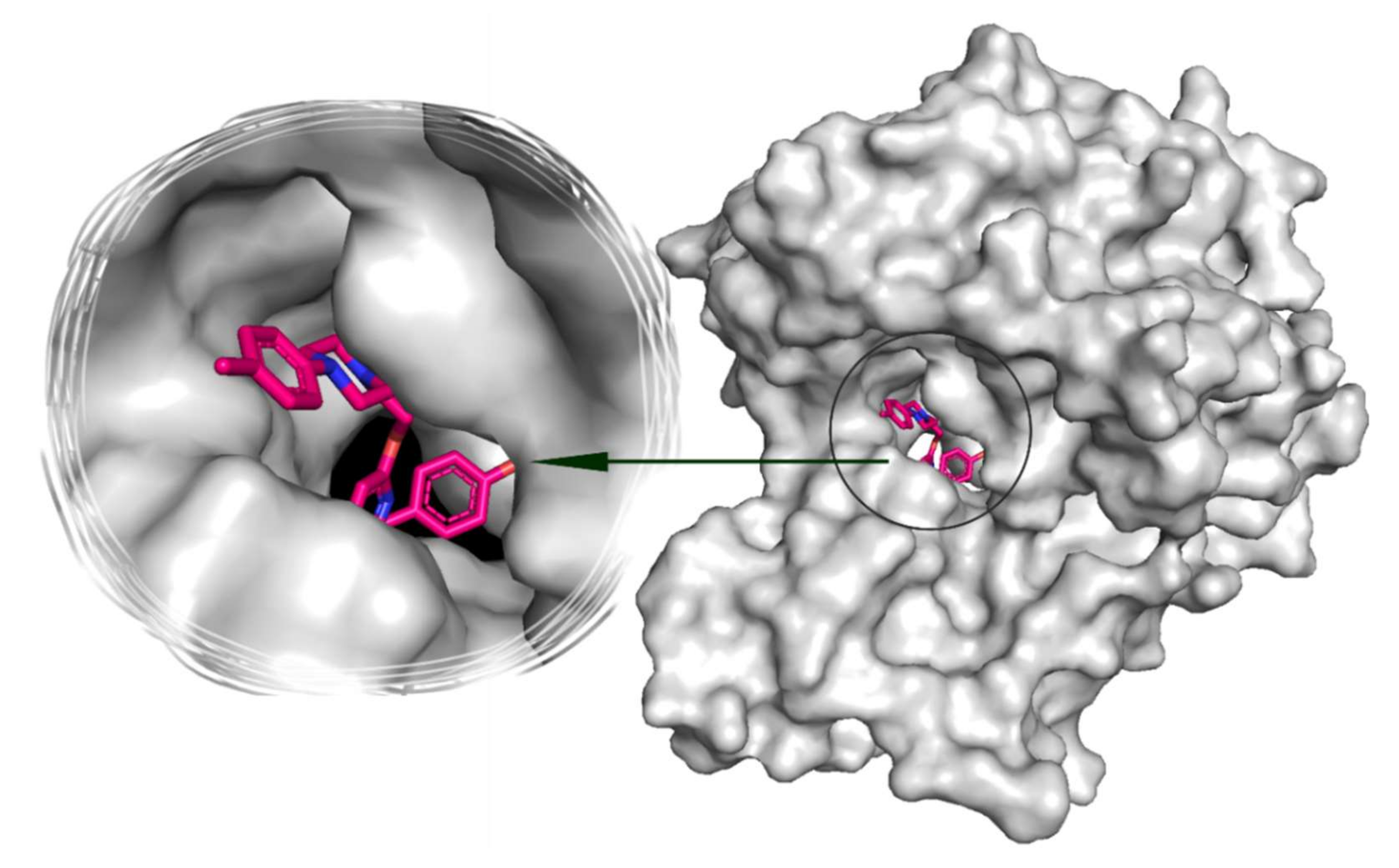
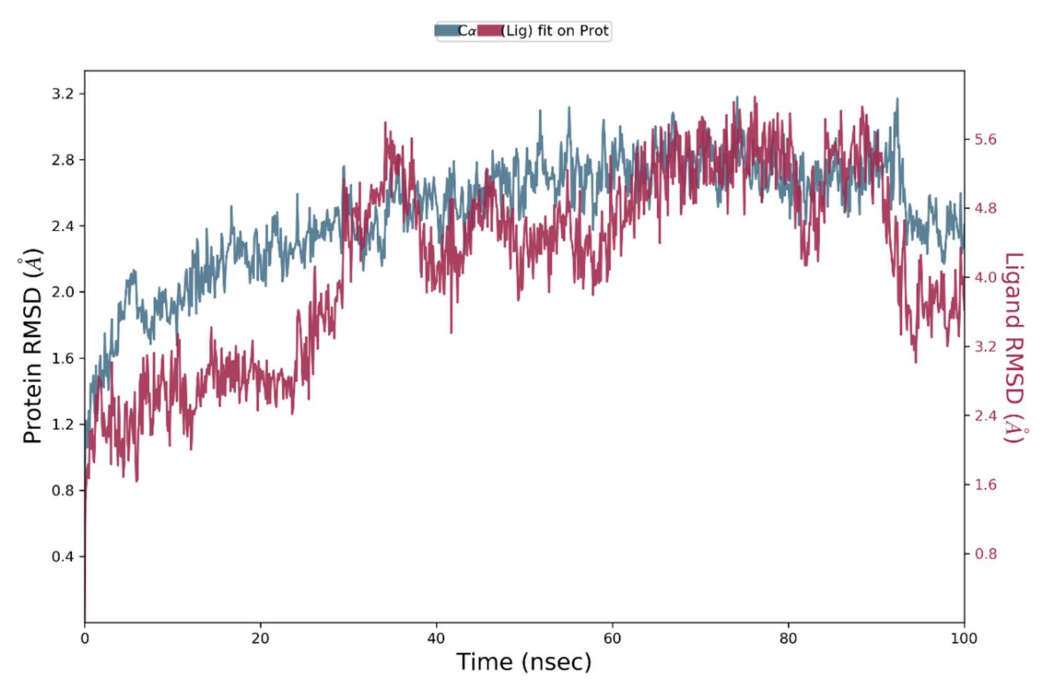
Compound 5e was studied using molecular docking modeling to assess the binding of compound 5e towards the catalytical grove of PARP1 [39]. Figure 6A depicts the binding pose of compound 5e with PARP1. Docking simulations revealed the prominent interactions of compound 5e with PARP1 active site residues (Figure 6B). The docking score was assigned to compound 5e that binds to the active site with −7.33 kcal/mol energy and forms potential hydrogen bonds. TYR49 is bonded with the nitrogen of the pyrimidine ring and ARG217 with the oxygen of methoxybenzene with the bond distances 2.50 and 3.56 Å (Figure 7), respectively. Furthermore, pi–pi stacking interactions exist with ILE218 and TYR235. In addition to the latter, hydrophobic interactions have been discovered in the active site [39]. 100 ns MD simulations were run for the molecule in the PARP1 binding pocket, and the associated coordinates throughout the MD simulation were gathered separately in the trajectory file. Figure 8 depicts the root-mean-square deviations (RMSDs) of Cα atoms in the investigated systems. After 30 ns of MD simulations, all of the studied systems have relaxed and show no significant fluctuations in RMSD values. The evaluated system exhibited an average RMSD of less than 3.0 Å. The LigFitProt RMSD plot displays the RMSD of ligands when the protein–ligand complex is initially aligned as a reference state with the backbone atoms of the protein.
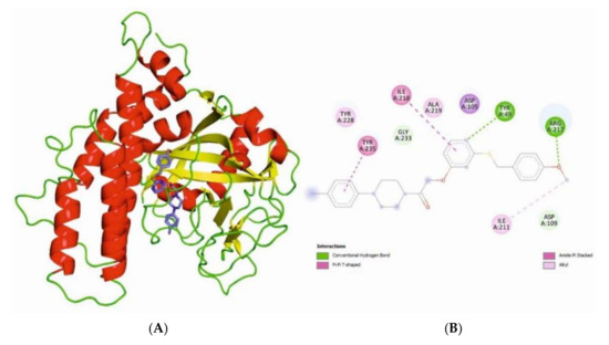
Figure 6.
Cartoon representation of PARP1 with compound 5e (A) and its 2D interactions (4HHY) (B).
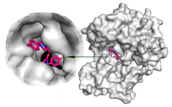
Figure 7.
3D surface view of PARP1 and compound 5e and a zoomed view of the compound 5e in the active site cavity.
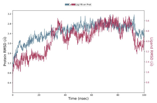
Figure 8.
RMSD plot of ligand 5e and PARP1.
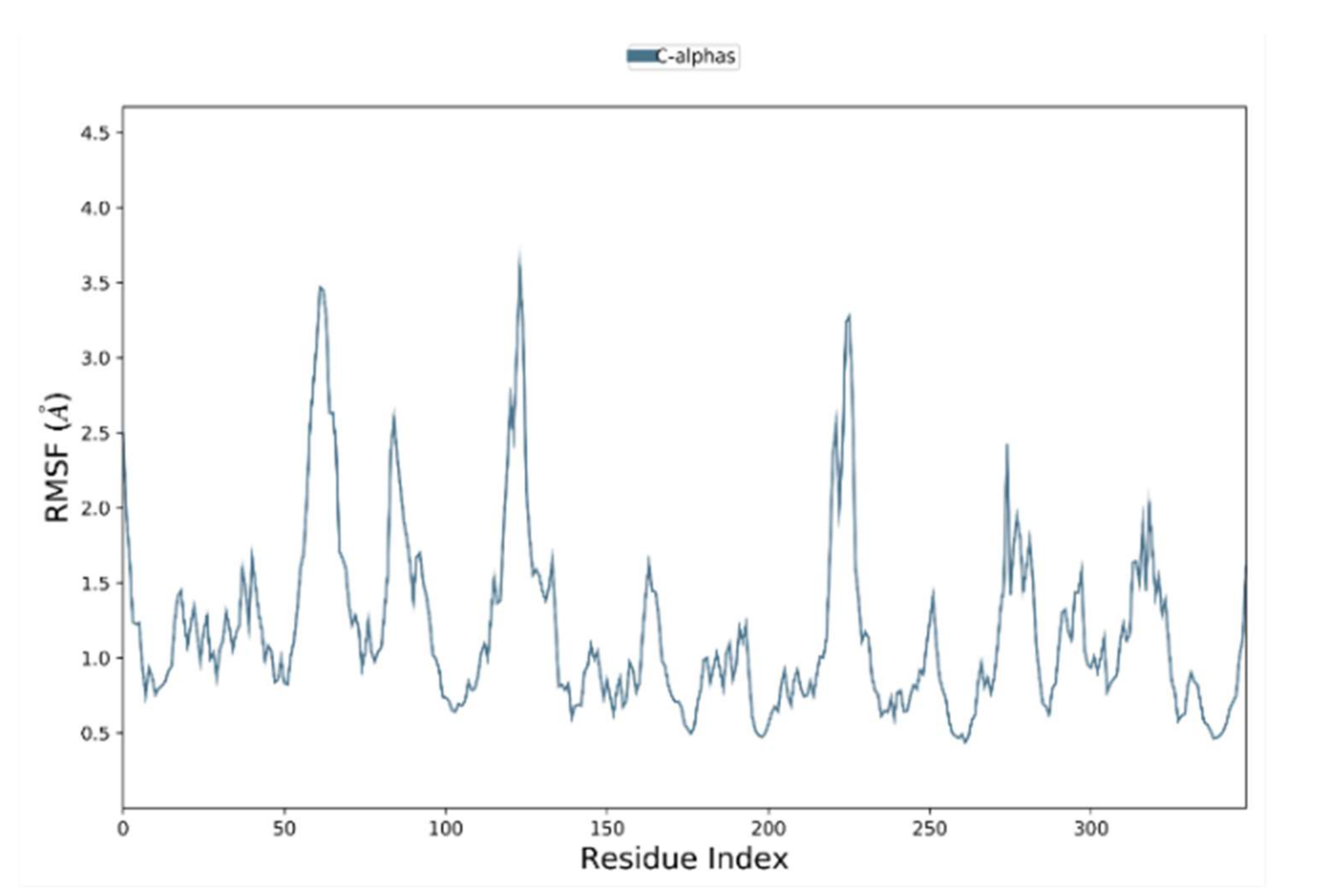
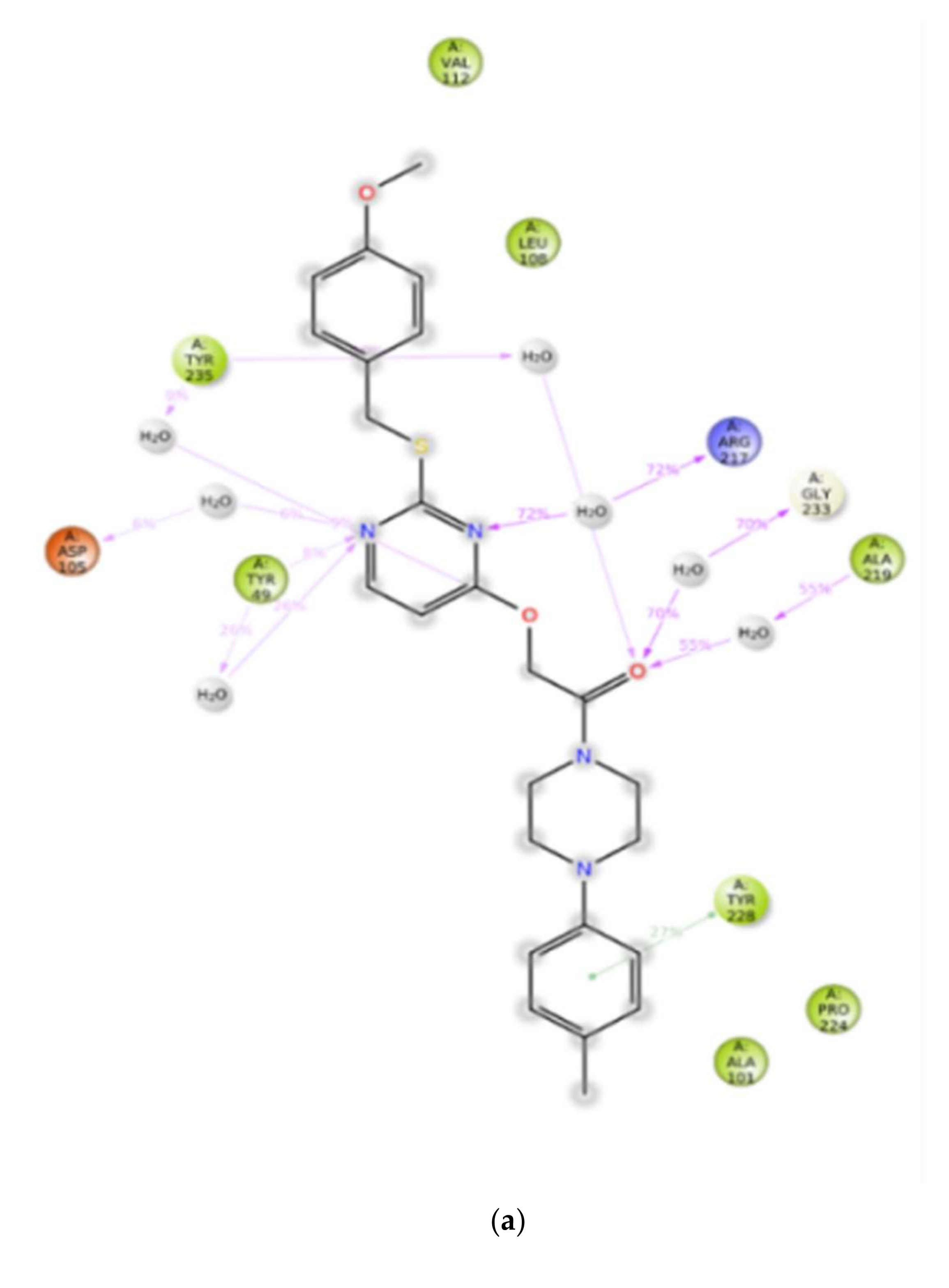
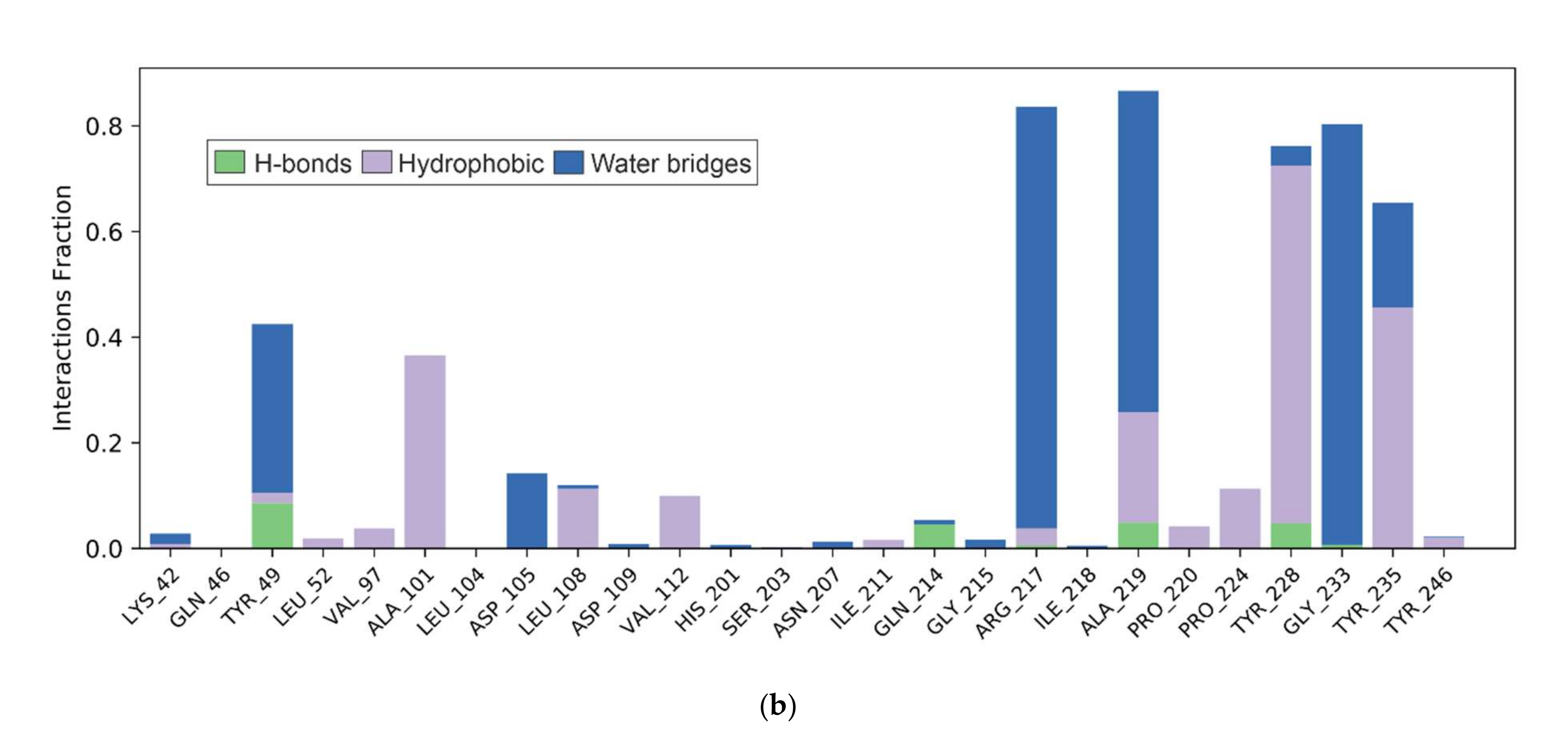
Root-mean-square fluctuation (RMSF) values were also calculated during simulations to investigate the effect of defined hits on the mobility of the target protein’s backbone atoms (Figure 9). Figure 10A depicts associations that appear in the chosen path for more than 6% of the simulation time (0.00–100.00 ns). Hydrogen bonds with TYR49 and π–π stacking interactions with TYR228 produce significant interactions in the molecule. ASP105, ARG217, ALA219, GLY233, and TYR235 have stable non-bonded chemical contacts throughout the simulations, as shown by the protein–ligand interactions graph Figure 10B. The stability between compound 5e and PARP1 is furthermore enhanced by the interaction with the amino acid residues ARG217, GLY233, ALA219, and TYR49 of PARP1 via water bridges [40].
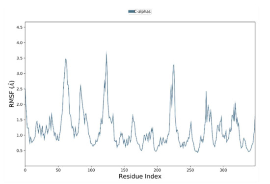
Figure 9.
RMSF plot of protein molecule (4HHY) PARP1.
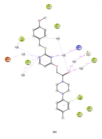
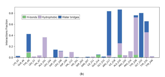
Figure 10.
(a) Compound 5e-PARP1 interaction and (b) histogram charts of the interaction percentages of the MD simulation for the complex.
3. Materials and Methods
All organic chemicals used were purchased from Sigma-Aldrich. 1H and 13C NMR spectra were recorded on a Bruker WH-200 (400 MHz) and JEOLJSM-ECS (400 MHz) spectrometer in CDCl3 as solvent, using TMS as an internal standard, and chemical shifts are expressed as ppm. High-resolution mass spectra were determined on a Bruker Daltonics instrument. Mass spectra were determined on an Agilent LC-MS and the elemental analyses were carried out using an Elemental Vario Cube CHNS rapid analyzer. The progress of the reaction was monitored by TLC precoated silica gel G plates [41].
3.1. Synthesis of Compound 2
To a stirred solution of 2-thiouracil, p-methoxy benzyl bromide in DMF was added potassium carbonate, and the reaction mixture was heated to 80 °C. The reaction was monitored by thin-layer chromatography, quenched with water, and extracted with ethyl acetate. The organic layer was washed with brine and dried over sodium sulfate and concentrated under vacuum. The crude product was purified by column chromatography with silica-60 to yield the alcohol 2 in 81% yield.
3.2. Synthesis of Compound 3
To a stirred solution of alcohol 2, tert-butyl bromoacetate in DMF was added potassium carbonate (1.2 eq), and the reaction mixture was heated to 80 °C. The reaction was monitored by thin-layer chromatography. After completion of the reaction, it was quenched with water and extracted with ethyl acetate. The organic layer was washed with brine and dried over sodium sulfate and concentrated under vacuum. The crude product was purified by column chromatography with silica-60 to afford the ester 3 in 85% yield.
3.3. Synthesis of Compound 4
To a stirred solution of ester 3 in dichloromethane, was added trifluoroacetic acid and stirred overnight. After completion of the reaction as monitored by thin-layer chromatography, it was quenched with sodium bicarbonate and extracted with ethyl acetate. The organic layer was washed with brine and dried over sodium sulfate and concentrated under vacuum. The crude product was purified by column chromatography with silica-60 to afford acid in 65% yield.
3.4. General Synthetic Method for the Preparation of 5a–5l
To a stirred solution of the acid (100 mg, 0.33 mol) in dry dichloromethane (4 mL) under nitrogen atmosphere, was added EDC. HCl (93.7 mg, 0.49 mol). Then, DMAP (4 mg, 10 mol%), amine derivative (130.8 mg, 0.49 mol) was added to the reaction mixture and stirred for five minutes. Triethylamine (0.15 mL, 0.99 mol) was then added slowly, dropwise, to the reaction mixture and it was stirred for 2–3 h. After completion of the reaction, as indicated by TLC, it was quenched with water (5 mL) and extracted with ethyl acetate (2 × 10 mL). The combined organic layers were washed with brine (5 mL) and dried over anhydrous sodium sulfate. Evaporation of the solvent gave the crude residue which was purified by silica gel column chromatography using petroleum ether/EtOAc (3:7) as eluent to furnish amides 5a–5l.
1-(4-(2,3-Dichlorophenyl)piperazin-1-yl)-2-((2-((4-methoxybenzyl)thio)pyrimidin-4-yl)oxy)ethanone (5a), Reddish yellow solid; MP: 145–148 °C; 88% yield; 1H NMR (400 MHz, CDCl3): δ 8.29 (d, J = 5.6 Hz, 1H), 7.30 (d, J = 8.4 Hz, 2H), 7.18 (d, J = 8.0 Hz, 1H), 7.11 (t, J = 8.0 Hz 1H), 6.83–6.78 (m, 3H), 6.58 (d, J = 5.6 Hz, 1H), 5.03 (s, 2H), 4.33 (s, 2H), 3.77 (s, 5H), 3.59 (s, 2H), 3.07 (s, 4H; 13C NMR (100 MHz, CDCl3): δ 173.1, 169.6, 167.3, 160.8, 159.7, 152.3, 150.3, 136.0, 132.0, 130.9, 129.7, 129.4, 127.2, 120.8, 120.5, 115.9, 105.9, 65.0, 57.2, 53.4, 47.1, 44.2, 41.2, 36.8; LCMS (ESI): m/z for C24H24Cl2N4O3S+H, calcd 519.0946; found: 519.0929.
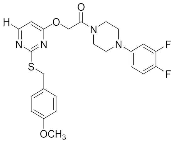
1-(4-(3,4-Difluorophenyl)piperazin-1-yl)-2-((2-((4-methoxybenzyl)thio)pyrimidin-4-yl)oxy)ethanone (5b), Yellow white solid; MP: 135–138 °C; 92% yield; 1H NMR (400 MHz, CDCl3): δ 8.29 (d, J = 5.7 Hz, 1H), 7.28 (s, 1H), 7.02 (d, J = 9.5 Hz, 2H), 6.80 (d, J = 8.5 Hz, 2H), 6.61–6.51 (m, 3H), 5.02 (s, 2H), 4.31 (s, 2H), 3.76 (d, 5H), 3.58 (s, 2H), 3.07 (d, J = 16.2 Hz, 4H); 13C NMR (100 MHz, CDCl3): δ 167.8, 165.5, 159, 158.0, 148.0, 130.0, 129.1, 117.5, 114.1, 112.5, 106.3, 104.1, 63.11, 55.2, 50.1, 49.8, 44.8, 41.9, 40.0, 35.1, 22.8; LCMS (ESI): m/z for C24H24F2N4O3S+H, calcd 487.1537; found: 487.0645.
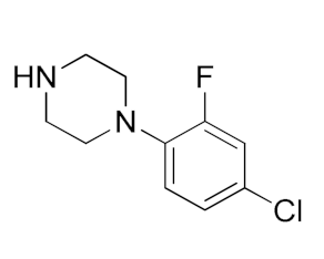
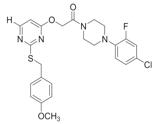
1-(4-(4-Chloro-2-fluorophenyl)piperazin-1-yl)-2-((2-((4-methoxybenzyl)thio)pyrimidin-4-yl)oxy)ethanone (5c), White solid; MP: 142–145 °C; 90% yield; 1H NMR (400 MHz, CDCl3): δ 8.29 (d, J = 5.6 Hz, 1H), 7.28 (d, J = 24.8 Hz, 2H), 7.05 (dd, J = 12, 2.4 Hz, 1H), 6.97 (d, J = 8.4 Hz, 1H) 6.82 (d, J = 8.8 Hz, 2H), 6.73 (m,1H), 6.58 (d, J = 5.6 Hz, 1H), 5.02 (s, 2H), 4.31 (s, 2H), 3.77 (s, 5H), 3.58 (m, 2H), 3.01 (m, 4H); 13C NMR (100 MHz, CDCl3): 171.2, 167.7, 165.3, 158.9, 157.8, 156.6, 154.1, 138.3, 138.2, 130.0, 129.0, 124.6, 124.5, 120.0, 117.1, 116.8, 114.0, 104.0, 63.0, 55.2, 50.7, 50.2, 42.0, 34.9.; LCMS (ESI): m/z for C24H24ClFN4O3S+H, calcd 503.1242; found: 503.0788.
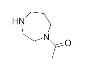
1-(4-Acetyl-1,4-diazepan-1-yl)-2-((2-((4-methoxybenzyl)thio)pyrimidin-4-yl)oxy)ethanone (5d), 1H NMR (400 MHz, CDCl3): δ 8.27 (d, J = 5.8 Hz, 1H), 7.29 (d, J = 8.4 Hz, 2H), 6.82 (d, J = 8.2 Hz, 2H), 6.56 (d, J = 5.9 Hz, 1H), 5.00–4.96 (m, 2H), 4.31 (d, J = 5.0 Hz, 2H), 3.77 (s, 3H), 3.63–3.47 (m, 8H), 2.07 (d, J = 3.4 Hz, 3H), 1.90 (d, J = 31.1 Hz, 2H); 13C NMR (100 MHz, CDCl3): δ δ171.2, 170.7, 166.9, 159.1, 157.7, 130.1, 129.3, 114.1, 104.2, 63.2, 62.9, 55.4, 49.2, 48.6, 45.1,44.4, 34.9; LCMS (ESI): m/z for C21H26N4O4S+H, calcd 431.1675; found: 431.0702.
2-((2-((4-Methoxybenzyl)thio)pyrimidin-4-yl)oxy)-1-(4-(p-tolyl)piperazin-1-yl)ethanone (5e), 1H NMR (400 MHz, CDCl3): δ 8.29 (d, J = 5.6 Hz, 1H), 7.29 (d, J = 16.0 Hz, 2H), 7.07 (d, J = 8.0 Hz, 2H), 6.87–6.74 (m, 4H), 6.57 (d, J = 5.6 Hz, 1H), 5.03 (s, 2H), 4.32 (s, 2H), 3.78 (s, 5H), 3.57 (S, 2H), 3.16–3.02 (m, 4H), 2.28 (s, 3H); 13C NMR (100 MHz, CDCl3): δ 171.4, 167.9, 165.5, 159.0, 157.9, 130.5, 130.2 129.9, 117.4, 114.1, 104.1, 63.3, 55.4, 50.4, 50.1, 45.0, 42.1, 35.0, 20.6; LCMS (ESI): m/z for C25H28N4O3S+H, calcd 465.1882; found: 465.0852.
1-(4-(2-Fluorophenyl)piperazin-1-yl)-2-((2-((4-methoxybenzyl)thio)pyrimidin-4-yl)oxy)ethanone (5f), 1H NMR (400 MHz, CDCl3): δ 8.28 (d, J = 5.6 Hz, 1H), 7.29 (d, J = 8.8 Hz, 2H), 7.05–6.94 (m, 4H), 6.86–6.81 (m, 3H), 6.57 (d, J = 5.6 Hz, 1H), 5.03 (s, 2H), 4.32 (s, 2H), 3.77 (s, 5H), 3.58 (s, 2H), 3.12–3.02 (m, 4H); 13C NMR (100 MHz, CDCl3): δ 171.2, 167.8, 165.3, 158.8, 157.8, 157.0, 139.4, 139.3, 130.1, 130.0, 129.5, 128.5, 125.1, 123.3, 119.3, 117.6, 116.3, 113.5, 103.5, 68.1, 63.1, 55.3, 50.8, 50.3, 45.0, 42.1, 34.8; LCMS (ESI): m/z for C24H25FN4O3S+H, calcd 469.1631; found: 469.2454.
2-((2-((4-Methoxybenzyl)thio)pyrimidin-4-yl)oxy)-N-(5-(trifluoromethyl)-1,3,4-thiadiazol-2-yl)acetamide (5g), 1H NMR (400 MHz, CDCl3): δ 8.33 (d, J = 5.3 Hz, 1H), 7.18 (d, J = 8.0 Hz, 2H), 6.74 (d, J = 8.0 Hz, 2H), 6.62 (d, J = 5.4 Hz, 1H), 5.21 (s, 2H), 4.23 (s, 2H), 3.73 (s, 3H); 13C NMR (100 MHz, CDCl3): δ 171.1, 166.6, 166.4, 161.0, 158.3, 157.7, 129.5, 129.3, 128.2, 113.4, 103.4, 63.6, 54.7, 34.2; LCMS (ESI): m/z for C17H14F3N5O3S2+H, calcd 458.0490; found: 458.1220.
N-(Furan-2-ylmethyl)-2-((2-((4-methoxybenzyl)thio)pyrimidin-4-yl)oxy)acetamide (5h), 1H NMR (400 MHz, CDCl3): δ 8.29 (d, J = 5.6 Hz, 1H), 7.40–7.26 (m, 3H), 6.83 (d, J = 8 Hz, 2H), 6.58 (brs, 1H), 6.48 (d, J = 6.0 Hz, 1H), 6.32 (s, 1H), 6.24 (s, 1H), 4.86 (s, 2H), 4.51 (d, J = 5.6 Hz, 2H), 4.31 (s, 2H), 3.78 (s, 3H); 13C NMR (100 MHz, CDCl3): δ 172.1, 167.0, 158.8, 158.1, 150.6, 142.3, 130.8, 130.1, 1291, 128.8, 114.5, 110.5, 107.7, 103.4, 68.1, 64.8, 55.2, 36.0, 34.8; LCMS (ESI): m/z for C19H19N3O4S+H, calcd 386.1096; found: 386.1792.
2-((2-((4-Methoxybenzyl)thio)pyrimidin-4-yl)oxy)-N-(1,2,3,4-tetrahydronaphthalen-1-yl)acetamide (5i), 1H NMR (400 MHz, CDCl3): δ 8.27 (d, J = 5.7 Hz, 1H), 7.70 (dd, J = 5.7, 3.3 Hz, 1H), 7.53 (dd, J = 5.7, 3.3 Hz, 1H), 7.34 (d, J = 8.6 Hz, 2H), 7.18 (s, 1H), 7.11 (s, 1H), 6.85 (d, J = 8.6 Hz, 2H), 6.44 (d, J = 5.7 Hz, 1H), 4.89 (d, J = 3.5 Hz, 2H), 4.35 (s, 2H), 4.22 (m,1H), 3.78 (s, 3H), 2.79 (dd, J = 12.4, 5.9 Hz, 2H), 1.81 (dd, J = 12.0, 6.7 Hz, 4H); 13C NMR (100 MHz, CDCl3): δ 171.9, 167.7, 167.1, 166.6, 158.8, 158.0, 137.63, 136.1, 130.84, 130.0, 129.2, 129.0, 128.43, 128.4, 128.2, 127.4, 126.4, 113.9, 103.5, 68.1, 55.3, 47.3, 38.7, 34.9, 20.1.
2-((2-((4-Chlorobenzyl)thio)-6-methylpyrimidin-4-yl)oxy)-1-(4-(3,4-dichlorophenyl)piperazin-1-yl)ethan-1-one (5j), Off white solid; MP: 135–138 °C; 88% yield; 1H NMR (400 MHz, CDCl3): δ 7.32 (d, J = 8.4 Hz, 2H), 7.24 (d, J = 8.2 Hz, 2H), 7.19 (dd, J = 8.0, 1.1 Hz, 1H), 7.10 (t, J = 8.0 Hz, 1H), 6.80 (dd, J = 8.0, 1.1 Hz, 1H), 6.42 (s, 1H), 4.99 (s, 2H), 4.32 (s, 2H), 3.76 (s, 2H), 3.57 (s, 2H), 3.03–2.94 (m, 4H), 2.38 (s, 3H); 13C NMR (100 MHz, CDCl3): δ 169.8, 168.4, 165.6, 150.3, 136.4, 134.2, 133.0, 131.1, 130.1, 128.2, 127.8, 127.5, 125.3, 118.8, 102.7, 63.1, 51.2, 50.9, 50.7, 45.1, 42.2, 34.5, 30.3, 23.8. LCMS (ESI): m/z for C24H23Cl3N4O2S, calcd 537.8890; found:537.0455.
1-(4-(4-Chlorophenyl)piperazin-1-yl)-2-((2-((4-fluorobenzyl)thio)-6-methylpyrimidin-4-yl)oxy)ethanone (5k), Off white solid; MP: 138–142 °C; 90% 1H NMR (400 MHz, CDCl3): δ 7.34 (dd, J = 8.1, 5.6 Hz, 2H), 7.22 (d, J = 8.8 Hz, 2H), 6.96 (t, J = 8.6 Hz, 2H), 6.82 (d, J = 8.5 Hz, 2H), 6.43 (s, 1H), 5.00 (s, 2H), 4.32 (s, 2H), 3.75 (s, 2H), 3.59 (s, 2H), 3.12 (d, J = 20.8 Hz, 4H), 2.40 (s, 3H). LCMS (ESI): m/z for C24H24ClFN4O2S, calcd 486.9894; found: 487.1152.
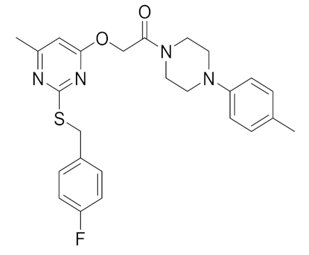
2-((2-((4-Fluorobenzyl)thio)-6-methylpyrimidin-4-yl)oxy)-1-(4-(p-tolyl)piperazin-1-yl)ethanone (5l), Yellowish white solid; MP: 134–137 °C; 88% yield; 1H NMR (400 MHz, CDCl3): δ 7.35 (dd, J = 8.4, 5.5 Hz, 2H), 7.09 (d, J = 8.3 Hz, 2H), 6.96 (t, J = 8.6 Hz, 2H), 6.83 (d, J = 5.1 Hz, 2H), 6.43 (s, 1H), 5.01 (s, 2H), 4.33 (s, 2H), 3.76 (s, 2H), 3.59 (s, 2H), 3.10 (d, J = 17.7 Hz, 4H), 2.40 (s, 3H), 2.29 (s, 3H). LCMS (ESI): m/z for C25H27FN4O2S, calcd 466.5709; found: 467.1684.
3.5. Cell Viability Assay
MCF-7 and MCF-10A cells were procured from Procell Life Science and Technology Co. LTD, China. Cells (2000) were cultured in MEM or Leibovitz’s L-15 medium enriched with 2% FBS, MCF-10A cells were cultured in DMEM with 5% horse serum (HS) (general culture: 2% HS was used for experimental conditions), 20 ng/mL EGF, 0.5μg/mL hydrocortisone, 10μg/mL insulin, 1% NEAA (non-essential amino acids); and maintained in a humidified atmosphere of 5% CO2 at 37 °C. DMSO-dissolved compounds were stored as a stock solution, and diluted with cell culture medium as required. Cells (4 × 10³) were cultured for 12 h in 96-well plates and treated with thiouracil amides or Olaparib at 0, 0.01, 0.1, 10, 100, and 1000 µM concentrations for 72 h. The inhibitory effect of the compounds was assessed using Alamar Blue [42,43,44,45].
3.6. Assay of PARP1 Catalytic Activity
The catalytic activity of PARP1 was determined using the PARP/Apoptosis Universal Colorimetric Assay Kit (4677-096-K, R&D Systems Ltd., Abingdon, UK) [46]. Compounds 5a or 5e or Olaparib were added to recombinant human PARP enzyme prior to evaluation as per manufacturer’s instructions.
3.7. Western Blot Analysis
Western blot analysis was performed to determine the levels of cleaved PARP1, and the phosphorylation ofH2AX in MCF-7 cells using previously reported methods [29]. Cells at 50–60% confluence were treated with compounds and incubated for 60 h in 2% FBS. MCF-7 cells were also treated with Olaparib, 5a, or 5e, and the levels of cleaved PARP1 and the phosphorylation of p-H2AX were determined using the respective antibodies, as previous [29].
3.8. CASPASE-3/7 Activity Assay
CASPASE-3/7 activity was determined by the Caspase-Glo® 3/7 assay kit (Promega corporation, Madison, WI, USA) according to the manufacturer’s instructions [30]. MCF-7 cells were seeded in 96-well plates and incubated overnight. Compounds or vehicle were added to cells after 12 h. After a 24 h incubation period, Caspase-Glo® 3/7 reagent and media (1:1) were added into each well and processed according to the manufacturer’s instructions.
3.9. DFT Calculations for the Compound 5e
Computational DFT studies for the 5e molecule were performed using Gaussian09 [47]. The molecule was built and visualized by Gaussview 5 program package. The molecular geometry of the 5e molecule was optimized at B3LYP (Becke’s three parameter (B3)) level with a 6-311+G (d,p) basis set [48].
3.10. Bioinformatics Studies for the Compound 5e
The Scripps Research Institute’s AutoDock Tools (ADT) (v1.5.7) [49,50] was used to generate grid and docking parameter files.The crystal structure of PARP1 with the ligand was used to retrieve the crystal structure of PARP1 with a ligand (PDB ID: 4HHY) and use it as afor the docking purpose (PDB ID: 4HHY). With the centroid rendered from inhibitor in the crystallographic structure, the grid box size of 65 × 65 × 65 Å with 0.481 Å spacing is defined. The empirical free-energy function and the Lamarckian genetic algorithm were used to perform molecular docking with the macromolecule, with an initial population of 150 randomly placed individuals, a maximum number of 2,500,000 energy evaluations, a mutation rate of 0.02, a crossover rate of 0.80, and 10 docking runs. Visualization plots were examined using BIOVIA Discovery Studio Visualizer (v21.10.20298) [51] and PyMOL (v2.5.2) [52,53].
3.11. Statistical Analysis
The data were analyzed by Student’s t-test and p < 0.05 was considered statistically significant (GraphPad Prism 5.0; GraphPad Software, La Jolla, CA, USA).
4. Conclusions
A series of thiouracil derivatives containing amide linkages were synthesized and screened for loss of viability of breast cancer cells. The compounds exhibited moderate to significant activity in MCF-7 cells. Compounds 5a and 5e inhibited PARP1 activity, produced PARP cleavage, and enhanced phosphorylation of H2AX. Computational methodologies demonstrated conclusively that compound 5e interacts with PARP.
Supplementary Materials
The following are available online at https://www.mdpi.com/article/10.3390/molecules27092848/s1, NMR spectra of Compounds 5a–5l.
Author Contributions
S.N.D., P.K.M., J.-R.Y., M.K.S., A.S., R.S. and O.N. performed the experiments. B.B., S.R., M.M., P.B.S., V.P., P.E.L., K.S.A., A.C. and S.A.A. designed the experiments. B.B. and P.E.L. provided resources and wrote the manuscript. All authors have read and agreed to the published version of the manuscript.
Funding
This work was supported by DBT-NER, and Vision Group on Science and Technology (CESEM), Government of Karnataka. This work was also supported by the Shenzhen Key Laboratory of Innovative Oncotherapeutics (ZDSYS20200820165400003) (Shenzhen Science and Technology Innovation Commission), China; Shenzhen Development and Reform Commission Subject Construction Project ([2017]1434), China; Overseas Research Cooperation Project (HW2020008) (Tsinghua Shenzhen International Graduate School), China; Tsinghua University Stable Funding Key Project (WDZC20200821150704001); the Shenzhen Bay Laboratory (21310031), China and TBSI Faculty Start-up Funds, China. This work was supported by a National Research Foundation of Korea (NRF) grant funded by the Korean government (MSIP) (NRF-2021R1I1A2060024). This project was supported by Researchers Supporting Project number (RSP-2022/5) King Saud University, Riyadh, Saudi Arabia.
Institutional Review Board Statement
Not applicable.
Informed Consent Statement
Not applicable.
Conflicts of Interest
The authors declare no conflict of interest.
Sample Availability
The synthesized compounds are available at Dr. Basappa Laboratory.
References
- Sonnenblick, A.; de Azambuja, E.; Azim, H.A., Jr.; Piccart, M. An update on PARP inhibitors—moving to the adjuvant setting. Nat. Rev. Clin. Oncol. 2015, 12, 27–41. [Google Scholar] [CrossRef] [PubMed]
- Beck, C.; Robert, I.; Reina-San-Martin, B.; Schreiber, V.; Dantzer, F. Poly(ADP-ribose) polymerases in double-strand break repair: Focus on PARP1, PARP2 and PARP3. Exp. Cell Res. 2014, 329, 18–25. [Google Scholar] [CrossRef] [PubMed]
- Kim, M.Y.; Zhang, T.; Kraus, W.L. Poly(ADP-ribosyl)ation by PARP-1, ‘PAR-laying’ NAD+ into a nuclear signal. Genes Dev. 2005, 19, 1951–1967. [Google Scholar] [CrossRef] [Green Version]
- Kraus, W.L. Transcriptional control by PARP-1, chromatin mod-ulation, enhancer-binding, coregulation, and insulation. Curr. Opin. Cell Biol. 2008, 20, 294–302. [Google Scholar] [CrossRef] [Green Version]
- Oza, A.M.; Tinker, A.V.; Oaknin, A.; Shapira-Frommer, R.; McNeish, I.A.; Swisher, E.M.; Ray-Coquard, I.; Bell-McGuinn, K.; Coleman, R.L.; O’Malley, D.M.; et al. Antitumor activity and safety of the PARP inhibitor rucaparib in patients with high-grade ovarian carcinoma and a germline or somatic BRCA1 or BRCA2 mutation: Integrated analysis of data from study 10 and ARIEL2. Gynecol. Oncol. 2017, 147, 267–275. [Google Scholar] [CrossRef] [Green Version]
- Kaye, S.B.; Lubinski, J.; Matulonis, U.; Ang, J.E.; Gourley, C.; Karlan, B.Y.; Amnon, A.; Bell-McGuinn, K.M.; Chen, L.M.; Friedlander, M.; et al. Phase II, open-label, randomized, multi- center study comparing the efficacy and safety of olaparib, a poly (ADP-ribose) polymerase inhibitor, and pegylated liposomal doxorubicin in patients with BRCA1 or BRCA2 mutations and recurrent ovarian cancer. J. Clin. Oncol. 2012, 30, 372–379. [Google Scholar] [CrossRef]
- Litton, J.K.; Rugo, H.S.; Ettl, J.; Hurvitz, S.A.; Gonçalves, A.; Lee, K.-H.; Fehrenbacher, L.; Yerushalmi, R.; Mina, L.A.; Martin, M.; et al. Talazoparib in patients with advanced breast cancer and a germline BRCA mutation. N. Engl. J. Med. 2018, 379, 753–763. [Google Scholar] [CrossRef]
- Swisher, E.M.; Lin, K.K.; Oza, A.M.; Scott, C.L.; Giordano, H.; Sun, J.; Konecny, G.E.; Coleman, R.L.; Tinker, A.V.; O’Malley, D.M.; et al. Rucaparib in relapsed, platinum-sensitive high-grade ovarian carcinoma (ARIEL2 Part 1): An international, multicentre, open-label, phase 2 trial. Lancet Oncol. 2017, 18, 75–87. [Google Scholar] [CrossRef] [Green Version]
- Schiewer, M.J.; Knudsen, K.E. Transcriptional roles of parp1 in cancer. Mol. Cancer Res. 2014, 12, 1069–1080. [Google Scholar] [CrossRef] [Green Version]
- Zhang, F.; Wang, Y.; Wang, L.; Luo, X.; Huang, K.; Wang, C.; Du, M.; Liu, F.; Luo, T.; Huang, D.; et al. Poly(adp-ribose) polymerase 1 is a key regulator of estrogen receptor alpha-dependent gene transcription. J. Biol. Chem. 2013, 288, 11348–11357. [Google Scholar] [CrossRef] [Green Version]
- Purnell, M.R.; Whish, W.J. Novel inhibitors of poly(ADP-ribose) synthetase. Biochem. J. 1980, 185, 775–777. [Google Scholar] [CrossRef] [PubMed]
- Du, Y.; Yamaguchi, H.; Wei-Chao, C.; Hsu, J.L.; Wang, H.-L.; Hsu, Y.-H.; Lin, W.-C.; Yu, W.-H.; Leonard, P.G.; Lee, G.R., 4th; et al. Blocking c-Met-mediated PARP1 phosphorylation enhances anti-tumor effects of PARP inhibitors. Nat. Med. 2016, 22, 194–201. [Google Scholar] [CrossRef] [PubMed]
- National Center for Biotechnology Information. PubChem Patent Summary for US-8071623-B2. 2022. Available online: https://pubchem.ncbi.nlm.nih.gov/patent/US-8071623-B2 (accessed on 12 January 2022).
- Berger, S.H.; Pittman, D.L.; Wyatt, M.D. Uracil in DNA: Consequences for carcinogenesis and chemotherapy. Biochem. Pharmacol. 2008, 76, 697–706. [Google Scholar] [CrossRef] [PubMed] [Green Version]
- Steinhagen, H.; Gerisch, M.; Mittendorf, J.; Schlemmer, K.-H.; Albrecht, B. Substituted uracil derivatives as potent inhibitors of poly(ADP-ribose)polymerase-1 (PARP-1). Bioorganic Med. Chem. Lett. 2002, 12, 3187–3190. [Google Scholar] [CrossRef]
- Weeks, L.; Zentner, G.; Scacheri, P.C.; Gerson, S.L. Uracil DNA glycosylase (UNG) loss enhances DNA double strand break formation in human cancer cells exposed to pemetrexed. Cell Death Dis. 2014, 5, e1045. [Google Scholar] [CrossRef] [Green Version]
- Pandey, V.; Wang, B.; Mohan, C.D.; Raquib, A.R.; Rangappa, S.; Srinivasa, V.; Fuchs, J.E.; Girish, K.S.; Zhu, T.; Bender, A.; et al. Discovery of a small-molecule inhibitor of specific serine residue BAD phosphorylation. Proc. Natl. Acad. Sci. USA 2018, 115, E10505–E10514. [Google Scholar] [CrossRef] [Green Version]
- Zhang, M.; Wang, B.; Chong, Q.-Y.; Pandey, V.; Guo, Z.; Chen, R.-M.; Wang, L.; Wang, Y.; Ma, L.; Kumar, A.P.; et al. A novel small-molecule inhibitor of trefoil factor 3 (TFF3) potentiates MEK1/2 inhibition in lung adenocarcinoma. Oncogenesis 2019, 8, 65. [Google Scholar] [CrossRef]
- Sulaiman, N.B.S.; Mohan, C.D.; Basappa, S.; Pandey, V.; Rangappa, S.; Bharathkumar, H.; Kumar, A.P.; Lobie, P.E.; Rangappa, K.S. An azaspirane derivative suppresses growth and induces apoptosis of ER-positive and ER-negative breast cancer cells through the modulation of JAK2/STAT3 signaling pathway. Int. J. Oncol. 2016, 49, 1221–1229. [Google Scholar] [CrossRef] [Green Version]
- Basappa; Sugahara, K.; Thimmaiah, K.N.; Bid, H.K.; Houghton, P.J.; Rangappa, K.S. Anti-tumor activity of a novel HS-mimetic-vascular endothelial growth factor binding small molecule. PLoS ONE 2012, 7, e39444. [Google Scholar] [CrossRef] [Green Version]
- Bharathkumar, H.; Mohan, C.D.; Rangappa, S.; Kang, T.; Keerthy, H.K.; Fuchs, J.E.; Kwon, N.H.; Bender, A.; Kim, S.; Basappa, B.; et al. Screening of quinoline, 1,3-benzoxazine, and 1,3-oxazine-based small molecules against isolated methionyl-tRNA synthetase and A549 and HCT116 cancer cells including an in silico binding mode analysis. Org. Biomol. Chem. 2015, 13, 9381–9387. [Google Scholar] [CrossRef] [Green Version]
- Baburajeev, C.P.; Mohan, C.D.; Rangappa, S.; Mason, D.J.; Fuchs, J.E.; Bender, A.; Barash, U.; Vlodavsky, I.; Basappa; Rangappa, K.S. Identification of Novel Class of Triazolo-Thiadiazoles as Potent Inhibitors of Human Heparanase and their Anticancer Activity. BMC Cancer 2017, 17, 235. [Google Scholar] [CrossRef] [PubMed] [Green Version]
- Vishwanath, D.; Girimanchanaika, S.S.; Dukanya, D.; Rangappa, S.; Yang, J.-R.; Pandey, V.; Lobie, P.E.; Basappa, B. Design and Activity of Novel Oxadiazole Based Compounds That Target Poly(ADP-ribose) Polymerase. Molecules 2022, 27, 703. [Google Scholar] [CrossRef] [PubMed]
- Ji, J.; Zhang, Y.; Redon, C.E.; Reinhold, W.C.; Chen, A.P.; Fogli, L.K.; Holbeck, S.L.; Parchment, R.E.; Hollingshead, M.; Tomaszewski, J.E.; et al. Phosphorylated fraction of H2AX as a measurement for DNA damage in cancer cells and potential applications of a novel assay. PLoS ONE 2017, 12, e0171582. [Google Scholar] [CrossRef] [PubMed]
- Mohan, C.D.; Anilkumar, N.C.; Rangappa, S.; Shanmugam, M.K.; Mishra, S.; Chinnathambi, A.; Alharbi, S.A.; Bhattacharjee, A.; Sethi, G.; Kumar, A.P.; et al. Novel 1,3,4-Oxadiazole Induces Anticancer Activity by Targeting NF-κB in Hepatocellular Carcinoma Cells. Front. Oncol. 2018, 8, 42. [Google Scholar] [CrossRef] [PubMed] [Green Version]
- Lee, J.H.; Rangappa, S.; Mohan, C.D.; Basappa; Sethi, G.; Lin, Z.-X.; Rangappa, K.S.; Ahn, K.S. Brusatol, a Nrf2 Inhibitor Targets STAT3 Signaling Cascade in Head and Neck Squamous Cell Carcinoma. Biomolecules 2019, 9, 550. [Google Scholar] [CrossRef] [PubMed] [Green Version]
- Lee, J.H.; Mohan, C.D.; Basappa, S.; Rangappa, S.; Chinnathambi, A.; Alahmadi, T.A.; Alharbi, S.A.; Kumar, A.P.; Sethi, G.; Ahn, K.S.; et al. The IκB Kinase Inhibitor ACHP Targets the STAT3 Signaling Pathway in Human Non-Small Cell Lung Carcinoma Cells. Biomolecules 2019, 9, 875. [Google Scholar] [CrossRef] [Green Version]
- Zeng, X.; Jiang, S.; Ruan, S.; Guo, Z.; Guo, J.; Liu, M.; Ye, C.; Dong, J. MAPK4 silencing together with a PARP1 inhibitor as a combination therapy in triple-negative breast cancer cells. Mol. Med. Rep. 2021, 24, 548. [Google Scholar] [CrossRef]
- Ke, B.; Li, A.; Fu, H.; Kong, C.; Liu, T.; Zhu, Q.; Zhang, Y.; Zhang, Z.; Chen, C.; Jin, C. PARP-1 inhibitors enhance the chemosensitivity of leukemia cells by attenuating NF-кB pathway activity and DNA damage response induced by Idarubicin. Acta Biochim. Biophys. Sin. 2022, 54, 1–8. [Google Scholar] [CrossRef]
- Bhat, A.; Tan, V.; Heng, B.; Chow, S.; Basappa, S.; Essa, M.M.; Chidambaram, S.B.; Guillemin, G.J. Papaverine, a Phosphodiesterase 10A Inhibitor, Ameliorates Quinolinic Acid-Induced Synaptotoxicity in Human Cortical Neurons. Neurotox Res. 2021, 39, 1238–1250. [Google Scholar] [CrossRef]
- Baburajeev, C.P.; Mohan, C.D.; Ananda, H.; Rangappa, S.; Fuchs, J.E.; Jagadish, S.; Siveen, K.S.; Chinnathambi, A.; Alharbi, S.A.; Zayed, M.E.; et al. Development of Novel Triazolo-Thiadiazoles from Heterogeneous “Green” Catalysis as Protein Tyrosine Phosphatase 1B Inhibitors. Sci. Rep. 2015, 5, 14195. [Google Scholar] [CrossRef] [Green Version]
- Naveenkumar, S.K.; Thushara, R.M.; Sundaram, M.S.; Hemshekhar, M.; Paul, M.; Thirunavukkarasu, C.; Basappa; Nagaraju, G.; Raghavan, S.C.; Girish, K.S.; et al. Unconjugated Bilirubin exerts Pro-Apoptotic Effect on Platelets via p38-MAPK activation. Sci. Rep. 2015, 5, 15045. [Google Scholar] [CrossRef] [PubMed] [Green Version]
- Chumadathil Pookunoth, B.; Eshwar Rao, S.; Deveshegowda, S.N.; Kashinath Metri, P.; Fazl-Ur-Rahman, K.; Periyasamy, G.; Virupaiah, G.; Priya, B.S.; Pandey, V.; Lobie, P.E.; et al. Development of a New Arylamination Reaction Catalyzed by Polymer Bound 1,3-(Bisbenzimidazolyl) Benzene Co(II) Complex and Generation of Bioactive Adamanate Amines. Catalysts 2020, 10, 1315. [Google Scholar] [CrossRef]
- Chandramohanadas, R.; Basappa; Russell, B.; Liew, K.; Yau, Y.H.; Chong, A.; Liu, M.; Gunalan, K.; Raman, R.; Renia, L.; et al. Small molecule targeting malaria merozoite surface protein-1 (MSP-1) prevents host invasion of divergent plasmodial species. J. Infect. Dis. 2014, 210, 1616–1626. [Google Scholar] [CrossRef] [PubMed]
- Neelgundmath, M.; Dinesh, K.R.; Mohan, C.D.; Li, F.; Dai, X.; Siveen, K.S.; Paricharak, S.; Mason, D.J.; Fuchs, J.E.; Sethi, G.; et al. Novel synthetic coumarins that targets NF-κB in Hepatocellular carcinoma. Bioorganic Med. Chem. Lett. 2015, 25, 893–897. [Google Scholar] [CrossRef] [PubMed]
- Fleming, I. Molecular Orbitals and Organic Chemical Reactions; Wiley: Hoboken, NJ, USA, 2010. [Google Scholar]
- Bowers, K.J.; Chow, D.E.; Xu, H.; Dror, R.O.; Eastwood, M.P.; Gregersen, B.A.; Klepeis, J.L.; Kolossvary, I.; Moraes, M.A.; Sacerdoti, F.D.; et al. Scalable algorithms for molecular dynamics simulations on commodity clusters. In Proceedings of the SC’06: 2006 ACM/IEEE Conference on Supercomputing, Tampa, FL, USA, 11–17 November 2006; IEEE: Manhattan, NY, USA, 2006; p. 43. [Google Scholar]
- Sahin, K.; Durdagi, S. Identifying new piperazine-based PARP1 inhibitors using text mining and integrated molecular modeling approaches. J. Biomol. Struct. Dyn. 2021, 39, 681–690. [Google Scholar] [CrossRef] [PubMed]
- Khamees, H.A.; Mohammed, Y.H.E.; Ananda, S.; Al-Ostoot, F.H.; Sangappa, Y.; Alghamdi, S.; Khanum, S.A.; Madegowda, M. Effect of o-difluoro and p-methyl substituents on the structure, optical properties and anti-inflammatory activity of phenoxy thiazole acetamide derivatives: Theoretical and experimental studies. J. Mol. Struct. 2020, 1199, 127024. [Google Scholar] [CrossRef]
- Poornima, C.S.; Dean, P.M. Hydration in drug design. 1. Multiple hydrogen-bonding features of water molecules in mediating protein-ligand interactions. J. Comput. Mol. Des. 1995, 9, 500–512. [Google Scholar] [CrossRef]
- Sadashiva, M.P.; Basappa, B.; NanjundaSwamy, S.; Li, F.; Manu, K.A.; Sengottuvelan, M.; Prasanna, D.S.; Anilkumar, N.C.; Sethi, G.; Sugahara, K.; et al. Anti-cancer activity of novel dibenzo[b,f]azepine tethered isoxazoline derivatives. BMC Chem. Biol. 2012, 12, 5. [Google Scholar] [CrossRef] [Green Version]
- Fongmoon, D.; Shetty, A.K.; Basappa; Yamada, S.; Sugiura, M.; Kongtawelert, P.; Sugahara, K. Chondroitinase-mediated Degradation of Rare 3-O-Sulfated Glucuronic Acid in Functional Oversulfated Chondroitin Sulfate K and E. J. Biol. Chem. 2007, 282, 36895–36904. [Google Scholar] [CrossRef] [Green Version]
- Priya, B.; Swamy, S.N.; Tejesvi, M.; Basappa; Sarala, G.; Gaonkar, S.; Naveen, S.; Prasad, J.S.; Rangappa, K. Synthesis, characterization, antimicrobial and single crystal X-ray crystallographic studies of some new sulfonyl, 4-chloro phenoxy benzene and dibenzoazepine substituted benzamides. Eur. J. Med. Chem. 2006, 41, 1262–1270. [Google Scholar] [CrossRef]
- Blanchard, V.; Chevalier, F.; Imberty, A.; Leeflang, B.R.; Basappa; Sugahara, K.; Kamerling, J.P. Conformational studies on five octasaccharides isolated from chondroitin sulfate using NMR spectroscopy and molecular modeling. Biochemistry 2007, 46, 1167–1175. [Google Scholar] [CrossRef] [PubMed]
- Basappa, B.; Pookunoth, B.C.; Kempasiddegowda, M.S.; Subbegowda, R.K.; Lobie, P.; Pandey, V. Novel Biphenyl Amines Inhibit Oestrogen Receptor (ER)-α in ER-Positive Mammary Carcinoma Cells. Molecules 2021, 26, 783. [Google Scholar] [CrossRef] [PubMed]
- Postawski, K.; Monist, M.; Keith, G. PARP-1 activity in normal and cancerous human endometrium and its relationship with quantity of abasic sites (AP). Ginekol. Pol. 2011, 82, 16–21. [Google Scholar] [PubMed]
- Frisch, M.J. Gaussian 09; Gaussian Inc.: Wallingford, CT, USA, 2009. [Google Scholar]
- El-Remaily, M.A.E.A.A.A.; El-Dabea, T.; Alsawat, M.; Mahmoud, M.H.H.; Alfi, A.A.; El-Metwaly, N.; Abu-Dief, A.M. Development of New Thiazole Complexes as Powerful Catalysts for Synthesis of Pyrazole-4-Carbonitrile Derivatives under Ultrasonic Irradiation Condition Supported by DFT Studies. ACS Omega 2021, 6, 21071–21086. [Google Scholar] [CrossRef] [PubMed]
- El-Remaily, M.A.; Soliman, A.M.; Khalifa, M.E.; El-Metwaly, N.M.; Alsoliemy, A.; El-Dabea, T.; Abu-Dief, A.M. Rapidly, highly yielded and green synthesis of dihydrotetrazolo [1,5-a] pyrimidine derivatives in aqueous media using recoverable Pd (II) thiazole catalyst accelerated by ultrasonic: Computational studies. Appl. Organomet. Chem. 2021, 36, e6320. [Google Scholar] [CrossRef]
- Lee, C.; Yang, W.; Parr, R.G. Development of the Colle-Salvetti correlation-energy formula into a functional of the electron density. Phys. Rev. B 1988, 37, 785–789. [Google Scholar] [CrossRef] [Green Version]
- Sanner, M.F. Python: A programming language for software integration and development. J. Mol. Graph. Model. 1999, 17, 57–61. [Google Scholar]
- BIOVIA Dassault Systèmes. Discovery Studio Visualizer, 21.1.0.20298; Dassault Systèmes: San Diego, CA, USA, 2020. [Google Scholar]
- Schrödinger, L.; DeLano, W. PyMOL. 2020. Available online: http://www.pymol.org/pymol (accessed on 2 January 2022).
Publisher’s Note: MDPI stays neutral with regard to jurisdictional claims in published maps and institutional affiliations. |
© 2022 by the authors. Licensee MDPI, Basel, Switzerland. This article is an open access article distributed under the terms and conditions of the Creative Commons Attribution (CC BY) license (https://creativecommons.org/licenses/by/4.0/).

