
Benzophenone Derivatives with Histamine H3 Receptor Affinity and Cholinesterase Inhibitory Potency as Multitarget-Directed Ligands for Possible Therapy of Alzheimer’s Disease

 , , , , , , , , , and
Abstract
:
1. Introduction
2. Results and Discussion
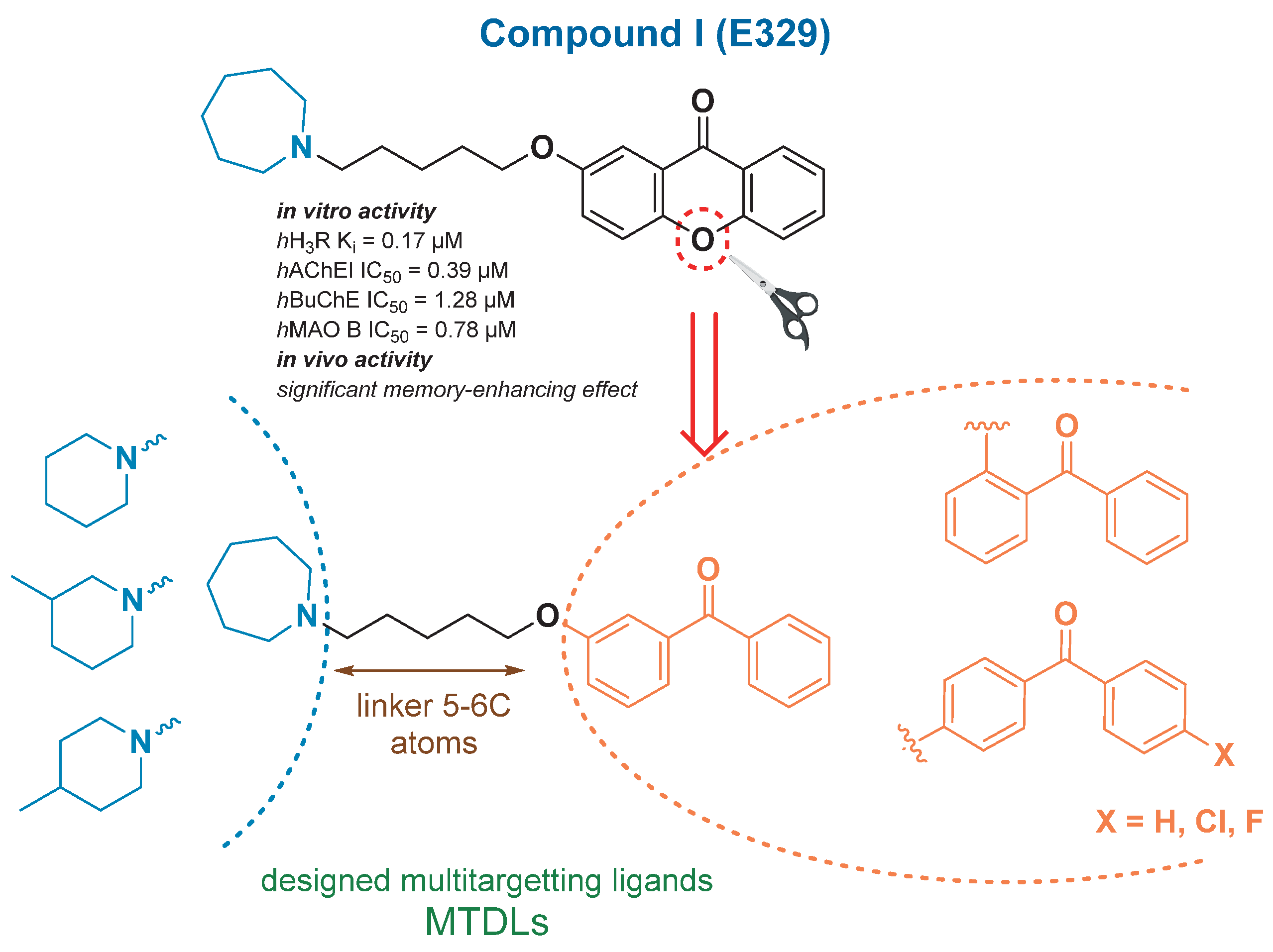
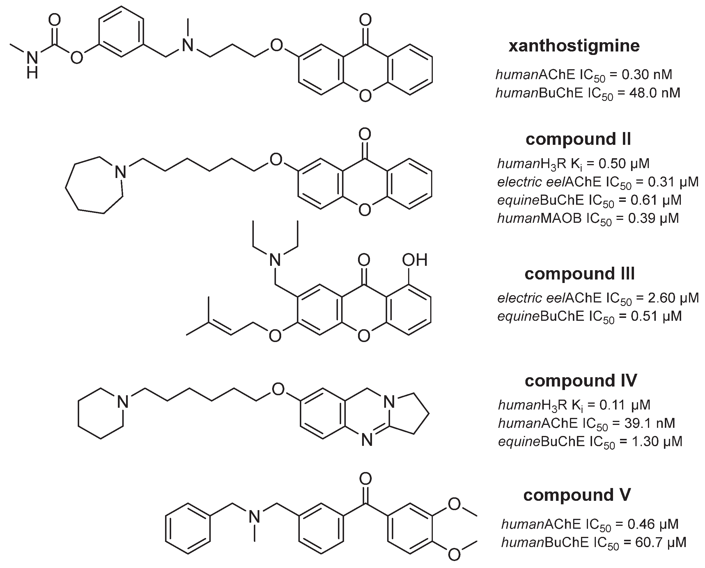
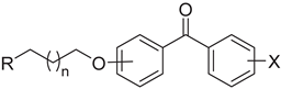
2.1. Rational Design
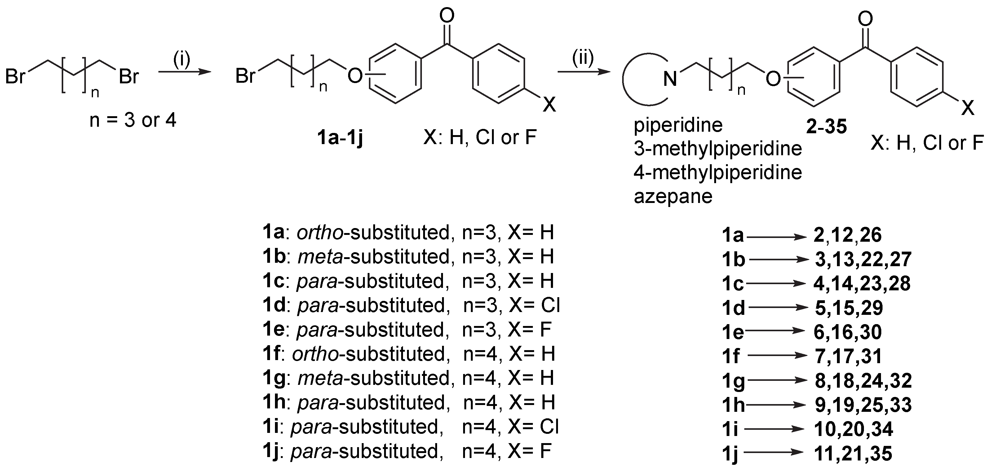
2.2. Synthesis of Designed Benzophenones
2.3. In Vitro Activity of Tested Benzophenones
2.3.1. Histamine H3 Receptor Affinity
2.3.2. Inhibition of eeAChE, eqBuChE, hAChE and hBuChE
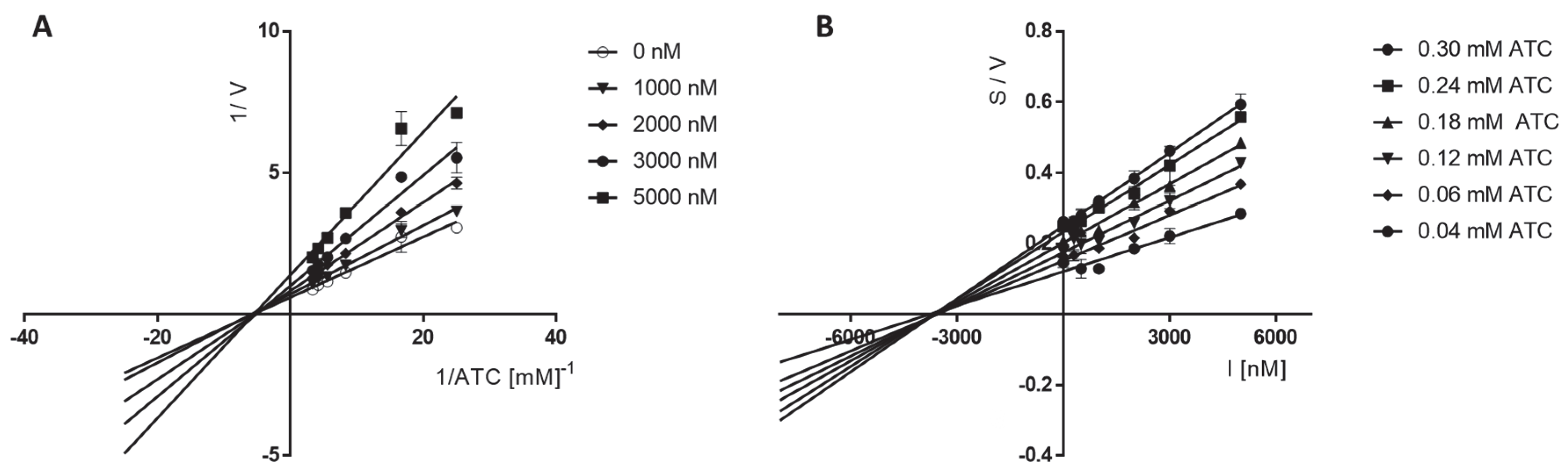
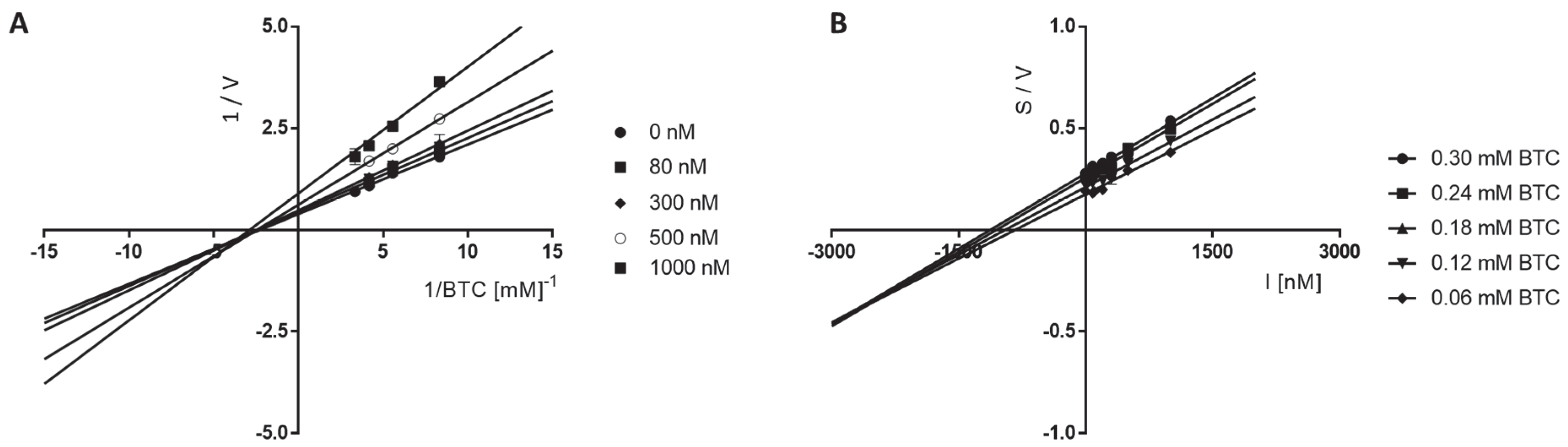
2.3.3. Kinetic Studies of eeAChE and eqBuChE Inhibition
2.3.4. Human MAO B Inhibitory Activity
2.4. In Silico Docking Studies
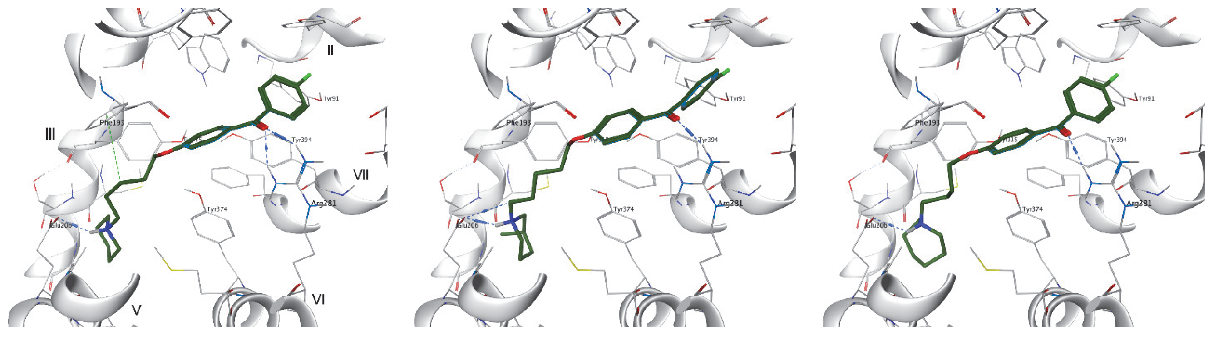
2.4.1. Molecular Modeling Studies to Histamine H3 Receptor
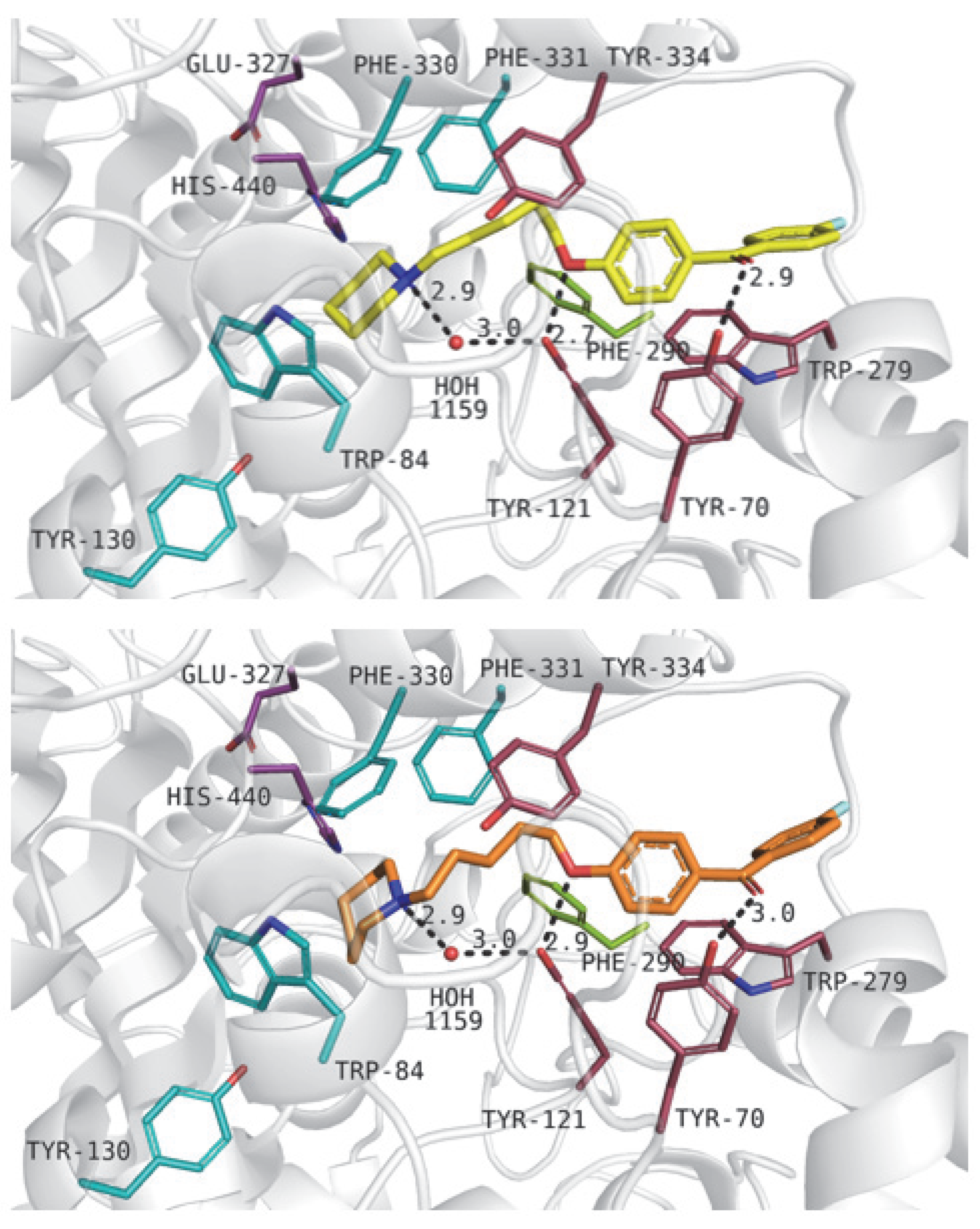
2.4.2. Analysis of Binding Mode within AChE
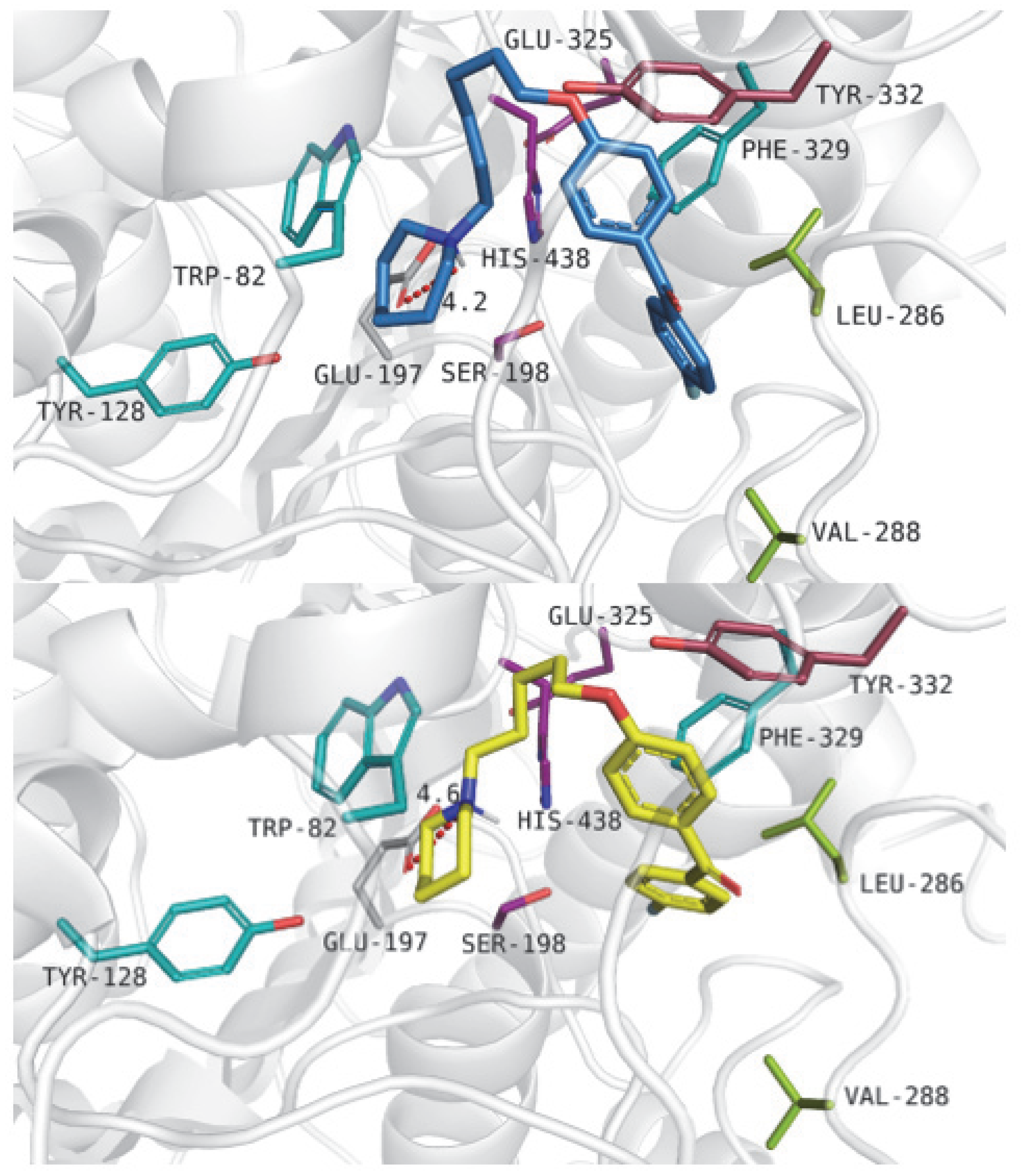
2.4.3. Analysis of Binding Mode within BuChE
2.4.4. Analysis of Binding Mode with Monoamine Oxidase B
2.5. Selected ADMET Properties
2.5.1. In Vitro Metabolic Stability of Compound 6
2.5.2. Neuroprotection Studies of Compound 6
2.5.3. The Permeability of Compound 6
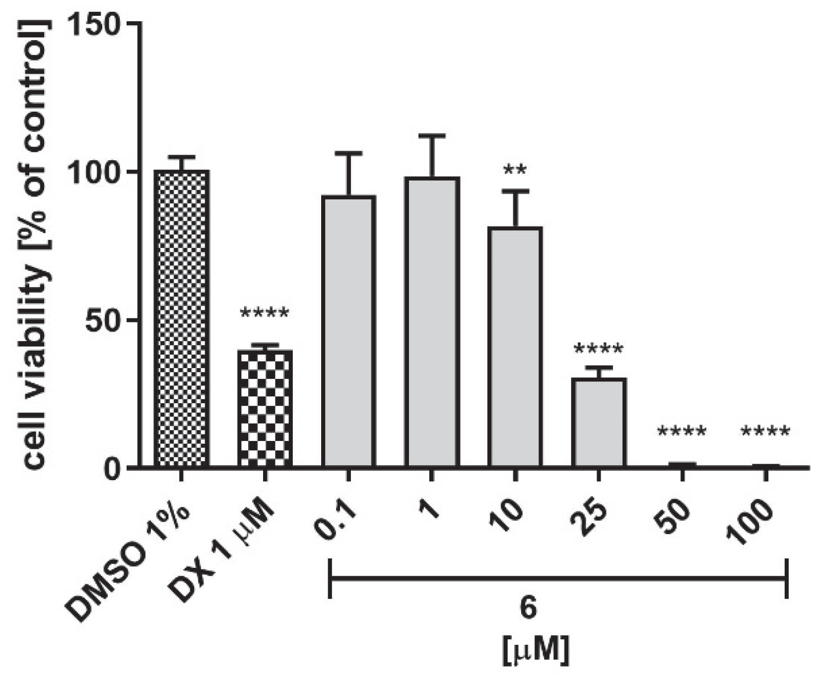
2.5.4. Hepatotoxicity Studies of 6
2.5.5. In Silico Prediction of Mutagenic Effect of Selected Compounds
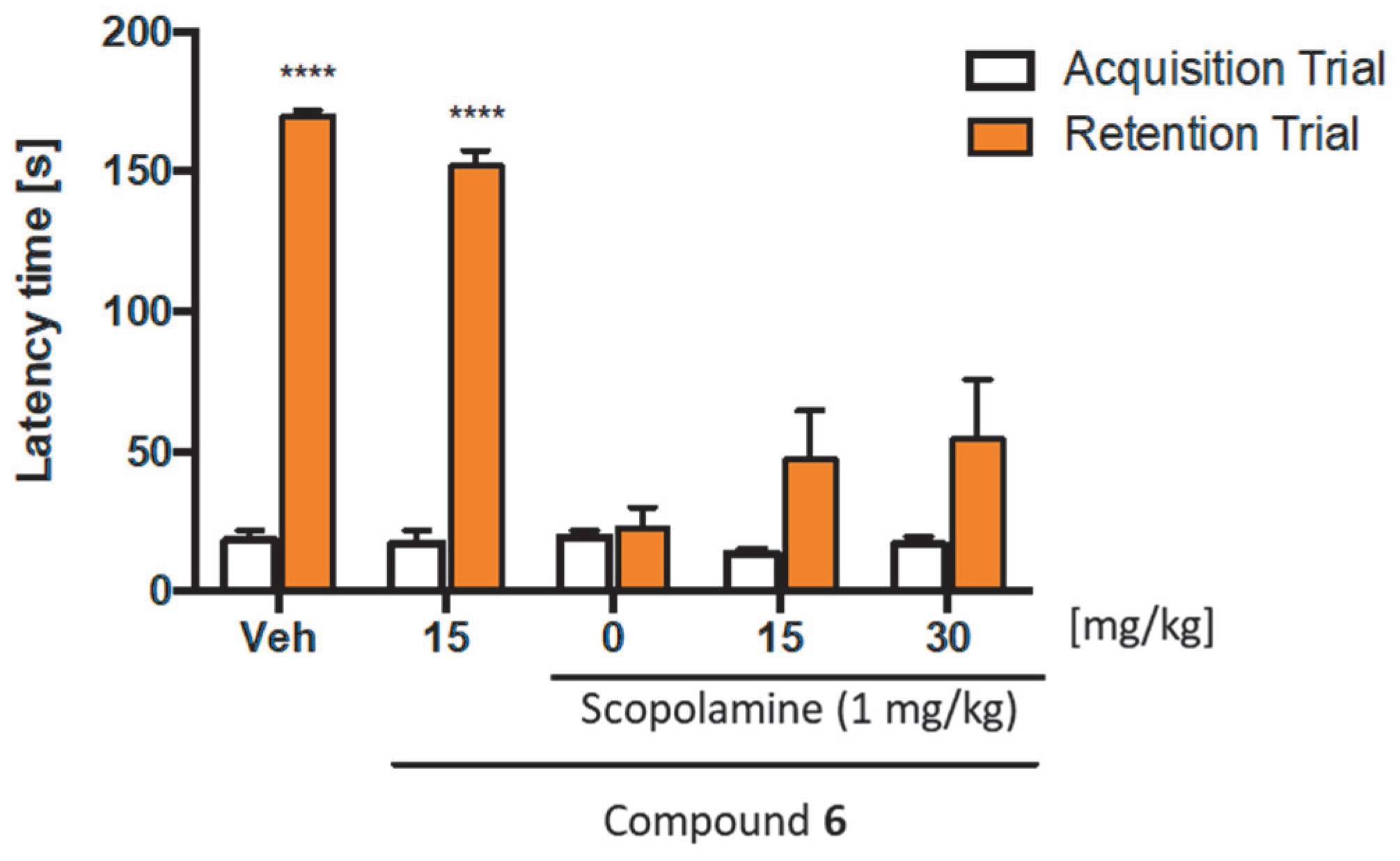
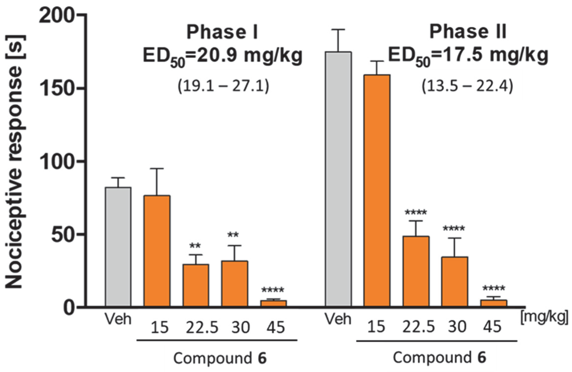
2.6. In Vivo Pharmacological Studies
3. Concluding Remarks
4. Experimental Section
4.1. Synthesis
General Procedure for the Synthesis of Final Compounds
4.2. Biochemical Assays
4.2.1. H3R Affinity
4.2.2. Inhibition of Cholinesterases
4.2.3. Kinetics of ChEs Inhibition
4.2.4. Inhibition of Human MAO B
4.3. Docking Studies
4.3.1. Molecular Modeling Studies to Histamine H3 Receptor
4.3.2. Molecular Modeling Studies to AChE, BuChE and MAO B
4.4. ADMET Properties
4.4.1. Metabolic Stability
4.4.2. Neuroprotection
4.4.3. Permeability
4.4.4. Hepatotoxicity
4.5. In Vivo Experiments
4.5.1. Passive Avoidance Test
4.5.2. Formalin Test in Mice
Supplementary Materials
Author Contributions
Funding
Institutional Review Board Statement
Informed Consent Statement
Data Availability Statement
Acknowledgments
Conflicts of Interest
Sample Availability
Abbreviations
References
- Handra, C.; Andreia Coman, O.; Coman, L.; Enache, T.; Stoleru, S.; Sorescu, A.-M.; Ghiță, I.; Fulga, I. The connection between different neurotransmitters involved in cognitive processes. Farmacia 2019, 67, 193–201. [Google Scholar] [CrossRef]
- Cummings, J.; Lee, G.; Zhong, K.; Fonseca, J.; Taghva, K. Alzheimer’s disease drug development pipeline: 2021. Alzheimer’s Dement. Transl. Res. Clin. Interv. 2021, 7, e12179. [Google Scholar] [CrossRef] [PubMed]
- Alvarez, E.O. The role of histamine on cognition. Behav. Brain Res. 2009, 199, 183–189. [Google Scholar] [CrossRef]
- Davies, P.; Maloney, A.J.F. Selective loss of central cholinergic neurons in Alzheimer’s Disease. Lancet 1976, 308, 1403. [Google Scholar] [CrossRef]
- Pohanka, M. Cholinesterases, a target of pharmacology and toxicology. Biomed. Pap. 2011, 155, 219–230. [Google Scholar] [CrossRef] [PubMed] [Green Version]
- Singh, M.; Jadhav, H.R. Histamine H3 Receptor Function and Ligands: Recent Developments. Mini Rev. Med. Chem. 2012, 13, 47–57. [Google Scholar] [CrossRef]
- Khanfar, M.A.; Affini, A.; Lutsenko, K.; Nikolic, K. Multiple Targeting Approaches on Histamine H3 Receptor Antagonists. Front. Neurosci. 2016, 10, 201. [Google Scholar] [CrossRef] [Green Version]
- Sadek, B.; Saad, A.; Sadeq, A.; Jalal, F.; Stark, H. Histamine H3 receptor as a potential target for cognitive symptoms in neuropsychiatric diseases. Behav. Brain Res. 2016, 312, 415–430. [Google Scholar] [CrossRef]
- Giacobini, E. Cholinesterase inhibitors: New roles and therapeutic alternatives. Pharmacol. Res. 2004, 50, 433–440. [Google Scholar] [CrossRef]
- Brioni, J.D.; Esbenshade, T.A.; Garrison, T.R.; Bitner, S.R.; Cowart, M.D. Discovery of histamine H3 antagonists for the treatment of cognitive disorders and Alzheimer’s disease. J. Pharmacol. Exp. Ther. 2011, 336, 38–46. [Google Scholar] [CrossRef]
- Zlomuzica, A.; Dere, D.; Binder, S.; De Souza Silva, M.A.; Huston, J.P.; Dere, E. Neuronal histamine and cognitive symptoms in Alzheimer’s disease. Neuropharmacology 2016, 106, 135–145. [Google Scholar] [CrossRef] [PubMed]
- Bautista-Aguilera, O.M.; Budni, J.; Mina, F.; Medeiros, E.B.; Deuther-Conrad, W.; Entrena, J.M.; Moraleda, I.; Iriepa, I.; Marco-Contelles, J. Contilisant, a Tetratarget Small Molecule for Alzheimer’s Disease Therapy Combining Cholinesterase, Monoamine Oxidase Inhibition, and H3R Antagonism with S1R Agonism Profile. J. Med. Chem. 2018, 61, 6937–6943. [Google Scholar] [CrossRef] [PubMed]
- Lopes, F.B.; Aranha, C.M.S.Q.; Fernandes, J.P.S. Histamine H3 receptor and cholinesterases as synergistic targets for cognitive decline: Strategies to the rational design of multitarget ligands. Chem. Biol. Drug Des. 2021, 98, 212–225. [Google Scholar] [CrossRef] [PubMed]
- Huang, W.; Tang, L.; Shi, Y.; Huang, S.; Xu, L.; Sheng, R.; Wu, P.; Li, J. Bioorganic & Medicinal Chemistry Searching for the Multi-Target-Directed Ligands against Alzheimer’s disease: Discovery of quinoxaline-based hybrid compounds with AChE, H3R and BACE 1 inhibitory activities. Bioorg. Med. Chem. 2011, 19, 7158–7167. [Google Scholar] [CrossRef]
- Galimberti, D.; Scarpini, E. Old and new acetylcholinesterase inhibitors for Alzheimer’s disease. Expert Opin. Investig. Drugs 2016, 25, 1181–1187. [Google Scholar] [CrossRef]
- Pepeu, G.; Giovannini, M.G. Cholinesterase inhibitors and memory. Chem. Biol. Interact. 2010, 187, 403–408. [Google Scholar] [CrossRef]
- Blanco-Silvente, L.; Castells, X.; Saez, M.; Barceló, M.A.; Garre-Olmo, J.; Vilalta-Franch, J.; Capellà, D. Discontinuation, Efficacy, and Safety of Cholinesterase Inhibitors for Alzheimer’s Disease: A Meta-Analysis and Meta-Regression of 43 Randomized Clinical Trials Enrolling 16,106 Patients. Int. J. Neuropsychopharmacol. 2017, 20, 519–528. [Google Scholar] [CrossRef] [Green Version]
- Mehta, M.; Adem, A.; Sabbagh, M. New acetylcholinesterase inhibitors for alzheimer’s disease. Int. J. Alzheimers Dis. 2012, 2012, 728983. [Google Scholar] [CrossRef] [Green Version]
- Łazewska, D.; Kieć-Kononowicz, K. New developments around histamine H3 receptor antagonists/inverse agonists: A patent review (2010-present). Expert Opin. Ther. Pat. 2014, 24, 89–111. [Google Scholar] [CrossRef]
- Maramai, S.; Benchekroun, M.; Gabr, M.T.; Yahiaoui, S. Multitarget Therapeutic Strategies for Alzheimer’s Disease: Review on Emerging Target Combinations. BioMed Res. Int. 2020, 2020, 5120230. [Google Scholar] [CrossRef]
- Bolognesi, M.L.; Simoni, E.; Rosini, M.; Minarini, A.; Tumiatti, V.; Melchiorre, C. Multitarget-Directed Ligands: Innovative Chemical Probes and Therapeutic Tools Against Alzheimer’s Disease. Curr. Top. Med. Chem. 2011, 11, 2797–2806. [Google Scholar] [CrossRef] [PubMed]
- Ramalakshmi, N.; Remya, R.S.; Nalini, C.N. Multitarget Directed Ligand Approaches for Alzheimer’s Disease: A Comprehensive Review. Mini Rev. Med. Chem. 2021, 21, 2361–2388. [Google Scholar] [CrossRef] [PubMed]
- Bembenek, S.D.; Keith, J.M.; Letavic, M.A.; Apodaca, R.; Barbier, A.J.; Dvorak, L.; Aluisio, L.; Miller, K.L.; Lovenberg, T.W.; Carruthers, N.I. Lead identification of acetylcholinesterase inhibitors–histamine H3 receptor antagonists from molecular modeling. Bioorg. Med. Chem. 2008, 16, 2968–2973. [Google Scholar] [CrossRef] [PubMed]
- Ghamari, N.; Dastmalchi, S.; Zarei, O.; Arias-Montaño, J.; Reiner, D.; Ustun-Alkan, F.; Stark, H.; Hamzeh-Mivehroud, M. In silico and in vitro studies of two non-imidazole multiple targeting agents at histamine H3 receptors and cholinesterase enzymes. Chem. Biol. Drug Des. 2020, 95, 279–290. [Google Scholar] [CrossRef] [PubMed]
- Incerti, M.; Flammini, L.; Saccani, F.; Morini, G.; Comini, M.; Coruzzi, M.; Barocelli, E.; Ballabeni, V.; Bertoni, S. Dual-Acting Drugs: An in vitro Study of Nonimidazole Histamine H3 Receptor Antagonists Combining Anticholinesterase Activity. ChemMedChem 2010, 5, 1143–1149. [Google Scholar] [CrossRef] [PubMed]
- Bajda, M.; Łażewska, D.; Godyń, J.; Zaręba, P.; Kuder, K.; Hagenow, S.; Łątka, K.; Stawarska, E.; Stark, H.; Kieć-Kononowicz, K.; et al. Search for new multi-target compounds against Alzheimer’s disease among histamine H3 receptor ligands. Eur. J. Med. Chem. 2020, 185, 111785. [Google Scholar] [CrossRef]
- Łażewska, D.; Jończyk, J.; Bajda, M.; Szałaj, N.; Więckowska, A.; Panek, D.; Moore, C.; Kuder, K.; Malawska, B.; Kieć-Kononowicz, K. Cholinesterase inhibitory activity of chlorophenoxy derivatives—Histamine H3 receptor ligands. Bioorg. Med. Chem. Lett. 2016, 26, 4140–4145. [Google Scholar] [CrossRef]
- Bajda, M.; Kuder, K.J.; Łewska, D.; Kieć-Kononowicz, K.; Wiȩckowska, A.; Ignasik, M.; Guzior, N.; Jończyk, J.; Malawska, B. Dual-Acting Diether Derivatives of Piperidine and Homopiperidine with Histamine H3 Receptor Antagonistic and Anticholinesterase Activity. Arch. Pharm. 2012, 345, 591–597. [Google Scholar] [CrossRef]
- Godyń, J.; Zaręba, P.; Łażewska, D.; Stary, D.; Reiner-Link, D.; Frank, A.; Latacz, G.; Mogilski, S.; Kaleta, M.; Doroz-Płonka, A.; et al. Cyanobiphenyls: Novel H3 receptor ligands with cholinesterase and MAO B inhibitory activity as multitarget compounds for potential treatment of Alzheimer’s disease. Bioorg. Chem. 2021, 114, 105129. [Google Scholar] [CrossRef]
- Łażewska, D.; Zaręba, P.; Godyń, J.; Doroz-Płonka, A.; Frank, A.; Reiner-Link, D.; Bajda, M.; Stary, D.; Mogilski, S.; Olejarz-Maciej, A.; et al. Biphenylalkoxyamine Derivatives–Histamine H3 Receptor Ligands with Butyrylcholinesterase Inhibitory Activity. Molecules 2021, 26, 3580. [Google Scholar] [CrossRef]
- Alachkar, A.; Łażewska, D.; Latacz, G.; Frank, A.; Siwek, A.; Lubelska, A.; Honkisz-Orzechowska, E.; Handzlik, J.; Stark, H.; Kieć-Kononowicz, K.; et al. Studies on Anticonvulsant Effects of Novel Histamine H3R Antagonists in Electrically and Chemically Induced Seizures in Rats. Int. J. Mol. Sci. 2018, 19, 3386. [Google Scholar] [CrossRef] [PubMed] [Green Version]
- Łażewska, D.; Bajda, M.; Kaleta, M.; Zaręba, P.; Doroz-Płonka, A.; Siwek, A.; Alachkar, A.; Mogilski, S.; Saad, A.; Kuder, K.; et al. Rational design of new multitarget histamine H3 receptor ligands as potential candidates for treatment of Alzheimer’s Disease. Eur. J. Med. Chem. 2020, 207, 112743. [Google Scholar] [CrossRef] [PubMed]
- Sander, K.; von Coburg, Y.; Camelin, J.C.; Ligneau, X.; Rau, O.; Schubert-Zsilavecz, M.; Schwartz, J.C.; Stark, H. Acidic elements in histamine H3 receptor antagonists. Bioorg. Med. Chem. Lett. 2010, 20, 1581–1584. [Google Scholar] [CrossRef]
- Wingen, K.; Stark, H. Scaffold variations in amine warhead of histamine H3 receptor antagonists. Drug Discov. Today Technol. 2013, 10, e483–e489. [Google Scholar] [CrossRef] [PubMed]
- Miles, J.A.; Ross, B.P. Recent Advances in Virtual Screening for Cholinesterase Inhibitors. Chem. Neurosci. 2020, 12, 30–41. [Google Scholar] [CrossRef] [PubMed]
- Cruz, M.I.; Cidade, H.; Pinto, M. Dual/multitargeted xanthone derivatives for Alzheimer’s disease: Where do we stand? Futur. Med. Chem. 2017, 9, 1611–1630. [Google Scholar] [CrossRef]
- Rizzo, S.; Cavalli, A.; Ceccarini, L.; Bartolini, M.; Belluti, F.; Bisi, A.; Andrisano, V.; Recanatini, M.; Rampa, A. Structure–Activity Relationships and Binding Mode in the Human Acetylcholinesterase Active Site of Pseudo-Irreversible Inhibitors Related to Xanthostigmine. ChemMedChem 2009, 4, 670–679. [Google Scholar] [CrossRef]
- Menéndez, C.A.; Biscussi, B.; Accordino, S.; Paula Murray, A.; Gerbino, D.C.; Appignanesi, G.A. Design, synthesis and biological evaluation of 1,3-dihydroxyxanthone derivatives: Effective agents against acetylcholinesterase. Bioorg. Chem. 2017, 75, 201–209. [Google Scholar] [CrossRef] [Green Version]
- Kou, X.; Song, L.; Wang, Y.; Yu, Q.; Ju, H.; Yang, A.; Shen, R. Design, synthesis and anti-Alzheimer’s disease activity study of xanthone derivatives based on multi-target strategy. Bioorgan. Med. Chem. Lett. 2020, 30, 126927. [Google Scholar] [CrossRef]
- Belluti, F.; Piazzi, L.; Bisi, A.; Gobbi, S.; Bartolini, M.; Cavalli, A.; Valenti, P.; Rampa, A. Design, synthesis, and evaluation of benzophenone derivatives as novel acetylcholinesterase inhibitors. Eur. J. Med. Chem. 2009, 44, 1341–1348. [Google Scholar] [CrossRef]
- Ellman, G.L.; Courtney, K.D.; Andres, V.; Featherstone, R.M. A new and rapid colorimetric determination of acetylcholinesterase activity. Biochem. Pharmacol. 1961, 7, 88–95. [Google Scholar] [CrossRef] [PubMed]
- Szczepańska, K.; Karcz, T.; Mogilski, S.; Siwek, A.; Kuder, K.J.; Latacz, G.; Kubacka, M.; Hagenow, S.; Lubelska, A.; Olejarz, A.; et al. Synthesis and biological activity of novel tert-butyl and tert-pentylphenoxyalkyl piperazine derivatives as histamine H3R ligands. Eur. J. Med. Chem. 2018, 152, 223–234. [Google Scholar] [CrossRef]
- Levoin, N.; Calmels, T.; Poupardin-Olivier, O.; Labeeuw, O.; Danvy, D.; Robert, P.; Berrebi-Bertrand, I.; Ganellin, C.R.; Schunack, W.; Stark, H.; et al. Refined Docking as a Valuable Tool for Lead Optimization: Application to Histamine H3 Receptor Antagonists. Arch. Pharm. 2008, 341, 610–623. [Google Scholar] [CrossRef] [PubMed]
- Bajda, M.; Wiȩckowska, A.; Hebda, M.; Guzior, N.; Sotriffer, C.A.; Malawska, B. Structure-based search for new inhibitors of cholinesterases. Int. J. Mol. Sci. 2013, 14, 5608–5632. [Google Scholar] [CrossRef] [PubMed] [Green Version]
- Kryger, G.; Silman, I.; Sussman, J.L. Structure of acetylcholinesterase complexed with E2020 (Ariceptρ): Implications for the design of new anti-Alzheimer drugs. Structure 1999, 7, 297–307. [Google Scholar] [CrossRef] [PubMed] [Green Version]
- MetaSite, Version 6.0.1; Molecular Discovery Ltd.: Borehamwood, UK.
- Wichur, T.; Godyń, J.; Góral, I.; Latacz, G.; Bucki, A.; Siwek, A.; Głuch-Lutwin, M.; Mordyl, B.; Śniecikowska, J.; Walczak, M.; et al. Development and crystallography-aided SAR studies of multifunctional BuChE inhibitors and 5-HT6R antagonists with β-amyloid anti-aggregation properties. Eur. J. Med. Chem. 2021, 225, 113792. [Google Scholar] [CrossRef] [PubMed]
- Kurnik-Łucka, M.; Latacz, G.; Martyniak, A.; Bugajski, A.; Kieć-Kononowicz, K.; Gil, K. Salsolinol—Neurotoxic or Neuroprotective? Neurotox. Res. 2020, 37, 286–297. [Google Scholar] [CrossRef] [Green Version]
- Chen, X.; Murawski, A.; Patel, K.; Crespi, C.L.; Balimane, P.V. A novel design of artificial membrane for improving the PAMPA model. Pharm. Res. 2008, 25, 1511–1520. [Google Scholar] [CrossRef]
- Paniagua, A.C.; Amariles, P. Hepatotoxicity by Drugs. In Pharmacokinetics and Adverse Effects of Drugs—Mechanisms and Risks Factors; Malangu, N., Ed.; IntechOpen: London, UK, 2017. [Google Scholar] [CrossRef] [Green Version]
- Schindler, U. Pre-clinical evaluation of cognition enhancing drugs, Prog. Neuropsychopharmacol. Biol. Psychiatry 1989, 13, 99–115. [Google Scholar] [CrossRef]
- Cheng, Y.-C.; Prusoff, W.H. Relationship between the inhibition constant (K1) and the concentration of inhibitor which causes 50 per cent inhibition (I50) of an enzymatic reaction. Biochem. Pharmacol. 1973, 22, 3099–3108. [Google Scholar]
- Molecular Operating Environment (MOE) 2019.1; Chemical Computing Group ULC: Montreal, QC, Canada, 2019.
- Hawkins, P.C.D.; Skillman, A.G.; Warren, G.L.; Ellingson, B.A.; Stahl, M.T. Conformer Generation with OMEGA: Algorithm and Validation Using High Quality Structures from the Protein Databank and Cambridge Structural Database. J. Chem. Inf. Model. 2010, 50, 572–584. [Google Scholar] [CrossRef] [PubMed]
- Salinas-Abarca, A.B.; Avila-Rojas, S.H.; Barragán-Iglesias, P. Formalin injection produces long-lasting hypersensitivity with characteristics of neuropathic pain. Eur. J. Pharmacol. 2017, 797, 83–93. [Google Scholar] [CrossRef] [PubMed]
- Muley, M.M.; Krustev, E.; Mcdougall, J.J.; Pain, C. Preclinical Assessment of Inflammatory Pain Models of Cutaneous Pain. CNS Neurosci. Ther. 2016, 22, 88–101. [Google Scholar] [CrossRef] [PubMed]


 | ||||||||
|---|---|---|---|---|---|---|---|---|
| R | Compd | n | Position a | X | hH3R Ki [nM] b | eeAChE IC50 [μM] c (% Inh. at 10 µM) | eqBuChE IC50 [μM] d (% Inh. at 10 µM) | hMAO B (%Inh. at 1 µM) e |
 | 2 | 3 | ortho | H | 371 [120;1147] | 22% ± 1 | 1.607 ± 0.060 | 23% ± 2 |
| 3 | 3 | meta | H | 210 [101;435] | 7.754 ± 0.329 | 4.006 ± 0.067 | 15% ± 1 | |
| 4 | 3 | para | H | 36 * [10;30] | 41% ± 1 | 2.477 ± 0.040 | 44% ± 1 | |
| 5 | 3 | para | 4-Cl | 20 [3;141] | 2.870 ± 0.071 | 1.685 ± 0.017 | 18% ± 0 | |
| 6 | 3 | para | 4-F | 8 [1;62] | 2.306 ± 0.004 hAChE f 9.585 ± 0.220 | 0.172 ± 0.005 hBuChE g 1.155 ± 0.034 | 25% ± 4 | |
| 7 | 4 | ortho | H | 161 [92;280] | 3% ± 0 | 2.108 ± 0.032 | 12% ± 0 | |
| 8 | 4 | meta | H | 76 * [54;108] | 3.627 ± 0.111 | 2.096 ± 0.060 | 15% ± 3 | |
| 9 | 4 | para | H | 41 * [26;66] | 3.733 ± 0.085 | 1.781 ± 0.034 | 9% ± 2 | |
| 10 | 4 | para | 4-Cl | 25 [11;58] | 2.669 ± 0.094 | 2.457 ± 0.061 | 39% ± 2 | |
| 11 | 4 | para | 4-F | 26 [9;72] | 3.098 ± 0.009 | 0.419 ± 0.002 | 28% ± 3 | |
 | 12 | 3 | ortho | H | 208 [80;543] | 16% ± 2 | 1.607 ± 0.061 | 9% ± 1 |
| 13 | 3 | meta | H | 78 [39;130] | 3.790 ± 0.104 | 3.424 ± 0.078 | 8% ± 6 | |
| 14 | 3 | para | H | 40 * [13;119] | 3.180 ± 0.083 | 3.781 ± 0.061 | 9% ± 4 | |
| 15 | 3 | para | 4-Cl | 37 [22;60] | 2.646 ± 0.087 | 2.562 ± 0.050 | 19% ± 9 | |
| 16 | 3 | para | 4-F | 12 [2;71] | 2.023 ± 0.006 | 0.311 ± 0.006 | 20% ± 3 | |
| 17 | 4 | ortho | H | 152 [72;322] | 20% ± 2 | 2.498 ± 0.028 | 8% ± 1 | |
| 18 | 4 | meta | H | 110 * [62;196] | 2.002 ± 0.030 | 2.895 ± 0.077 | 27% ± 7 | |
| 19 | 4 | para | H | 53 * [31;89] | 2.106 ± 0.050 | 1.978 ± 0.034 | 7% ± 1 | |
| 20 | 4 | para | 4-Cl | 44 [26;75] | 2.737 ± 0.098 | 2.523 ± 0.031 | 17% ± 2 | |
| 21 | 4 | para | 4-F | 75 [36;154] | 1.999 ± 0.046 | 2.560 ± 0.040 | 23% ± 6 | |
 | 22 | 3 | meta | H | 298 [192;462] | 4.451 ± 0.094 | 1.705 ± 0.046 | 32% ± 2 |
| 23 | 3 | para | H | 67 [61;74] | 2.603 ± 0.072 | 1.567 ± 0.035 | 27% ± 2 | |
| 24 | 4 | meta | H | 106 [89;127] | 2.457 ± 0.070 | 1.063 ± 0.043 | 20% ± 3 | |
| 25 | 4 | para | H | 53 [34;82] | 2.692 ± 0.047 | 1.556 ± 0.050 | 22% ± 12 | |
 | 26 | 3 | ortho | H | 302 [46;1995] | 24% ± 3 | 0.384 ± 0.008 | 3% ± 0 |
| 27 | 3 | meta | H | 307 [188;502] | 3.661 ± 0.092 | 0.901 ± 0.010 | 14% ± 2 | |
| 28 | 3 | para | H | 39 * [11;142] | 4.630 ± 0.100 | 0.678 ± 0.007 | 29% ± 1 | |
| 29 | 3 | para | 4-Cl | 66 [5;892] | 2.244 ± 0.035 | 0.676 ± 0.003 | 26% ± 1 | |
| 30 | 3 | para | 4-F | 13 [2;92] | 1.110 ± 0.003 | 0.224 ± 0.003 | 30% ± 1 | |
| 31 | 4 | ortho | H | 162 [82;319] | 11% ± 1 | 0.317 ± 0.003 | 5% ± 1 | |
| 32 | 4 | meta | H | 70 * [44;170] | 1.551 ± 0.034 | 0.616 ± 0.002 | 17% ± 4 | |
| 33 | 4 | para | H | 41 * [33;50] | 1.810 ± 0.038 | 1.281 ± 0.038 | 23% ± 1 | |
| 34 | 4 | para | 4-Cl | 133 [35;508] | 2.033 ± 0.018 | 0.867 ± 0.002 | 30% ± 2 | |
| 35 | 4 | para | 4-F | 35 [6;204] | 1.654 ± 0.004 | 0.161 ± 0.004 | 24% ± 0 | |
| Reference | pitolisant | 12 ± 3 ** | 3% ± 0 | 8.400 ± 0.180 | 2% ± 3 | |||
| tacrine | nt | 0.024 ± 0.001 | 0.015 ± 0.001 | nt | ||||
| rasagiline h | nt | nt | nt | 25 ± 7 | ||||
| safinamide | nt | nt | nt | 98% ± 4 7.6 ± 1 i | ||||
| Substrate | Molecular Mass (m/z) | % Remaining | Molecular Mass of the Metabolite (m/z) | Metabolic Pathway |
|---|---|---|---|---|
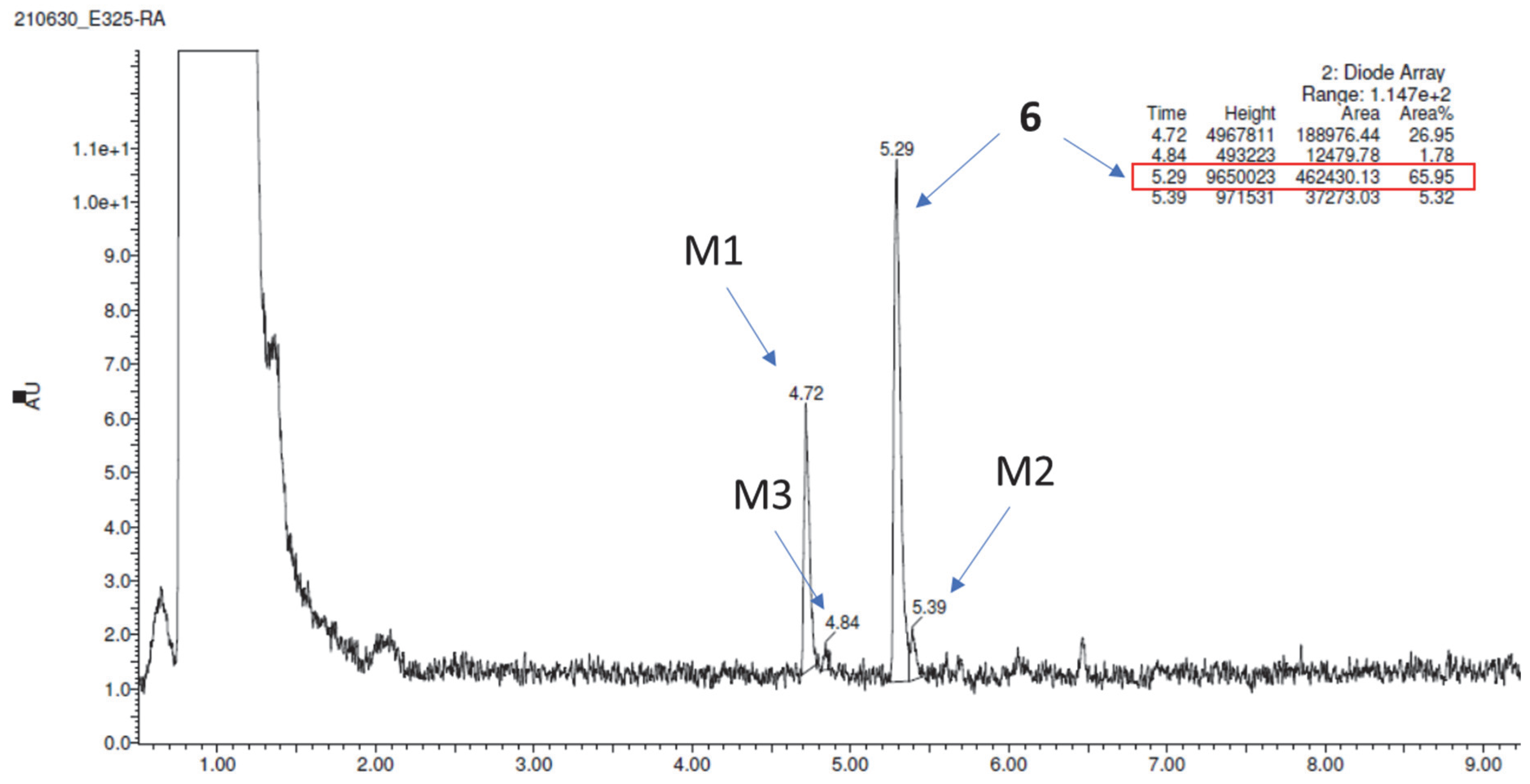
| 6 | 370.33 | 65.95 | 372.39 (M1) 386.35 (M2) 388.41 (M3) | reduction of carbonyl group hydroxylation reduction of carbonyl group, hydroxylation |
| Verapamil (control) * | 455.54 | 23.93 | 441.42 (M1) | demethylation |
| 441.42 (M2) | demethylation | |||
| 291.35 (M3) | defragmentation | |||
| 293.34 (M4) | defragmentation/hydroxylation | |||
| 277.33 (M5) | defragmentation |
| Compound | Pe n,o [10−6 cm/s] ± SD |
|---|---|
| 6 | 6.3 ± 0.6 |
| Caffeine | 15.1 ± 0.4 |
Disclaimer/Publisher’s Note: The statements, opinions and data contained in all publications are solely those of the individual author(s) and contributor(s) and not of MDPI and/or the editor(s). MDPI and/or the editor(s) disclaim responsibility for any injury to people or property resulting from any ideas, methods, instructions or products referred to in the content. |
© 2022 by the authors. Licensee MDPI, Basel, Switzerland. This article is an open access article distributed under the terms and conditions of the Creative Commons Attribution (CC BY) license (https://creativecommons.org/licenses/by/4.0/).
Share and Cite
Godyń, J.; Zaręba, P.; Stary, D.; Kaleta, M.; Kuder, K.J.; Latacz, G.; Mogilski, S.; Reiner-Link, D.; Frank, A.; Doroz-Płonka, A.; et al. Benzophenone Derivatives with Histamine H3 Receptor Affinity and Cholinesterase Inhibitory Potency as Multitarget-Directed Ligands for Possible Therapy of Alzheimer’s Disease. Molecules 2023, 28, 238. https://doi.org/10.3390/molecules28010238
Godyń J, Zaręba P, Stary D, Kaleta M, Kuder KJ, Latacz G, Mogilski S, Reiner-Link D, Frank A, Doroz-Płonka A, et al. Benzophenone Derivatives with Histamine H3 Receptor Affinity and Cholinesterase Inhibitory Potency as Multitarget-Directed Ligands for Possible Therapy of Alzheimer’s Disease. Molecules. 2023; 28(1):238. https://doi.org/10.3390/molecules28010238
Chicago/Turabian StyleGodyń, Justyna, Paula Zaręba, Dorota Stary, Maria Kaleta, Kamil J. Kuder, Gniewomir Latacz, Szczepan Mogilski, David Reiner-Link, Annika Frank, Agata Doroz-Płonka, and et al. 2023. "Benzophenone Derivatives with Histamine H3 Receptor Affinity and Cholinesterase Inhibitory Potency as Multitarget-Directed Ligands for Possible Therapy of Alzheimer’s Disease" Molecules 28, no. 1: 238. https://doi.org/10.3390/molecules28010238