Review

Honey Quality Control: Review of Methodologies for Determining Entomological Origin

by
Saeed Mohamadzade Namin
1,
Sampat Ghosh
1 and
Chuleui Jung
1,2,*
1
Agricultural Science and Technology Institute, Andong National University, Andong 36729, Republic of Korea
2
Department of Plant Medicals, Andong National University, Andong 36729, Republic of Korea
*
Author to whom correspondence should be addressed.
Molecules 2023, 28(10), 4232; https://doi.org/10.3390/molecules28104232
Submission received: 14 April 2023 / Revised: 17 May 2023 / Accepted: 19 May 2023 / Published: 22 May 2023

Abstract

:
Honey is a widely consumed natural product, and its entomological origin can significantly influence its market value. Therefore, traceability of the entomological origin of honey should also be considered in honey quality control protocols. Although several methods exist, such as physicochemical characterization and bioactivity profiling of honey of different entomological origins, the most promising three methods for entomological authentication of honey include protein-based identification, chemical profiling, and a DNA-based method. All of these methods can be applied for reliable identification of the entomological origin of honey. However, as the honey is a complex matrix, the inconsistency of the results obtained by these methods is a pragmatic challenge, and therefore, the use of each method in all the cases is questionable. Most of these methodologies can be used for authentication of newly harvested honey and it is worth understanding the possibility of using these methods for authentication of relatively old samples. Most probably, using DNA-based methods targeting small fragments of DNA can provide the best result in old samples, however, the species-specific primers targeting short fragments are limited and not available for all species. Therefore, using universal primers in combination with a DNA metabarcoding approach can be a good solution that requires further investigation. This present article describes the applications of different methods, their pros, and their cons to identify honey based on entomological origin.

Graphical Abstract

1. Introduction

Honey is a sweet natural product that is produced by managed and wild bees, derived from the nectar of flowers. It is made up of various components such as sugar, protein, vitamins, minerals, aromatic substances, polyphenols, pigments, beeswax, and pollen that contribute to its color, smell, and flavor [1,2]. However, honey adulteration is a growing concern, including the production of honey by feeding bees with commercial industrial sugar [3], the addition of foreign sugar, as well as mislabeling. The botanical, geographical, and entomological origin of honey greatly influences its price [4,5,6,7,8,9,10]. Among honeybees, the European honeybee, Apis mellifera, and the Eastern honeybee, A. cerana, are two dominant species for honey production [11].
Apis cerana is native to temperate and tropical Asia and is widespread from Afghanistan to far east Russia and Japan, north into the foothills of the Himalayan mountains, and south through Indonesia [12,13]. This species was introduced to New Guinea in the late 1970s from Indonesia and became invasive to Australia in 1998 [14], and eradication attempts against it remain unsuccessful [14,15,16]. European honeybee, A. mellifera, is native to Europe, the Middle East, and Africa, and was introduced to other continents in the 17th century. It is now widespread around the world [17,18]. Because A. mellifera is more productive and suitable for modern beekeeping, it has been introduced into many A. cerana habitats, such as China, South Korea, and some Southeast Asian countries [19,20]. Afterwards, the number of A. cerana colonies declined due to high interspecific competition for the same niche as A. mellifera. Other than managed honeybees, honey can be harvested from the colonies of wild Apis bees and stingless bees. As a result of recent discoveries about the benefits of honey consumption, the demand for natural honey has increased and the price of natural honey is five to seven times higher than A. mellifera honey [8,11]. Therefore, honey from A. cerana and wild bees is very prone to adulteration, either by mislabeling or mixing the natural honey with cheaper A. mellifera honey to gain a higher profit [8,11].
In addition, honey in Europe is produced by A. mellifera. However, based on morphological [12] and molecular analyses [21], about ten subspecies of A. mellifera are present in Europe, belonging to three different lineages: The African (A), the Eastern European (C), and the Western European (M). Understanding the lineage of honeybee from which the honey originates is helpful in identifying the geographical origin of the honey, which is necessary for quality control assessment of honey samples. While various methodologies are used to identify the entomological origin of honey, a comprehensive review of these methods is currently unavailable. Hence, the objective of this review is to provide a summary of various methodologies used to identify the entomological origin of honey, assess their respective advantages and disadvantages, and help researchers choose the most appropriate method for their sample identification. Furthermore, this review may offer valuable insights into the development of novel methodologies.

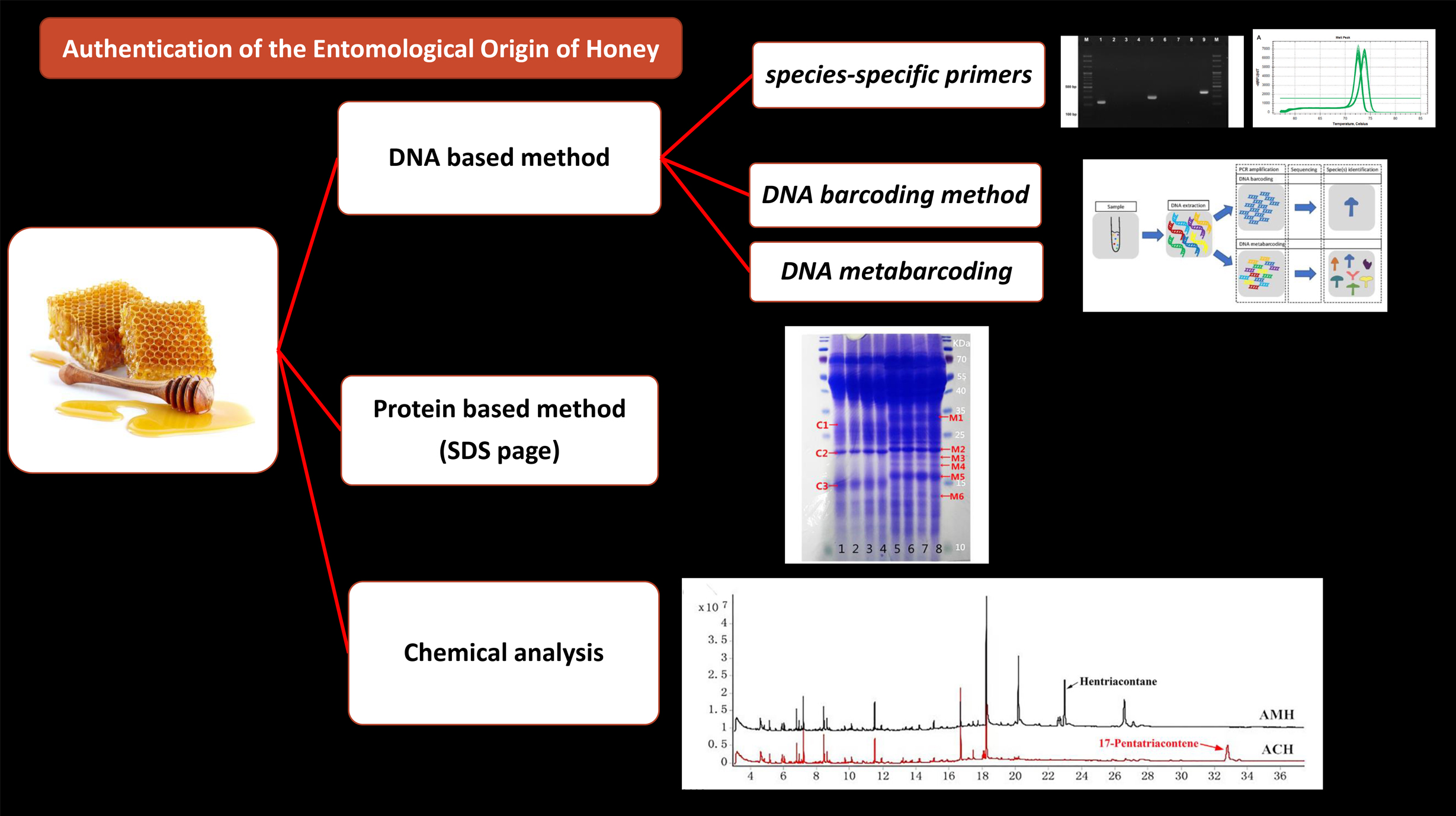
2. Identification Methods of Entomological Origin of Honey

2.1. Protein Based Method

Sodium Dodecyl Sulfate Polyacrylamide Gel Electrophoresis (SDS-PAGE) is a popular technique for separating proteins based on their mass. In an electric field, proteins migrate through the gel from the negatively charged electrode toward the positively charged electrode, allowing for separation of proteins based on molecular weight. SDS-PAGE, as depicted in Figure 1A, has frequently been used to identify the entomological origin of honey from both managed honeybees and stingless bees [22,23]. Despite the limited amount of protein in honey, some studies have been conducted on its protein profile using methods such as Matrix-Assisted Laser Desorption Ionization Time of Flight Mass Spectrometry (MALDI-TOF-MS) and Liquid Chromatography Mass Spectrometry (LC-MS/MS) [1,24,25]. However, in the majority of cases, these studies have not specifically focused on distinguishing the entomological source of honey. The Major Royal Jelly Proteins (MRJPs) are a group of nine homologous proteins (MRJP1–MRJP9) [26,27,28] secreted by worker bees. Along with pollen proteins, they are significant protein resources in honey [1,29]. As the MRJPs of different bee species are specific and they remain in the honey, there is a possibility of using such a protein to identify the entomological origin of honey. Previous studies have shown that the protein profile of SDS-PAGE is different between honey samples from A. cerana and A. mellifera [22]. Moreover, Won et al. [1] showed that MRJP1 has different molecular weights in different honeybees (56 kDa in A. cerana and 59 kDa in A. mellifera) despite having similar primary structure. By producing an artificial marker protein from Escherichia coli and co-electrophoresing it with honey samples, they successfully discriminated the entomological origin of honey samples by SDS–PAGE and found several adulterated honey samples. Additionally, Zhang et al. [30] indicated that three species-specific bands with molecular weight between 15.0 and 29.4 KDa are present in A. cerana honey, while A. mellifera honey is characterized by the appearance of six species-specific bands with molecular weights between 13.8 and 33.1 KDa.

2.2. Physicochemical Properties and Bioactive Characterization of Honey to Determine Entomological Origin

The chemical properties of honey have been used to identify the entomological origin of honey. Zannat et al. [31] showed that the physicochemical profile of honey such as moisture, formaldehyde and Hydroxymethylfurfural (HMF), pH, and color were key determinants to identify the entomological origin of Bangladesh honey samples, such as A. cerana, A. dorsata, and A. mellifera honey. This method can be applied to detect the mislabeling of honey. Furthermore, it seems that Principle Component Analysis (PCA) involving the physicochemical properties of honey can be useful for identification of the entomological origin of honey samples regionally, but the results cannot always be extended to other regions due to the high variation in the chemical properties of honey from different localities. For instance, the moisture content of honey samples from Bangladesh were 21.3, 19, and 17% from A. dorsata, A. cerana and A. mellifera, respectively. However, the reported amount of moisture for A. cerana honey from Thailand was 21.2% [32] which is comparable with the amount of moisture in A. dorsata honey from Bangladesh. Furthermore, moisture content of A. mellifera honey from Thailand was reported as 18.8%, which was similar to the amount of moisture in A. cerana honey from Bangladesh. On the other hand, using the color of honey as a method of recognition cannot be recommended due to its variation based on the floral source of honey, storage duration, and storage conditions. However, since multidimensional analysis, such as PCA, is required for categorization of honey samples with different origins, more studies are required to understand the possibility of the usage of this method in identification of the entomological origin of honey.
Another attempt to identify the entomological origin of honey is bioactive characterization [33,34,35,36] and mineral content [37,38]. Kelulut honey, produced by stingless bees Heterotrigona itama, was found distinguishable from Apis spp. honey based on characteristics such as high moisture, free acidity, color intensity, and antioxidant capacities measured by AEAC and FRAP assays [34]. Findings of the study by Wu et al. [36] illustrated that Lepidotrigona flavibasis honey exhibited the highest antioxidant activity (in terms of DPPH and FRAP assay), proline content, flavonoid content, and phenolic content among A. cerana cerana, A. dorsata, and L. flavibasis honey samples. Rodríguez-Malavera et al. [33] characterized 16 honey samples from 10 stingless bee species (Melipona crinita, M. eburnea, M. grandis, M. illota, Nannotrigona melanocera, Partamona epiphytophila, Ptilotrigona lurida, Scaptotrigona polystica, Scaura latitarsis, and Tetragonisca angustula) based on physicochemical properties (color and moisture), biochemical components (such as flavonoid, polyphenol, nitrites and protein contents), and bioactive properties (including antibacterial and antioxidant activities). Although the Trolox Equivalent Antioxidant Capacities (TEAC) measured by decoloration of the ABTS + ● radical cation were found to be different for honey samples from different bee species, the sample number of individual honey types was quite limited [33]. Biluca et al. [39] demonstrated the difference in moisture content and acidity on the 35 honey samples from stingless bees in Brazil compared to values of Apis mellifera. Although the study indicated higher antioxidant activities and phenolic contents of the stingless bee honey, there was no specific marker for identifying its entomological origin [39]. A review by Ávila et al. [35] also demonstrated the broader biological activities of stingless bees compared to Apis mellifera honey. Kek et al. [37] indicated the possibility of distinguishing honey from different bee species such as Apis spp. and Heterotrigona spp. based chemical composition and mineral contents. However, although honey of different bee species is characterized with different physicochemical properties, bioactive characteristics, and minerals profile, it is often influenced by geographical location, floral origin, and storage conditions.

2.3. Chemical Profiling to Authenticate Entomological Origin of Honey

Due to intricate chemical makeup of honey with variations of physicochemical properties, integration of chemical analyses with compound profiling are employed in scientific studies on the authentication of honey. Figure 1B represents the typical approach of sample processing and chromatographic techniques used widely in scientific studies.
The analysis of volatile compounds is an alternative and promising approach in the authentication of honey samples of different entomological origin. Volatile compounds have a high vapor pressure at room temperature, and therefore, easily evaporate and turn into gas. Besides contributing to the unique fragrance, flower volatiles play vital role in attracting pollinators. Several studies have indicated that these volatile compounds and metabolites remain consistent in flowers, nectar, or honeydew which can be traced back to the specific floral source of the honey. For instance, 2-methoxybenzoic acid and 3-phenyllactic acid are biomarkers for Manuka honey, and methyl anthranilate is a marker for citrus honey [38,40]. Volatile compounds also have been reported for identifying the entomological differences of honey [41,42]. Gas Chromatography Mass Spectrometry (GC-MS) is a widely used technique which requires complex sample processing and pretreatment. A well-accepted method for the extraction of volatile compounds prior to separation and identification is Headspace Solid Phase Micro Extraction (HS-SPME). However, this technique requires rigorous testing and optimization of various factors, such as the type of fibers and ionic strength of the extraction medium, to ensure accurate and reliable results. Sharin et al. [43] discriminated Malaysian stingless bee honey according to the different entomological origin (Heterotrigona bakei, Geniotrigona thoracica, Tetrigona binghami) based on volatile compound profiling. Partial Least Square Discriminant Analysis (PLS-DA) model based on physicochemical properties such as pH, moisture, total soluble solid, ash, electric conductivity, and 13 significant volatile compounds, clearly differentiated the honey samples (n = 75) according to three stingless bee species. T. binghami honey is marked by significantly higher electrical conductivity, moisture and ash content, and high abundance of 2,6,6-trimethyl-1-cyclohexene-1-carboxaldehyde,2,6,6-trimethyl-1-cyclohexene-1-acetaldehyde and ethyl 2-(5-methyl-5-vinyltetrahydrofuran-2-yl) propan-2-yl carbonate. Copaene was proposed as a chemical marker for G. thoracica honey. In this study, prior to GC-MS analysis, chemical methods were carried out to isolate maximum number of potential volatile compounds from the complex honey matrices. One of the challenges of using GC-MS is inconsistency in results due to variations in the complexity in the type of food samples, including honey, and the pretreatment process. On the other hand, Headspace Gas Chromatography Ion mobility Spectrometry (HS-GC-IMS) is a method with no prior complex pretreatment of the sample. Mass spectrometry separates compounds by their mass-to-charge ratio, while Ion Mobility Spectrometry (IMS) is a post-ionization separation technique that separates ions by their size, shape, and charge. Briefly, the separation method works by driving ions through a gas-filled chamber using an electric field, and the ions separate based on their shape, with the more compact ions moving faster than those with an open structure. In a study with lychee honey, 20 from Apis cerana and 20 from Apis mellifera, from Guangdong province in southern China, the presence of volatiles such as 1-nonanol, phenethyl acetate, and 1-heptanol were significantly higher in Apis cerana honey and therefore, can be used as markers for cerana honey [42]. Differences of volatile compounds were found between Apis cerana and Apis mellifera honeycomb [44]. Some scientific studies used beeswax to differentiate the entomological origin of honeybees and stingless bees. Xu et al. [45] demonstrated that the differences between the beeswax of A. cerana cerana and A. mellifera ligustica were mainly based on the content and composition of hydrocarbons, monoesters, and free acids. After GC-MS/MS analysis of honey from A. mellifera and A. cerana colonies, Zhang et al. [30] found that Hentriacontane and 17-Pentatriacontene were the characteristic components of A. mellifera and A. cerana, respectively.
Metabolomics approaches are also used to discriminate honey of different entomological origin. A comprehensive LC-MS based metabolomics approach revealed that 3-amino-2-naphthoic acid and methyl indole-3-acetate are potential markers for Apis cerana honey, as these compounds were present in higher amounts in Apis cerana honey. On the other hand, kynurenic acid was determined as a marker for Apis mellifera honey [46]. In this study [46], Electrospray Ionization Quadrupole Orbitrap High-Resolution Mass Spectrometry was used. 1H NMR-based metabolomics approach is also used to authenticate entomological as well as floral origin of honey [47,48,49]. Aqueous (in D2O) as well as chloroform extracts of honey were prepared, subjected to 1H NMR analysis followed by chemometric analysis Orthogonal Projection to Latent Structure Discrimination Analysis (OPLS-DA) [47]. Unique metabolic fingerprints were obtained for different honey types such as Apis mellifera, Geotrigona-Trigona, Melipona, and Scaptotrigona [47]. Geotrigona-Trigona honeys contained relatively low quantities of fructose and glucose, and higher contents of di- and tri-saccharides such as raffinose, isomaltose, isomaltotriose, melibiose, and palatinose; Melipona pot-honey contained relatively low amount of turanose and of di- and tri-saccharides in comparison to the other honeys. On the other hand, Apis mellifera honey was characterized by higher contents of glucose and fructose. Concerning to the minor chemical compounds, Geotrigona-Trigona honey differed from other types of honeys in the content of 3-hydroxy-2-butanone and 2,3-butanediol [47]. Schievano et al. [48] used NMR-based metabolomics approach to 67 samples to classify them according to entomological origins i.e., Apis mellifera, Melipona aff. Fuscopilosa, and Trigona clavipes. Another study [49] employed 1H NMR Spectroscopy and Ultra-High Performance Liquid Chromatography coupled with Quadrupole Time of Flight Mass Spectrometry (UHPLC-QTOF-MS) followed by chemometrics analysis to classify raw stingless bee honey samples based on entomological origin using. The NMR spectral data indicated presence of d-Fructofuranose in Heterotrigona itama honey, β-d-Glucose, d-Xylose, and α-d-Glucose in Geniotrogona thoracica honey, and l-Lactic acid, Acetic acid, and l-Alanine in Trigona apicalis honey [49].
Fourier Transform Infrared (FTIR) spectroscopy is also used for entomological differentiation of honey. FTIR spectral data subjected to PCA showed distinguished clusters for A. mellifera, A.dorsata, A. cerana, and Trigona honey samples collected from different provinces of Philippine [50]. FTIR spectroscopy was also applied in order to authenticate honey i.e., differentiation between real and fake honey [51]. The study [51] also depicted that the wavelength range 1600–1700 cm−1 was the best to classify the Apis spp. and Tetragonula spp. honey samples.

2.4. DNA-Based Method

Recently, DNA-based methods are considered as a rapid, accurate, and suitable tool for species identification in animal products and processed foods [52,53,54,55]. This method relies on the identification of insect DNA present within the honey sample, as represented in Figure 1C. It is more precise, reliable, quick, and cost effective for analyzing large sample sizes compared to previously described methods [10]. Most DNA is located in the cell nucleus (nuclear DNA), but a small amount of DNA can also be found in the mitochondria (mitochondrial DNA or mtDNA). The method is based on Polymerase Chain Reaction (PCR) and the different honey samples can be identified according to DNA barcoding or by analyzing the pattern of bands on gel electrophoresis.

2.4.1. DNA Barcoding

DNA barcoding is a molecular method that involves amplifying a short region of the target genome through PCR and comparing the resulting sequences with reference sequences available in public databases, such as GenBank, to identify any biological species [56]. It has been widely used for species-level identification and has contributed to the genetic traceability of livestock, agricultural crops, and their food products [56,57]. Recently, DNA barcoding has been developed to identify the entomological origin of honey from A. dorsata, A. mellifera, A. cerana, and Heterotrigona itama [58]. Several sets of primers have been designed to amplify the Cytochrome Oxidase I (COI) or 16S rRNA part of mitochondrial DNA (mtDNA) (Table 1). After sequencing, the entomological origin of honey can be identified [58,59]. Furthermore, this Sanger sequencing-based technique has been used to distinguish honey samples derived from different mitotypes (C1 and C2) of the C lineage of Apis mellifera based on the sequence of 170 bp obtained from the primer set targeting tRNAleu-COX2 intergenic region of mtDNA [60]. Although this method has been successful in identifying the species of honeybees, it has limitations in identifying the entomological origin of honey in counterfeit mixed honey samples due to the constraints of the Sanger sequencing approach.

2.4.2. Usage of Species-Specific Primers

Zhang et al. [10] developed a DNA-based method for identifying honey samples from A. cerana and A. mellifera using two sets of species-specific primers that target MRJP2. The method involves a double PCR followed by gel electrophoresis, which results in an amplicon size of 212 bp for A. cerana and 560 bp for A. mellifera (Table 2). The specificity of the primer sets also enables the detection of honey fraud using a multiplex PCR, where both sets of primers are used simultaneously in one PCR reaction to detect DNA from both species. This method successfully differentiated honey originated from A. cerana javana and A. mellifera in Indonesia. However, the authors mentioned that M-F and M-R primer set has to be used with caution since PCR amplicons were observed while using these primers and the DNA extracted from A. cerana honey in low annealing temperature [62].
Mitochondrial DNA is present in most cells of the organism in relatively high copy numbers. mtDNA is inherited maternally and characterized by a high genetic variation between related species and a low intraspecific variation [64,65,66]. Therefore, it is suitable for taxonomic purposes, such as species identification through DNA barcoding and phylogenetic analysis. Among all mitochondrial genes, COI has shown to be the most interesting and largely used, given its lower mutation rates and high incidence of nucleotide substitution at the third codon position when compared to other protein-coding genes [67,68]. Kim et al. [8] developed two sets of species-specific primers targeting 133 bp and 178 bp of COI gene for A. mellifera and one set of primers with amplicon size of 178 bp for amplification of only A. cerana DNA (Table 2). Although the sizes of the amplicons are suitable enough for relatively old honey samples as well, Zhang et al. [10] claimed that the A. mellifera primer sets are not specific enough for distinguishing of A. cerana honey originated from China. Later, Soares et al. [11] designed species-specific primers targeting 111 bp of the tRNAleu-cox2 intergenic region to detect A. cerana DNA through PCR. Recently, Mohamadzade Namin et al. [63] developed three sets of species-specific primers targeting NADH Dehydrogenase 2 (ND2) region of mtDNA (Table 2) in order to distinguish entomological origin of major types of honey in Asian market from A. cerana, A. dorsata, and A. mellifera. The results of the specificity test indicated the possibility of differentiation of honey samples based on gel electrophoresis pattern (223, 301, and 376 bp for A. cerana, A. dorsata, and A. mellifera, respectively).

2.4.3. Real-Time PCR-Based Methods

High-Resolution Melting Analysis

Quantitative analysis of DNA fragment melt curves after PCR amplification is known as High-Resolution Melt Analysis (HRM Analysis). HRM Analysis is a modern version of amplicon melting analysis. To perform HRM, a real-time PCR detection system with exceptional thermal stability and sensitivity, as well as dedicated software, is required. The use of high-quality qPCR instruments and DNA-binding dyes has enabled the identification of even minor variations in nucleic acid sequences by precisely melting double-stranded PCR amplicons [69]. The species-specific primers developed by Zhang et al. [10] to distinguish A. cerana honey and A. mellifera honey through gel electrophoresis pattern are also applicable in Real-Time PCR assay through melting curve analysis as a single melt peak for each species was observed (76.4 °C for A. cerana and 82.9 °C for A. mellifera). In addition, the melting curve analysis demonstrated the possibility of using the primer sets developed by Mohamadzade Namin et al. [63] to identify the entomological origin of honey from A. cerana (Tm: 71.9 °C), A. dorsata (Tm: 69.2 °C), and A. mellifera (Tm: 72.4 °C). Another set of primer was developed by Soares et al. [11] to amplify 140 bp of 16S rRNA gene of the mtDNA to differentiate A. cerana and A. mellifera honey through high-resolution melting analysis of Real-time PCR.
This method has been used by Soares et al. [70] to distinguish the entomological origin of honey from different lineages of European honeybee, A. mellifera. In their study [70], a two-step method was employed that involved a PCR approach for identifying the A lineage through agarose gel electrophoresis, followed by a real-time PCR that distinguished between the M and C lineages using their high-resolution melting profile. The developed primers targeting 150 bp of the COI gene can be applied to differentiate lineage A, C, and M from various European countries. Since the nucleotide substitutions is the source of the differences between different clusters in the high-resolution melting curve analysis, the sequencing of PCR amplicons can be the alternative method when the Real-Time PCR is not available [71].

Loop-Mediated Isothermal Amplification (LAMP) Technology

LAMP is another Real-Time PCR (qPCR) technique which has been used to distinguish the entomological origin of honey. Introduced in 2000, LAMP improves the sensitivity and specificity of nucleic acid amplification efficacy [72]. Besides, the ability of LAMP to synthesize DNA using auto-cycling strand displacement activity at a constant temperature (60–65 °C) has rendered expensive thermocyclers and laborious technical optimization of cycling conditions unnecessary and significantly shortened the amplification duration as well [72,73]. As a result of these distinctive characteristics, LAMP has been integrated into the development of diagnostic assays for detecting a wide range of medically significant communicable diseases [74]. Recently, Gao et al. [75] designed specific LAMP primers that target the MRJP2 gene. The developed primers effectively amplify the target gene and successfully distinguish between the A. cerana and A. mellifera honey with the detection sensitivity of 4 ng/μL and 1 ng/μL for A. cerana honey and A. mellifera honey, respectively.

2.4.4. Length Polymorphism of PCR Product

Variation of the amplicon sizes resulting from the PCR reaction can also be applied as a technique to identify the entomological origin of honey. Utzeri et al. [76] applied a primer set that targeted the tRNAleu-COX2 intergenic mtDNA to identify the lineage origin of European honeybee from which honey samples were collected. The primers produced fragments of 85 bp for A. mellifera C lineage (including A. mellifera ligustica), 138 bp for M lineage (including A. mellifera mellifera), or 152 bp for A lineage (honeybee subspecies of African origin and A. mellifera siciliana). These different fragment sizes can be distinguished through a high percentage agarose gel electrophoresis. Honey originated from A. mellifera iberiensis showed either 152 bp or 138 bp or both fragment sizes, which is consistent with the hybrid origin of Iberian honeybee populations [76].

2.4.5. DNA Metabarcoding Approach

DNA metabarcoding is a next-generation sequencing approach allowing the identification of the origin of samples with multiple sources [77,78,79]. To prepare the metabarcoding library, two rounds of PCR amplification are needed. The first PCR involves the use of primers with overhanging sequences for amplification, followed by the attachment of a specific tag to the amplicons during the second PCR. These tags serve as a unique identifier for each sample, making it easy to track and distinguish them. The resulting sequences then undergo various bioinformatics processes, including quality control, trimming, merging, dereplication, and chimera checking. The filtered sequences are then compared to reference libraries to facilitate identification. Prosser and Hebert [61] recently developed a metabarcoding-based approach to identify the entomological origin of honey using COI gene (Table 1). This method was successfully applied to distinguish the entomological origin of A. mellifera honey and stingless bee, Melipona beecheii, honey, and there is perhaps a possibility of using the same primer set to detect the other Apis honeybee species. The methodology was assessed using pure honey samples and it would be interesting to assess the possibility of using this method to understand the level of adulteration in the case of mixed honey samples. Further research is required to determine the possibility of using the same primer set to distinguish other Apis and non-Apis honeybees. One potential limitation of this approach is the possibility of mismatches in the amplification site of phylogenetically distant groups, which could affect its accuracy. Despite these limitations, DNA metabarcoding has significant potential as a tool for identifying the origin of honey samples from different bees.

3. Discussion

We have reviewed various methodologies that have been used for identifying the entomological origin of honey. Most previous studies have focused on differentiating honey in specific regions, and further investigation is required to extend the applicability of these methodologies to other geographical locations.
Although DNA-based methods are considered fast and accurate for entomological origin identification, DNA degradation is one of the most crucial limitations of using these methods for old or processed honey samples [8,76,80,81]. Honey is a complex matrix and the presence of H2O2 in combination with other components can accelerate the degradation of the DNA remaining inside honey. On the other hand, the acidic pH of honey (ranging from 3.06 to 4.23) [37] depurates DNA and leads to subsequent strand cleavage, which causes dwindling of DNA strands and unsuccessful PCR [82,83,84]. Utzeri et al. [76] also reported that the E2 forward and H2 reverse primers [21], targeting 500–1000 bp of the COI-COII section of the mtDNA, cannot be used in entomological origin authentication analysis due to the lack of amplification. Undoubtedly, shorter amplicons are more accessible using DNA-based methods, however, the age range of honey to which such a methodology can be applied is not completely clear and investigation of the pace of DNA degradation inside honey is crucial. Additionally, designing of the species-specific primers targeting a short fragment of DNA is not possible for all honeybees and stingless bees. However, DNA metabarcoding can be a good solution by which one set of primers can be applied to all honeybee and stingless bee species. As honey from one honeybee species can be stolen by conspecific workers of other colonies or other species [85,86], it is possible that small amounts of honey with a different origin may be present in studied samples. Therefore, the sensitivity of the methodologies should be taken into consideration, and the detection of small amounts of other honey should not be considered honey fraud. It is essential to optimize the methodologies to minimize false positives and false negatives. Quantitative methods that provide the honey content of different species are highly valuable. One option is to analyze the intensity of bands using available software, such as ImageJ, in PCR-based methods following electrophoresis. Additionally, the number of sequence reads provided by metabarcoding analysis can be converted to a percentage and used in quantitative analysis, which requires further investigation. While metabarcoding can sometimes lead to overestimation of some species due to bias in amplification, other advanced methods such as whole-genome shotgun sequencing can provide more accurate quantitative results [87]. These methods should be investigated in future studies.
Proteomics has been utilized in various fields, including food authentication, and has the potential for determining the entomological origin of honey. Previous studies have shown promising results in using protein analysis for honey authentication, and further research could lead to even better results. However, while state-of-the-art technologies can accurately differentiate the origin of honey, they may not be practical for routine testing due to their high cost and time-consuming nature. Therefore, proteomics and genomics may be more appropriate for specific situations, such as when there is a suspicion of honey adulteration or for research purposes.

4. Conclusions

The authentication of the entomological origin of honey is an essential part of ensuring the quality, safety, and authenticity of honey for consumers. The detection of honeybee DNA or secretions inside honey allows for the tracing of the entomological origin of honey. Several methods have been developed to authenticate the entomological origin of honey, including protein-based, beeswax-based, and DNA-based analysis. Each of these methods has its strengths and weaknesses and may be more suitable for certain types of honey or specific situations. It is important to have standardized procedures and criteria for the authentication of honey regardless of the methodology used. This can ensure that the results are accurate, reliable, and consistent across different geographical regions. By implementing reliable methods and standards, consumers can be confident that they are purchasing high-quality honey from a trusted source.

Author Contributions

S.M.N. and S.G. collected data, provided figures and tables, and wrote the manuscript. C.J. and S.M.N. provided financial support for the study and C.J. supervised this research. All authors edited the final version of the manuscript. All authors have read and agreed to the published version of the manuscript.

Funding

This study was supported by the BSRP through the National Research Foundation of Korea (NRF), Ministry of Education (NRF-2018R1A6A1A03024862). This study was partially supported by the Rural Development Administration (RDA, PJ01476102).

Institutional Review Board Statement

Not applicable.

Informed Consent Statement

Not applicable.

Data Availability Statement

Data available on request to the authors.

Conflicts of Interest

The authors declare no conflict of interest.

Sample Availability

Not applicable.

References

  1. Won, S.-R.; Lee, D.-C.; Ko, S.H.; Kim, J.-W.; Rhee, H.-I. Honey major protein characterization and its application to adulteration detection. Food Res. Int. 2008, 41, 952–956. [Google Scholar] [CrossRef]
  2. Pavlova, T.; Stamatovska, V.; Kalevska, T.; Dimov, I.; Nakov, G. Quality characteristics of honey: A review. Proc. Univ. RUSE 2018, 57, 31–37. [Google Scholar]
  3. Guler, A.; Kocaokutgen, H.; Garipoglu, A.V.; Onder, H.; Ekinci, D.; Biyik, S. Detection of adulterated honey produced by honeybee (Apis mellifera L.) colonies fed with different levels of commercial industrial sugar (C3 and C4 plants) syrups by the carbon isotope ratio analysis. Food Chem. 2014, 155, 155–160. [Google Scholar] [CrossRef] [PubMed]
  4. Ghosh, S.; Lee, D.G.; Jung, C. A comparative study of the two different methods IRMS and CRDS for estimation of δ13C (%) of Honey Samples. J. Apic. 2018, 33, 99–105. [Google Scholar] [CrossRef]
  5. Anklam, E. A review of the analytical methods to determine the geographical and botanical origin of honey. Food Chem. 1998, 63, 549–562. [Google Scholar] [CrossRef]
  6. Aries, E.; Burton, J.; Carrasco, L.; De Rudder, O.; Maquet, A. Scientific Support to the Implementation of a Coordinated Control Plan with a View to Establishing the Prevalence of Fraudulent Practices in the Marketing of Honey. NSANTE/2015; JRC Technical Report 2016, JRC104749. Available online: https://ec.europa.eu/food/sites/food/files/safety/docs/oc_control-progs_honey_jrc-tech-report_2016.pdf (accessed on 14 December 2018).
  7. Wu, L.; Du, B.; Vander Heyden, Y.; Chen, L.; Zhao, L.; Wang, M.; Xue, X. Recent advancements in detecting sugar-based adulterants in honey–A challenge. TrAC Trends Anal. Chem. 2017, 86, 25–38. [Google Scholar] [CrossRef]
  8. Kim, C.-K.; Lee, D.-C.; Choi, S.-H. Detection of Korean Native Honey and European Honey by Using Duplex Polymerase Chain Reaction and Immunochromatographic Assay. Korean J. Food Sci. Anim. Resour. 2017, 37, 599. [Google Scholar] [CrossRef]
  9. Oroian, M.; Ropciuc, S.; Paduret, S.; Todosi, E. Rheological analysis of honeydew honey adulterated with glucose, fructose, inverted sugar, hydrolysed inulin syrup and malt wort. LWT 2018, 95, 1–8. [Google Scholar] [CrossRef]
  10. Zhang, Y.-Z.; Wang, S.; Chen, Y.-F.; Wu, Y.-Q.; Tian, J.; Si, J.-J.; Zhang, C.-P.; Zheng, H.-Q.; Hu, F.-L. Authentication of Apis cerana honey and Apis mellifear honey based on major royal jelly protein 2 gene. Molecules 2019, 24, 289. [Google Scholar] [CrossRef]
  11. Soares, S.; Grazina, L.; Mafra, I.; Costa, J.; Pinto, M.A.; Duc, H.P.; Oliveira, M.B.P.; Amaral, J.S. Novel diagnostic tools for Asian (Apis cerana) and European (Apis mellifera) honey authentication. Food Res. Int. 2018, 105, 686–693. [Google Scholar] [CrossRef]
  12. Ruttner, F. Biogeography and Taxonomy of Honeybees; Springer: Berlin/Heidelberg, Germany, 1988. [Google Scholar]
  13. Hepburn, C.; Radloff, S.E. Honeybees of Asia; Springer: Berlin/Heidelberg, Germany, 2011. [Google Scholar]
  14. Anderson, D.L.; Annand, N.; Lacey, M.; Ete, S. Control of Asian Honey Bees in Solomon Islands; Australian Centre for International Agricultural Research (ACIAR): Canberra, Australia, 2012. [Google Scholar]
  15. Barry, S.; Cook, D.; Duthie, R.; Clifford, D.; Anderson, D. Future Surveillance Needs for Honeybee Biosecurity; Rural Industries Research and Development Corporation: Canberra, Australia, 2010. [Google Scholar]
  16. Shield, J. The Asian Honey Bee: Report of an Incursion in Cairns 2007: Technical Aspects of the Response; Department of Primary Industries and Fisheries: Brisbane, Australia, 2007; pp. 1–106. [Google Scholar]
  17. Sammataro, D.; Avitabile, A. The Beekeeper’s Handbook, 3rd ed.; Comstock Publishing Associates: Ithaca, NY, USA, 1998. [Google Scholar]
  18. Winston, M.; Dropkin, J.; Taylor, O. Demography and life history characteristics of two honey bee races (Apis mellifera). Oecologia 1981, 48, 407–413. [Google Scholar] [CrossRef] [PubMed]
  19. Partap, L.; Verma, L. Asian bees and beekeeping: Issues and initiatives. In Proceedings of the 4th Asian Apiculture Association International Conference, Kathmandu, Nepal, 23–28 March 1998; pp. 3–14. [Google Scholar]
  20. Verma, L. Beekeeping in Integrated Mountain Development; Oxford & IBH Publishing: New Delhi, India, 1991; pp. 1–237. [Google Scholar]
  21. Garnery, L.; Cornuet, J.M.; Solignac, M. Evolutionary history of the honey bee Apis mellifera inferred from mitochondrial DNA analysis. Mol. Ecol. 1992, 1, 145–154. [Google Scholar] [CrossRef] [PubMed]
  22. Lee, D.C.; Lee, S.Y.; Cha, S.H.; Choi, Y.S.; Rhee, H.I. Discrimination of native bee-honey and foreign bee-honey by SDS–PAGE. Korean J. Food Sci. 1998, 30, 1–5. [Google Scholar]
  23. Ramón-Sierra, J.M.; Ruiz-Ruiz, J.C.; Ortiz-Vázquez, E. Electrophoresis characterization of protein as a method to establish the entomological origin of stingless bee honeys. Food Chem. 2015, 138, 43–48. [Google Scholar] [CrossRef]
  24. Chua, L.S.; Lee, J.Y.; Chan, G.F. Honey protein extraction and determination by mass spectrometry. Anal. Bioanal. Chem. 2013, 405, 3063–3074. [Google Scholar] [CrossRef]
  25. Erban, T.; Shcherbachenko, E.; Talacko, P.; Harant, K. A single honey proteome dataset for identifying adulteration by foreign analyses and mining various protein markers natural to honey. J. Proteom. 2021, 239, 104157. [Google Scholar] [CrossRef]
  26. Schmitzová, J.; Klaudiny, J.; Albert, Š.; Schröder, W.; Schreckengost, W.; Hanes, J.; Júdová, J.; Simúth, J. A family of major royal jelly proteins of the honeybee Apis mellifera L. Cell. Mol. Life Sci. 1998, 54, 1020–1030. [Google Scholar] [CrossRef]
  27. Albert, Š.; Klaudiny, J. The MRJP/YELLOW protein family of Apis mellifera: Identification of new members in the EST library. J. Insect Physiol. 2004, 50, 51–59. [Google Scholar] [CrossRef]
  28. Drapeau, M.D.; Albert, Š.; Kucharski, R.; Prusko, C.; Maleszka, R. Evolution of the Yellow/Major Royal Jelly Protein family and the emergence of social behavior in honey bees. Genome Res. 2006, 16, 1385–1394. [Google Scholar] [CrossRef]
  29. Di Girolamo, F.; D’Amato, A.; Righetti, P.G. Assessment of the floral origin of honey via proteomic tools. J. Proteom. 2012, 75, 3688–3693. [Google Scholar] [CrossRef]
  30. Zhang, Y.-Z.; Chen, Y.-F.; Wu, Y.-Q.; Si, J.-J.; Zhang, C.-P.; Zheng, H.-Q.; Hu, F.-L. Discrimination of the entomological origin of honey according to the secretions of the bee (Apis cerana or Apis mellifera). Food Res. Int. 2019, 116, 362–369. [Google Scholar] [CrossRef] [PubMed]
  31. Zannat, R.; Rahman, M.M.; Joaty, J.Y.; Miah, M.R.U.; Al Mamun, M.A.; Hassan, J. Towards authentication of entomological origin of honey in Bangladesh through molecular and biochemical approaches. J. Agric. Food Res. 2023, 12, 100543. [Google Scholar] [CrossRef]
  32. Buawangpong, N.; Burgett, M. Capped honey moisture content from four honey bee species; Apis dorsata F., Apis florea F., Apis cerana F. and Apis mellifera L. (Hymenoptera: Apidae) in Northern Thailand. J. Apic. 2019, 34, 157–160. [Google Scholar] [CrossRef]
  33. Rodríguez-Malavera, A.J.; Rasmussenb, C.; Gutiérrezc, M.G.; Gild, F.; Nievesd, B.; Vitc, P. Properties of honey from ten species of Peruvian stingless bees. Nat. Prod. Commun. 2009, 4, 1221–1226. [Google Scholar] [CrossRef]
  34. Kek, S.P.; Chin, N.L.; Tan, S.W.; Yusof, Y.A.; Chua, L.S. Classification of honey from its bee origin via chemical profiles and mineral content. Food Anal. Methods 2017, 10, 19–30. [Google Scholar] [CrossRef]
  35. Ávila, S.; Beux, M.R.; Ribani, R.H.; Zambiazi, R.C. Stingless bee honey: Quality parameters, bioactive compounds, health-promotion properties and modification detection strategies. Trends Food Sci. Technol. 2018, 81, 37–50. [Google Scholar] [CrossRef]
  36. Wu, J.; Han, B.; Zhao, S.; Zhong, Y.; Han, W.; Gao, J. Bioactive characterization of multifloral honeys from Apis cerana, Apis dorsata, and Lepidoptrigona flavibasis. Food Res. Int. 2022, 161, 111808. [Google Scholar] [CrossRef]
  37. Kek, S.P.; Chin, N.L.; Yusof, Y.A.; Tan, S.W.; Chua, L.S. Classification of entomological origin of honey based on its physicochemical and antioxidant properties. Int. J. Food Prop. 2017, 20, S2723–S2738. [Google Scholar] [CrossRef]
  38. Beitlich, N.; Koelling-Speer, I.; Oelschlaegel, S.; Speer, K. Differentiation of Manuka Honey from Kanuka Honey and from Jelly Bush Honey using HS-SPME-GC/MS and UHPLC-PDA-MS/MS. J. Agric. Food Chem. 2014, 62, 6435–6444. [Google Scholar] [CrossRef]
  39. Biluca, F.C.; Braghini, F.; Gonzaga, L.V.; Costa, A.C.O.; Fett, R. Physico-chemical profiles, minerals and bioactive compounds of stingless bee honey (Meliponinae). J. Food Compos. Anal. 2016, 50, 61–69. [Google Scholar] [CrossRef]
  40. Alissandrakis, E.; Tarantilis, P.A.; Harizanis, P.C.; Polissiou, M. Comparison of the volatile composition in thyme honeys from several origins in Greece. J. Agric. Food Chem. 2007, 55, 8152–8157. [Google Scholar] [CrossRef] [PubMed]
  41. da Costa, A.C.V.; Sousa, J.M.B.; Bezerra, T.K.A.; da Silva, F.L.H.; Pastore, G.M.; da Silva, M.A.A.P.; Madruga, M.S. Volatile profile of monofloral honeys produced in Brazilian semiarid region by stingless bees and key volatile compounds. LWT-Food Sci. Technol. 2018, 94, 198–207. [Google Scholar] [CrossRef]
  42. Wang, X.; Rogers, K.M.; Li, Y.; Yang, S.; Chen, L.; Zhou, J. Untargeted and targeted discrimination of honey collected by Apis cerana and Apis mellifera based on volatiles using HS-GC-IMS and HS-SPME-GC-MS. J. Agric. Food Chem. 2019, 67, 12144–12152. [Google Scholar] [CrossRef] [PubMed]
  43. Sharin, S.N.; Sani, M.S.A.; Jaafar, M.A.; Yuswan, M.Y.; Kassim, N.K.; Manaf, Y.N.; Wasoh, H.; Zaki, N.N.M.; Hashim, A.M. Discrimination of Malaysian stingless bee honey from different entomological origins based on physicochemical properties and volatile compound profiles using chemometrics and machine learning. Food Chem. 2021, 346, 128654. [Google Scholar] [CrossRef] [PubMed]
  44. Guo, X.; Liang, Y.; Yi, S.; Qiu, S.; Liu, M.; Ning, F.; Luo, L. Honeycomb, a new food resource with health care function: The difference of volatile compounds found in Apis cerana and Apis mellifera honeycombs. Foods 2022, 11, 3204. [Google Scholar] [CrossRef]
  45. Xu, J.; Zhou, Q.; Lin, S.; Luo, L. Study on the composition of beeswax of Apis cerana cerana and Apis mellifera ligustica. Chromatography 1989, 7, 175–176. [Google Scholar]
  46. Wang, X.; Li, Y.; Chen, L.; Zhou, J. Analytical strategies for LC-MS based untargeted and targeted metabolomics approaches reveal the entomological origins of honey. J. Agric. Food Chem. 2022, 70, 1358–1366. [Google Scholar] [CrossRef]
  47. Zuccato, V.; Finotello, C.; Menegazzo, I.; Peccolo, G.; Schievano, E. Entomological authentication of stingless bee honey by 1H NMR-based metabolomics approach. Food Control 2017, 82, 145–153. [Google Scholar] [CrossRef]
  48. Schievano, E.; Stocchero, M.; Zuccato, V.; Conti, I.; Piana, L. NMR assessment of European acacia honey origin and composition of EU-blend based on geographical floral markers. Food Chem. 2019, 288, 96–101. [Google Scholar] [CrossRef]
  49. Razali, M.T.A.; Zainal, Z.A.; Maulidiani, M.; Shaari, K.; Zamri, Z.; Idrus, M.Z.M.; Khatib, A.; Abas, F.; Ling, Y.S.; Rui, L.L.; et al. Classification of raw stingless bee honeys by bee species origins using the NMR- and LC-MS-based metabolomics approach. Molecules 2018, 23, 216. [Google Scholar] [CrossRef]
  50. Grabato, J.R.; Pilario, K.E.; Micor, J.R.L.; Mojica, E.-R.E. Geographical and entomological differentiation pf Phillipine honey by multivariate analysis of FTIR spectra. J. Food Compos. Anal. 2022, 114, 104853. [Google Scholar] [CrossRef]
  51. Sahlan, M.; Karwita, S.; Gozan, M.; Hermansyah, H.; Yohda, M.; Yoo, Y.J.; Pratami, D.K. Identification and classification of honey’s authenticity by attenuated total reflectance Fourier-transform infrared spectroscopy and chemometric method. Vet. World 2019, 12, 1304–1310. [Google Scholar] [CrossRef] [PubMed]
  52. Bottero, M.T.; Dalmasso, A. Animal species identification in food products: Evolution of biomolecular methods. Vet. J. 2011, 190, 34–38. [Google Scholar] [CrossRef] [PubMed]
  53. Kumar, A.; Kumar, R.R.; Sharma, B.D.; Gokulakrishnan, P.; Mendiratta, S.K.; Sharma, D. Identification of species origin of meat and meat products on the DNA basis: A review. Crit. Rev. Food Sci. Nutr. 2015, 55, 1340–1351. [Google Scholar] [CrossRef]
  54. Amaral, J.; Meira, L.; Oliveira, M.; Mafra, I. Advances in authenticity testing for meat speciation. In Advances in Food Authenticity Testing; Elsevier: Amsterdam, The Netherlands, 2016; pp. 369–414. [Google Scholar]
  55. Willette, D.A.; Simmonds, S.E.; Cheng, S.H.; Esteves, S.; Kane, T.L.; Nuetzel, H.; Pilaud, N.; Rachmawati, R.; Barber, P.H. Using DNA barcoding to track seafood mislabeling in Los Angeles restaurants. Conserv. Biol. 2017, 31, 1076–1085. [Google Scholar] [CrossRef] [PubMed]
  56. Bruni, I.; Galimberti, A.; Caridi, L.; Scaccabarozzi, D.; De Mattia, F.; Casiraghi, M.; Labra, M. A DNA barcoding approach to identify plant species in multiflower honey. Food Chem. 2015, 170, 308–315. [Google Scholar] [CrossRef]
  57. Barcaccia, G.; Lucchin, M.; Cassandro, M. DNA Barcoding as a molecular tool to track down Mislabeling and food piracy. Diversity 2015, 8, 2. [Google Scholar] [CrossRef]
  58. Kek, S.P.; Chin, N.L.; Tan, S.W.; Yusof, Y.A.; Chua, L.S. Molecular identification of honey entomological origin based on bee mitochondrial 16S rRNA and COI gene sequences. Food Control 2017, 78, 150–159. [Google Scholar] [CrossRef]
  59. Schnell, I.B.; Fraser, M.; Willerslev, E.; Gilbert, M.T. Characterisation of insect and plant origins using DNA extracted from small volumes of bee honey. Arthropod-Plant Interact. 2010, 4, 107–116. [Google Scholar] [CrossRef]
  60. Utzeri, V.J.; Ribani, A.; Taurisano, V.; Fontanesi, L. Entomological authentication of honey based on a DNA method that distinguishes Apis mellifera mitochondrial C mitotypes: Application to honey produced by A. m. ligustica and A. m. carnica. Food Control. 2022, 134, 108713. [Google Scholar] [CrossRef]
  61. Prosser, S.W.; Hebert, P.D. Rapid identification of the botanical and entomological sources of honey using DNA metabarcoding. Food Chem. 2017, 214, 183–191. [Google Scholar] [CrossRef] [PubMed]
  62. Raffiudin, R.; Shullia, N.I.; Febiriani, T.V.; Nisa, N.R.; Rahmadini, J.; Purwanto, H. Entomological origin detection of honey from Apis mellifera and A. cerana javana in Indonesia based on the Major Royal Jelly Protein 2 (mrjp2) gene. J. Apic. Res. 2023, 62, 330–333. [Google Scholar] [CrossRef]
  63. Mohamadzade Namin, S.; Yeasmin, F.; Choi, H.W.; Jung, C. DNA-Based Method for Traceability and Authentication of Apis cerana and A. dorsata Honey (Hymenoptera: Apidae), Using the NADH dehydrogenase 2 Gene. Foods 2022, 11, 928. [Google Scholar] [CrossRef] [PubMed]
  64. Moritz, C.; Dowling, T.E.; Brown, W.M. Evolution of animal mitochondrial DNA: Relevance for population biology and systematics. Ann. Rev. Ecol. Syst. 1987, 18, 269–292. [Google Scholar] [CrossRef]
  65. Song, S.; Pursell, Z.F.; Copeland, W.C.; Longley, M.J.; Kunkel, T.A.; Mathews, C.K. DNA precursor asymmetries in mammalian tissue mitochondria and possible contribution to mutagenesis through reduced replication fidelity. Proc. Natl. Acad. Sci. USA 2005, 102, 14. [Google Scholar] [CrossRef]
  66. Zink, R.M.; Barrowclough, G.E. Mitochondrial DNA under siege in avian phylogeography. Mol. Ecol. 2008, 17, 2107–2121. [Google Scholar] [CrossRef]
  67. Yi, S.; Ellsworth, D.L.; Li, W.H. Slow molecular clocks in old world monkeys, apes and humans. Mol. Biol. Evol. 2002, 19, 2191–2198. [Google Scholar] [CrossRef]
  68. McClellan, D.A. The codon-degeneracy model of molecular evolution. J. Mol. Evol. 2000, 50, 131–140. [Google Scholar] [CrossRef]
  69. Garritano, S.; Gemignani, F.; Voegele, C.; Nguyen-Dumont, T.; Calvez-Kelm, F.L.; De Silva, D.; Lesueur, F.; Landi, S.; Tavtigian, S.V. Determining the effectiveness of high resolution melting analysis for SNP genotyping and mutation scanning at the TP53 locus. BMC Genet. 2009, 10, 5. [Google Scholar] [CrossRef]
  70. Soares, S.; Grazina, L.; Mafra, I.; Costa, J.; Pinto, M.A.; Oliveira, M.B.P.O.; Amaral, J.S. Towards honey authentication: Differentiation of Apis mellifera subspecies in European honey based on mitochondrial DNA marker. Food Chem. 2019, 283, 294–301. [Google Scholar] [CrossRef]
  71. Honrado, M.; Lopes, A.R.; Pinto, M.A.; Amaral, J.S. A novel real-time PCR coupled with high resolution melting analysis as a simple and fast tool for the entomological authentication of honey by targeting Apis mellifera mitochondrial DNA. Food Res. Int. 2022, 161, 111761. [Google Scholar] [CrossRef] [PubMed]
  72. Notomi, T.; Okayama, H.; Masubuchi, H.; Yonekawa, T.; Watanabe, K.; Amino, N.; Hase, T. Loop-mediated isothermal amplification of DNA. Nucleic Acids Res. 2000, 28, E63. [Google Scholar] [CrossRef] [PubMed]
  73. Nagamine, K.; Hase, T.; Notomi, T. Accelerated reaction by loop-mediated isothermal amplifcation using loop primers. Mol. Cell. Probes 2002, 16, 223–229. [Google Scholar] [CrossRef] [PubMed]
  74. Foo, P.C.; Najian, A.B.N.; Muhamad, N.A.; Ahamad, M.; Mohamed, M.; Yean, C.Y.; Lim, B.H. Loop-mediated isothermal amplification (LAMP) reaction as viable PCR substitute for diagnostic applications: A comparative analysis study of LAMP, comventional PCR, nested PCR (nPCR0 and real-time PCR (qPCR) based on Entamoeba histolytica DNA derived from faecal sample. BMC Biotechnol. 2020, 20, 34. [Google Scholar]
  75. Gao, J.; Jin, X.; Gong, B.; Li, J.; Chen, A.; Tan, J.; Wang, J. Identification of insect sources of honey in China based on real-time fluorescent LAMP technology. J. Food Compos. Anal. 2023, 115, 104875. [Google Scholar] [CrossRef]
  76. Utzeri, V.J.; Ribani, A.; Fontanesi, L. Authentication of honey based on a DNA method to differentiate Apis mellifera subspecies: Application to Sicilian honey bee (A. m. siciliana) and Iberian honey bee (A. m. iberiensis) honeys. Food Control 2018, 91, 294–301. [Google Scholar] [CrossRef]
  77. Hawkins, J.; de Vere, N.; Griffith, A.; Ford, C.R. Using DNA metabarcoding to Identify the floral composition of honey: A new tool for investigating honey bee foraging preferences. PLoS ONE 2015, 10, e0134735. [Google Scholar] [CrossRef]
  78. de Vere, N.; Jones, L.E.; Gilmore, T.; Moscrop, J.; Lowe, A.; Smith, D.; Hegarty, M.J.; Creer, S.; Ford, C.R. Using DNA metabarcoding to investigate honey bee foraging reveals limited flower use despite high floral availability. Sci. Rep. 2017, 7, 42838. [Google Scholar] [CrossRef]
  79. Mohamadzade Namin, S.; Kim, M.-J.; Son, M.; Jung, C. Honey DNA metabarcoding revealed foraging resource partitioning between Korean native and introduced honey bees (Hymentoptera: Apidae). Sci. Rep. 2022, 12, 14394. [Google Scholar] [CrossRef]
  80. Bovo, S.; Utzeri, V.J.; Ribani, A.; Cabbri, R.; Fontanesi, L. Shortgun sequencing of honey DNA can describe honey bee derived environmental signatures and the honey bee hologenome complexity. Sci. Rep. 2020, 10, 9279. [Google Scholar] [CrossRef]
  81. Brudzynski, K.; Abubaker, K.; Miotto, D. Unraveling a mechanism of honey antibacterial action: Polyphenol/H2O2-induced oxidative effect on bacterial cell growth and on DNA degradation. Food Chem. 2012, 133, 329–336. [Google Scholar] [CrossRef] [PubMed]
  82. Gryson, N. Effect of food processing on plant DNA degradation and PCR-based GMO analysis: A review. Anal. Bioanal. Chem. 2010, 396, 2003–2022. [Google Scholar] [CrossRef]
  83. Kek, S.P.; Chin, N.L.; Tan, S.W.; Yusof, Y.A.; Chua, L.S. Comparison of DNA extraction methods for entomological origin identification of honey using simple additive weighting method. Int. J. Food Sci. Technol. 2018, 53, 2490–2499. [Google Scholar] [CrossRef]
  84. Turci, M.; Sardaro, M.L.S.; Visioli, G.; Maestri, E.; Marmiroli, M.; Marmiroli, N. Valuation of DNA extraction procedures for traceability of various tomato products. Food Control 2010, 21, 143–149. [Google Scholar] [CrossRef]
  85. Hyatt, S. Asian Honey Bee (Apis cerana Javana) in Cairns, Far North Queensland; Report of Field Observations April 2007–September 2011; State of Queensland, Department of Employment, Economic Development and Innovation: Brisbane, Australia, 2012; pp. 1–24. [Google Scholar]
  86. Zheng, H.; Cao, L.; Huang, S.; Neumann, P.; Hu, F. Current Status of the Beekeeping Industry in China. In Asian Beekeeping in the 21st Century; Chantawannakul, P., Williams, G., Neumann, P., Eds.; Springer: Singapore, 2018. [Google Scholar]
  87. Bell, K.L.; Petit, R.A.; Cutler, A.; Dobbs, E.K.; Macpherson, J.M.; Read, T.D.; Burgess, K.S.; Brosi, B.J. Comparing whole-genome shotgun sequencing and DNA metabarcoding approaches for species identification and quantification of pollen species mixtures. Ecol. Evol. 2021, 11, 16082–16098. [Google Scholar] [CrossRef]
Figure 1. Schematic representation of the commonly used sample processing and techniques for the study of the entomological origin of honey. (A) Protein-based method, (B) Chemical-based methods emphasizing on different extraction methods, separation techniques, and Detection of identification of compounds, (C) DNA-based methods.
Molecules 28 04232 g001
Table 1. The primer sets which have been applied in the identification of the entomological origin of honey samples through DNA barcoding or metabarcoding approach.
PrimerPrimr SequenceTarget GeneSize of Amplicon (bp)Method of DiscriminationReference
COI-300FGGATTTATTGTCTGAGCACATCCOI316DNA barcodingKek et al., [58]
COI-300RTTGCGAATACTGCTCCTATT
16S-300FGGACGATAAGACCCTATAGAA16S rRNA296–307
16S-300RTTGTTAAAAGTCGAACAGAC
AncientLepF2ATTRRWRATGATCAARTWTATAATCOI120DNA metabarcodingProsser and Hebert, [61].
Apis238_R1TAATCAAAATCTAATATTATTTATTCG
Table 2. Species-specific primer sets for identification of honey samples through PCR or Real-Time PCR.
PrimerPrimr SequenceTarget SpeciesTarget GeneSize of Amplicon (bp)Method of DiscriminationReference
M2-FCATCACTTGAATGATTAAATTTTTTACCAA. melliferaCOI206Duplex PCRKim et al., [8]
M2-RTTATTTTTAAATTAATATGAATTAAGTGGGG
M5-FCACTTGAATGATTAAATTTTTTACCACCTA. melliferaCOI133
M5-RCTTAAGTTCAATGCACTTATTCTGCC
C6-FGGAGGGGGAGATCCAATTTAA. ceranaCOI178
C5-RACCTAATATTGCGTAAATTATACCTAGATTT
AC1-FTCTGAATTCAAACTCAAAGTAAAAA. ceranatRNAleu-cox2 111PCRSuares et al., [11]
AC1-RATAATATGAGTTTGATTCTTGAAA
AM1-FAGCTAATTAAAACAACAATACABoth species16S rRNA140Melting curve analysis by Real-Time PCR
AM1-RAAGGTAGTAAATGTTGAATCATT
C-FTTTAACAATAAAAATAATCAGAAGAA. ceranaMRJP2212Duplex PCR Zhang et al., [10]
C-RTTACATCCTAATTGATTTTAATGCG
M-FGCCATCCCTTGAAATTGTCACTCGTA. melliferaMRJP2560Melting curve analysis by Real-Time PCR
M-RTCTGCAAACGACCAATCAGGATAT
AC-FTCATTAGRTTTTACAAAATCWGATCA A. ceranaNADH2223Duplex PCRMohamadzade Namin et al., [63]
AC-RCTTATAACTAAATATGTTAATGATCATA
AM-FCYATTAGATTTACTAAAACAGATACTA. melliferaNADH2376Duplex PCR
AM-RATAATTAAATGAATATAAAATAATTATAGCA
AD-FTATATTAATTGTTATAACTTACATAAATAAA. dorsataNADH2301Duplex PCR
AD-RGGATTAAGAATATATAATATTCATATTTT
Disclaimer/Publisher’s Note: The statements, opinions and data contained in all publications are solely those of the individual author(s) and contributor(s) and not of MDPI and/or the editor(s). MDPI and/or the editor(s) disclaim responsibility for any injury to people or property resulting from any ideas, methods, instructions or products referred to in the content.

