Article

Design and Synthesis of Brefeldin A-Isothiocyanate Derivatives with Selectivity and Their Potential for Cervical Cancer Therapy

1
Key Laboratory of Structure-Based Drug Design & Discovery, Ministry of Education, School of Traditional Chinese Materia Medica, Shenyang Pharmaceutical University, Shenyang 110016, China
2
Institute of Applied Ecology, Chinese Academy of Sciences, Shenyang 110016, China
*
Authors to whom correspondence should be addressed.
Molecules 2023, 28(11), 4284; https://doi.org/10.3390/molecules28114284
Submission received: 27 April 2023 / Revised: 20 May 2023 / Accepted: 20 May 2023 / Published: 23 May 2023
(This article belongs to the Special Issue Natural Compounds: A Lead for Drug Discovery and Development)

Abstract

:
Brefeldin A has a wide range of anticancer activity against a variety of tumor cells. Its poor pharmacokinetic properties and significant toxicity seriously hinder its further development. In this manuscript, 25 brefeldin A-isothiocyanate derivatives were designed and synthesized. Most derivatives showed good selectivity between HeLa cells and L-02 cells. In particular, 6 exhibited potent antiproliferative activity against HeLa cells (IC50 = 1.84 μM) with no obvious cytotoxic activity to L-02 (IC50 > 80 μM). Further cellular mechanism tests indicated that 6 induced HeLa cell cycle arrest at G1 phase. Cell nucleus fragmentation and decreased mitochondrial membrane potential suggested 6 could induce apoptosis in HeLa cells through the mitochondrial-dependent pathway.

1. Introduction

In 2020, there were approximately 19.3 million new cancer cases and nearly 10 million cancer deaths in 185 countries worldwide [1]. The top ten new cancer cases were breast (11.7%), lung (11.4%), colorectal (10.0%), prostate (7.3%), stomach (5.6%), liver (5.1%), esophagus (3.1%), cervical (3.1%), thyroid (3.0%) and bladder (3.0%) cancers in turn. The top ten cancer deaths were lung (18.0%), colorectal (9.4%), liver (8.3%), stomach (7.7%), breast (6.9%), esophagus (5.5%), pancreas (4.7%), prostate (3.8%), cervical (3.4%) and leukemia (3.1%) cancers in turn. Compared with 2020, the number of new cancer cases worldwide in 2040 is expected to increase by 47% to 28.4 million. At present, small molecule chemotherapy drugs widely used as anticancer agents in clinic still have common side effects that cannot be ignored [2]. For example, doxorubicin causes cardiotoxicity and neurotoxicity [3], cisplatin causes nephrotoxicity [4] and 5-fluorouracil causes intestinal mucositis and severe diarrhea [5]. Both usual adverse events and frequent drug resistance have promoted the development of new anticancer agents.
Natural products play important roles in drug development. In the field of anticancer drugs, 39% of 260 small molecules approved therapeutic agents originated from unaltered natural products or natural product derivatives [6]. Brefeldin A (BFA, Figure 1) is a naturally occurring macrolide antibiotic that revealed significant cytotoxic effects against various mammalian cancer cell lines [7]. BFA and its derivatives are isolated from the fermentation broth of Penicillium decumbens [8], Curvularia lunata [9], Penicillium camemberti [10], Eupenicillium brefeldianum [11], Penicillium janthinellum [12], etc. BFA promotes endoplasmic reticulum stress by inhibiting protein transport from endoplasmic reticulum to Golgi apparatus, leading to the accumulation of misfolded proteins in endoplasmic reticulum [13]. Further studies have shown that BFA acts to stabilize the ARF-GDP-Sec7 domain protein complex and noncompetitively inhibits the activation of ADP-ribosylation factor 1 (ARF1) by guanine nucleotide exchange factors (GEFs) [14,15]. As a prospective anticancer agent, BFA exhibited its activity through different mechanisms of action, including disrupting cell cycle, cell apoptosis and blocking the mitophagy process [16,17,18]. However, its poor pharmacokinetic properties and remarkable toxicity seriously hindered its clinical application [19,20,21,22]. Hence, BFA is a promising lead compound to be modified and BFA derivatives were designed and synthesized to overcome its defects [23,24,25,26].
Isothiocyanates (ITCs) are produced by enzymatic hydrolysis of glucosinolates, important secondary metabolites in Brassicaceae [27,28]. Representative ITCs with anticancer activity include allyl isothiocyanate (AITC), erucin, sulforaphane (SFN), sulforaphene, 6-(methylsulfinyl)hexyl isothiocyanate, benzyl isothiocyanate (BITC) and phenethyl isothiocyanate (PEITC) [29,30]. ITCs exert anticancer effects or cancer prevention by regulating different signaling pathways and protein expression [30,31,32,33,34]. 1,4,5,8-naphthalenetetracarboxylic diimide-ITC derivative A (0.5 μM) (Figure 1) induced apoptosis by activating caspase-8, decreased mitochondrial membrane potential and induced cell cycle arrest in G1 phase in human T lymphoblastoid cell line Jurkat [35]. In a series of synthesized artemisinin-ITC derivatives, all derivatives showed more effective cytotoxicity against glioblastoma multiforme U87 cells than the lead compound dihydroartemisinin in vitro [29]. Among them, compound B (IC50 = 7.41 μM) (Figure 1) with the strongest cytotoxic activity induced cell apoptosis by up-regulating the expression levels of caspase-9, caspase-3, cytochrome c, proapoptotic protein Bax, and down-regulating the expression levels of anti-apoptotic protein Bcl-2. Furthermore, compound B led to autophagic cell death and inhibited cell migration.
In addition, ITCs have been shown to be hydrogen sulfide (H2S) donors that slowly transport exogenous H2S [36]. H2S, as the third gasotransmitter discovered after carbon monoxide and nitric oxide, participates in regulating various physiological and pathological processes [37,38,39,40]. Some studies have shown that ITCs reduced adverse reactions and side effects of marketed drugs. ITCs alleviated paclitaxel or oxaliplatin-induced neuropathic pain through their H2S-releasing properties and activation of Kv7 channels [41]. SFN was involved in protecting the gastrointestinal mucosa from oxidative damage induced by non-steroidal anti-inflammatory drugs [42]. AITC protected acetaminophen-induced liver injury by activating NRF2 to reduce spontaneous degradation of hepatocytes [43]. Based on the principle of prodrug, compound C (Figure 1) was synthesized by replacing the free amino group of memantine with isothiocyanate [44]. Compound C released H2S through a L-cysteine-mediated mechanism, had a protective effect on neuronal inflammation and induced a sharp decline in reactive oxygen species (ROS) production. Moreover, compound C reduced Aβ(1-42) self-induced aggregation, protected human neurons and rat microglia from Aβ oligomer-induced damage and promoted autophagy. Besides, erucin and SFN-rivastigmine hybrids demonstrated antioxidant and neuroprotective activities in human neuronal cells [45].
Based on the above-mentioned discoveries, we designed and synthesized 25 novel BFA-ITC derivatives to reduce the toxicity of BFA. Specifically, 4-carboxyphenyl isothiocyanate was chosen from ITCs and it was directly or through different linkers connected to the 4-OH and/or 7-OH of BFA. The antiproliferative activities of BFA, 4-carboxyphenyl isothiocyanate, positive drug paclitaxel and all target derivatives against human liver cancer HepG2, triple-negative breast cancer MDA-MB-231, melanoma A375, lung cancer A549, cervical cancer HeLa and normal liver L-02 cell lines were tested. Significantly, 6 exhibited potent antiproliferative activity against HeLa cells (IC50 = 1.84 μM) with no obvious cytotoxic activity to L-02 (IC50 > 80 μM), the selectivity index between L-02 and HeLa (SIHeLa) was more than 43. Thus, the effects of 6 on cell cycle, apoptosis and mitochondrial membrane potential were further studied in HeLa cell line. As one of the common female malignant tumors, the incidence of cervical cancer is still high, especially in low-middle-income countries. The treatment of cervical cancer is mainly based on surgery and radiotherapy, but the cure rate of advanced patients is very low. According to our in vitro results, 6 shows a good anticancer effect on cervical cancer HeLa cells and potent selectivity, which seems to have the potential for cervical cancer therapy. In summary, 6 is worthy of further study as a potential cervical cancer therapeutic agent.

2. Results and Discussion

2.1. Chemistry

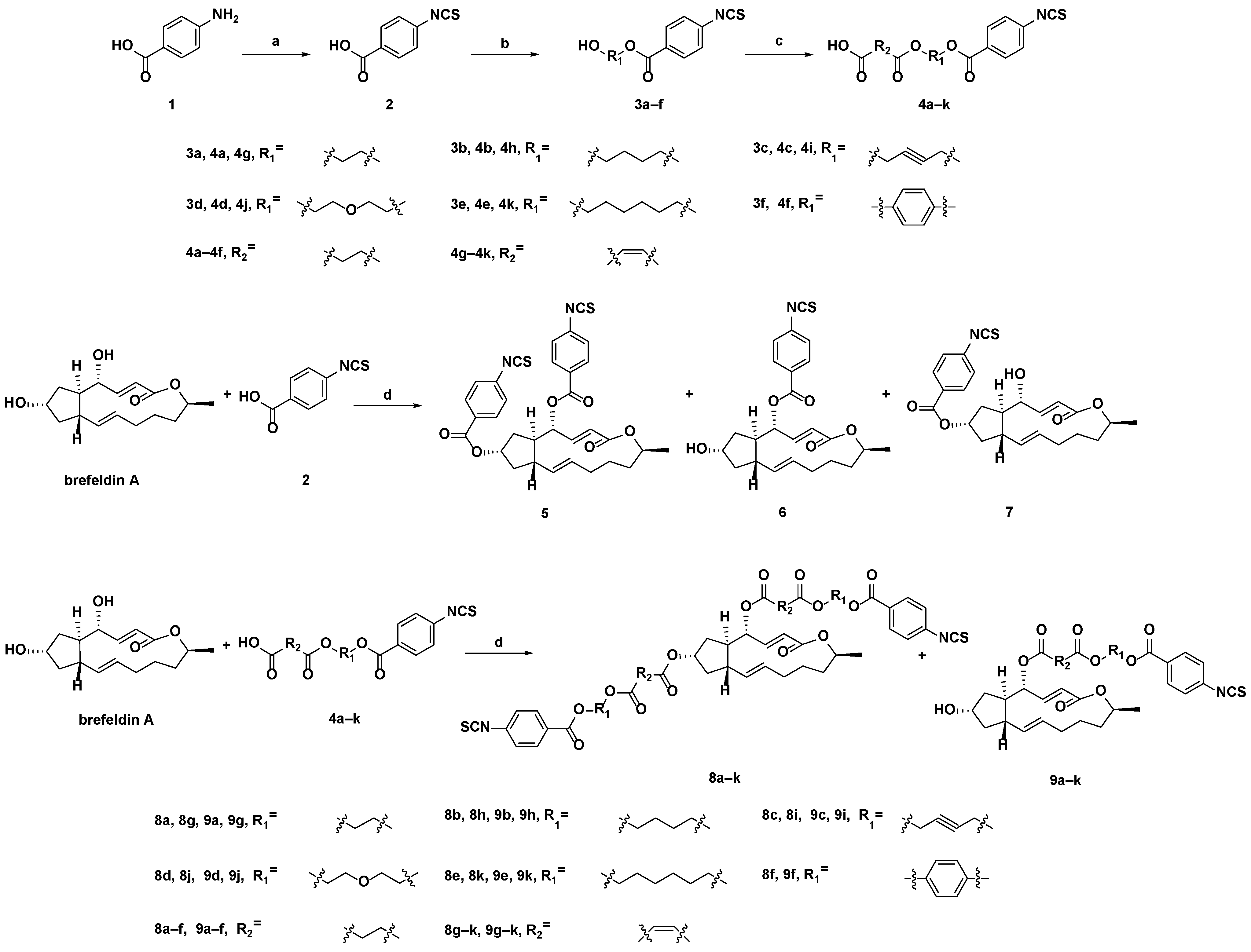
The synthesis strategy of the target compounds is shown in Scheme 1. 4-Carboxyphenyl isothiocyanate 2 was synthesized by the reaction of 4-aminobenzoic acid with 1,1′-thiocarbonyldiimidazole in the mixture of anhydrous dichloromethane (DCM) and triethylamine (TEA). Under the catalysis of 4-dimethylaminopyridine (DMAP) and N-(3-dimethylaminopropyl)-N’’-ethylcarbodiimidehydrochloride (EDCI), 4-carboxyphenyl isothiocyanate reacted with ethylene glycol, 1,4-butanediol, 2-butyne-1,4-diol, diethylene glycol, 1,6-hexanediol and hydroquinone in DCM at room temperature to obtain intermediates 3af. 3af were reacted with succinic anhydride or maleic anhydride at room temperature in the presence of DMAP and TEA in DCM to obtain intermediates 4ak. The intermediates 2 and 4ak were dissolved in DCM, and EDCI, DMAP and BFA were added in turn to obtain the target derivatives 5, 6, 7, 8ak and 9ak at room temperature.

2.2. Biological Evaluation

2.2.1. Antiproliferative Activity

As shown in Table 1, all derivatives, the lead compound BFA, 2 and positive control paclitaxel were tested for antiproliferative activity in HepG2, MDA-MB-231, A375, A549 and HeLa tumor cells and human liver cells L-02. Except for 4,7-OH disubstituted BFA derivatives 5 and 8ak, all derivatives showed good antiproliferative activity against five tumor cell lines. For most active compounds, the lowest IC50 value occurred on the HeLa cell line, the antiproliferative activity of 9b and 9j towards HeLa cells was similar to that shown on A375 and MDA-MBA-231 cell lines. The IC50 values of 4-OH monosubstituted derivatives 6 and 9ak and 7-OH monosubstituted derivative 7 against HeLa cells were 1.84, 2.69, 2.28, 3.63, 0.99, 0.81, 13.12, 3.63, 9.68, 6.59, 1.71, 2.39 and 3.02 μM, respectively. The structure-activity relationship analysis showed that the cytotoxic activity of 9f with hydroquinone as the linker was the weakest, compared with 9ae. The cytotoxic activity of derivatives 9ae linked by succinic anhydride was generally higher than that of derivatives 9gk linked by maleic anhydride. By comparing the antiproliferative activity of derivatives 9ae linked with succinic anhydride, it was found that 9d (R1 = diethylene glycol) had the best cytotoxic activity. Among the derivatives 9gk with maleic anhydride as the linker, 9j with the same diethylene glycol linker had the best cytotoxic activity. While 6 and 7 were directly linked by the lead compound BFA and 2 through ester bonds, and their activity against cancer cell lines was quite different. Specifically, the antiproliferative activity of 6 against five tumor cell lines was generally stronger than that of 7, and the toxicity to L-02 cell line was much weaker than that of 7.
Although the antiproliferative activity of the derivatives against five tumor cell lines was not comparable to that of BFA and paclitaxel, their SIHeLa were generally good, especially for 6, 8d, 8h, 9d, 9e and 9k, whose SIHeLa were all greater than 9, suggesting good selectivity. Among them, 6 showed the best selectivity to HeLa cells, and its IC50 value to HeLa cells was 1.84 μM. In L-02 cells, 6 showed no obvious cytotoxic activity and its IC50 value to L-02 cells was greater than 80 μM. Thus, 6 was selected for preliminary antiproliferative mechanism study in HeLa cells.

2.2.2. 6 Induced HeLa Cell Cycle Arrest in G1 Phase

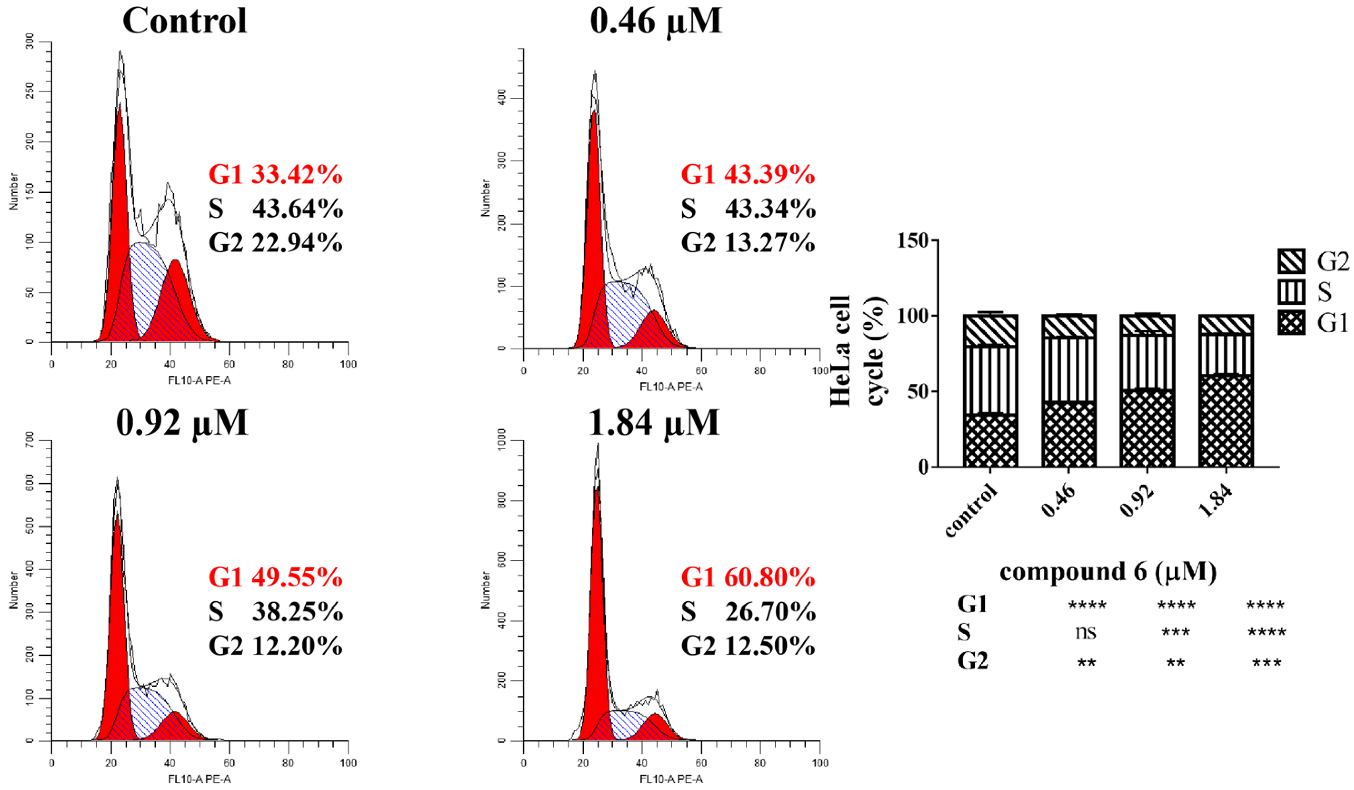
Propidium iodide (PI) flow cytometric assay was performed to test the DNA distribution at each stage to evaluate the regulatory effect of 6 on HeLa cell cycle. As shown in Figure 2, after HeLa cells were incubated with 6 (0, 0.46, 0.92 and 1.84 µM) for 48 h, cells arrested in G1 phase gradually increased from 33.42% of the control group to 43.39%, 49.55% and 60.80%, respectively. Accordingly, cells in S phase and G2 phase were down-regulated from 43.64% and 22.94% to 26.70% and 12.50%, respectively. 6 induced HeLa cell cycle arrest in G1 phase, indicating that HeLa cells could not pass through the checkpoint at the end of G1 phase for subsequent DNA replication and mitosis, which may lead to apoptosis.

2.2.3. 6 Induced HeLa Cell Nucleus Fragmentation

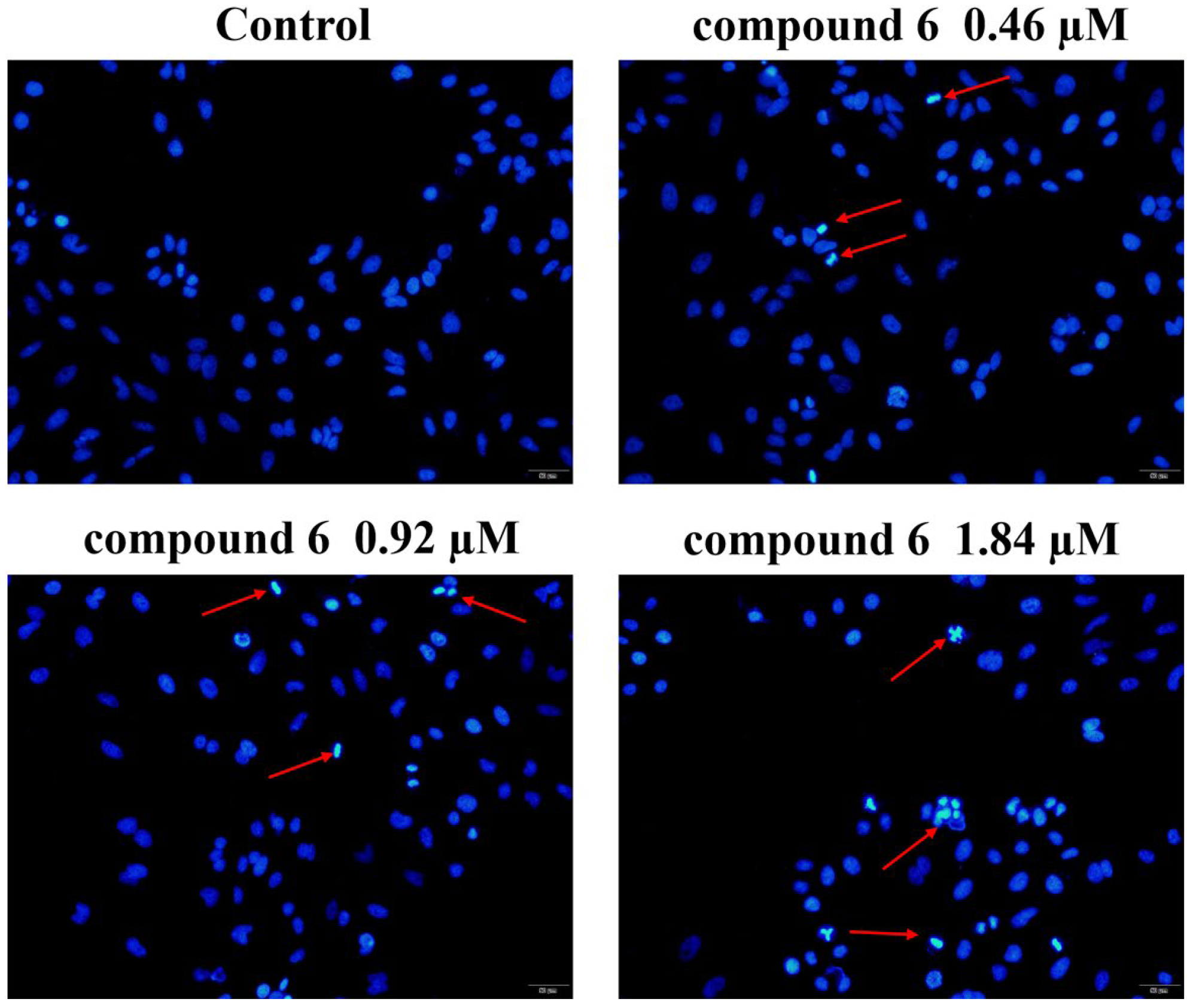
Deviant morphological characteristics such as pyknosis and nuclear fragmentation often occur in the early stage of apoptosis. As shown in Figure 3, Hoechst33258 staining result showed that with the increase of 6 (0, 0.46, 0.92 and 1.84 µM), the bright spots with higher fluorescence intensity gradually appeared in the HeLa cells, indicating the occurrence of nuclear fragmentation. Hoechst33258 staining indicated that 6 induced HeLa cell apoptosis.

2.2.4. 6 Induced Apoptosis in HeLa Cells

Annexin V-fluorescein isothiocyanate (FITC) and PI were used to further detect apoptosis. As shown in Figure 4, after HeLa cells were incubated with 6 (0, 0.46, 0.92 and 1.84 µM) for 48 h, the percentage of cells in the early and late stages of apoptosis increased from 3.21% to 16.04%, 26.41% and 39.03%, respectively. Combined with the results of Hoechst33258 staining, it was confirmed that 6 induced apoptosis of HeLa cells in a dose-dependent manner.

2.2.5. 6 Decreased Mitochondrial Membrane Potential in HeLa Cells

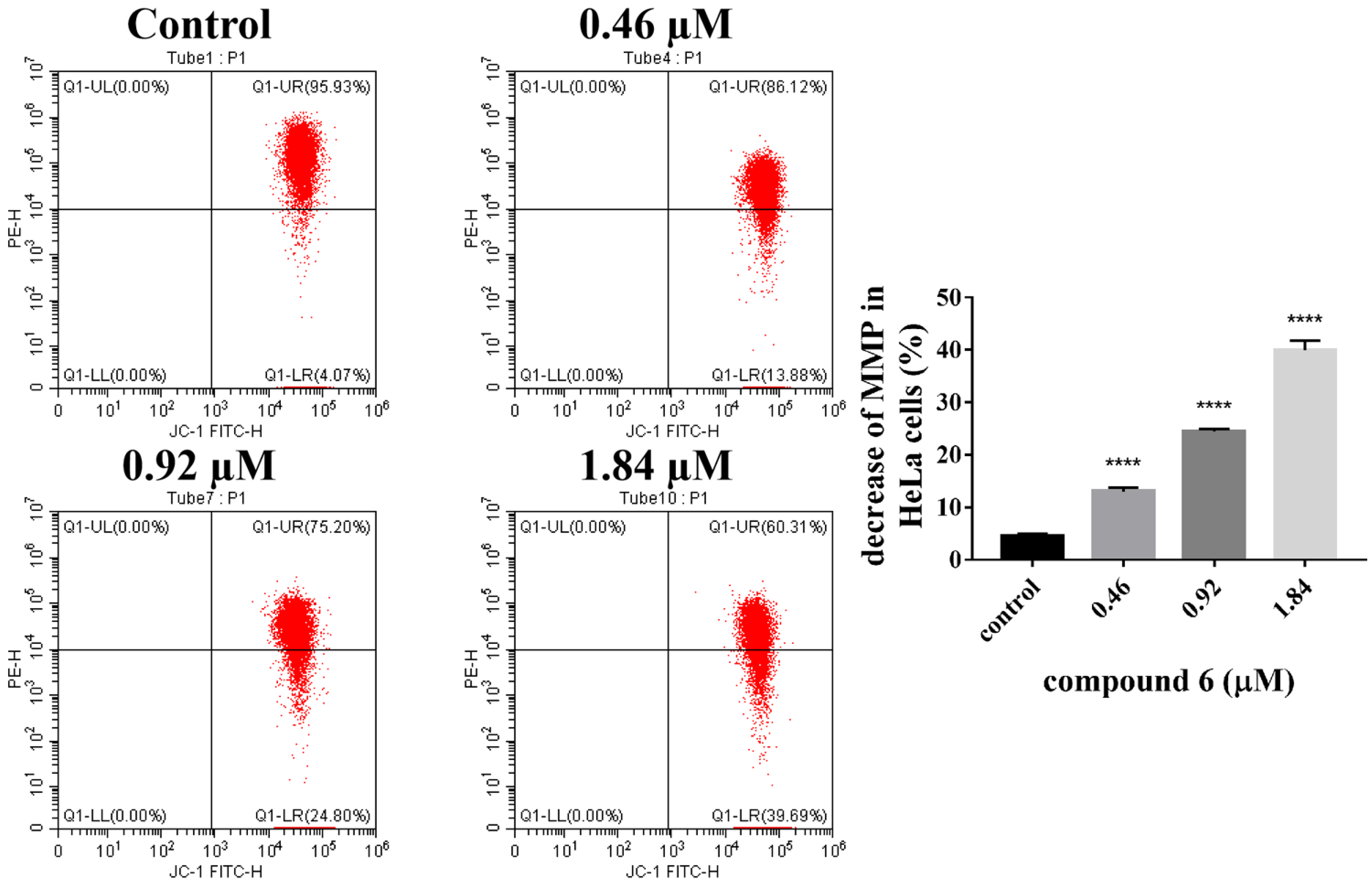
The destruction of mitochondrial membrane potential is a landmark event in the early stage of apoptosis. 5,5′,6,6′-tetrachloro-1,1′,3,3′-tetraethylbenzimi-dazolylcarbocyanine iodide (JC-1) is a membrane permeable cationic dye that selectively enters mitochondria and emits green fluorescence in cells with decreased mitochondrial membrane potential. As shown in Figure 5, with the increase of 6 (0, 0.46, 0.92 and 1.84 µM), the green fluorescent indicating that HeLa cells gradually increased from 4.07% to 13.88%, 24.80% and 39.69%, indicating HeLa cells with decreased mitochondrial membrane potential increased significantly with the increase of 6 concentration.

3. Materials and Methods

3.1. Chemistry

BFA was provided by Dr. Huaqi Pan from Shenyang Institute of Applied Ecology, Chinese Academy of Sciences, and the specific production process referred to the patent [46]. Anhydrous DCM was prepared based on standard methods, and other chemical materials and reagents were purchased from commercial suppliers. The reaction products were purified by 200–300 mesh silica gel column chromatography. The analysis of thin-layer chromatography was carried out on silica gel GF254 plate. With tetramethylsilane (TMS) as the internal standard, 1H nuclear magnetic resonance (NMR) and 13C NMR spectra of all derivatives were measured on a Bruker (Billerica, MA, USA) 600 MHz spectrometer (AVANCE III HD 600 MHz), and the chemical shifts data were reported in δ. High-resolution electrospray ionization mass spectrometry (HR-ESIMS) spectra were determined on an Waters (Milford, MA, USA) XEVO G2-XS QToF apparatus.

3.1.1. Synthesis of Intermediates 4ak

4-Aminobenzoic acid (1.2 g, 8.8 mmol) and 1,1′-thiocarbonyldiimidazole (2.1 g, 11.8 mmol) were dissolved in 30 mL anhydrous DCM, and then TEA (1.4 mL, 10.0 mmol) was added to react for 1 h under ice bath. 12 mL of n-hexane solution containing 3 mL hydrochloric acid was added and the mixture was keep at room temperature for 2 h. After 2 h, the reaction mixture was poured into 30 mL of ice water. After the solid was completely precipitated, the cake was filtered and washed several times, and the rice white powder 4-carboxyphenyl isothiocyanate 2 (1.5 g) was obtained, crude yield: 95%.
2 (179.2 mg, 1.0 mmol) and ethylene glycol (112 μL, 2.0 mmol) were dissolved in 20 mL DCM, and EDCI (575.1 mg, 3.0 mmol) and a catalytic amount of DMAP (7.2 mg, 0.06 mmol) were added to react for 12 h at room temperature. After the reaction was completed, 40 mL water was added to the reaction solution, extracted three times with DCM, the combined DCM was washed once with saturated sodium chloride solution, dried with Na2SO4, filtered, concentrated under reduced pressure, and purified by silica gel column chromatography (petroleum ether (PE): ethyl acetate (EA) = 4:1) to obtain colorless powder intermediate 3a 130.2 mg, yield: 60%.
3a (130.2 mg, 0.6 mmol) and succinic anhydride (100.1 mg, 1.0 mmol) were dissolved in 10 mL DCM, and then TEA (278 μL, 2.0 mmol) and a catalytic amount of DMAP (7.2 mg, 0.06 mmol) were added at room temperature. After 12 h, 20 mL of water was added, extracted three times with DCM, the combined DCM was washed once with saturated sodium chloride solution, dried with Na2SO4, filtered and concentrated under reduced pressure to obtain intermediate 4a 160.5 mg, crude yield: 85%.
Referring to the above reaction, ethylene glycol was replaced with 1,4-butanediol, 2-butyne-1,4-diol, diethylene glycol, 1,6-hexanediol or hydroquinone to obtain intermediates 3bf. The structures of 3af were characterized by 1H NMR (Figures S1–S6). Then 3af were reacted with succinic anhydride or maleic anhydride to obtain intermediates 4ak. The crude yields of 4bk were between 64–88%.

2-Hydroxyethyl 4-Isothiocyanatobenzoate (3a)

White powder, yield: 60%. 1H NMR (CDCl3, 600 MHz), δ: 8.05 (2H, dt, J = 8.6, 1.8 Hz, Ar-H), 7.27 (2H, dt, J = 8.7, 1.7 Hz, Ar-H), 4.46 (2H, m, -COOCH2-), 3.96 (2H, m, -OCH2-).

4-Hydroxybutyl 4-Isothiocyanatobenzoate (3b)

White powder, yield: 54%. 1H NMR (CDCl3, 600 MHz), δ: 8.02 (2H, dt, J = 8.8, 2.0 Hz, Ar-H), 7.26 (2H, dt, J = 8.7, 1.9 Hz, Ar-H), 4.36 (2H, t, J = 6.6 Hz, -COOCH2-), 3.72 (2H, t, J = 6.5 Hz, -OCH2-), 1.87 (2H, m, -CH2-), 1.72 (2H, m, -CH2-).

4-Hydroxybut-2-yn-1-yl 4-Isothiocyanatobenzoate (3c)

White powder, yield: 65%. 1H NMR (CDCl3, 600 MHz), δ: 8.05 (2H, dt, J = 8.7, 2.0 Hz, Ar-H), 7.27 (2H, dt, J = 8.7, 2.0 Hz, Ar-H), 4.96 (2H, t, J = 1.8 Hz, -COOCH2C≡), 4.33 (2H, t, J = 1.8 Hz, -OCH2C≡).

2-(2-Hydroxyethoxy)ethyl 4-Isothiocyanatobenzoate (3d)

White powder, yield: 67%. 1H NMR (CDCl3, 600 MHz), δ: 8.05 (2H, dt, J = 8.6, 1.9 Hz, Ar-H), 7.27 (2H, dt, J = 8.7, 1.9 Hz, Ar-H), 4.50 (2H, m, -COOCH2-), 3.84 (2H, m, -OCH2-), 3.76 (2H, m, -OCH2-), 3.65 (2H, m, -OCH2-).

6-Hydroxyhexyl 4-Isothiocyanatobenzoate (3e)

White powder, yield: 64%. 1H NMR (CDCl3, 600 MHz), δ: 8.02 (2H, dt, J = 8.7, 1.9 Hz, Ar-H), 7.26 (2H, dt, J = 8.7, 2.0 Hz, Ar-H), 4.31 (2H, t, J = 6.7 Hz, -COOCH2-), 3.65 (2H, t, J = 6.5 Hz, -OCH2-), 1.78 (2H, m, -CH2-), 1.60 (2H, m, -CH2-), 1.46 (4H, m, 2×-CH2-).

4-Hydroxyphenyl 4-Isothiocyanatobenzoate (3f)

White powder, yield: 36%. 1H NMR (DMSO-d6, 600 MHz), δ: 9.51 (1H, s, Ar-OH), 8.14(2H, dt, J = 8.7, 1.9 Hz, Ar-H), 7.60 (2H, dt, J = 8.7, 1.9 Hz, Ar-H), 7.06 (2H, dt, J = 8.9, 2.9 Hz, Ar-H), 6.81 (2H, dt, J = 9.0, 2.9 Hz, Ar-H).

3.1.2. Synthesis of Compounds 57

BFA (56.1 mg, 0.2 mmol) and 4-carboxyphenyl isothiocyanate (53.8 mg, 0.3 mmol) were dissolved in 10 mL of DCM, and EDCI (191.7 mg, 1.0 mmol) and DMAP (2.4 mg, 0.02 mmol) were added to react for 12 h at room temperature. After the reaction was completed, 20 mL of water was added, extracted three times with DCM, the combined DCM was washed once with saturated sodium chloride solution, dried with Na2SO4, filtered, concentrated under reduced pressure, and gradient eluted with silica gel column chromatography (PE:EA = 10:1–4:1) to obtain compounds 57, respectively. The structures of 57 were characterized by 1H NMR, 13C NMR, and HR-ESIMS (Figures S7–S15).

(1R,2E,6S,10E,11aS,13S,14aR)-6-methyl-4-oxo-1,6,7,8,9,11a,12,13,14,14a-decahydro-4H-cyclopenta[f][1]oxacyclotridecine-1,13-diyl bis(4-isothiocyanatobenzoate) (5)

White oil, yield: 23%. 1H NMR (CDCl3, 600 MHz), δ: 8.05 (2H, d, J = 8.6 Hz, Ar-H), 8.00 (2H, d, J = 8.6 Hz, Ar-H), 7.35 (1H, dd, J = 15.7, 3.4 Hz, H-3), 7.28 (4H, m, Ar-H), 5.79 (2H, m, H-2, H-11), 5.56 (1H, ddd, J = 10.4, 3.1, 1.7 Hz, H-4), 5.40 (1H, m, H-7), 5.30 (1H, dd, J = 15.1, 9.6 Hz, H-10), 4.88 (1H, m, H-15), 2.67–0.93 (15H, m, H-5, H-6a, H-6b, H-8a, H-8b, H-9, H-12a, H-12b, H-13a, H-13b, H-14a, H-14b, -CH3); 13C NMR (CDCl3, 150 MHz), δ: 165.5, 164.8, 164.2, 146.6, 138.1, 138.0, 136.2, 135.8, 135.4, 131.6, 131.2(×2), 131.0 (×2), 128.2, 127.9, 125.8 (×2), 125.7 (×2), 118.8, 77.1, 76.3, 72.0, 50.1, 44.2, 40.1, 38.5, 34.1, 31.8, 26.5, 20.7; HR-ESIMS m/z calcd for C32H30N2NaO6S2 [M + Na]+ 625.1437, found 625.1489.

(1R,2E,6S,10E,11aS,13S,14aR)-13-Hydroxy-6-methyl-4-oxo-1,6,7,8,9,11a,12,13,14,14a-decahydro-4H-cyclopenta[f][1]oxacyclotridecin-1-yl 4-isothiocyanatobenzoate (6)

White oil, yield: 12%. 1H NMR (CDCl3, 600 MHz), δ: 8.06 (2H, d, J = 8.6 Hz, Ar-H), 7.33 (1H, dd, J = 15.7, 3.3 Hz, H-3), 7.29 (2H, d, J = 8.6 Hz, Ar-H), 5.74 (2H, m, H-2, H-11), 5.56 (1H, ddd, J = 10.5, 3.1, 1.9 Hz, H-4), 5.34 (1H, dd, J = 15.3, 9.6 Hz, H-10), 4.85 (1H, m, H-15), 4.34 (1H, m, H-7), 2.55–0.90 (15H, m, H-5, H-6a, H-6b, H-8a, H-8b, H-9, H-12a, H-12b, H-13a, H-13b, H-14a, H-14b, -CH3); 13C NMR (CDCl3, 150 MHz), δ: 165.6, 164.3, 146.9, 138.1, 136.3, 136.1, 131.2 (×2), 130.8, 128.0, 125.8 (×2), 118.4, 77.3, 72.4, 71.9, 49.7, 44.3, 43.2, 41.1, 34.1, 31.8, 26.6, 20.8; HR-ESIMS m/z calcd for C24H27NNaO5S [M + Na]+ 464.1502, found 464.1505.

(1R,2E,6S,10E,11aS,13S,14aR)-1-Hydroxy-6-methyl-4-oxo-1,6,7,8,9,11a,12,13,14,14a-decahydro-4H-cyclopenta[f][1]oxacyclotridecin-13-yl 4-isothiocyanatobenzoate (7)

White oil, yield: 7%. 1H NMR (CDCl3, 600 MHz), δ: 8.00 (2H, dt, J = 8.6, 2.0 Hz, Ar-H), 7.37 (1H, dd, J = 15.7, 3.1 Hz, H-3), 7.27 (2H, dt, J = 8.6, 2.0 Hz, Ar-H), 5.94 (1H, dd, J = 15.7, 1.9 Hz, H-2), 5.75 (1H, m, H-11), 5.40 (1H, m, H-7), 5.24 (1H, dd, J = 15.2, 9.4 Hz, H-10), 4.88 (1H, m, H-15), 4.18 (1H, m, H-4), 2.50–0.92 (15H, m, H-5, H-6a, H-6b, H-8a, H-8b, H-9, H-12a, H-12b, H-13a, H-13b, H-14a, H-14b, -CH3); 13C NMR (CDCl3, 150 MHz), δ: 166.0, 164.9, 151.1, 137.9, 135.8, 135.7, 131.2, 131.0 (×2), 129.0, 125.7 (×2), 117.9, 76.3, 75.8, 71.7, 52.4, 44.0, 40.2, 38.8, 34.1, 31.8, 26.6, 20.8; HR-ESIMS m/z calcd for C24H27NNaO5S [M + Na]+ 464.1502, found 464.1498.

3.1.3. Synthesis of Compounds 8ak and 9ak

BFA (56.1 mg, 0.2 mmol) and 4a (160.5 mg) were dissolved in 10 mL of DCM, EDCI (191.7 mg, 1.0 mmol) and a catalytic amount of DMAP (2.4 mg, 0.02 mmol) were added to react for 12 h at room temperature. After the reaction was completed, 20 mL of water was added, extracted three times with DCM the combined DCM was washed once with saturated sodium chloride solution, dried with Na2SO4, filtered, concentrated under reduced pressure, and gradient eluted with silica gel column chromatography (DCM:MeOH = 400:1–150:1) to obtain compounds 8a and 9a, respectively. All other derivatives were obtained by referring to the synthesis method of compounds 8a and 9a. The structures of 8ak and 9ak were characterized by 1H NMR, 13C NMR, and HR-ESIMS (Figures S16–S81).

Bis(2-((4-isothiocyanatobenzoyl)oxy)ethyl) O,O’-((1R,2E,6S,10E,11aS,13S,14aR)-6-methyl-4-oxo-1,6,7,8,9,11a,12,13,14,14a-decahydro-4H-cyclopenta[f][1]oxacyclotridecine-1,13-diyl) disuccinate (8a)

Colorless oil, yield: 25%. 1H NMR (CDCl3, 600 MHz), δ: 8.03 (4H, m, Ar-H), 7.27 (4H, m, Ar-H), 7.20 (1H, dd, J = 15.7, 3.4 Hz, H-3), 5.71 (2H, m, H-2, H-11), 5.22 (2H, m, H-4, H-10), 5.10 (1H, m, H-7), 4.85 (1H, m, H-15), 4.51 (4H, m, 2×-COOCH2-), 4.44 (4H, m, 2×-COOCH2-), 2.64 (8H, m, 4×-COCH2-), 2.46–0.89 (15H, m, H-5, H-6a, H-6b, H-8a, H-8b, H-9, H-12a, H-12b, H-13a, H-13b, H-14a, H-14b, -CH3); 13C NMR (CDCl3, 150 MHz), δ: 172.1, 171.8, 171.6, 171.1, 165.5, 165.1 (×2), 146.6, 138.0 (×2), 135.9, 135.8, 135.5, 131.3, 131.2 (×4), 128.3 (×2), 125.7 (×4), 118.5, 76.6, 75.6, 71.8, 62.9 (×2), 62.4, 62.3, 49.7, 44.0, 40.0, 38.2, 34.1, 31.8, 29.2, 28.9 (×2), 28.8, 26.5, 20.7; HR-ESIMS m/z calcd for C44H46N2NaO14S2 [M + Na]+ 913.2283, found 913.2337.

(1R,2E,6S,10E,11aS,13S,14aR)-13-Hydroxy-6-methyl-4-oxo-1,6,7,8,9,11a,12,13,14,14a-decahydro-4H-cyclopenta[f][1]oxacyclotridecin-1-yl (2-((4-isothiocyanatobenzoyl)oxy)ethyl) succinate (9a)

Colorless oil, yield: 18%. 1H NMR (CDCl3, 600 MHz), δ: 8.04 (2H, d, J = 8.5 Hz, Ar-H), 7.28 (2H, d, J = 8.5 Hz, Ar-H), 7.22 (1H, dd, J = 15.8, 3.4 Hz, H-3), 5.70 (2H, m, H-2, H-11), 5.27 (2H, m, H-4, H-10), 4.85 (1H, m, H-15), 4.52 (2H, t, J = 4.7 Hz, -COOCH2-), 4.45 (2H, m, -COOCH2-), 4.30 (1H, m, H-7), 2.70 (4H, m, 2×-COCH2-), 2.45–0.89 (15H, m, H-5, H-6a, H-6b, H-8a, H-8b, H-9, H-12a, H-12b, H-13a, H-13b, H-14a, H-14b, -CH3); 13C NMR (CDCl3, 150 MHz), δ: 171.8, 171.1, 165.6, 165.2, 146.9, 138.0, 136.3, 135.9, 131.3 (×2), 130.7, 128.3, 125.7 (×2), 118.3, 76.7, 72.3, 71.9, 63.0, 62.3, 49.5, 44.3, 43.1, 40.9, 34.1, 31.8, 29.0, 28.9, 26.6, 20.8; HR-ESIMS m/z calcd for C30H35NNaO9S [M + Na]+ 608.1925, found 608.1930.

Bis(4-((4-isothiocyanatobenzoyl)oxy)butyl) O,O’-((1R,2E,6S,10E,11aS,13S,14aR)-6-methyl-4-oxo-1,6,7,8,9,11a,12,13,14,14a-decahydro-4H-cyclopenta[f][1]oxacyclotridecine-1,13-diyl) disuccinate (8b)

Colorless oil, yield: 28%. 1H NMR (CDCl3, 600 MHz), δ: 8.02 (4H, m, Ar-H), 7.27 (4H, m, Ar-H), 7.21 (1H, dd, J = 15.6, 3.2 Hz, H-3), 5.71 (2H, m, H-2, H-11), 5.24 (2H, m, H-4, H-10), 5.13 (1H, m, H-7), 4.85 (1H, m, H-15), 4.34 (4H, m, 2×-COOCH2-), 4.16 (4H, m, 2×-COOCH2-), 2.64 (8H, m, 4×-COCH2-), 2.47–0.89 (23H, m, H-5, H-6a, H-6b, H-8a, H-8b, H-9, H-12a, H-12b, H-13a, H-13b, H-14a, H-14b, -CH3, 4×-CH2-); 13C NMR (CDCl3, 150 MHz), δ: 172.2, 172.0, 171.7, 171.2, 165.5, 165.3 (×2), 146.7, 137.9, 137.8, 135.7, 135.6, 135.6, 131.2, 131.0 (×4), 128.8, 128.7, 125.6 (×4), 118.5, 76.6, 75.5, 71.8, 64.8, 64.7, 64.3, 64.1, 49.7, 44.0, 40.0, 38.2, 34.1, 31.7, 29.3, 29.0 (×3), 26.5, 25.3(×4), 20.7; HR-ESIMS m/z calcd for C48H54N2NaO14S2 [M + Na]+ 969.2909, found 969.2952.

(1R,2E,6S,10E,11aS,13S,14aR)-13-Hydroxy-6-methyl-4-oxo-1,6,7,8,9,11a,12,13,14,14a-decahydro-4H-cyclopenta[f][1]oxacyclotridecin-1-yl (4-((4-isothiocyanatobenzoyl)oxy)butyl) succinate (9b)

Colorless oil, yield: 15%. 1H NMR (CDCl3, 600 MHz), δ: 8.03 (2H, d, J = 8.5 Hz, Ar-H), 7.27 (2H, d, J = 8.5 Hz, Ar-H), 7.22 (1H, dd, J = 15.7, 3.3 Hz, H-3), 5.69 (2H, m, H-2, H-11), 5.29 (2H, m, H-4, H-10), 4.85 (1H, m, H-15), 4.33 (3H, m, H-7, -COOCH2-), 4.17 (2H, t, J = 6.1 Hz, -COOCH2-), 2.67 (4H, m, 2×-COCH2-), 2.45–0.89 (19H, m, H-5, H-6a, H-6b, H-8a, H-8b, H-9, H-12a, H-12b, H-13a, H-13b, H-14a, H-14b, -CH3, 2×-CH2-); 13C NMR (CDCl3, 150 MHz), δ: 172.0, 171.2, 165.6, 165.4, 147.0, 137.8, 136.3, 135.7, 131.0 (×2), 130.7, 128.8, 125.7 (×2), 118.3, 76.7, 72.3, 71.8, 64.8, 64.2, 49.5, 44.3, 43.1, 40.9, 34.0, 31.8, 29.0 (×2), 26.6, 25.3 (×2), 20.8; HR-ESIMS m/z calcd for C32H39NNaO9S [M + Na]+ 636.2238, found 636.2252.

Bis(4-((4-isothiocyanatobenzoyl)oxy)but-2-yn-1-yl) O,O’-((1R,2E,6S,10E,11aS,13S,14aR)-6-methyl-4-oxo-1,6,7,8,9,11a,12,13,14,14a-decahydro-4H-cyclopenta[f][1]oxacyclotridecine-1,13-diyl) disuccinate (8c)

Colorless oil, yield: 13%. 1H NMR (CDCl3, 600 MHz), δ: 8.05 (4H, m, Ar-H), 7.28 (4H, m, Ar-H), 7.21 (1H, dd, J = 15.7, 3.4 Hz, H-3), 5.71 (2H, m, H-2, H-11), 5.23 (2H, m, H-4, H-10), 5.14 (1H, m, H-7), 4.95 (4H, m, 2×-COOCH2C≡), 4.86 (1H, m, H-15), 4.76 (4H, m, 2×-COOCH2C≡), 2.69 (6H, m, 3×-COCH2-), 2.61 (2H, m, -COCH2-), 2.48–0.91 (15H, m, H-5, H-6a, H-6b, H-8a, H-8b, H-9, H-12a, H-12b, H-13a, H-13b, H-14a, H-14b, -CH3); 13C NMR (CDCl3, 150 MHz), δ: 171.5 (×2), 171.2, 171.1, 165.5, 164.6 (×2), 146.6, 138.2, 138.1, 136.1 (×2), 135.6, 131.3 (×5), 127.9, 127.8, 125.7 (×4), 118.5, 81.0 (×2), 80.7 (×2), 76.7, 75.6, 71.8, 52.9, 52.8, 52.4, 52.3, 49.7, 44.0, 40.0, 38.2, 34.1, 31.7, 29.2, 28.8 (×3), 26.5, 20.7; HR-ESIMS m/z calcd for C48H46N2NaO14S2 [M + Na]+ 961.2283, found 961.2315.

(1R,2E,6S,10E,11aS,13S,14aR)-13-Hydroxy-6-methyl-4-oxo-1,6,7,8,9,11a,12,13,14,14a-decahydro-4H-cyclopenta[f][1]oxacyclotridecin-1-yl (4-((4-isothiocyanatobenzoyl)oxy)but-2-yn-1-yl) succinate (9c)

Colorless oil, yield: 32%. 1H NMR (CDCl3, 600 MHz), δ: 8.06 (2H, d, J = 8.3 Hz, Ar-H), 7.28 (2H, d, J = 8.3 Hz, Ar-H), 7.22 (1H, dd, J = 15.7, 3.3 Hz, H-3), 5.69 (2H, m, H-2, H-11), 5.29 (2H, m, H-4, H-10), 4.96 (2H, s, -COOCH2C≡), 4.85 (1H, m, H-15), 4.77 (2H, s, -COOCH2C≡), 4.31 (1H, m, H-7), 2.71 (4H, m, 2×-COCH2-), 2.45–0.90 (15H, m, H-5, H-6a, H-6b, H-8a, H-8b, H-9, H-12a, H-12b, H-13a, H-13b, H-14a, H-14b, -CH3); 13C NMR (CDCl3, 150 MHz), δ: 171.3, 171.0, 165.6, 164.7, 146.9, 138.1, 136.3, 136.1, 131.3 (×2), 130.7, 127.9, 125.7 (×2), 118.4, 81.0, 80.7, 72.4, 71.9, 52.9, 52.4, 49.5, 44.3, 43.1, 40.9, 34.1, 31.8, 28.9, 28.8, 26.6, 20.8; HR-ESIMS m/z calcd for C32H35NNaO9S [M + Na]+ 632.1925, found 632.1942.

Bis(2-(2-((4-isothiocyanatobenzoyl)oxy)ethoxy)ethyl) O,O’-((1R,2E,6S,10E,11aS,13S,14aR)-6-methyl-4-oxo-1,6,7,8,9,11a,12,13,14,14a-decahydro-4H-cyclopenta[f][1]oxacyclotridecine-1,13-diyl) disuccinate (8d)

Colorless oil, yield: 17%. 1H NMR (CDCl3, 600 MHz), δ: 8.04 (4H, m, Ar-H), 7.27 (4H, m, Ar-H), 7.21 (1H, dd, J = 15.7, 3.4 Hz, H-3), 5.71 (2H, m, H-2, H-11), 5.23 (2H, m, H-4, H-10), 5.13 (1H, m, H-7), 4.85 (1H, m, H-15), 4.47 (4H, m, 2×-COOCH2-), 4.26 (4H, m, 2×-COOCH2-), 3.82 (4H, m, 2×-OCH2-), 3.73 (4H, m, 2×-OCH2-), 2.63 (8H, m, 4×-COCH2-), 2.48–0.91 (15H, m, H-5, H-6a, H-6b, H-8a, H-8b, H-9, H-12a, H-12b, H-13a, H-13b, H-14a, H-14b, -CH3); 13C NMR (CDCl3, 150 MHz), δ: 172.2, 171.9, 171.6, 171.1, 165.5, 165.3 (×2), 146.7, 137.9, 137.8, 135.8, 135.7, 135.6, 131.2, 131.1 (×4), 128.6, 128.5, 125.7 (×4), 118.5, 76.6, 75.5, 71.8, 69.0 (×4), 64.3, 64.2, 63.8, 63.7, 49.7, 44.0, 40.0, 38.2, 34.1, 31.7, 29.2, 28.9 (×3), 26.5, 20.7; HR-ESIMS m/z calcd for C48H54N2NaO16S2 [M + Na]+ 1001.2807, found 1001.2847.

(1R,2E,6S,10E,11aS,13S,14aR)-13-Hydroxy-6-methyl-4-oxo-1,6,7,8,9,11a,12,13,14,14a-decahydro-4H-cyclopenta[f][1]oxacyclotridecin-1-yl (2-(2-((4-isothiocyanatobenzoyl)oxy)ethoxy)ethyl) succinate (9d)

Colorless oil, yield: 25%. 1H NMR (CDCl3, 600 MHz), δ: 8.05 (2H, d, J = 8.3 Hz, Ar-H), 7.28 (2H, d, J = 8.3 Hz, Ar-H), 7.23 (1H, dd, J = 16.0, 2.5 Hz, H-3), 5.69 (2H, m, H-2, H-11), 5.28 (2H, m, H-4, H-10), 4.85 (1H, m, H-15), 4.48 (2H, m, -COOCH2-), 4.29 (3H, m, H-7, -COOCH2-), 3.83 (2H, m, -OCH2-), 3.74 (2H, m, -OCH2-), 2.67 (4H, m, 2×-COCH2-), 2.44–0.92 (15H, m, H-5, H-6a, H-6b, H-8a, H-8b, H-9, H-12a, H-12b, H-13a, H-13b, H-14a, H-14b, -CH3); 13C NMR (CDCl3, 150 MHz), δ: 172.0, 171.1, 165.6, 165.4, 147.0, 137.8, 136.3, 135.7, 131.2 (×2), 130.6, 128.5, 125.7 (×2), 118.3, 76.7, 72.3, 71.8, 69.0 (×2), 64.2, 63.7, 49.5, 44.3, 43.1, 41.0, 34.0, 31.8, 29.0, 28.9, 26.6, 20.8; HR-ESIMS m/z calcd for C32H39NNaO10S [M + Na]+ 652.2187, found 652.2188.

Bis(6-((4-isothiocyanatobenzoyl)oxy)hexyl) O,O’-((1R,2E,6S,10E,11aS,13S,14aR)-6-methyl-4-oxo-1,6,7,8,9,11a,12,13,14,14a-decahydro-4H-cyclopenta[f][1]oxacyclotridecine-1,13-diyl) disuccinate (8e)

Colorless oil, yield: 23%. 1H NMR (CDCl3, 600 MHz), δ: 8.02 (4H, m, Ar-H), 7.27 (4H, m, Ar-H), 7.22 (1H, dd, J = 15.7, 3.4 Hz, H-3), 5.71 (2H, m, H-2, H-11), 5.25 (2H, m, H-4, H-10), 5.14 (1H, m, H-7), 4.85 (1H, m, H-15), 4.31 (4H, m, 2×-COOCH2-), 4.10 (4H, m, 2×-COOCH2-), 2.64 (8H, m, 4×-COCH2-), 2.49–0.90 (31H, m, H-5, H-6a, H-6b, H-8a, H-8b, H-9, H-12a, H-12b, H-13a, H-13b, H-14a, H-14b, -CH3, 8×-CH2-); 13C NMR (CDCl3, 150 MHz), δ: 172.3, 172.0, 171.7, 171.2, 165.5, 165.4 (×2), 146.8, 137.8, 137.7, 135.7, 135.6, 135.5, 131.2, 131.0 (×4), 129.0, 128.9, 125.6 (×4), 118.5, 76.6, 75.5, 71.8, 65.3, 65.2, 64.7, 64.6, 49.7, 44.1, 40.0, 38.2, 34.1, 31.8, 29.3, 29.1, 29.0 (×2), 28.6 (×2), 28.5 (×2), 26.5, 25.7 (×2), 25.6 (×2), 20.7; HR-ESIMS m/z calcd for C52H62N2NaO14S2 [M + Na]+ 1025.3535, found 1025.3533.

(1R,2E,6S,10E,11aS,13S,14aR)-13-Hydroxy-6-methyl-4-oxo-1,6,7,8,9,11a,12,13,14,14a-decahydro-4H-cyclopenta[f][1]oxacyclotridecin-1-yl (6-((4-isothiocyanatobenzoyl)oxy)hexyl) succinate (9e)

Colorless oil, yield: 26%. 1H NMR (CDCl3, 600 MHz), δ: 8.02 (2H, d, J = 8.5 Hz, Ar-H), 7.27 (2H, d, J = 8.5 Hz, Ar-H), 7.23 (1H, dd, J = 15.7, 3.4 Hz, H-3), 5.69 (2H, m, H-2, H-11), 5.29 (2H, m, H-4, H-10), 4.84 (1H, m, H-15), 4.31 (3H, m, H-7, -COOCH2-), 4.11 (2H, t, J = 6.1 Hz, -COOCH2-), 2.66 (4H, m, 2×-COCH2-), 2.45–0.89 (23H, m, H-5, H-6a, H-6b, H-8a, H-8b, H-9, H-12a, H-12b, H-13a, H-13b, H-14a, H-14b, -CH3, 4×-CH2-); 13C NMR (CDCl3, 150 MHz), δ: 172.1, 171.3, 165.6, 165.5, 147.0, 137.7, 136.3, 135.6, 131.0 (×2), 130.7, 129.0, 125.6 (×2), 118.3, 76.6, 72.3, 71.8, 65.3, 64.7, 49.5, 44.3, 43.1, 40.9, 34.0, 31.8, 29.1, 29.0, 28.5 (×2), 26.6, 25.6 (×2), 20.8; HR-ESIMS m/z calcd for C34H43NNaO9S [M + Na]+ 664.2551, found 664.2558.

Bis(4-((4-isothiocyanatobenzoyl)oxy)phenyl) O,O’-((1R,2E,6S,10E,11aS,13S,14aR)-6-methyl-4-oxo-1,6,7,8,9,11a,12,13,14,14a-decahydro-4H-cyclopenta[f][1]oxacyclotridecine-1,13-diyl) disuccinate (8f)

Colorless oil, yield: 14%. 1H NMR (CDCl3, 600 MHz), δ: 8.17 (4H, m, Ar-H), 7.33 (4H, m, Ar-H), 7.22 (5H, m, H-3, Ar-H), 7.15 (4H, m, Ar-H), 5.72 (2H, m, H-2, H-11), 5.32 (1H, ddd, J = 10.1, 3.1, 1,6 Hz, H-4), 5.23 (1H, dd, J = 15.3, 9.6 Hz, H-10), 5.18 (1H, m, H-7), 4.86 (1H, m, H-15), 2.89 (4H, m, 2×-COCH2-), 2.79 (2H, m, -COCH2-), 2.70 (2H, m, -COCH2-), 2.51–0.91 (15H, m, H-5, H-6a, H-6b, H-8a, H-8b, H-9, H-12a, H-12b, H-13a, H-13b, H-14a, H-14b, -CH3); 13C NMR (CDCl3, 150 MHz), δ: 171.5, 171.0, 170.8, 170.5, 165.5, 163.8 (×2), 148.2 (×2), 148.1 (×2), 146.6, 138.3 (×2), 136.4 (×2), 135.6, 131.7 (×4), 131.3, 127.8 (×2), 125.9 (×4), 122.5 (×8), 118.6, 76.7, 75.8, 71.8, 49.8, 44.1, 40.0, 38.3, 34.1, 31.7, 29.3 (×2), 29.1, 28.9, 26.5, 20.7; HR-ESIMS m/z calcd for C52H46N2NaO14S2 [M + Na]+ 1009.2283, found 1009.2352.

(1R,2E,6S,10E,11aS,13S,14aR)-13-Hydroxy-6-methyl-4-oxo-1,6,7,8,9,11a,12,13,14,14a-decahydro-4H-cyclopenta[f][1]oxacyclotridecin-1-yl (4-((4-isothiocyanatobenzoyl)oxy)phenyl) succinate (9f)

Colorless oil, yield: 8%. 1H NMR (CDCl3, 600 MHz), δ: 8.18 (2H, d, J = 8.6 Hz, Ar-H), 7.33 (2H, d, J = 8.6 Hz, Ar-H), 7.22 (3H, m, H-3, Ar-H), 7.17 (2H, m, Ar-H), 5.71 (2H, m, H-2, H-11), 5.30 (2H, m, H-4, H-10), 4.86 (1H, m, H-15), 4.28 (1H, m, H-7), 2.87 (4H, m, 2×-COCH2-), 2.45–0.90 (15H, m, H-5, H-6a, H-6b, H-8a, H-8b, H-9, H-12a, H-12b, H-13a, H-13b, H-14a, H-14b, -CH3); 13C NMR (CDCl3, 150 MHz), δ: 171.0, 170.6, 165.6, 163.9, 148.2, 148.1, 146.9, 138.3, 136.4, 136.3, 131.7 (×2), 130.7, 127.8, 125.9 (×2), 122.5 (×4), 118.4, 76.8, 72.3, 71.9, 49.5, 44.3, 43.1, 41.0, 34.1, 31.8, 29.2, 29.0, 26.6, 20.8; HR-ESIMS m/z calcd for C34H35NNaO9S [M + Na]+ 656.1925, found 656.1943.

Bis(2-((4-isothiocyanatobenzoyl)oxy)ethyl) O,O’-((1R,2E,6S,10E,11aS,13S,14aR)-6-methyl-4-oxo-1,6,7,8,9,11a,12,13,14,14a-decahydro-4H-cyclopenta[f][1]oxacyclotridecine-1,13-diyl) dimaleate (8g)

Colorless oil, yield: 38%. 1H NMR (CDCl3, 600 MHz), δ: 8.03 (4H, m, Ar-H), 7.28 (4H, m, Ar-H), 7.22 (1H, dd, J = 15.8, 2.8 Hz, H-3), 6.88 (4H, m, 4×-C=CH-), 5.71 (2H, m, H-2, H-11), 5.37 (1H, d, J = 9.8 Hz, H-4), 5.22 (2H, m, H-7, H-10), 4.87 (1H, m, H-15), 4.57 (8H, m, 4×-COOCH2-), 2.56–0.91 (15H, m, H-5, H-6a, H-6b, H-8a, H-8b, H-9, H-12a, H-12b, H-13a, H-13b, H-14a, H-14b, -CH3); 13C NMR (CDCl3, 150 MHz), δ: 165.3, 165.1 (×2), 164.6, 164.3, 164.2, 163.5, 145.8, 138.1, 138.0, 136.0 (×2), 135.2, 134.3, 133.9, 133.3, 133.0, 131.7, 131.2 (×4), 128.1 (×2), 125.8 (×2), 125.7 (×2), 118.9, 76.4, 72.0, 63.1, 63.0, 62.7 (×2), 49.6, 44.0, 39.9, 38.3, 34.1, 31.8, 26.4, 20.7; HR-ESIMS m/z calcd for C44H42N2NaO14S2 [M + Na]+ 909.1970, found 909.2030.

(1R,2E,6S,10E,11aS,13S,14aR)-13-Hydroxy-6-methyl-4-oxo-1,6,7,8,9,11a,12,13,14,14a-decahydro-4H-cyclopenta[f][1]oxacyclotridecin-1-yl (2-((4-isothiocyanatobenzoyl)oxy)ethyl) maleate(9g)

Colorless oil, yield: 14%. 1H NMR (CDCl3, 600 MHz), δ: 8.05 (2H, d, J = 8.6 Hz, Ar-H), 7.29 (2H, d, J = 8.6 Hz, Ar-H), 7.24 (1H, m, H-3), 6.92 (2H, m, 2×-C=CH-), 5.70 (2H, m, H-2, H-11), 5.33 (2H, m, H-4, H-10), 4.86 (1H, m, H-15), 4.58 (4H, m, 2×-COOCH2-), 4.33 (1H, m, H-7), 2.50–0.92 (15H, m, H-5, H-6a, H-6b, H-8a, H-8b, H-9, H-12a, H-12b, H-13a, H-13b, H-14a, H-14b, -CH3); 13C NMR (CDCl3, 150 MHz), δ: 165.5, 165.1, 164.4, 163.6, 146.3, 138.0, 136.2, 136.0, 133.7, 133.6, 131.2 (×2), 130.8, 128.1, 125.8 (×2), 118.6, 77.5, 72.4, 72.0, 63.1, 62.8, 49.5, 44.3, 43.2, 41.0, 34.1, 31.8, 26.6, 20.8; HR-ESIMS m/z calcd for C30H33NNaO9S [M + Na]+ 606.1768, found 606.1790.

Bis(4-((4-isothiocyanatobenzoyl)oxy)butyl) O,O’-((1R,2E,6S,10E,11aS,13S,14aR)-6-methyl-4-oxo-1,6,7,8,9,11a,12,13,14,14a-decahydro-4H-cyclopenta[f][1]oxacyclotridecine-1,13-diyl) dimaleate (8h)

Colorless oil, yield: 28%. 1H NMR (CDCl3, 600 MHz), δ: 8.02 (4H, m, Ar-H), 7.26 (5H, m, H-3, Ar-H), 6.87 (4H, m, 4×-C=CH-), 5.73 (2H, m, H-2, H-11), 5.39 (1H, d, J = 10.3 Hz, H-4), 5.24 (2H, m, H-7, H-10), 4.87 (1H, m, H-15), 4.33 (8H, m, 4×-COOCH2-), 2.56–0.91 (23H, m, H-5, H-6a, H-6b, H-8a, H-8b, H-9, H-12a, H-12b, H-13a, H-13b, H-14a, H-14b, -CH3, 4×-CH2-); 13C NMR (CDCl3, 150 MHz), δ: 165.3 (×3), 164.8, 164.6, 164.3, 163.6, 145.9, 137.9, 137.8, 135.7 (×2), 135.2, 134.5, 133.8, 133.6, 132.8, 131.6, 131.0 (×4), 128.7 (×2), 125.7 (×4), 118.9, 77.2, 76.3, 72.0, 64.9, 64.8, 64.6 (×2), 49.7, 44.1, 40.0, 38.3, 34.1, 31.7, 26.4, 25.3 (×4), 20.7; HR-ESIMS m/z calcd for C48H50N2NaO14S2 [M + Na]+ 965.2596, found 965.2650.

(1R,2E,6S,10E,11aS,13S,14aR)-13-Hydroxy-6-methyl-4-oxo-1,6,7,8,9,11a,12,13,14,14a-decahydro-4H-cyclopenta[f][1]oxacyclotridecin-1-yl (4-((4-isothiocyanatobenzoyl)oxy)butyl) maleate (9h)

Colorless oil, yield: 25%. 1H NMR (CDCl3, 600 MHz), δ: 8.03 (2H, d, J = 8.6 Hz, Ar-H), 7.26 (3H, m, H-3, Ar-H), 6.89 (2H, m, 2×-C=CH-), 5.70 (2H, m, H-2, H-11), 5.33 (2H, m, H-4, H-10), 4.86 (1H, m, H-15), 4.33 (5H, m, H-7, 2×-COOCH2-), 2.50–0.91 (19H, m, H-5, H-6a, H-6b, H-8a, H-8b, H-9, H-12a, H-12b, H-13a, H-13b, H-14a, H-14b, -CH3, 2×-CH2-); 13C NMR (CDCl3, 150 MHz), δ: 165.5, 165.4, 164.7, 163.8, 146.3, 137.8, 136.2, 135.7, 134.3, 133.0, 131.0 (×2), 130.8, 128.7, 125.7 (×2), 118.5, 77.4, 72.3, 72.0, 64.9, 64.7, 49.5, 44.3, 43.2, 41.0, 34.1, 31.8, 26.6, 25.3 (×2), 20.8; HR-ESIMS m/z calcd for C32H37NNaO9S [M + Na]+ 634.2081, found 634.2088.

Bis(4-((4-isothiocyanatobenzoyl)oxy)but-2-yn-1-yl) O,O’-((1R,2E,6S,10E,11aS,13S,14aR)-6-methyl-4-oxo-1,6,7,8,9,11a,12,13,14,14a-decahydro-4H-cyclopenta[f][1]oxacyclotridecine-1,13-diyl) dimaleate (8i)

Colorless oil, yield: 26%. 1H NMR (CDCl3, 600 MHz), δ: 8.05 (4H, m, Ar-H), 7.28 (4H, m, Ar-H), 7.23 (1H, J = 15.7, 3.5 Hz, H-3), 6.89 (4H, m, 4×-C=CH-), 5.73 (2H, m, H-2, H-11), 5.38 (1H, d, J = 10.3, 3.2, 1.7 Hz, H-4), 5.24 (2H, m, H-7, H-10), 4.97 (4H, m, 2×-COOCH2C≡), 4.88 (5H, m, H-15, 2×-COOCH2C≡), 2.56–0.93 (15H, m, H-5, H-6a, H-6b, H-8a, H-8b, H-9, H-12a, H-12b, H-13a, H-13b, H-14a, H-14b, -CH3); 13C NMR (CDCl3, 150 MHz), δ: 165.3, 164.6 (×2), 164.1, 164.0, 163.8, 163.4, 145.8, 138.2, 138.1, 136.2, 136.1, 135.2, 134.7, 133.7, 133.5, 132.6, 131.7, 131.3 (×4), 127.8 (×2), 125.7 (×4), 118.9, 81.3, 81.2, 80.4 (×2), 76.4, 72.0, 53.0, 52.9, 52.8 (×2), 49.7, 44.0, 39.9, 38.4, 34.1, 31.7, 26.4, 20.7; HR-ESIMS m/z calcd for C48H42N2NaO14S2 [M + Na]+ 957.1970, found 957.2020.

(1R,2E,6S,10E,11aS,13S,14aR)-13-Hydroxy-6-methyl-4-oxo-1,6,7,8,9,11a,12,13,14,14a-decahydro-4H-cyclopenta[f][1]oxacyclotridecin-1-yl (4-((4-isothiocyanatobenzoyl)oxy)but-2-yn-1-yl) maleate (9i)

Colorless oil, yield: 22%. 1H NMR (CDCl3, 600 MHz), δ: 8.06 (2H, dt, J = 8.6, 2.0 Hz, Ar-H), 7.28 (2H, dt, J = 8.6, 2.0 Hz, Ar-H), 7.25 (1H, m, H-3), 6.93 (2H, m, 2×-C=CH-), 5.70 (2H, m, H-2, H-11), 5.36 (1H, ddd, J = 10.5, 3.2, 1.8 Hz, H-4), 5.31 (1H, dd, J = 15.2, 9.7 Hz, H-10), 4.98 (2H, t, J = 1.7 Hz, -COOCH2C≡), 4.87 (3H, m, H-15, -COOCH2C≡), 4.33 (1H, m, H-7), 2.49–0.89 (15H, m, H-5, H-6a, H-6b, H-8a, H-8b, H-9, H-12a, H-12b, H-13a, H-13b, H-14a, H-14b, -CH3); 13C NMR (CDCl3, 150 MHz), δ: 165.4, 164.6, 163.9, 163.6, 146.2, 138.1, 136.2, 136.1, 133.9, 133.4, 131.3 (×2), 130.9, 127.8, 125.8 (×2), 118.6, 81.3, 80.4, 77.5, 72.3, 72.0, 53.0, 52.8, 49.5, 44.3, 43.2, 41.0, 34.1, 31.8, 26.6, 20.8; HR-ESIMS m/z calcd for C32H33NNaO9S [M + Na]+ 630.1768, found 630.1790.

Bis(2-(2-((4-isothiocyanatobenzoyl)oxy)ethoxy)ethyl) O,O’-((1R,2E,6S,10E,11aS,13S,14aR)-6-methyl-4-oxo-1,6,7,8,9,11a,12,13,14,14a-decahydro-4H-cyclopenta[f][1]oxacyclotridecine-1,13-diyl) dimaleate (8j)

Colorless oil, yield: 18%. 1H NMR (CDCl3, 600 MHz), δ: 8.03 (4H, m, Ar-H), 7.26 (5H, m, H-3, Ar-H), 6.86 (4H, m, 4×-C=CH-), 5.72 (2H, m, H-2, H-11), 5.39 (1H, d, J = 10.3 Hz, H-4), 5.24 (2H, m, H-7, H-10), 4.87 (1H, m, H-15), 4.48 (4H, m, 2×-COOCH2-), 4.38 (4H, m, 2×-COOCH2-), 3.81 (8H, m, 4×-OCH2-), 2.56–0.93 (15H, m, H-5, H-6a, H-6b, H-8a, H-8b, H-9, H-12a, H-12b, H-13a, H-13b, H-14a, H-14b, -CH3); 13C NMR (CDCl3, 150 MHz), δ: 165.3 (×3), 164.8, 164.5, 164.2, 163.6, 145.9, 137.9, 137.8, 135.8, 135.7, 135.3, 134.3, 134.0, 133.3, 133.0, 131.7, 131.1 (×4), 128.5 (×2), 125.7 (×4), 118.9, 76.3, 72.0, 69.1 (×2), 68.8 (×2), 64.3, 64.2, 64.1 (×2), 49.7, 44.1, 40.0, 38.3, 34.1, 31.7, 26.5, 20.7; HR-ESIMS m/z calcd for C48H50N2NaO16S2 [M + Na]+ 997.2494, found 997.2576.

(1R,2E,6S,10E,11aS,13S,14aR)-13-Hydroxy-6-methyl-4-oxo-1,6,7,8,9,11a,12,13,14,14a-decahydro-4H-cyclopenta[f][1]oxacyclotridecin-1-yl (2-(2-((4-isothiocyanatobenzoyl)oxy)ethoxy)ethyl) maleate (9j)

Colorless oil, yield: 29%. 1H NMR (CDCl3, 600 MHz), δ: 8.04 (2H, dt, J = 8.6, 2.0 Hz, Ar-H), 7.27 (2H, dt, J = 8.6, 2.0 Hz, Ar-H), 7.25 (1H, dd, J = 15.6, 3.4 Hz, H-3), 6.90 (2H, m, 2×-C=CH-), 5.70 (2H, m, H-2, H-11), 5.34 (2H, m, H-4, H-10), 4.86 (1H, m, H-15), 4.49 (2H, t, J = 4.8 Hz, -COOCH2-), 4.40 (2H, t, J = 4.7 Hz, -COOCH2-), 4.33 (1H, m, H-7), 3.82 (4H, m, 2×-OCH2-), 2.49–0.92 (15H, m, H-5, H-6a, H-6b, H-8a, H-8b, H-9, H-12a, H-12b, H-13a, H-13b, H-14a, H-14b, -CH3); 13C NMR (CDCl3, 150 MHz), δ: 165.5, 165.3, 164.6, 163.7, 146.3, 137.8, 136.2, 135.7, 134.1, 133.2, 131.1 (×2), 130.8, 128.5, 125.7 (×2), 118.6, 77.4, 72.4, 72.0, 69.1, 68.8, 64.3, 64.2, 49.5, 44.3, 43.1, 41.0, 34.1, 31.8, 26.6, 20.8; HR-ESIMS m/z calcd for C32H37NNaO10S [M + Na]+ 650.2030, found 650.2050.

Bis(6-((4-isothiocyanatobenzoyl)oxy)hexyl) O,O’-((1R,2E,6S,10E,11aS,13S,14aR)-6-methyl-4-oxo-1,6,7,8,9,11a,12,13,14,14a-decahydro-4H-cyclopenta[f][1]oxacyclotridecine-1,13-diyl) dimaleate (8k)

Colorless oil, yield: 27%. 1H NMR (CDCl3, 600 MHz), δ: 8.02 (4H, m, Ar-H), 7.26 (5H, m, H-3, Ar-H), 6.86 (4H, m, 4×-C=CH-), 5.73 (2H, m, H-2, H-11), 5.38 (1H, ddd, J = 10.3, 3.2, 1.7 Hz, H-4), 5.23 (2H, m, H-7, H-10), 4.87 (1H, m, H-15), 4.32 (4H, m, 2×-COOCH2-), 4.22 (4H, m, 2×-COOCH2-), 2.56–0.93 (31H, m, H-5, H-6a, H-6b, H-8a, H-8b, H-9, H-12a, H-12b, H-13a, H-13b, H-14a, H-14b, -CH3, 8×-CH2-); 13C NMR (CDCl3, 150 MHz), δ: 165.4 (×2), 165.3, 164.9, 164.7, 164.4, 163.7, 145.9, 137.8, 137.7, 135.6, 135.5, 135.3, 134.7, 133.8, 133.6, 132.6, 131.6, 131.0 (×4), 128.9 (×2), 125.6 (×4), 118.8, 77.1, 76.3, 72.0, 65.4, 65.3, 65.2 (×2), 49.7, 44.1, 40.0, 38.3, 34.1, 31.8, 28.6 (×2), 28.4 (×2), 26.5, 25.7 (×2), 25.6 (×2), 20.7; HR-ESIMS m/z calcd for C52H58N2NaO14S2 [M + Na]+ 1021.3222, found 1021.3292.

(1R,2E,6S,10E,11aS,13S,14aR)-13-Hydroxy-6-methyl-4-oxo-1,6,7,8,9,11a,12,13,14,14a-decahydro-4H-cyclopenta[f][1]oxacyclotridecin-1-yl (6-((4-isothiocyanatobenzoyl)oxy)hexyl) maleate (9k)

Colorless oil, yield: 21%. 1H NMR (CDCl3, 600 MHz), δ: 8.03 (2H, d, J = 8.5 Hz, Ar-H), 7.27 (3H, m, H-3, Ar-H), 6.88 (2H, m, 2×-C=CH-), 5.71 (2H, m, H-2, H-11), 5.33 (2H, m, H-4, H-10), 4.86 (1H, m, H-15), 4.33 (3H, m, H-7, -COOCH2-), 4.23 (2H, t, J = 6.6 Hz, -COOCH2-), 2.49–0.91 (23H, m, H-5, H-6a, H-6b, H-8a, H-8b, H-9, H-12a, H-12b, H-13a, H-13b, H-14a, H-14b, -CH3, 4×-CH2-); 13C NMR (CDCl3, 150 MHz), δ: 165.5 (×2), 164.7, 163.8, 146.4, 137.7, 136.2, 135.6, 134.5, 132.8, 131.0 (×2), 130.8, 128.9, 125.7 (×2), 118.5, 77.4, 72.3, 72.0, 65.4, 65.2, 49.5, 44.3, 43.1, 41.0, 34.1, 31.8, 28.6, 28.4, 26.6, 25.7, 25.6, 20.8; HR-ESIMS m/z calcd for C34H41NNaO9S [M + Na]+ 662.2394, found 662.2433.

3.2. Cell Viability Analysis

CCK-8 assay was used to detect the cytotoxic activity of all derivatives against HepG2, MDA-MB-231, A375, A549 and HeLa tumor cells and human liver cells L-02. All cells were cultured in Dulbecco’s Modified Eagle’s Medium (DMEM) (Burlington, MA, USA) or Roswell Park Memorial Institute (RPMI)-1640 medium containing 10% fetal bovine serum and 1% penicillin/streptomycin in an incubator at 37 °C and 5% CO2. Cells were digested with 0.25% trypsin with 0.02% EDTA (in PBS), and centrifuged to collect cells. Cell suspension with a concentration of 8 × 10⁴ cells/mL was prepared after cell counting. 100 μL cell suspension was added to each well of the 96-well plate and then placed in an incubator overnight. Most of the derivatives were configured into 80 mM, 20 mM or 4 mM stock solution in DMSO, and the stock solution concentration of BFA and paclitaxel was 0.4 mM. The stock solutions were dilute it in the medium (DMSO < 1‰). The medium in each well was discarded, and 100 μL of gradient diluted derivative solution was added (the control group was added with serum-free medium). After 48 h, the medium in each well was discarded, 100 μL serum-free medium containing 10% CCK-8 was added to each well and incubated for 1 h, the absorbance value at 450 nm wavelength was measured by a microplate reader, and the inhibition rate was calculated. The IC50 values of the derivatives for each cell were calculated by GraphPad.Prism. v7.0.

3.3. Cell Cycle Analysis

Cell cycle was detected by PI single staining method. Cell suspension with a concentration of 2 × 10⁵ cells/mL was prepared after cell counting. 1 mL cell suspension was added to each well of the 6-well plate and then placed in an incubator overnight. The medium in each well was discarded, and 1 mL of gradient diluted derivative solution 6 (0, 0.46, 0.92 and 1.84 µM) was added (the control group was added with serum-free medium). According to the manual of Cell Cycle Detection Kit KGA511, after 48 h, HeLa cells were collected and fixed with 70% ethanol for 2 h. After centrifugation, 100 μL RNase A (working concentration 20 μg/mL) was added and placed in a 37 °C water bath for 30 min. Then 400 μL PI staining (working concentration 50 μg/mL) was added and mixed, centrifugal tube was placed in the dark at 4 °C for 30 min. Red fluorescence at the excitation wavelength of 488 nm was recorded by flow cytometry.

3.4. Hoechst 33258 Staining Assay

HeLa cells in 6-well plates were collected after administration. According to the manual of Apoptotic Cell Hoechst 33258 Detection Kit KGA211, HeLa cells were fixed with paraformaldehyde for 30 min. After centrifugation, HeLa cells were soaked in PBS for 5 min and washed 3 times. An appropriate amount of Hoechst 33258 staining solution (working concentration 0.5 μg/mL) was added so that fully covers the HeLa cells and then the cells were left at room temperature for 10 min. After dyeing, HeLa cells were soaked in PBS for 5 min and washed 3 times. Observation under an Olympus (Tokyo, Japan) IX73 fluorescence microscope after sealing with an anti-fluorescence quenching sealing solution (glycerol:PBS = 1:9).

3.5. Cell Apoptosis Analysis

According to the manual of Annexin V-FITC/PI Apoptosis Detection Kit KGA105, HeLa cells in 6-well plates were collected by 0.25% trypsin without EDTA (in PBS) digestion after 48 h administration by the above method. After washing with PBS 3 times, 500 μL Binding Buffer was added to suspend HeLa cells. Then Annexin V-FITC and PI were added successively in dark for 15 min, and HeLa cell apoptosis was detected by BD FACSVerse flow cytometer.

3.6. Mitochondrial Membrane Potential Analysis

Mitochondrial membrane potential was detected by the JC-1 staining method. According to the manual of JC-1 Apoptosis Detection Kit KGA601, HeLa cells in 6-well plates were collected by 0.25% trypsin without EDTA (in PBS) digestion after 48 h administration. The Incubation Buffer was prepared according to the instructions, mixing and preheating to 37 °C. 500 μL 1 × Incubation Buffer was mixed with 1 μL JC-1 (working concentration 10 μg/mL) to suspend HeLa cells and then the cells were incubated for 20 min. Cells were collected by centrifugation at room temperature, washed twice with 1 × Incubation Buffer and then detected by BD FACSVerse flow cytometer.

4. Conclusions

By using BFA as a lead compound, 25 novel BFA-isothiocyanate derivatives 5, 6, 7, 8ak and 9ak were designed and synthesized. All derivatives were tested for their cytotoxic activity against HepG2, MDA-MB-231, A375, A549 and HeLa tumor cell lines and human liver cells L-02. Their selectivity index between L-02 and HeLa was generally good, suggesting good selectivity. Among them, 6 showed an antiproliferative effect against HeLa cells with an IC50 value of 1.84 μM, and showed the best selectivity between L-02 and HeLa (SIHeLa > 43) with an IC50 value to L-02 cells greater than 80 μM. Further cellular mechanism tests found that 6 induced HeLa cell cycle arrest in G1 phase, cell nucleus fragmentation, and decreased mitochondrial membrane potential in HeLa cells. In short, 6 might induce apoptosis of HeLa cells through a mitochondrial-dependent pathway. Therefore, as a candidate anticancer compound, 6 is worthy of further research such as exploring its anticancer signaling pathways and anticancer activity and safety in vivo.

Supplementary Materials

The following supporting information can be downloaded at: https://www.mdpi.com/article/10.3390/molecules28114284/s1, Figures S1–S6: 1H NMR of 3af, Figures S7–S81: 1H NMR, 13C NMR and HR-ESIMS of compounds 5, 6, 7, 8ak and 9ak.

Author Contributions

Validation, formal analysis and writing—original draft preparation, M.W.; methodology and formal analysis X.C.; validation, investigation and software, Y.Q.; validation and investigation, Q.M.; methodology and resources, H.P.; methodology, data curation and visualization, H.L.; conceptualization, project administration and supervision, H.H.; conceptualization, project administration and funding acquisition, D.L. All authors have read and agreed to the published version of the manuscript.

Funding

This research was funded by Liao Ning Revitalization Talents Program (XLYC2007095), Natural Science Foundation of Liao Ning Province (2022-YQ-17) and Career Development Support Plan for Young and Middle-aged Teachers in Shenyang Pharmaceutical University.

Institutional Review Board Statement

Not applicable.

Informed Consent Statement

Not applicable.

Data Availability Statement

Data are contained within the article or Supplementary Materials.

Conflicts of Interest

The authors declare no conflict of interest.

Abbreviations

AITC: allyl isothiocyanate; ARF1, ADP-ribosylation factor 1; BFA, brefeldin A; BITC, benzyl isothiocyanate; CCK-8, Cell Counting Kit-8; DCM, dichloromethane; DMAP, 4-dimethylaminopyridine; DMEM, Dulbecco’s Modified Eagle’s Medium; DMSO, dimethyl sulfoxide; EA, ethyl acetate; EDCI, N-(3-dimethylaminopropyl)-N’’-ethylcarbodiimidehydrochloride; EDTA, ethylenediaminetetraacetic acid; GEFs, guanine nucleotide exchange factors; FITC, fluorescein isothiocyanate; H2S, hydrogen sulfide; ITCs, isothiocyanates; JC-1, 5,5′,6,6′-tetrachloro-1,1′,3,3′-tetraethylbenzimi-dazolylcarbocyanine iodide; NMR, nuclear magnetic resonance; NRF2, nuclear factor erythroid-2 related factor 2; PE, petroleum ether; PEITC, phenethyl isothiocyanate; PI, propidium iodide; ROS, reactive oxygen species; rt, room temperature; RPMI, Roswell Park Memorial Institute; SFN, sulforaphane; SI, selectivity index; TEA, triethylamine; TMS, tetramethylsilane.

References

  1. Sung, H.; Ferlay, J.; Siegel, R.L.; Laversanne, M.; Soerjomataram, I.; Jemal, A.; Bray, F. Global cancer statistics 2020: GLOBOCAN estimates of incidence and mortality worldwide for 36 cancers in 185 countries. CA Cancer J. Clin. 2021, 71, 209–249. [Google Scholar] [CrossRef] [PubMed]
  2. Meinardi, M.T.; Gietema, J.A.; van Veldhuisen, D.J.; van der Graaf, W.T.; de Vries, E.G.; Sleijfer, D.T. Long-term chemotherapy-related cardiovascular morbidity. Cancer Treat. Rev. 2000, 26, 429–447. [Google Scholar] [CrossRef] [PubMed]
  3. Liao, D.; Shangguan, D.; Wu, Y.; Chen, Y.; Liu, N.; Tang, J.; Yao, D.; Shi, Y. Curcumin protects against doxorubicin induced oxidative stress by regulating the Keap1-Nrf2-ARE and autophagy signaling pathways. Psychopharmacology 2023, 240, 1179–1190. [Google Scholar] [CrossRef] [PubMed]
  4. Tayem, Y.; Green, C.J.; Motterlini, R.; Foresti, R. Isothiocyanate-cysteine conjugates protect renal tissue against cisplatin-induced apoptosis via induction of heme oxygenase-1. Pharmacol. Res. 2014, 81, 1–9. [Google Scholar] [CrossRef] [PubMed]
  5. Jonan, S.; Hamouda, N.; Fujiwara, A.; Iwata, K.; Fujita, T.; Kato, S.; Amagase, K. Alleviative effects of glutamate against chemotherapeutic agent-induced intestinal mucositis. J. Physiol. Pharmacol. 2022, 73, 539–546. [Google Scholar] [CrossRef]
  6. Newman, D.J.; Cragg, G.M. Natural products as sources of new drugs over the nearly four decades from 01/1981 to 09/2019. J. Nat. Prod. 2020, 83, 770–803. [Google Scholar] [CrossRef]
  7. Anadu, N.O.; Davisson, V.J.; Cushman, M. Synthesis and anticancer activity of brefeldin A ester derivatives. J. Med. Chem. 2006, 49, 3897–3905. [Google Scholar] [CrossRef]
  8. Singleton, V.L.; Bohonos, N.; Ullstrup, A.J. Decumbin, A new compound from a species of Penicillium. Nature 1958, 181, 1072–1073. [Google Scholar] [CrossRef]
  9. Sam, C.T. Effect of brefeldin A and actinomycin D on culture growth and brefeldin A yield in Curvularia lunata. Folia Microbiol. 1978, 23, 133–136. [Google Scholar] [CrossRef]
  10. Abraham, W.R.; Arfmann, H.A. Penicillium camemberti a new source of brefeldin A. Planta Med. 1992, 58, 484. [Google Scholar] [CrossRef]
  11. Wang, Y.J.; Wu, Y.F.; Xue, F.; Wu, Z.X.; Xue, Y.P.; Zheng, Y.G.; Shen, Y.C. Isolation of brefeldin A from Eupenicillium brefeldianum broth using macroporous resin adsorption chromatography. J. Chromatogr. B Analyt. Technol. Biomed. Life Sci. 2012, 895–896, 146–153. [Google Scholar] [CrossRef]
  12. Zeng, F.; Chen, C.; Al Chnani, A.A.; Zhou, Q.; Tong, Q.; Wang, W.; Zang, Y.; Gong, J.; Wu, Z.; Liu, J.; et al. Dibrefeldins A and B, a pair of epimers representing the first brefeldin A dimers with cytotoxic activities from Penicillium janthinellum. Bioorg. Chem. 2019, 86, 176–182. [Google Scholar] [CrossRef]
  13. Lippincott-Schwartz, J.; Yuan, L.C.; Bonifacino, J.S.; Klausner, R.D. Rapid redistribution of Golgi proteins into the ER in cells treated with brefeldin A: Evidence for membrane cycling from Golgi to ER. Cell 1989, 56, 801–813. [Google Scholar] [CrossRef]
  14. Donaldson, J.G.; Finazzi, D.; Klausner, R.D. Brefeldin A inhibits Golgi membrane-catalysed exchange of guanine nucleotide onto ARF protein. Nature 1992, 360, 350–352. [Google Scholar] [CrossRef]
  15. Mossessova, E.; Corpina, R.A.; Goldberg, J. Crystal structure of ARF1*Sec7 complexed with brefeldin A and its implications for the guanine nucleotide exchange mechanism. Mol. Cell 2003, 12, 1403–1411. [Google Scholar] [CrossRef]
  16. Alexander, B.; Fishman, A.I.; Green, D.; Choudhury, M.S.; Konno, S. Attenuation of androgenic regulation by brefeldin A in androgen-responsive prostate cancer cells. Urol. Oncol. 2013, 31, 104–109. [Google Scholar] [CrossRef]
  17. Wlodkowic, D.; Skommer, J.; Pelkonen, J. Brefeldin A triggers apoptosis associated with mitochondrial breach and enhances HA14-1- and anti-Fas-mediated cell killing in follicular lymphoma cells. Leuk. Res. 2007, 31, 1687–1700. [Google Scholar] [CrossRef]
  18. Wang, J.; Fang, Y.; Yan, L.; Yuan, N.; Zhang, S.; Xu, L.; Nie, M.; Zhang, X.; Wang, J. Erythroleukemia cells acquire an alternative mitophagy capability. Sci. Rep. 2016, 6, 24641. [Google Scholar] [CrossRef]
  19. Phillips, L.R.; Wolfe, T.L.; Malspeis, L.; Supko, J.G. Analysis of brefeldin A and the prodrug breflate in plasma by gas chromatography with mass selective detection. J. Pharm. Biomed. Anal. 1998, 16, 1301–1309. [Google Scholar] [CrossRef]
  20. Kikuchi, S.; Shinpo, K.; Tsuji, S.; Yabe, I.; Niino, M.; Tashiro, K. Brefeldin A-induced neurotoxicity in cultured spinal cord neurons. J. Neurosci. Res. 2003, 71, 591–599. [Google Scholar] [CrossRef]
  21. Tanii, I.; Yoshinaga, K.; Toshimori, K. The effects of brefeldin A on acrosome formation and protein transport to the acrosome in organ cultures of rat seminiferous tubules. J. Electron. Microsc. 1998, 47, 161–167. [Google Scholar] [CrossRef] [PubMed]
  22. Ignashkova, T.I.; Gendarme, M.; Peschk, K.; Eggenweiler, H.M.; Lindemann, R.K.; Reiling, J.H. Cell survival and protein secretion associated with Golgi integrity in response to Golgi stress-inducing agents. Traffic 2017, 18, 530–544. [Google Scholar] [CrossRef] [PubMed]
  23. Paek, S.M. Recent synthesis and discovery of brefeldin A analogs. Mar. Drugs 2018, 16, 133. [Google Scholar] [CrossRef] [PubMed]
  24. Han, T.; Tian, K.; Pan, H.; Liu, Y.; Xu, F.; Li, Z.; Uchita, T.; Gao, M.; Hua, H.; Li, D. Novel hybrids of brefeldin A and nitrogen mustards with improved antiproliferative selectivity: Design, synthesis and antitumor biological evaluation. Eur. J. Med. Chem. 2018, 150, 53–63. [Google Scholar] [CrossRef]
  25. Jiang, Y.Y.; Gao, Y.; Liu, J.Y.; Xu, Y.; Wei, M.Y.; Wang, C.Y.; Gu, Y.C.; Shao, C.L. Design and characterization of a natural Arf-GEFs inhibitor prodrug CHNQD-01255 with potent anti-hepatocellular carcinoma efficacy in vivo. J. Med. Chem. 2022, 65, 11970–11984. [Google Scholar] [CrossRef]
  26. Jiang, Y.Y.; Wu, S.; Wu, Y.W.; Gao, Y.; Chong, D.; Sun, C.; Wei, M.Y.; Gu, Y.C.; Shao, C.L.; Gu, Y. New brefeldin A-cinnamic acid ester derivatives as potential antitumor agents: Design, synthesis and biological evaluation. Eur. J. Med. Chem. 2022, 240, 114598. [Google Scholar] [CrossRef]
  27. Soundararajan, P.; Kim, J.S. Anti-carcinogenic glucosinolates in cruciferous vegetables and their antagonistic effects on prevention of cancers. Molecules 2018, 23, 2983. [Google Scholar] [CrossRef]
  28. Ramirez, D.; Abellan-Victorio, A.; Beretta, V.; Camargo, A.; Moreno, D.A. Functional ingredients from Brassicaceae species: Overview and perspectives. Int. J. Mol. Sci. 2020, 21, 1998. [Google Scholar] [CrossRef]
  29. Nyein, C.M.; Zhong, X.; Lu, J.; Luo, H.; Wang, J.; Rapposelli, S.; Li, M.; Ou-Yang, Y.; Pi, R.; He, X. Synthesis and anti-glioblastoma effects of artemisinin-isothiocyanate derivatives. RSC Adv. 2018, 8, 40974–40983. [Google Scholar] [CrossRef]
  30. Yano, S.; Wu, S.; Sakao, K.; Hou, D.X. Involvement of ERK1/2-mediated ELK1/CHOP/DR5 pathway in 6-(methylsulfinyl)hexyl isothiocyanate-induced apoptosis of colorectal cancer cells. Biosci. Biotechnol. Biochem. 2019, 83, 960–969. [Google Scholar] [CrossRef]
  31. Chikara, S.; Nagaprashantha, L.D.; Singhal, J.; Horne, D.; Awasthi, S.; Singhal, S.S. Oxidative stress and dietary phytochemicals: Role in cancer chemoprevention and treatment. Cancer Lett. 2018, 413, 122–134. [Google Scholar] [CrossRef]
  32. Singh, D.; Arora, R.; Bhatia, A.; Singh, H.; Singh, B.; Arora, S. Molecular targets in cancer prevention by 4-(methylthio)butyl isothiocyanate—A comprehensive review. Life Sci. 2020, 241, 117061. [Google Scholar] [CrossRef]
  33. Dinh, T.N.; Parat, M.O.; Ong, Y.S.; Khaw, K.Y. Anticancer activities of dietary benzyl isothiocyanate: A comprehensive review. Pharmacol. Res. 2021, 169, 105666. [Google Scholar] [CrossRef]
  34. Ioannides, C.; Konsue, N. A principal mechanism for the cancer chemopreventive activity of phenethyl isothiocyanate is modulation of carcinogen metabolism. Drug Metab. Rev. 2015, 47, 356–373. [Google Scholar] [CrossRef]
  35. Minarini, A.; Milelli, A.; Tumiatti, V.; Ferruzzi, L.; Marton, M.R.; Turrini, E.; Hrelia, P.; Fimognari, C. Design, synthesis and biological evaluation of new naphtalene diimides bearing isothiocyanate functionality. Eur. J. Med. Chem. 2012, 48, 124–131. [Google Scholar] [CrossRef]
  36. Citi, V.; Martelli, A.; Testai, L.; Marino, A.; Breschi, M.C.; Calderone, V. Hydrogen sulfide releasing capacity of natural isothiocyanates: Is it a reliable explanation for the multiple biological effects of Brassicaceae? Planta Med. 2014, 80, 610–613. [Google Scholar] [CrossRef]
  37. Hu, X.; Xiao, Y.; Sun, J.; Ji, B.; Luo, S.; Wu, B.; Zheng, C.; Wang, P.; Xu, F.; Cheng, K. New possible silver lining for pancreatic cancer therapy: Hydrogen sulfide and its donors. Acta Pharm. Sin. B 2021, 11, 1148–1157. [Google Scholar] [CrossRef]
  38. Wallace, J.L.; Nagy, P.; Feener, T.D.; Allain, T.; Ditrói, T.; Vaughan, D.J.; Muscara, M.N.; de Nucci, G.; Buret, A.G. A proof-of-concept, Phase 2 clinical trial of the gastrointestinal safety of a hydrogen sulfide-releasing anti-inflammatory drug. Br. J. Pharmacol. 2020, 177, 769–777. [Google Scholar] [CrossRef]
  39. Paul, B.D.; Snyder, S.H. Gasotransmitter hydrogen sulfide signaling in neuronal health and disease. Biochem. Pharmacol. 2018, 149, 101–109. [Google Scholar] [CrossRef]
  40. Karwi, Q.G.; Whiteman, M.; Wood, M.E.; Torregrossa, R.; Baxter, G.F. Pharmacological postconditioning against myocardial infarction with a slow-releasing hydrogen sulfide donor, GYY4137. Pharmacol. Res. 2016, 111, 442–451. [Google Scholar] [CrossRef]
  41. Di Cesare Mannelli, L.; Lucarini, E.; Micheli, L.; Mosca, I.; Ambrosino, P.; Soldovieri, M.V.; Martelli, A.; Testai, L.; Taglialatela, M.; Calderone, V.; et al. Effects of natural and synthetic isothiocyanate-based H2S-releasers against chemotherapy-induced neuropathic pain: Role of Kv7 potassium channels. Neuropharmacology 2017, 121, 49–59. [Google Scholar] [CrossRef] [PubMed]
  42. Yanaka, A. Role of sulforaphane in protection of gastrointestinal tract against H. pylori and NSAID-induced oxidative stress. Curr. Pharm. Des. 2017, 23, 4066–4075. [Google Scholar] [CrossRef]
  43. Kim, M.W.; Kang, J.H.; Jung, H.J.; Park, S.Y.; Phan, T.H.L.; Namgung, H.; Seo, S.Y.; Yoon, Y.S.; Oh, S.H. Allyl isothiocyanate protects acetaminophen-induced liver injury via NRF2 activation by decreasing spontaneous degradation in hepatocyte. Nutrients 2020, 12, 3585. [Google Scholar] [CrossRef] [PubMed]
  44. Sestito, S.; Daniele, S.; Pietrobono, D.; Citi, V.; Bellusci, L.; Chiellini, G.; Calderone, V.; Martini, C.; Rapposelli, S. Memantine prodrug as a new agent for Alzheimer’s disease. Sci. Rep. 2019, 9, 4612. [Google Scholar] [CrossRef] [PubMed]
  45. Sestito, S.; Pruccoli, L.; Runfola, M.; Citi, V.; Martelli, A.; Saccomanni, G.; Calderone, V.; Tarozzi, A.; Rapposelli, S. Design and synthesis of H2S-donor hybrids: A new treatment for Alzheimer’s disease? Eur. J. Med. Chem. 2019, 184, 111745. [Google Scholar] [CrossRef]
  46. Pan, H.Q.; Hu, J.C.; Wang, S.J. A Plant Endophytic Fungus Eupenicillium brefeldianum F4a and Its Application. Chinese Patent No. ZL201410072690.1, 2 September 2015. [Google Scholar]
Figure 1. The chemical structures of brefeldin A and representative ITC derivatives.
Molecules 28 04284 g001
Scheme 1. Synthesis of 5, 6, 7, 8ak and 9ak. Reagents and conditions: (a) 1,1′-thiocarbonyldiimidazole, TEA, anhydrous DCM, 0 °C, 1 h, then hydrochloric acid, n-hexane, rt, 2 h; (b) corresponding diol, DMAP, EDCI, anhydrous DCM, rt, 12 h; (c) succinic anhydride or maleic anhydride, TEA, DMAP, anhydrous DCM, rt, 12 h; (d) DMAP, EDCI, anhydrous DCM, rt, 12 h.
Molecules 28 04284 sch001
Figure 2. 6 induced HeLa cell cycle arrest in G1 phase. HeLa cells were fixed with 70% ethanol after 48 h administration of 6 (0, 0.46, 0.92 and 1.84 µM). PI staining was performed after RNase A treatment, and red fluorescence at the excitation wavelength of 488 nm was recorded by flow cytometry. Data are represented as mean ± SD of three independent experiments. ** p < 0.01, *** p < 0.001, **** p < 0.0001, ns—not significant.
Molecules 28 04284 g002
Figure 3. 6 induced chromatin condensation and nuclear fragmentation in HeLa cells. HeLa cells were fixed with paraformaldehyde after 48 h administration of 6 (0, 0.46, 0.92 and 1.84 µM) and then an appropriate amount of Hoechst 33258 staining solution was added and placed at room temperature for 10 min. Observation of staining by fluorescence microscope.
Molecules 28 04284 g003
Figure 4. 6 induced apoptosis in HeLa cells. HeLa cells were collected by 0.25% trypsin (without ethylenediaminetetraacetic acid (EDTA)) digestion after 48 h administration of 6 (0, 0.46, 0.92 and 1.84 µM). Annexin V-FITC and PI were added successively in dark for 15 min, and apoptosis was detected by flow cytometry. Data are represented as mean ± SD of three independent experiments. **** p < 0.0001.
Molecules 28 04284 g004
Figure 5. 6 induced the decrease of mitochondrial membrane potential in HeLa cells. Cells were collected by 0.25% trypsin (without EDTA) digestion after 48 h administration of 6 (0, 0.46, 0.92 and 1.84 µM). HeLa cells were suspended in a mixture of 1×Incubation Buffer and JC-1, and incubated for 20 min. The fluorescence changes of JC-1 were detected by flow cytometry. Data are represented as mean ± SD of three independent experiments. **** p < 0.0001.
Molecules 28 04284 g005
Table 1. The antiproliferative effects of the target compounds, parent compounds and positive control against different cell lines.
Compounda IC50 (μM)b SIHeLa
HepG2MDA-MB-231A375A549HeLaL-02
BFA0.12 ± 0.010.054 ± 0.0010.039 ± 0.0010.056 ± 0.0020.044 ± 0.0040.094 ± 0.0042.14
paclitaxel0.55 ± 0.030.33 ± 0.030.059 ± 0.0010.014 ± 0.0020.034 ± 0.0020.13 ± 0.003.82
2>10082.06 ± 2.7042.99 ± 3.3479.25 ± 2.5245.73 ± 1.7467.82 ± 2.001.48
5>40>40>4020.9 ± 1.42>40>80c NC
620.76 ± 1.226.83 ± 0.613.08 ± 0.437.18 ± 0.511.84 ± 0.08>80>43.48
7>4017.03 ± 1.215.78 ± 0.1020.35 ± 1.313.02 ± 0.238.60 ± 0.712.85
8a>40>40>40>4011.98 ± 1.47>80>6.68
8b>40>40>40>4016.41 ± 1.35>80>4.88
8c>40>40>40>4039.03 ± 1.43>80>2.05
8d>40>4022.77 ± 1.58>407.67 ± 0.33>80>10.43
8e>40>40>40>4026.78 ± 0.70>80>2.99
8f>40>40>40>40>40>80c NC
8g>40>40>40>409.74 ± 0.37>80>8.21
8h>40>40>40>408.48 ± 0.72>80>9.43
8i>40>40>40>40>40>80c NC
8j>40>40>4023.79 ± 0.0230.59 ± 2.1221.27 ± 0.160.70
8k>40>40>40>40>40>80c NC
9a5.93 ± 0.038.04 ± 0.012.07 ± 0.098.29 ± 0.102.69 ± 0.3015.48 ± 1.225.75
9b6.54 ± 0.182.23 ± 0.042.79 ± 0.252.84 ± 0.022.28 ± 0.0410.56 ± 0.604.63
9c8.84 ± 0.289.77 ± 0.2210.41 ± 0.1613.19 ± 0.033.63 ± 0.1517.37 ± 0.444.79
9d2.34 ± 0.312.05 ± 0.272.93 ± 0.122.85 ± 0.100.99 ± 0.029.18 ± 0.139.27
9e5.77 ± 0.071.56 ± 0.053.87 ± 0.352.59 ± 0.130.81 ± 0.037.87 ± 0.339.72
9f27.73 ± 0.4732.08 ± 1.0321.31 ± 0.3316.18 ± 0.0613.12 ± 0.4525.1 ± 0.821.91
9g12.8 ± 0.157.51 ± 0.0711.88 ± 0.5410.42 ± 0.593.63 ± 0.2320.88 ± 0.495.75
9h13.27 ± 0.105.71 ± 0.2011.2 ± 0.298.54 ± 0.269.68 ± 0.2427.81 ± 1.392.87
9i11.87 ± 0.3210.00 ± 0.269.10 ± 0.4610.3 ± 0.356.59 ± 0.7528.14 ± 0.144.27
9j3.69 ± 0.142.27 ± 0.031.5 ± 0.054.61 ± 0.121.71 ± 0.0210.76 ± 0.576.29
9k3.28 ± 0.272.61 ± 0.087.27 ± 0.578.34 ± 0.242.39 ± 0.1023.95 ± 1.2310.02
a IC50: half inhibitory concentrations measured by the Cell Counting Kit-8 (CCK-8) assay. The values are expressed as average ± standard deviation of three independent experiments. b SIHeLa: selectivity index between L-02 and HeLa. It was calculated as: SI = IC50(L-02)/IC50(HeLa). c NC: not calculated.
Disclaimer/Publisher’s Note: The statements, opinions and data contained in all publications are solely those of the individual author(s) and contributor(s) and not of MDPI and/or the editor(s). MDPI and/or the editor(s) disclaim responsibility for any injury to people or property resulting from any ideas, methods, instructions or products referred to in the content.