Review

On the Use of Deuterated Organic Solvents without TMS to Report 1H/13C NMR Spectral Data of Organic Compounds: Current State of the Method, Its Pitfalls and Benefits, and Related Issues

by
Ryszard B. Nazarski
Theoretical and Structural Chemistry Group, Department of Physical Chemistry, Faculty of Chemistry, University of Lodz, 163/165 Pomorska, 90-236 Łódź, Poland
Molecules 2023, 28(11), 4369; https://doi.org/10.3390/molecules28114369
Submission received: 29 April 2023 / Revised: 22 May 2023 / Accepted: 24 May 2023 / Published: 26 May 2023
(This article belongs to the Special Issue New Insights into Nuclear Magnetic Resonance (NMR) Spectroscopy)

Abstract

:
The quite popular, simple but imperfect method of referencing NMR spectra to residual 1H and 13C signals of TMS-free deuterated organic solvents (referred to as Method A) is critically discussed for six commonly used NMR solvents with respect to their δH and δC values that exist in the literature. Taking into account the most reliable data, it was possible to recommend ‘best’ δX values for such secondary internal standards. The position of these reference points on the δ scale strongly depends on the concentration and type of analyte under study and the solvent medium used. For some solvents, chemically induced shifts (CISs) of residual 1H lines were considered, also taking into account the formation of 1:1 molecular complexes (for CDCl3). Typical potential errors that can occur as a result of improper application of Method A are considered in detail. An overview of all found δX values adopted by users of this method revealed a discrepancy of up to 1.9 ppm in δC reported for CDCl3, most likely caused by the CIS mentioned above. The drawbacks of Method A are discussed in relation to the classical use of an internal standard (Method B), two ‘instrumental’ schemes in which Method A is often implicitly applied, that is, the default Method C using 2H lock frequencies and Method D based on Ξ values, recommended by the IUPAC but only occasionally used for 1H/13C spectra, and external referencing (Method E). Analysis of current needs and opportunities for NMR spectrometers led to the conclusion that, for the most accurate application of Method A, it is necessary to (a) use dilute solutions in a single NMR solvent and (b) to report δX data applied for the reference 1H/13C signals to the nearest 0.001/0.01 ppm to ensure the precise characterization of new synthesized or isolated organic systems, especially those with complex or unexpected structures. However, the use of TMS in Method B is strongly recommended in all such cases.

Graphical Abstract

1. Introduction

Nuclear magnetic resonance (NMR) spectroscopy is undoubtedly the most reliable and rich source of information on the structure, dynamics, and reactivity of any chemical system, especially in solution. The use of deuterated solvents to provide the 2H field-frequency lock for today’s spectrometers is evident. CDCl3 and DMSO-d6 are the most commonly used due to their very good solubilizing properties. The latter is more versatile but more expensive and difficult to remove from the NMR sample.
The only question that remains is how to relate the resonance signals in the NMR spectra of the analytes under study to proper zero-frequency reference materials, which are critical in this analytical technique. The debate on standardizing NMR chemical shifts is ongoing [1,2]. Interestingly, new reference materials are constantly being proposed, such as relatively chemically inert cubane, which appears to be an ideal internal standard for reactions monitored with 1H and/or 13C spectra [3]; new related results on this topic and their discussion are presented in Supplementary Materials.
A recently published article by Guzman and Hoye [4] discussed typical situations that chemists face daily during the NMR spectral characterization of new products or isolates from natural resources. Highly reliable δH data were obtained with a coaxial tube arrangement that shows that internal tetramethylsilane (TMS) is superior to the residual CHCl3 signal for routine reference of 1H spectra taken in CDCl3. It was found that, as a result of intermolecular interactions between TMS (or residual CHCl3) and analytes of various types, the lines of the former shift to a higher or lower frequency (lower or higher field) as the concentration of the NMR sample increases, confirming previous results by Hatada and Kitayama [5]. However, this chemically induced shift (CIS) was much smaller for TMS for the vast majority of analytes tested. Other authors [6,7] have already noted work [4] and recognized it as a call to return to the roots of NMR [6], namely to the originally [8] proposed use of TMS as an internal reference standard confirmed in later papers [9,10].
Guzman and Hoye [4] provided beneficially strong evidence for the superiority of using internal TMS for 1H NMR spectra taken in CDCl3. However, the author felt that a broader look at this convenient but essentially unregulated practice of recording spectra in TMS-free NMR solvents and then reporting the resulting spectral data on the TMS scale is needed. Guzman and Hoye [4] did not relate their study to important 13C nuclei and limited themselves to a single solvent (CDCl3).
Accordingly, this short review addresses some of the issues that have emerged from a thorough literature review on the subject; and may be of interest to the chemistry community at large. Here, a great set of different ‘reference’ δX data (X = H and C), applied in the aforementioned referencing practice, is critically discussed for the six most commonly used deuterated solvents [11]. At the same time, other important questions related to this topic are considered. The idea behind this paper was to organize (and in some cases even correct) existing information in the literature on the title method of standardizing NMR spectra and several other closely related issues. To the best of the author’s knowledge, no such coverage of all these topics has been presented to date.
In a recent paper by Napolitano et al. [7], which deals with the chemical shift referencing strategy proposed by them for neat organic liquids using benchtop NMR systems, the following statement was made: “Unfortunately, relinquishing the use of deuterated solvents and other additives such as TMS can give rise to inconsistencies in chemical shift referencing.” The authors wrote this sentence quoting two research papers [4,12] but no review articles. The purpose of this publication is to partially fill the research gap regarding non-aqueous solutions in six selected deuterated solvents.
However, it should be noted that some issues related to the topics discussed here, such as a more accurate analysis of the sources of CISs resulting from changes in the analyte type versus changes involving the reference material used or comparison of results or errors arising from the application of all four Methods A–E (see Table 1), are beyond the scope of this review, which is, in the vast majority, based only on the analysis of data and results already published in the literature.

2. Search Results and Discussion

Internal TMS in CDCl3 is a perfect standard for δ values of three key NMR nuclei, namely, 1H, 13C, and 29Si. Its strong and sharp line is distinct from other signals in the vast majority of samples analyzed. Therefore, the IUPAC 2001 definition gives δX = 0 ppm for the 1H line of TMS [in a 1% (v/v) CDCl3 solution] as the primary reference for all these nuclei using the common cylindrical NMR tubes [1]. It is worth noting that δC of 0.74 ppm was found for such a solution using a spherical ampule; hence, this value is free of the effects of bulk magnetic susceptibility (BMS) [13].
However, the use of TMS as a reference material for the spectral characterization of silyl derivatives can sometimes be difficult, especially with older NMR spectrometers, due to the possible overlap of 1H resonance signals. A good example of this is the case of compounds containing the tert-butyldimethylsilyl (t-BuCH2Si, TBS) protecting group [14,15,16]. In the first work [14], no method was given to characterize the 400/100 MHz 1H/13C spectra of the obtained product, while, in the second [15], the corresponding NMR spectra were referenced to residual 1H and 13C solvent signals used as internal standards (hereafter referred to as two variants of Method A). In contrast, in a recent study [16] conducted on a 600 MHz system, a classical approach with internal TMS (Method B) was applied.
Generally, TMS is fairly neutral to typical organic compounds. However, there may be slight differences in its δH values, especially in the presence of aromatic systems in the analyzed solutions [5,8,9,10,17,18,19]. Much larger changes were found for its δC data [19,20,21,22].

2.1. Some History

It has been known for about 55 years [23,24,25,26,27,28] that the experimental δX values are sometimes indirectly calibrated to the TMS line at 0 ppm in NMR solvents using the aforementioned Method A, in which the δX data were adopted from the available literature [24,25,29,30]. The requirement introduced in most reputable journals to include copies of NMR spectra taken for all new compounds/isolates has revealed the widespread use of this simple but imperfect method.
According to this search, the first compilation of δH data for residual impurities of available deuterated NMR solvents was performed at Merck Sharp & Dohme of Canada, Ltd. (hereafter referred to as MSD), Pointe-Claire, Dorval, Quebec, Canada. Most likely, it originated as a specification of the supplied solvents to identify the 1H lines of their incompletely deuterated components. This data set was already included in a textbook by Silverstein et al., in its 1967 edition [31]. In fact, this year a study [23] was published using the residual 1H signal of CDCl3 as an internal standard. The extension of the above set to cover the 13C data was also carried out by the Isotope Division of MSD using 100/25 MHz 1H/13C spectra of solutions containing 5% TMS (v/v). Indeed, two articles [24,32] have been found in which NMR spectra were referenced to solvent signals whose δXs were taken from undated reference data [33] provided by MSD. All these values have also been quoted in some books [26,28,34,35]. Such δX data were therefore widely available in the second half of the 1980s. For their original list shown in ref. [34], see Table S1 of Supplementary Materials. This data set is still available online (but its source is not provided) [33,36]. Its update [37,38], based on 200/50 MHz NMR spectra taken for more dilute solutions of TMS, has already been carried out at Cambridge Isotope Laboratories, Inc. (Tewksbury, MA, USA), which acquired the assets of MSD Isotopes (its main global competitor) in 1993 from Merck Frosst of Canada. It should be noted that the book [39] was erroneously cited in ref. [38] as the source of reference δX data instead of ref. [37]. Only the melting and boiling points of the deuterated NMR solvents are given in ref. [37] were taken from this book.
Other similar tables covering TMS-doped solvents have been published in a few books or booklets [5,40,41,42,43,44,45,46,47,48], sometimes with erroneous δXs [47] (see Tables S2 and S4). However, a significant spate of articles reporting NMR data found by Method A did not occur until after the publication of a highly cited work by Gottlieb et al. [49]. It should be highlighted that δC data for some solvents have been originally published in this work with quite large uncertainties, for example of ±0.06 ppm for CDCl3 and DMSO-d6. Their article, which was in part an update of Fletton and Page’s early paper on 1H NMR data only [50], was later expanded to include additional impurities and solvents [51,52]. Similar δX sets have also been reported by other authors [53,54]. Evidently, in light of all of the above facts, some researchers have found the application of Method A for referencing NMR spectra to be completely correct [5,26,28,55].
Undoubtedly, due to its ease of use, some Ph.D. students around the world were eager to use Method A to record and report NMR spectra. As a result, the determined spectral data found their way into their theses and later into related original research articles. It follows that the reviewers involved were not opposed to this reference scheme. Over time, this became widespread and fully accepted [38,55,56,57,58,59]. Not surprisingly, the past decade has seen a plethora of articles in which chemical shifts were indirectly referenced to TMS, using Method A. Its implicit use in the increasingly applied post-processing NMR spectral data MestReNova program [60] has further popularized this very simple procedure.

2.2. Methods for 1H/13C Chemical Shift Reference

In Hoffman’s article [61], the following can be read: “The common practice today is to measure the chemical shift relative to the solvent peak in the proton spectrum, to the signal of TMS… or to rely on the spectrometer to set the frequency relative to the deuterium signal of the solvent” (referred to herein as Method A, the 1H variant of Method B and Method C, respectively). Two additional reference schemes, namely Methods D and E, are also in normal use (see Table 1).
In fact, all current NMR instruments can ‘lock’ on the 2H signal of deuterated solvents [5,55,61,62,63,64,65,66,67]; therefore, the addition of internal reference standards is really not required [55,65,66,67]. Such a by solvent or solvent-based scheme can be used in Method C for all NMR-active nuclei. This approach, which is usually treated as default in the protocols of all modern spectrometers, uses the 2H lock frequencies of the NMR solvents (see Table S5) to reference δX data instead of the 1H frequency of TMS (as recommended by the IUPAC). However, as is the case with the others, this approach has some benefits and drawbacks [12,61]. The resulting spectra are typically calibrated using Method A or some third-party post-processing packages, such as the aforementioned MestReNova program [60], which also implicitly uses this procedure, as well as the δX values of refs. [37,49]. Similar to the case of ref. [38] (vide supra), in MestReNova the book [39] is erroneously cited as the source of the reference δX data instead of ref. [37]. It should also be emphasized that the CIL’s NMR Solvent Data Chart [37] contains such values for pure TMS-doped solvents, which is a completely different case from those that occur in real situations (vide infra). If TMS is present in the sample, Method B can be used.
The specific variant of Method C discussed above is the substitution method. Its application involves using a separate standard NMR tube with the reference standard and then directly recording the spectrum of the analyte in a solvent without reference material. Any changes in locking and shimming between the two samples should be avoided. However, the results obtained in this way are not accurate, even if the same deuterated solvent is used in both cases, mainly due to the lack of adjustment performed for the second sample and the CIS and/or BMS effects that can also occur for more concentrated solutions [2]. Consequently, the use of this approach is currently not recommended in most situations [68].
The second ‘instrumental’ scheme (Method D), which is very rarely used for 1H/13C nuclei, is recommended by the IUPAC for indirect referencing of δXs of all NMR-active nuclei. This accurate unified scale of chemical shifts is based on predetermined ratios of appropriate absolute frequencies (Ξ values), with a primary reference of TMS in CDCl3 (vide supra) [1,2,69,70,71,72,73,74,75,76]. However, sometimes, non-TMS signals are used as secondary internal standards [71,72,73]. For example, 1H NMR spectra were calibrated to the TMS scale using the central signal of the CHClF2 triplet as an internal secondary reference standard set to 7.21 ppm [70]. In turn, in ref. [72], the 14N chemical shifts were referenced indirectly using a 1H NMR frequency of solid adamantane at 1.8 ppm.
The application of Method D includes several NMR-active nuclei essential in medical and biological sciences, namely 11B, 15N, 19F, and 31P [12,69,70,71,72,73,74,75,76]. More specifically, in Bruker NMR systems, having two spectra, for example, 1H and, e.g., 14N or 19F, after referencing the first (in Method A or, much better, using Method B), the second spectrum is usually standardized indirectly [73] with respect to the 1H frequency using the xiref referencing macro implemented in TopSpin software. Importantly, it avoids the widespread, but now not-recommended, use of primary external standards, namely 15% BF3·OEt2 in CDCl3, neat CH3NO2, neat CFCl3, and 85% H3PO4, respectively [12,77,78,79], which is usually less accurate and sometimes very dangerous. In fact, it can be assumed that the death of an academic chemist as a result of exposure to liquid Me2Hg (external reference for 199Hg NMR spectroscopy) accelerated the introduction of this universal procedure at the expense of using the highly problematic Method E [80,81,82]. The application of Method D, which is available in MestReNova [60], is explained in numerous articles [1,2,12,19,61,69,70,71,72,73], books [38,65,83], manuals [60], and websites [74,75,76]. One of the δC data sets discussed in the Supplementary Materials was found in this way (see footnote f to Table S4).
The use of the external references mentioned above is the domain of Method E. In this approach, the reference material (pure liquid or solution) is usually placed in an inner capillary of a two-tube coaxial arrangement. Sometimes, dedicated reference standards have been proposed or used. For example, Batley and Redmond [78] suggested the application of an aqueous solution of tetrahydroxyphosphonium perchlorate as an external secondary 31P standard, with δP = 0.09 ppm, instead of a well-known primary standard, which is 85% phosphoric acid, with δP = −0.73 ppm, for measurements in aqueous solutions using standard cylindrical NMR tubes. In estimating the two aforementioned reference δP data, these authors used corrections for the difference in BMS values between the aqueous sample and the reference material. In turn, in the work [84], the 19F NMR chemical shifts were referenced against CFCl3 using BF3∙Et2O as an external secondary standard with δF = −153.0 ppm.
Generally, Method E is currently rather discouraged, as a tedious (usually not very accurate) BMS correction must be used when applied correctly (vide supra/infra). Its use is common in different host–guest [85,86] or pH-dependent 1H/13C NMR titrations [87,88]. However, in this way, somewhat inaccurate δX data are usually obtained, even when the analyte and reference standard used are dissolved in the same solvent, due to gradual changes in the magnetic susceptibility of the titrated solution when successive aliquots of titrant are added. In general, to nullify the difference in BMS between the two solutions in question, a coaxial arrangement of two cells would have to be applied, with the sample and reference material being placed in two perfectly spherical containers [21,22,77].
The non-typical use of the magic-angle spinning (MAS) technique for NMR referencing in solution should still be discussed here for the sake of completeness. The signal positions found by this approach using standard cylindrical tubes are practically free of the effects of BMS and solvent influence. Therefore, with the use of this non-standard method, it was possible to determine the ‘absolute’ δX values for benzene (C6H6) or chloroform (CHCl3) and TMS (δX = 0 ppm), as well as their subsequent changes when mixed within these two pairs of pure liquids [89]. The δX data for TMS in the solutions thus formed were not zero, as expected. The following data, δH = 0.100 ppm and δC = 0.554 ppm, were found for the CHCl3 solution.
Indeed, the chemical literature is highly non-heterogeneous with regard to NMR referencing, as noted by Pauli et al. [57]. Various aspects of this topic are described on the Chemical Shift Referencing website [68], along with tips on how to properly describe the use of all five Methods A–E in a scientific article.

2.3. NMR Solvent Signals as Secondary Internal References

There are many δX data available in the literature for residual 1H and 13C signals from deuterated solvents doped with TMS that can be used to indirectly reference the observed NMR signals in Method A. The problem is that these δX values differ from each other and can change strongly depending on the type and concentration of the analyte under study [4,5,90,91]. The influence of these two main factors is shown in Figure 1 [5] for several typical compounds of different types dissolved in CDCl3. The impact of the measurement temperature is fairly low.
The results of the search performed for six commonly used TMS-doped NMR solvents are given in Tables S2 and S4 of Supplementary Materials. There is no agreement on the exact δX values of these internal reference signals in dilute solutions. Therefore, one of the goals of this work was to propose exact δX values—considering the most reliable data taken from the literature.
These recommended values originally given in the two tables mentioned above are summarized here in Table 2. Unfortunately, these δX data are, with few exceptions, available without stated uncertainties. Therefore, the values currently proposed were obtained by averaging relatively new data, which appears to be the most reliable. Furthermore, the δX data that differed significantly from the other data were not taken into account.
All details of the δX data collected in this search (including some specific examples of 1H and 13C NMR spectra discussed in the context of using deuterated solvent signals as secondary internal standards) and reported variously directed CIS effects can be found in the Supplementary Materials. In general, binary mixtures of NMR solvents were not considered in this review, except for the three interesting exemplary cases related to refs. [27,92,93] (vide infra).

2.4. Reference of NMR Spectra Using Method A–Current State

In his book, Jacobsen [92] presents several 13C{1H} spectra with the CDCl3 signal set to 77.00 ppm. However, he recommends the use of Method D implemented in current versions of software on modern NMR instruments [69,70,71,72,73,74,75,76] and some software packages, such as MestReNova [60], because 13C signals from deuterated solvents are no longer used for reference in the unified Ξ scale (vide supra). Finally, he writes: “Old habits die hard, however, and most organic chemists are still using the deuterated solvent peak as a reference” [92] (p. 130). Surprisingly, in this case, the author did not recommend the classical Method B using internal TMS. One might think that only a negligible fraction of NMR spectrometer operators use Method D to record spectra for 13C nuclei. Moreover, not all scientists applying NMR spectroscopy in their research use the MestReNova program.
The above citation from ref. [92] perfectly reflects the current state of the practice of internal referencing δX data using Method A, which is likely to be increasingly applied. It should be emphasized that it was certainly not the intention of the authors of the works [49,51,52,53,54] to introduce this simple procedure into widespread use. They only proposed an easy way to facilitate the identification of common trace impurities in 1H and 13C NMR spectra, such as silicone grease [94].
Most of the authors of the articles [49,51,52,53,54] describe the variability of δH data for mobile hydrogen atoms. In some cases, a similar though usually much smaller variation may also apply to non-mobile protons, e.g., CHCl3 in CDCl3 [4]. In their book, Richards and Hollerton [55] misleadingly wrote that the residual 1H signals of CDCl3, CD3OD, and DMSO-d6 “are perfectly solid in terms of their shifts.” It is important to remember that all these reported δX values are only approximate, in part due to the method used to find them [49]. For example, the δH value for CH3CN in the CDCl3 solution is reported to be 1.98 [53], 2.00 [95], or even 2.10 ppm [49,51]. Furthermore, to ensure the unambiguous identification of observed resonance signals as specific impurities, a set of 1H and 13C spectra recorded for the same NMR sample should be analyzed simultaneously [53].
The truth is that, on the Internet and in some books, Method A is explicitly mentioned or even recommended [55]. For example, on the University of Reading (UK) website, you can read that “if TMS is absent from the deuteriated solvent, then the residual protons in the deuteriated solvent can also be used as a secondary reference” (original spelling used) [96]. The identification of such residual lines was discussed in an NMR course for students [97]. Additionally, Leonard et al. [98] mentioned “…the resonance signal for residual CHCl3, a peak that is often used as a reference point in 1H NMR spectra.” In turn, Armarego and Chai in their very useful book [99] wrote the following about 13C signals from deuterated NMR solvents: “In some instances these minor signals have been very useful as internal standards for reporting the chemical shifts of substances, thus avoiding contamination from other added standards, particularly if the samples need to be used for further studies.” However, the authors do not refer to such cases.
Method A (especially its 1H variant) is fully applicable to a variety of routine situations, e.g., recording working spectra in optimal reaction conditions or as part of quality control procedures in the chemical or pharmaceutical industry, among others. However, this simple procedure, in its current form, is usually insufficient for a proper spectroscopic description of all new organic compounds with complex or unexpected molecular structures, especially the isolates from various natural sources.
In general, it is good practice to use TMS-doped deuterated solvents in Method B to prepare the so-called ‘NMR spectra for publication.’ In fact, internal TMS is “as much an analyte as the actual analyte” [18]. The decisive factor here is the relative insensitivity of δH for TMS and, to a lesser extent, its δC values to CIS effects [4,19,20,21,22]. It is usually best to use fresh solvents, e.g., CDCl3 stabilized with silver ribbon as a halogen radical scavenger, with a non-minimal amount of TMS, if possible. Therefore, a good alternative is to purchase this compound in small ampoules. This avoids contact with air oxygen and the influence of moisture, which quickly accumulates inside a bottle taken out of the refrigerator. The storage and purification of highly deteriorated CDCl3 are discussed in the Supplementary Materials.
In fact, poor storage of CDCl3 leads to its acidification. Recently, Teipel et al. [93] showed that the use of this non-fresh solvent in a mixture with CD3OD (2:1–1:1, v/v) to record spectra of various fat extracts (from fish, hen eggs, or coffee) led to some irregular 1H NMR signal shifts of these organic materials, which were attributed to the effect of a wet acidic CDCl3 present in this binary solvent mixture. According to the authors, there are no reports in the literature regarding 1H NMR signal shifts of analytes due to numerous aggressive CDCl3 decomposition products, especially DCl/HCl. This literature search confirms this fact. For some comments on storage, numerous possible undesirable side reactions when using ‘old’ acidic CDCl3, and its purification, see the short discussion in the Supplementary Materials.
Commonly used deuterated NMR solvents typically containing 0.03% v/v TMS are only slightly more expensive than their TMS-free counterparts. For those considered here, the price of the former is only 1.05–1.2 times higher. The exception here is CDCl3 with silver foil as a stabilizer, which is 1.35 times more expensive than this solvent without a stabilizer. This applies to bottles as well as ampoules. These latter are a bit more expensive. You can also purchase cheaper pure NMR solvents and add TMS directly to the analyzed sample to be dissolved if necessary. A bottle of TMS is generally accessible in most laboratories, as validated by the author of this review’s experiences during his postdoctoral training. Therefore, the issue is probably not the price of solvents with TMS but acquired habits (the long use of Method A) that are difficult to eradicate.
Doping NMR solvents with vapor from a TMS bottle introduced into the sample solution using a Pasteur pipette [100,101] is usually not sufficient to record 13C spectra. The authors of [102] used a ’mixed’ A/B method. The 1H NMR spectra taken in CDCl3 were referenced to internal TMS, but the δC data were reportedly relative to the solvent signal at 77.0 ppm, although the TMS line is visible in most of the 13C{1H} spectra provided.
Finally, it should be mentioned that 1H NMR spectra in NMR solvents containing TMS are sometimes quite unexpectedly referenced with respect to residual signals. For example, in [103], the spectrum of product 1 (Figure 2) in CDCl3 was related to the CHCl3 line at 7.26 ppm [49,51], although TMS was visible at δH −0.08 ppm (Figure S5). The colors of this spectrum strongly suggest that it was analyzed with MestReNova [60]. The line at −0.01 ppm (instead of 0.07 ppm), which most likely comes from silicone grease [49,51], reinforces this notion. Due to some structural similarities between 1 and hexamethylphosphoramide (HMPA, 2), which induces a large high-frequency shift of the CHCl3 signal [4], it is clear that the δH values reported in [103] were underestimated by 0.08 ppm.
A similar case appears in [104], in which “NMR spectra were recorded …. using CDCl3 as the solvent. Chemical shifts were reported in parts per million (ppm) using TMS as the internal standard (1H NMR: δ = 7.26 ppm, 13C NMR: δ = 77.16 ppm).” The above sentence should be regarded as a great mental shortcut that should not appear in a scientific publication. The 1H NMR spectrum of the concentrated solution of compound 3, referenced in this way, indicates the TMS line at ~0.23 ppm (see Figure S6). The associated 13C spectrum shows the TMS signal at 0 ppm. Therefore, it can be assumed that, in this case, a corresponding solute–solvent complex was formed (an analog of the well-known molecular system 4 (Figure 3) [105]), which caused a large CIS of the CHCl3 line from the usual value of 7.26 ppm [49,51] to ~7.03 ppm. Consequently, the δH data reported for 3 were overestimated by ~0.23 ppm. Furthermore, it is likely that there were some difficulties in recognizing the weak CHCl3 line among the numerous CarH signals due to the high concentration of the analyzed sample.
Apparently, the authors of refs. [103,104] recognized the superiority of a secondary reference standard over a primary standard. The two aforementioned oppositely directed biases due to CIS effects gave a total difference of ΔδH = 0.31 ppm for the CHCl3 line. Perhaps this is a partial answer to an important question formulated in the Supplementary Materials about the origin of a large difference in the extreme ‘reference’ δH values reported by the users of Method A (a range of 7.19 to 7.30 ppm); see Table S2.
In the case of complex 4 (characterized by a large low-frequency CIS effect) [4], interestingly, it was helpful in theoretically verifying the case of an analogously formed acetone–chloroform complex (complex 5) [106], also showing a very large CIS effect for the CHCl3 signal, but in the opposite direction (Figure 1). A brief description of GIAO-DFT-NMR calculations now performed for these two hydrogen-bonded molecular species and a discussion of the results obtained can be found in the computational part of the Supplementary Materials. The high predictive power of the applied theoretical approach was demonstrated.
Chemical shift referencing becomes complicated when binary mixtures of organic solvents must be used, so Method B with a reference material explicitly added to the NMR sample is usually strongly recommended in all such cases. In fact, for this type of tertiary mixture, the analyte has a significant effect on δXs of both the solvents, as well as the secondary internal standard. Thus, in [27], the residual 1H line at 7.25 ppm of CDCl3, as the main component of the binary solvent mixture, was adopted as an internal reference, although the 2:1 mixture of CDCl3 and CD3OD was used. As a result, the residual CHD2OD and CD3OH protons appeared at 3.09 and 4.25 ppm, respectively, and not at 3.306 and 4.848 ppm—typically observed for pure CD3OD doped with TMS (Table 2). Therefore, it could be assumed that the δH for the CHCl3 line in this NMR sample was, in fact, different from the assumed δH of 7.25 ppm and that the δH data reported for the analytes studied are subject to some errors.
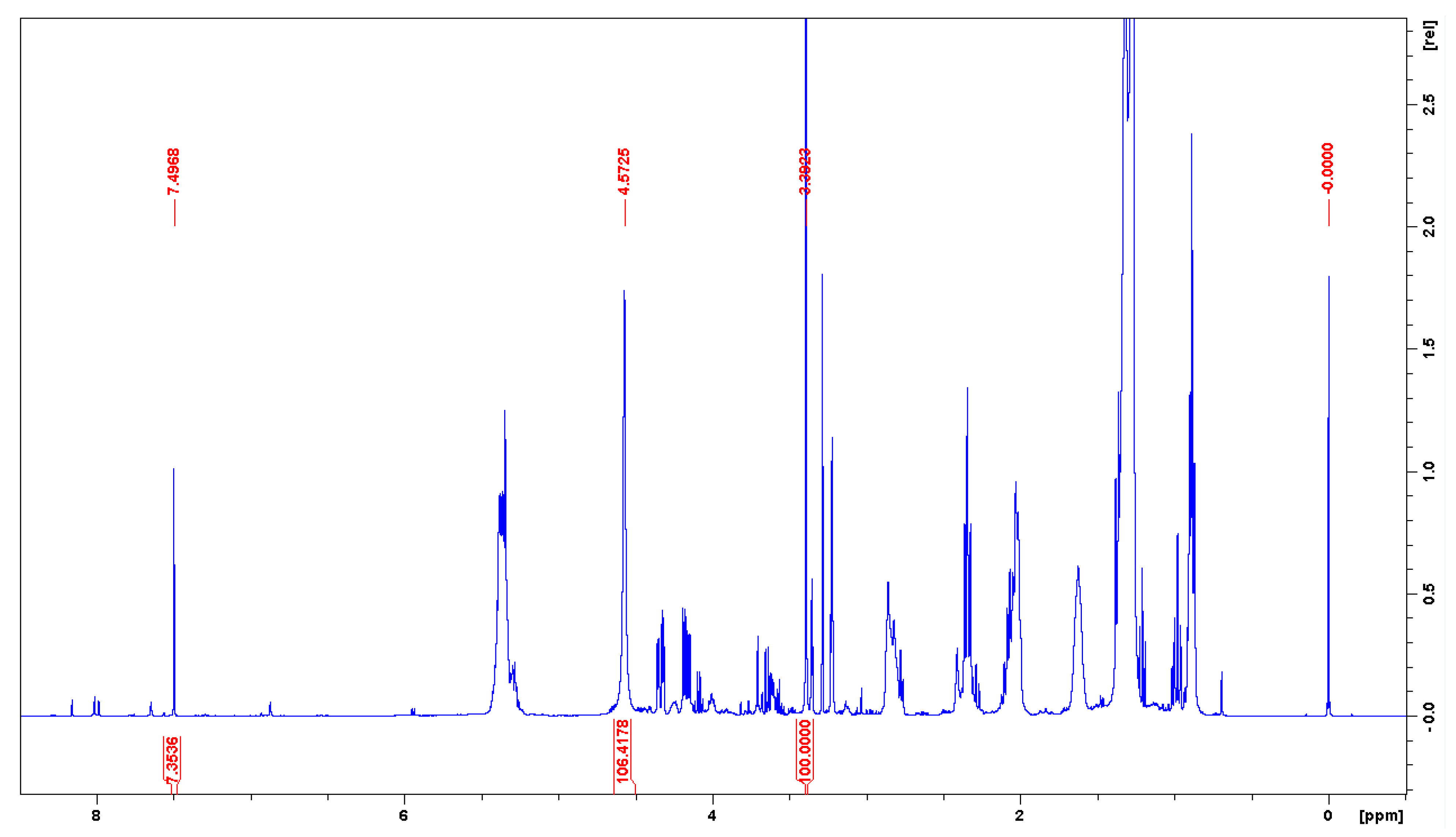
The analysis performed in this work concerning a series of 1H NMR spectra recently recorded for fat fish extracts [93,107], also taken in the binary mixture (2:1, v/v) of CDCl3 and CD3OD, confirmed the previous assumption regarding the δH data in the article [27]. Figure 4 shows three important signals of the mixture discussed; however, in this specific case, this solvent mixture also contained TMS. These proton signals from CHCl3, probably a mixture of H2O/HOD in equilibrium exchange with CD3OH, and CHD2OD were found at 7.497 (7.493), 4.5725 (4.572), and 3.392 (3.392) ppm, respectively. The above values refer to the initial use of wet and deteriorated CDCl3 (Figure 4), while those in parentheses were taken from a very similar 1H NMR spectrum (not shown) obtained finally in deacidified CDCl3. At this point, it should be mentioned that the signal attributed to the water present in CD3OD is observed at 4.87 ppm [49,51,54].
More specifically, both of the spectra mentioned above were taken initially using unpurified (slightly acidic) CDCl3 and then using this solvent mitigated by mixing with an aqueous solution of disodium carbonate [93]. This procedure resulted in a 2.5-fold increase in signal intensity at ~4.57 ppm and a minimal high-field shift of the CHCl3 line. The CHD2OD signal did not change position. Taking into account the differences in δH values between CHCl3 and the other two signals in question, the results are 2.93 and 4.11 ppm. Interestingly, very similar differences in such δH data apply to the article [27], that is, 3.00 and 4.16 ppm (vide supra). Small differences (approximately 6 ppm) may be due to the different types of analytes in the NMR samples discussed. This finding suggests that in the case of ref. [27], the CHCl3 line was present at ~7.22 ppm and not 7.25 ppm.
Identical circumstances refer to 13C NMR spectra, which are performed very often in CDCl3 containing a small amount of DMSO-d6 to completely dissolve the analyte under study. For example, after adding five drops of it to the indole-2-carboxylic acid sample dissolved in 0.7 mL of CDCl3, the signal of the added cosolvent occurred at 38.97 ppm (instead of the typical δC of 39.46 ppm, see Table 2) when a δC of 77.00 ppm was assumed for the dominant signal of CDCl3 [92] (pp. 171–172). The absence of TMS in the NMR sample makes it impossible to answer the question of whether the presence of the aforementioned analyte changed the position of the 13C signal from CDCl3.
Another problem arises when δD data [5,61,63,76] (see Table S5), stored on NMR spectrometer computers and used for automatic lock corrections [61,62], are indirectly used to reference residual 1H and 13C solvent signals. For example, in a series of 1H spectra shown in [108], δHs of 7.262 and 3.341 ppm (found currently after averaging) were given for the residual signals of CDCl3 and CD3OD, respectively. However, all related 13C{1H} spectra omit the δC values of the solvent signals. The absence of these data greatly increases the uncertainty of the published spectral characteristics of the products described. In contrast, in [109], δC values of ~47.58 and 47.85 ppm obtained from the raw calculation (Table S4, row for the years 2003–2022) were used to calibrate the 13C signals of CD3OD instead of the typical value of 49.04 ppm. As a result, the δC data reported for the isolates under study are greatly underestimated.

2.5. Proposals Based on Current Needs and Opportunities for NMR Spectrometers

The comparison of δX data measured for synthesized or isolated organic species with the values reported in the literature for solutions in the same NMR solvents should not be difficult. There may be only minor discrepancies, mainly due to different sample concentrations and/or probe temperatures. Furthermore, all these differences, e.g., of ±0.1 ppm for 1H spectra and ±2.0 ppm (or even ±2.5 ppm) [56] for 13C spectra, are usually more or less systematic. A certain problem may be the very large difference in the concentrations of the solutions. However, all of these facts are well known among synthetic chemists. In addition to NMR spectra, they usually know the results obtained from the use of other analytical techniques. Thus, there is little doubt that the compounds being compared are identical.
However, for δX values reported for solutions in the other NMR solvent(s), it will be advisable to use some corrections to compare these data, since the chemical shift of TMS is not zero in all solvents [2,17,18,19,62,89]; δH = 0 ppm is only in dilute CDCl3 solution (vide supra). Its δH has been found to be between −0.8 and 0.2 ppm depending on the solvent; δH = −0.1277 ppm is for liquid TMS [91]. Generally, the TMS line can vary by more than 1 ppm for 1H spectra and 4 or even 5 ppm for 13C spectra [19,20,21]. Examples of measured ‘absolute’ solvent-induced shifts of the 13C signal of TMS dissolved in CDCl3 and CD3OD are 0.74 ppm [13] and −0.74 [110] ppm, respectively. The former δC is equal to the difference between the ‘absolute’ [13,89] and ‘observed’ δC proposed here for CDCl3, ΔδC = 77.75–77.01 = 0.74 (Table 2). The consideration of such δC data for other NMR solvents without TMS discussed here suggests particularly large TMS–solvent interactions for DMSO-d6, ΔδC = 40.76–39.46 =1.30 ppm (Table S4). Such changes in δC values, calculated analogously for (CD3)2CO and C6D6, in particular, are much smaller (0.31/0.22 and 0.10 ppm, respectively).
Another marginal issue is the frequent complexity of 1H NMR spectra of some organic species, which are characterized by strongly coupled spin systems and cannot be analyzed on the basis of first-order assumptions. As a rule, the authors of typical articles do not analyze such spectra and only provide ranges of the observed multiplets. For example, such fragments “4.17–4.12 (m, 1H, H-6b)” and “2.36–1.45 (m, 14H, 5 × CH2ad, 4 × CHad)” can be found in the NMR spectral description of a new bicyclic product 6 (Figure 5) [16], despite the fact that its spectrum was measured on a 600-megahertz machine. This example simultaneously demonstrates the use of a current one- and two-decimal standard for reporting δC and δH data, respectively [59,111]. Unfortunately, such δX values, which are usually measured with much higher precision, are rounded off without apparent need at the spectrum analysis stage. MestReNova [60] processes 1H NMR spectra in this manner. Accordingly, for more complex spectra, the ranges of multiplets are provided using this program.
Occasionally, you may encounter a qualitatively different issue when the reported molecular structure is questionable. In fact, many structural revisions based on theoretical predictions of the values of NMR parameters have been reported in the literature in recent years. Most of these structural misassignments concern complex organic compounds, especially those isolated from various natural resources (see, e.g., ref. [112]).
Figure 5. Compounds discussed in refs. [16,113].
Molecules 28 04369 g005
However, it turns out that the incorrect assignment of 1H NMR signals can also concern very simple molecules. For example, an erroneous assignment of α-ring protons of the cationic part of ionic system 7 has recently been corrected by DFT-NMR calculations [113]. Furthermore, a full spin analysis of the spectrum of species 7 was carried out via line shape-based iterative refinement and subsequent spectrum simulation. Therefore, in this case, all δH and JHH data were found with a precision of 0.0001 ppm and 0.01 Hz, respectively. Such an analysis is usually the exception rather than the rule (vide supra). However, occasionally, by carefully processing raw 1H NMR spectra using classical approaches [114,115,116,117], one can easily obtain δHC and JHH values with a precision of 0.001/0.01 ppm and 0.01 Hz, respectively [118]. Typically, zero-filling and resolution enhancement with the Lorentzian-to-Gaussian function is performed prior to the Fourier transformation of the spectrum [55,114,115,116,117]; this approach is also available in [60].
It is important to realize that a complete set of the δH and JHH data, pertaining to each complex organic species, including isolates from natural sources, can serve as its unique 1H NMR fingerprint [57], especially when these parameters were obtained by 1H NMR iterative full spin analysis. Therefore, the Raw Data Initiative highly recommends depositing original properly referenced spectra (or even raw data such as free induction decay (FID) files) for all new natural products for possible structural reanalysis in the future [111,119,120]. Similar suggestions can also be found elsewhere [121]. It should be noted that this approach is fully in accordance with the latest research data policy of the American Chemical Society [122].
Recently, the notion that, in all such cases, the δHs should be provided with at least three but preferably four decimal places when using high-field superconducting magnets to obtain sets of parameters that are suitable as numerical substitutes for current 1H NMR spectra is commonplace [111]. The NMReDATA Initiative presented an identical view on the deposition of computer records of such spectra for all new complex synthetic and natural organic systems [123]. Publishing δH values with a precision of 0001 ppm is recommended.
The use of the δH and δC data with higher precision than is currently suggested is particularly important for all species with high C/H ratios or with proton deficiency and many heteroatoms, or more generally, for organic systems that provide a small number of δX data. A suitable method to use in such cases is the multinuclear method [124], which includes a non-1:1 correlation between theoretical predictions and experimental δX data. This approach uses δH values multiplied by an adjustable factor n, for example, n = 10. In this case, the δHC data measured for the same NMR samples containing TMS and reported to the nearest 0.001/0.01 ppm are mandatory.
An application of 13C signals from deuterated solvents to standardize the δC values is justified in part because the range of typical δC data is approximately 20 times larger than the δH range that suffers from a much lower spectral dispersion of δH data. For this reason, except for the cases of some specific isomeric systems, the reported δC values rounded to one decimal place are recommended [59].
However, given the large inaccuracies in the δX values used in Method A (see the Supplementary Materials for all the details) and the relatively high dependence of δC on the type/concentration of the analyte studied (vide supra), it is crucial that the adopted ‘reference’ δX data are given accurately in publications. Therefore, it should be mandatory that these δH and δC values should be provided within 0.001 and 0.01 ppm because this affects the precision of δX data reported. The authors of [125] provide a positive example here. Unfortunately, in many of the articles cited above and recently published, such δX data do not appear at all [23,29,30,58,126,127,128]. It should be stressed that the aforementioned precision is achieved seamlessly on all 400-MHz NMR machines with an inherently lower digital resolution.
Finally, it should be noted that Burns and Reynolds in their book [129] recommend that δH and δC values for all new organic species or isolates should be given in three and two decimal places, respectively, with the last place subscripted to indicate uncertainty due to sample concentration and/or probe temperature. Furthermore, they considered Methods A and B equivalent because, in their opinion, residual 1H and 13C solvent signals have similar uncertainties when used in place of TMS.
Recent findings by Guzman and Hoye [4] did not confirm the aforementioned view for 1H NMR spectra in CDCl3. In addition, most of the cases discussed in this work related to the standardization of 13C NMR spectra using solvent signals do not seem to agree with the above opinion [129]. However, only a more detailed investigation of this issue can provide a reliable view of the matter.

3. Summary and Conclusions

This short review article sketches the most likely ‘birth’ of Method A for popular, simple but not perfect referencing of NMR spectra, involving their indirect standardization using residual 1H and 13C signals from deuterated solvents as secondary reference points on the chemical shift scale. Undoubtedly, the inclusion of this procedure in the arsenal of various NMR spectroscopy methods had to be preceded by the introduction of pulsed Fourier-transform instruments in the early 1970s. All documents and information found in the literature review indicated that Method A became widespread as a result of the initiative of NMR users, especially synthetic organic chemists. The obvious catalysts for this process were the acceptance of TMS as an internal zero-point standard for 13C spectra and the common availability of ‘reference’ δH and then δC data as secondary internal standards. The increasingly popular use of this method to record and report 1H and 13C NMR spectral data is briefly described here.
Regarding the new results obtained now, all ‘reference’ δX data found in the literature concerning six common NMR solvents doped with TMS were considered and can be found in the Supplementary Materials (Tables S2 and S4). This allowed the proposal of their ‘best’ values for use in Method A, which are summarized in Table 2. The large variability of these δX data was also analyzed in detail. The influence of the concentration and type of the analyte tested is greatest, while the directions and magnitudes of such CIS result from various solvent–analyte interactions that occurred in a particular case. The large δH changes in the position of the CHCl3 line observed in the presence of acetone or benzene in TMS-doped CDCl3 [4,5] were reproduced very well in the current DFT-NMR calculations carried out for hydrogen-bonded complexes 4 and 5 formed in this solvent (see Supplementary Materials). Therefore, it is clear that dilute solutions should generally be applied when Method A is used to minimize all such CIS effects for real NMR samples as much as possible.
As for the current state of Method A, its practical application is shown here in the convention of good and numerous bad exemplary cases. Statistically, the use of this simple method is generally acceptable, but there is still plenty of room for improvement. Focusing only on the procedures for post-processing the recorded NMR spectra, it is possible to mention some basic problems and pitfalls associated with Method A, which are mainly due to the mistakes of the researchers themselves as end users.
In particular, this includes the adoption of inappropriate ‘reference’ δX data, which leads to an over- or under-estimation of δX data finally reported. Similar problems always occur when mixtures of NMR solvents are used. In such cases, it is necessary to use Method B (vide supra). The adopted secondary ‘reference’ data or their sources from the literature are often not given at all or only partially, which should not be the case. This is especially true for solvent signals in published copies of NMR spectra, often without providing their δX values. Similar problems arise, for example, when using MestReNova [60] without knowing the limitations of Method A.
The recommended use of the above program [60] leads to the generation of 1H NMR spectral data in the form of a list of signals for which the number of protons and possibly JHH data are given. Sometimes, only the ranges of multiplets are given for certain spectrum regions (vide supra). In general, 1H/13C spectra are readily available for inclusion in a publication after analysis, but their precision strongly depends on the researcher as the end user. He must be fully aware of the limitations of Method A. The lack of a primary TMS signal makes it difficult to use. As a result, the reported δX data may be subject to some errors and the published spectra may lack important reference values for the NMR solvent signals adopted in the analysis performed. Unfortunately, the belief in this type of user-friendly software is often too high. Different examples of such issues can be found in the Search Results and Discussion section.
It is easy to list numerous drawbacks or pitfalls as well as only a few benefits of using Method A. The first is mainly due to the absence of generally accepted rules for its application. In particular, this includes the lack of a formal obligation to provide the exact δX values of the reference 1H and 13C solvent signals used to report spectral data for all new organic species. The second issue is due to possible signal overlap. In fact, there may be problems with the spectra of aromatics referenced by applying the residual 1H lines from CDCl3 or C6D6 (see above for an exemplary case of species 3). The use of TMS or cubane as an internal reference can be useful in all such situations (for the latter, see Table S3 and the related discussion). However, the main problem is the occurrence of numerous CIS effects, especially when using quite concentrated NMR samples. These effects can often lead to significant changes in the δX values adopted for the secondary references in question to lower or higher frequencies, depending on the type of analyte.
It should also be highlighted that, in the absence of TMS in the NMR sample, Method A is used indirectly in Method C. More importantly, it is also applied in an IUPAC-recommended Method D used to reference spectra involving some NMR-active nuclei that are different from 13C (vide supra). Strictly, 13C spectra can also be standardized in this way, but solvent signals are generally used for this purpose. Therefore, the correct referencing of the initial 1H spectra using Method A is decisive in all these cases.
In view of all the foregoing facts, it should be mandatory to give the reference δH and δC data adopted in Method A with a precision of 0.001 and 0.01 ppm, respectively, and report the spectral data for the products or isolates with the same precision. If there is any doubt about the rounding of some values (up or down), it is recommended that one should insert the digit 5 in the additional decimal position given in the subscript. To avoid prolonging the main text of the publication, the δX data with such precision has been included in the Supplementary Materials section. The above precision requests may be seen to be somewhat excessive, especially for organic chemists, who usually have additional experimental data at their disposal when identifying the synthesized products. However, as noted earlier, these requirements particularly apply to all new complex or unexpected structures. In such cases, identical requirements should also be applied to the use of Methods B–D.
One of the few advantages of Method A (used as mentioned above) is that its application avoids contamination of the samples being analyzed by TMS. It is likely that its use could be useful in some cases where the NMR samples under study need to be used for further special studies, as suggested in ref. [99]. Indisputably, silyl derivatives possessing signals with δHs very close to 0 ppm may be obvious targets for this approach (vide supra). Using the classical iteration of Method B with an internal TMS can be difficult in such cases. This may be especially true for spectra recorded on older 200-MHz spectrometers.
According to the results of this literature review, there is a significant discrepancy of 1.7 and even 1.9 ppm in the δC values for DMSO-d6 and CDCl3, respectively, applied by the users of Method A (Table S4, two lines at the bottom); these results are in line with the large uncertainties in δC values for these solvents reported in ref. [49]. Smaller differences in the δCs concern CD3OD and especially CD3CN. One can only assume that the sources of the aforementioned differences in δC data are the CIS effects resulting from the presence of various analytes in the NMR samples studied. These findings suggest the need for a systematic investigation of 13C{1H} spectra recorded in all solvents discussed here for analytes of different types, simultaneously using 13C signals from TMS and NMR solvent, analogously to the study performed for 1H NMR spectra in CDCl3 [4]. A similar investigation carried out more than two decades ago produced unsatisfactory results [56].
The careful and correct application of each of the four Methods A–D to fairly dilute solutions of different organic species in deuterated organic solvents should, in principle, lead to very similar δH and δC data for analytes of different types. To the author’s knowledge, to date, no comparative studies similar to those performed for 19F NMR spectra [12] have been carried out.
One might be tempted to conclude that the daily practice of NMR spectroscopy has, so far, outgrown the recommendations of the IUPAC and other similar regulations that have not yet taken into account the widespread use of Method A, in which experimentally measured δX data are related to internal TMS at 0 ppm, albeit indirectly.
Therefore, it is probably time for an in-depth discussion and formulation of some undoubtedly needed rules for the best possible use of Method A. Its application will probably expand due to the increasing frequency of milligram-scale chemical syntheses and the growing use of benchtop NMR instruments [7,130]. Indeed, by using the latter relatively inexpensive devices, residual 1H signals from deuterated solvents are frequently applied [130]. In general, a proper routine use of Method A for the referencing of 1H and 13C spectra with all of the above requirements seems to have great potential, especially in the context of the increasingly widespread use of cryogenically cooled probes of classical NMR spectrometers, which allows for the analysis of very dilute solutions.
However, for all newly synthesized or isolated organic species, especially those with complex or unexpected molecular structures, the use of TMS in Method B is strongly recommended to ensure the precise 1H and 13C NMR spectroscopic characterization of the organic systems mentioned above.

Supplementary Materials

The following supporting information can be downloaded at: https://www.mdpi.com/article/10.3390/molecules28114369/s1, The experimental ‘reference’ δH and δC values from the literature for six deuterated solvents without TMS, including MSD data (Table S1), a continuation of the subsection on solvent signals as secondary NMR standards, including different CIS effects, a discussion of cubane and cyclohexane as internal references (Table S3), two new subsections on 1H and 13C spectra (Tables S2 and S4 covering 45 sets of δX data) including δD values for six solvents considered (Table S5), stacked 1H NMR spectra from the ‘titration’ of residual incompletely deuterated components of the four deuterated solvents with tetra-n-butylammonium chloride as a titrant (Figures S1–S4) and two cases of not correctly analyzed 1H NMR spectra (Figures S5 and S6); an additional section with description and discussion of the results of GIAO-DFT-NMR calculations performed now for hydrogen-bonded complexes 4 and 5 (Tables S6 and S7, Figure S7); [131,132,133,134,135,136,137,138,139,140,141,142,143,144,145,146,147,148,149,150,151,152,153,154,155,156,157,158,159,160,161,162,163,164,165,166,167,168,169,170,171,172,173,174,175,176,177,178,179,180,181,182,183,184,185,186] references to the Supplementary Materials part (PDF).

Funding

This research received no external funding.

Institutional Review Board Statement

Not applicable.

Informed Consent Statement

Not applicable.

Data Availability Statement

Not applicable.

Acknowledgments

The author wishes to express his thanks to Stefan T. Berger (Universität Leipzig, Germany), Gerhard Hägele (Heinrich-Heine-Universität Düsseldorf, Germany), Roy E. Hoffman (The Hebrew University of Jerusalem), Janez Plavec (Slovenian NMR Center, Ljubljana, Slovenia), Cheng-Wei T. Chang (Utah State University, USA), and Włodzimierz Makulski (University of Warsaw, Poland) for correspondence and/or submitted documents or scans thereof. The author thanks Paris E. Georghiou (Memorial University of Newfoundland, Canada) for sending some documents and copies of the 1H NMR spectra (including the FID files) and Thomas Kuballa [Chemisches und Veterinäruntersuchungsamt (CVUA) Karlsruhe, Germany] for sending the FID files for a series of 1H NMR spectra. All information provided by Kevin Millis (Cambridge Isotope Laboratories, Inc., Tewksbury, MA, USA), Katja Glock, and Peter Conradi (Merck KGaA, Darmstadt, Germany) is appreciated. The author extends his thanks to Konrad Kowalski, Andrzej Jóźwiak, Piotr Matczak, Zbigniew Malinowski, and Paweł Tokarz of this Faculty for their helpful discussion and comments on the early draft of the manuscript. The author also thanks the anonymous reviewers, one in particular, for their valuable suggestions that significantly improved the quality of the article. The author is also grateful to the University of Lodz for partial financial support. Part of the research (calculations for complexes 4 and 5) was supported by the PLGrid Infrastructure at ACC Cyfronet (Prometheus supercomputer; AGH, Kraków, Poland).

Conflicts of Interest

The author declares no conflict of interest.

Sample Availability

Not applicable.

References

  1. Harris, R.K.; Becker, E.D.; de Menezes, S.M.C.; Goodfellow, R.; Granger, P. NMR nomenclature. Nuclear spin properties and conventions for chemical shifts (IUPAC Recommendations 2001). Pure Appl. Chem. 2001, 73, 1795–1818. [Google Scholar] [CrossRef]
  2. Harris, R.K.; Becker, E.D.; de Menezes, S.M.C.; Granger, P.; Hoffman, R.E.; Zilm, K.W. Further conventions for NMR shielding and chemical shifts (IUPAC Recommendations 2008). Pure Appl. Chem. 2008, 80, 59–84. [Google Scholar] [CrossRef]
  3. Chalmers, B.A.; Chen, A.P.-J.; Savage, G.P.; Williams, C.M. Cubane: A New NMR Internal Standard. Aust. J. Chem. 2010, 63, 1108–1110. [Google Scholar] [CrossRef]
  4. Guzman, A.L.; Hoye, T.R. TMS is Superior to Residual CHCl3 for Use as the Internal Reference for Routine 1H NMR Spectra Recorded in CDCl3. J. Org. Chem. 2022, 87, 905–909. [Google Scholar] [CrossRef]
  5. Hatada, K.; Kitayama, T. NMR Spectroscopy of Polymers; Springer: Berlin, Germany, 2004; pp. 1–42. [Google Scholar]
  6. Guizzetti, S.; Schwindeman, J.A.; Daniels, D.S.B.; Douglas, J.J.; Kosanovich, A.; Zhao, W.; Kelly, C.B.; Knight, J. Some Items of Interest to Process R&D Chemists and Engineers. Org. Process Res. Dev. 2022, 26, 239–250. [Google Scholar] [CrossRef]
  7. Napolitano, J.G.; Yang, C.; Conklin, B.; He, Y.; Ochoa, J.L. Toward the Development of Rapid, Automated Identification Tests for Neat Organic Liquids Using Benchtop NMR Instrumentation. Anal. Chem. 2022, 94, 16095–16102. [Google Scholar] [CrossRef]
  8. Tiers, G.V.D. Proton nuclear resonance spectroscopy. I. Reliable shielding values by “internal referencing” with tetramethylsilane. J. Phys. Chem. 1958, 62, 1151–1152. [Google Scholar] [CrossRef]
  9. Bacon, R.; Maciel, G.E. Solvent Effects on the Five Shielding Constants In Tetramethylsilane and Cyclohexane. J. Am. Chem. Soc. 1973, 95, 2413–2426. [Google Scholar] [CrossRef]
  10. Sugiura, M.; Takao, N.; Ueji, S. A New Method for Differentiating Between Solvent Effect Mechanisms on 13C Chemical Shifts. Org. Magn. Reson. 1982, 18, 128–133. [Google Scholar] [CrossRef]
  11. Residual Deuterated Solvent Peak in 1H and Deuterated Peak in 13C{1H} NMR Spectra. Available online: https://cheminfographic.wordpress.com/2020/06/01/residual-deuterated-solvent-peak-in-1h-and-deuterated-peak-in-13c-nmr-spectra (accessed on 21 May 2023).
  12. Rosenau, C.P.; Jelier, B.J.; Gossert, A.D.; Togni, A. Exposing the Origins of Irreproducibility in Fluorine NMR Spectroscopy. Angew. Chem. Int. Ed. 2018, 57, 9528–9533. [Google Scholar] [CrossRef]
  13. Jackowski, K.; Makulski, W. 13C shielding scale for MAS NMR spectroscopy. Magn. Reson. Chem. 2011, 49, 600–602. [Google Scholar] [CrossRef] [PubMed]
  14. Ge, H.; Spletstoser, J.T.; Yang, Y.; Kayser, M.; Georg, G.I. Synthesis of Docetaxel and Butitaxel Analogues through Kinetic Resolution of Racemic β-Lactams with 7-O-Triethylsilylbaccatin III. J. Org. Chem. 2007, 72, 756–759. [Google Scholar] [CrossRef] [PubMed]
  15. Totani, K.; Shinoda, Y.; Shiba, M.; Iwamoto, S.; Koizumi, A.; Matsuzaki, Y.; Hirano, M. Silyl-assisted 1,2-cis-α-glucosylation for the synthesis of a triglucoside moiety in high-mannose-type oligosaccharides. RSC Adv. 2015, 5, 75918–75922. [Google Scholar] [CrossRef]
  16. Cloutier, M.; Lavoie, S.; Gauthier, C. C7 Epimerization of Benzylidene-Protected β-D-Idopyranosides Brings Structural In-sights into Idose Conformational Flexibility. J. Org. Chem. 2022, 87, 12932–12953. [Google Scholar] [CrossRef] [PubMed]
  17. Laszlo, P.; Speert, A.; Ottinger, R.; Reisse, J. Reconsideration of the Internal Tetramethylsilane Reference for Proton Magnetic Resonance Studies. J. Chem. Phys. 1968, 48, 1732–1735. [Google Scholar] [CrossRef]
  18. Jutila, M. Influence of an Internal Reference on 1H NMR Solvent Shifts. Determination of Reference Independent ASIS Values. Acta Chem. Scand. B 1981, 35, 503–506. [Google Scholar] [CrossRef]
  19. Hoffman, R.E. Variations on the chemical shift of TMS. J. Magn. Reson. 2003, 163, 325–331. [Google Scholar] [CrossRef]
  20. Ziessow, D.; Carroll, M. Referencing 13C NMR Medium Shifts. Ber. Bunsenges. Phys. Chem. 1972, 76, 61–64. [Google Scholar]
  21. Moreau-Descoings, M.C.; Goethals, G.; Seguin, J.P.; Doucet, J.P. Measurements of actual association 13C shifts: Medium effects on the TMS carbons. Spectrochim. Acta A 1987, 43, 17–20. [Google Scholar] [CrossRef]
  22. de Kowalewski, D.G.; de los Santos, C.; Marceca, E. Use of an Internal Reference in 13C Chemical Shift Measurements. Magn. Reson. Chem. 1990, 48, 1–4. [Google Scholar] [CrossRef]
  23. Crawford, R.J.; Erickson, G.L. Thermolysis of cis- and trans-4-Deuterio-3-methyl-l-pyrazolin. J. Am. Chem. Soc. 1967, 89, 3907–3908. [Google Scholar] [CrossRef]
  24. Cane, D.J.; Graham, W.A.G.; Vancea, L. Synthesis and nuclear magnetic resonance study of the compounds (2-X-1,3-dithiane) (X = H, Me, SiMe3, GeMe3, SnMe3, PbMe3) and their tetracarbonyliron complexes. Can. J. Chem. 1978, 56, 1538–1544. [Google Scholar] [CrossRef]
  25. Huckerby, T.N. Accurate Referencing of 13C NMR Spectra from Glycosaminoglycan and other Polysaccharides in Aqueous Medium. Org. Magn. Reson. 1983, 21, 67–70. [Google Scholar] [CrossRef]
  26. Kalinowski, H.-O.; Berger, S.; Braun, S. 13C-NMR-Spektroskopie; Georg Thieme Verlag: Stuttgart, Germany, 1984; pp. 73–74. [Google Scholar]
  27. Abraham, W.; Wertz, P.W.; Downing, D.T. Linoleate-rich acylglucosylceramides of pig epidermis: Structure determination by proton magnetic resonance. J. Lipid Res. 1985, 26, 761–766. [Google Scholar] [CrossRef]
  28. Kalinowski, H.-O.; Berger, S.; Braun, S. Carbon-13C NMR Spectroscopy; John Wiley & Sons: Chichester, UK, 1988; pp. 85–86. [Google Scholar]
  29. Bercaw, J.E.; Davies, D.L.; Wolczanski, P.T. Reactions of Alkyl and Hydride Derivatives of Permethylscandocene and -zirconocene with Nitriles and Amines. Catalytic Hydrogenation of tert-Butyl Cyanide with Permethylscandocene Hydride. Organometallics 1986, 5, 443–450. [Google Scholar] [CrossRef]
  30. Yamamoto, Y.; Noda, S.; Nanai, N.; Okura, I.; Inoue, Y. 1H NMR Conformational Study of Viologen-Linked Porphyrins. Bull. Chem. Soc. Jpn. 1989, 62, 2152–2158. [Google Scholar] [CrossRef]
  31. Silverstein, R.M.; Bassler, G.C. Spectrometric Identification of Organic Compounds, 2nd ed.; John Wiley & Sons, Inc.: New York, NY, USA, 1967; p. 135. [Google Scholar]
  32. Granger, D.D. 13C NMR of Crosslinked Poly(Methacrylic Anhydride). In Solid State NMR of Polymers; A collection of papers presented at The Third Chemical Congress of North America (Toronto, ON, Canada, 5–10 June 1988); Mathias, L.J., Ed.; Springer Science+Business Media: New York, NY, USA, 1991; pp. 179–200. [Google Scholar]
  33. Deuterated NMR Solvents–Handy Reference Data. Available online: https://scs.illinois.edu/system/files/inline-files/Deuterated_Solvents.pdf (accessed on 21 May 2023).
  34. Kegley, S.E.; Pinhas, A.R. Problems and Solutions in Organometallic Chemistry; University Science Books: Mill Valley, CA, USA, 1986; p. 9. [Google Scholar]
  35. Breitmaier, E.; Voelter, W. 13C NMR Spectroscopy. Methods and Applications, 3rd ed.; VCH Verlagsgesellschaft mbH: Weinheim, Germany, 1987; p. 109. [Google Scholar]
  36. Deuterated NMR Solvent Table. Available online: https://web.stanford.edu/group/chem-NMR/help_docs/nmr_solvents.htm (accessed on 21 May 2023).
  37. CIL’s NMR Solvent Data Chart. Available online: https://www.isotope.com (accessed on 21 May 2023).
  38. Zerbe, O.; Jurt, S. Applied NMR Spectroscopy for Chemists and Life Scientists; Wiley-VCH Verlag GmbH & Co. KGaA: Weinheim, CA, USA, 2014; pp. 26−28, 70–71. [Google Scholar]
  39. Budavari, S.; O’Neil, M.J.; Smith, A.; Heckelman, P.E. The Merck Index, an Encyclopedia of Chemicals, Drugs, and Biologicals, 11th ed; Merck & Co., Inc.: Rahway, NJ, USA, 1989. [Google Scholar]
  40. Gordon, A.J.; Ford, R.A. The Chemist’s Companion—A Handbook of Practical Data, Techniques, and References; John Wiley & Sons: New York, NY, USA, 1972; pp. 249–251. [Google Scholar]
  41. Pretsch, E.; Clerc, T.; Seibl, J.; Simon, W. Tabellen zur Strukturaufklärung Organischer Verbindungen mit Spektroskopischen Methoden; Springer: Berlin, Germany, 1976; pp. C250–C260. [Google Scholar]
  42. Breitmaier, E.; Voelter, W. 13C NMR Spectroscopy. Methods and Applications in Organic Chemistry, 2nd ed.; Verlag Chemie: Weinheim, CA, USA, 1978; p. 69. [Google Scholar]
  43. Handbuch der Instrumentellen Analytik NMR-Spektroskopie; Merck: Darmstadt, Germany, 1988.
  44. Pouchert, C.J.; Behnke, J. (Eds.) The Aldrich Library of 13C and 1H FT NMR Spectra; Aldrich Chemical Company, Inc.: Milwaukee, WI, USA, 1993; Volume 3, pp. 620A–625A. [Google Scholar]
  45. Spectroscopy Catalogue; Sigma-Aldrich Co.: St. Louis, MO, USA, 1997; p. 37.
  46. Bruno, T.J.; Svoronos, P.D.N. CRC Handbook of Basic Tables for Chemical Analysis, 2nd ed.; CRC Press: Boca Raton, FL, USA, 2003. [Google Scholar]
  47. Balci, M. Basic 1H- and 13C-NMR Spectroscopy; Elsevier: London, UK, 2005; p. 35. [Google Scholar]
  48. Bruno, T.J.; Svoronos, P.D.N. CRC Handbook of Basic Tables for Chemical Analysis, 3rd ed.; CRC Press: Boca Raton, FL, USA, 2011; pp. 484–486. [Google Scholar]
  49. Gottlieb, H.E.; Kotlyar, V.; Nudelman, A. NMR Chemical Shifts of Common Laboratory Solvents as Trace Impurities. J. Org. Chem. 1997, 62, 7512–7515. [Google Scholar] [CrossRef]
  50. Fletton, R.A.; Page, J.E. Proton Chemical Shifts for Solvents and other Simple Substances. Analyst 1971, 96, 370–373. [Google Scholar] [CrossRef]
  51. Fulmer, G.R.; Miller, A.J.M.; Sherden, N.H.; Gottlieb, H.E.; Nudelman, A.; Stoltz, B.M.; Bercaw, J.E.; Goldberg, K.I. NMR Chemical Shifts of Trace Impurities: Common Laboratory Solvents, Organics, and Gases in Deuterated Solvents Relevant to the Organometallic Chemist. Organometallics 2010, 29, 2176–2179. [Google Scholar] [CrossRef]
  52. Gottlieb, H.E.; Graczyk-Millbrandt, G.; Inglis, G.G.A.; Nudelman, A.; Perez, D.; Qian, Y.; Shuster, L.E.; Sneddon, H.F.; Upton, R.J. Development of GSK’s NMR guides—A tool to encourage the use of more sustainable solvents. Green Chem. 2016, 18, 3867–3878. [Google Scholar] [CrossRef]
  53. Jones, I.C.; Sharman, G.J.; Pidgeon, J. 1H and 13C NMR data to aid the identification and quantification of residual solvents by NMR spectroscopy. Magn. Reson. Chem. 2005, 43, 497–509. [Google Scholar] [CrossRef] [PubMed]
  54. Babij, N.R.; McCusker, E.O.; Whiteker, G.T.; Canturk, B.; Choy, N.; Creemer, L.C.; De Amicis, C.V.; Hewlett, N.M.; Johnson, P.L.; Knobelsdorf, J.A.; et al. NMR Chemical Shifts of Trace Impurities: Industrially Preferred Solvents Used in Process and Green Chemistry. Org. Process Res. Dev. 2016, 20, 661–667. [Google Scholar] [CrossRef]
  55. Richards, S.A.; Hollerton, J.C. Essential Practical NMR for Organic Chemistry; John Wiley & Sons: Chichester, UK, 2011; pp. 16−18, 31–36. [Google Scholar]
  56. Grzonka, M.; Davies, A.N. Empirical Investigation on the Reproducibility of 13C NMR Shift Values. J. Chem. Inf. Comput. Sci. 1998, 38, 1096–1101. [Google Scholar] [CrossRef]
  57. Pauli, G.F.; Gödecke, T.; Jaki, B.U.; Lankin, D.C. Quantitative 1H NMR. Development and Potential of an Analytical Method: An Update. J. Nat. Prod. 2012, 75, 834–851. [Google Scholar] [PubMed]
  58. Alexandri, E.; Ahmed, R.; Siddiqui, H.; Choudhary, M.I.; Tsiafoulis, C.G.; Gerothanassis, I.P. High Resolution NMR Spectroscopy as a Structural and Analytical Tool for Unsaturated Lipids in Solution. Molecules 2017, 22, 1663. [Google Scholar] [CrossRef]
  59. NMR Guidelines for ACS Journals. According to these guidelines, residual 1H impurities of deuterated NMR solvents are allowed to be applied as internal reference standards. Available online: http://pubsapp.acs.org/paragonplus/submission/acs_nmr_guidelines.pdf (accessed on 21 May 2023).
  60. MestReNova Version 14.3.1. Mestrelab Research S.L.: Santiago de Compostela, Spain, 2023. Available online: https://mestrelab.com/software/mnova (accessed on 21 May 2023).
  61. Hoffman, R.E. Standardization of chemical shifts of TMS and solvent signals in NMR solvents. Magn. Reson. Chem. 2006, 44, 606–616. [Google Scholar] [CrossRef]
  62. Granger, P.; Bourdonneau, M.; Assémat, O.; Piotto, M. NMR Chemical Shift Measurements Revisited: High Precision Measure-ments. Concepts Magn. Reson. A 2007, 30, 184–193. [Google Scholar] [CrossRef]
  63. NMR Solvent Reference Shift. Version 4.2. Available online: http://chem.ch.huji.ac.il/nmr/software/solvent.html (accessed on 21 May 2023).
  64. Hoffman, R.; (The Hebrew University of Jerusalem, Jerusalem, Israel). Personal Communications, 2022–2023.
  65. Akitt, J.W.; Mann, B.E. NMR and Chemistry: An Introduction to Modern NMR Spectroscopy, 4th ed.; CRC Press: Boca Raton, FL, USA, 2000; pp. 37–38. [Google Scholar]
  66. ur-Rahman, A.; Choudhary, M.I.; tul-Wahab, A. Solving Problems with NMR Spectroscopy, 2nd ed.; Elsevier Academic Press: Amsterdam, The Netherlands, 2016; pp. 22–23. [Google Scholar]
  67. Chemical Shift Referencing Your NMR Spectra. Available online: http://mangia.caltech.edu/NMRshifts.html (accessed on 21 May 2023).
  68. Chemical Shift Referencing. Available online: https://nmr.chem.ucsb.edu/protocols/refppm.html (accessed on 21 May 2023).
  69. Georgiou, M.; Wöckel, S.; Konstanzer, V.; Dechert, S.; John, M.; Meyer, F. Structural Variations in Tetrasilver(I) Complexes of Pyrazolate-bridged Compartmental N-Heterocyclic Carbene Ligands. Z. Naturforsch. B 2009, 64, 1542–1552. [Google Scholar] [CrossRef]
  70. Mulloyarova, V.V.; Ustimchuk, D.O.; Filarowski, A.; Tolstoy, P.M. H/D Isotope Effects on 1H-NMR Chemical Shifts in Cyclic Heterodimers and Heterotrimers of Phosphinic and Phosphoric Acids. Molecules 2020, 25, 1907. [Google Scholar] [CrossRef]
  71. Caló, F.P.; Bistoni, G.; Auer, A.A.; Leutzsch, M.; Fürstner, A. Triple Resonance Experiments for the Rapid Detection of 103Rh NMR Shifts: A Combined Experimental and Theoretical Study into Dirhodium and Bismuth−Rhodium Paddlewheel Complexes. J. Am. Chem. Soc. 2021, 143, 12473–12479. [Google Scholar] [CrossRef]
  72. Bakardjiev, M.; Holub, J.; Bavol, D.; Vrána, J.; Samsonov, M.A.; Růžička, A.; Růžičková, Z.; Fanfrlík, J.; Hnyk, D. Thiaborane Icosahedral Barrier Increased by the Functionalization of all Terminal Hydrogens in closo-1-SB11H11. Inorg. Chem. 2021, 60, 8428–8431. [Google Scholar] [CrossRef]
  73. Khalaji, M.; Paluch, P.; Potrzebowski, M.J.; Dudek, M.K. Narrowing down the conformational space with solid-state NMR in crystal structure prediction of linezolid cocrystals. Solid State Nucl. Magn. Reson. 2022, 121, 101813. [Google Scholar] [CrossRef]
  74. The Unified Scale for Referencing in NMR. New IUPAC Recommendations [revised (cgf): 26 July 2010]. Available online: http://www2.chem.wisc.edu/~cic/nmr/Guides/Other/Xi_chem_shift_scale.pdf (accessed on 21 May 2023).
  75. Automatic Heteronuclear Referencing with setref. (Varian/Agilent instruments). Available online: https://lsa.umich.edu/content/dam/chem-assets/chem-docs/setref.pdf (accessed on 21 May 2023).
  76. Chemical Shift Referencing. Available online: http://chem.ch.huji.ac.il/nmr/whatisnmr/chemshift.html (accessed on 21 May 2023).
  77. Witanowski, M.; Stefaniak, L.; Kamieński, B.; Biernat, S.; Webb, G.A. Influence of Some Paramagnetic Relaxation Reagents on Nitrogen Nuclear Shielding. J. Magn. Reson. 1981, 43, 456–462. [Google Scholar] [CrossRef]
  78. Batley, M.; Redmond, J.W. 31P NMR Reference Standards for Aqueous Samples. J. Magn. Reson. 1982, 49, 172–174. [Google Scholar] [CrossRef]
  79. Ksenofontov, A.A.; Isaev, Y.I.; Lukanov, M.M.; Makarov, D.M.; Eventova, V.A.; Khodov, I.A.; Berezin, M.B. Accurate prediction of 11B NMR chemical shift of BODIPYs via machine learning. Phys. Chem. Chem. Phys. 2023, 25, 9472–9481. [Google Scholar] [CrossRef]
  80. Live, D.; Harris, R.K. Hazardous references. Chem. Eng. News 1997, 75, 7. [Google Scholar]
  81. Zee, D.Z.; Singer, C.P.; O’Halloran, T.V. Chemical-Shift Standards for 199Hg NMR Spectroscopy, 25 Years Later. Inorg. Chem. 2022, 61, 13657–13661. [Google Scholar] [CrossRef]
  82. Blayney, M.B.; Nierenberg, D.; O’Halloran, T.V.; Wilcox, D.E.; Winn, J.S. Twenty-Five Years Ago−Remembering the Life and Loss of Professor Karen, E. Wetterhahn. ACS Chem. Health Saf. 2022, 29, 325–326. [Google Scholar] [CrossRef]
  83. Silverstein, R.M.; Webster, F.X.; Kiemle, D.J.; Bryce, L.B. Spectrometric Identification of Organic Compounds, 8th ed.; John Wiley & Sons, Inc.: Hoboken, NJ, USA, 2015; pp. 298, 320–324. [Google Scholar]
  84. Smith, J.E.; Yang, H.; Gabbaï, F.P. An Electrophilic, Intramolecularly Base-Stabilized Platinum−Antimony Complex. Organometallics 2021, 40, 3886–3892. [Google Scholar] [CrossRef]
  85. Sleem, H.F.; Dawe, L.N.; Rahman, S.; Georghiou, P.E. Halide ion effect on the chloroform chemical shift in supramolecular complexation studies with tetra-n-butylammonium salts: A 1H NMR and X-ray study. Supramol. Chem. 2014, 26, 579–582. [Google Scholar] [CrossRef]
  86. Assiri, Y.; Rahman, S.; Georghiou, P.E. Halide ion effect on the 1H NMR chemical shifts of the residual protons in commonly employed deuterated solvents with tetra-n-butylammonium chloride—Part 2. Supramol. Chem. 2016, 28, 6–9. [Google Scholar] [CrossRef]
  87. Sroczyński, D.; Grzejdziak, A.; Nazarski, R.B. Basic Properties and Protonation Mechanism of Some Tetraaza Macrocyclic Ligands. J. Incl. Phenom. Macrocycl. Chem. 1999, 35, 251–260. [Google Scholar] [CrossRef]
  88. Nazarski, R.B. Assignment of pH-dependent NMR spectra of the scorpiand macrocycle: An application of titration profiles and spectral linewidths. Magn. Reson. Chem. 2003, 41, 70–74. [Google Scholar] [CrossRef]
  89. Hayashi, S.; Yanagisawa, M.; Hayamizu, K. Nuclear Magnetic Resonance Chemical Shifts of Pure Organic-Solvents Determined by Magic Angle Spinning. Anal. Sci. 1991, 7, 955–957. [Google Scholar] [CrossRef]
  90. Hoffman, R.E.; Becker, E.D. Temperature dependence of the 1H chemical shift of tetramethylsilane in chloroform, methanol, and dimethylsulfoxide. J. Magn. Reson. 2005, 176, 87–98. [Google Scholar] [CrossRef] [PubMed]
  91. Hoffman, R. Magnetic susceptibility measurement by NMR: 2. The magnetic susceptibility of NMR solvents and their chemical shifts. J. Magn. Reson. 2022, 335, 107105. [Google Scholar] [CrossRef]
  92. Jacobsen, N.E. NMR Data Interpretation Explained: Understanding 1D and 2D NMR Spectra of Organic Compounds and Natural Products; John Wiley & Sons Inc.: Hoboken, NJ, USA, 2017; Chapter 4. [Google Scholar]
  93. Teipel, J.; Gottstein, V.; Hölzle, E.; Kaltenbach, K.; Lachenmeier, D.W.; Kuballa, T. An Easy and Reliable Method for the Mitigation of Deuterated Chloroform Decomposition to Stabilise Susceptible NMR Samples. Chemistry 2022, 4, 776–785. [Google Scholar] [CrossRef]
  94. Nazarski, R.B.; Gralak, D.K.; Kudzin, Z.H. Physical Image vs. Structure Relation, 5. Multinuclear NMR Structural Study on Some Hydrazones of O,O-Dialkyl 1-Oxoalkanephosphonates. Bull. Pol. Acad. Sci. Chem. 2000, 48, 27–33. [Google Scholar]
  95. Spectral Database for Organic Compounds. National Institute of Advanced Industrial Science and Technology (AIST), Japan. Available online: https://sdbs.db.aist.go.jp/sdbs/cgi-bin/direct_frame_top.cgi (accessed on 21 May 2023).
  96. NMR Solvents. Available online: https://www.reading.ac.uk/caf/-/media/project/functions/research/chemical-analysis-facility/documents/nmrsamplepreparationpdf (accessed on 21 May 2023).
  97. Wietstock, S.M.; Peterson, K.A.; Goodenough-Lashua, D.A.M.; Miller, D.A.; Johnson, J.F. NMR Spectroscopy in the Undergraduate Curriculum at the University of Notre Dame. In NMR Spectroscopy in the Undergraduate Curriculum; ACS Symposium Series; American Chemical Society: Washington, DC, USA, 2013; Volume 1128, Chapter 18; p. 279. [Google Scholar]
  98. Leonard, J.; Lygo, B.; Procter, G. Advanced Practical Organic Chemistry, 3rd ed.; CRC Press Taylor & Francis Group, LLC: Boca Raton, FL, USA, 2013; p. 279. [Google Scholar]
  99. Armarego, W.L.F.; Chai, C.L.L. Purification of Laboratory Chemicals, 7th ed.; Elsevier Inc.: Amsterdam, The Netherlands, 2013; p. 34. [Google Scholar]
  100. Bogdan, A.R.; Davies, N.L.; James, K. Comparison of diffusion coefficients for matched pairs of macrocyclic and linear molecules over a drug-like molecular weight range. Org. Biomol. Chem. 2011, 9, 7727–7733. [Google Scholar] [CrossRef]
  101. NMR Solvents & Referencing. Available online: http://www.chm.bris.ac.uk/nmr/nmrweb/nmrsolvents.htm (accessed on 21 May 2023).
  102. Hoye, R.C.; Anderson, G.L.; Brown, S.G.; Schultz, E.E. Total Synthesis of Clathculins A and B. J. Org. Chem. 2010, 75, 7400–7403. [Google Scholar] [CrossRef]
  103. Morodo, R.; Riva, R.; van den Akker, N.M.S.; Molin, D.G.M.; Jérôme, C.; Monbaliu, J.-C.M. Accelerating the end-to-end production of cyclic phosphate monomers with modular flow chemistry. Chem. Sci. 2022, 13, 10699–10706. [Google Scholar] [CrossRef]
  104. Yuan, S.; Liu, X.; Huang, Z.; Gui, S.; Diao, Y.; Peng, Y.-Y.; Ding, Q. Tetrabutylammonium Chloride-Induced Cascade Radical Addition/Cyclization of O-Isocyanodiaryl Amines: A Novel Protocol for the Synthesis of 11-Trifluoromethylated Dibenzodiazepines. J. Org. Chem. 2022, 87, 16542–16549. [Google Scholar] [CrossRef]
  105. Tsuzuki, S.; Honda, K.; Uchimaru, T.; Mikami, M.; Tanabe, K. The Interaction of Benzene with Chloro- and Fluoromethanes: Effects of Halogenation on CH/π Interaction. J. Phys. Chem. A 2002, 106, 4423–4428. [Google Scholar] [CrossRef]
  106. Golubev, V.A.; Gurina, D.L.; Kumeev, R.S. Self-Diffusion and Heteroassociation in an Acetone–Chloroform Mixture at 298 K. Russ. J. Phys. Chem. A 2018, 92, 75–78. [Google Scholar] [CrossRef]
  107. Kuballa, T.; (Chemisches und Veterinäruntersuchungsamt (CVUA), Karlsruhe, Germany). Personal Communications, 2023.
  108. Zhu, G.-L.; Wan, L.-S.; Peng, X.-R.; Shi, Q.-Q.; Li, X.-N.; Chen, J.-C.; Zhou, L.; Qiu, M.-H. Cytotoxic Limonoids from the Twigs and Leaves of Toona ciliata. J. Nat. Prod. 2019, 82, 2419–2429. [Google Scholar] [CrossRef]
  109. Choi, B.-K.; Phan, T.H.T.; Hwang, S.; Oh, D.-C.; Kang, J.S.; Lee, H.-S.; Ngo, T.D.N.; Tran, T.T.V.; Shin, H.J. Resorcinosides A and B, Glycosylated Alkylresorcinols from a Marine-Derived Strain of the Fungus Penicillium janthinellum. J. Nat. Prod. 2019, 82, 3186–3190. [Google Scholar] [CrossRef]
  110. Morcombe, C.R.; Zilm, K.W. Chemical shift referencing in MAS solid state NMR. J. Magn. Reson. 2003, 162, 479–486. [Google Scholar] [CrossRef]
  111. Pauli, G.F.; Chen, S.-N.; Lankin, D.C.; Bisson, J.; Case, R.J.; Chadwick, L.R.; Gödecke, T.; Inui, T.; Krunic, A.; Jaki, B.U.; et al. Essential Parameters for Structural Analysis and Dereplication by 1H NMR Spectroscopy. J. Nat. Prod. 2014, 77, 1473–1487. [Google Scholar] [CrossRef]
  112. Nazarski, R.B. Summary of DFT calculations coupled with current statistical and/or artificial neural network (ANN) methods to assist experimental NMR data in identifying diastereomeric structures. Tetrahedron Lett. 2021, 71, 152548. [Google Scholar] [CrossRef]
  113. Hägele, G.; Nazarski, R.B.; Schmitz, A.; Xing, S.; Janiak, C. 1H NMR spectra, structure, and conformational exchange of S-n-alkyltetrahydrothiophenium cations of some ionic liquids. Phosphorus Sulfur Silicon Relat. Elem. 2022, 197, 788–798. [Google Scholar] [CrossRef]
  114. Lindon, J.C.; Ferrige, A.G. Digitisation and data processing in Fourier transform NMR. Prog. Nucl. Magn. Reson. Spectrosc. 1980, 14, 27–66. [Google Scholar] [CrossRef]
  115. Kupka, T.; Dzięgielewski, J.O. Improvement in the evaluation of quantitative data in FT NMR spectroscopy by the convolution difference resolution enhancement (CDRE) technique. Magn. Reson. Chem. 1988, 26, 353–357. [Google Scholar] [CrossRef]
  116. Anet, F.A.L.; O’Leary, D.J. H-D coupling constants and deuterium isotope effects on the proton chemical shifts in partially deuteriated methanes. Tetrahedron Lett. 1989, 30, 2755–2758. [Google Scholar] [CrossRef]
  117. Nazarski, R.B.; Leśniak, S. Physical Image vs. Structure Relation, 4. Configuration and Conformation Determination of Some Bicyclic Lactams by 1H NMR and Theoretical Methods. Bull. Pol. Acad. Sci. Chem. 2000, 48, 19–25. [Google Scholar]
  118. Nazarski, R.B.; Wałejko, P.; Witkowski, S. Multi-conformer molecules in solutions: An NMR-based DFT/MP2 conformational study of two glucopyranosides of a vitamin E model compound. Org. Biomol. Chem. 2016, 14, 3142–3158. [Google Scholar] [CrossRef] [PubMed]
  119. Pauli, G.F.; Niemitz, M.; Bisson, J.; Lodewyk, M.W.; Soldi, C.; Shaw, J.T.; Tantillo, D.J.; Saya, J.M.; Vos, K.; Kleinnijenhuis, R.A.; et al. Toward Structural Correctness: Aquatolide and the Importance of 1D Proton NMR FID Archiving. J. Org. Chem. 2016, 81, 878–889. [Google Scholar] [CrossRef]
  120. McAlpine, J.B.; Chen, S.-N.; Kutateladze, A.; MacMillan, J.B.; Appendino, G.; Barison, A.; Beniddir, M.A.; Biavatti, M.W.; Bluml, S.; Boufridi, A.; et al. The value of universally available raw NMR data for transparency, reproducibility, and integrity in natural product research. Nat. Prod. Rep. 2019, 36, 35–107. [Google Scholar] [CrossRef]
  121. Available online: https://pubsapp.acs.org/paragonplus/submission/fid_for_publication_joceah_orlef7.pdf (accessed on 21 May 2023).
  122. ACS Research Data Policy. Available online: https://publish.acs.org/publish/data_policy (accessed on 21 May 2023).
  123. Pupier, M.; Nuzillard, J.-M.; Wist, J.; Schlörer, N.E.; Kuhn, S.; Erdelyi, M.; Steinbeck, C.; Williams, A.J.; Butts, C.; Claridge, T.D.W.; et al. NMReDATA, a standard to report the NMR assignment and parameters of organic compounds. Magn. Reson. Chem. 2018, 56, 703–715. [Google Scholar] [CrossRef]
  124. Nazarski, R.B.; Justyna, K.; Leśniak, S.; Chrostowska, A. A Benefit of Using the IDSCRF- over UFF-Radii Cavities and Why Joint Correlations of NMR Chemical Shifts Can Be Advantageous: Condensed Pyridines as an IEF-PCM/GIAO/DFT Case Study. J. Phys. Chem. A 2016, 120, 9519–9528. [Google Scholar] [CrossRef]
  125. Fuentes-Monteverde, J.C.C.; Nath, N.; Forero, A.M.; Balboa, E.M.; Navarro-Vázquez, A.; Griesinger, C.; Jiménez, C.; Rodríguez, J. Connection of Isolated Stereoclusters by Combining 13C-RCSA, RDC, and J-Based Configurational Analyses and Structural Revision of a Tetraprenyltoluquinol Chromane Meroterpenoid from Sargassum muticum. Mar. Drugs 2022, 20, 462. [Google Scholar] [CrossRef]
  126. Tay, B.; van Meurs, M.; Tan, J.; Ye, S.; Borgna, A.; van Herk, A.M.; Selvaratnam, S.; Wang, C.; Taniguchi, S.; Suzuki, Y.; et al. Imidazolium-Catalyzed Formation of Bisphenol A Polycarbonate with a Reduced Level of Branching. Ind. Eng. Chem. Res. 2021, 60, 17928–17941. [Google Scholar] [CrossRef]
  127. Aghazada, S.; Munz, D.; Heinemann, F.W.; Scheurer, A.; Meyer, K. A Crystalline Iron Terminal Methylidene. J. Am. Chem. Soc. 2021, 143, 17219–17225. [Google Scholar] [CrossRef]
  128. Polukeev, A.V.; Wallenberg, R.; Uhlig, J.; Hulteberg, C.P.; Wendt, O.F. Iridium-Catalyzed Dehydrogenation in a Continuous Flow Reactor for Practical On-Board Hydrogen Generation From Liquid Organic Hydrogen Carriers. ChemSusChem 2022, e202200085. [Google Scholar] [CrossRef]
  129. Burns, D.C.; Reynolds, W.F. Optimizing NMR Methods for Structure Elucidation: Characterizing Natural Products and Other Organic Compounds; The Royal Society of Chemistry: Cambridge, UK, 2019; Chapter 5. [Google Scholar]
  130. Duchowny, A.; Adams, A. Compact NMR Spectroscopy for Low-Cost Identification and Quantification of PVC Plasticizers. Molecules 2021, 26, 1221. [Google Scholar] [CrossRef]
  131. Levy, G.C.; Nelson, G.L. Carbon-13 Nuclear Magnetic Resonance for Organic Chemists; Wiley-Interscience: New York, NY, USA, 1972; p. 23. [Google Scholar]
  132. Levy, G.C.; Cargioli, J.D. Carbon-13 Chemical Shifts on the TMS Scale. J. Magn. Reson. 1972, 6, 143–144. [Google Scholar] [CrossRef]
  133. Jackowski, K.; Jaszuński, M.; Wilczek, M. Alternative Approach to the Standardization of NMR Spectra. Direct Measurement of Nuclear Magnetic Shielding in Molecules. J. Phys. Chem. A 2010, 114, 2471–2475. [Google Scholar] [CrossRef]
  134. Garbacz, P.; Jackowski, K. Referencing of 1H and 13C NMR shielding measurements. Chem. Phys. Lett. 2019, 728, 148–152. [Google Scholar] [CrossRef]
  135. Reichardt, C.; Welton, T. Solvents and Solvent Effects in Organic Chemistry, 4th ed.; Wiley-VCH Verlag GmbH & Co. KGaA: Weinheim, Germany, 2011; pp. 564–567. [Google Scholar]
  136. Reichardt, C. Solvents and Solvent Effects in Organic Chemistry, 1st ed.; Verlag Chemie GmbH: Weinheim, Germany, 1979; pp. 277–278. [Google Scholar]
  137. Claridge, T.D.W. High-Resolution NMR Techniques in Organic Chemistry, 2nd ed.; Elsevier Ltd.: Amsterdam, The Netherlands, 2009; p. 60. [Google Scholar]
  138. Claridge, T.D.W. High-Resolution NMR Techniques in Organic Chemistry, 3rd ed.; Elsevier Ltd.: Amsterdam, The Netherlands, 2016; p. 90. [Google Scholar]
  139. Pretsch, E. ; Bühlmann, P; Badertscher, M. Structure Determination of Organic Compounds. Tables of Spectral Data, 4th ed.; Springer: Berlin, Germany, 2009; pp. 153–154, 239–240. [Google Scholar]
  140. Pretsch, E. ; Bühlmann, P; Badertscher, M. Structure Determination of Organic Compounds. Tables of Spectral Data, 5th ed.; Springer GmbH Germany: Berlin, Germany, 2020; pp. 160–162, 251–252. [Google Scholar]
  141. Metz, K.R. Nuclear Magnetic Resonance (NMR) Spectroscopy. In Handbook of Measurement in Science and Engineering; Kutz, M., Ed.; John Wiley & Sons, Inc.: Hoboken, NJ, USA, 2016; Volume 3, p. 2547. [Google Scholar]
  142. NMR Deuterated Solvent Properties Reference Chart. Available online: https://www.sigmaaldrich.com/PL/pl/technical-documents/technical-article/analytical-chemistry/nuclear-magnetic-resonance/nmr-deuterated-solvent-properties-reference (accessed on 21 May 2023).
  143. Sleem, H.F. Synthesis of New Macrocyclic Polyamides and Polysulfonamides and a Study of their Complexation Behavior using 1H-NMR and Mass Spectrometry. Ph.D. Thesis, Memorial University of Newfoundland, St. John’s, NL, Canada, 2013; pp. 144–159. [Google Scholar]
  144. Assiri, Y. Complexation Properties of Upper- and Lower-rim Functionalized Calix[4]arenes. M.Sc. Thesis, Memorial University of Newfoundland, St. John’s, NL, Canada, 2014. Chapter 3. pp. 76–95. [Google Scholar]
  145. Georghiou, P.E.; (Memorial University of Newfoundland, St. John’s, Canada). Personal Communication, 2022.
  146. Desando, M.A.; Lahajnar, G.; Plavec, J. Molecular Interactions and Mechanisms in the 1H NMR Relaxation of Residual CHCl3 in Deuterochloroform Solution of a Two-Chain Ionic Surfactant. J. Solution Chem. 2018, 47, 1246–1268. [Google Scholar] [CrossRef]
  147. Wu, Z.; Jäger, M.; Buter, J.; Minnaard, A.J. A protecting group-free synthesis of the Colorado potato beetle pheromone. Beilstein J. Org. Chem. 2013, 9, 2374–2377. [Google Scholar] [CrossRef]
  148. Chan, D.; Cronin, L.; Duckett, S.B.; Hupfield, P.; Perutz, R.N. Synthesis, structure and reactivity of N,O-metallacyclic (dicarbonyldiazene) platinum complexes. New J. Chem. 1998, 22, 511–516. [Google Scholar] [CrossRef]
  149. Popova, T.; Dymova, M.A.; Koroleva, L.S.; Zakharova, O.D.; Lisitskiy, V.A.; Raskolupova, V.I.; Sycheva, T.; Taskaev, S.; Silnikov, V.N.; Godovikova, T.S. Homocystamide Conjugates of Human Serum Albumin as a Platform to Prepare Bimodal Multidrug Delivery Systems for Boron Neutron Capture Therapy. Molecules 2021, 26, 6537. [Google Scholar] [CrossRef] [PubMed]
  150. Adamson, J.; Nazarski, R.B.; Jarvet, J.; Pehk, T.; Aav, R. Shortfall of B3LYP in Reproducing NMR JCH Couplings in Some Isomeric Epoxy Structures with Strong Stereoelectronic Effects: A Benchmark Study on DFT Functionals. ChemPhysChem 2018, 19, 631–642. [Google Scholar] [CrossRef] [PubMed]
  151. Ciechańska, M.; Jóźwiak, A.; Nazarski, R.B.; Skorupska, E.A. Unexpected Rearrangement of Dilithiated Isoindoline-1,3-diols into 3-Aminoindan-1-ones via N-Lithioaminoarylcarbenes: A Combined Synthetic and Computational Study. J. Org. Chem. 2019, 84, 11425–11440. [Google Scholar] [CrossRef] [PubMed]
  152. Muhamadejev, R.; Melngaile, R.; Paegle, P.; Zibarte, I.; Petrova, M.; Jaudzems, K.; Veliks, J. Residual Solvent Signal of CDCl3 as a qNMR Internal Standard for Application in Organic Chemistry Laboratory. J. Org. Chem. 2021, 86, 3890–3896. [Google Scholar] [CrossRef]
  153. Deuterated Chloroform. Available online: https://www.ckgas.com/wp-content/uploads/2015/04/deuterated_chloroform.pdf (accessed on 21 May 2023).
  154. Kawai, S. Discussion on Decomposition of Chloroform. J. Pharm. Soc. Jpn. 1966, 86, 1125–1132. [Google Scholar] [CrossRef]
  155. Alapi, T.; Dombi, A. Direct VUV photolysis of chlorinated methanes and their mixtures in an oxygen stream using an ozone producing low-pressure mercury vapour lamp. Chemosphere 2007, 67, 693–701. [Google Scholar] [CrossRef]
  156. Hungerford, N.L.; McKinney, A.R.; Stenhouse, A.M.; McLeod, M.D. Selective manipulation of steroid hydroxyl groups with boronate esters: Efficient access to antigenic C-3 linked steroid−protein conjugates and steroid sulfate standards for drug detection. Org. Biomol. Chem. 2006, 4, 3951–3959. [Google Scholar] [CrossRef]
  157. Miles, W.H.; Duca, D.G.; Selfridge, B.R.; Palha De Sousa, C.A.; Hamman, K.B.; Goodzeit, E.O.; Freedman, J.T. Amine-catalyzed epimerization of γ-hydroxybutenolides. Tetrahedron Lett. 2007, 48, 7809–7812. [Google Scholar] [CrossRef]
  158. Dias, D.A.; Urban, S. Phytochemical Investigation of the Australian Lichens Ramalina glaucescens and Xanthoria parietina. Nat. Prod. Commun. 2009, 4, 959–964. [Google Scholar] [CrossRef]
  159. Dialer, L.O.; Selivanova, S.V.; Müller, C.J.; Müller, A.; Stellfeld, T.; Graham, K.; Dinkelborg, L.M.; Krämer, S.D.; Schibli, R.; Reiher, M.; et al. Studies toward the Development of New Silicon-Containing Building Blocks for the Direct 18F-Labeling of Peptides. J. Med. Chem. 2013, 56, 7552–7563. [Google Scholar] [CrossRef]
  160. Inayoshi, T.; Hirata, K.; Watanabe, T.; Yamazaki, M. Syntheses and properties of a new series of tetrathiafulvalene derivatives incorporating fused ethyleneoxymethylene and ethylenethiomethylene units, and their charge–transfer complexes. Synth. Met. 2015, 205, 162–177. [Google Scholar] [CrossRef]
  161. Sedrik, R.; Bonjour, O.; Laanesoo, S.; Liblikas, I.; Pehk, T.; Jannasch, P.; Vares, L. Chemically Recyclable Poly(β-thioether ester)s Based on Rigid Spirocyclic Ketal Diols Derived from Citric Acid. Biomacromolecules 2022, 23, 2685–2696. [Google Scholar] [CrossRef]
  162. Gunda, P.; Russon, L.M.; Lakshman, M.K. Pd-Catalyzed Amination of Nucleoside Arylsulfonates to yield N6-Aryl-2,6-Diaminopurine Nucleosides. Angew. Chem. Int. Ed. 2004, 43, 6372–6377. [Google Scholar] [CrossRef]
  163. Salih, A.M.; Ahmad, M.B.; Ibrahim, N.A.; Dahlan, K.Z.H.M.; Tajau, R.; Mahmood, M.H.; Yunus, W.M.Z.W. Synthesis of Radiation Curable Palm Oil−Based Epoxy Acrylate: NMR and FTIR Spectroscopic Investigations. Molecules 2015, 20, 14191–14211. [Google Scholar] [CrossRef]
  164. Nguyen, T.V.T.; Wodrich, M.D.; Waser, J. Substrate-controlled C–H or C–C alkynylation of cyclopropanes: Generation of aryl radical cations by direct light activation of hypervalent iodine reagents. Chem. Sci. 2022, 13, 12831–12839. [Google Scholar] [CrossRef]
  165. Zhong, T.; Wolcott, M.P.; Liu, H.; Wang, J. Propionylation-modified chitin with improved solubility in green ethanol/water binary solvents for sustainable film and coating applications. J. Clean. Prod. 2020, 250, 119458. [Google Scholar] [CrossRef]
  166. Mascitti, A.; Lupacchini, M.; Guerra, R.; Taydakov, I.; Tonucci, L.; d’Alessandro, N.; Lamaty, F.; Martinez, J.; Colacino, E. Poly(ethylene glycol)s as grinding additives in the mechanochemical preparation of highly functionalized 3,5-disubstituted hydantoins. Beilstein J. Org. Chem. 2017, 13, 19–25. [Google Scholar] [CrossRef]
  167. Nyquist, R.A.; Putzig, C.L.; Hasha, D.L. Solvent Effect Correlations for Acetone: IR versus NMR Data for the Carbonyl Group. Appl. Spectrosc. 1989, 43, 1049–1053. [Google Scholar] [CrossRef]
  168. Handa, M.; Kataoka, M.; Wakaumi, M.; Sasaki, Y. Physical and Donor–Acceptor Properties of 3-Propyl-4-ethylsydnone. Bull. Chem. Soc. Jpn. 1997, 70, 315–320. [Google Scholar] [CrossRef]
  169. He, X.; Yan, Z.; Hu, X.; Zuo, Y.; Jiang, C.; Jin, L.; Shang, Y. FeCl3-Catalyzed Cascade Reaction: An Efficient Approach to Functionalized Coumarin Derivatives. Synth. Commun. 2014, 44, 1507–1514. [Google Scholar] [CrossRef]
  170. Ghadimi, H.; Ghani, S.A.; Amiri, I.S. Electrochemistry of Dihydroxybenzene Compounds. Carbon Based Electrodes and Their Uses in Synthesis and Sensors; Elsevier: Amsterdam, The Netherlands, 2017; Chapter 3; pp. 47–49. [Google Scholar]
  171. Breitmaier, E.; Voelter, W. 13C NMR Spectroscopy. Methods and Applications, 1st ed.; Verlag Chemie GmbH: Weinheim, Germany, 1974; p. 70. [Google Scholar]
  172. Pretsch, E.; Clerc, T.; Seibl, J.; Simon, W. Tabellen zur Strukturaufklärung organischer Verbindungen mit spektroskopischen Methoden; Springer: Berlin, Germany, 1981. [Google Scholar]
  173. NMR-Solvents. Available online: https://www.science-and-fun.de/tools/solvents (accessed on 21 May 2023).
  174. Skorupska, E.A.; Nazarski, R.B.; Ciechańska, M.; Jóźwiak, A.; Kłys, A. Dynamic 1H NMR spectroscopic study of hindered internal rotation in selected N,N-dialkyl isonicotinamides: An experimental and DFT analysis. Tetrahedron 2013, 69, 8147–8154. [Google Scholar] [CrossRef]
  175. Wei, J.; Zhou, X.; Dong, M.; Yang, L.; Zhao, C.; Lu, R.; Bao, G.; Hu, F. Metabolites and novel compounds with anti-microbial or antiaging activities from Cordyceps fumosorosea. AMB Express 2022, 12, 40. [Google Scholar] [CrossRef] [PubMed]
  176. Manwill, P.K.; Kalsi, M.; Wu, S.; Martinez Rodriguez, E.J.; Cheng, X.; Piermarini, P.M.; Rakotondraibe, H.L. Semi-synthetic cinnamodial analogues: Structural insights into the insecticidal and antifeedant activities of drimane sesquiterpenes against the mosquito Aedes aegypti. PLoS Negl. Trop. Dis. 2020, 14, e0008073. [Google Scholar] [CrossRef] [PubMed]
  177. HyperChemTM-Molecular Modeling System Release 8.0.10 for Windows; Hypercube, Inc.: Gainesville, FL, USA, 2011.
  178. Kadam, R.U.; Garg, D.; Schwartz, J.; Visini, R.; Sattler, M.; Stocker, A.; Darbre, T.; Reymond, J.-L. CH−π “T-Shape” Interaction with Histidine Explains Binding of Aromatic Galactosides to Pseudomonas aeruginosa Lectin LecA. ACS Chem. Biol. 2013, 8, 1925–1930. [Google Scholar] [CrossRef]
  179. Frisch, M.J.; Trucks, G.W.; Schlegel, H.B.; Scuseria, G.E.; Robb, M.A.; Cheeseman, J.R.; Scalmani, G.; Barone, V.; Petersson, G.A.; Nakatsuji, H.; et al. Gaussian 16, Revision C.01; Gaussian, Inc.: Wallingford, CT, USA, 2019. [Google Scholar]
  180. PCMODEL for Windows. Version 8.50.0. Molecular Modeling Software for Windows Operating System, Apple Macintosh OS, Linux and Unix; Serena Software: Bloomington, IN, USA, 2003.
  181. Rauhut, G.; Puyear, S.; Wolinski, K.; Pulay, P. Comparison of NMR Shieldings Calculated from Hartree-Fock and Density Functional Wave Functions Using Gauge-Including Atomic Orbitals. J. Phys. Chem. 1996, 100, 6310–6316. [Google Scholar] [CrossRef]
  182. Tomasi, J.; Mennucci, B.; Cancès, E. The IEF Version of the PCM Solvation Method: An Overview of a New Method Addressed to Study Molecular Solutes at the QM ab Initio Level. J. Mol. Struct. Theochem 1999, 464, 211–226. [Google Scholar] [CrossRef]
  183. Michalik, E.; Nazarski, R.B. Synthesis, complete NMR assignments, and NOE versus GIAO data assisted ab initio modelling the overall conformations of amide 3,4′-diquinolinyl sulfides in solution. Another approach to analysis of flexible systems. Tetrahedron 2004, 60, 9213–9222. [Google Scholar] [CrossRef]
  184. Chemcraft, Version 1.8 (built 523b)-A graphical visualization program for quantum chemistry computation; Chemcraft: High Point, NC, USA, 2023.
  185. Makulski, W.; Jackowski, K. 1H, 13C and 29Si magnetic shielding in gaseous and liquid tetramethylsilane. J. Magn. Reson. 2020, 313, 106716. [Google Scholar] [CrossRef]
  186. Rzepiela, K.; Kaminský, J.; Buczek, A.; Broda, M.A.; Kupka, T. Electron Correlation or Basis Set Quality: How to Obtain Converged and Accurate NMR Shieldings for the Third-Row Elements? Molecules 2022, 27, 8230. [Google Scholar] [CrossRef]
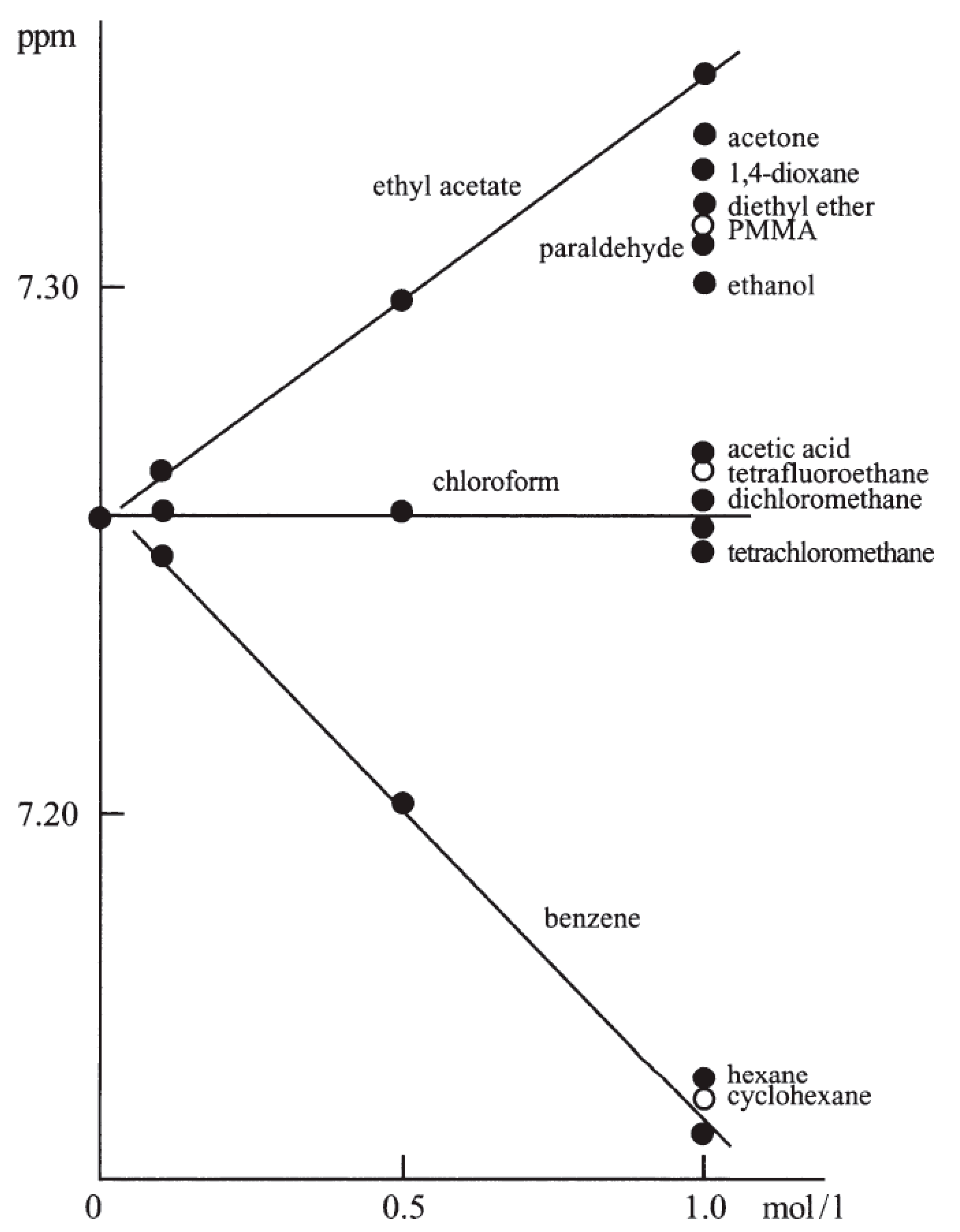
Figure 1. Chemical shifts of the CHCl3 line at different concentrations of various analytes studied in CDCl3 solution, PMMA = poly(methyl methacrylate) [5]. Reproduced with permission from Springer Nature.
Molecules 28 04369 g001
Figure 2. Compounds discussed in refs. [4,103,104].
Molecules 28 04369 g002
Figure 3. Hydrogen-bonded complexes discussed in refs. [105,106].
Molecules 28 04369 g003
Figure 4. 400 Hz 1H NMR spectrum of fat extract from trout taken in the mixture of CDCl3/CD3OD (2:1, v/v) containing internal TMS [107]. Only important signals are integrated; see text.
Molecules 28 04369 g004
Table 1. Five standard 1H and 13C NMR chemical shift reference methods a.
MethodDescriptionNotes
AResidual 1H and 13C signals from NMR solvents are used as internal standards–two variants (1H and 13C)A simplified, formally unregulated but widely applied method, the use of which is discussed in detail here
BBy using internal references (mainly TMS)Standard approach–a codex of chemistry (TMS)
CUse 2H known lock frequencies of the NMR solventsDefault method on all modern NMR spectrometers
DAn accurate IUPAC-recommended general scheme for indirect referencing based on Ξ valuesUsed for all NMR-observable nuclei, but only very rarely routinely for 1H and 13C
EUse of external standards (typically) in coaxial tubesUsed most often for the 11B, 15N, 19F, 31P nuclei, etc.
a By using standard cylindrical NMR tubes.
Table 2. Recommended δX values for 1H (residual) and 13C signals of six common NMR solvents measured vs. internal TMS, ppm.
CDCl3(CD3)2CO(CD3)2SOC6D6CD3CNCD3OD
δH7.2602.0532.5027.1561.9393.3064.848 a,b
δC77.0129.83206.15 b39.46128.031.36118.3649.04
a The CD3OH signal. b The application of this signal in Method A is usually not recommended due to the high variability of its δX value (see Supplementary Materials).
Disclaimer/Publisher’s Note: The statements, opinions and data contained in all publications are solely those of the individual author(s) and contributor(s) and not of MDPI and/or the editor(s). MDPI and/or the editor(s) disclaim responsibility for any injury to people or property resulting from any ideas, methods, instructions or products referred to in the content.