
Biosensors with Metal Ion–Phosphate Chelation Interaction for Molecular Recognition

Abstract
:1. Introduction
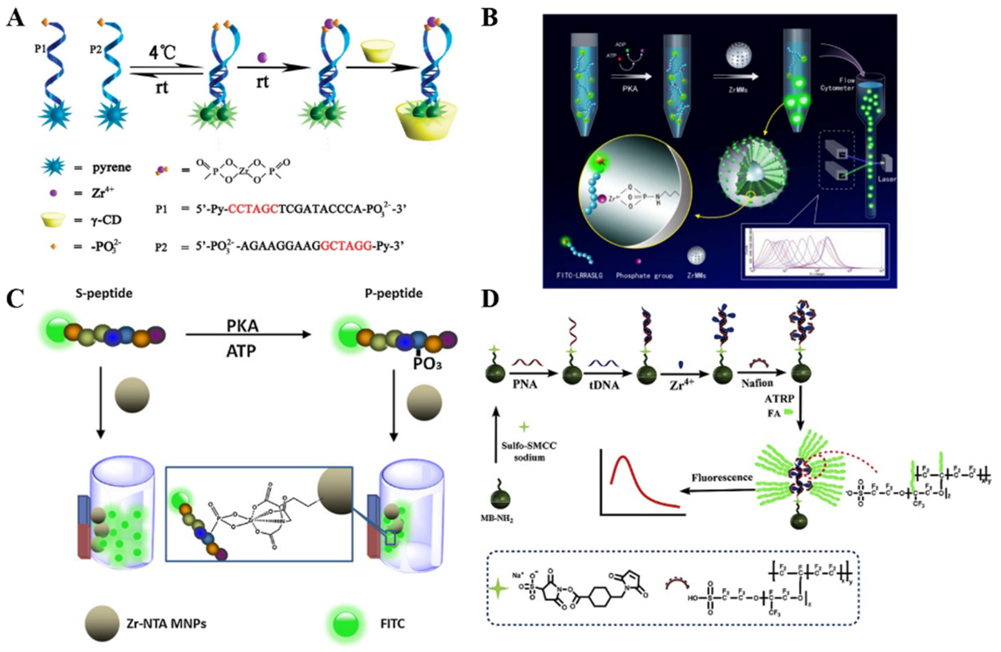
2. Phos-Tag
2.1. Electrochemical Methods
2.2. Fluorescence Methods
3. Metal Ions
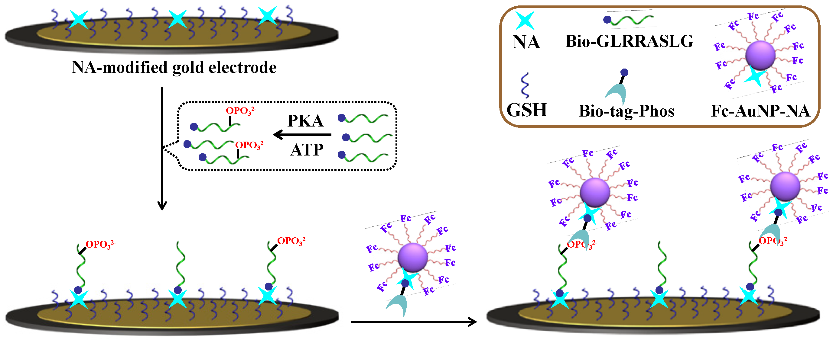
3.1. Electrochemical Biosensors


| Method | Substrate | Signal Report | Metal Ions | Target | Linear Range | Detection Limit | Ref. |
|---|---|---|---|---|---|---|---|
| EC | PNA | SI-eATRP of FMMA | Zr4+ | DNA | 1 × 10⁻⁵–10 pM | 3.2 aM | [83] |
| EC | PNA | SI-eATRP of C3H4O/silver | Zr4+ | DNA | 1 × 10⁻⁷–1 nM | 0.89 aM | [84] |
| EC | Peptide-AuE | eATRP of FMMA | Zr4+ | PKA | 0–1.4 × 10² U/mL | 1.63 U/mL | [85] |
| EC | Initiator | ARGET ATRP of FMMA | Zr4+ | ALP | 20–2 × 10² U/mL | 1.64 U/mL | [86] |
| EC | Peptide | RAFT of FMMA | Zr4+ | DNA | 1 × 10⁻⁵–1 nM | 10.73 aM | [87] |
| EC | Peptide | RAFT of FMMA | Zr4+ | PKA | 0–0.14 U/mL | 1.05 U/mL | [88] |
| EC | Peptide | TEMPO | Zr4+ | DNA | 1 × 10⁻²–1 × 10² nM | 2.57 pM | [89] |
| EC | PNA | SI-eATRP of FMMA | Zr4+ | DNA | 1 × 10⁻⁵– 0.1 nM | 72 aM | [90] |
| EC | PNA | GO-eATRP of FMMA | Zr4+ | DNA | 1 × 10⁻³–1 × 10² fM | 0.213 aM | [91] |
| EC | PNA | Rose bengal-mediated photoATRP of FMMA | Zr4+ | DNA | 1–1 × 10⁵ fM | 0.115 fM | [92] |
| EC | Peptide | pDNA + RCA | Zr4+ | PKA | 5–5 × 10² U/mL | 0.5 U/mL | [93] |
| EC | Peptide | pDNA + TMSDR/HCR | Zr4+ | PKA | 5 × 10⁻²–1 × 10² U/mL | 20 mU/mL | [94] |
| EC | DNA | psDNA + Fc-SWNTs | Zr4+ | PNK | 1 × 10⁻²–10 U/mL | 10 mU/mL | [95] |
| EC | Peptide | pDNA-AuNPs | Zr4+ | PKA | 0–2.5 × 10² U/mL | 0.15 U/mL | [96] |
| EC | Peptide | DNA-AuNPs | Zr4+ | PKA | 0.1–40 U/mL | 30 mU/mL | [97] |
| ECL | Kemptide-MBs | pDNA/TBR-cysteamine-AuNP | Zr4+ | PKA | 1 × 10⁻²–50 U/mL | 5 mU/mL | [98] |
| ECL | Kemptide | pDNA/XOD-AuNPs | Zr4+ | PKA | 0.1–10 U/mL | 90 mU/mL | [99] |
| ECL | Peptide-GQDs | pDNA/G-quadruplex–hemin DNAzyme- AuNPs | Zr4+ | PKA | 5 × 10⁻²–5 U/mL | 40 mU/mL | [100] |
| ECL | Peptide | Ru(II)-SiO2 NPs | Zr4+ | PKA | 1 × 10⁻²–1 U/mL | 5 mU/mL | [101] |
| PEC | Peptide-AuNPs | P-g-C3N4 | Zr4+ | PKA | 5 × 10⁻²–50 U/mL | 77 mU/mL | [102] |
| PEC | Kemptide TiO2 | pDNA-AuNPs | Zr4+ | PKA | 8 × 10⁻³–1 U/mL | 5 mU/mL | [103] |
| FL | – | phosphorylated and pyrene-labeled DNA | Zr4+ | Zr4+ | 0.5–1 × 10²μM | 200 nM | [104] |
| FL | – | FITC-peptide/polymer | Zr4+ | PKA | 0.5–1 × 10³ U/mL | 0.2 U/mL | [105] |
| FL | SiO2 | FITC-peptide | Zr4+ | PKA | 1 × 10⁻²–50 U/mL | 6 mU/mL | [106] |
| FL | NTA-MNPs | FITC-peptide | Zr4+ | PKA | Not reported | 0.8 U/mL | [107] |
| FL | PNA-MBs | ATRP of FITC-o-acrylate | Zr4+ | DNA | 0.1 fM–0.1 nM | 35.5 aM | [108] |
| FL | – | DNA-QDs/Cy5-peptide | Zr4+ | PKA | 3 × 10⁻²–100 U/mL | 0.882 mU/mL | [109] |
| FL | – | Peptide−GQD | Zr4+ | CK2 | 0.1–1 U/mL | 30 mU/mL | [110] |
| FL | – | Peptide-AuNCs | Zr4+ | CK2 | 8 × 10⁻²–2 U/mL | 27 mU/mL | [111] |
| FL | Zr4+-MBs | peptide/avidin-UCNPs | Zr4+ | PKA | 5 × 10⁻²–0.2 U/mL | 20 mU/mL | [112] |
| FL | DNA-MBs | TRITC-peptide | Dy3+ | PKA | 0.5–4 U/mL | 0.12 U/mL | [113] |
| FL | – | TRITC-peptide/UCNPs | – | PKA | 0.1–10 U/mL | 50 mU/mL | [114] |
| Color | – | ATP-AuNPs | – | Zr4+ | 0.5–1 × 10² μM | 95 nM | [115] |
| Color | – | Citrate-AuNPs | Zr4+ | ATP | 0.1–15 μM | 28 nM | [76] |
| Color | – | AuNPs | Zr4+ | 1B | 5–1.8 × 10² mU/mL | 1.7 mU/mL | [116] |
| PGM | Zr4+-MBs | Invertase | Zr4+ | PKA | 0.1–10 U/mL | 0.1 U/mL | [117] |
3.2. Fluorescence Biosensors
3.3. Other Metal Ion-Based Methods
4. MOFs
5. Conclusions
Author Contributions
Funding
Institutional Review Board Statement
Informed Consent Statement
Data Availability Statement
Conflicts of Interest
References
- Justino, C.I.L.; Freitas, A.C.; Pereira, R.; Duarte, A.C.; Rocha Santos, T.A.P. Recent developments in recognition elements for chemical sensors and biosensors. TrAC Trends Anal. Chem. 2015, 68, 2–17. [Google Scholar] [CrossRef]
- Mamardashvili, G.; Kaigorodova, E.; Lebedev, I.; Mamardashvili, N. Molecular recognition of imidazole-based drug molecules by Cobalt(III)- and Zinc(II)-coproporphyrins in aqueous media. Molecules 2023, 28, 964. [Google Scholar] [CrossRef] [PubMed]
- Wang, X.; Lu, X.; Chen, J. Development of biosensor technologies for analysis of environmental contaminants. Trends Environ. Anal. Chem. 2014, 2, 25–32. [Google Scholar] [CrossRef]
- Jones, A.; Dhanapala, L.; Kankanamage, R.N.T.; Kumar, C.V.; Rusling, J.F. Multiplexed immunosensors and immunoarrays. Anal. Chem. 2020, 92, 345–362. [Google Scholar] [CrossRef] [PubMed]
- de Carvalho, C.C. Enzymatic and whole cell catalysis: Finding new strategies for old processes. Biotechnol. Adv. 2011, 29, 75–83. [Google Scholar] [CrossRef] [PubMed]
- Sochor, J.; Zitka, O.; Hynek, D.; Jilkova, E.; Krejcova, L.; Trnkova, L.; Adam, V.; Hubalek, J.; Kynicky, J.; Vrba, R.; et al. Bio-sensing of cadmium(II) ions using Staphylococcus aureus. Sensors 2011, 11, 10638–10663. [Google Scholar] [CrossRef]
- Song, K.M.; Lee, S.; Ban, C. Aptamers and their biological applications. Sensors 2012, 12, 612–631. [Google Scholar] [CrossRef]
- Zhang, K.; Li, H.; Wang, W.; Cao, J.; Gan, N.; Han, H. Application of multiplexed mptasensors in food contaminants detection. ACS Sens. 2020, 5, 3721–3738. [Google Scholar] [CrossRef]
- Algieri, C.; Drioli, E.; Guzzo, L.; Donato, L. Bio-mimetic sensors based on molecularly imprinted membranes. Sensors 2014, 14, 13863–13912. [Google Scholar] [CrossRef]
- Justino, C.I.L.; Duarte, A.C.; Rocha-Santos, T.A.P. Analytical applications of affibodies. TrAC Trends Anal. Chem. 2015, 65, 73–82. [Google Scholar] [CrossRef]
- Shrivas, K.; Nirmalkar, N.; Deb, M.K.; Dewangan, K.; Nirmalkar, J.; Kumar, S. Application of functionalized silver nanoparticles as a biochemical sensor for selective detection of lysozyme protein in milk sample. Spectrochim. Acta A Mol. Biomol. Spectrosc. 2019, 213, 127–133. [Google Scholar] [CrossRef]
- Carreira-Barral, I.; Fernández-Pérez, I.; Mato-Iglesias, M.; de Blas, A.; Platas-Iglesias, C.; Esteban-Gómez, D. Recognition of AMP, ADP and ATP through cooperative binding by Cu(II) and Zn(II) complexes containing urea and/or phenylboronic acid moieties. Molecules 2018, 23, 479. [Google Scholar] [CrossRef]
- Wang, J.; Zhu, X.; Tu, Q.; Guo, Q.; Zarui, C.S.; Momand, J.; Sun, X.Z.; Zhou, F. Capture of p53 by electrodes modified with consensus DNA duplexes and amplified voltammetric detection using ferrocene-capped gold nanoparticle/streptavidin conjugates. Anal. Chem. 2008, 80, 769–774. [Google Scholar] [CrossRef] [PubMed]
- Zhang, Y. Boronic acid-functionalized nanomaterials for the design of electrochemical biosensors. Int. J. Electrochem. Sci. 2022, 17, 220661–220673. [Google Scholar] [CrossRef]
- Qin, X.; Zhang, Z.; Shao, H.; Zhang, R.; Chen, L.; Yang, X. Boronate affinity material-based sensors for recognition and detection of glycoproteins. Analyst 2020, 145, 7511–7527. [Google Scholar] [CrossRef]
- Bian, Z.; Liu, A.; Li, Y.; Fang, G.; Yao, Q.; Zhang, G.; Wu, Z. Boronic acid sensors with double recognition sites: A review. Analyst 2020, 145, 719–744. [Google Scholar] [CrossRef] [PubMed]
- Li, H.; He, H.; Liu, Z. Recent progress and application of boronate affinity materials in bioanalysis. TrAC Trends Anal. Chem. 2021, 140, 116271–116295. [Google Scholar] [CrossRef]
- Wang, H.; Bie, Z.; Lü, C.; Liu, Z. Magnetic nanoparticles with dendrimer-assisted boronate avidity for the selective enrichment of trace glycoproteins. Chem. Sci. 2013, 4, 4298–4303. [Google Scholar] [CrossRef]
- Zhou, J.; Xu, X.; Liu, W.; Liu, X.; Nie, Z.; Qing, M.; Nie, L.; Yao, S. Graphene oxide-peptide nanocomplex as a versatile fluorescence probe of protein kinase activity based on phosphorylation protection against carboxypeptidase digestion. Anal. Chem. 2013, 85, 5746–5754. [Google Scholar] [CrossRef] [PubMed]
- Freeman, R.; Finder, T.; Gill, R.; Willner, I. Probing protein kinase (CK2) and alkaline phosphatase with CdSe/ZnS quantum dots. Nano Lett. 2010, 10, 2192–2196. [Google Scholar] [CrossRef]
- Agnes, R.S.; Jernigan, F.; Shell, J.R.; Sharma, V.; Lawrence, D.S. Suborganelle sensing of mitochondrial cAMP-dependent protein kinase activity. J. Am. Chem. Soc. 2010, 132, 6075–6080. [Google Scholar] [CrossRef]
- Bai, J.; Zhao, Y.; Wang, Z.; Liu, C.; Wang, Y.; Li, Z. Dual-readout fluorescent assay of protein kinase activity by use of TiO2-coated magnetic microspheres. Anal. Chem. 2013, 85, 4813–4821. [Google Scholar] [CrossRef] [PubMed]
- Song, W.; Liang, R.-P.; Wang, Y.; Zhang, L.; Qiu, J.-D. Gold nanoclusters-based dual-emission ratiometric fluorescence probe for monitoring protein kinase. Sens. Actuat. B Chem. 2016, 226, 144–150. [Google Scholar] [CrossRef]
- Kim, S.; Eom, M.S.; Kim, S.K.; Seo, S.H.; Han, M.S. A highly sensitive gold nanoparticle-based colorimetric probe for pyrophosphate using a competition assay approach. Chem. Commun. 2013, 49, 152–154. [Google Scholar] [CrossRef] [PubMed]
- Kim, S.; Eom, M.S.; Seo, S.H.; Han, M.S. Highly sensitive gold nanoparticle-based colorimetric probe for phytate detection with high selectivity over various phosphate derivatives. Tetrahedron Lett. 2013, 54, 5284–5287. [Google Scholar] [CrossRef]
- Zhou, Y.; Yin, H.; Zhao, W.-W.; Ai, S. Electrochemical, electrochemiluminescent and photoelectrochemical bioanalysis of epigenetic modifiers: A comprehensive review. Coordin. Chem. Rev. 2020, 424, 213519–213562. [Google Scholar] [CrossRef]
- Kinoshita, E.; Kinoshita-Kikuta, E.; Koike, T. Advances in Phos-tag-based methodologies for separation and detection of the phosphoproteome. Biochim. Biophys. Acta 2015, 1854, 601–608. [Google Scholar] [CrossRef]
- Hirano, H.; Shirakawa, J. Recent developments in Phos-tag electrophoresis for the analysis of phosphoproteins in proteomics. Expert Rev. Proteomic. 2022, 19, 103–114. [Google Scholar] [CrossRef]
- Kinoshita, E.; Kinoshita-Kikuta, E.; Koike, T. History of Phos-tag technology for phosphoproteomics. J. Proteom. 2022, 252, 104432–104439. [Google Scholar] [CrossRef]
- Kinoshita-Kikuta, E.; Koike, T.; Kinoshita, E. Recent advances in the Phos-tag technique focused on the analysis of phosphoproteins in a bacterial two-component system. J. Proteom. 2022, 252, 104429–104433. [Google Scholar] [CrossRef]
- Kinoshita, E.; Takahashi, M.; Takeda, H.; Shiro, M.; Koike, T. Recognition of phosphate monoester dianion by an alkoxide-bridged dinuclear zinc(II) complex. Dalton. Trans. 2004, 8, 1189–1193. [Google Scholar] [CrossRef] [PubMed]
- Sugiyama, Y.; Uezato, Y. Analysis of protein kinases by Phos-tag SDS-PAGE. J. Proteom. 2022, 255, 104485–104490. [Google Scholar] [CrossRef]
- Hisanaga, S.I.; Krishnankutty, A.; Kimura, T. In vivo analysis of the phosphorylation of tau and the tau protein kinases Cdk5-p35 and GSK3 beta by using Phos-tag SDS-PAGE. J. Proteom. 2022, 262, 104591–104597. [Google Scholar] [CrossRef] [PubMed]
- Kinoshita-Kikuta, E.; Kinoshita, E.; Koike, T. Phos-tag beads as an immunoblotting enhancer for selective detection of phosphoproteins in cell lysates. Anal. Biochem. 2009, 389, 83–85. [Google Scholar] [CrossRef] [PubMed]
- Barbieri, C.M.; Stock, A.M. Universally applicable methods for monitoring response regulator aspartate phosphorylation both in vitro and in vivo using Phos-tag-based reagents. Anal. Biochem. 2008, 376, 73–82. [Google Scholar] [CrossRef]
- Yamada, S.; Nakamura, H.; Kinoshita, E.; Kinoshita-Kikuta, E.; Koike, T.; Shiro, Y. Separation of a phosphorylated histidine protein using phosphate affinity polyacrylamide gel electrophoresis. Anal. Biochem. 2007, 360, 160–162. [Google Scholar] [CrossRef]
- Shin, I.S.; Chand, R.; Lee, S.W.; Rhee, H.W.; Kim, Y.S.; Hong, J.I. Homogeneous electrochemical assay for protein kinase activity. Anal. Chem. 2014, 86, 10992–10995. [Google Scholar] [CrossRef]
- Chand, R.; Han, D.; Shin, I.-S.; Hong, J.-I.; Kim, Y.-S. Gold nanoparticle enhanced electrochemical assay for protein kinase activity using a synthetic chemosensor on a microchip. J. Electrochem. Soc. 2015, 162, B89–B93. [Google Scholar] [CrossRef]
- Kinoshita, E.; Kinoshita-Kikuta, E.; Sugiyama, Y.; Fukada, Y.; Ozeki, T.; Koike, T. Highly sensitive detection of protein phosphorylation by using improved Phos-tag Biotin. Proteomics 2012, 12, 932–937. [Google Scholar] [CrossRef] [PubMed]
- Kinoshita, E.; Kinoshita-Kikuta, E.; Koike, T. Sandwich assay for phosphorylation of protein multiplexes by using antibodies and Phos-tag. Anal. Biochem. 2013, 438, 104–106. [Google Scholar] [CrossRef] [PubMed]
- Liu, X.J.; Li, X.Y.; Gao, X.; Ge, L.; Sun, X.Z.; Li, F. A universal paper-based electrochemical sensor for Zero-background assay of diverse biomarkers. ACS Appl. Mater. Interfaces 2019, 11, 15381–15388. [Google Scholar] [CrossRef] [PubMed]
- Jing, L.; Xie, C.; Li, Q.; Yang, M.; Li, S.; Li, H.; Xia, F. Electrochemical biosensors for the analysis of breast cancer biomarkers: From design to application. Anal. Chem. 2022, 94, 269–296. [Google Scholar] [CrossRef]
- Chang, Y.; Ma, X.; Sun, T.; Liu, L.; Hao, Y. Electrochemical detection of kinase by converting homogeneous analysis into heterogeneous assay through avidin-biotin interaction. Talanta 2021, 234, 122649–122654. [Google Scholar] [CrossRef] [PubMed]
- Yin, H.; Wang, H.; Jiang, W.; Zhou, Y.; Ai, S. Electrochemical immunosensor for N6-methyladenosine detection in human cell lines based on biotin-streptavidin system and silver-SiO2 signal amplification. Biosens. Bioelectron. 2017, 90, 494–500. [Google Scholar] [CrossRef] [PubMed]
- Zhang, Q.; Li, X.; Li, B.; Yin, H.; Ai, S. An electrochemical biosensor for the activity assay of polynucleotide kinase and inhibitor screening. Anal. Methods 2015, 7, 9984–9991. [Google Scholar] [CrossRef]
- Yang, Z.; Jiang, W.; Liu, F.; Zhou, Y.; Yin, H.; Ai, S. A novel electrochemical immunosensor for the quantitative detection of 5-hydroxymethylcytosine in genomic DNA of breast cancer tissue. Chem. Commun. 2015, 51, 14671–14673. [Google Scholar] [CrossRef]
- Zhou, Y.; Yin, H.; Li, X.; Li, Z.; Ai, S.; Lin, H. Electrochemical biosensor for protein kinase A activity assay based on gold nanoparticles-carbon nanospheres, phos-tag-biotin and β-galactosidase. Biosens. Bioelectron. 2016, 86, 508–515. [Google Scholar] [CrossRef]
- Yin, H.; Wang, M.; Li, B.; Yang, Z.; Zhou, Y.; Ai, S. A sensitive electrochemical biosensor for detection of protein kinase A activity and inhibitors based on Phos-tag and enzymatic signal amplification. Biosens. Bioelectron. 2015, 63, 26–32. [Google Scholar] [CrossRef]
- Wang, H.; Zhang, Q.; Yin, H.; Wang, M.; Jiang, W.; Ai, S. Photoelectrochemical immunosensor for methylated RNA detection based on g-C3N4/CdS quantum dots heterojunction and Phos-tag-biotin. Biosens. Bioelectron. 2017, 95, 124–130. [Google Scholar] [CrossRef]
- Zhou, Y.; Jiang, W.; Wu, H.; Liu, F.; Yin, H.; Lu, N.; Ai, S. Amplified electrochemical immunoassay for 5-methylcytosine using a nanocomposite prepared from graphene oxide, magnetite nanoparticles and beta-cyclodextrin. Microchim. Acta 2019, 186, 488–497. [Google Scholar] [CrossRef]
- Yin, H.; Sun, B.; Dong, L.; Li, B.; Zhou, Y.; Ai, S. A signal “on” photoelectrochemical biosensor for assay of protein kinase activity and its inhibitor based on graphite-like carbon nitride, Phos-tag and alkaline phosphatase. Biosens. Bioelectron. 2015, 64, 462–468. [Google Scholar] [CrossRef] [PubMed]
- Zhou, Y.; Wang, M.; Yang, Z.; Yin, H.; Ai, S. A Phos-tag-based photoelectrochemical biosensor for assay of protein kinase activity and inhibitors. Sens. Actuat. B Chem. 2015, 206, 728–734. [Google Scholar] [CrossRef]
- Wang, H.; Qi, C.; He, W.; Wang, M.; Jiang, W.; Yin, H.; Ai, S. A sensitive photoelectrochemical immunoassay of N6-methyladenosine based on dual-signal amplification strategy: Ru doped in SiO2 nanosphere and carboxylated g-C3N4. Biosens. Bioelectron. 2018, 99, 281–288. [Google Scholar] [CrossRef] [PubMed]
- Ai, L.; Wang, Y.; Zhou, Y.; Yin, H. Photoelectrochemical biosensor for N6-methyladenosine detection based on enhanced photoactivity of TiO2-X and MoS2 nanocomposite. J. Electroanal. Chem. 2021, 895, 115444–115452. [Google Scholar] [CrossRef]
- Yin, H.; Wu, H.; Chen, Y.; Li, F.; Wang, J.; Ai, S. Photoelectrochemical determination of the activity of histone acetyltransferase and inhibitor screening by using MoS2 nanosheets. Microchim. Acta 2019, 186, 663–671. [Google Scholar] [CrossRef]
- Li, B.; Yin, H.; Zhou, Y.; Wang, M.; Wang, J.; Ai, S. Photoelectrochemical detection of miRNA-319a in rice leaf responding to phytohormones treatment based on CuO-CuWO4 and rolling circle amplification. Sens. Actuat. B Chem. 2018, 255, 1744–1752. [Google Scholar] [CrossRef]
- Sui, C.; Yin, H.; Wang, L.; Zhou, Y.; Ai, S. Electrochemiluminescence biosensor for DNA hydroxymethylation detection based on enzyme-catalytic covalent bonding reaction of -CH2OH and thiol functionalized Fe3O4 magnetic beads. Biosens. Bioelectron. 2020, 150, 111908–111914. [Google Scholar] [CrossRef]
- Jiang, W.; Wu, L.; Duan, J.; Yin, H.; Ai, S. Ultrasensitive electrochemiluminescence immunosensor for 5-hydroxymethylcytosine detection based on Fe3O4@SiO2 nanoparticles and PAMAM dendrimers. Biosens. Bioelectron. 2018, 99, 660–666. [Google Scholar] [CrossRef]
- Jiang, S.; Geng, Y.X.; Liu, W.J.; Wang, Z.Y.; Zhang, C.Y. Construction of a phos-tag-directed self-assembled fluorescent magnetobiosensor for the simultaneous detection of multiple protein kinases. J. Mater. Chem. B 2022, 10, 9992–10000. [Google Scholar] [CrossRef]
- Jiang, S.; Wang, P.; Li, C.C.; Cui, L.; Li, Y.; Zhang, C.Y. Development of a phos-tag-based fluorescent biosensor for sensitive detection of protein kinase in cancer cells. J. Mater. Chem. B 2022, 10, 3260–3267. [Google Scholar] [CrossRef]
- Zhao, Y.C.; Xiang, J.Z.; Cheng, H.; Liu, X.J.; Li, F. Flexible photoelectrochemical biosensor for ultrasensitive microRNA detection based on concatenated multiplex signal amplification. Biosens. Bioelectron. 2021, 194, 113581. [Google Scholar] [CrossRef] [PubMed]
- Li, Z.; Lu, J.; Wei, W.; Tao, M.; Wang, Z.; Dai, Z. Recent advances in electron manipulation of nanomaterials for photoelectrochemical biosensors. Chem. Commun. 2022, 58, 12418–12430. [Google Scholar] [CrossRef]
- del Barrio, M.; Luna-López, G.; Pita, M. Enhancement of biosensors by implementing photoelectrochemical processes. Sensors 2020, 20, 3281. [Google Scholar] [CrossRef] [PubMed]
- Wang, H.; Yin, H.; Huang, H.; Li, K.; Zhou, Y.; Waterhouse, G.I.N.; Lin, H.; Ai, S. Dual-signal amplified photoelectrochemical biosensor for detection of N6-methyladenosine based on BiVO4-110-TiO2 heterojunction, Ag+-mediated cytosine pairs. Biosens. Bioelectron. 2018, 108, 89–96. [Google Scholar] [CrossRef] [PubMed]
- Zhao, W.W.; Xu, J.J.; Chen, H.Y. Photoelectrochemical enzymatic biosensors. Biosens. Bioelectron. 2017, 92, 294–304. [Google Scholar] [CrossRef]
- Li, H.Y.; Yang, Q.T.; Wang, Z.X.; Li, F. Iridium complex with specific intercalation in G-quadruplex: A phosphorescence and electrochemiluminescence dual-mode homogeneous biosensor for enzyme-free and label-free detection of microRNA. ACS Sens. 2023, 8, 1529–1535. [Google Scholar] [CrossRef] [PubMed]
- Wei, Y.; Qi, H.; Zhang, C. Recent advances and challenges in developing electrochemiluminescence biosensors for health analysis. Chem. Commun. 2023, 59, 3507–3522. [Google Scholar] [CrossRef]
- Takiyama, K.; Kinoshita, E.; Kinoshita-Kikuta, E.; Fujioka, Y.; Kubo, Y.; Koike, T. A Phos-tag-based fluorescence resonance energy transfer system for the analysis of the dephosphorylation of phosphopeptides. Anal. Biochem. 2009, 388, 235–241. [Google Scholar] [CrossRef]
- Somura, M.; Takiyama, K.; Kinoshita-Kikuta, E.; Kinoshita, E.; Koike, T. A Phos-tag-based fluorescence resonance energy transfer system for the analysis of the kinase reaction of a substrate peptide. Anal. Methods 2011, 3, 1303–1309. [Google Scholar] [CrossRef]
- Shiba, A.; Kinoshita-Kikuta, E.; Kinoshita, E.; Koike, T. TAMRA/TAMRA fluorescence quenching systems for the activity assay of alkaline phosphatase. Sensors 2017, 17, 1877–1888. [Google Scholar] [CrossRef]
- Kinoshita-Kikuta, E.; Kurosaki, H.; Kunisada, N.; Kinoshita, E.; Koike, T. A phos-tag-based fluorescence quenching system for activity assay and inhibitor screening for alkaline phosphatase. Am. J. Anal. Chem. 2014, 5, 796–804. [Google Scholar] [CrossRef]
- Rhee, H.W.; Lee, S.H.; Shin, I.S.; Choi, S.J.; Park, H.H.; Han, K.; Park, T.H.; Hong, J.I. Detection of kinase activity using versatile fluorescence quencher probes. Angew. Chem. Int. Ed. 2010, 49, 4919–4923. [Google Scholar] [CrossRef] [PubMed]
- Nobori, T.; Kishimura, A.; Mori, T.; Katayama, Y. A FRET-based protein kinase assay using Phos-tag-modified quantum dots and fluorophore-labeled peptides. Anal. Sci. 2021, 37, 1361–1366. [Google Scholar] [CrossRef] [PubMed]
- Andersson, L.; Porath, J. Isolation of phosphoproteins by immobilized metal (Fe3+) affinity chromatography. Anal. Biochem. 1986, 154, 250–254. [Google Scholar] [CrossRef] [PubMed]
- Zondlo, S.C.; Gao, F.; Zondlo, N.J. Design of an encodable tyrosine kinase-inducible domain: Detection of tyrosine kinase activity by terbium luminescence. J. Am. Chem. Soc. 2010, 132, 5619–5621. [Google Scholar] [CrossRef]
- Qi, W.; Liu, Z.; Zhang, W.; Halawa, M.I.; Xu, G. Visual and plasmon resonance absorption sensor for adenosine triphosphate based on the high affinity between phosphate and Zr(IV). Sensors 2016, 16, 1674–1684. [Google Scholar] [CrossRef]
- Monot, J.; Petit, M.; Lane, S.M.; Guisle, I.; Leger, J.; Tellier, C.; Talham, D.R.; Bujoli, B. Towards zirconium phosphonate-based microarrays for probing DNA-protein interactions: Critical influence of the location of the probe anchoring groups. J. Am. Chem. Soc. 2008, 130, 6243–6251. [Google Scholar] [CrossRef]
- Nonglaton, G.; Benitez, I.O.; Guisle, I.; Pipelier, M.; Leger, J.; Dubreuil, D.; Tellier, C.; Talham, D.R.; Bujoli, B. New approach to oligonucleotide microarrays using zirconium phosphonate-modified surfaces. J. Am. Chem. Soc. 2004, 126, 1497–1502. [Google Scholar] [CrossRef]
- Fang, C.; Fan, Y.; Kong, J.; Gao, Z.; Balasubramanian, N. Electrical detection of oligonucleotide using an aggregate of gold nanoparticles as a conductive tag. Anal. Chem. 2008, 80, 9387–9394. [Google Scholar] [CrossRef]
- Wang, M.; Wang, G.X.; Xiao, F.N.; Zhao, Y.; Wang, K.; Xia, X.H. Sensitive label-free monitoring of protein kinase activity and inhibition using ferric ions coordinated to phosphorylated sites as electrocatalysts. Chem. Commun. 2013, 49, 8788–8790. [Google Scholar] [CrossRef]
- Wieckowska, A.; Li, D.; Gill, R.; Willner, I. Following protein kinase acivity by electrochemical means and contact angle measurements. Chem. Commun. 2008, 44, 2376–2378. [Google Scholar] [CrossRef]
- Zhu, X.; Liu, Z.; Li, J.; Li, Z.; Si, F.; Yang, H.; Kong, J. Dual signal amplification based on polysaccharide-initiated ring-opening polymerization and click polymerization for exosomes detection. Talanta 2021, 233, 122531–122538. [Google Scholar] [CrossRef]
- Hu, Q.; Han, D.; Gan, S.; Bao, Y.; Niu, L. Surface-initiated-reversible-addition-fragmentation-chain-transfer polymerization for electrochemical DNA biosensing. Anal. Chem. 2018, 90, 12207–12213. [Google Scholar] [CrossRef] [PubMed]
- Hao, L.; Zhao, L.; Li, G.; Li, Y.; Ma, L.; Liu, Y.; Wang, W.; Kong, J. Ultrasensitive detection of CYFRA 21-1 DNA via SI-RAFT based in-situ metallization signal amplification. Microchem. J. 2020, 158, 105216–105223. [Google Scholar] [CrossRef]
- Hu, Q.; Wang, Q.; Jiang, C.; Zhang, J.; Kong, J.; Zhang, X. Electrochemically mediated polymerization for highly sensitive detection of protein kinase activity. Biosens. Bioelectron. 2018, 110, 52–57. [Google Scholar] [CrossRef] [PubMed]
- Li, X.; Lu, J.; Li, Z.; Yang, H.; Li, W.; Liu, Y.; Miao, M. Electrochemical detection of alkaline phosphatase activity via atom transfer radical polymerization. Bioelectrochemistry 2022, 144, 107998–108004. [Google Scholar] [CrossRef] [PubMed]
- Li, X.; Zhang, Y.; Hao, L.; Liu, Y.; Wang, X.; Yang, H.; Kong, J. Ultrasensitive label-free detection for lung cancer CYFRA 21-1 DNA based on ring-opening polymerization. Talanta 2021, 223, 121730–121739. [Google Scholar] [CrossRef] [PubMed]
- Hu, Q.; Kong, J.; Han, D.; Bao, Y.; Zhang, X.; Zhang, Y.; Niu, L. Ultrasensitive peptide-based electrochemical detection of protein kinase activity amplified by RAFT polymerization. Talanta 2020, 206, 120173–120178. [Google Scholar] [CrossRef]
- Wang, C.; Liu, J.; Kong, J.; Zhang, X. Nitronyl nitroxide monoradical TEMPO as new electrochemical label for ultrasensitive detection of nucleic acids. Anal. Chim. Acta 2020, 1136, 19–24. [Google Scholar] [CrossRef]
- Hu, Q.; Wang, Q.; Sun, G.; Kong, J.; Zhang, X. Electrochemically mediated surface-initiated de novo growth of polymers for amplified electrochemical detection of DNA. Anal. Chem. 2017, 89, 9253–9259. [Google Scholar] [CrossRef]
- Liu, Q.; Liu, J.; Yang, H.; Wang, X.; Kong, J.; Zhang, X. Highly sensitive lung cancer DNA detection via GO enhancing eATRP signal amplification. Microchem. J. 2021, 160, 105766–105773. [Google Scholar] [CrossRef]
- Hu, Y.; Yu, S.; Ma, N.; Kong, J.; Zhang, X. Rose bengal-mediated photoinduced atom transfer radical polymerization for high sensitivity detection of target DNA. Talanta 2023, 254, 124104–124110. [Google Scholar] [CrossRef]
- Miao, P.; Ning, L.; Li, X.; Li, P.; Li, G. Electrochemical strategy for sensing protein phosphorylation. Bioconjug. Chem. 2012, 23, 141–145. [Google Scholar] [CrossRef] [PubMed]
- Cheng, W.; Ma, J.; Xiang, L.; Sun, Y.; Huang, W.; Zhang, Z.; Kong, D.; Li, J. Zr4+-mediated hybrid chain reaction and its application for highly sensitive electrochemical detection of protein kinase A. Bioelectrochemistry 2021, 140, 107796–107801. [Google Scholar] [CrossRef] [PubMed]
- Wang, Y.; He, X.; Wang, K.; Ni, X.; Su, J.; Chen, Z. Ferrocene-functionalized SWCNT for electrochemical detection of T4 polynucleotide kinase activity. Biosens. Bioelectron. 2012, 32, 213–218. [Google Scholar] [CrossRef] [PubMed]
- Xu, X.; Nie, Z.; Chen, J.; Fu, Y.; Li, W.; Shen, Q.; Yao, S. A DNA-based electrochemical strategy for label-free monitoring the activity and inhibition of protein kinase. Chem. Commun. 2009, 45, 6946–6948. [Google Scholar] [CrossRef]
- Wang, Z.; Sun, N.; He, Y.; Liu, Y.; Li, J. DNA assembled gold nanoparticles polymeric network blocks modular highly sensitive electrochemical biosensors for protein kinase activity analysis and inhibition. Anal. Chem. 2014, 86, 6153–6159. [Google Scholar] [CrossRef]
- Zhao, Z.; Zhou, X.; Xing, D. Highly sensitive protein kinase activity assay based on electrochemiluminescence nanoprobes. Biosens. Bioelectron. 2012, 31, 299–304. [Google Scholar] [CrossRef]
- Wang, Z.; Yan, Z.; Sun, N.; Liu, Y. Multiple signal amplification electrogenerated chemiluminescence biosensors for sensitive protein kinase activity analysis and inhibition. Biosens. Bioelectron. 2015, 68, 771–776. [Google Scholar] [CrossRef]
- Liu, J.; He, X.; Wang, K.; He, D.; Wang, Y.; Mao, Y.; Shi, H.; Wen, L. A highly sensitive electrochemiluminescence assay for protein kinase based on double-quenching of graphene quantum dots by G-quadruplex-hemin and gold nanoparticles. Biosens. Bioelectron. 2015, 70, 54–60. [Google Scholar] [CrossRef]
- Chen, Z.; He, X.; Wang, Y.; Wang, K.; Du, Y.; Yan, G. Ru(II) encapsulated phosphorylate-terminated silica nanoparticles-based electrochemiluminescent strategy for label-free assay of protein kinase activity and inhibition. Biosens. Bioelectron. 2013, 41, 519–525. [Google Scholar] [CrossRef] [PubMed]
- Li, X.; Zhou, Y.; Xu, Y.; Xu, H.; Wang, M.; Yin, H.; Ai, S. A novel photoelectrochemical biosensor for protein kinase activity assay based on phosphorylated graphite-like carbon nitride. Anal. Chim. Acta 2016, 934, 36–43. [Google Scholar] [CrossRef]
- Yan, Z.; Wang, Z.; Miao, Z.; Liu, Y. Dye-sensitized and localized surface plasmon resonance enhanced visible-light photoelectrochemical biosensors for highly sensitive analysis of protein kinase activity. Anal. Chem. 2016, 88, 922–929. [Google Scholar] [CrossRef] [PubMed]
- Meng, H.M.; Fu, T.; Zhang, X.B.; Wang, N.N.; Tan, W.; Shen, G.L.; Yu, R.Q. Efficient fluorescence turn-on probe for zirconium via a target-triggered DNA molecular beacon strategy. Anal. Chem. 2012, 84, 2124–2128. [Google Scholar] [CrossRef]
- Bai, J.; Liu, C.; Yang, T.; Wang, F.; Li, Z. A versatile platform for highly sensitive detection of kinase activity based on metal ion-mediated FRET using an anionic conjugated polymer. Chem. Commun. 2013, 49, 3887–3889. [Google Scholar] [CrossRef]
- Ren, W.; Liu, C.; Lian, S.; Li, Z. Flow cytometry-assisted mix-and-read assay for ultrasensitive detection of protein kinase activity by use of Zr4+-functionalized mesoporous SiO2 microspheres. Anal. Chem. 2013, 85, 10956–10961. [Google Scholar] [CrossRef] [PubMed]
- Tan, P.; Lei, C.; Liu, X.; Qing, M.; Nie, Z.; Guo, M.; Huang, Y.; Yao, S. Fluorescent detection of protein kinase based on zirconium ions-immobilized magnetic nanoparticles. Anal. Chim. Acta 2013, 780, 89–94. [Google Scholar] [CrossRef]
- Zhang, J.; Liu, Q.; Ba, Y.; Cheng, J.; Yang, H.; Cui, Y.; Kong, J.; Zhang, X. F-containing initiatior for ultrasensitive fluorescent detection of lung cancer DNA via atom transfer radical polymerization. Anal. Chim. Acta 2020, 1094, 99–105. [Google Scholar] [CrossRef]
- Li, Y.; Liu, Q.; Cui, L.; Liu, W.; Qiu, J.G.; Zhang, C.Y. Zirconium ion-mediated assembly of a single quantum dot-based nanosensor for kinase assay. Chem. Commun. 2021, 57, 6376–6379. [Google Scholar] [CrossRef]
- Wang, Y.; Zhang, L.; Liang, R.P.; Bai, J.M.; Qiu, J.D. Using graphene quantum dots as photoluminescent probes for protein kinase sensing. Anal. Chem. 2013, 85, 9148–9155. [Google Scholar] [CrossRef]
- Song, W.; Wang, Y.; Liang, R.P.; Zhang, L.; Qiu, J.D. Label-free fluorescence assay for protein kinase based on peptide biomineralized gold nanoclusters as signal sensing probe. Biosens. Bioelectron. 2015, 64, 234–240. [Google Scholar] [CrossRef]
- Yang, W.; Wang, Y.; Chang, L.; Liu, C.; Bai, J.; Li, Z. Highly sensitive detection of protein kinase activity using upconversion luminescent nanoparticles. RSC Adv. 2014, 4, 14546–14549. [Google Scholar] [CrossRef]
- Zhang, X.; Liu, C.; Wang, H.; Wang, H.; Li, Z. Rare earth ion mediated fluorescence accumulation on a single microbead: An ultrasensitive strategy for the detection of protein kinase activity at the single-cell level. Angew. Chem. Int. Ed. 2015, 54, 15186–15190. [Google Scholar] [CrossRef] [PubMed]
- Liu, C.; Chang, L.; Wang, H.; Bai, J.; Ren, W.; Li, Z. Upconversion nanophosphor: An efficient phosphopeptides-recognizing matrix and luminescence resonance energy transfer donor for robust detection of protein kinase activity. Anal. Chem. 2014, 86, 6095–6102. [Google Scholar] [CrossRef] [PubMed]
- Qi, W.; Zhao, J.; Zhang, W.; Liu, Z.; Xu, M.; Anjum, S.; Majeed, S.; Xu, G. Visual and surface plasmon resonance sensor for zirconium based on zirconium-induced aggregation of adenosine triphosphate-stabilized gold nanoparticles. Anal. Chim. Acta 2013, 787, 126–131. [Google Scholar] [CrossRef] [PubMed]
- Zhang, J.; Lv, J.; Wang, X.; Li, D.; Wang, Z.; Li, G. A simple and visible colorimetric method through Zr4+-phosphate coordination for the assay of protein tyrosine phosphatase 1B and screening of its inhibitors. Analyst 2015, 140, 5716–5723. [Google Scholar] [CrossRef]
- Yang, W.; Lu, X.; Wang, Y.; Sun, S.; Liu, C.; Li, Z. Portable and sensitive detection of protein kinase activity by using commercial personal glucose meter. Sens. Actuat. B Chem. 2015, 210, 508–512. [Google Scholar] [CrossRef]
- Rininsland, F.; Xia, W.; Wittenburg, S.; Shi, X.; Stankewicz, C.; Achyuthan, K.; McBranch, D.; Whitten, D. Metal ion-mediated polymer superquenching for highly sensitive detection of kinase and phosphatase activities. Proc. Natl. Acad. Sci. USA 2004, 101, 15295–15300. [Google Scholar] [CrossRef] [PubMed]
- Shen, C.; Zhang, K.; Gao, N.; Wei, S.; Liu, G.; Chai, Y.; Yang, M. Colorimetric and electrochemical determination of the activity of protein kinase based on retarded particle growth due to binding of phosphorylated peptides to DNA—capped silver nanoclusters. Microchim. Acta 2016, 183, 2933–2939. [Google Scholar] [CrossRef]
- Ding, Y.; Kang, W.; Xiao, X.; Wang, K.; Wang, N.; Zhang, H.; Yang, M.; Shen, G. Using carbon quantum dots as selective photoluminescent probes for protein kinase assay. Aust. J. Chem. 2015, 68, 1249–1254. [Google Scholar] [CrossRef]
- Shiosaki, S.; Nobori, T.; Mori, T.; Toita, R.; Nakamura, Y.; Kim, C.W.; Yamamoto, T.; Niidome, T.; Katayama, Y. A protein kinase assay based on FRET between quantum dots and fluorescently-labeled peptides. Chem. Commun. 2013, 49, 5592–5594. [Google Scholar] [CrossRef] [PubMed]
- Lian, S.; Yang, W.; Wang, Y.; Liu, C.; Li, Z. Sensitive detection of hexokinase activity by use of Zr4+-coated magnetic beads coupled with phenylboronic acid-functionalized upconversion nanophosphors. Analyst 2014, 139, 5582–5586. [Google Scholar] [CrossRef]
- Geng, H.; Vilms Pedersen, S.; Ma, Y.; Haghighi, T.; Dai, H.; Howes, P.D.; Stevens, M.M. Noble metal nanoparticle biosensors: From fundamental studies toward point-of-care diagnostics. Acc. Chem. Res. 2022, 55, 593–604. [Google Scholar] [CrossRef] [PubMed]
- Deng, J.; Jiang, Q.; Wang, Y.; Yang, L.; Yu, P.; Mao, L. Real-time colorimetric assay of inorganic pyrophosphatase activity based on reversibly competitive coordination of Cu2+ between cysteine and pyrophosphate ion. Anal. Chem. 2013, 85, 9409–9415. [Google Scholar] [CrossRef]
- Deng, H.H.; Luo, B.Y.; He, S.B.; Chen, R.T.; Lin, Z.; Peng, H.P.; Xia, X.H.; Chen, W. Redox recycling-triggered peroxidase-like activity enhancement of bare gold nanoparticles for ultrasensitive colorimetric detection of rare-earth Ce3+ ion. Anal. Chem. 2019, 91, 4039–4046. [Google Scholar] [CrossRef]
- Lien, C.W.; Tseng, Y.T.; Huang, C.C.; Chang, H.T. Logic control of enzyme-like gold nanoparticles for selective detection of lead and mercury ions. Anal. Chem. 2014, 86, 2065–2072. [Google Scholar] [CrossRef]
- Shi, Y.; Wang, J.; Mu, K.; Liu, S.; Yang, G.; Zhang, M.; Yang, J. Copper (II) ion-modified gold nanoclusters as peroxidase mimetics for the colorimetric detection of pyrophosphate. Sensors 2021, 21, 5538–5546. [Google Scholar] [CrossRef] [PubMed]
- Guan, Y.; Lu, Y.; Sun, J.; Zhao, J.; Huang, W.; Zhang, X.; Liu, Y. Redox recycling-activated signal amplification of peroxidase-like catalytic activity based on bare gold nanoparticle–metal ion ensembles as colorimetric sensor array for ultrasensitive discrimination of phosphates. ACS Sustain. Chem. Eng. 2021, 9, 9802–9812. [Google Scholar] [CrossRef]
- Xiang, Y.; Lu, Y. Using personal glucose meters and functional DNA sensors to quantify a variety of analytical targets. Nat. Chem. 2011, 3, 697–703. [Google Scholar] [CrossRef]
- Zhang, J.; Lan, T.; Lu, Y. Translating in vitro diagnostics from centralized laboratories to point-of-care locations using commercially-available handheld meters. TrAC Trends Anal. Chem. 2020, 124, 115782. [Google Scholar] [CrossRef]
- Zhang, H.; Chen, G.Y.; Qian, Z.M.; Li, W.J.; Li, C.H.; Hu, Y.J.; Yang, F.Q. A portable personal glucose meter method for enzyme activity detection and inhibitory activity evaluation based on alkaline phosphatase-mediated reaction. Anal. Bioanal. Chem. 2021, 413, 2457–2466. [Google Scholar] [CrossRef]
- Chang, J.F.; Lv, W.X.; Li, Q.; Li, H.Y.; Li, F. One-step synthesis of methylene blue-encapsulated ZIF for dual-signal fluorescent and homogeneous electrochemical biosensing. Anal. Chem. 2020, 92, 8959–8964. [Google Scholar] [CrossRef]
- Qin, J.; Li, J.; Zeng, H.; Tang, J.; Tang, D. Recent advances in metal-organic framework-based photoelectrochemical and electrochemiluminescence biosensors. Analyst 2023, 148, 2200–2213. [Google Scholar] [CrossRef] [PubMed]
- Gu, C.; Bai, L.; Pu, L.; Gai, P.; Li, F. Highly sensitive and stable self-powered biosensing for exosomes based on dual metal-organic frameworks nanocarriers. Biosens. Bioelectron. 2021, 176, 112907. [Google Scholar] [CrossRef]
- Wang, Z.; Fu, Y.; Kang, Z.; Liu, X.; Chen, N.; Wang, Q.; Tu, Y.; Wang, L.; Song, S.; Ling, D.; et al. Organelle-specific triggered release of immunostimulatory oligonucleotides from intrinsically coordinated DNA-metal-organic frameworks with soluble exoskeleton. J. Am. Chem. Soc. 2017, 139, 15784–15791. [Google Scholar] [CrossRef]
- Wang, S.; Morris, W.; Liu, Y.; McGuirk, C.M.; Zhou, Y.; Hupp, J.T.; Farha, O.K.; Mirkin, C.A. Surface-specific functionalization of nanoscale metal-organic frameworks. Angew. Chem. Int. Ed. 2015, 54, 14738–14742. [Google Scholar] [CrossRef] [PubMed]
- Fu, Y.; Kang, Z.; Cao, W.; Yin, J.; Tu, Y.; Li, J.; Guan, H.; Wang, Y.; Wang, Q.; Kong, X. Defect-assisted loading and docking conformations of pharmaceuticals in metal-organic frameworks. Angew. Chem. Int. Ed. 2021, 60, 7719–7727. [Google Scholar] [CrossRef] [PubMed]
- Dai, G.; Li, Z.; Luo, F.; Ai, S.; Chen, B.; Wang, Q. Electrochemical determination of Salmonella typhimurium by using aptamer-loaded gold nanoparticles and a composite prepared from a metal-organic framework (type UiO-67) and graphene. Microchim. Acta 2019, 186, 620–628. [Google Scholar] [CrossRef] [PubMed]
- Wei, Y.P.; Zhang, Y.W.; Mao, C.J. A silver nanoparticle-assisted signal amplification electrochemiluminescence biosensor for highly sensitive detection of mucin 1. J. Mater. Chem. B 2020, 8, 2471–2475. [Google Scholar] [CrossRef]
- Li, Y.; Hu, M.; Huang, X.; Wang, M.; He, L.; Song, Y.; Jia, Q.; Zhou, N.; Zhang, Z.; Du, M. Multicomponent zirconium-based metal-organic frameworks for impedimetric aptasensing of living cancer cells. Sens. Actuat. B Chem. 2020, 306, 127608–127618. [Google Scholar] [CrossRef]
- Zhang, Z.H.; Duan, F.H.; Tian, J.Y.; He, J.Y.; Yang, L.Y.; Zhao, H.; Zhang, S.; Liu, C.S.; He, L.H.; Chen, M.; et al. Aptamer-embedded zirconium-based metal-organic framework composites prepared by de novo bio-inspired approach with enhanced biosensing for detecting trace analytes. ACS Sens. 2017, 2, 982–989. [Google Scholar] [CrossRef]
- Wang, S.; McGuirk, C.M.; Ross, M.B.; Wang, S.; Chen, P.; Xing, H.; Liu, Y.; Mirkin, C.A. General and direct method for preparing oligonucleotide-functionalized metal-organic framework nanoparticles. J. Am. Chem. Soc. 2017, 139, 9827–9830. [Google Scholar] [CrossRef] [PubMed]
- Wang, S.; Chen, Y.; Wang, S.; Li, P.; Mirkin, C.A.; Farha, O.K. DNA-functionalized metal-organic framework nanoparticles for intracellular delivery of proteins. J. Am. Chem. Soc. 2019, 141, 2215–2219. [Google Scholar] [CrossRef] [PubMed]
- Zhang, G.Y.; Zhuang, Y.H.; Shan, D.; Su, G.F.; Cosnier, S.; Zhang, X.J. Zirconium-based porphyrinic metal-organic framework (PCN-222): Enhanced photoelectrochemical response and its application for label-free phosphoprotein detection. Anal. Chem. 2016, 88, 11207–11212. [Google Scholar] [CrossRef]
- Yan, Z.; Wang, F.; Deng, P.; Wang, Y.; Cai, K.; Chen, Y.; Wang, Z.; Liu, Y. Sensitive electrogenerated chemiluminescence biosensors for protein kinase activity analysis based on bimetallic catalysis signal amplification and recognition of Au and Pt loaded metal-organic frameworks nanocomposites. Biosens. Bioelectron. 2018, 109, 132–138. [Google Scholar] [CrossRef] [PubMed]
- Zhang, G.Y.; Cai, C.; Cosnier, S.; Zeng, H.B.; Zhang, X.J.; Shan, D. Zirconium-metalloporphyrin frameworks as a three-in-one platform possessing oxygen nanocage, electron media, and bonding site for electrochemiluminescence protein kinase activity assay. Nanoscale 2016, 8, 11649–11657. [Google Scholar] [CrossRef]
- Wang, Y.; Yin, H.; Li, X.; Waterhouse, G.I.N.; Ai, S. Photoelectrochemical immunosensor for N6-methyladenine detection based on Ru@UiO-66, Bi2O3 and Black TiO2. Biosens. Bioelectron. 2019, 131, 163–170. [Google Scholar] [CrossRef]
- Yu, K.; Wei, T.; Li, Z.; Li, J.; Wang, Z.; Dai, Z. Construction of molecular sensing and logic systems based on site-occupying effect-modulated MOF-DNA interaction. J. Am. Chem. Soc. 2020, 142, 21267–21271. [Google Scholar] [CrossRef] [PubMed]
- Wang, C.; Tang, G.; Tan, H. Pyrophosphate ion-triggered competitive displacement of ssDNA from a metal-organic framework and its application in fluorescent sensing of alkaline phosphatase. J. Mater. Chem. B 2018, 6, 7614–7620. [Google Scholar] [CrossRef] [PubMed]
- Wang, L.; Hu, Z.; Wu, S.; Pan, J.; Xu, X.; Niu, X. A peroxidase-mimicking Zr-based MOF colorimetric sensing array to quantify and discriminate phosphorylated proteins. Anal. Chim. Acta 2020, 1121, 26–34. [Google Scholar] [CrossRef]
- Song, Z.; Li, Y.; Teng, H.; Ding, C.; Xu, G.; Luo, X. Designed zwitterionic peptide combined with sacrificial Fe-MOF for low fouling and highly sensitive electrochemical detection of T4 polynucleotide kinase. Sens. Actuat. B Chem. 2020, 305, 127329–127337. [Google Scholar] [CrossRef]
- Sun, Z.; Wang, L.; Wu, S.; Pan, Y.; Dong, Y.; Zhu, S.; Yang, J.; Yin, Y.; Li, G. An electrochemical biosensor designed by using Zr-based metal-organic frameworks for the detection of glioblastoma-derived exosomes with practical application. Anal. Chem. 2020, 92, 3819–3826. [Google Scholar] [CrossRef]
- Lei, M.; Ge, F.; Zheng, H. Stable Cd metal-organic framework as a multiresponsive luminescent biosensor for rapid, accurate, and recyclable detection of hippuric acid, nucleoside phosphates, and Fe3+ in urine and serum. Inorg. Chem. 2022, 61, 11243–11251. [Google Scholar] [CrossRef] [PubMed]
- Zhao, P.; Liu, Y.; He, C.; Duan, C. Synthesis of a lanthanide metal-organic framework and its fluorescent detection for phosphate group-based molecules such as adenosine triphosphate. Inorg. Chem. 2022, 61, 3132–3140. [Google Scholar] [CrossRef]
- Zhang, Y.-R.; Xie, X.-Z.; Yin, X.-B.; Xia, Y. Flexible ligand for metal-organic frameworks with simultaneous large-pore and antenna effect emission. Chem. Eng. J. 2022, 443, 136532–136539. [Google Scholar] [CrossRef]
- Yang, C.; Wang, K.; Li, Z.; Mo, L.; Lin, W. A two-photon metal-organic framework nanoprobe with catalytic hairpin assembly for amplified MicroRNA imaging in living cells and tissues. Sens. Actuat. B Chem. 2022, 359, 131593–131600. [Google Scholar] [CrossRef]
- Meng, H.M.; Shi, X.; Chen, J.; Gao, Y.; Qu, L.; Zhang, K.; Zhang, X.B.; Li, Z. DNA amplifier-functionalized metal-organic frameworks for multiplexed detection and imaging of intracellular mRNA. ACS Sens. 2020, 5, 103–109. [Google Scholar] [CrossRef] [PubMed]
- Li, S.; Liu, X.; Chai, H.; Huang, Y. Recent advances in the construction and analytical applications of metal-organic frameworks-based nanozymes. TrAC Trends Anal. Chem. 2018, 105, 391–403. [Google Scholar] [CrossRef]
- Wang, F.; Chen, L.; Liu, D.; Ma, W.; Dramou, P.; He, H. Nanozymes based on metal-organic frameworks: Construction and prospects. TrAC Trends Anal. Chem. 2020, 133, 116080–116096. [Google Scholar] [CrossRef]
- Niu, X.; Li, X.; Lyu, Z.; Pan, J.; Ding, S.; Ruan, X.; Zhu, W.; Du, D.; Lin, Y. Metal-organic framework based nanozymes: Promising materials for biochemical analysis. Chem. Commun. 2020, 56, 11338–11353. [Google Scholar] [CrossRef]
- Huang, X.; Zhang, S.; Tang, Y.; Zhang, X.; Bai, Y.; Pang, H. Advances in metal–organic framework-based nanozymes and their applications. Coordin. Chem. Rev. 2021, 449, 214216–214239. [Google Scholar] [CrossRef]
- Ye, K.; Wang, L.; Song, H.; Li, X.; Niu, X. Bifunctional MIL-53(Fe) with pyrophosphate-mediated peroxidase-like activity and oxidation-stimulated fluorescence switching for alkaline phosphatase detection. J. Mater. Chem. B 2019, 7, 4794–4800. [Google Scholar] [CrossRef] [PubMed]
- Wang, J.; Li, W.; Zheng, Y.-Q. Colorimetric assay for the sensitive detection of phosphate in water based on metal–organic framework nanospheres possessing catalytic activity. New J. Chem. 2020, 44, 19683–19689. [Google Scholar] [CrossRef]
- Wang, Y.; Zhu, Y.; Binyam, A.; Liu, M.; Wu, Y.; Li, F. Discovering the enzyme mimetic activity of metal-organic framework (MOF) for label-free and colorimetric sensing of biomolecules. Biosens. Bioelectron. 2016, 86, 432–438. [Google Scholar] [CrossRef] [PubMed]
- Wang, C.; Gao, J.; Cao, Y.; Tan, H. Colorimetric logic gate for alkaline phosphatase based on copper (II)-based metal-organic frameworks with peroxidase-like activity. Anal. Chim. Acta 2018, 1004, 74–81. [Google Scholar] [CrossRef] [PubMed]


| Method | Substrate | Signal Report | Target | Linear Range | Detection Limit | Ref. |
|---|---|---|---|---|---|---|
| EC | Avidin | Fc/neutravidin-AuNPs | PKA | 5 × 10⁻³–20 U/mL | 5 × 10⁻³ U/mL | [43] |
| EC | Antibody | Avidin/AgNPs-SiO2 | m6A | 0.2–5 × 10² nM | 7.8 × 10⁻² nM | [44] |
| EC | DNA/AuNPs | Biotin-ALP | PNK | 0–5 U/mL | 2.75 × 10⁻³ U/mL | [45] |
| EC | Antibody-graphene | Avidin-ALP | 5hmC | 0.1–30 nM | 3.25 × 10⁻² nM | [46] |
| EC | Peptide/AuNPs/CNs | SA-SiO2/β-galactosidase | PKA | 5 × 10⁻²–1 × 10² U/mL | 1.4 × 10⁻² U/mL | [47] |
| EC | AuNPs | Avidin-HRP | PKA | 0.5–25 U/mL | 0.15 U/mL | [48] |
| PEC | Antibody-g-C3N4/CdS | Avidin-CuO | RNA | 1 × 10⁻²–10 nM | 3.53 pM | [49] |
| PEC | Antibody-β-CD-GO/Fe3O4 | PAMAM-avidin-ALP | 5hmC | 1 × 10⁻²–50 nM | 3.2 pM | [50] |
| PEC | Peptide-g-C3N4/AuNPs | Avidin-ALP | PKA | 5 × 10⁻²–1 × 10² U/mL | 1.5 × 10⁻² U/mL | [51] |
| PEC | Peptide-Bi2S3 | SA | PKA | 5 × 10⁻²–1 × 10² U/mL | 1.7 × 10⁻² U/mL | [52] |
| PEC | Antibody-g-C3N4/CdS | Avidin/Ru-SiO2 | m6A | 1 × 10⁻²–10 nM | 3.23 pM | [53] |
| PEC | AuNPs/TiO2−x/MoS2 | Avidin-ALP | m6A | 0.3–1 × 10² nM | 0.14 nM | [54] |
| PEC | AuNPs/MoS2/graphene | SA-β-galactosidase | HAT | 0.3–1 × 10² nM | 0.14 nM | [55] |
| PEC | CuO-CuWO4 | Avidin-ALP | miRNA | 1 × 10⁻⁶–0.1 nM | 0.47 fM | [56] |
| ECL | MPBA-PDA@Fe3O4 | Ru-avidin | 5hmC | 1 × 10⁻²–5 × 10² nM | 2.86 pM | [57] |
| ECL | Antibody-Fe3O4@SiO2/GO | Avidin/Ru-PAMAM | 5hmC | 0.1–30 nM | 4.7 × 10⁻² nM | [58] |
| FL | - | FITC/Cy5-peptide | PKA | 1 × 10⁻⁴–10 nM | 1 fM | [59] |
| FL | SA-coated MBs | Cy5/BHQ2-DNA | PKA | 1 × 10⁻⁴–10 U/mL | 1.98 × 10⁻⁵ U/mL | [60] |
| Method | Substrate | Signal Report | Target | Linear Range | Detection Limit | Ref. |
|---|---|---|---|---|---|---|
| ECL | DNA | Eu@Zr-MOF | Mucin 1 | 1.13 × 10⁻⁴–0.113 ng/mL | 0.37 fg/mL | [139] |
| ECL | Zr-PCN-222 | – | Protein | 0–2 μg/mL | 0.13 μg/mL | [144] |
| ECL | Kemptide-GO | AuNPs-PtNPs@Zr-UiO-66 | PKA | 1.5 × 10⁻²–0.25 U/mL | 9 mU/mL | [145] |
| ECL | Kemptide-AuNPs | Zr-ZnTCPP-MOF-525 | PKA | 1 × 10⁻²–20 U/mL | 5 mU/mL | [146] |
| PEC | AuNPs/Bi2O3/B-TiO2 | Ru@Zr-UiO-66 | m6A | 5 × 10⁻²–30 nM | 16.7 pM | [147] |
| FL | Zr-PCN-224 | TAMRA-DNA | ALP | 0.5−10 and 10−100 U/L | 0.19 U/L | [148] |
| FL | Ce-MOFs | FITC-DNA | ALP | 2−1 × 10² U/L | 0.18 U/L | [149] |
| Color | – | Zr-MOFs | α-casein | 0.17–5 μg/mL | 0.16 μg/mL | [150] |
Disclaimer/Publisher’s Note: The statements, opinions and data contained in all publications are solely those of the individual author(s) and contributor(s) and not of MDPI and/or the editor(s). MDPI and/or the editor(s) disclaim responsibility for any injury to people or property resulting from any ideas, methods, instructions or products referred to in the content. |
© 2023 by the authors. Licensee MDPI, Basel, Switzerland. This article is an open access article distributed under the terms and conditions of the Creative Commons Attribution (CC BY) license (https://creativecommons.org/licenses/by/4.0/).
Share and Cite
Ma, X.; Hao, Y.; Dong, X.; Xia, N. Biosensors with Metal Ion–Phosphate Chelation Interaction for Molecular Recognition. Molecules 2023, 28, 4394. https://doi.org/10.3390/molecules28114394
Ma X, Hao Y, Dong X, Xia N. Biosensors with Metal Ion–Phosphate Chelation Interaction for Molecular Recognition. Molecules. 2023; 28(11):4394. https://doi.org/10.3390/molecules28114394
Chicago/Turabian StyleMa, Xiaohua, Yuanqiang Hao, Xiaoxiao Dong, and Ning Xia. 2023. "Biosensors with Metal Ion–Phosphate Chelation Interaction for Molecular Recognition" Molecules 28, no. 11: 4394. https://doi.org/10.3390/molecules28114394