

Phytochemical Composition and Antioxidant and Anti-Inflammatory Activities of Humboldtia sanjappae Sasidh. & Sujanapal, an Endemic Medicinal Plant to the Western Ghats


Abstract
:1. Introduction
2. Results and Discussion
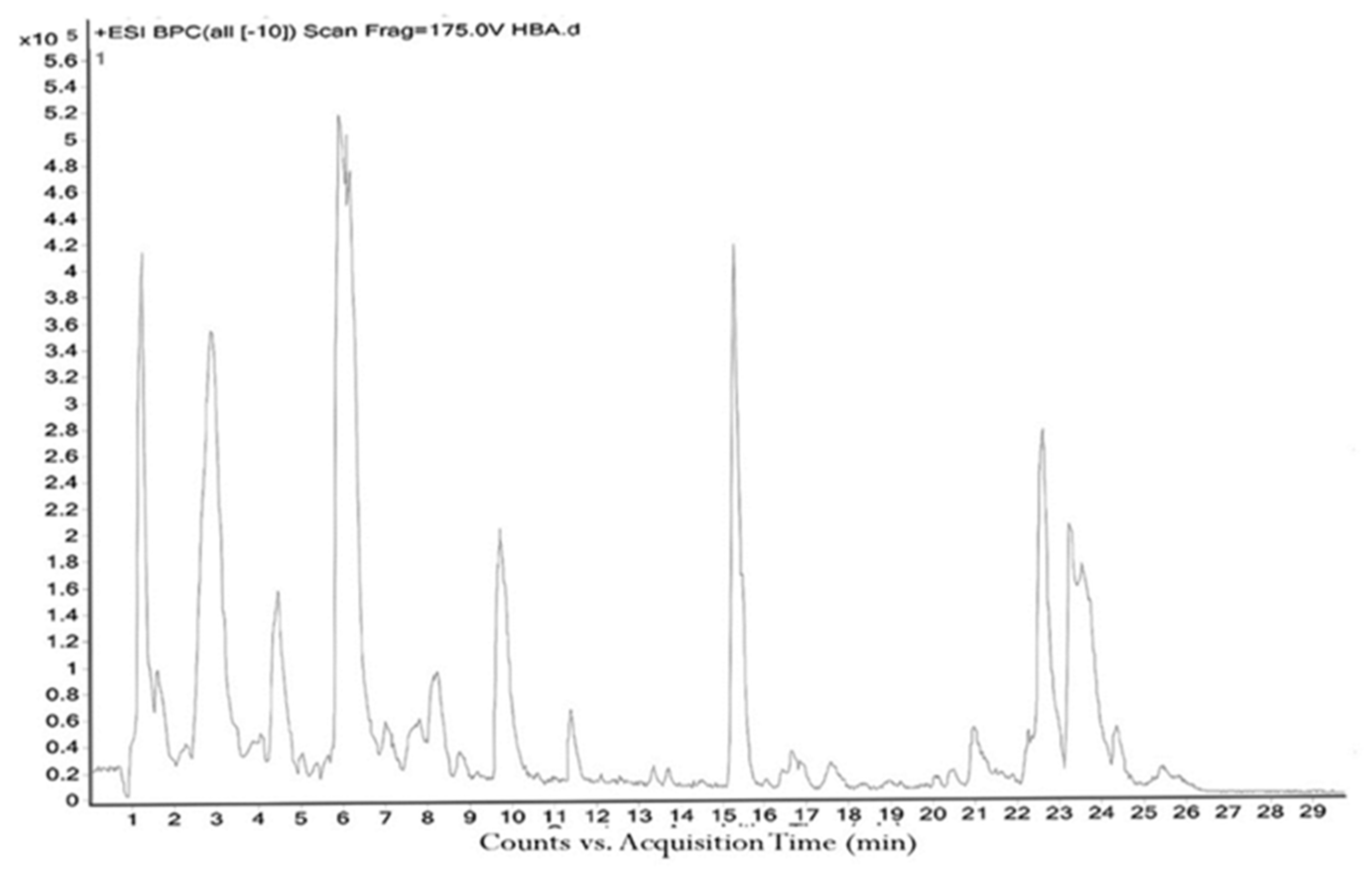
2.1. Quantitative and Qualitative Estimation of Phytochemicals in H. sanjappae
2.2. In Vitro Antioxidant Activities of H. sanjappae Extract
2.3. Enzyme Inhibitory Properties of H. sanjappae Ethanol Extract
2.4. Anti-Inflammatory Activity of H. sanjappae
2.5. Antibacterial Activity of H. sanjappae
3. Materials and Methods
3.1. Humboldtia sanjappae Collection and Extraction
3.2. Phytochemical Analysis of Humboldtia sanjappae
3.3. Analysis of the Antioxidant Activity of H. sanjappae Ethanol Extract
3.4. Analysis of the H. sanjappae Ethanol Extract on Activities of Enzymes
3.5. Effect of H. sanjappae Ethanol Extract on Lipopolysaccharide-Induced Anti-Inflammatory Activity in Macrophages
3.6. Antibacterial Activity of H. sanjappae Ethanol Extract
3.7. Statistical Analysis
4. Conclusions
Supplementary Materials
Author Contributions
Funding
Institutional Review Board Statement
Informed Consent Statement
Data Availability Statement
Acknowledgments
Conflicts of Interest
Sample Availability
References
- Kumar, V.; Bishayee, K.; Park, S.; Lee, U.; Kim, J. Oxidative stress in cerebrovascular disease and associated diseases. Front. Endocrinol. 2023, 14, 1124419. [Google Scholar] [CrossRef]
- Jia, D.; Nagaoka, Y.; Katsumata, M.; Orsulic, S. Inflammation is a key contributor to ovarian cancer cell seeding. Sci. Rep. 2018, 8, 12394. [Google Scholar] [CrossRef]
- Hausmann, S.; Kong, B.; Michalski, C.; Erkan, M.; Friess, H. The role of inflammation in pancreatic cancer. Inflamm. Cancer 2014, 816, 129–151. [Google Scholar]
- Mastinu, A.; Bonini, S.A.; Premoli, M.; Maccarinelli, G.; Mac Sweeney, E.; Zhang, L.; Lucini, L.; Memo, M. Protective Effects of Gynostemma pentaphyllum (var. Ginpent) against Lipopolysaccharide-Induced Inflammation and Motor Alteration in Mice. Molecules 2021, 26, 570. [Google Scholar] [CrossRef] [PubMed]
- Rahman, S.; Atikullah, M.; Islam, M.N.; Mohaimenul, M.; Ahammad, F.; Islam, M.S.; Saha, B.; Rahman, H. Anti-inflammatory, antinociceptive and antidiarrhoeal activities of methanol and ethyl acetate extract of Hemigraphis alternata leaves in mice. Clin. Phytoscience 2019, 5, 16. [Google Scholar] [CrossRef]
- Golia, E.; Limongelli, G.; Natale, F.; Fimiani, F.; Maddaloni, V.; Pariggiano, I.; Bianchi, R.; Crisci, M.; D’Acierno, L.; Giordano, R. Inflammation and cardiovascular disease: From pathogenesis to therapeutic target. Curr. Atheroscler. Rep. 2014, 16, 435. [Google Scholar] [CrossRef]
- Iqbal, J.; Abbasi, B.A.; Mahmood, T.; Kanwal, S.; Ali, B.; Shah, S.A.; Khalil, A.T. Plant-derived anticancer agents: A green anticancer approach. Asian Pac. J. Trop. Biomed. 2017, 7, 1129–1150. [Google Scholar] [CrossRef]
- Kasote, D.M.; Katyare, S.S.; Hegde, M.V.; Bae, H. Significance of antioxidant potential of plants and its relevance to therapeutic applications. Int. J. Biol. Sci. 2015, 11, 982. [Google Scholar] [CrossRef]
- Oguntibeju, O.O. Medicinal plants with anti-inflammatory activities from selected countries and regions of Africa. J. Inflamm. Res. 2018, 11, 307. [Google Scholar] [CrossRef] [PubMed]
- Coulibaly, A.Y.; Hashim, R.; Sulaiman, S.F.; Sulaiman, O.; Ang, L.Z.P.; Ooi, K.L. Bioprospecting medicinal plants for antioxidant components. Asian Pac. J. Trop. Med. 2014, 7, S553–S559. [Google Scholar] [CrossRef] [PubMed]
- Okach, D.; Nyunja, A.; Opande, G. Phytochemical screening of some wild plants from Lamiaceae and their role in traditional medicine in Uriri District-Kenya. Int. J. Herb. Med. 2013, 1, 135–143. [Google Scholar]
- Liu, C.H.; Abrams, N.D.; Carrick, D.M.; Chander, P.; Dwyer, J.; Hamlet, M.R.; Macchiarini, F.; PrabhuDas, M.; Shen, G.L.; Tandon, P. Biomarkers of chronic inflammation in disease development and prevention: Challenges and opportunities. Nat. Immunol. 2017, 18, 1175–1180. [Google Scholar] [CrossRef]
- Nunes, C.d.R.; Barreto Arantes, M.; Menezes de Faria Pereira, S.; Leandro da Cruz, L.; de Souza Passos, M.; Pereira de Moraes, L.; Vieira, I.J.C.; Barros de Oliveira, D. Plants as sources of anti-inflammatory agents. Molecules 2020, 25, 3726. [Google Scholar] [CrossRef] [PubMed]
- Zammel, N.; Saeed, M.; Bouali, N.; Elkahoui, S.; Alam, J.M.; Rebai, T.; Kausar, M.A.; Adnan, M.; Siddiqui, A.J.; Badraoui, R. Antioxidant and anti-Inflammatory effects of Zingiber officinale roscoe and Allium subhirsutum: In silico, biochemical and histological Study. Foods 2021, 10, 1383. [Google Scholar] [CrossRef] [PubMed]
- Apaza Ticona, L.; Pérez-Uz, B.; García Esteban, M.T.; Aguilar Rico, F.; Slowing, K. Anti-melanogenic and Anti-inflammatory Activities of Hibiscus sabdariffa. Rev. Bras. De Farmacogn. 2022, 32, 127–132. [Google Scholar] [CrossRef]
- Ďuračková, Z. Some current insights into oxidative stress. Physiol. Res. 2010, 59, 459–469. [Google Scholar] [CrossRef]
- Rodriguez-Garcia, I.; Silva-Espinoza, B.A.; Ortega-Ramirez, L.A.; Leyva, J.M.; Siddiqui, M.W.; Cruz-Valenzuela, M.R.; Gonzalez-Aguilar, G.A.; Ayala-Zavala, J.F. Oregano Essential Oil as an Antimicrobial and Antioxidant Additive in Food Products. Crit. Rev. Food Sci. Nutr. 2016, 56, 1717–1727. [Google Scholar] [CrossRef]
- Mhatre, S.; Srivastava, T.; Naik, S.; Patravale, V. Antiviral activity of green tea and black tea polyphenols in prophylaxis and treatment of COVID-19: A review. Phytomedicine 2021, 85, 153286. [Google Scholar] [CrossRef]
- Alagumanivasagam, G.; Veeramani, P. A review on medicinal plants with hypolipidemic activity. Int. J. Pharm. Anal. Res. 2015, 4, 129–134. [Google Scholar]
- Fu, W.; Zhuang, W.; Zhou, S.; Wang, X. Plant-derived neuroprotective agents in Parkinson’s disease. Am. J. Transl. Res. 2015, 7, 1189. [Google Scholar]
- Shamsudin, N.F.; Ahmed, Q.U.; Mahmood, S.; Shah, S.A.A.; Sarian, M.N.; Khattak, M.M.A.K.; Khatib, A.; Sabere, A.S.M.; Yusoff, Y.M.; Latip, J. Flavonoids as Antidiabetic and Anti-Inflammatory Agents: A Review on Structural Activity Relationship-Based Studies and Meta-Analysis. Int. J. Mol. Sci. 2022, 23, 12605. [Google Scholar] [CrossRef]
- Gangaram, S.; Naidoo, Y.; Dewir, Y.H.; El-Hendawy, S. Phytochemicals and Biological Activities of Barleria (Acanthaceae). Plants 2021, 11, 82. [Google Scholar] [CrossRef]
- Olivia, N.U.; Goodness, U.C.; Obinna, O.M. Phytochemical profiling and GC-MS analysis of aqueous methanol fraction of Hibiscus asper leaves. Future J. Pharm. Sci. 2021, 7, 59. [Google Scholar] [CrossRef]
- Njamen, D.; Djiogue, S.; Zingue, S.; Mvondo, M.A.; Nkeh-Chungag, B.N. In vivo and in vitro estrogenic activity of extracts from Erythrina poeppigiana (Fabaceae). J. Complement. Integr. Med. 2013, 10, 63–73. [Google Scholar] [CrossRef] [PubMed]
- Nair, R.V.; Jayasree, D.V.; Biju, P.G.; Baby, S. Anti-inflammatory and anticancer activities of erythrodiol-3-acetate and 2, 4-di-tert-butylphenol isolated from Humboldtia unijuga. Nat. Prod. Res. 2018, 34, 2319–2322. [Google Scholar] [CrossRef]
- Kumar, J.K.; Prasad, A.D.; Chaturvedi, V. Phytochemical screening of five medicinal legumes and their evaluation for in vitro anti-tubercular activity. Ayu 2014, 35, 98. [Google Scholar]
- Pavithra, G.; Naik, A.S.; Siddiqua, S.; Vinayaka, K.; TR, P.K.; Mukunda, S. Antioxidant and antibacterial activity of flowers of Calycopteris floribunda (Roxb.) Poiret, Humboldtia brunonis Wall and Kydia calycina Roxb. Int. J. Drug Dev. Res. 2013, 5, 301–310. [Google Scholar]
- Sindhu, S.; Manorama, S.; Sumathi, P.; Adira, S. Antimicrobial studies on the endemic medicinal plant Humboldtia brunonis wall. (Caesalpiniaceae). Int. J. Pharmaceut. Sci. Health Care 2014, 4. [Google Scholar]
- Asirvatham, R.; Yesudanam, S. Neuropharmacological study of Humboldtia vahliana Wight. Sch. Acad. J. Pharm. 2018, 7, 171–183. [Google Scholar]
- Sanjappa, M. A revision of the genus Humboldtia Vahl (Leguminosae-Caesalpinioideae). Blumea Biodivers. Evol. Biogeogr. Plants 1986, 31, 329–339. [Google Scholar]
- Asirvatham, R.; Yesudanam, S. Evaluation of antioxidant potential of Humboldtia Vahliana Wight in Neuropharmacological screening on mice. J. Int. Res. Med. Pharm. Sci. 2017, 11, 41–53. [Google Scholar]
- John, B.; Sulaiman, C.; George, S.; Reddy, V. Total phenolics and flavonoids in selected medicinal plants from Kerala. Int. J. Pharm. Pharm. Sci. 2014, 6, 406–408. [Google Scholar]
- Sheik, S.; Chandrashekar, K. Antimicrobial and antioxidant activities of Kingiodendron pinnatum (DC.) Harms and Humboldtia brunonis Wallich: Endemic plants of the Western Ghats of India. J. Natl. Sci. Found. Sri Lanka 2014, 42, 307. [Google Scholar] [CrossRef]
- Nisbet, L.J.; Moore, M. Will natural products remain an important source of drug research for the future? Curr. Opin. Biotechnol. 1997, 8, 708–712. [Google Scholar] [CrossRef] [PubMed]
- Nagabhushan, R.K.; Raveesha, A. Ethnobotanical survey and scientific validation of medicinal plants used in the treatment of fungal infections in Agumbe region of Western Ghats, India. Int. J. Pharm. Pharm. Sci. 2015, 7, 273–277. [Google Scholar]
- Vijayan, A.; Liju, V.B.; John, R.J.V.; Parthipan, B.; Renuka, C. Traditional Remedies of Kani Tribes of Kottoor Reserve Forest, Agasthyavanam, Thiruvananthapuram, Kerala; CSIR: New Delhi, India, 2007. [Google Scholar]
- Al-Khayri, J.M.; Sahana, G.R.; Nagella, P.; Joseph, B.V.; Alessa, F.M.; Al-Mssallem, M.Q. Flavonoids as potential anti-inflammatory molecules: A review. Molecules 2022, 27, 2901. [Google Scholar] [CrossRef]
- Ghannadi, A.; Hajhashemi, V.; Jafarabadi, H. An investigation of the analgesic and anti-inflammatory effects of Nigella sativa seed polyphenols. J. Med. Food 2005, 8, 488–493. [Google Scholar] [CrossRef]
- Li, X.-W.; Chen, H.-P.; He, Y.-Y.; Chen, W.-L.; Chen, J.-W.; Gao, L.; Hu, H.-Y.; Wang, J. Effects of Rich-Polyphenols Extract of Dendrobium loddigesii on Anti-Diabetic, Anti-Inflammatory, Anti-Oxidant, and Gut Microbiota Modulation in db/db Mice. Molecules 2018, 23, 3245. [Google Scholar] [CrossRef]
- Ginwala, R.; Bhavsar, R.; Chigbu, D.G.I.; Jain, P.; Khan, Z.K. Potential Role of Flavonoids in Treating Chronic Inflammatory Diseases with a Special Focus on the Anti-Inflammatory Activity of Apigenin. Antioxidants 2019, 8, 35. [Google Scholar] [CrossRef]
- Bettaieb, A.; Cremonini, E.; Kang, H.; Kang, J.; Haj, F.G.; Oteiza, P.I. Anti-inflammatory actions of (−)-epicatechin in the adipose tissue of obese mice. Int. J. Biochem. Cell Biol. 2016, 81, 383–392. [Google Scholar] [CrossRef]
- Reagan, L.P.; Magarinos, A.M.; McEWEN, B.S. Neurological changes induced by stress in streptozotocin diabetic rats. Ann. N. Y. Acad. Sci. 1999, 893, 126–137. [Google Scholar] [CrossRef] [PubMed]
- Freitas, L.M.; Antunes, F.T.T.; Obach, E.S.; Correa, A.P.; Wiiland, E.; de Mello Feliciano, L.; Reinicke, A.; Amado, G.J.V.; Grivicich, I.; Fialho, M.F.P. Anti-inflammatory effects of a topical emulsion containing Helianthus annuus oil, glycerin, and vitamin B3 in mice. J. Pharm. Investig. 2021, 51, 223–232. [Google Scholar] [CrossRef]
- Ou, Z.; Zhao, J.; Zhu, L.; Huang, L.; Ma, Y.; Ma, C.; Luo, C.; Zhu, Z.; Yuan, Z.; Wu, J. Anti-inflammatory effect and potential mechanism of betulinic acid on λ-carrageenan-induced paw edema in mice. Biomed. Pharmacother. 2019, 118, 109347. [Google Scholar] [CrossRef] [PubMed]
- Kwon, Y.-I.I.; Vattem, D.A.; Shetty, K. Evaluation of clonal herbs of Lamiaceae species for management of diabetes and hypertension. Asia Pac. J. Clin. Nutr. 2006, 15, 107. [Google Scholar] [PubMed]
- Shai, L.; Magano, S.; Lebelo, S.; Mogale, A. Inhibitory effects of five medicinal plants on rat alpha-glucosidase: Comparison with their effects on yeast alpha-glucosidase. J. Med. Plants Res. 2011, 5, 2863–2867. [Google Scholar]
- Matu, E.N.; Van Staden, J. Antibacterial and anti-inflammatory activities of some plants used for medicinal purposes in Kenya. J. Ethnopharmacol. 2003, 87, 35–41. [Google Scholar] [CrossRef]
- Frieri, M.; Kumar, K.; Boutin, A. Antibiotic resistance. J. Infect. Public Health 2017, 10, 369–378. [Google Scholar] [CrossRef]
- MacGowan, A.; Macnaughton, E. Antibiotic resistance. Medicine 2017, 45, 622–628. [Google Scholar] [CrossRef]
- Alibi, S.; Crespo, D.; Navas, J. Plant-derivatives small molecules with antibacterial activity. Antibiotics 2021, 10, 231. [Google Scholar] [CrossRef]
- Guimarães, A.C.; Meireles, L.M.; Lemos, M.F.; Guimarães, M.C.C.; Endringer, D.C.; Fronza, M.; Scherer, R. Antibacterial activity of terpenes and terpenoids present in essential oils. Molecules 2019, 24, 2471. [Google Scholar] [CrossRef]
- Simirgiotis, M.J.; Burton, D.; Parra, F.; López, J.; Muñoz, P.; Escobar, H.; Parra, C. Antioxidant and antibacterial capacities of Origanum vulgare L. essential oil from the arid Andean Region of Chile and its chemical characterization by GC-MS. Metabolites 2020, 10, 414. [Google Scholar] [CrossRef]
- El Moussaoui, A.; Jawhari, F.Z.; Almehdi, A.M.; Elmsellem, H.; Benbrahim, K.F.; Bousta, D.; Bari, A. Antibacterial, antifungal and antioxidant activity of total polyphenols of Withania frutescens L. Bioorganic Chem. 2019, 93, 103337. [Google Scholar] [CrossRef]
- Reglodi, D.; Renaud, J.; Tamas, A.; Tizabi, Y.; Socías, S.B.; Del-Bel, E.; Raisman-Vozari, R. Novel tactics for neuroprotection in Parkinson’s disease: Role of antibiotics, polyphenols and neuropeptides. Prog. Neurobiol. 2017, 155, 120–148. [Google Scholar] [CrossRef]
- Ramata-Stunda, A.; Petriņa, Z.; Valkovska, V.; Borodušķis, M.; Gibnere, L.; Gurkovska, E.; Nikolajeva, V. Synergistic effect of polyphenol-rich complex of plant and green propolis extracts with antibiotics against respiratory infections causing bacteria. Antibiotics 2022, 11, 160. [Google Scholar] [CrossRef]
- Haghjoo, B.; Lee, L.H.; Habiba, U.; Tahir, H.; Olabi, M.; Chu, T.-C. The synergistic effects of green tea polyphenols and antibiotics against potential pathogens. Adv. Biosci. Biotechnol. 2013, 4, 959. [Google Scholar] [CrossRef]
- Usman, M.; Khan, W.R.; Yousaf, N.; Akram, S.; Murtaza, G.; Kudus, K.A.; Ditta, A.; Rosli, Z.; Rajpar, M.N.; Nazre, M. Exploring the phytochemicals and anti-cancer potential of the members of Fabaceae family: A comprehensive review. Molecules 2022, 27, 3863. [Google Scholar] [CrossRef]
- Zonyane, S.; Fawole, O.A.; La Grange, C.; Stander, M.A.; Opara, U.L.; Makunga, N.P. The implication of chemotypic variation on the anti-oxidant and anti-cancer activities of Sutherlandia frutescens (L.) R.Br.(Fabaceae) from different geographic locations. Antioxidants 2020, 9, 152. [Google Scholar] [CrossRef]
- Borquaye, L.S.; Doetse, M.S.; Baah, S.O.; Mensah, J.A. Anti-inflammatory and anti-oxidant activities of ethanolic extracts of Tamarindus indica L. (Fabaceae). Cogent Chem. 2020, 6, 1743403. [Google Scholar] [CrossRef]
- Abdulkhaleq, L.A.; Assi, M.A.; Noor, M.H.M.; Abdullah, R.; Saad, M.Z.; Taufiq-Yap, Y.H. Therapeutic uses of epicatechin in diabetes and cancer. Vet. World 2017, 10, 869–872. [Google Scholar] [CrossRef] [PubMed]
- Dong, H.; Yang, X.; He, J.; Cai, S.; Xiao, K.; Zhu, L. Enhanced antioxidant activity, antibacterial activity and hypoglycemic effect of luteolin by complexation with manganese (II) and its inhibition kinetics on xanthine oxidase. RSC Adv. 2017, 7, 53385–53395. [Google Scholar] [CrossRef]
- Alshehri, S.; Imam, S.S.; Altamimi, M.A.; Hussain, A.; Shakeel, F.; Elzayat, E.; Mohsin, K.; Ibrahim, M.; Alanazi, F. Enhanced dissolution of luteolin by solid dispersion prepared by different methods: Physicochemical characterization and antioxidant activity. ACS Omega 2020, 5, 6461–6471. [Google Scholar] [CrossRef] [PubMed]
- Kim, N.M.; Kim, J.; Chung, H.Y.; Choi, J.S. Isolation of luteolin 7-O-rutinoside and esculetin with potential antioxidant activity from the aerial parts of Artemisia montana. Arch. Pharmacal Res. 2000, 23, 237–239. [Google Scholar] [CrossRef] [PubMed]
- Xu, H.; Linn, B.S.; Zhang, Y.; Ren, J. A review on the antioxidative and prooxidative properties of luteolin. React. Oxyg. Species 2019, 7, 136–147. [Google Scholar] [CrossRef]
- Guo, Y.; Liu, Y.; Zhang, Z.; Chen, M.; Zhang, D.; Tian, C.; Liu, M.; Jiang, G. The antibacterial activity and mechanism of action of luteolin against Trueperella pyogenes. Infect. Drug Resist. 2020, 13, 1697–1711. [Google Scholar] [CrossRef]
- Çetinkaya, M.; Baran, Y. Therapeutic Potential of Luteolin on Cancer. Vaccines 2023, 11, 554. [Google Scholar] [CrossRef]
- Potočnjak, I.; Šimić, L.; Gobin, I.; Vukelić, I.; Domitrović, R. Antitumor activity of luteolin in human colon cancer SW620 cells is mediated by the ERK/FOXO3a signaling pathway. Toxicol. Vitr. 2020, 66, 104852. [Google Scholar] [CrossRef]
- Cavia-Saiz, M.; Busto, M.D.; Pilar-Izquierdo, M.C.; Ortega, N.; Perez-Mateos, M.; Muñiz, P. Antioxidant properties, radical scavenging activity and biomolecule protection capacity of flavonoid naringenin and its glycoside naringin: A comparative study. J. Sci. Food Agric. 2010, 90, 1238–1244. [Google Scholar] [CrossRef]
- Patel, K.; Singh, G.K.; Patel, D.K. A review on pharmacological and analytical aspects of naringenin. Chin. J. Integr. Med. 2018, 24, 551–560. [Google Scholar] [CrossRef]
- Ismail, N.H.; Mohamad, H.; Mohidin, A.; Lajis, N.H. Antioxidant activity of anthraquinones from Morinda elliptica. Nat. Prod. Sci. 2002, 8, 48–51. [Google Scholar]
- Chee, C.W.; Zamakshshari, N.H.; Lee, V.S.; Abdullah, I.; Othman, R.; Lee, Y.K.; Hashim, N.M.; Rashid, N.N. Morindone from Morinda citrifolia as a potential antiproliferative agent against colorectal cancer cell lines. PLoS ONE 2022, 17, e0270970. [Google Scholar] [CrossRef]
- Guil-Guerrero, J.; Martínez-Guirado, C.; del Mar Rebolloso-Fuentes, M.; Carrique-Pérez, A. Nutrient composition and antioxidant activity of 10 pepper (Capsicum annuun) varieties. Eur. Food Res. Technol. 2006, 224, 1–9. [Google Scholar] [CrossRef]
- Sun, T.; Xu, Z.; Wu, C.T.; Janes, M.; Prinyawiwatkul, W.; No, H. Antioxidant activities of different colored sweet bell peppers (Capsicum annuum L.). J. Food Sci. 2007, 72, S98–S102. [Google Scholar] [CrossRef] [PubMed]
- Ahmad, M.F.; Wahab, S.; Ahmad, F.A.; Ashraf, S.A.; Abullais, S.S.; Saad, H.H. Ganoderma lucidum: A potential pleiotropic approach of ganoderic acids in health reinforcement and factors influencing their production. Fungal Biol. Rev. 2022, 39, 100–125. [Google Scholar] [CrossRef]
- Dubois-Deruy, E.; Peugnet, V.; Turkieh, A.; Pinet, F. Oxidative stress in cardiovascular diseases. Antioxidants 2020, 9, 864. [Google Scholar] [CrossRef] [PubMed]
- Ndrepepa, G. Myeloperoxidase–A bridge linking inflammation and oxidative stress with cardiovascular disease. Clin. Chim. Acta 2019, 493, 36–51. [Google Scholar] [CrossRef] [PubMed]
- Hayes, J.D.; Dinkova-Kostova, A.T.; Tew, K.D. Oxidative stress in cancer. Cancer Cell 2020, 38, 167–197. [Google Scholar] [CrossRef]
- Yaribeygi, H.; Sathyapalan, T.; Atkin, S.L.; Sahebkar, A. Molecular mechanisms linking oxidative stress and diabetes mellitus. Oxidative Med. Cell. Longev. 2020, 2020, 8609213. [Google Scholar] [CrossRef]
- Alqahtani, A.S.; Hidayathulla, S.; Rehman, M.T.; ElGamal, A.A.; Al-Massarani, S.; Razmovski-Naumovski, V.; Alqahtani, M.S.; El Dib, R.A.; AlAjmi, M.F. Alpha-Amylase and Alpha-Glucosidase Enzyme Inhibition and Antioxidant Potential of 3-Oxolupenal and Katononic Acid Isolated from Nuxia oppositifolia. Biomolecules 2019, 10, 61. [Google Scholar] [CrossRef]
- Semaan, D.G.; Igoli, J.O.; Young, L.; Marrero, E.; Gray, A.I.; Rowan, E.G. In vitro anti-diabetic activity of flavonoids and pheophytins from Allophylus cominia Sw. on PTP1B, DPPIV, alpha-glucosidase and alpha-amylase enzymes. J. Ethnopharmacol. 2017, 203, 39–46. [Google Scholar] [CrossRef]
- Feunaing, R.T.; Tamfu, A.N.; Gbaweng, A.J.Y.; Mekontso Magnibou, L.; Ntchapda, F.; Henoumont, C.; Laurent, S.; Talla, E.; Dinica, R.M. In vitro Evaluation of α-amylase and α-glucosidase Inhibition of 2,3-Epoxyprocyanidin C1 and Other Constituents from Pterocarpus erinaceus Poir. Molecules 2022, 28, 126. [Google Scholar] [CrossRef]
- Facchin, B.M.; dos Reis, G.O.; Vieira, G.N.; Mohr, E.T.B.; da Rosa, J.S.; Kretzer, I.F.; Demarchi, I.G.; Dalmarco, E.M. Inflammatory biomarkers on an LPS-induced RAW 264.7 cell model: A systematic review and meta-analysis. Inflamm. Res. 2022, 71, 741–758. [Google Scholar] [CrossRef] [PubMed]
- Hoppstädter, J.; Dembek, A.; Linnenberger, R.; Dahlem, C.; Barghash, A.; Fecher-Trost, C.; Fuhrmann, G.; Koch, M.; Kraegeloh, A.; Huwer, H.; et al. Toll-Like Receptor 2 Release by Macrophages: An Anti-inflammatory Program Induced by Glucocorticoids and Lipopolysaccharide. Front. Immunol. 2019, 10, 1634. [Google Scholar] [CrossRef] [PubMed]
- Kaneko, N.; Kurata, M.; Yamamoto, T.; Morikawa, S.; Masumoto, J. The role of interleukin-1 in general pathology. Inflamm. Regen. 2019, 39, 12. [Google Scholar] [CrossRef]
- Pyrillou, K.; Burzynski, L.C.; Clarke, M.C.H. Alternative Pathways of IL-1 Activation, and Its Role in Health and Disease. Front. Immunol. 2020, 11, 613170. [Google Scholar] [CrossRef] [PubMed]
- Bent, R.; Moll, L.; Grabbe, S.; Bros, M. Interleukin-1 Beta—A Friend or Foe in Malignancies? Int. J. Mol. Sci. 2018, 19, 2155. [Google Scholar] [CrossRef]
- Chen, J.; Wang, W.; Ni, Q.; Zhang, L.; Guo, X. Interleukin 6-regulated macrophage polarization controls atherosclerosis-associated vascular intimal hyperplasia. Front. Immunol. 2022, 13, 952164. [Google Scholar] [CrossRef]
- Hirani, D.; Alvira, C.M.; Danopoulos, S.; Milla, C.; Donato, M.; Tian, L.; Mohr, J.; Dinger, K.; Vohlen, C.; Selle, J.; et al. Macrophage-derived IL-6 trans-signalling as a novel target in the pathogenesis of bronchopulmonary dysplasia. Eur. Respir. J. 2022, 59, 2002248. [Google Scholar] [CrossRef] [PubMed]
- Rose-John, S.; Jenkins, B.J.; Garbers, C.; Moll, J.M.; Scheller, J. Targeting IL-6 trans-signalling: Past, present and future prospects. Nat. Rev. Immunol. 2023, 23, 666–681. [Google Scholar] [CrossRef]
- Chen, J.; Wei, Y.; Yang, W.; Huang, Q.; Chen, Y.; Zeng, K.; Chen, J. IL-6: The Link Between Inflammation, Immunity and Breast Cancer. Front. Oncol. 2022, 12, 903800. [Google Scholar] [CrossRef]
- Rašková, M.; Lacina, L.; Kejík, Z.; Venhauerová, A.; Skaličková, M.; Kolář, M.; Jakubek, M.; Rosel, D.; Smetana, K., Jr.; Brábek, J. The Role of IL-6 in Cancer Cell Invasiveness and Metastasis-Overview and Therapeutic Opportunities. Cells 2022, 11, 3698. [Google Scholar] [CrossRef]
- Al Obeed, O.A.; Alkhayal, K.A.; Al Sheikh, A.; Zubaidi, A.M.; Vaali-Mohammed, M.A.; Boushey, R.; McKerrow, J.H.; Abdulla, M.H. Increased expression of tumor necrosis factor-α is associated with advanced colorectal cancer stages. World J. Gastroenterol. 2014, 20, 18390–18396. [Google Scholar] [CrossRef]
- Zhao, P.; Zhang, Z. TNF-α promotes colon cancer cell migration and invasion by upregulating TROP-2. Oncol. Lett. 2018, 15, 3820–3827. [Google Scholar] [CrossRef] [PubMed]
- Król, M.; Kepinska, M. Human Nitric Oxide Synthase—Its Functions, Polymorphisms, and Inhibitors in the Context of Inflammation, Diabetes and Cardiovascular Diseases. Int. J. Mol. Sci. 2021, 22, 56. [Google Scholar] [CrossRef] [PubMed]
- Iwata, M.; Inoue, T.; Asai, Y.; Hori, K.; Fujiwara, M.; Matsuo, S.; Tsuchida, W.; Suzuki, S. The protective role of localized nitric oxide production during inflammation may be mediated by the heme oxygenase-1/carbon monoxide pathway. Biochem. Biophys. Rep. 2020, 23, 100790. [Google Scholar] [CrossRef] [PubMed]
- Liu, D.; Guo, Y.; Wu, P.; Wang, Y.; Golly, M.K.; Ma, H. The necessity of walnut proteolysis based on evaluation after in vitro simulated digestion: ACE inhibition and DPPH radical-scavenging activities. Food Chem. 2020, 311, 125960. [Google Scholar] [CrossRef]
- Lai, S.-C.; Ho, Y.-L.; Huang, S.-C.; Huang, T.-H.; Lai, Z.-R.; Wu, C.-R.; Lian, K.-Y.; Chang, Y.-S. Antioxidant and antiproliferative activities of Desmodium triflorum (L.) DC. Am. J. Chin. Med. 2010, 38, 329–342. [Google Scholar] [CrossRef]
- Konaté, K.; Souza, A.; Coulibaly, A.; Meda, N.; Kiendrebeogo, M.; Lamien-Meda, A.; Millogo-Rasolodimby, J.; Lamidi, M.; Nacoulma, O. In vitro antioxidant, lipoxygenase and xanthine oxidase inhibitory activities of fractions from Cienfuegosia digitata Cav. Sida alba L. and Sida acuta Burn f.(Malvaceae). Pak. J. Biol. Sci. PJBS 2010, 13, 1092–1098. [Google Scholar]
- Tanaka, T.; Narazaki, M.; Kishimoto, T. IL-6 in inflammation, immunity, and disease. Cold Spring Harb. Perspect. Biol. 2014, 6, a016295. [Google Scholar] [CrossRef]
- Samaraweera, U.; Sotheeswaran, S.; Uvais, M.; Sultanbawa, S. 3,5,7,3′, 5′-Pentahydroxyflavan and 3α-methoxyfriedelan from Humboldtia laurifolia. Phytochemistry 1983, 22, 565–567. [Google Scholar] [CrossRef]
- Dyamavvanahalli, S.L.; Raveesha, K.A.; Nagabhushan, S. Bioprospecting of selected medicinal plants for antibacterial activity against some pathogenic bacteria. J. Med. Plants Res. 2011, 5, 4087–4093. [Google Scholar]
- Yadav, R.; Agarwala, M. Phytochemical analysis of some medicinal plants. J. Phytol. 2011, 3, 10–14. [Google Scholar]
- Harborne, A. Phytochemical Methods a Guide to Modern Techniques of Plant Analysis; Springer Science & Business Media: London, UK, 1998. [Google Scholar]
- Singleton, V.L.; Rossi, J.A. Colorimetry of total phenolics with phosphomolybdic-phosphotungstic acid reagents. Am. J. Enol. Vitic. 1965, 16, 144–158. [Google Scholar] [CrossRef]
- Zhishen, J.; Mengcheng, T.; Jianming, W. The determination of flavonoid contents in mulberry and their scavenging effects on superoxide radicals. Food Chem. 1999, 64, 555–559. [Google Scholar] [CrossRef]
- House, N.C.; Puthenparampil, D.; Malayil, D.; Narayanankutty, A. Variation in the polyphenol composition, antioxidant, and anticancer activity among different Amaranthus species. S. Afr. J. Bot. 2020, 135, 408–412. [Google Scholar] [CrossRef]
- Webber, D.M.; Wallace, M.A.; Burnham, C.D. Stop Waiting for Tomorrow: Disk Diffusion Performed on Early Growth Is an Accurate Method for Antimicrobial Susceptibility Testing with Reduced Turnaround Time. J. Clin. Microbiol. 2022, 60, e0300720. [Google Scholar] [CrossRef]
- Morgan, B.L.; Depenbrock, S.; Martínez-López, B. Identifying Associations in Minimum Inhibitory Concentration Values of Escherichia coli Samples Obtained From Weaned Dairy Heifers in California Using Bayesian Network Analysis. Front. Vet. Sci. 2022, 9, 771841. [Google Scholar] [CrossRef] [PubMed]
- Di Simone, S.C.; Acquaviva, A.; Libero, M.L.; Chiavaroli, A.; Recinella, L.; Leone, S.; Brunetti, L.; Politi, M.; Giannone, C.; Campana, C.; et al. The association of Tanacetum parthenium and Salix alba extracts reduces cortex serotonin turnover, in an ex vivo experimental model of migraine. Processes 2022, 10, 280. [Google Scholar] [CrossRef]
- Iskandar, N.N.; Iriawati, I. Vinblastine and Vincristine production on Madagascar Periwinkle (Catharanthus roseus (L.) G. Don) callus culture treated with polethylene glycol. Makara J. Sci. 2016, 20, 7–16. [Google Scholar] [CrossRef]
- Rao, P.; Knaus, E.E. Evolution of nonsteroidal anti-inflammatory drugs (NSAIDs): Cyclooxygenase (COX) inhibition and beyond. J. Pharm. Pharm. Sci. 2008, 11, 81s–110s. [Google Scholar] [CrossRef]
- Khumalo, G.P.; Van Wyk, B.E.; Feng, Y.; Cock, I.E. A review of the traditional use of southern African medicinal plants for the treatment of inflammation and inflammatory pain. J. Ethnopharmacol. 2022, 283, 114436. [Google Scholar] [CrossRef] [PubMed]
- Bharadwaj, K.C.; Gupta, T.; Singh, R.M. Alkaloid group of Cinchona officinalis: Structural, synthetic, and medicinal aspects. In Synthesis of Medicinal Agents from Plants; Elsevier: Amsterdam, The Netherlands, 2018; pp. 205–227. [Google Scholar]


| Sl. No. | RT | m/z a | m/z b | Name of the Compound | Fragments | Mol. Wt. | Chemical Formula | Structure |
|---|---|---|---|---|---|---|---|---|
| 1 | 3.575 | 577.14 | 577.14 | Richotomine | 483.13, 315.11, 197.80 | 532.14 | C30H20N4O6 | 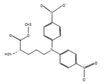 |
| 2 | 3.773 | 579.15 | 579.15 | Procyanidin B7 | 579.14, 443.15, 383.12, 227.17 | 578.14 | C30H26O12 |  |
| 3 | 4.166 | 289.07 | 289.07 | (−)−Epicatechin | 289.07, 226.07 | 290.08 | C15H14O6 |  |
| 4 | 4.633 | 513.14 | 513.14 | 2″,6″−Di-O-acetylononin | 513.14, 289.07 | 514.15 | C26H26O11 | 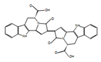 |
| 5 | 5.908 | 494.24 | 494.24 | Ryanodine | 494.23, 189.07 | 493.23 | C25H35NO9 |  |
| 6 | 5.919 | 465.16 | 465.16 | Pomiferin | 421.16, 213.07 | 420.16 | C25H24O6 |  |
| 7 | 6.312 | 549.22 | 549.22 | Cymorcin diglucoside | 431.10, 253.03 | 490.21 | C22H34O12 |  |
| 8 | 8.763 | 287.05 | 287.06 | Maritimetin | 287.05, 283.15, 267.15 | 286.05 | C15H10O6 |  |
| 9 | 9.101 | 285.04 | 285.04 | Luteolin | 285.04, 215.09 | 286.05 | C15H10O6 |  |
| 10 | 9.475 | 271.06 | 271.06 | Naringenin | 271.06, 259.12, 248.97 | 272.07 | C15H12O5 |  |
| 11 | 9.681 | 271.06 | 271.06 | Coriandrone E | 251.16, 179.10 | 248.07 | C13H12O5 |  |
| 12 | 10.067 | 271.06 | 271.06 | Morindone | 271.05, 267.15, 253.14 | 270.05 | C15H10O5 |  |
| 13 | 10.197 | 301.07 | 301.07 | (+)-Sophorol | 271.05, 301.06, 295.18, 277.17 | 300.06 | C16H12O6 |  |
| 14 | 10.485 | 269.08 | 269.08 | Formononetin | 258.04, 179.10, 139.15 | 268.07 | C16H12O4 |  |
| 15 | 11.846 | 283.19 | 283.19 | Lactapiperanol C | 279.09, 265.17 | 282.18 | C16H26O4 |  |
| 16 | 15.229 | 507.23 | 507.22 | Limonoate | 507.22, 351.25, 238.12 | 506.22 | C26H34O10 |  |
| 17 | 16.573 | 295.23 | 295.23 | 17−Hydroxylinolenic acid | 295.22, 284.32, 277.21 | 294.22 | C18H30O3 |  |
| 18 | 19.676 | 645.42 | 645.42 | Capsanthin 5,6−epoxide | 529.30, 403.26, 238.89 | 600.42 | C40H56O4 |  |
| 19 | 21.644 | 593.27 | 593.27 | Ganoderic acid F | 415.35, 227.17, 570.41 | 570.28 | C32H42O9 |  |
| 20 | 22.179 | 471.35 | 471.35 | delta−Maslinic acid | 471.85, 311.17, 248.97 | 472.36 | C30H48O4 |  |
| 21 | 23.291 | 413.26 | 413.27 | D8’−Merulinic acid A | 391.28, 279.15, 149.02 | 390.28 | C24H38O4 |  |
| Assay | µg Equivalent/mg of Extract |
|---|---|
| Total phenolic content | 378.77 ± 6.62 |
| Total flavonoid content | 204.76 ± 6.10 |
| Activity | IC50 Value(µg/mL) | ||
|---|---|---|---|
| HSE | Ascorbic Acid | Acarbose | |
| DPPH scavenging | 6.53 ± 1.49 | 2.11 ± 0.25 | >200 |
| FRAP value | 8.46 ± 1.38 | 4.15 ± 0.47 | >200 |
| α-amylase | 47.60 ± 0.19 | 33.92 ± 2.54 | 122.18 ± 3.08 |
| α-glucosidase | 32.09 ± 0.54 | 29.85 ± 2.01 | 103.45 ± 2.68 |
| IL-1β (pg/mg Protein) | IL-6 (pg/mg Protein) | TNF-α (pg/mg Protein) | NO (µM/mg Protein) | |
|---|---|---|---|---|
| Untreated | 64.6 ± 1.9 | 133.4 ± 5.8 | 115.2 ± 3.1 | 10.7 ± 0.64 |
| LPS Control | 573.4 ± 4.5 | 628.5 ± 8.2 | 856.0 ± 11.2 | 75.2 ± 2.1 |
| Aspirin (1 mM) | 147.5 ± 5.1 *** | 209.5 ± 9.1 *** | 247.5 ± 6.3 *** | 22.7 ± 1.7 *** |
| HSE 5 µg/mL | 403.7 ± 6.2 ** | 507.1 ± 8.1 * | 715.2 ± 8.8 * | 58.8 ± 3.4 * |
| HSE 10 µg/mL | 298.5 ± 8.4 *** | 388.4 ± 2.8 *** | 602.8 ± 5.2 *** | 42.3 ± 1.9 *** |
| HSE 20 µg/mL | 156.2 ± 3.4 *** | 291.3 ± 6.6 *** | 493.7 ± 6.4 *** | 30.7 ± 2.5 *** |
| Bacteria | Zone of Inhibition (mm) | MIC Concentration (mg/mL) | ||
|---|---|---|---|---|
| HSE | GM (20 µg) | HSE | GM | |
| Escherichia coli | 18.5 ± 0.2 | 21.7 ± 0.5 | 1.50 ± 0.01 | 0.325 |
| Pseudomonas aeruginosa | 24.1 ± 0.3 | 22.1 ± 0.1 | 0.625 ± 0.02 | 0.325 |
| Staphylococcus aureus | 20.6 ± 0.3 | 19.7 ± 0.2 | 1.25 ± 0.04 | 0.625 |
| Salmonella enterica | 22.1 ± 0.1 | 20.5 ± 0.2 | 1.00 ± 0.01 | 0.625 |
Disclaimer/Publisher’s Note: The statements, opinions and data contained in all publications are solely those of the individual author(s) and contributor(s) and not of MDPI and/or the editor(s). MDPI and/or the editor(s) disclaim responsibility for any injury to people or property resulting from any ideas, methods, instructions or products referred to in the content. |
© 2023 by the authors. Licensee MDPI, Basel, Switzerland. This article is an open access article distributed under the terms and conditions of the Creative Commons Attribution (CC BY) license (https://creativecommons.org/licenses/by/4.0/).
Share and Cite
Sidhic, J.; George, S.; Alfarhan, A.; Rajagopal, R.; Olatunji, O.J.; Narayanankutty, A. Phytochemical Composition and Antioxidant and Anti-Inflammatory Activities of Humboldtia sanjappae Sasidh. & Sujanapal, an Endemic Medicinal Plant to the Western Ghats. Molecules 2023, 28, 6875. https://doi.org/10.3390/molecules28196875
Sidhic J, George S, Alfarhan A, Rajagopal R, Olatunji OJ, Narayanankutty A. Phytochemical Composition and Antioxidant and Anti-Inflammatory Activities of Humboldtia sanjappae Sasidh. & Sujanapal, an Endemic Medicinal Plant to the Western Ghats. Molecules. 2023; 28(19):6875. https://doi.org/10.3390/molecules28196875
Chicago/Turabian StyleSidhic, Jameema, Satheesh George, Ahmed Alfarhan, Rajakrishnan Rajagopal, Opeyemi Joshua Olatunji, and Arunaksharan Narayanankutty. 2023. "Phytochemical Composition and Antioxidant and Anti-Inflammatory Activities of Humboldtia sanjappae Sasidh. & Sujanapal, an Endemic Medicinal Plant to the Western Ghats" Molecules 28, no. 19: 6875. https://doi.org/10.3390/molecules28196875