Review

Recent Advances on Small-Molecule Antagonists Targeting TLR7

1
Faculty of Pharmacy, Montpellier University, 34093 Montpellier, France
2
School of Life Sciences, Shanghai Normal University, Shanghai 200234, China
3
Institut des Biomolécules Max Mousseron IBMM, Ecole Nationale Supérieure de Chimie de Montpellier ENSCM, Montpellier University, Centre National de La Recherche Scientifique CNRS, 34093 Montpellier, France
*
Author to whom correspondence should be addressed.
Molecules 2023, 28(2), 634; https://doi.org/10.3390/molecules28020634
Submission received: 28 November 2022 / Revised: 28 December 2022 / Accepted: 30 December 2022 / Published: 7 January 2023

Abstract

:
Toll-like receptor 7 (TLR7) is a class of pattern recognition receptors (PRRs) recognizing the pathogen-associated elements and damage and as such is a major player in the innate immune system. TLR7 triggers the release of pro-inflammatory cytokines or type-I interferons (IFN), which is essential for immunoregulation. Increasing reports also highlight that the abnormal activation of endosomal TLR7 is implicated in various immune-related diseases, carcinogenesis as well as the proliferation of human immunodeficiency virus (HIV). Hence, the design and development of potent and selective TLR7 antagonists based on small molecules or oligonucleotides may offer new tools for the prevention and management of such diseases. In this review, we offer an updated overview of the main structural features and therapeutic potential of small-molecule antagonists of TLR7. Various heterocyclic scaffolds targeting TLR7 binding sites are presented: pyrazoloquinoxaline, quinazoline, purine, imidazopyridine, pyridone, benzanilide, pyrazolopyrimidine/pyridine, benzoxazole, indazole, indole, and quinoline. Additionally, their structure-activity relationships (SAR) studies associated with biological activities and protein binding modes are introduced.

1. Introduction

Toll-like receptors (TLRs) are a major family of prototypical pattern recognition receptors (PPRs) and type I membranous glycoproteins [1]. TLRs recognize pathogen-associated molecular patterns (PAMPs) and damage-associated molecular patterns (DAMPs). TLRs can initiate antimicrobial host defense responses to restrain pathogenic replication in the innate immune system [2,3]. The engagement of PAMP-PRR interaction allows TLRs to activate downstream signaling molecules of host defense responses [4]. To date, ten TLR subtypes (TLRs 1–10) have been identified in mammals. They are expressed in various innate immune cells, including dendritic cells (DCs), macrophages, and B cells as well as other cell types, such as epithelial cells, endothelial cells, and fibroblasts [5,6]. Notably, TLR7 and TLR9 are only expressed in plasmacytoid DCs rather than myeloid DCs [7]. Several nucleotide-sensing intracellular TLRs, including TLR3, TLR7, TLR8, and TLR9, originally synthesized in the endoplasmic reticulum (ER), are finally transferred to the endosomal compartments [8,9].
TLR7 are selectively activated by guanosine and uridine-containing single-stranded RNA (ssRNA) from viruses, bacteria, endogenous RNA, and oligoribonucleotides in the DCs’ endolysosomes [10]. TLR7 also respond to various chemical ligands, such as small heterocyclic molecules. Such recognition promotes the release of pro-inflammatory cytokines, chemokines, and type-I interferons (IFN), which are involved in the up-regulation of inflammatory reactions [11].
TLR7 is a key determinant of the protective immunity, conversely, its dysregulation is linked to the susceptibility of inflammatory diseases, such as lupus, caused by activation of host-origin nucleic-sensing pathways via TLR7 [12]. Therefore, the rational design of antagonist ligands is a primary focus for the management of autoimmune disorders, cancers, virus infection, and other potential TLR7-associated clinical disorders [13,14].
The development of small-molecule antagonists of TLR7 might start with the identification of chemical scaffolds by high-throughput screening (HTS), or chemical switches, that transform an existing agonist to an antagonist [15]. After the determination of the chemical scaffolds, structure-activity relationship (SAR) studies are then applied to optimize their antagonistic activities and might be followed by the co-crystallization of the inhibitor/TLR7 complexes [16]. Biological studies usually use HEK-Blue or HEK293 cell lines, which are engineered to overexpress TLR7 to indirectly report the nuclear factor-κB (NF-κB) translocation to the cell nucleus. Tests, such as secreted embryonic alkaline phosphatase (SEAP)-driven assay, cell proliferation assay, isothermal titration calorimetry (ITC), and immunoblotting, are also used in in vitro validations. A non-selective TLR7 agonist R848 (Resiquimod) can be commonly used as a positive control [17]. The inhibition of TLR7-induced pro-inflammatory cytokines can be measured through a real-time polymerase chain reaction (RT-PCR) [18].

2. TLR7 Main Features

2.1. Structural Studies of TLR7

TLR7 is characterized by three distinct domains (Figure 1a) [19]. Firstly, an N-terminal ectodomain (ECD) contains 26 leucine-rich repeats (LRRs). They form a large parallel β-sheet, which lies in the inner part of the ECD allowing a protein–protein interaction between the two monomers. Opposite the β-sheet, α-helices form the convex surface. The different widths between the β-sheet and the α-helices might explain why the ECD structure is curved [20]. The unstructured Z-loop region between LRR14 and LRR15 is particularly important for TLR dimerization. TLR7 requires proteolytic cleavage at the Z-loop for its activation; TLR7 with an uncleaved Z-loop is unable to form the dimer and recognize any microbial RNA [21]. After proteolytic cleavage at the Z-loop region, the N-terminal remains connected with the C-terminal of TLR7 through a disulfide bond between Cys98 (N-ter) and Cys475 (C-ter) [22].
Following ligand stimulation, two ECDs of TLR7 form an m-shape symmetrical homodimer, due to the proximity of the LRR loops. This stage is crucial to trigger a downstream signal transduction [25,26]. Moreover, TLR7 has been considered as a dual receptor. Small-molecule ligands insert into the binding site within the dimerization interface, whereas the binding of oligonucleotides is found at the concave surface, respectively (Figure 1b) [24,27].
Additionally, a transmembrane (TM) domain consisting of a single long transmembrane helix inserts into the lipid endomembrane, due to hydrophobic forces [23]. Finally, the cytoplasmic toll/interleukin-1 receptor (TIR) homology domain, a horseshoe structure, is highly conserved in the TLRs. The TIR domain is taken into consideration for interacting with the other TIR-containing systems to activate a signaling cascade through recruitment of adaptor proteins. In TLR1 and TLR10, the TIR domain has a central parallel five-stranded β-sheet flanked with five α-helices. Inside of the TIR domain, a BB-loop connects a β strand and an α helix, which play a crucial role in the formation of the dimer and activation of downstream signaling. At present, the structure of TIR domain has not been reported yet in TLR7 [28,29].

2.2. TLR7 Signaling Pathways

The homodimerization of TLR7 allows to initiate the myeloid differentiation primary response 88 (MyD88)-dependent pathway in the plasmacytoid DCs (Figure 1c) [30]. In the signaling pathway, TLR7 cooperates with protein kinases, transcription factors, and adaptor proteins [31].
Upon ligand binding, TLR7 firstly moves toward the MyD88 adaptor-like protein (MAL) and interacts with MyD88 [32]. Then, MyD88 recruits interleukin-1 receptor-associated kinase (IRAK) family members and forms a large intracellular oligomeric signaling complex: the myddosome. During the formation of the myddosome, IRAK4 activates the autophosphorylation of IRAK1, which is then released to interact with the tumor necrosis factor receptor–associated factor 6 (TRAF6) [33]. TRAF6 functions as an E3 ubiquitin ligase and promotes the non-degradative K63-linked ubiquitination of growth factor-β-activated kinase 1 (TAK1).
Subsequently, the poly-ubiquitinated TAK1 is activated after the formation of a complex with TAK1-binding proteins (TAB1, TAB2, and TAB3) [34]. TAK1 downstream cascades are then divided into two different signaling pathways: NF-κB pathway and mitogen-activated protein kinases (MAPK) pathway [35]. TAK1 phosphorylates IκB kinase β (IKKβ). IKKβ forms a complex with a catalytic subunit IKKα and a regulatory subunit NEMO termed as IKKγ. The IKK complex then phosphorylates NF-κB inhibitory protein IκBα. The inhibitory protein IKK family inactivates and keeps apart the transcription factor NF-κB dimer [36]. Subsequently, the degradation of both IκBα and IκBβ allows the nuclear translocation of the NF-κB, which stimulates the genes encoding IFNs and pro-inflammatory cytokines [37]. Additionally, TAK1 activates the AP-1 transcription factor, which leads to an increased expression of cytokines and IFNβ in the nucleus via the MAPK signaling pathway [1].

3. TLR7 Implication in a Variety of Clinical Diseases

3.1. Autoimmune Disorders

TLR7 MyD88-dependant signaling pathway drives the production of type 1 IFN in human pDCs and is implicated in the pathogenesis of autoimmune diseases [38]. The abnormal immune system turns its defenses against pathogens upon normal physiological components of the body [39].
Among them, systemic lupus erythematosus (SLE) is a polygenic autoimmune disease characterized by the elevation of two cell types; they are autoreactive age-associated B cells (ABCs) and extrafollicular helper T cells [40]. Additionally, SLE is also associated with the production of antinuclear autoantibodies in multiple organs [41]. Abnormal resistance to the degradation of self-derived RNA activates the TLR7 MyD88 signaling pathway and increases the production of pro-inflammatory cytokines [42]. In SLE pathogenesis, TLR7 can induce the transcription of IFN-stimulated genes (ISGs), which can up-regulate type 1 IFN and activate B cells [43,44]. Several data support the link between TLR7 signaling and B cell activation and production of autoantibodies [45,46]. Similarly, the TLR7Y264H variant resulted in the activation of DCs to release serum lgG in mouse and caused severe lupus in child [39,47]. Up-regulation of TLR7 also increases IFN-β production in pDCs of SLE patients [48]. A recent study also revealed that such a variant increased the affinity of TLR7 for guanosine and cGMP and caused B-cell driven autoimmunity [49]. The TLR7 copy number tightly correlates with disease severity in SLE [50]. In addition, enhanced TLR7 signaling is associated with the differentiation of inflammatory hemophagocytes (iHPCs), which are responsible for anemia and thrombocytopenia in immunity-related diseases [51].
For the management of autoimmune disorders, several studies report the use of synthesized oligonucleotides to act as immunosuppressor of TLR7 [52,53]. Among them, IMO-3100, a TLR7/9 dual antagonist of TLR7 and TLR9 can block the expression of IFN-β, TNF-α, and interleukin 17 (IL-17) and attenuate SLE, rheumatoid arthritis (RA) in a murine model [54]. IMO-3100 significantly improved the expression profile of disease-related MAD-3 genes involved in spindle-assembly checkpoint (SAC) [55]. Recently, IMO-3100 was evaluated in a 4-week phase 2 trial in psoriasis patients (NCT01622348). IMO-9200, a trinary antagonist of TLRs 7/8/9, ameliorates SLE progression in mouse model and shows safety and tolerance in healthy subjects [56]. IMO-8400 is also a dual antagonist of TLRs 7/9 that completed a phase 2 trial in psoriasis pathogenesis (NCT01899729) and dermatomyositis (NCT02612857). The macromolecules will not be described in detail in this review, as its main scope and purpose is to focus on small molecules.
Endosomal TLR7 is also implicated in the pathogenesis of organ-specific type 1 diabetes (T1D), which leads to insufficient insulin secretion and hyperglycemia [57]. T1D occurs as a consequence of the immune destruction of insulin-producing β-islet cells within the pancreas [58,59]. Further to the cytokines release, CD4+ helper T cells promote CD8+ cytotoxic T cells responses, which lead to the β-islet cells blast. Consequently, the loss of β-islet cells makes it difficult for the body to metabolize glucose [60,61]. TLR7 stimulates the up-regulation of a proinflammatory cytokine and type I/II IFNs and accelerates spontaneous onset of autoimmune diabetes [59]. Additionally, TLR7 deficiency suppresses the development of T1D by altering B-cell functions and immunoglobulin production in diabetogenic mouse. Meanwhile, TLR7 deficiency limits the number of CD4+ T cells and reduces the proliferation of antigen-specific CD8+ T cells [58].
Sjogren’s syndrome (SS) is also associated with TLR7 expression [62,63]. SS is a rheumatoid autoimmune disorder characterized by dry eyes and a dry mouth, and often accompanies lupus and rheumatoid arthritis. In this disease, the immune system destroys moisture-secreting glands, such as salivary and lacrimal glands. Patients with SS exhibit increased secretion of inflammatory cytokines in line with TLR7 and TLR9 activation. Such stimulation in peripheral blood B-cells indicates altered TLR signaling [64]. Therefore, the development of antagonists targeting TLR7 might prove beneficial for the treatment of SLE, T1D, and SS.

3.2. Immuno-Oncology

TLRs are also involved in the development of various tumors [65]. Either hyperactivation or hypoactivation of TLRs increase the survival and metastasis of a tumor. On the one hand, elevated expression of TLRs signaling induces the production of cytokines and stimulates immune cells, such as DCs, to foster tumor immunotherapy [66,67]. On the other hand, activation of the TLR7 Myd88 signaling pathway induces chronic inflammation, which is an important factor for further putative tumorigenesis and tumor progression [68]. Additionally, TLRs aberrant stimulation could be involved in the early initiation, carcinogenesis, and therapeutic resistance in several types of cancer, such as gastrointestinal malignancies, melanoma, and esophageal cancer [69,70,71]. Dysregulation of TLRs could also enhance immune escape and angiogenesis [72].
Furthermore, TLR7 overexpression is related to high cell proliferation in lung cancer as well as pancreatic cancer [70,72]. Up-regulation of TLR7 decreases the expression of several antitumor molecules implicated in apoptosis. Moreover, increased TLR7 accelerates the proliferation of human CD4+ T helper cells and induces the production of IL-10, IL-2, and IFNγ, and leads to chemoresistance in primary tumors [73,74]. Finally, TLR7 and TLR8 stimulation are associated with immune evasion in line with an increase in the nuclear factor NF-κB and cyclooxygenase-2 (COX-2) expression [75].

3.3. Antiviral Immunotherapy and Infection

Endosomal TLRs might promote human immunodeficiency virus type 1 (HIV-1) replication and latency reversal via the stimulation of inflammatory responses [76]. TLR7 overexpression leads to the hypo-responsiveness of CD4+ T cells, and the production of IFN-α in HIV-1 replication [77,78]. TLR7 engagement in CD4+ T cells results in the dephosphorylation of transcription factor NFATc2 and then induces an anergic gene-expression program. In contrast, the anergy of CD4+ T cells could be eliminated by silencing TLR7 [79]. Furthermore, TLR7 and TLR9 were found to be involved in T cell CD95/Fas-mediated apoptosis by inducing Type 1 IFN upon exposure to HIV-1. This enhanced apoptosis has been shown to be inhibited by a phosphonothioate deoxyribose compound acting as a TLR7/9 specific antagonist [80]. Furthermore, an oligonucleotide TLR7/9 antagonist had the potential to abolish the production of virus-induced chemokines, such as interferon gamma-induced protein 10 (IP-10) in HIV-1 viremia [81].
By blocking related cytokines, influenza-related immunopathology could be moderated [82]. The mortality of viral respiratory diseases is often associated with the ‘cytokine storm’ along with an excessive pro-inflammatory cytokine production [83]. TLR7 antagonism demonstrated an adjustive role in the protection of an IFN-1-driven cytokine storm produced by pDCs and monocytes. TLR7 abrogation also reduced the number of lung neutrophils and attenuated inflammation and mortality in influenza in a murine model [84].
TLR7 is also implicated in other infection diseases, such as Pseudomonas aeruginosa pneumonia or Helicobacter pylori infection, and TLR7 or TLR7/8 antagonist might play a positive role in bacterial recognition and treatment [85,86,87].

3.4. Others

TLR7 is involved in the pathogenesis of knee osteoarthritis (OA) pain induced by microRNAs (miRNAs) [88]. TLR7 can detect the GU-rich motif of miRNAs; therefore, removal of this TLR recognition motif eliminates OA and blocks the analgesic effect [89]. Clinical data in humans have also suggested a possible involvement of TLR7 in atherosclerotic lesions characterized by the accumulations of lipid, cells and matrix components [90]. The knockout of TLR7 in vivo demonstrated a protective effect toward the atherosclerotic lesions by constraining inflammatory macrophage activation and cytokine production [91].

4. Small Molecule Antagonistic Ligands of TLR7

4.1. Imiquimod Analogs

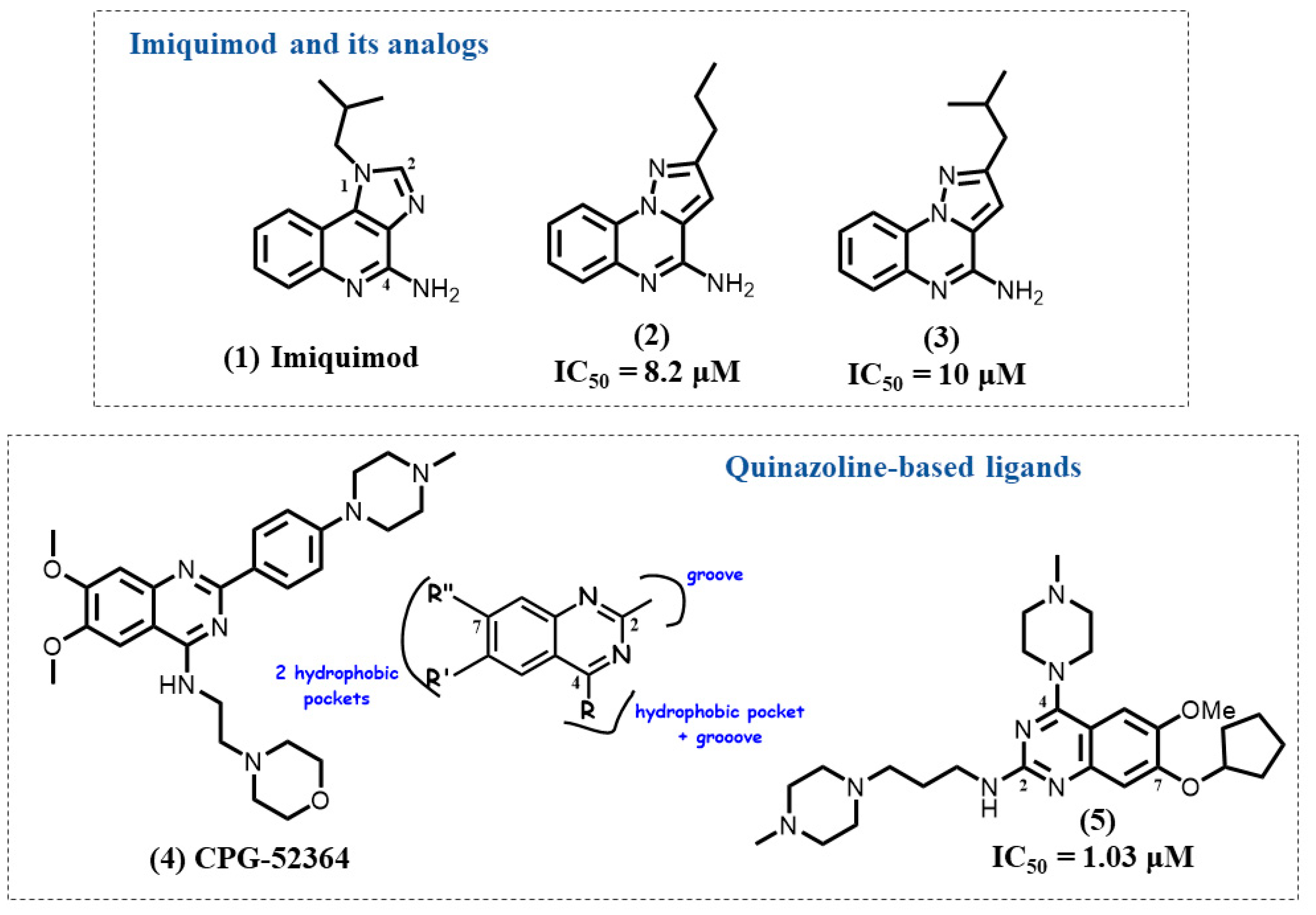
Imiquimod 1 was approved by FDA in 1997 and EMA in 1998 (Figure 2) [92]. Imiquimod activates a series of cytokines, such as interferon-α (IFN-α) and IL-6 via TLR7 [93]. At present, imiquimod is commonly used to treat genital warts and superficial basal cell carcinoma (BCC) under the brand name Aldara [94,95].
Recently, our team synthesized three heterocyclic series derived from the chemical scaffold of imiquimod (imidazo [4,5-c]quinoline): imidazo [1,2-a]pyrazine, imidazo [1,5-a]quinoxaline, and pyrazolo [1,5-a]quinoxaline [96]. Although the heterocycle core is different from the one from imiquimod, the compounds possess some structural similarities, including an amine group and a hydrophobic alkyl chain. Most interestingly, compounds did not show any agonist property but potent and specific antagonistic activities. Among these heterocyclic series, two pyrazolo [1,5-a]quinoxaline derivatives 2 and 3 were identified as lead compounds with IC50 = 8.2 μM and IC50 = 10 μM for TLR7 inhibition activity while they did not exhibit any activity on TLR8. In comparison to the imidazo [1,2-a]pyrazine series, the antagonistic activity of pyrazolo [1,5-a]quinoxaline compounds was increased when their alkyl chains reached 4–5 carbon atoms maximum. Meanwhile, the presence of a butyl or an isobutyl chain, such as in compounds 2 and 3, was shown to be beneficial to obtain the greatest antagonistic potency.
Docking simulation was also performed in the active region of TLR8 (PDB ID: 5WYZ), due to the high sequence similarity and similar antagonistic site between TLR7 and TLR8. Thereafter, a box covering the likely TLR7 binding antagonist sites was applied, which allowed to determine various chemical interactions located in the interface of TLR7 dimer. The tricyclic scaffold of the pyrazoloquinoxaline forms a face-to-face π-π stacking with Phe1302, followed by additional van der Waals forces with Tyr242. In addition, the alkyl chain was engaged into the hydrophobic pocket of TLR7. In particular, Thr384 of TLR7 was indicated to explain the selectivity in comparison with Ile403 of TLR8 containing a bulky side chain, regardless of the high degree of similarity between TLR7 and TLR8. In in vitro tests, compounds 2 and 3 inhibited the expression of TNF-α and interleukin 6 (IL-6) in HEK-blue-hTLR7/8 cells. Additionally, compounds 2 and 3 selectively inhibited NF-κB translocation in TLR7 MyD88 signaling pathways.
A recent study showed also that TLR7 can adopt both imidazo [4,5-c]quinoline-based agonists and antagonists. A C2-alkyl substitution on the tricyclic core can interact with a hydrophobic area of the receptor at the dimer interface and appeared to be a determinant for its activity [97].

4.2. Quinazoline-Based Ligands

The quinazoline derivative CPG-52364 4 is a potent trinary TLR7/8/9 antagonist with a ratio for TLR7/9 antagonism of 0.8 (Figure 2) [54]. CPG-52364 was tested for tolerability in chronic autoimmune diseases, including SLE, but unfortunately failed in phase 1 clinical trials (NCT00547014). The reason of discontinuation remains unpublished.
Recently, some new TLR7 antagonists with the same quinazoline scaffold were synthesized [98]. The development of diverse TLR7 antagonists was mainly accomplished through random screening. To correlate TLR7 antagonistic activity with the structural features in different chemotypes, the authors derived a hypothetical binding model based on molecular docking analysis along with molecular dynamics (MD) simulation studies by using the homologue protein hTLR8 (PDB ID: 3W3J). Binding studies exhibited different pockets, grooves, and a central cavity where the ligand could interact with specific residues of its receptor through hydrophobic and hydrogen bonds. Molecular docking analyses determined the presence of three hydrophobic pockets and two small grooves in the binding site of TLR7 antagonist 4.
Such studies paved the way for the rational design of several chemotypes. Based on the structural insight gained, TLR7 antagonists with the quinazoline core were designed to better understand the engagement mode of the molecules within the diverse protein pockets. The biological evaluation of the synthesized molecules was performed in TLR7-reporter HEK293 cells as well as in pDCs. The study provided a rational design approach thus facilitating further development of novel small molecule hTLR7 antagonists based on different chemical scaffolds. Among them, the best active molecule 5 showed TLR7 inhibition with an IC50 of 1.03 μM, and the suppression of IFN-α induction in response to TLR7 activation in pDCs with an IC50 of 1.42 μM. Separate substitution of quinazoline at C2 or C7 position with a flexible linker and the presence of other small hydrophobic groups were shown to be beneficial for achieving potent TLR7 antagonism. Meanwhile, the cooperation of C2, C4, and C7 substitution could lead to an increase in antagonistic activity of TLR7.
With the most potent antagonist of quinazoline derivatives, molecular dynamics simulation studies show highly stable interactions, which lead to a steady-state fold of TLR7. This study highlights the crucial role of hydrophobic interactions around the central cavity of the antagonist site with important residues, such as Tyr356, Thr525, and Gln354 in the binding mode of the most potent antagonist derivatives.

4.3. Purine-Based Ligands

TLR7 can also respond to guanosine and to small molecules containing a purine core. Such data were of great value for rational drug design [24,99]. An adenine scaffold was previously reported in a series of TLR7 agonists [100]. Hydroxyl groups at C8 and C2 were demonstrated to be important to boost IFN activity [101]. Meanwhile, an amino group at C6 and the introduction of an alkylamino group on the N9 position increased aqueous solubility [102]. Such chemical modification on the adenine core were determined to be useful for antagonism, as well as drug delivery and absorption.
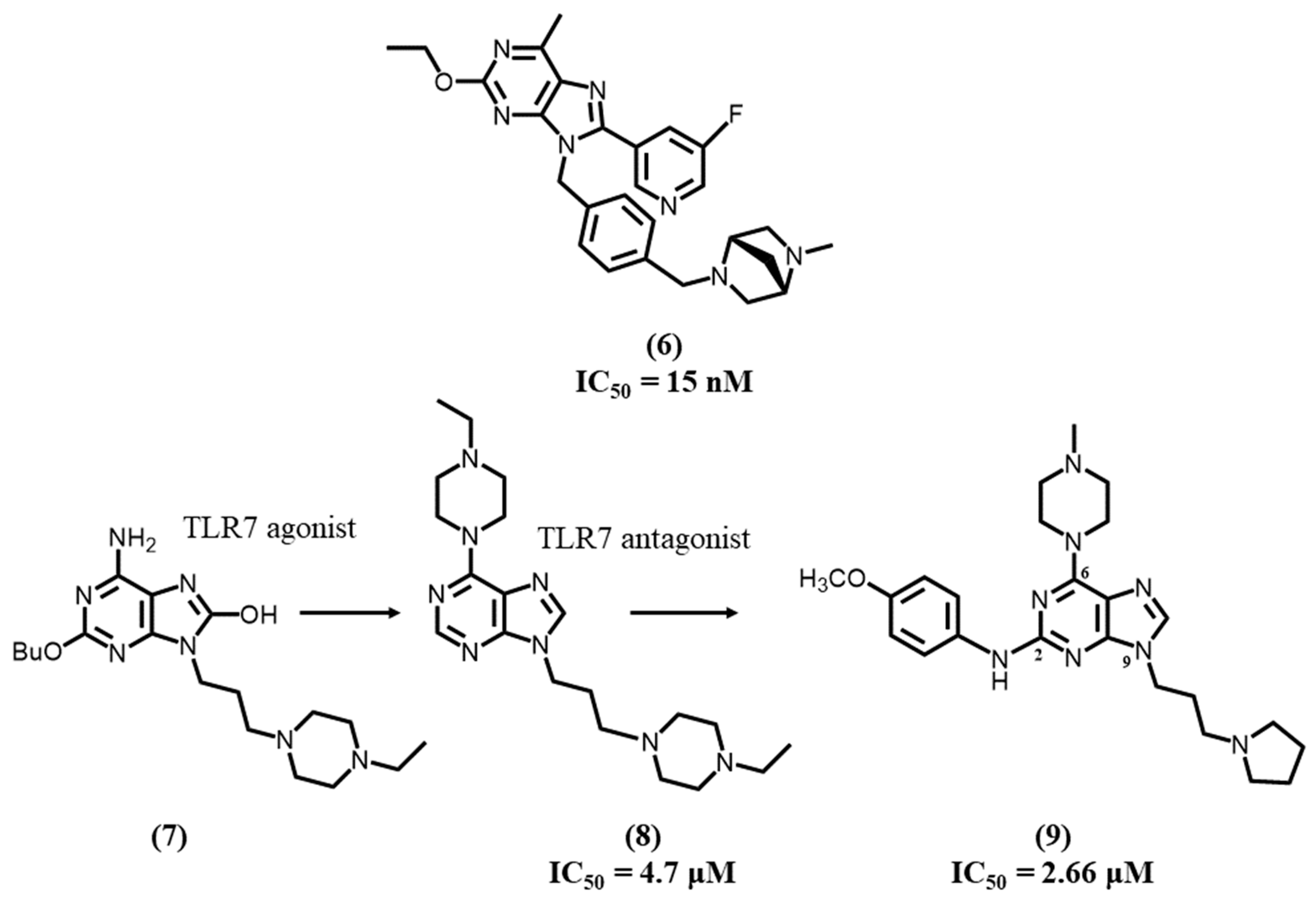
In 2020, Tojo S. et al. developed, for potential SLE applications, a series of TLR7 antagonists based on the 8-oxoadenine core using a chemical switch approach [103]. Based on SAR studies, the authors emphasized that the C8 position of adenine could be substituted from an 8-oxo to an 8-pyridyl group, which allowed the conversion of an agonist into an antagonist. Additionally, the C2 side chain and 8-pyridyl moiety were further optimized, and an amine was introduced on the para-position of the 9-phenyl group in order to increase solubility. As a result, a potent TLR7 antagonist 6 based on a 6-methyl derivative was successfully synthesized with high inhibitory activity (IC50 = 15 nM), selectivity and good solubility (>0.15 mg/mL at pH 7.4) (Figure 3).
Crystallographic studies showed that the most active TLR antagonist 6 interacted at the dimerization interface of the TLR7 protein and induced an open form conformation of the dimer. Such an open form, which shows large differences at the interface compared to the closed form, appears to prevent TLR activation. The 6-methyl adenine and 8-fluoropyridine rings of the most active antagonistic compound are capable to form π-π stacking with Phe408, Phe506, Phe507, Phe349, Phe351, Leu353, and Val381. A hydrogen bond was observed with Gln354, and additional interactions were depicted with Gln323, Phe349, Tyr264, Asn265, Phe351, Ser530, and Tyr579. In vitro results showed that compound 6 totally blocked IFN-α production induced by R848, which suggested compound 6 was specific in inhibiting TLR7. Furthermore, in vivo studies showed compound 6 significantly decreased proteinuria and prevented death in SLE mice.
In 2020, Mukherjee A. et al. reported a very simple modification at the 2-position of 9-alkylpiperazinyl guanosine derivatives induced to a chemical switch from a TLR7 agonist 7 to a clinically relevant antagonist [15].
The deletion of a butoxy moiety afforded compounds with antagonist activity, leading to a potent TLR7 inhibitor 8 after having further increased lipophilic interactions by modifying the amino group on the 6-position. Compound 8 exhibited an IC50 of 4.7 μM on TLR7 and induced inhibition of IL-6 transcription and proinflammatory cytokines, such as TNF-α. It also showed significant potency on an in vivo rodent model of psoriasis. In a molecular docking study, the team used the homology model of TLR7 (PDB ID: 5WYZ) and showed that the purine core formed π-π stacking interactions between two hydrophobic residues, Phe329 and Tyr1374. Meanwhile, ethylpiperazine at C6 formed an important hydrogen bonding between the terminal nitrogen and Ser452. Additionally, the alkyl linker on N9 interacted with Leu331 and Phe329 in a hydrophobic cavity.
In a further study, the same team developed an orally bioavailable dual TLR7/9 antagonist 9, with IC50 values of 0.08 and 2.66 μM against TLR9 and TLR7, respectively [104]. They highlighted several suitable substitutions at C2, C6, and N9 positions that could exhibit considerable efficacy in TLR7 and TLR9 antagonism. Compared to compound 8, compound 9 bears a 4-methoxybenzyl group in the C2 position and a pyrrolidine group, instead of pyrazine, at N9. After in vivo evaluation, compound 9 was suggested to be available as a therapeutic candidate in autoimmune disorders after being shown to have therapeutic effects on psoriasis in mice.

4.4. Imidazopyridine-Based Ligands

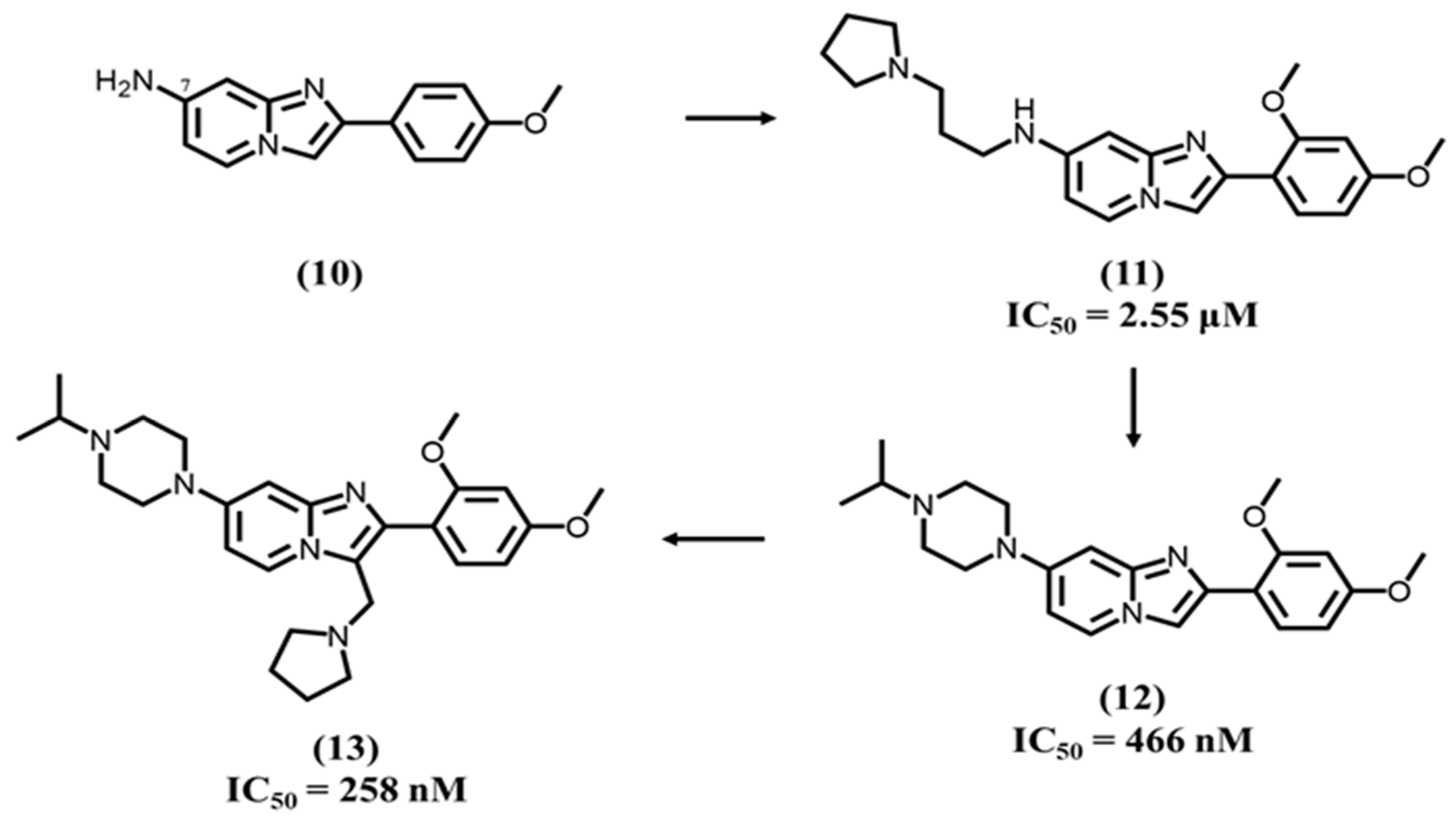
Recently, Das N. et al. developed a potent dual TLR7/9 antagonist based on a 2-phenylimidazopyridine heterocyclic core with a bridgehead nitrogen, which exhibited IC50 values of 0.47 and 0.04 μM against TLR7 and TLR9, respectively [105]. They initially intended to explore minimal pharmacophoric features around the basic core 10 (Figure 4). The presence of two methoxy groups at C2 and C4 positions 11 on the phenyl group and a NH2 function at C7 position of the heterocyclic platform showed interesting TLR7 antagonistic activity with an IC50 of 2.55 μM. Among these modifications, the NH2 group at the C7 position acts as a hydrogen bond acceptor, which stabilizes the pharmacophore in the binding site.
Concurrently, the imidazopyridine core forms π-π interactions with the central hydrophobic cavity. The oxygen atom of the methoxy group at the C4 position forms a hydrogen bond contributing to the stabilization of the ligand-receptor combination. Di-methoxy substitution on the 2-phenyl ring leads to a compound 12, which exhibited TLR9 and TLR7 inhibition with IC50 values of 40 and 466 nM, respectively. After replacement of the NH2 group by an alkyl-substituted piperazine and the introduction of a pyrrolidine cycle at position 3 of the imidazopyridine core, Das N. et al. obtained a lead compound 13, which exhibited TLR9 and TLR7 inhibition with IC50 values of 15 and 258 nM, respectively.

4.5. Pyridone-Based Ligands

Knoepfel T. et al. developed a selective dual TLR7/8 antagonist further with high-throughput screens (HTS) using TR-FRET (time-resolved fluorescence resonance energy transfer) assays with a biotinylated TLR8 protein and Eu-labeled streptavidin [16]. Consequently, they first identified a hit compound 14 with a quinazoline core (Figure 5) forming face-to-edge stacking located in a hydrophobic pocket and a pyridin-2-one group for hydrogen interactions. The hit-to-lead optimization process showed that 8-methyl substitution 15 increased the cellular potency and affinity toward TLR8 but was detrimental to TLR7 antagonistic activity. Therefore, even though TLR7 and TLR8 have a high degree of structural similarity, two main differences were noted on two non-conserved amino acids between TLR7 and TLR8. In comparison to TLR8 (PDB ID: 6TY5), alanine 518 is a serine (530) in one of the TLR7 monomers, and glutamic acid 427 is a valine (430) in TLR7 in the other one. Considering such differences, they changed the quinazoline core by an indazole to avoid the interaction with Glu427 of TLR8 and a steric clash with Ser530 of TLR7. The next step was to separate TLR7/8 balanced activity from TLRs 4/9. A 6-methyl substitution 16 on the pyridone exhibited desired TLR7/8 potency without any TLR4/9 profile and showed the C6 position of the pyridone was crucial for TLR4/9 activity. For the final step of their lead optimization, they focused on the binding to TLR8, since TLR7 activity was still maintained in order to obtain a dual TLR7/8 antagonist 17, with very low IC50s = 0.62 and 1.5 nM for TLR7 and TLR8, respectively.

4.6. Benzanilide-Based Ligands

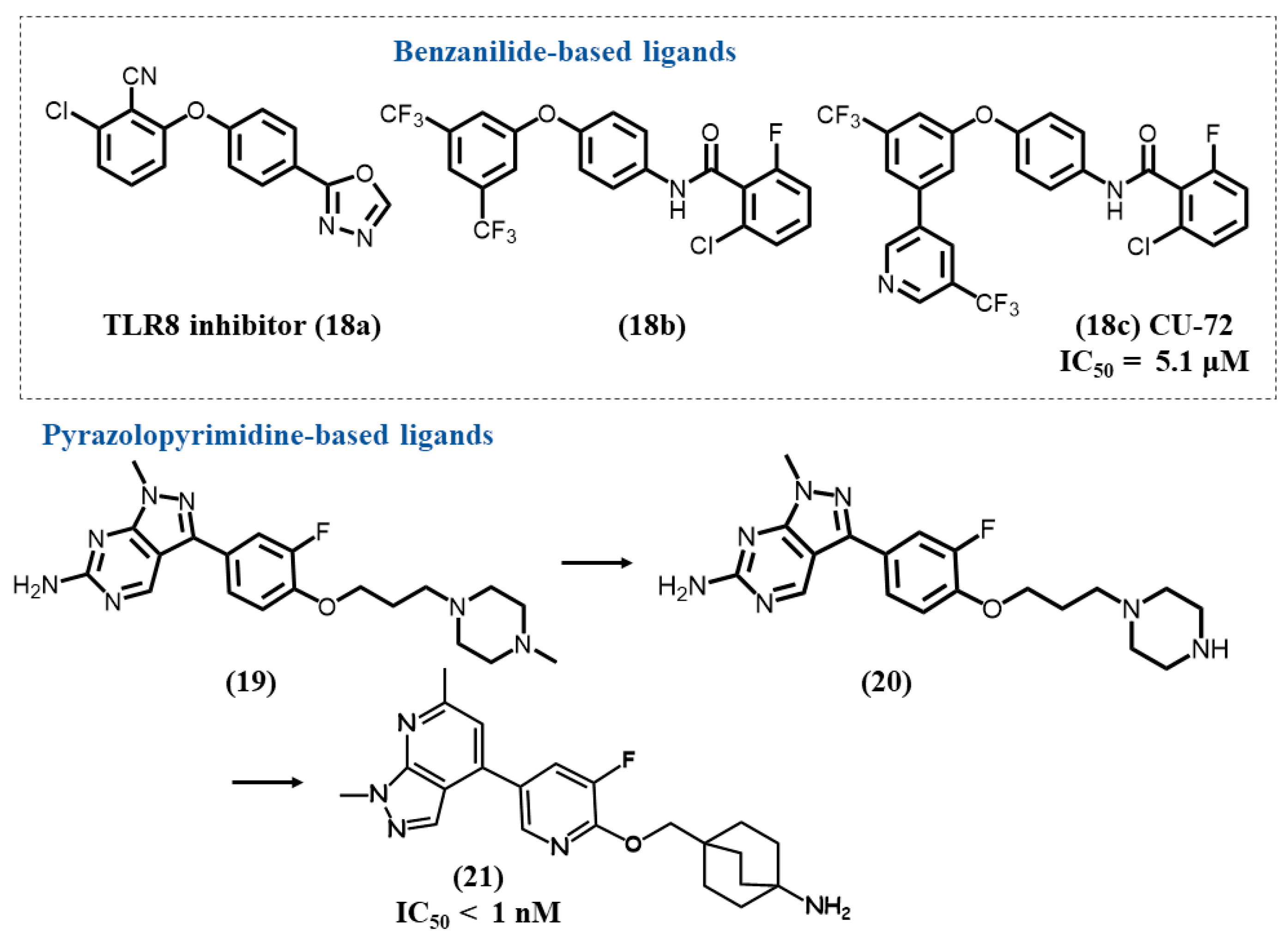
Padilla-Salinas R. et al. performed a first high-throughput screening (HTS) targeting TLR8, which allowed them to identify two first hits, a non-benzanilide TLR8-specific inhibitor 18a and a 2,6-dihalogenobenzanilide derivative 18b with TLR7/8 dual inhibitory activity (Figure 6) [106]. A series of SAR studies mainly performed on the benzanilide derivative showed the importance of the presence of three closed linked phenyl rings, with a separation by an amide group for two of them, one of the phenyl rings being substituted by two trifluoromethyl groups. Furthermore, an optimal distance had to be maintained between the different aromatic rings. Benzanilide ring modulation was intolerant for TLR7. Replacement of a trifluoromethyl group by a trifluoromethyl pyridine led to the TLRs 7/8 antagonist CU-72 18c, which showed micromolar inhibitory activities on TLR7 (IC50 = 5.1 μM) and TLR8 (IC50 = 2.87 μM). In vitro studies suggested CU-72 could act for the inhibition of ssRNA-sensing pathways at low concentrations. Unfortunately, the compound shows low solubility and remains to be further optimized for extended biological evaluation.

4.7. Pyrazolopyrimidine/Pyridine-Based Ligands

Alper P.B. et al., using a large screening study performed on a databank of two million compounds, further identified a first hit pyrazolopyrimidine compound 19 with a piperazine moiety (Figure 6) as a good inhibitor of the TLR7 agonist imiquimod on IL6 production [107]. TLR7 antagonistic activity was then shown to be significantly increased by replacement of the piperazine by a piperidine ring 20 and then a bicyclo [2,2,2]octane. The next step was to focus on chemical modulations to improve the oral exposure and solubility. Replacement of the pyrazolopyrimidine scaffold by a pyrazolopyridine one and a change in the attachment position to the heterocycle platform afforded the lead compound 21 (IC50 < 1 nM).

4.8. Benzoxazole-Based Ligands

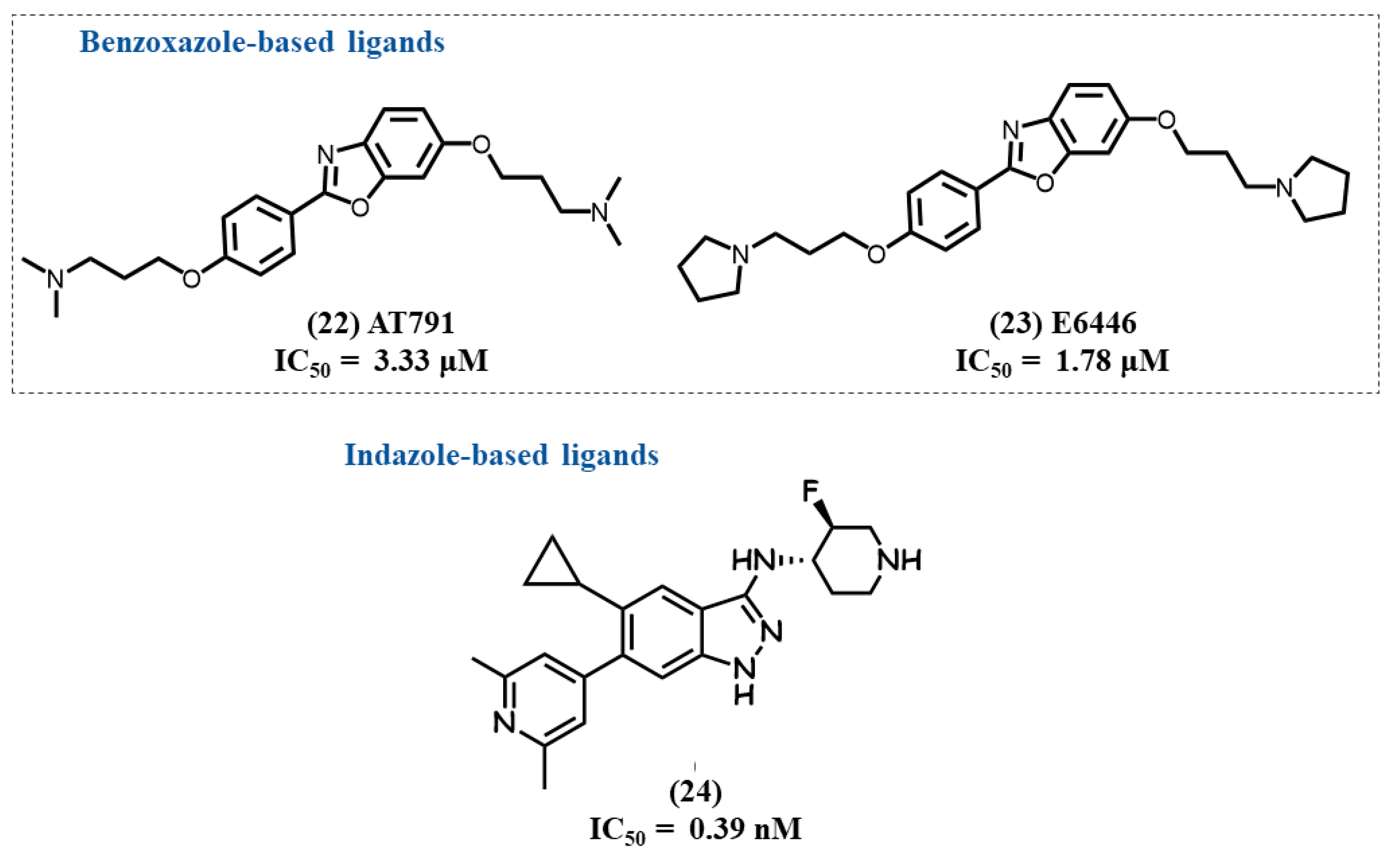
Lamphie, M. et al. studied the activity and mechanism of the action of two potent and selective benzo[d]oxazole TLR7/9 antagonists, AT791 22 and E6446 23 (Figure 7) [108]. The two compounds were initially developed to block TLR9 DNA-induced stimulation, but they were also shown to inhibit TLR7 stimulation. Such inhibition does not appear to be linked to a direct binding to TLRs but by their accumulation as lipophilic weak bases in endosomes, as, for example, antimalarial drugs and, more interestingly, their high and selective affinity for nucleic acids.

4.9. Indazole-Based Ligands

Betschart C. et al. reported a potent and selective dual TLR7/8 antagonist 24 with IC50 values of 0.39 and 0.08 nM on TLR7 and TLR8, respectively (Figure 7) [109]. Their study started from a fragment-based screening, and hits were optimized following SAR and co-crystallography with TLR8 (PDB ID: 7R53 for compound 24). Such latter data showed that compound 24 stabilizes the inactive conformation of TLR8, and indazole formed two key hydrogen bonds with Gly351 in one monomer and Ser516 in the other one. The cyclopropyl ring of antagonist 24 occupies a nearby hydrophobic pocket. In in vitro and in vivo studies, compound 24 showed inhibition of TLR7-dependent IFNα release.

4.10. Indole-Based Ligands

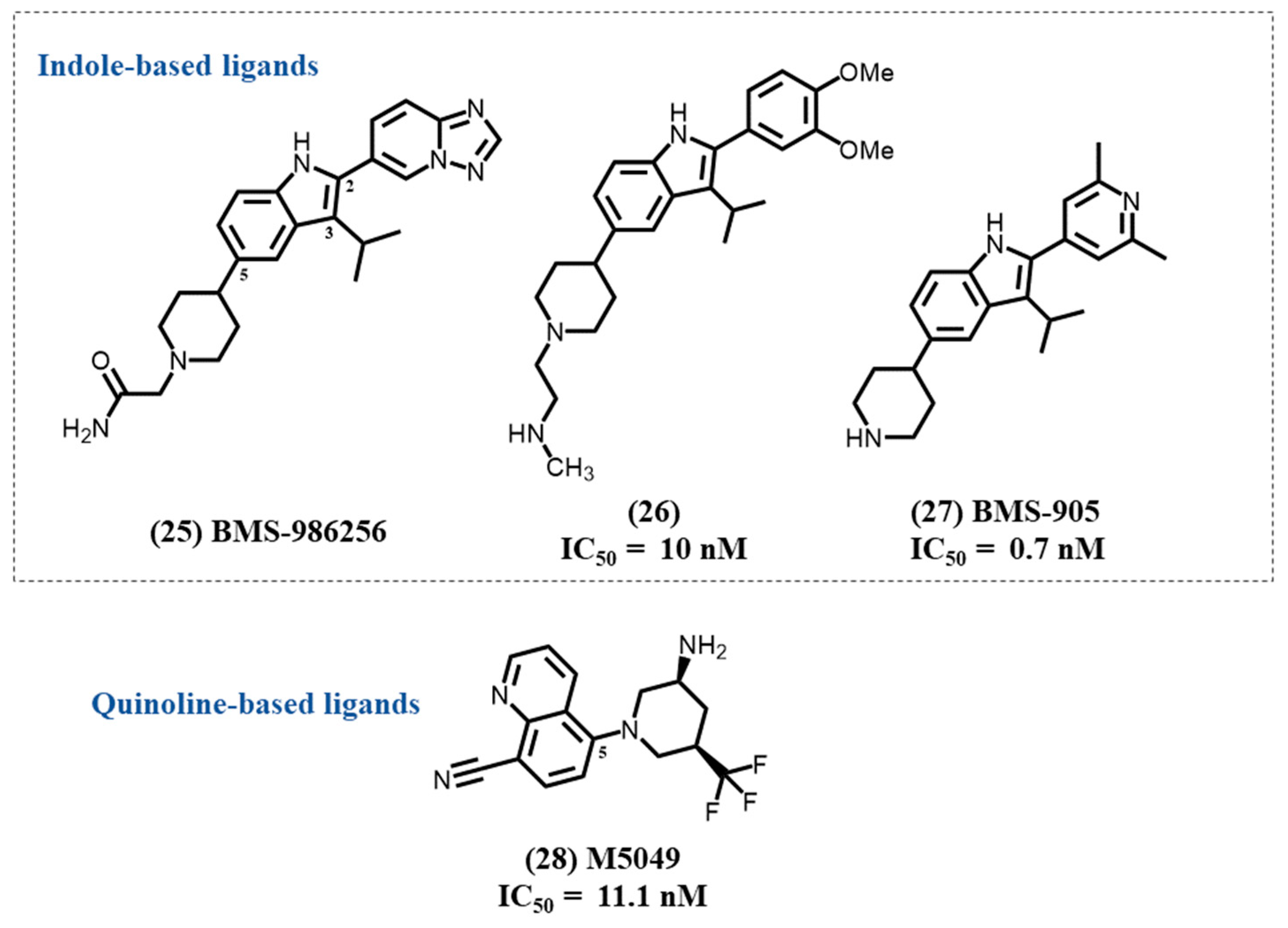
Afimetoran 25 (BMS-986256) is an oral potent inhibitor of TLR 7/8 in clinical trials (Figure 8). It is an indole-based compound with two heterocyclic substituents, a triazolopyridine- and a piperidine-based structures. Afimetoran had the ability to prevent lupus symptoms in mouse models and reverse organ damage, and it showed promising PK data as well as safety profiles in a phase 1 trial (NCT04493541). A recent phase 2 study evaluates its efficacy in patients with systemic lupus erythematosus (NCT04895696).
In 2020, a BMS group of medicinal chemists reported the discovery process of a series of indole-based TLR antagonists, with the exclusion of BMS-986256 [110]. Their SAR study concentrates on tri-substituted indole derivatives with, for the most active compounds, a dimethoxy phenyl substitution on position 2, a short alkyl group on position 3, and the piperidinyl heterocycle grafted on position 5. The most active compound 26 was determined to be a very potent and dual TLR7/8 inhibitor with IC50 values of 10 and 17 nM, respectively. It was also shown to have a much lower effect on TLR9 activity.
Recently, in continuum of this first SAR study, the same group developed a 2-pyridinyl-indole series that allows them to afford a compound 27, BMS-905, another dual inhibitor of TLR7/8 [111]. In this study, they demonstrated that the aryl substitution on position 2 of the indole was important for ligand–protein interactions. They firstly determined the engagement of the heteroatom with various pyridinyl isomers. The 2′,6′-dimethyl groups on the pyridinyl group induced the greatest potency in both TLR7 and TLR8. The substitution of the C3 position of the indole by an isopropyl was shown to be essential for selectivity toward TLR9. With these changes, BMS-905 was discovered as a second potent TLR7/8 dual antagonist lead, with IC50 values of 0.7 and 3.2 nM for TLR7 and TLR8, respectively. It is interesting to note that two of the three main structural features of compound 26 and BMS-905 can also be found in BMS-986256 with an important change on position 2 by replacement of the phenyl or pyridinyl group by the triazolopyridinyl heterocycle.

4.11. Quinoline-Based Ligands

Enpatoran 28 (M5049) is a 5-piperidylquinoline derivative developed as a potent and selective dual TLR7/8 antagonist with IC50 values of 11.1 and 24.1 nM for TLR7 and TLR8, respectively (Figure 8). M5049 appears to bind the open form of the TLR dimer and stabilizes the inactive state of the receptor, which prevents its activation [112].
In in vitro and in vivo tests, enpatoran shows a therapeutic effect in SLE, rare cutaneous lupus erythematosus (CLE), and other autoimmune diseases by suppressing the pathologic activity of ribonucleic acid–containing immune complexes. In comparation to placebo, M5049 showed promising drug-like properties with good tolerance and promising PK without significant dose-limiting adverse events in healthy volunteers. Further studies focus on the evaluation of its potential efficacy in patients with autoimmune diseases, such as lupus, with high TLR7 and TLR8 expression [113].
Additionally, TLR7 is also involved in coronavirus disease 2019 (COVID-19), due to the participation of TLR7 in the invasion and infection of the virus [114,115]. Therefore, enpatoran also underwent a phase 2 trial in order to evaluate its safety and efficacy in COVID-19 pneumonia patients (NCT04448756). High exposure (100 mg) to enpatoran reduced the inflammatory response that leads to the cytokine storm in some cases, and was recommended to reduce acute hyperinflammation caused by COVID-19 [116].

5. Conclusions

TLR7 is crucial for recognizing xenobiotics containing PAMPs in the innate immunity system. The TLR7 overexpression, caused by the presence and abnormal degradation of self-RNA, activates MyD88-dependent signaling pathway and guides the host defensive responses. Hence, TLR7 is an attractive target for drug design studies and development. In this review, we provide general information for potential therapeutic treatments, including autoimmune disorders, cancer, and antiviral immunotherapy. Various heterocyclic aromatic scaffolds of small molecule TLR7 antagonists are described, many of them bearing a five-membered cycle. The obtention of the lead compounds include SAR optimization, and some of them are X-ray co-crystallography data. For small molecule antagonists, most of the studies state that TLR7 antagonists occupy the same binding site as agonists and change the closed-form conformation of TLR7 to an opened-form conformation. Several small molecule TLR7 antagonists have undergone clinical trial; the first results appear to assess their safety of use in patients. Afimetoran, an indole-based TLR7 antagonist, and Enpatoran, a quinoline derivative, are under phase II clinical investigation in patients with SLE or other immune related diseases.

Author Contributions

Conceptualization, H.Z. and P.-A.B.; methodology, H.Z., P.W. and P.-A.B.; formal analysis, H.Z.; investigation, H.Z.; resources, P.-A.B.; data curation, P.W.; writing—original draft preparation, H.Z.; writing—review and editing, H.Z. and P.-A.B.; visualization, H.Z.; supervision, P.-A.B.; project administration, P.-A.B. All authors have read and agreed to the published version of the manuscript.

Funding

This research received no external funding.

Institutional Review Board Statement

Not applicable.

Informed Consent Statement

Not applicable.

Data Availability Statement

Not applicable.

Acknowledgments

The authors would like to thank Yunru Chen for assistance in language editing.

Conflicts of Interest

The authors declare no conflict of interest.

References

  1. Kawai, T.; Akira, S. The Role of Pattern-Recognition Receptors in Innate Immunity: Update on Toll-like Receptors. Nat. Immunol. 2010, 11, 373–384. [Google Scholar] [CrossRef] [PubMed]
  2. Fitzgerald, K.A.; Kagan, J.C. Toll-like Receptors and the Control of Immunity. Cell 2020, 180, 1044–1066. [Google Scholar] [CrossRef] [PubMed]
  3. Pandey, S.; Kawai, T.; Akira, S. Microbial Sensing by Toll-Like Receptors and Intracellular Nucleic Acid Sensors. Cold Spring Harb. Perspect. Biol. 2015, 7, a0162462015. [Google Scholar] [CrossRef] [PubMed] [Green Version]
  4. Heil, F.; Hemmi, H.; Hochrein, H.; Ampenberger, F.; Kirschning, C.; Akira, S.; Lipford, G.; Wagner, H.; Bauer, S. Species-Specific Recognition of Single-Stranded RNA via Toll-like Receptor 7 and 8. Science 2004, 303, 1526–1529. [Google Scholar] [CrossRef] [PubMed] [Green Version]
  5. Lauterbach, M.A.; Hanke, J.E.; Serefidou, M.; Mangan, M.S.J.; Kolbe, C.-C.; Hess, T.; Rothe, M.; Kaiser, R.; Hoss, F.; Gehlen, J.; et al. Toll-like Receptor Signaling Rewires Macrophage Metabolism and Promotes Histone Acetylation via ATP-Citrate Lyase. Immunity 2019, 51, 997–1011.e7. [Google Scholar] [CrossRef]
  6. Corzo, C.A.; Varfolomeev, E.; Setiadi, A.F.; Francis, R.; Klabunde, S.; Senger, K.; Sujatha-Bhaskar, S.; Drobnick, J.; Do, S.; Suto, E.; et al. The Kinase IRAK4 Promotes Endosomal TLR and Immune Complex Signaling in B Cells and Plasmacytoid Dendritic Cells. Sci. Signal. 2020, 13, eaaz1053. [Google Scholar] [CrossRef]
  7. Jarrossay, D.; Napolitani, G.; Colonna, M.; Sallusto, F.; Lanzavecchia, A. Specialization and Complementarity in Microbial Molecule Recognition by Human Myeloid and Plasmacytoid Dendritic Cells. Eur. J. Immunol. 2001, 31, 3388–3393. [Google Scholar] [CrossRef]
  8. Gay, N.J.; Symmons, M.F.; Gangloff, M.; Bryant, C.E. Assembly and Localization of Toll-like Receptor Signalling Complexes. Nat. Rev. Immunol. 2014, 14, 546–558. [Google Scholar] [CrossRef]
  9. Pelka, K.; Bertheloot, D.; Reimer, E.; Phulphagar, K.; Schmidt, S.V.; Christ, A.; Stahl, R.; Watson, N.; Miyake, K.; Hacohen, N.; et al. The Chaperone UNC93B1 Regulates Toll-like Receptor Stability Independently of Endosomal TLR Transport. Immunity 2018, 48, 911–922.e7. [Google Scholar] [CrossRef]
  10. Akira, S.; Uematsu, S.; Takeuchi, O. Pathogen Recognition and Innate Immunity. Cell 2006, 124, 783–801. [Google Scholar] [CrossRef]
  11. Swiecki, M.; Colonna, M. The Multifaceted Biology of Plasmacytoid Dendritic Cells. Nat. Rev. Immunol. 2015, 15, 471–485. [Google Scholar] [CrossRef]
  12. Junt, T.; Barchet, W. Translating Nucleic Acid-Sensing Pathways into Therapies. Nat. Rev. Immunol. 2015, 15, 529–544. [Google Scholar] [CrossRef]
  13. O’reilly, S.; Duffy, L. Toll-like Receptors in the Pathogenesis of Autoimmune Diseases: Recent and Emerging Translational Developments. ITT 2016, 5, 69–80. [Google Scholar] [CrossRef] [Green Version]
  14. Patinote, C.; Karroum, N.B.; Moarbess, G.; Cirnat, N.; Kassab, I.; Bonnet, P.-A.; Deleuze-Masquéfa, C. Agonist and Antagonist Ligands of Toll-like Receptors 7 and 8: Ingenious Tools for Therapeutic Purposes. Eur. J. Med. Chem. 2020, 193, 112238. [Google Scholar] [CrossRef]
  15. Mukherjee, A.; Raychaudhuri, D.; Sinha, B.P.; Kundu, B.; Mitra, M.; Paul, B.; Bandopadhyay, P.; Ganguly, D.; Talukdar, A. A Chemical Switch for Transforming a Purine Agonist for Toll-like Receptor 7 to a Clinically Relevant Antagonist. J. Med. Chem. 2020, 63, 4776–4789. [Google Scholar] [CrossRef]
  16. Knoepfel, T.; Nimsgern, P.; Jacquier, S.; Bourrel, M.; Vangrevelinghe, E.; Glatthar, R.; Behnke, D.; Alper, P.B.; Michellys, P.-Y.; Deane, J.; et al. Target-Based Identification and Optimization of 5-Indazol-5-Yl Pyridones as Toll-like Receptor 7 and 8 Antagonists Using a Biochemical TLR8 Antagonist Competition Assay. J. Med. Chem. 2020, 63, 8276–8295. [Google Scholar] [CrossRef]
  17. Hu, Z.; Zhang, T.; Jiang, S.; Yin, H. Protocol for Evaluation and Validation of TLR8 Antagonists in HEK-Blue Cells via Secreted Embryonic Alkaline Phosphatase Assay. STAR Protoc. 2022, 3, 101061. [Google Scholar] [CrossRef]
  18. van der Fits, L.; Mourits, S.; Voerman, J.S.A.; Kant, M.; Boon, L.; Laman, J.D.; Cornelissen, F.; Mus, A.-M.; Florencia, E.; Prens, E.P.; et al. Imiquimod-Induced Psoriasis-Like Skin Inflammation in Mice Is Mediated via the IL-23/IL-17 Axis. J. Immunol. 2009, 182, 5836–5845. [Google Scholar] [CrossRef] [Green Version]
  19. Zhang, Z.; Ohto, U.; Shimizu, T. Toward a Structural Understanding of Nucleic Acid-sensing Toll-like Receptors in the Innate Immune System. FEBS Lett. 2017, 591, 3167–3181. [Google Scholar] [CrossRef] [Green Version]
  20. Bell, J.K.; Mullen, G.E.D.; Leifer, C.A.; Mazzoni, A.; Davies, D.R.; Segal, D.M. Leucine-Rich Repeats and Pathogen Recognition in Toll-like Receptors. Trends Immunol. 2003, 24, 528–533. [Google Scholar] [CrossRef]
  21. Maeda, K.; Akira, S. TLR7 Structure: Cut in Z-Loop. Immunity 2016, 45, 705–707. [Google Scholar] [CrossRef] [PubMed] [Green Version]
  22. Kanno, A.; Yamamoto, C.; Onji, M.; Fukui, R.; Saitoh, S.; Motoi, Y.; Shibata, T.; Matsumoto, F.; Muta, T.; Miyake, K. Essential Role for Toll-like Receptor 7 (TLR7)-Unique Cysteines in an Intramolecular Disulfide Bond, Proteolytic Cleavage and RNA Sensing. Int. Immunol. 2013, 25, 413–422. [Google Scholar] [CrossRef] [PubMed] [Green Version]
  23. Ishida, H.; Asami, J.; Zhang, Z.; Nishizawa, T.; Shigematsu, H.; Ohto, U.; Shimizu, T. Cryo-EM Structures of Toll-like Receptors in Complex with UNC93B1. Nat. Struct. Mol. Biol. 2021, 28, 173–180. [Google Scholar] [CrossRef]
  24. Zhang, Z.; Ohto, U.; Shibata, T.; Krayukhina, E.; Taoka, M.; Yamauchi, Y.; Tanji, H.; Isobe, T.; Uchiyama, S.; Miyake, K.; et al. Structural Analysis Reveals That Toll-like Receptor 7 Is a Dual Receptor for Guanosine and Single-Stranded RNA. Immunity 2016, 45, 737–748. [Google Scholar] [CrossRef] [PubMed] [Green Version]
  25. Shibata, T.; Ohto, U.; Nomura, S.; Kibata, K.; Motoi, Y.; Zhang, Y.; Murakami, Y.; Fukui, R.; Ishimoto, T.; Sano, S.; et al. Guanosine and Its Modified Derivatives Are Endogenous Ligands for TLR7. Int. Immunol. 2016, 28, 211–222. [Google Scholar] [CrossRef] [Green Version]
  26. Miyake, K.; Shibata, T.; Ohto, U.; Shimizu, T.; Saitoh, S.-I.; Fukui, R.; Murakami, Y. Mechanisms Controlling Nucleic Acid-Sensing Toll-like Receptors. Int. Immunol. 2018, 30, 43–51. [Google Scholar] [CrossRef] [PubMed] [Green Version]
  27. Wang, Y.; Zhang, S.; Li, H.; Wang, H.; Zhang, T.; Hutchinson, M.R.; Yin, H.; Wang, X. Small-Molecule Modulators of Toll-like Receptors. Acc. Chem. Res. 2020, 53, 1046–1055. [Google Scholar] [CrossRef]
  28. Lushpa, V.A.; Goncharuk, M.V.; Lin, C.; Zalevsky, A.O.; Talyzina, I.A.; Luginina, A.P.; Vakhrameev, D.D.; Shevtsov, M.B.; Goncharuk, S.A.; Arseniev, A.S.; et al. Modulation of Toll-like Receptor 1 Intracellular Domain Structure and Activity by Zn2+ Ions. Commun. Biol. 2021, 4, 1003. [Google Scholar] [CrossRef]
  29. Nyman, T.; Stenmark, P.; Flodin, S.; Johansson, I.; Hammarström, M.; Nordlund, P. The Crystal Structure of the Human Toll-like Receptor 10 Cytoplasmic Domain Reveals a Putative Signaling Dimer. J. Biol. Chem. 2008, 283, 11861–11865. [Google Scholar] [CrossRef] [Green Version]
  30. Kim, T.H.; Oh, D.S.; Jung, H.E.; Chang, J.; Lee, H.K. Plasmacytoid Dendritic Cells Contribute to the Production of IFN-β via TLR7-MyD88-Dependent Pathway and CTL Priming during Respiratory Syncytial Virus Infection. Viruses 2019, 11, 730. [Google Scholar] [CrossRef]
  31. Li, D.; Wu, M. Pattern Recognition Receptors in Health and Diseases. Sig Transduct Target. Ther. 2021, 6, 291. [Google Scholar] [CrossRef]
  32. AbdAllah, N.B.; Toraih, E.A.; Al Ageeli, E.; Elhagrasy, H.; Gouda, N.S.; Fawzy, M.S.; Helal, G.M. MYD88, NFKB1, and IL6 Transcripts Overexpression Are Associated with Poor Outcomes and Short Survival in Neonatal Sepsis. Sci. Rep. 2021, 11, 13374. [Google Scholar] [CrossRef]
  33. Balka, K.R.; Nardo, D. Understanding Early TLR Signaling through the Myddosome. J. Leukoc. Biol. 2019, 105, 339–351. [Google Scholar] [CrossRef]
  34. Chen, Z.J. Ubiquitination in Signaling to and Activation of IKK: Ubiquitin-Mediated Activation of IKK. Immunol. Rev. 2012, 246, 95–106. [Google Scholar] [CrossRef] [Green Version]
  35. Kawasaki, T.; Kawai, T. Toll-Like Receptor Signaling Pathways. Front. Immunol. 2014, 5, 461. [Google Scholar] [CrossRef] [Green Version]
  36. Tang, J.-R.; Michaelis, K.A.; Nozik-Grayck, E.; Seedorf, G.J.; Hartman-Filson, M.; Abman, S.H.; Wright, C.J. The NF-ΚB Inhibitory Proteins IκBα and IκBβ Mediate Disparate Responses to Inflammation in Fetal Pulmonary Endothelial Cells. J. Immunol. 2013, 190, 2913–2923. [Google Scholar] [CrossRef] [Green Version]
  37. Liu, T.; Zhang, L.; Joo, D.; Sun, S.-C. NF-ΚB Signaling in Inflammation. Sig. Transduct. Target. Ther. 2017, 2, 17023. [Google Scholar] [CrossRef] [Green Version]
  38. Farrugia, M.; Baron, B. The Role of Toll-Like Receptors in Autoimmune Diseases through Failure of the Self-Recognition Mechanism. Int. J. Inflam. 2017, 2017, 8391230. [Google Scholar] [CrossRef] [Green Version]
  39. Wang, T.; Marken, J.; Chen, J.; Tran, V.B.; Li, Q.-Z.; Li, M.; Cerosaletti, K.; Elkon, K.B.; Zeng, X.; Giltiay, N.V. High TLR7 Expression Drives the Expansion of CD19+CD24hiCD38hi Transitional B Cells and Autoantibody Production in SLE Patients. Front. Immunol. 2019, 10, 1243. [Google Scholar] [CrossRef] [Green Version]
  40. Illescas-Montes, R.; Corona-Castro, C.C.; Melguizo-Rodríguez, L.; Ruiz, C.; Costela-Ruiz, V.J. Infectious Processes and Systemic Lupus Erythematosus. Immunology 2019, 158, 153–160. [Google Scholar] [CrossRef]
  41. Zhou, Y.; Yuan, J.; Pan, Y.; Fei, Y.; Qiu, X.; Hu, N.; Luo, Y.; Lei, W.; Li, Y.; Long, H.; et al. T Cell CD40LG Gene Expression and the Production of IgG by Autologous B Cells in Systemic Lupus Erythematosus. Clin. Immunol. 2009, 132, 362–370. [Google Scholar] [CrossRef] [PubMed] [Green Version]
  42. Marshak-Rothstein, A.; Rifkin, I.R. Immunologically Active Autoantigens: The Role of Toll-Like Receptors in the Development of Chronic Inflammatory Disease. Annu. Rev. Immunol. 2007, 25, 419–441. [Google Scholar] [CrossRef] [PubMed]
  43. Kiefer, K.; Oropallo, M.A.; Cancro, M.P.; Marshak-Rothstein, A. Role of Type I Interferons in the Activation of Autoreactive B Cells. Immunol. Cell Biol 2012, 90, 498–504. [Google Scholar] [CrossRef] [PubMed] [Green Version]
  44. Wirth, J.R.; Molano, I.; Ruiz, P.; Coutermarsh-Ott, S.; Cunningham, M.A. TLR7 Agonism Accelerates Disease and Causes a Fatal Myeloproliferative Disorder in NZM 2410 Lupus Mice. Front. Immunol. 2020, 10, 3054. [Google Scholar] [CrossRef]
  45. Hwang, S.-H.; Lee, H.; Yamamoto, M.; Jones, L.A.; Dayalan, J.; Hopkins, R.; Zhou, X.J.; Yarovinsky, F.; Connolly, J.E.; Curotto de Lafaille, M.A.; et al. B Cell TLR7 Expression Drives Anti-RNA Autoantibody Production and Exacerbates Disease in Systemic Lupus Erythematosus–Prone Mice. J. Immunol. 2012, 189, 5786–5796. [Google Scholar] [CrossRef] [PubMed] [Green Version]
  46. Walsh, E.R.; Pisitkun, P.; Voynova, E.; Deane, J.A.; Scott, B.L.; Caspi, R.R.; Bolland, S. Dual Signaling by Innate and Adaptive Immune Receptors Is Required for TLR7-Induced B-Cell–Mediated Autoimmunity. Proc. Natl. Acad. Sci. USA 2012, 109, 16276–16281. [Google Scholar] [CrossRef] [Green Version]
  47. Deane, J.A.; Pisitkun, P.; Barrett, R.S.; Feigenbaum, L.; Town, T.; Ward, J.M.; Flavell, R.A.; Bolland, S. Control of Toll-like Receptor 7 Expression Is Essential to Restrict Autoimmunity and Dendritic Cell Proliferation. Immunity 2007, 27, 801–810. [Google Scholar] [CrossRef] [Green Version]
  48. Sakata, K.; Nakayamada, S.; Miyazaki, Y.; Kubo, S.; Ishii, A.; Nakano, K.; Tanaka, Y. Up-Regulation of TLR7-Mediated IFN-α Production by Plasmacytoid Dendritic Cells in Patients with Systemic Lupus Erythematosus. Front. Immunol. 2018, 9, 1957. [Google Scholar] [CrossRef] [Green Version]
  49. Brown, G.J.; Cañete, P.F.; Wang, H.; Medhavy, A.; Bones, J.; Roco, J.A.; He, Y.; Qin, Y.; Cappello, J.; Ellyard, J.I.; et al. TLR7 Gain-of-Function Genetic Variation Causes Human Lupus. Nature 2022, 605, 349–356. [Google Scholar] [CrossRef]
  50. Vanpouille-Box, C.; Hoffmann, J.A.; Galluzzi, L. Pharmacological Modulation of Nucleic Acid Sensors—Therapeutic Potential and Persisting Obstacles. Nat. Rev. Drug Discov. 2019, 18, 845–867. [Google Scholar] [CrossRef]
  51. Akilesh, H.M.; Buechler, M.B.; Duggan, J.M.; Hahn, W.O.; Matta, B.; Sun, X.; Gessay, G.; Whalen, E.; Mason, M.; Presnell, S.R.; et al. Chronic TLR7 and TLR9 Signaling Drives Anemia via Differentiation of Specialized Hemophagocytes. Science 2019, 363, eaao5213. [Google Scholar] [CrossRef]
  52. Rimbach, K.; Kaiser, S.; Helm, M.; Dalpke, A.H.; Eigenbrod, T. 2’-O-Methylation within Bacterial RNA Acts as Suppressor of TLR7/TLR8 Activation in Human Innate Immune Cells. J. Innate Immun. 2015, 7, 482–493. [Google Scholar] [CrossRef]
  53. Schmitt, F.C.F.; Freund, I.; Weigand, M.A.; Helm, M.; Dalpke, A.H.; Eigenbrod, T. Identification of an Optimized 2′- O -Methylated Trinucleotide RNA Motif Inhibiting Toll-like Receptors 7 and 8. RNA 2017, 23, 1344–1351. [Google Scholar] [CrossRef] [Green Version]
  54. Lai, C.-Y.; Su, Y.-W.; Lin, K.-I.; Hsu, L.-C.; Chuang, T.-H. Natural Modulators of Endosomal Toll-Like Receptor-Mediated Psoriatic Skin Inflammation. J. Immunol. Res. 2017, 2017, 1–15. [Google Scholar] [CrossRef] [Green Version]
  55. Suárez-Fariñas, M.; Arbeit, R.; Jiang, W.; Ortenzio, F.S.; Sullivan, T.; Krueger, J.G. Suppression of Molecular Inflammatory Pathways by Toll-Like Receptor 7, 8, and 9 Antagonists in a Model of IL-23-Induced Skin Inflammation. PLoS ONE 2013, 8, e84634. [Google Scholar] [CrossRef] [Green Version]
  56. Zhu, F.; Wang, D.; Jiang, W.; Bhagat, L.; Agrawal, S. Sa1757 Targeting Innate Immune Receptors to Treat Inflammatory Bowel Disease: Preclinical Activity of IMO-9200, an Antagonist of TLRS 7, 8, and 9 in Mouse Models of Colitis. Gastroenterology 2015, 148, S-324. [Google Scholar] [CrossRef]
  57. Pearson, J.A.; Tai, N.; Ekanayake-Alper, D.K.; Peng, J.; Hu, Y.; Hager, K.; Compton, S.; Wong, F.S.; Smith, P.C.; Wen, L. Norovirus Changes Susceptibility to Type 1 Diabetes by Altering Intestinal Microbiota and Immune Cell Functions. Front. Immunol. 2019, 10, 2654. [Google Scholar] [CrossRef]
  58. Huang, J.; Peng, J.; Pearson, J.A.; Efthimiou, G.; Hu, Y.; Tai, N.; Xing, Y.; Zhang, L.; Gu, J.; Jiang, J.; et al. Toll-like Receptor 7 Deficiency Suppresses Type 1 Diabetes Development by Modulating B-Cell Differentiation and Function. Cell Mol. Immunol. 2021, 18, 328–338. [Google Scholar] [CrossRef]
  59. Lee, A.S.; Ghoreishi, M.; Cheng, W.K.; Chang, T.-Y.E.; Zhang, Y.Q.; Dutz, J.P. Toll-like Receptor 7 Stimulation Promotes Autoimmune Diabetes in the NOD Mouse. Diabetologia 2011, 54, 1407–1416. [Google Scholar] [CrossRef] [Green Version]
  60. Tomita, T. Apoptosis of Pancreatic β-Cells in Type 1 Diabetes. Bosn J. Basic Med. Sci. 2017, 17, 183–193. [Google Scholar] [CrossRef]
  61. Eizirik, D.L.; Mandrup-Poulsen, T. A Choice of Death—The Signal-Transduction of Immune-Mediated Beta-Cell Apoptosis. Diabetologia 2001, 44, 2115–2133. [Google Scholar] [CrossRef] [PubMed]
  62. Wang, Y.; Roussel-Queval, A.; Chasson, L.; Hanna Kazazian, N.; Marcadet, L.; Nezos, A.; Sieweke, M.H.; Mavragani, C.; Alexopoulou, L. TLR7 Signaling Drives the Development of Sjögren’s Syndrome. Front. Immunol. 2021, 12, 676010. [Google Scholar] [CrossRef] [PubMed]
  63. Alexopoulou, L. Nucleic Acid-Sensing Toll-like Receptors: Important Players in Sjögren’s Syndrome. Front. Immunol. 2022, 13, 980400. [Google Scholar] [CrossRef] [PubMed]
  64. Karlsen, M.; Jonsson, R.; Brun, J.G.; Appel, S.; Hansen, T. TLR-7 and -9 Stimulation of Peripheral Blood B Cells Indicate Altered TLR Signalling in Primary Sjögren’s Syndrome Patients by Increased Secretion of Cytokines. Scand. J. Immunol. 2015, 82, 523–531. [Google Scholar] [CrossRef] [PubMed] [Green Version]
  65. Rakoff-Nahoum, S.; Medzhitov, R. Toll-like Receptors and Cancer. Nat. Rev. Cancer 2009, 9, 57–63. [Google Scholar] [CrossRef] [PubMed]
  66. Javaid, N.; Choi, S. Toll-like Receptors from the Perspective of Cancer Treatment. Cancers 2020, 12, 297. [Google Scholar] [CrossRef] [Green Version]
  67. Urban-Wojciuk, Z.; Khan, M.M.; Oyler, B.L.; Fåhraeus, R.; Marek-Trzonkowska, N.; Nita-Lazar, A.; Hupp, T.R.; Goodlett, D.R. The Role of TLRs in Anti-Cancer Immunity and Tumor Rejection. Front. Immunol. 2019, 10, 2388. [Google Scholar] [CrossRef]
  68. Grimmig, T.; Matthes, N.; Hoeland, K.; Tripathi, S.; Chandraker, A.; Grimm, M.; Moench, R.; Moll, E.-M.; Friess, H.; Tsaur, I.; et al. TLR7 and TLR8 Expression Increases Tumor Cell Proliferation and Promotes Chemoresistance in Human Pancreatic Cancer. Int. J. Oncol. 2015, 47, 857–866. [Google Scholar] [CrossRef] [Green Version]
  69. Fukata, M.; Abreu, M.T. Role of Toll-like Receptors in Gastrointestinal Malignancies. Oncogene 2008, 27, 234–243. [Google Scholar] [CrossRef] [Green Version]
  70. Zhang, M.; Yan, Z.; Wang, J.; Yao, X. Toll-like Receptors 7 and 8 Expression Correlates with the Expression of Immune Biomarkers and Positively Predicts the Clinical Outcome of Patients with Melanoma. OTT 2017, 10, 4339–4346. [Google Scholar] [CrossRef]
  71. Sheyhidin, I. Overexpression of TLR3, TLR4, TLR7 and TLR9 in Esophageal Squamous Cell Carcinoma. WJG 2011, 17, 3745. [Google Scholar] [CrossRef]
  72. Keshavarz, A.; Pourbagheri-Sigaroodi, A.; Zafari, P.; Bagheri, N.; Ghaffari, S.H.; Bashash, D. Toll-like Receptors (TLRs) in Cancer; with an Extensive Focus on TLR Agonists and Antagonists. IUBMB Life 2021, 73, 10–25. [Google Scholar] [CrossRef]
  73. Chatterjee, S.; Crozet, L.; Damotte, D.; Iribarren, K.; Schramm, C.; Alifano, M.; Lupo, A.; Cherfils-Vicini, J.; Goc, J.; Katsahian, S.; et al. TLR7 Promotes Tumor Progression, Chemotherapy Resistance, and Poor Clinical Outcomes in Non–Small Cell Lung Cancer. Cancer Res. 2014, 74, 5008–5018. [Google Scholar] [CrossRef] [Green Version]
  74. Caron, G.; Duluc, D.; Frémaux, I.; Jeannin, P.; David, C.; Gascan, H.; Delneste, Y. Direct Stimulation of Human T Cells via TLR5 and TLR7/8: Flagellin and R-848 Up-Regulate Proliferation and IFN-γ Production by Memory CD4+ T Cells. J. Immunol. 2005, 175, 1551–1557. [Google Scholar] [CrossRef] [Green Version]
  75. Liu, B.; Qu, L.; Yan, S. Cyclooxygenase-2 Promotes Tumor Growth and Suppresses Tumor Immunity. Cancer Cell Int. 2015, 15, 106. [Google Scholar] [CrossRef] [Green Version]
  76. Meås, H.Z.; Haug, M.; Beckwith, M.S.; Louet, C.; Ryan, L.; Hu, Z.; Landskron, J.; Nordbø, S.A.; Taskén, K.; Yin, H.; et al. Sensing of HIV-1 by TLR8 Activates Human T Cells and Reverses Latency. Nat. Commun. 2020, 11, 147. [Google Scholar] [CrossRef] [Green Version]
  77. Fugger, L.; Jensen, L.T.; Rossjohn, J. Challenges, Progress, and Prospects of Developing Therapies to Treat Autoimmune Diseases. Cell 2020, 181, 63–80. [Google Scholar] [CrossRef]
  78. Patamawenu, A.A.; Wright, N.E.; Shofner, T.; Evans, S.; Manion, M.M.; Doria-Rose, N.; Migueles, S.A.; Mendoza, D.; Peterson, B.; Wilhelm, C.; et al. Toll-like Receptor 7-Adapter Complex Modulates Interferon-α Production in HIV-Stimulated Plasmacytoid Dendritic Cells. PLoS ONE 2019, 14, e0225806. [Google Scholar] [CrossRef] [Green Version]
  79. Dominguez-Villar, M.; Gautron, A.-S.; de Marcken, M.; Keller, M.J.; Hafler, D.A. TLR7 Induces Anergy in Human CD4+ T Cells. Nat. Immunol. 2015, 16, 118–128. [Google Scholar] [CrossRef]
  80. Fraietta, J.A.; Mueller, Y.M.; Yang, G.; Boesteanu, A.C.; Gracias, D.T.; Do, D.H.; Hope, J.L.; Kathuria, N.; McGettigan, S.E.; Lewis, M.G.; et al. Type I Interferon Upregulates Bak and Contributes to T Cell Loss during Human Immunodeficiency Virus (HIV) Infection. PLoS Pathog. 2013, 9, e1003658. [Google Scholar] [CrossRef]
  81. Simmons, R.P.; Scully, E.P.; Groden, E.E.; Arnold, K.B.; Chang, J.J.; Lane, K.; Lifson, J.; Rosenberg, E.; Lauffenburger, D.A.; Altfeld, M. HIV-1 Infection Induces Strong Production of IP-10 through TLR7/9-Dependent Pathways. AIDS 2013, 27, 2505–2517. [Google Scholar] [CrossRef] [Green Version]
  82. Gu, Y.; Zuo, X.; Zhang, S.; Ouyang, Z.; Jiang, S.; Wang, F.; Wang, G. The Mechanism behind Influenza Virus Cytokine Storm. Viruses 2021, 13, 1362. [Google Scholar] [CrossRef] [PubMed]
  83. Liu, Q.; Zhou, Y.; Yang, Z. The Cytokine Storm of Severe Influenza and Development of Immunomodulatory Therapy. Cell Mol. Immunol. 2016, 13, 3–10. [Google Scholar] [CrossRef] [PubMed] [Green Version]
  84. Rappe, J.C.F.; Finsterbusch, K.; Crotta, S.; Mack, M.; Priestnall, S.L.; Wack, A. A TLR7 Antagonist Restricts Interferon-Dependent and -Independent Immunopathology in a Mouse Model of Severe Influenza. J. Exp. Med. 2021, 218, e20201631. [Google Scholar] [CrossRef] [PubMed]
  85. Xu, H.; Huang, L.; Luo, Q.; Tu, Q.; Liu, J.; Yu, R.; Huang, J.; Chen, T.; Yin, Y.; Cao, J. Absence of Toll-like receptor 7 protects mice against Pseudomonas aeruginosa pneumonia. Int. Immunopharmacol. 2021, 96, 107739. [Google Scholar] [CrossRef]
  86. Mancuso, G.; Gambuzza, M.; Midiri, A.; Biondo, C.; Papasergi, S.; Akira, S.; Teti, G.; Beninati, C. Bacterial recognition by TLR7 in the lysosomes of conventional dendritic cells. Nat. Immunol. 2009, 10, 587–594. [Google Scholar] [CrossRef]
  87. Lee, C.Y.Q.; Chan, Y.T.; Cheok, Y.Y.; Tan, G.M.Y.; Tang, T.F.; Cheong, H.C.; Vadivelu, J.; Abdullah, S.; Looi, C.Y.; Wong, W.F. Helicobacter pylori Infection Elicits Type I Interferon Response in Human Monocytes via Toll-Like Receptor 8 Signaling. J. Immunol. Res. 2022, 2022, 3861518. [Google Scholar] [CrossRef]
  88. Chen, X.; Liang, H.; Zhang, J.; Zen, K.; Zhang, C.-Y. MicroRNAs Are Ligands of Toll-like Receptors. RNA 2013, 19, 737–739. [Google Scholar] [CrossRef] [Green Version]
  89. Hoshikawa, N.; Sakai, A.; Takai, S.; Suzuki, H. Targeting Extracellular MiR-21-TLR7 Signaling Provides Long-Lasting Analgesia in Osteoarthritis. Mol. Ther. Nucleic Acids 2020, 19, 199–207. [Google Scholar] [CrossRef]
  90. Liu, C.-L.; Santos, M.M.; Fernandes, C.; Liao, M.; Iamarene, K.; Zhang, J.-Y.; Sukhova, G.K.; Shi, G.-P. Toll-like Receptor 7 Deficiency Protects Apolipoprotein E-Deficient Mice from Diet-Induced Atherosclerosis. Sci. Rep. 2017, 7, 847. [Google Scholar] [CrossRef]
  91. Salagianni, M.; Galani, I.E.; Lundberg, A.M.; Davos, C.H.; Varela, A.; Gavriil, A.; Lyytikäinen, L.-P.; Lehtimäki, T.; Sigala, F.; Folkersen, L.; et al. Toll-Like Receptor 7 Protects From Atherosclerosis by Constraining “Inflammatory” Macrophage Activation. Circulation 2012, 126, 952–962. [Google Scholar] [CrossRef] [Green Version]
  92. Hemmi, H.; Kaisho, T.; Takeuchi, O.; Sato, S.; Sanjo, H.; Hoshino, K.; Horiuchi, T.; Tomizawa, H.; Takeda, K.; Akira, S. Small Anti-Viral Compounds Activate Immune Cells via the TLR7 MyD88–Dependent Signaling Pathway. Nat. Immunol. 2002, 3, 196–200. [Google Scholar] [CrossRef]
  93. Sauder, D.N. Imiquimod: Modes of Action. Br. J. Derm. 2003, 149, 5–8. [Google Scholar] [CrossRef]
  94. Buck, H.W. Imiquimod (Aldara? Cream). Infect. Dis. Obstet. Gynecol. 1998, 6, 49–51. [Google Scholar] [CrossRef]
  95. Marks, R.; Gebauer, K.; Shumack, S.; Amies, M.; Bryden, J.; Fox, T.L.; Owens, M.L. Imiquimod 5% Cream in the Treatment of Superficial Basal Cell Carcinoma: Results of a Multicenter 6-Week Dose-Response Trial. J. Am. Acad Derm. 2001, 44, 807–813. [Google Scholar] [CrossRef]
  96. Bou Karroum, N.; Moarbess, G.; Guichou, J.-F.; Bonnet, P.-A.; Patinote, C.; Bouharoun-Tayoun, H.; Chamat, S.; Cuq, P.; Diab-Assaf, M.; Kassab, I.; et al. Novel and Selective TLR7 Antagonists among the Imidazo [1,2- a ]Pyrazines, Imidazo [1,5- a ]Quinoxalines, and Pyrazolo [1,5- a ]Quinoxalines Series. J. Med. Chem. 2019, 62, 7015–7031. [Google Scholar] [CrossRef]
  97. Yang, M.; Larson, P.G.; Brown, L.; Schultz, J.R.; Kucaba, T.A.; Griffith, T.S.; Ferguson, D.M. Toll-like Receptor 7 and 8 Imidazoquinoline-Based Agonist/Antagonist Pairs. Bioorg. Med. Chem. Lett. 2022, 59, 128548. [Google Scholar] [CrossRef]
  98. Pal, S.; Paul, B.; Bandopadhyay, P.; Preethy, N.; Sarkar, D.; Rahaman, O.; Goon, S.; Roy, S.; Ganguly, D.; Talukdar, A. Synthesis and Characterization of New Potent TLR7 Antagonists Based on Analysis of the Binding Mode Using Biomolecular Simulations. Eur. J. Med. Chem. 2021, 210, 112978. [Google Scholar] [CrossRef]
  99. Dharmapuri, S.; Aurisicchio, L.; Neuner, P.; Verdirame, M.; Ciliberto, G.; La Monica, N. An Oral TLR7 Agonist Is a Potent Adjuvant of DNA Vaccination in Transgenic Mouse Tumor Models. Cancer Gene Ther. 2009, 16, 462–472. [Google Scholar] [CrossRef]
  100. Hirota, K.; Kazaoka, K.; Niimoto, I.; Kumihara, H.; Sajiki, H.; Isobe, Y.; Takaku, H.; Tobe, M.; Ogita, H.; Ogino, T.; et al. Discovery of 8-Hydroxyadenines as a Novel Type of Interferon Inducer. J. Med. Chem. 2002, 45, 5419–5422. [Google Scholar] [CrossRef]
  101. Bazin, H.G.; Li, Y.; Khalaf, J.K.; Mwakwari, S.; Livesay, M.T.; Evans, J.T.; Johnson, D.A. Structural Requirements for TLR7-Selective Signaling by 9-(4-Piperidinylalkyl)-8-Oxoadenine Derivatives. Bioorg. Med. Chem. Lett. 2015, 25, 1318–1323. [Google Scholar] [CrossRef] [PubMed] [Green Version]
  102. Nakamura, T.; Wada, H.; Kurebayashi, H.; McInally, T.; Bonnert, R.; Isobe, Y. Synthesis and Evaluation of 8-Oxoadenine Derivatives as Potent Toll-like Receptor 7 Agonists with High Water Solubility. Bioorg. Med. Chem. Lett. 2013, 23, 669–672. [Google Scholar] [CrossRef] [PubMed]
  103. Tojo, S.; Zhang, Z.; Matsui, H.; Tahara, M.; Ikeguchi, M.; Kochi, M.; Kamada, M.; Shigematsu, H.; Tsutsumi, A.; Adachi, N.; et al. Structural Analysis Reveals TLR7 Dynamics Underlying Antagonism. Nat. Commun. 2020, 11, 5204. [Google Scholar] [CrossRef] [PubMed]
  104. Kundu, B.; Raychaudhuri, D.; Mukherjee, A.; Sinha, B.P.; Sarkar, D.; Bandopadhyay, P.; Pal, S.; Das, N.; Dey, D.; Ramarao, K.; et al. Systematic Optimization of Potent and Orally Bioavailable Purine Scaffold as a Dual Inhibitor of Toll-Like Receptors 7 and 9. J. Med. Chem. 2021, 64, 9279–9301. [Google Scholar] [CrossRef] [PubMed]
  105. Das, N.; Bandopadhyay, P.; Roy, S.; Sinha, B.P.; Dastidar, U.G.; Rahaman, O.; Pal, S.; Ganguly, D.; Talukdar, A. Development, Optimization, and In Vivo Validation of New Imidazopyridine Chemotypes as Dual TLR7/TLR9 Antagonists through Activity-Directed Sequential Incorporation of Relevant Structural Subunits. J. Med. Chem. 2022, 65, 11607–11632. [Google Scholar] [CrossRef]
  106. Padilla-Salinas, R.; Anderson, R.; Sakaniwa, K.; Zhang, S.; Nordeen, P.; Lu, C.; Shimizu, T.; Yin, H. Discovery of Novel Small Molecule Dual Inhibitors Targeting Toll-Like Receptors 7 and 8. J. Med. Chem. 2019, 62, 10221–10244. [Google Scholar] [CrossRef]
  107. Alper, P.B.; Deane, J.; Betschart, C.; Buffet, D.; Collignon Zipfel, G.; Gordon, P.; Hampton, J.; Hawtin, S.; Ibanez, M.; Jiang, T.; et al. Discovery of Potent, Orally Bioavailable in Vivo Efficacious Antagonists of the TLR7/8 Pathway. Bioorg. Med. Chem. Lett. 2020, 30, 127366. [Google Scholar] [CrossRef]
  108. Lamphier, M.; Zheng, W.; Latz, E.; Spyvee, M.; Hansen, H.; Rose, J.; Genest, M.; Yang, H.; Shaffer, C.; Zhao, Y.; et al. Novel Small Molecule Inhibitors of TLR7 and TLR9: Mechanism of Action and Efficacy In Vivo. Mol. Pharm. 2014, 85, 429–440. [Google Scholar] [CrossRef] [Green Version]
  109. Betschart, C.; Faller, M.; Zink, F.; Hemmig, R.; Blank, J.; Vangrevelinghe, E.; Bourrel, M.; Glatthar, R.; Behnke, D.; Barker, K.; et al. Structure-Based Optimization of a Fragment-like TLR8 Binding Screening Hit to an In Vivo Efficacious TLR7/8 Antagonist. ACS Med. Chem. Lett. 2022, 13, 658–664. [Google Scholar] [CrossRef]
  110. Mussari, C.P.; Dodd, D.S.; Sreekantha, R.K.; Pasunoori, L.; Wan, H.; Posy, S.L.; Critton, D.; Ruepp, S.; Subramanian, M.; Watson, A.; et al. Discovery of Potent and Orally Bioavailable Small Molecule Antagonists of Toll-like Receptors 7/8/9 (TLR7/8/9). ACS Med. Chem. Lett. 2020, 11, 1751–1758. [Google Scholar] [CrossRef]
  111. Sreekantha, R.K.; Mussari, C.P.; Dodd, D.S.; Pasunoori, L.; Hegde, S.; Posy, S.L.; Critton, D.; Ruepp, S.; Subramanian, M.; Salter-Cid, L.M.; et al. Identification of 2-Pyridinylindole-Based Dual Antagonists of Toll-like Receptors 7 and 8 (TLR7/8). ACS Med. Chem. Lett. 2022, 13, 812–818. [Google Scholar] [CrossRef]
  112. Vlach, J.; Bender, A.T.; Przetak, M.; Pereira, A.; Deshpande, A.; Johnson, T.L.; Reissig, S.; Tzvetkov, E.; Musil, D.; Morse, N.T.; et al. Discovery of M5049: A Novel Selective Toll-Like Receptor 7/8 Inhibitor for Treatment of Autoimmunity. J. Pharm. Exp. Ther. 2021, 376, 397–409. [Google Scholar] [CrossRef]
  113. Port, A.; Shaw, J.V.; Klopp-Schulze, L.; Bytyqi, A.; Vetter, C.; Hussey, E.; Mammasse, N.; Ona, V.; Bachmann, A.; Strugala, D.; et al. Phase 1 Study in Healthy Participants of the Safety, Pharmacokinetics, and Pharmacodynamics of Enpatoran (M5049), a Dual Antagonist of Toll-like Receptors 7 and 8. Pharm. Res. Perspect. 2021, 9, e00842. [Google Scholar] [CrossRef]
  114. Khanmohammadi, S.; Rezaei, N. Role of Toll-like Receptors in the Pathogenesis of COVID-19. J. Med. Virol. 2021, 93, 2735–2739. [Google Scholar] [CrossRef]
  115. Dyavar, S.R.; Singh, R.; Emani, R.; Pawar, G.P.; Chaudhari, V.D.; Podany, A.T.; Avedissian, S.N.; Fletcher, C.V.; Salunke, D.B. Role of Toll-like Receptor 7/8 Pathways in Regulation of Interferon Response and Inflammatory Mediators during SARS-CoV2 Infection and Potential Therapeutic Options. Biomed. Pharm. 2021, 141, 111794. [Google Scholar] [CrossRef]
  116. Klopp-Schulze, L.; Shaw, J.V.; Dong, J.Q.; Khandelwal, A.; Vazquez-Mateo, C.; Goteti, K. Applying Modeling and Simulations for Rational Dose Selection of Novel Toll-Like Receptor 7/8 Inhibitor Enpatoran for Indications of High Medical Need. Clin. Pharma. Ther. 2022, 112, 297–306. [Google Scholar] [CrossRef]
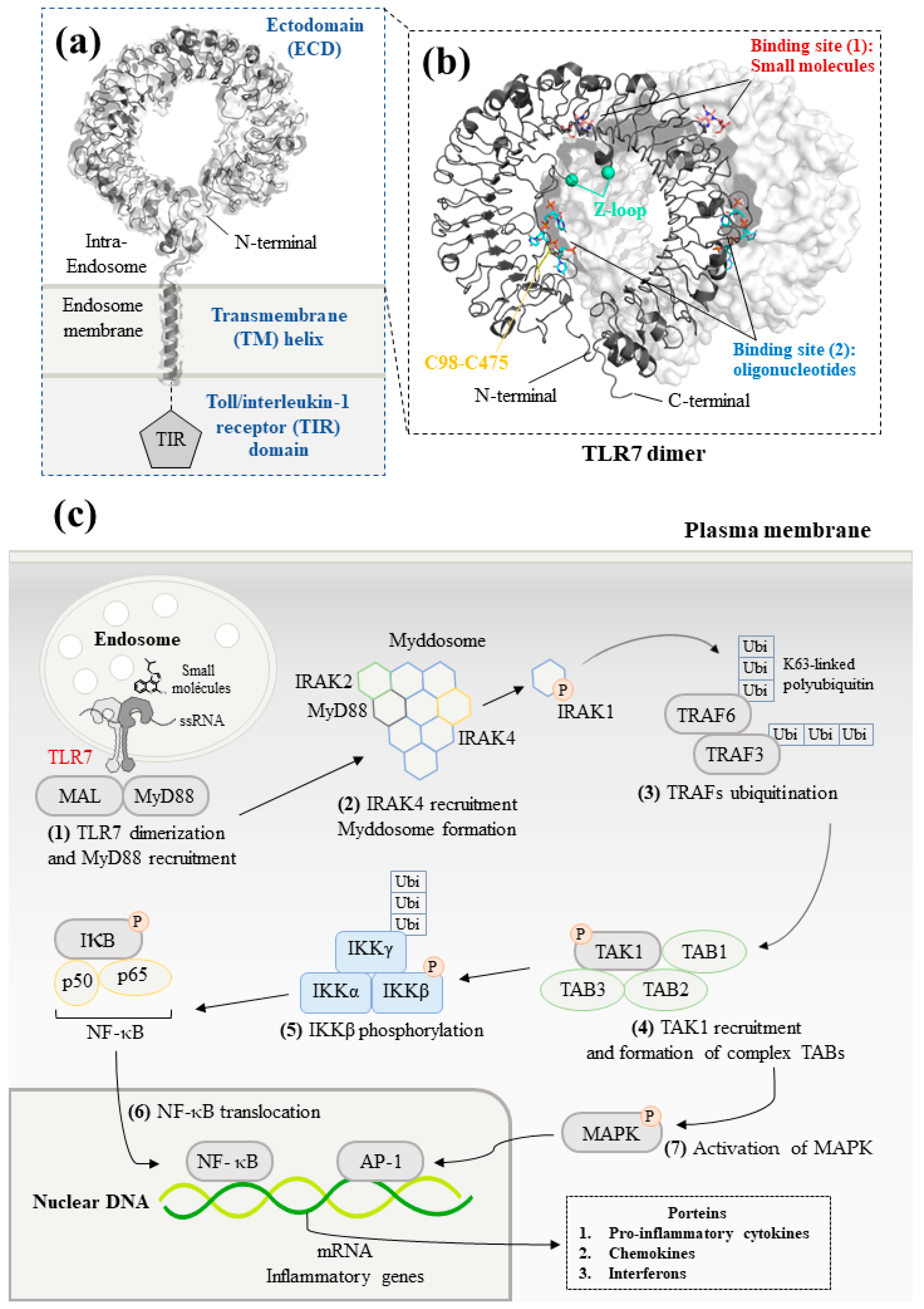
Figure 1. Structure and signaling pathways of the toll-like receptor 7 (TLR7): (a) Schematic representation of TLR7. The N-terminal ectodomain (ECD) locates in the endosome. The transmembrane helix consists of an α-helix across the endosomal membrane. The details of the structure of toll/interleukin-1 receptor (TIR) domain are not illustrated due to the lack of data for TLR7. Data are from reference [23] (PDB ID: 7CYN). (b) Front view of the ECD dimer. The cleaved Z-loop and a disulfide bridge between Cys98 and Cys475 are shown in cyan and yellow, respectively. The binding site of small molecules and synthetized oligonucleotides are illustrated in red and blue, respectively. Data are from reference [24] (PDB ID: 5GMG). (c) TLR7-mediated MyD88-dependent signaling pathways. Firstly, the recognition between small-molecule ligands or ssRNA and TLR7 allows to initiate downstream signaling. MyD88 then forms the myddosome with IRAK4, which phosphorylates and releases IRAK1. After that, TRAF6 is auto-ubiquitinated and activates TAK1. TAK1 forms a complex with TAB1/2/3. Finally, translocation of NF-κB and activation of MAPK signaling pathway generates innate immune responses that lead to the production of pro-inflammatory cytokines and IFNs.
Molecules 28 00634 g001
Figure 2. Imiquimod analogs and quinazoline derivatives.
Molecules 28 00634 g002
Figure 3. Structure-activities relation (SAR) studies and chemical structures for purine derivatives.
Molecules 28 00634 g003
Figure 4. SAR studies on imidazopyridine derivatives.
Molecules 28 00634 g004
Figure 5. SAR studies with pyridone derivatives.
Molecules 28 00634 g005
Figure 6. Structures of benzanilide-based ligands and SAR studies of pyrazolopyrimidine/pyridine derivatives.
Molecules 28 00634 g006
Figure 7. Benzoxazole and indazole derivatives.
Molecules 28 00634 g007
Figure 8. Indole and quinoline TLR7 antagonist derivatives.
Molecules 28 00634 g008
Disclaimer/Publisher’s Note: The statements, opinions and data contained in all publications are solely those of the individual author(s) and contributor(s) and not of MDPI and/or the editor(s). MDPI and/or the editor(s) disclaim responsibility for any injury to people or property resulting from any ideas, methods, instructions or products referred to in the content.