Article

Novel Strigolactone Mimics That Modulate Photosynthesis and Biomass Accumulation in Chlorella sorokiniana

by
Daria Gabriela Popa
1,2,†,
Florentina Georgescu
3,†,
Florea Dumitrascu
4,*,
Sergiu Shova
5,
Diana Constantinescu-Aruxandei
1,
Constantin Draghici
4,
Lucian Vladulescu
3 and
Florin Oancea
1,2,*
1
Bioproducts Team, Bioresources Department, National Institute for Research & Development in Chemistry and Petrochemistry—ICECHIM, Splaiul Independenței Nr. 202, Sector 6, 060021 Bucharest, Romania
2
Faculty of Biotechnologies, University of Agronomic Sciences and Veterinary Medicine of Bucharest, Bd. Mărăști Nr. 59, Sector 1, 011464 Bucharest, Romania
3
Enpro Soctech Com., Str. Elefterie Nr. 51, Sector 5, 050524 Bucharest, Romania
4
“Costin D. Nenițescu” Institute of Organic and Supramolecular Chemistry, Romanian Academy, Splaiul Independentei Nr. 202B, Sector 6, 060023 Bucharest, Romania
5
“Petru Poni” Institute of Macromolecular Chemistry, Romanian Academy, Aleea Grigore Ghica Voda Nr. 41-A, 700487 Iaşi, Romania
*
Authors to whom correspondence should be addressed.
These authors contributed equally to this work.
Molecules 2023, 28(20), 7059; https://doi.org/10.3390/molecules28207059
Submission received: 10 September 2023 / Revised: 3 October 2023 / Accepted: 6 October 2023 / Published: 12 October 2023
(This article belongs to the Special Issue Advances in Heterocyclic Synthesis)

Abstract

:
In terrestrial plants, strigolactones act as multifunctional endo- and exo-signals. On microalgae, the strigolactones determine akin effects: induce symbiosis formation with fungi and bacteria and enhance photosynthesis efficiency and accumulation of biomass. This work aims to synthesize and identify strigolactone mimics that promote photosynthesis and biomass accumulation in microalgae with biotechnological potential. Novel strigolactone mimics easily accessible in significant amounts were prepared and fully characterized. The first two novel compounds contain 3,5-disubstituted aryloxy moieties connected to the bioactive furan-2-one ring. In the second group of compounds, a benzothiazole ring is connected directly through the cyclic nitrogen atom to the bioactive furan-2-one ring. The novel strigolactone mimics were tested on Chlorella sorokiniana NIVA-CHL 176. All tested strigolactones increased the accumulation of chlorophyll b in microalgae biomass. The SL-F3 mimic, 3-(4-methyl-5-oxo-2,5-dihydrofuran-2-yl)-3H-benzothiazol-2-one (7), proved the most efficient. This compound, applied at a concentration of 10−7 M, determined a significant biomass accumulation, higher by more than 15% compared to untreated control, and improved the quantum yield efficiency of photosystem II. SL-F2 mimic, 5-(3,5-dibromophenoxy)-3-methyl-5H-furan-2-one (4), applied at a concentration of 10−9 M, improved protein production and slightly stimulated biomass accumulation. Potential utilization of the new strigolactone mimics as microalgae biostimulants is discussed.

1. Introduction

Strigolactones (SLs) are carotenoid-derived compounds that act as signaling molecules with an increased importance in plant science. The first strigolactone was isolated from root exudates of cotton in 1966 by Cook, who named it strigol [1] and proposed its structure [2]. SLs have been identified as phytohormones with an important role in controlling the plant architecture [3,4,5,6,7,8,9,10,11,12,13] and in plant response to biotic and abiotic stress [14,15,16,17]. These molecules are exo-signals in the rhizosphere, triggering the germination of seeds of parasitic weeds [18,19,20,21,22,23,24,25], acting as branching factors for arbuscular mycorrhizal (AM) fungi [25,26,27], promotor of rhizobia nodulation [28,29,30], regulator of rhizosphere microbiome [31,32], plant neighbor detector [33,34,35]. All these properties stimulated the interest in identifying the various roles played by SLs in (parasitic plant) seed germination, plant development and its response to environmental factors, and molecular receptors involved in the signaling mechanism [36,37,38].
The initial discovered natural SLs (canonical SLs) contain a tricyclic lactone (ABC-rings) connected using an enol ether unit to a furan-2-one ring (D). Usually, these SLs (also called canonical) are classified based on the stereochemistry of the BC-ring junction into two major groups, one with the parent structure of strigol and the other with the parent structure of oronbanchol—Figure 1 [39].
The structure-activity relation studies showed that the presence of the bioactiphore CD part, the stereochemistry, and the methyl substituent at C-4′ of the D-ring are essential structural features for natural SLs activity as germination stimulants for the seeds of parasitic weeds [22,40]. The SL bioactivity is not influenced by the structural changes of A, B, and C rings or by the substitution of an oxygen atom by the nitrogen atom as in imino SL analogs [41] and strigolactam analogs [42], or by a sulfur atom [6].
In the last decade, new stimulants of the parasitic weed seed germination were discovered and included in the non-canonical strigolactone category [43]. Such strigolactones retain methylated butenolide D-ring but lacks the A-, B- or C-rings.
Only tiny amounts of natural SLs have been isolated from plant root exudates. These compounds have structures that are too complex for large-scale syntheses. Therefore, a significant number of simplified synthetic SL analogs and mimics were synthesized in order to understand their diverse structure-activities relationship and detect the corresponding receptors in plants. The SL analogs retain the bioactiphore part of natural SLs and have a considerable degree of molecular freedom in the AB-rings of the molecule [22,38,40,41,42,44,45,46,47,48,49,50,51,52]. In SL mimics, the bioactive D-ring is directly connected to aroyloxy, (hetero)aryloxy, arylthio, or (hetero)aromatic moieties. They mimic various biological activities of natural SLs even if they do not have the typical structural features of natural SLs [6,52,53,54,55,56,57,58]. SL analogs and mimics also have the potential for the development of practical application of strigolactones for parasitic weed control [22] or as plant biostimulants, to improve symbiosis efficiencies, to enhance crop tolerance to biotic and abiotic stress (including soil pollutants) and to regulate cultivated plant architecture [59,60].
It seems that the initial function of strigolactones in terrestrial ecosystems was as exo-signals for recruiting plant-beneficial microorganisms [61,62]. 5-Deoxystrigol determines an akin effect on aquatic ecosystems, inducing the formation of a symbiotic consortium between Scenedesmus obliquus, Ganoderma lucidum, and bacteria from activated sludge and enhancing their biotechnological performance for pollutant removal and biogas upgrading [63]. The strigolactone analog GR24 induced symbiotic pellet formation between Chlorella vulgaris, G. lucidum, and endophytic bacteria and improved their efficiency in nutrient removal from biogas slurry and fixation of CO2 from biogas stream [64,65,66]. GR24 has a similar boosting effect on photosynthetic activities in plants [67,68] and microalgae [64]. Therefore, SLs have potential applications in microalgae biotechnology as well. However, despite their application at very low concentrations, 5-deoxystrigol, and GR24 are too challenging to synthesize at the kilograms level required for large-scale application, and more affordable compounds are needed to exploit the SL potential for microalgae biotechnology.
Continuing our interest in novel bioactive compounds for photosynthetic organism development [58,69,70], we present here novel SL mimics, easily accessible in significant amounts from simple and commercially available starting compounds such as 3,5-disubstituted phenols or 2-hydroxybenzothiazole derivatives and 3-methyl-5H-furan-2-one. SL mimics derived from phenols, usually named debranones, have an extensive range of SL-like activity [6,53,54]. Benzothiazole is a privileged bicyclic ring system with numerous biological properties [71,72]. Novel SL-like bioactive compounds containing substituted aryloxy moieties connected to the bioactive 3-methyl-5H-furan-2-one ring, as well as a benzothiazole ring connected directly via the cyclic nitrogen atom to the bioactive 3-methyl-5H-furan-2-one ring have been synthesized and investigated for their effect on microalgae photosynthesis and biomass, photosynthetic pigments, and protein accumulations.

2. Results

2.1. Synthesis of New Strigolactone Mimics

The first two SL mimics are derived from 3,5-disubstituited phenols and contain the bioactive 3-methyl-5H-furan-2-one ring linked to an aryloxy moiety. They have been synthesized with good yields by the coupling reactions of 3,5-disubstituted phenols with a common intermediate 5-bromo-3-methyl-5H-furan-2-one prepared by the bromination of 3-methyl-2-furanone [73]. Thus, the reaction of 3,5-dimethylphenol 1a with the 5-bromo-3-methyl-5H-furan-2-one 2 in acetone, in the presence of potassium carbonate, led to SL mimic SL-F1, 5-(3,5-dimethylphenoxy)-3-methyl-5H-furan-2-one 3. Under the same reaction conditions, 3,5-dibromophenol 1b reacts with 5-bromo-3-methyl-5H-furan-2-one 2 to give the SL mimic SL-F2, 5-(3,5-dibromophenoxy)-3-methyl-5H-furan-2-one 4 (Scheme 1). The structures of these new SL mimics 3 (SL-F1) and 4 (SL-F2) (See Appendix A, Table A1) have been confirmed by chemical and spectral analyses (FTIR spectroscopy 1H NMR, 13C NMR) shown in the Supplementary Information (Figures S1–S3).
The next SL mimics contain a bioactive benzothiazole unit connected to the bioactive 3-methyl-5H-furan-2-one structure. The starting compound, commercially available 2-hydroxybenzothiazole 5, could have enol–keto tautomeric forms, usually the keto tautomeric form being the most stable. Consequently, two different compounds could be formed in the reaction of 2-hydroxybenzothiazole 5 with 5-bromo-3-metyl-furan-2-one 2. The first possible SL mimic, 5-(benzothiazol-2-yloxy)-3-methyl-5H-furan-2-one 6, in which the two bioactive structural units are connected via an ether link, is the result of the coupling reaction of the enol-tautomeric form 5a with the intermediate 2. The second one, 3-(4-methyl-5-oxo-2,5-dihydrofuran-2-yl)-3H-benzothiazol-2-one 7, in which the 3-methyl-5H-furan-2-one unit is directly connected with the nitrogen atom from the benzotiazole ring, is the alkylation product of keto-tautomeric form 5b with the intermediate 2. By the reaction of commercial product 2-hydroxybenzothiazole 5 with 5-bromo-3-methyl-5H-furan-2-one 2 in anhydrous acetone in the presence of anhydrous potassium carbonate at room temperature, we have isolated and completely characterized only the N-alkylation product SL-F3, 3-(4-methyl-5-oxo-2,5-dihydrofuran-2-yl)-3H-benzothiazol-2-one 7 derived from the keto-form, 3H-benzothiazol-2-one 5b (Scheme 2).
The next starting compound, 6-bromo-3H-benzothiazol-2-one 8, was obtained by bromination with molecular bromine of the commercial product 3H-benzothiazol-2-one 5b. The bromination was performed with molecular bromine in glacial acetic acid at room temperature, an improved procedure for the synthesis of bromine-substituted benzothiazoles [74]. The regioselectivity of bromination was deduced by NMR spectroscopy, and the structure was confirmed by single-crystal X-ray analysis. According to X-ray crystallography (Table S1 in Supplementary Materials), compound 8 crystallizes in the C2/c space group of a monoclinic system with one molecule as an asymmetric unit. It is worth mentioning that each pair of centrosymmetrically related molecules is associated with a dimeric derivative via the cyclic H-bonding synthon, where N-H acts as a donor while the oxygen atom is the acceptor of the proton. The further interaction of the dimeric units occurs via π-π stacking with the centroid-to-centroid distance of 3.9602(3) Å. It determines the formation of the one-dimensional supramolecular array, as shown in Figure 2b.
The reaction of the common intermediate 5-bromo-3-methyl-5H-furan-2-one 2 with 6-bromo-3H-benzothiazol-2-one 8 led easily to the N-alkylation product SL-F5, 6-bromo-3-(4-methyl-5-oxo-2,5-dihydrofuran-2-yl)-3H-benzothiazol-2-one 9 (Scheme 3). In the same way, the reaction of the 5-bromo-3-methyl-5H-furan-2-one 2 with 6-nitro-3H-benzothiazol-2-one 10 [75] prepared by the reaction of 3H-benzothiazol-2-one 5b with nitric acid in acetic acid, gave the N-alkylation product SL-F6, 6-nitro-3-(4-methyl-5-oxo-2,5-dihydrofuran-2-yl)-3H-benzothiazol-2-one 11 (Scheme 3).
The structures of N-alkylation products 7 (SL-F3), 9 (SL-F5), and 11 (SL-F6) (see Appendix A, Table A1) have been confirmed by chemical and spectral analysis. The IR spectra (Figure S4 in Supplementary Materials) showed the presence of two carbonyl bands at about 1755–1769 cm−1 (C=O from furanone ring) and 1673–1690 cm−1 (C=O from benzothiazole ring). The 1H NMR spectra of these compounds revealed the presence of all characteristic protons (Figures S5a–S7a). 13C NMR spectra clearly indicated the presence of two carbonyl carbons at about 169.1–170.0 ppm and 170.5–171.2 ppm for all these SL mimics (Figures S5b–S7b). The alkylation with bromolactone 2 at the nitrogen atom of the benzothiazole ring was confirmed by the single-crystal X-ray analysis for compound 7 (SL-F3). The results of the X-ray diffraction study for seven are presented in Figure 3a and Table S2 (Supplementary Information).
The crystal structure is based on an extended system of C-H···O hydrogen bonding. As shown in Figure 3b, each molecule is interacting via six H-bonds with six adjacent symmetrically related molecules, which determined the formation of two-dimensional supramolecular layers parallel to the 011 plane. A view of the 2D supramolecular architecture is depicted in Figure 3c. The crystal structure of compound 7 (SL-F3) is built up from the packing of discrete supramolecular layers (Figure S8 in Supplementary Information).

2.2. The Biological Activity of Synthesized Compounds

The microalgae growth was monitored by reading the optical density at 750 nm. Figure 4 illustrates the dynamics of the optical densities at four representative moments of tested microalgae growth at 2, 5, 9, and 14 days of cultivation.
Significant differences between treatments were observed since the fifth day of Ch. sorokiniana NIVA-CHL 176 microalgae cultivation in the presence of the newly synthesized SL mimics at two concentrations, 10−7 and 10−9 M, respectively. These concentrations were selected based on preliminary trials that corroborated the existing data on the effects of natural strigolactone, 5-deoxystrigol, and SL analog GR 24 on microalgae [63,64]. The new mimic SL-F5, applied at a concentration of 10−9 M (V4c2), recorded the significantly highest absorbance compared to the solvent control (SC). The mimic SL-F3 applied at 10−7 M (V3c1) also determined a significant stimulation of microalgae growth compared to SC. The rest of the treatments, including GR24, had a marginally significant growth stimulation.
Between 5 and 9 days of culturing, the SL mimic treatments were less effective in stimulation of microalgae. At the 9th day of growth, most treatments did not record statistically significant differences between treatments and SC, except the mimic SL-F2 applied at 10−7 M (V2c1), which determined a marginally significant stimulation compared to the effect of solvent applied at 10−7 M (SC).
At the end of the culturing period, at 14 days, the cell growth upon some treatments was significantly higher, while upon others, the cell growth was slightly lower than in controls. The treatment that stood out as inducing the highest significant cell growth was V3c1, i.e., the strigolactone mimic SL-F3, at the higher concentration tested, 10−7 M, stimulated microalgae cell growth the most.
The SL analog GR24 did not determine statistically relevant effects on Ch. sorokiniana NIVA-CHL 176 after 9 and 14 days. The mimic SL-F6 has a dual response. Applied at the higher concentration, 10−7 M (V5c1), this mimic showed a marginally significant stimulative effect after 14 days. Applied at the lower concentration, 10−9 M, SL-F6 determined an inhibition of microalgae development that is statistically relevant compared to itself at the higher concentration and to SC.
We measured the chlorophyll fluorescence as an indicator of photosynthetic energy conversion in microalgae (Figure 5) at the end of the cultivation period (after 14 days). The results pointed towards SL-F3 as being the most effective in microalgae stimulation. At the higher concentration tested, 10−7 M, this SL mimic showed a statistically significant improvement in the functioning of system II in tested microalgae cells. The positive effect was at the limit of statistical significance for SL-F3 at 10−9 M and for SL-F5 at 10−9 M.
The content of chlorophyll a (ChlA), chlorophyll b (ChlB), total carotenoids, and total photosynthetic pigments are presented in Figure 6.
Regarding chlorophyll a (Figure 6a), a significant stimulatory effect was observed when treating Ch. sorokiniana with SL-F5 at the lower concentration, 10−9 M, (V4c2), compared to the controls. At 10−7 M concentration, two other strigolactone mimics, SL-F3 (V3c1) and SL-F2 (V2c1), induced the next higher ChlA than the rest of the variants and determined a slight increase in ChlA, but not statistically significant compared to SC.
Chlorophyll b, usually found in small amounts in microalgae cells, varied from 0.1 mg/L in controls to 1.4 mg/L in V4c2 (SL-F5). These variations (Figure 6b) show that all tested synthetic strigolactones, GR24 analogs, and the newly synthesized SL mimics determined a statistically significant increase in chlorophyll b accumulation in the biomass of treated microalgae.
On the other hand, the impact of SL mimics on the total carotenoids extracted from Ch. sorokiniana (Figure 6c) was not significant. However, in the case of SL-F3 applied at the higher concentration, 10−7 M (V3c1), the highest carotenoid content among all variants was obtained.
In the case of the total pigment accumulation (Figure 6d), the highest quantity of pigments, more than 6.5 mg/L, was found in the culture treated with SL-F5 at 10−9 M(V4c2)—61.8% increase compared to C and CS (approx. 4 mg/L). ChlA and ChlB have the largest share in this sum, significantly increasing compared to controls. A similar higher total pigment accumulation was observed for the treatments with SL-F1, SL-F2, and SL-F3 at the higher concentration tested (10−7 M)—with an increase of 38.5%, 48.3%, and 43.8%, respectively, compared to controls. These newly synthesized SL mimics showed higher stimulatory effects than the strigolactone analog (GR24, c1, and c2).
The average quantities of microalgae biomass obtained for each treatment are represented in Figure 7, expressed as grams of dry biomass per liter of growing media.
The results were similar to those determined when the optical densities were used to estimate the cell dynamics and accumulation of Ch. sorokiniana. In the case of microalgae in the genus Chlorella, light attenuation is determined mainly by scattering, which is at least 50 times higher than the absorption from photosynthetic pigments [76]. The determination of dried biomass confirmed the stimulatory effect of SL-F3 at the concentration of 10−7 M (V3c1) in Ch. sorokiniana. This increase was more than 20% compared to other SL mimics that determined a slight inhibition of biomass accumulation, i.e., SL-F1 applied at a lower concentration, 10−9 M (V1c2) and SL-F2 applied at both concentrations (V2c1 and V2c2). The higher biomass accumulation was also significant compared to controls (around 11%) and GR24 reference strigolactone analog. The next highest biomass production was induced by V5c1, similar to optical density, but the difference in biomass production was not statistically significant compared to the controls.
We determined the soluble proteins from the dried microalgae biomass using Bradford and Biuret, shown in Figure 8 and Figure 9, respectively.
There was a significant difference between soluble proteins determined by Biuret and Bradford methods. This difference is due to the limitations of these methods used for protein quantification in microalgae [77]. The Bradford assay does not detect peptides with a molecular mass lower than 3–5 KDa [78] and underestimates the soluble protein content in microalgae [79]. The Biuret method interferes with polyphenols [80] and starch [81] and overestimates the soluble protein content in microalgae [82]. Despite these limitations, both methods indicate that SL-F2 (V2c2), applied at the lower concentration (10−7 M), significantly increases soluble protein accumulation compared to both C and SC. Other differences in protein concentrations were obtained for the treatments with SL mimics derived from benzothiazoles, SL-F3 and SL-F6, especially when applied at the lower concentration, 10−9 M, and SL-F1 at 10−7 M, that stimulated accumulation of Bradford-reactive soluble proteins, without influencing or slightly decreasing the content of compounds giving biuret reaction. These differences are influenced by the effect of the solvent used for SL solubilization (DMSO—SC), which significantly enhanced the accumulation of the Bradford reactive proteins compared to the non-treated control (C)—by 12.8%.

3. Discussion

Strigolactones are complex molecules with multiple biological functions and high molecular diversity [83,84]. The natural strigolactones are challenging to extract and purify from plant tissues [85]. Their preparation from root exudates of plants cultivated in aeroponic conditions is limited to the species-specific exuded strigolactones—e.g., maize-produced non-canonical zealactones when cultivated in aeroponic conditions [86].
The chemical synthesis of canonical natural strigolactones is challenging [87], and preparing the ABC scaffolds requires a long route, which makes it hard to upscale to multigram synthesis. The chemical synthesis of non-canonical SLs is also challenging, involving numerous steps, with a low SL final yield, and starting from highly purified organic compounds that are laborious to prepare [88]. For example, a synthesis of carlactone, a non-canonical strigolactone that is also an intermediary in the biosynthesis of canonic strigolactones, starting from 2,6-dimethylcyclohexanone, requires seven steps, with a final yield of 0.4% [89].
As was mentioned, synthetic SL analogs and mimics were previously proposed, being easier to prepare than natural SLs and with significant biological activity [90]. For the most used strigolactone analog, GR24, a synthetic route suitable for multigram preparation, was developed [91]. However, the cost for the synthesis of GR24 is still high, and new synthetic SL analogs and mimics are needed for practical applications of strigolactones [59,87].
The target of this work was to prepare novel strigolactone mimics easily accessible in significant amounts from simple and available starting materials that could be used as microalgae biostimulants. The first two novel compounds contain 3,5-disubstituted aryloxy moieties connected to the bioactive furan-2-one ring. A benzothiazole ring connected directly via the cyclic nitrogen atom to the bioactive furan-2-one ring is characteristic of the second group of compounds. The newly synthesized SL mimics modulate the photosynthesis and biomass accumulation in the Ch. sorokiniana NIVA-CHL 176 strain. Ch. sorokiniana NIVA-CHL 176 is a robust microalgal strain that has the potential for phytoremediation [92], carbon sequestration [93], and functional food/feed production [94].
All tested strigolactones, GR24 analog, and the newly synthesized SL mimics significantly increase the synthesis of chlorophyll b. Chlorophyll b enhances the photosynthesis efficiency in microalgae [95], and in Ch. vulgaris, it was shown to act as a photoprotecting pigment [96]. The SL-F3 mimic, 3-(4-methyl-5-oxo-2,5-dihydrofuran-2-yl)-3H-benzothiazol-2-one (compound 7), proved to be the most efficient microalgae biostimulant. This compound, applied at a concentration of 10−7 M, determined a significant biomass accumulation, higher by more than 15% compared to untreated control, and also improved the quantum yield efficiency of photosystem II. SL-F2 mimic, 5-(3,5-dibromophenoxy)-3-methyl-5H-furan-2-one (compound 4), applied at a concentration of 10−9 M, improved protein production and slightly stimulated biomass accumulation.
“Microalgae biostimulants” is a category of products that has similar functions on microalgae biotechnology that plant biostimulants have in agriculture. Plant biostimulants are agricultural inputs that fulfill one of the following agricultural functions: enhance/benefit nutrient uptake and utilization, increase cultivated plant tolerance to abiotic stress, and improve crop quality traits due to an increased accumulation of (phyto)nutrients in harvest [97]. The mirror concept of “microalgae biostimulants” is still new and not yet largely used. Our group described as ”microalgae biostimulants” the humic acids [98,99] and selenium—betaine combination [100] that determine microalgae effects analog to those exerted by plant biostimulants on crops. An international group centered at Malaysian universities utilizes the term ”microalgae biostimulants” to describe the effects of feather hydrolysate [101] and antioxidant extract from onion peels [102] on microalgae. The functions defined for microalgae biostimulants could significantly contribute to the development of microalgae technology.
Abiotic stress, including light stress, is used to trigger the accumulation of compounds of interest in microalgae [103,104]. However, such stressful condition limits microalgae productivity and biosynthesis efficiency [105]. Phytohormones were proposed to optimize the application of the stress conditions while maintaining microalgae photosynthetic productivity [106,107]. Microalgae biostimulants seem to be more efficient tools than phytohormones to balance the increased accumulation of the compounds of interest in stress conditions and photosynthetic efficiency. By definition, their function is to increase microalgae tolerance to abiotic stress and to promote the accumulation of compounds of interest (“microalgae biomass quality”).
Strigolactones are not known to be present in microalgae, neither as phytohormones nor as quorum-sensing signals. Strigolactones first appeared in terrestrial plants, in bryophytes, acting as quorum-sensing signals [108] and for the recruitments of the mycorrhizae fungi [62,109]. The non-canonical strigolactones are present in larger quantities in root exudates and are mainly involved in rhizosphere signaling [43,110]. The canonical strigolactone evolved as hormonal signaling in flowering plants, maintaining their function as rhizosphere exo-signals [61]. The presence of the ABC rings and the stereochemistry seem to be essential for the proliferation of AM fungi hyphae [8,27]. In rice, canonical strigolactones are more efficient as rhizosphere signals than determinants of tillering [111]. The root-exuded strigolactones act similarly to a quorum-sensing signal, being a cue for the presence of the neighboring plants [33,34]
The function of strigolactones in terrestrial plants seems to be as an integrator of metabolic and nutritional signals [83] and an orchestrator of response to abiotic stress [16,17]. For example, strigolactones modulate the synthesis of chlorophyll b in a stress-specific manner. In maize plants subject to drought stress (and consequent oxidative stress), strigolactones increase the synthesis of chlorophyll b, which protects the photosystems against (photo)oxidative stress [112]. In cucumber plant subjected to low light stress (and without photooxidative stress), the strigolactone application decreases the chlorophyll b synthesis, promoting the synthesis of the more efficient chlorophyll a pigment [113]. A similar modulating effect was shown in the present work for microalgae submitted to a continuous light stress (100 μmol/m2·s)—an increase in the synthesis of the chlorophyll b photoprotective pigment in the case of continuous illumination and photooxidative stress. The continuous illumination for a photoperiod longer than 2 min was proved to determine light stress in Ch. vulgaris strain SAG 12A, reducing the efficiency of light harvesting systems [96].
The application of exogenous GR24 to Artemisia annua increased the quantum yield efficiency of the photosystem II, determined by chlorophyll fluorescence, Fv/Fm [114]. Our results showed a similar effect for SL mimics derived from benzothiazoles. The SL-F3 mimic, 3-(4-methyl-5-oxo-2,5-dihydrofuran-2-yl)-3H-benzothiazol-2-one (compound 7), has the highest effect, statistically significant compared to control and other SL treatments, in enhancing the quantum yield efficiency of photosystem II. Chlorophyll fluorescence is a an efficient tool for non-invasive monitoring microalgae photosynthetic efficiency in various conditions [115]. We used the pulse-amplitude modulated (PAM) chlorophyll fluorescence induction, i.e., assay of the chlorophyll fluorescence after adaptation to dark (no active photosynthetic centers) and after a saturation pulse—all photosynthetic centers saturated after an actinic pulse [116]. The quantum yield efficiency of photosystem II provides information related to the integrity and photochemical efficiency of the photosynthetic apparatus [117].
In plants, the application of the strigolactone analog GR24 determines an improvement in crop quality traits. In corn, the foliar application of GR24 in saline condition stress increased the number of grain per cob and the cob diameter [118]. In Artemisia annua the application of GR24 leads to an increased artemisin accumulation [114]. In our experiments, SL-F2 applied at the lower concentration (10−7 M), significantly increased the soluble protein accumulation (determined by the Bradford and Biuret methods). The treatments with SL mimics derived from benzothiazoles, SL-F3 and SL-F6, especially at the lower concentration, 10−9 M, and SL-F1 at 10−7 M, stimulate accumulation of the Bradford-reactive soluble proteins. These characteristics are important considering the potential use of Ch. sorokiniana NIVA-CHL 176 for production of functional foods/feeds.
Previous studies demonstrated the effects of the strigolactones analog GR24 [64,66,119] and of a natural strigolactone, 5-deoxystrigol [63], i.e., molecules that include A, B, C, and D rings, on microalgae,. The present study demonstrates, for the first time to the best of our knowledge, that strigolactone mimics, retaining only the butenolide D ring, are also active on microalgae. This study has practical applicationa, the newly synthesized SL mimics being candidates for the development of efficient microalgae biostimulant formulations. However, the mechanisms behind these results, through which the novel SLs mimics biostimulate microalgae needs further investigations. There are no studies related to strigolactone receptors from microalgae or related to the biological significance of the microalgae response to signaling molecules specific to terrestrial plants.

4. Materials and Methods

4.1. Materials

To assesses the biological effects of the newly synthesized strigolactones we used the Chlorella sorokiniana NIVA-CHL 176 strain, from the Norwegian Culture Collection of Algae, NORCCA. The reference strigolactone analog racemic (±) GR24 was purchased from StrigoLab (Turin, Italy). For preparing the BG11 growth media, we used chemicals with quality appropiate for the cultivation of the microalgae. Sodium nitrate (NaNO3), magnesium sulphate (MgSO4·7H2O), Calcium chloride (CaCl2·2H2O), Iron(III) chloride, cobalt(II) nitrate (Co(NO3)2·6H2O) were purchased from Merck (Merck Group, Darmstadt, Germany), potassium dihydrogen phosphate (KHPO4), citric acid, boric acid (H3BO3), zinc sulphate (ZnSO4·7H2O), sodium molybdate (Na2MoO4·2H2O) and cobalt(II) nitrate (Co(NO3)2·6H2O were supplied by Scharlau (Scharlab, Barcelona, Spain), EDTA disodium salt Na2EDTA·2H2O was purchased from Fluka (Honeywell, Morris Plains, NJ, USA), Manganese chloride (MnCl2·4H2O) was supplied by Carl Roth (Karlsruhe, Germany), copper sulphate (CuSO4·5H2O) was purchased from Chimopar. (Bucharest, Romania). The reagents used for biochemical analysis and extractions were analytical quality (p.a.). Dimethyl sulfoxide (DMSO) was purchased from Merck (Darmstadt), sodium hydroxide was supplied by Chimopar (Bucharest), and Bovine serum albumin (BSA) was purchased from Carl Roth (Karlsruhe).

4.2. General Information

The common intermediate, 5-bromo-3-methyl-5H-furan-2-one 2, was obtained with good yield by the bromination of 3-methyl-5H-furan-2-one with N-bromosuccinimide in CCl4, in the presence of benzoyl peroxide [73] and it was used as crude reaction product (94–95%).
The melting points (mp) were determined on a Boetius apparatus and are uncorrected. The IR spectra were registered on a Fourier-transform (FT)-IR Vertex 70 spectrometer (Bruker Optik GmbH, Ettlingen, Germany) in ATR modes. The NMR spectra were recorded on a Varian Gemini 300 BB instrument, operating at 300.1 MHz and 75.5 MHz for 1H and 13C nuclei, respectively, or on a Bruker Avance III instrument, operating at 500 MHz, and 100 MHz, respectively. Chemical shifts are reported as δ (ppm) and were referenced to internal TMS for 1H chemical shifts and to the internal deuterated solvent for 13C chemical shifts (CDCl3 referenced at 77.0 ppm) and unambiguously assigned based on additional COSY, HSQC/HETCOR and HMBC experiments. The elemental analysis was carried out on a COSTECH Instruments EAS32 apparatus. Satisfactory microanalyses for all new compounds were obtained.

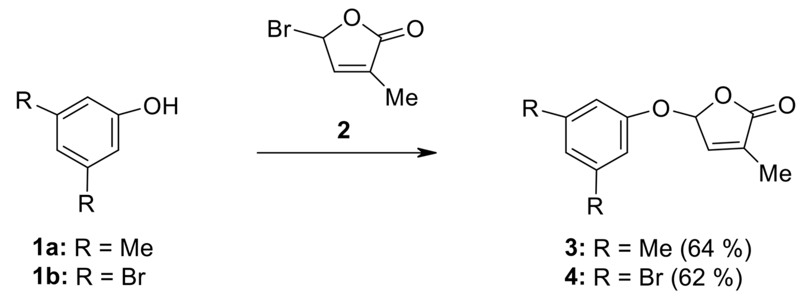
4.3. General Procedure for Preparation of SL Mimics Derived from 3,5-Disubstituted Phenols

To a solution of 3,5-disubstituted phenol 1a or 1b (10 mmol) in 20 mL acetone anhydrous potassium carbonate (1.52 g, 11 mmol) was added, then crude 5-bromo-3-methyl-5H-furan-2-one 2 (2.21 g, 12.7 mmol) was slowly added under stirring at room temperature. The stirring was continued for 16 h at room temperature. After cooling, the reaction mixture was filtered off to remove the potassium carbonate and the solvent was evaporated. The residue was chromatographed on a silicagel/alumina 70–230 mesh packed column eluting with dichloromethane and the final compounds were obtained after the complete removal of the eluting solvent.
  • 5-(3,5-Dimethylphenoxy)-3-methyl-5H-furan-2-one (3) [SL-F1]. Viscous, pale yellow liquid after purification using column chromatography. Yield 64%. Anal. Calcd. for C13H14O3 (218.25): C 71.54, H 6.47%; Found: C 71.62, H 6.56%. ATR-FTIR (solid): 1771 cm−1 (νC=O). 1H NMR (300 MHz, CDCl3) δ (ppm): 2.12 (s, 3H, Me), 2.44 (s, 6H, 2Me, phenyl), 6.38 (s, 1H, H-5), 6.87 (3H, H-4, 2H phenyl), 7.10 (s, 1H, phenyl). 13C NMR (75 MHz, CDCl3) δ (ppm): 10.5 (Me), 21.3 (2Me, phenyl), 99.1 (C-5), 114.4 (C-2, C-6, phenyl), 125.1(C-4, phenyl), 133.9 (C-3), 139.4 (C-3, C-5, phenyl), 142.6 (C-4), 156.4 (C-1, phenyl), 171.4 (C=O).
  • 5-(3,5-Dibromophenoxy)-3-methyl-5H-furan-2-one (4) [SL-F2]. The compound was crystallized from cyclohexane as white crystals with mp 88–91 °C; yield 62%. Anal. Calcd. for C11H8Br2O3 (347.99): C 37.97, H 2.32%; Found: C 38.91, H 2.40%. ATR-FTIR (solid): 1776 cm−1 (νC=O). 1H NMR (300 MHz, CDCl3) δ (ppm): 2.02 (t, 3H, Me, J = 1.4 Hz), 6.23 (quintet, J = 1.4 Hz, 1H, H-5), 6.97 (quintet, J = 1.5 Hz, 1H, H-4), 7.24 (d, 2H, J = 1.4 Hz, H-2, H-6, phenyl), 7.24 (t, 1H, J = 1.4 Hz, H-4 phenyl). 13C NMR (75 MHz, CDCl3) δ (ppm): 10.8 (Me), 98.5 (C-5), 113.2 (C-2, C-6, phenyl), 123.3 (C-3, C-5, phenyl), 129.4 (C-4, phenyl), 134.9 (C-3), 141.7 (C-4), 157.3 (C-1, phenyl), 170.9 (C=O).

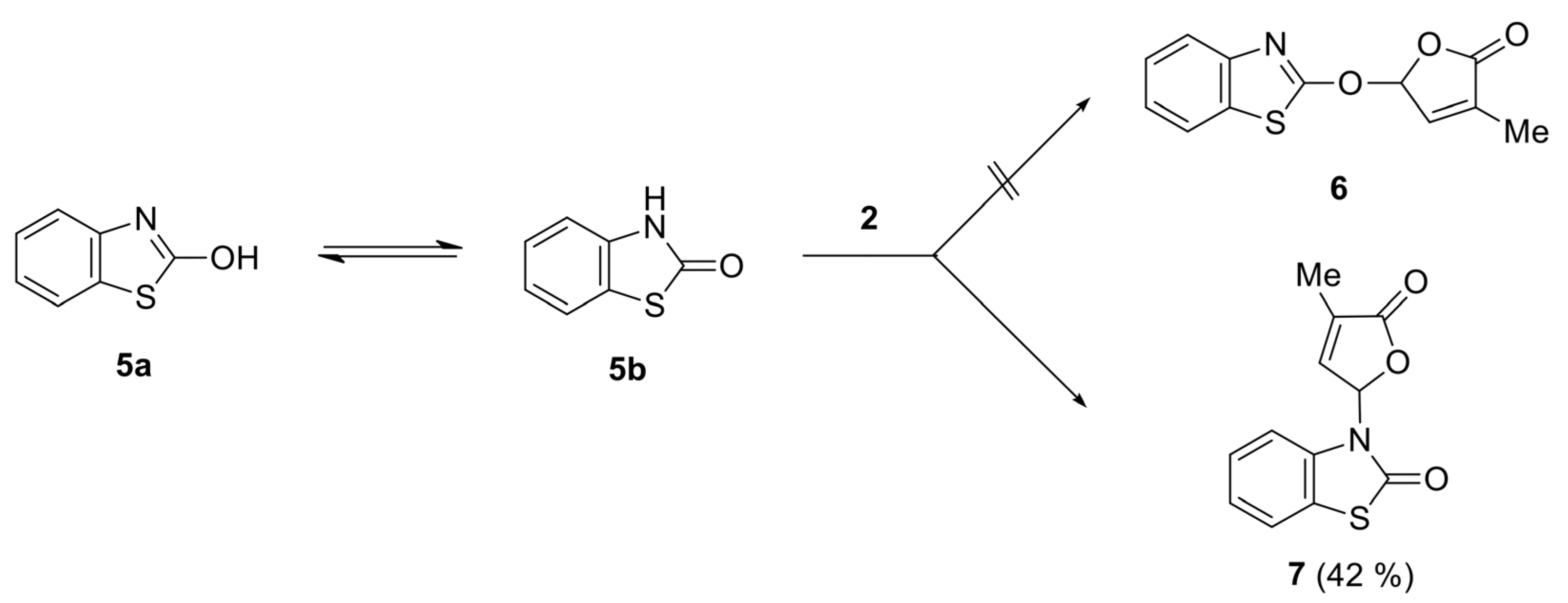
4.4. General Procedure for Preparation of SL Mimics Derived from Benzothiazoles

6-Bromo-3H-benzothiazol-2-one 8 was prepared by treating the commercially product 3H-benzothiazol-2-one 5 with bromine in acetic acid [74]. 5-Nitro-3H-benzothiazol-2-one 10 [75] was prepared by treating the commercially product 3H-benzothiazol-2-one 5 with nitric acid in acetic acid.
Anhydrous potassium carbonate (1.52 g, 11 mmol) was added into a solution of (10 mmol) 2-hydroxybenzothiazole 5, 6-bromo-2-hydroxybenzothiazole 8 or 6-nitro-2-hydroxybenzotiazole 10 in 20 mL acetone, then crude 5-bromo-3-methyl-5H-furan-2-one 2 (2.21 g, 12.7 mmol) was slowly added under stirring at room temperature. The stirring was continued for 16 h at room temperature. After cooling, the reaction mixture was filtered off to remove the potassium carbonate and the solvent was evaporated. The residue was chromatographed on an alumina-packed column eluting with dichloromethane and the final compounds were crystallized from a suitable solvent.
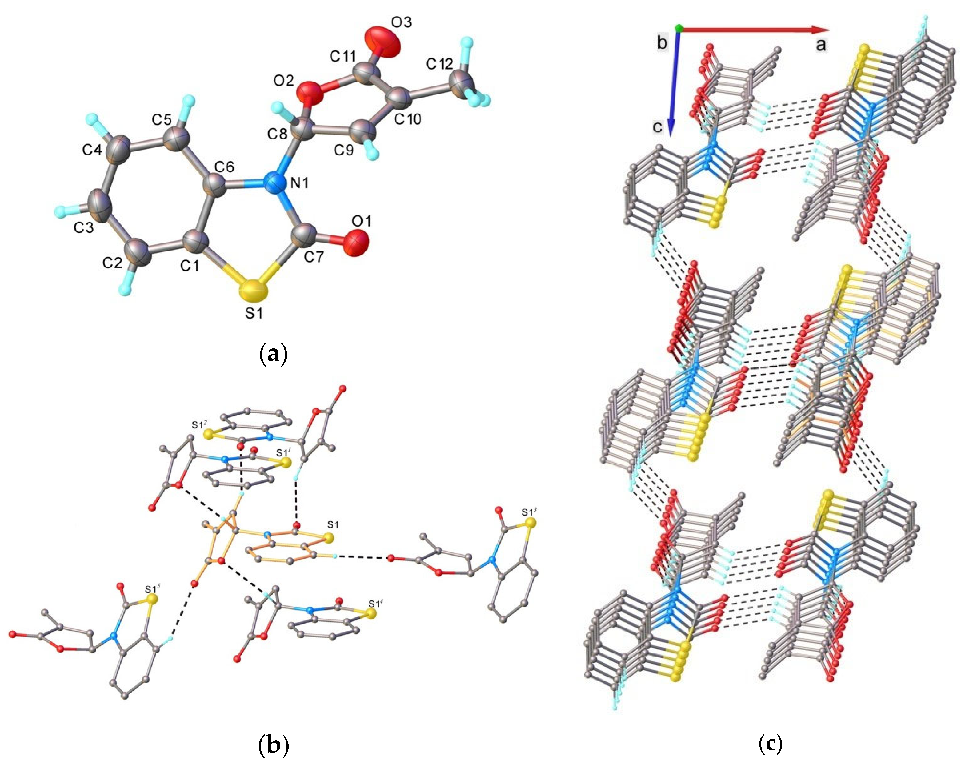
  • 3-(4-Methyl-5-oxo-2,5-dihydrofuran-2-yl)-3H-benzothiazol-2-one (7) [SL-F3]. The compound was crystallized from 2-propanol as white crystals with mp 153–155 °C; yield 42%. Anal. Calcd. for C12H9NO3S (M 247.27): C 58.29, H 3.67, N 5.66%; Found: C 58.21, H 3.42, N 5.71%. ATR-FTIR (solid): 1764 cm−1 (νC=O, lactone), 1673 cm−1 (νC=O, lactam). 1H NMR (300 MHz, CDCl3) δ (ppm): 2.28 (s, 3H, Me), 7.07 (d, 1H, J = 7.7 Hz, benzothiazole), 7.16 (bs, 1H, H-5), 7.35–7.42 (m, 3H, H-4 and 2H-benzothiazole), 7.58–7.60 (m, 1H, benzothiazole). 13C NMR (75 MHz, CDCl3) δ (ppm): 10.5 (Me), 81.5 (C-5), 111.9 (C-4, benzothiazole), 122.0 (C-7a, benzothiazole), 123.0, 124.2, 126.8 (C-5, C-6, C-7, benzothiazole), 134.4 (C-3), 135.0 (C-3a, benzothiazole), 143.1 (C-4), 170.0, 171.2 (2 C=O).
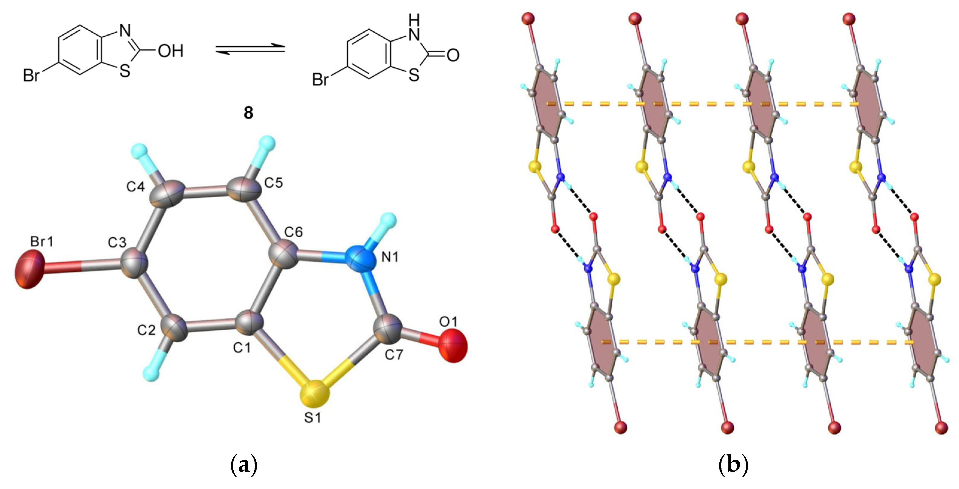
  • 6-Bromo-3-(4-methyl-5-oxo-2,5-dihydrofuran-2-yl)-3H-benzothiazol-2-one (9) [SL-F5]. The compound was crystallized from 2-propanol as white crystals with mp 176–177 °C, yielding 44%. Anal. Calcd. for C12H8BrNO3S (M 326.17): C 44.19, H 2.47, N 4.29%; Found: C 44.14, H 2.53, N 4.34. ATR-FTIR (solid): 1755 cm−1 (νC=O, lactone), 1690 cm−1 (νC=O, lactam). 1H NMR (300 MHz, CDCl3) δ (ppm): 2.27 (t, 3H, Me, J = 1.9 Hz), 6.94 (d, 1H, H-4, J = 8.8 Hz, benzothiazole), 7.10 (quintet, J = 1.9 Hz, 1H, H-5), 7.34 (quintet, J = 1.6 Hz, 1H, H-4), 7.53 (dd, 1H, H-5, J = 8.8, 1.9 Hz, benzothiazole), 7.72 (d, 1H, H-7, J = 1.9 Hz, benzothiazole). 13C NMR (75 MHz, CDCl3) δ (ppm): 11.1 (Me), 81.5 (C-5), 113.1 (C-4, benzothiazole), 117.0 (C-6, benzothiazole), 124.0 (C-7a, benzothiazole), 124.5, 128.8 (C-5, C-7, benzothiazole), 134.0 (C-3), 134.6 (C-3a, benzothiazole), 142.7 (C-4), 169.2, 170.9 (2 C=O).
  • 6-Nitro-3-(4-methyl-5-oxo-2,5-dihydrofuran-2-yl)-3H-benzothiazol-2-one (11) [SL-F6]. The compound was crystallized from acetonitrile as white crystals with mp 183–185 °C, yielding 39%. Anal. Calcd. for C12H8N2O5S (M 292.27): C 49.31, H 2.76, N 9.58%; Found: C 49.52, H 2.87, N 9.34%. ATR-FTIR (solid): 1769 cm−1 (νC=O, lactone), 1730 cm−1 (νC=O, lactam). 1H NMR (500 MHz, CDCl3) δ (ppm): 2.13 (t, 3H, Me, J = 1.7 Hz), 6.98 (t, J = 1.8 Hz, 1H, H-5), 7.05 (d, 1H, H-4, J = 9.0 Hz, benzothiazole), 7.23 (t, J = 1.7 Hz, 1H, H-4), 8.19 (dd, 1H, H-5, J = 9.0, 1.3 Hz, benzothiazole), 8.36 (d, 1H, H-7, J = 1.3 Hz, benzothiazole). 13C NMR (100 MHz, CDCl3) δ (ppm): 10.9 (Me), 81.4 (C-5), 111.6 (C-4, benzothiazole), 118.0 (C-5, benzothiazole), 122.8 (C-7, benzothiazole), 123.3 (C-7a, benzothiazole), 134.9 (C-3), 139.6 (C-3a, benzothiazole), 142.1 (C-4), 144.1(C-5, benzothiazole), 169.1, 170.5 (2 C=O).

4.5. Single Crystal X-ray Diffraction

The X-ray diffraction measurements were carried out with a Rigaku Oxford—Diffraction XCALIBUR E CCD diffractometer (Rigaku, Tokyo, Japan) equipped with graphite-monochromated MoKα radiation. The unit cell determination and data integration were carried out using the CrysAlis package of Oxford Diffraction (CrysAlisPro Software system, version 1.171.41.64, Rigaku Corporation, Oxford, UK). The structures were solved by Intrinsic Phasing using Olex2 software version 1.5 [120] with the SHELXT structure solution program [121] and refined using full-matrix least-squares on F2 with SHELXL-2015 [122] using an anisotropic model for non-hydrogen atoms. All H atoms were introduced in idealized positions (dCH = 0.96 Å) using the riding model. The H-atom attached to nitrogen were localized from difference Fourier maps, and their positional parameters were verified according to H-bonds geometry. The molecular plots were obtained using the Olex2 program. The crystallographic data and refinement details are quoted in Table S1, while bond lengths and angles are summarized in Tables S2 and S3 in the Supplementary Information (SI).

4.6. Biological Activities Tests

4.6.1. Bioassay of Strigolactones Mimics

The Ch. sorokiniana NIVA-CHL 176 strain was cultivated in the presence of the strigolactone mimics, and several parameters specific for microalgae growth and development, and biotechnological application were determined: absorbance at 750 nm, dry biomass, photosynthetic pigments, soluble protein content.

4.6.2. Growth Media

Microalgae cultures were grown in BG11 nutrient medium [123], which was prepared using stock solutions, the final concentration being: 17.6 mM NaNO3, 0.23 mM KHPO4, 0.3 mM MgSO4·7H2O, 0.24 mM CaCl2·2H2O, 0.31 mM Citric Acid·H2O, 0.021 mM FeCl3, 0.19 mM Na2EDTA·2H2O, 0.046 mM H3BO3, 9mM MnCl2·4H2O, 0.77mM ZnSO4·7H2O, 1.6 mM Na2MoO4·2H2O, 0.3 mM CuSO4·5H2O, 0.17 mM Co(NO3)2·6H2O. The medium was sterilized in an autoclave (MLS 375 1L, PHCBi, Wood Dale, IL, USA) at 121 °C for 15 min, and the Erlenmeyer flasks used for culturing were sterilized in a universal laboratory oven (UN 75, Memmert, Schwabach, Germany) at 180 °C for 3 h.

4.6.3. Treatment Solutions

To solubilize the analog strigolactones SL-F1 (V1), SL-F2 (V2), SL-F3 (V3), SL-F5 (V4), and SL-F6 (V5), we used dimethyl sulfoxide (DMSO) as solvent. Five stock solutions of 1 mM were prepared, considering the molar mass of each chemical. The treatments were done at two SL concentrations: the final concentrations tested were 1 × 10⁻⁷ M (c1) and 1 × 10⁻⁹ M (c2), prepared from the stock solution by dilutions with BG11 medium. We also included a solvent control (SC, 10−7 M DMSO) and a reference product, strigolactone analog GR24 (C-GR24). The treatments were assessed in triplicate in a final culturing volume of 25 mL in 50 mL sterilized Erlenmeyer flasks.

4.6.4. Culturing Conditions

The growth medium was inoculated at a final concentration of 1% microalgae biomass with the microalgae Ch. sorokiniana using a fresh culture in a laminar flow microbiological hood (Bio 2 Advantage Plus, Azbil Telstar, Terrassa, Spain) to avoid contamination. The cultures were kept in a growth chamber (AlgaeTron AG-230-ECO, Photon Systems Instruments, Drásov, Czech Republic) under controlled conditions of temperature and light (25° ± 1° C, illumination with white, fluorescent lamp at 120 μmol/m2·s, with a light/dark photoperiod of 14/10 h) and under continuous orbital shaking of 130–150 rpm (Unimax 1010 orbital shaker, Heidolph, Schwabach, Germany), for 14 days.

4.6.5. Growth Parameters
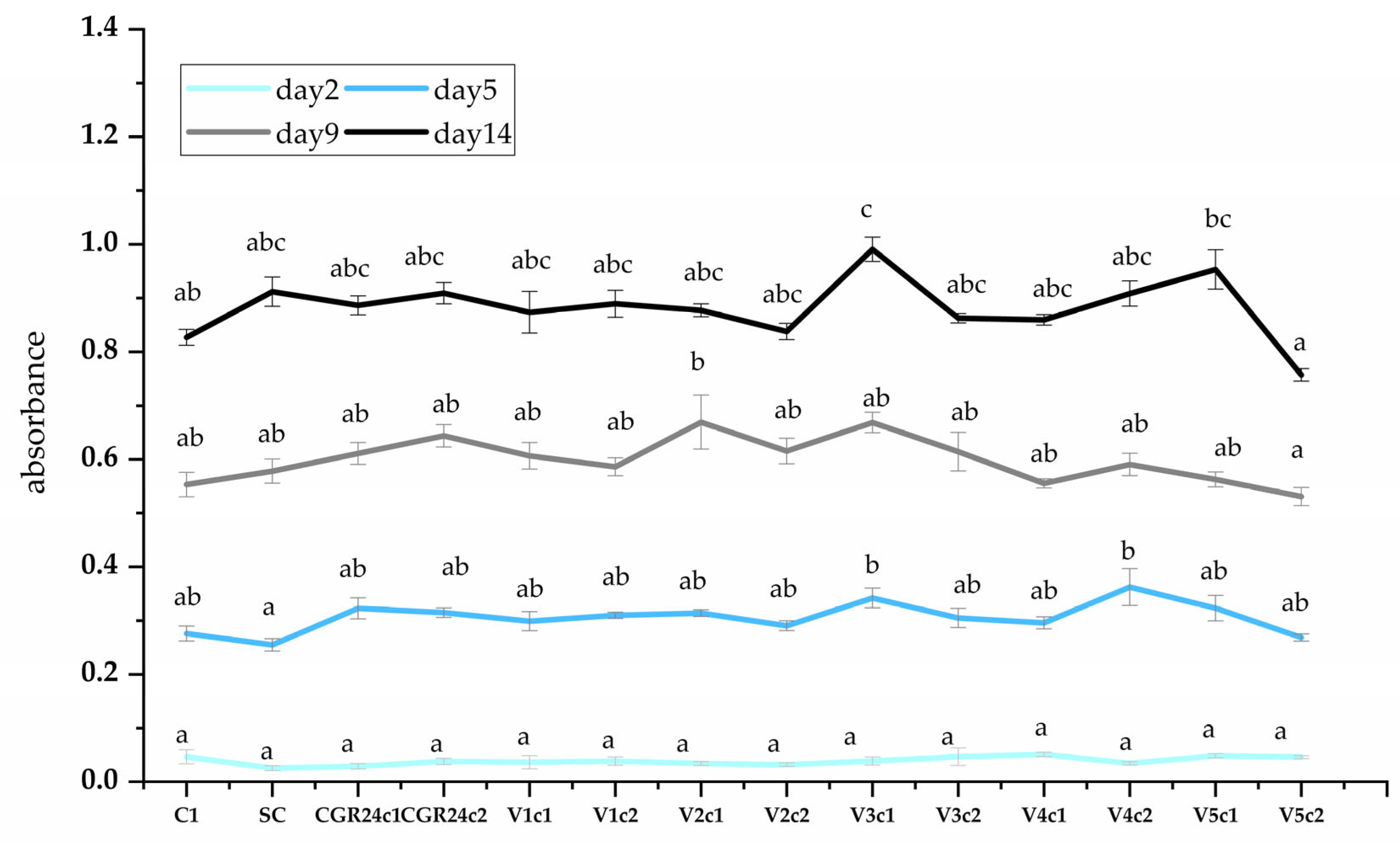
We determined the microalgae growth by measuring the optical density on a microplate reader (CLARIOStar, BMG Labtech, Ortenberg, Germany) at 750 nm of the aseptically taken samples from microalgae culture [124]. The readings were performed in sterile 96-well plates in triplicates.

4.6.6. Biomass

A volume of 22 mL of Ch. sorokiniana cultures was added to a pre-weighed (W1) Falcon tube and centrifuged with a Universal 320 R centrifuge (Hettich, Tuttlingen, Germany), at 5550× g for 5 min. After discarding the supernatant, the sediment was dried at 80 °C to a constant weight in a universal laboratory oven (UN 75, Memmert, Schwabach, Germany). The dried biomass accumulation (g/L) of microalgae was assessed in triplicate using the following formula [123]:
W = (W2 − W1) × 1000/V
where W1 represents the empty Falcon tube weight (g), W2 represents the Falcon tube weight, including microalgae-dried biomass, and V represents the culture volume tested. The biomass was used for the determination of the soluble proteins.

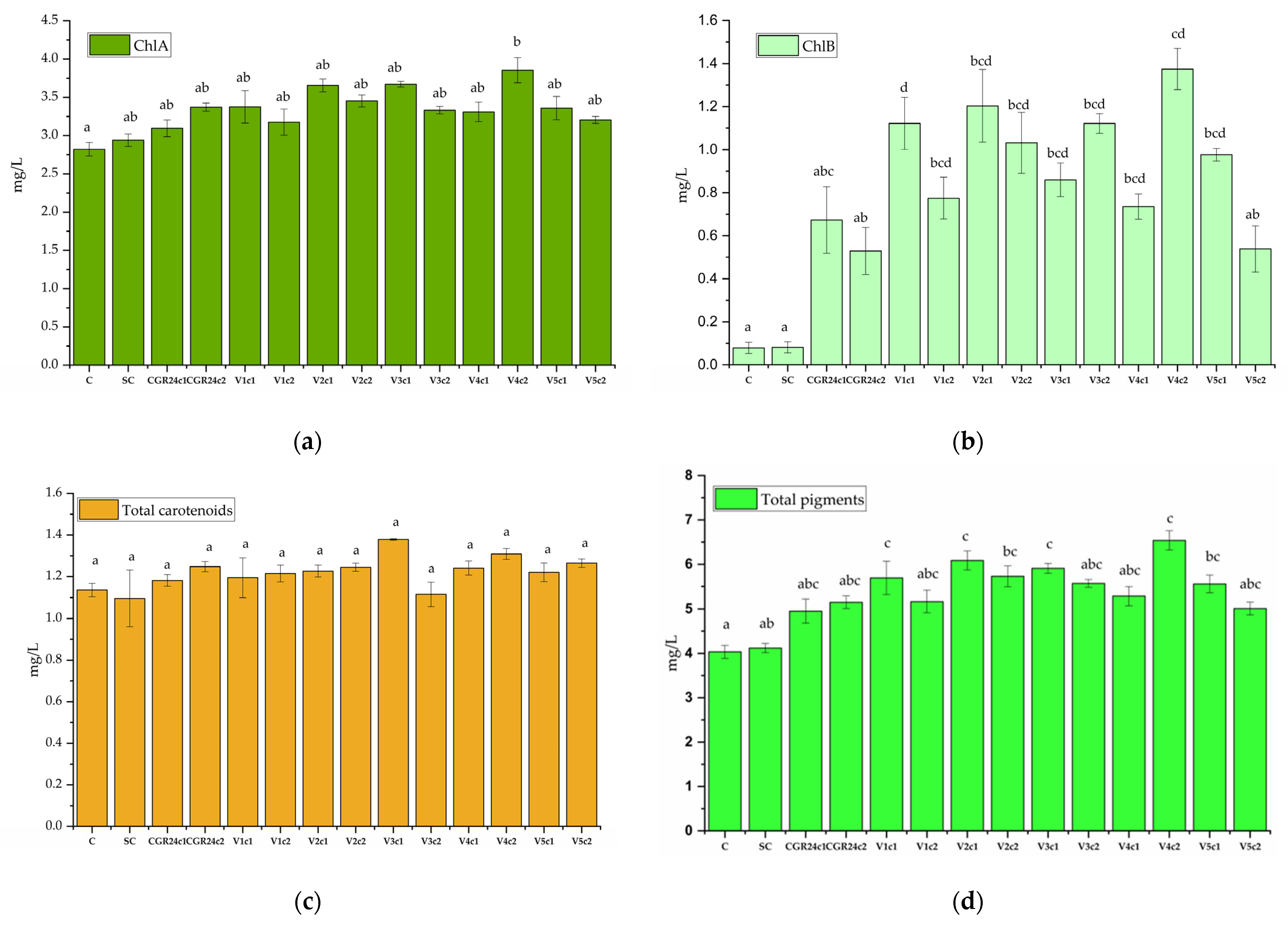
4.6.7. Pigment Concentrations after Extraction

For pigment measuring, we used the method described by Chai [125]. Briefly, 2 mL of each sample was centrifuged at 7400× g for 3 min. Over the remaining pellet, we added 2 mL of DMSO preheated to 60 °C and vortex for 10 min. Before reading the absorbance with a UV-Vis spectrophotometer (CLARIOstar, BMG Labtech, Ortenberg, Germany), the Eppendorf tubes were centrifuged using the same parameters mentioned above. We measured the absorbance at three different wavelengths, 480 nm, 649 nm, and 665 nm, necessary for the calculation of the content of chlorophyll a, chlorophyll b, total carotenoids, and total pigments in the samples, according to the following formulas:
Chlorophyll a (ChlA) (mg/L) = 12.47 × (OD665) − 3.62 × (OD649)
Chlorophyll b (ChlB) (mg/L) = 25.06 × (OD649) − 6.5 × (OD665)
Total carotenoid (mg/L) = [1000 (OD480) − 1.29 (ChlA) − 53.78 (ChlB)]/220
Total pigments (mg/L) = (1) + (2) + (3)

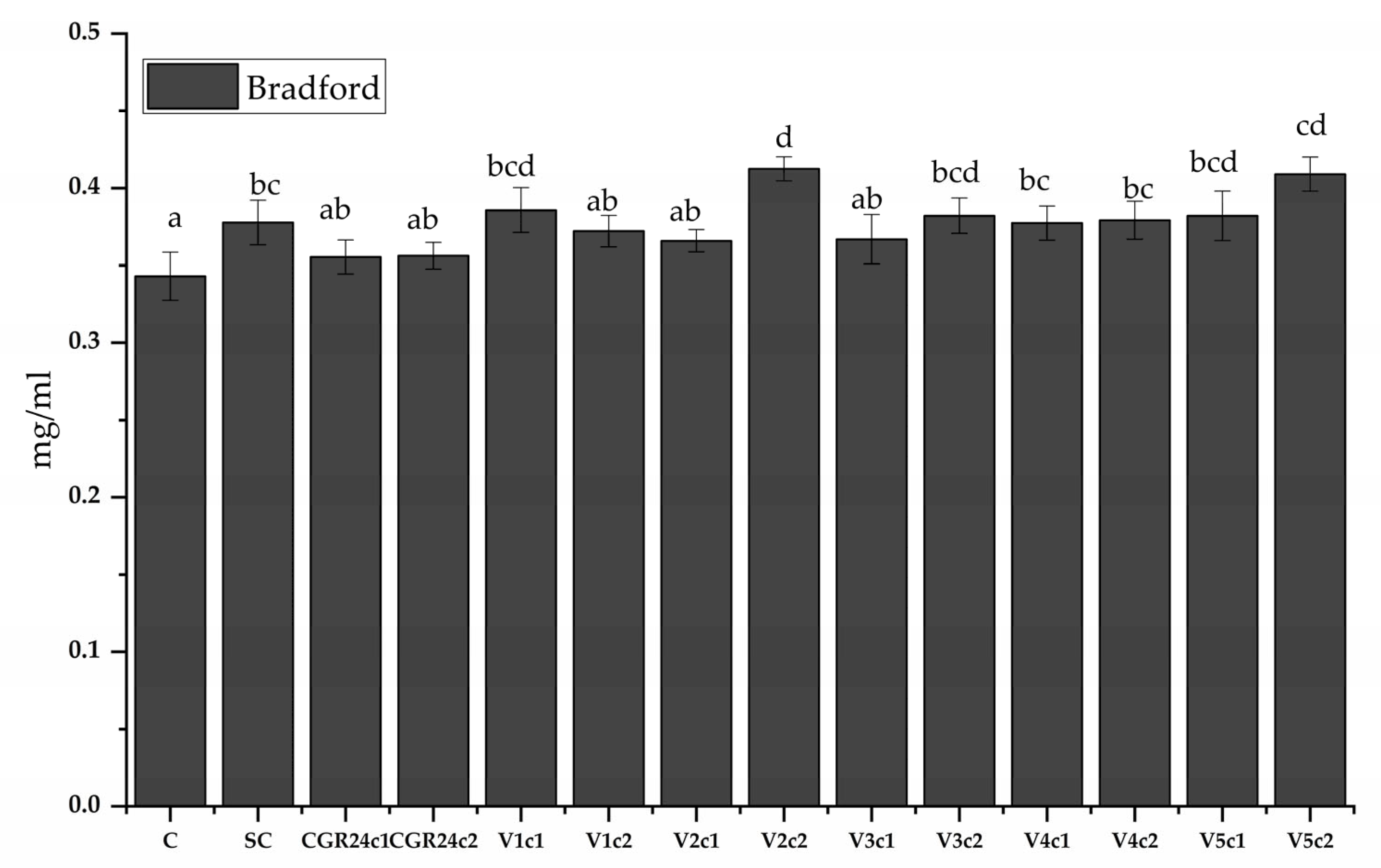
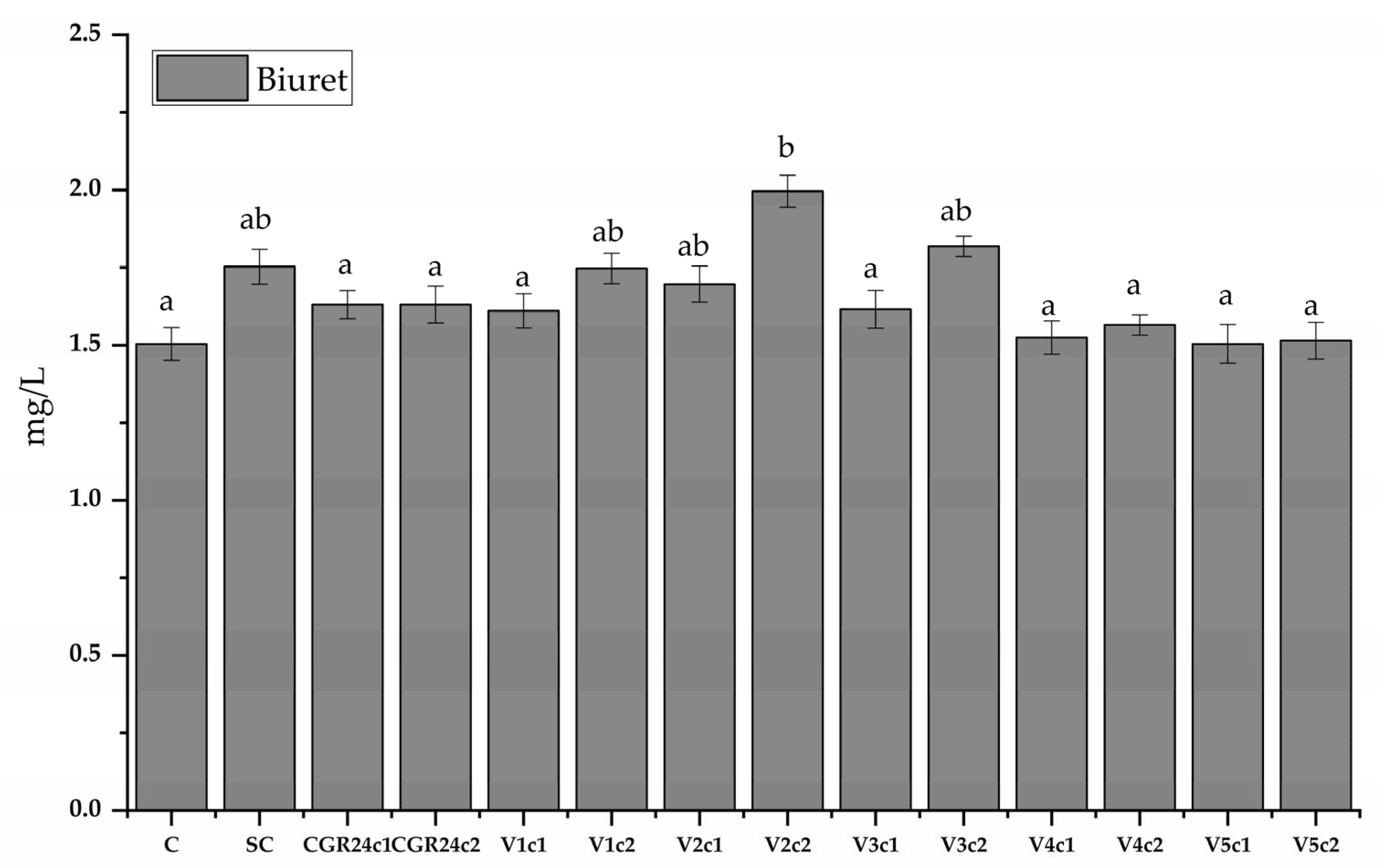
4.6.8. Protein Content

For protein extraction, we used freeze-dried biomass obtained after 14 days of cultivation. Basically, for solubilization, we left 5 mg microalgae powder suspended in 50 μL of 1 M NaOH for 10 min in a water bath at 80 °C. Then, we added 450 μL of distilled water, and the resulting solution was centrifuged for 30 min at 12,000× g (in a Universal 320 R centrifuge, Hettich, Tuttlingen, Germany), and the supernatant was moved to another tube. The above procedure was repeated twice, and the last extract was combined. The protein content was determined using two methods, Bradford and Biuret. For the Bradford method, we used bovine serum albumin (BSA) as standard in a concentration series of 0–10 µg/mL [95]. Briefly, over 200 µL of a sample, we added 50 µL of Bradford Reagent (Bradford Reagent, Sigma, Merck Group, Darmstadt, Germany), incubated for 5 min, and read the absorbance in a 96 well plate reader (CLARIOStar, BMG Labtech, Ortenberg, Germany) at a wavelength λ of 595 nm. For Biuret, the BSA standard curve was 0–10 mg/mL. Briefly, we added 200 µL of Biuret reagent to 40 µL of the sample; we incubated it in the dark at room temperature for 30 min, and then the absorbance (CLARIOStar, BMG Labtech, Ortenberg, Germany) was read at the wavelength λ = 555 nm.

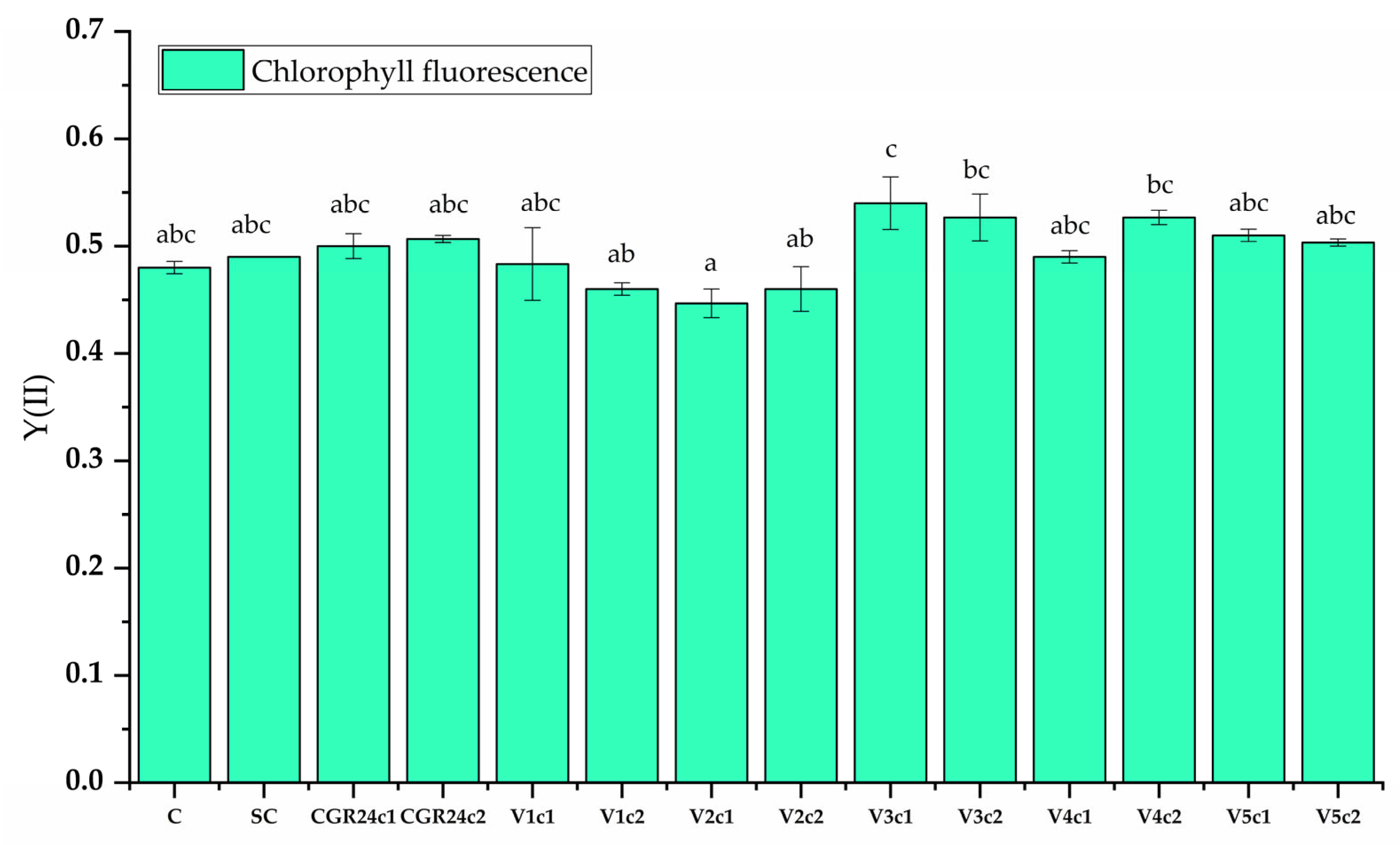
4.6.9. Chlorophyll Fluorescence

The chlorophyll fluorescence was measured using a handheld PAM fluorometer (PSI AquaPen AP 110/P, Photon Systems Instruments, Drásov, Czech Republic), according to manufacturer instructions, after microalgae adaptation to dark for 5 min. We calculated the maximum quantum yield of PSII photochemistry φP0 according to the following formula [126]:
φP0 = Fv/Fm
where Fv—maximum variable fluorescence, Fm-Fo; Fm—maximum chlorophyll a fluorescence (after actinic flash); Fo—minimum chlorophyll a fluorescence (after dark adaptation).

4.7. Statistical Analysis

The data were subjected to one-way ANOVA (with significance p ≤ 0.05) data analysis, and the mean differences were compared using the Tukey HSD post hoc test. All studies were conducted in triplicate, and the values were expressed as mean ± standard error. The statistical analysis was performed using IBM SPSS statistics software (SPSS version 26.0)

5. Conclusions

Novel strigolactone mimics that contain aryloxy groups or a benzothiazole scaffold linked to the bioactive 3-methyl-5H-furan-2-one unit have been synthesized and characterized. The first two strigolactone mimics bearing aryloxy groups were obtained by the coupling reactions of the corresponding 3,5-disubstituted phenols with 5-bromo-3-methyl-5H-furan-2-one in the presence of a basic catalyst. Three new synthetic strigolactone mimics in which the nitrogen atom from the benzothiazole nucleus is directly linked to 3-methyl-5H-furan-2-one ring are the results of the N-akylating reactions of the keto-tautomeric forms of 2-hydroxybenzothiazole derivatives, namely 3H-benzothiazol-2-one, 6-bromo-3H-benzothazol-2-one and 6-nitro-3H-benzothazol-2-one, respectively, with 5-bromo-3-methyl-5H-furan-2-one.
The novel strigolactone mimics modulate the photosynthesis and biomass accumulation in Ch. sorokiniana NIVA-CHL 176 and could be considered microalgae biostimulants. Our experiments demonstrate that their effects include stimulation of biomass accumulation (as a result on an enhanced nutrient uptake and nutrient use), stimulation of the photoprotective photosynthetic pigments (chlorophyll b) synthesis, and enhanced quantum yield efficiency in photosystem II (as a result of increased tolerance to the stress resulted from the application of light for long photoperiod) and improved biomass quality, due to the increased content of soluble proteins.
Beside the practical importance, as a tool to improve exploitation of microalgae biotechnology potential, our results suggest that the perception of strigolactones by microalgae is similar to that of other organisms, being related to the methylated butenolide D-ring.

Supplementary Materials

The following supporting information can be downloaded at: https://www.mdpi.com/article/10.3390/molecules28207059/s1, Figure S1. IR spectrum of (a) compound 3 (SL-F1) and (b) compound 4 (SL-F2). Figure S2. (a) The 1H NMR spectrum for compound 3 (SL-F1), recorded in CDCl3; (b) The 13C NMR spectrum for compound 3 (SL-F1), recorded in CDCl3. Figure S3. (a) The 1H NMR spectrum for compound 4 (SL-F2) recorded in CDCl3; (b) The 13C NMR spectrum for compound 4 (SL-F2) recorded in CDCl3. Figure S4. IR spectrum of (a) compound 9 (SL-F5) and (b) compound 11 (SL-F6). Figure S5. (a) The 1H NMR spectrum for compound 7 (SL-F3) recorded in CDCl3; (b) The 13C NMR spectrum for compound 7 (SL-F3) recorded in CDCl3. Figure S6. (a) The 1H NMR spectrum for compound 9 (SL-F5) recorded in CDCl3. (b) The 13C NMR spectrum for compound 9 (SL-F5) recorded in CDCl3. Figure S7. (a) The 1H NMR spectrum for compound 11 (SL-F6) recorded in CDCl3. (b) The 13C NMR spectrum for compound 11 (SL-F6) recorded in CDCl3. Figure S8. Space-filling representation of the crystal structure 7 showing the packing of 2D layers. Table S1. Bond distances (Å) and angles (°) of compound 8. Table S2. Bond distances (Å) and angles (°) of compound 7 (SL-F3). Table S3. Bond distances (Å) and angles (°) of compound 7 (SL-F3).

Author Contributions

Conceptualization, F.G., F.D. and F.O.; methodology, F.G., D.G.P. and C.D.; validation, D.C.-A., F.G. and F.D.; formal analysis, S.S.; investigation, D.G.P., F.G., C.D. and S.S.; resources, L.V. and F.O.; data curation, S.S.; writing—original draft preparation, F.G. and D.G.P.; writing—review and editing, D.C.-A. and F.O.; visualization, S.S., F.G. and D.C.-A.; supervision, F.O.; project administration, L.V. and F.O.; funding acquisition, L.V. and F.O. All authors have read and agreed to the published version of the manuscript.

Funding

This research was funded by project POC-A1-A1.2.3-G-2015-P_40_352-SECVENT, Sequential processes to close bioeconomy side stream and innovative bioproducts resulted from these, contract 81/2016, SMIS 105684, funded by Cohesion Funds of the European Union, subsidiary project 1500/2020 BioLignol. The APC was funded by the same project.

Institutional Review Board Statement

Not applicable.

Informed Consent Statement

Not applicable.

Data Availability Statement

X-ray diffraction data can be obtained free of charge via https://www.ccdc.cam.ac.uk/structures/, accessed on 10 September 2023 (or from the Cambridge Crystallographic Data Centre, 12 Union Road, Cambridge CB2 1EZ, UK; Fax: (+44) 1223-336-033; or [email protected]).

Acknowledgments

The authors thank Marius Ghiurea for his support on the determination of the quantum yield of Photosystem II, Bogdan Trică for the assistance with the statistical analysis, and Alina Vasilescu for the support with NMR analysis.

Conflicts of Interest

The authors declare no conflict of interest.

Sample Availability

Samples of the new strigolactone mimics are available from the authors.

Appendix A

The acronyms used for strigolactone mimics and analogs are summarized in Table A1.
Table A1. Chemical names, compound numbers in the paper, and compounds code for synthetic compounds with strigolactone activities.
Compound Number (Synthesis Series)Compound Code as Strigolactone Mimic/AnalogChemical Name
3SL-F15-(3,5-dimethylphenoxy)-3-methylfuran-2(5H)-one
4SL-F25-(3,5-dibromophenoxy)-3-methylfuran-2(5H)-one
7SL-F33-(4-methyl-5-oxo-2,5-dihydrofuran-2-yl)-3H-benzothiazol-2-one
9SL-F56-bromo-3-(4-methyl-5-oxo-2,5-dihydrofuran-2-yl)-3H-benzothiazol-2-one
11SL-F66-nitro-3-(4-methyl-5-oxo-2,5-dihydrofuran-2-yl)-3H-benzothiazol-2-one
-GR24 (strigolactone analog)—reference compound(rac)-GR24; [(±)-(3aR,8bS,E)-3-(((4-methyl-5-oxo-2,5-dihydrofuran-2-yl)oxy)methylene)-3,3a,4,8b-tetrahydro-2H-indeno [1,2-b]furan-2-one]

References

  1. Cook, C.E.; Whichard, L.P.; Turner, B.; Wall, M.E.; Egley, G.H. Germination of Witchweed (Striga lutea Lour.): Isolation and Properties of a Potent Stimulant. Science 1966, 154, 1189–1190. [Google Scholar] [CrossRef] [PubMed]
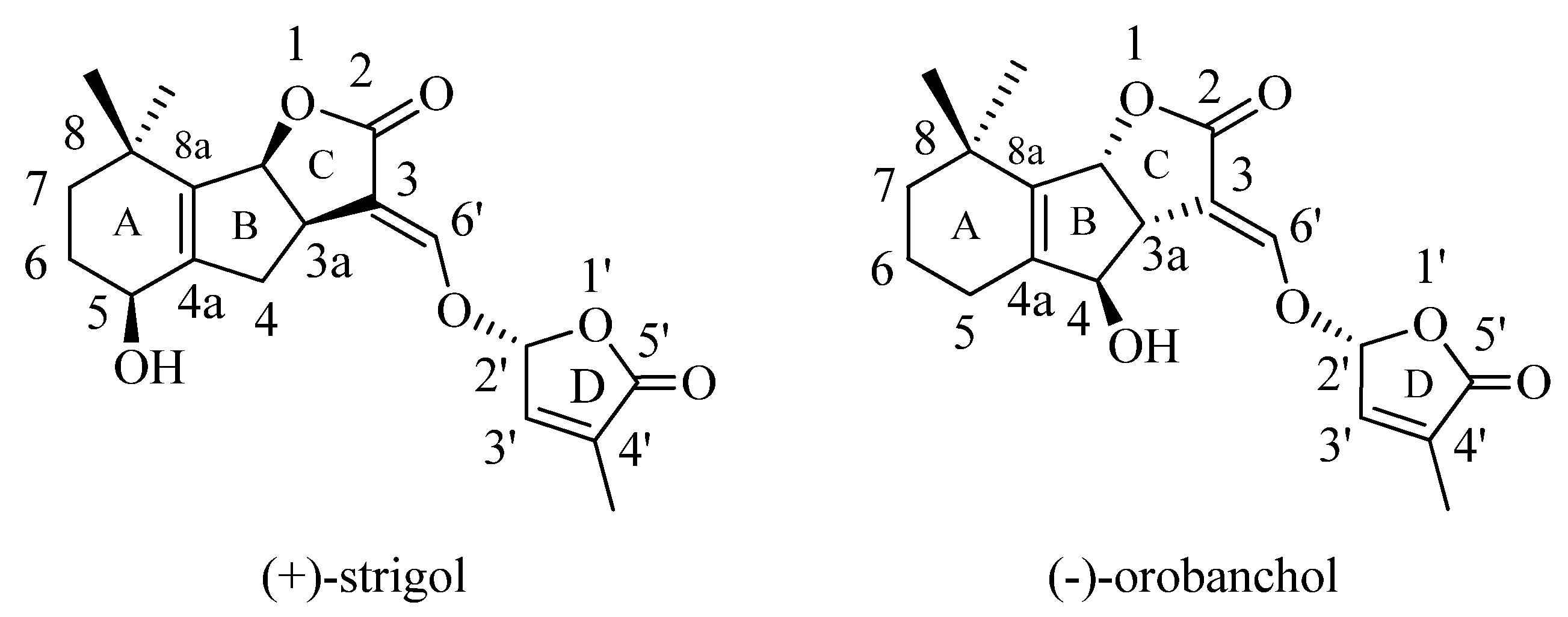
  2. Cook, C.E.; Whichard, L.P.; Wall, M.; Egley, G.H.; Coggon, P.; Luhan, P.A.; McPhail, A.T. Germination stimulants. II. Structure of strigol, a potent seed germination stimulant for witchweed (Striga lutea). J. Am. Chem. Soc. 1972, 94, 6198–6199. [Google Scholar] [CrossRef]
  3. Besserer, A.; Puech-Pages, V.; Kiefer, P.; Gomez-Roldan, V.; Jauneau, A.; Roy, S.; Portais, J.C.; Roux, C.; Becard, G.; Sejalon-Delmas, N. Strigolactones stimulate arbuscular mycorrhizal fungi by activating mitochondria. PLoS Biol. 2006, 4, e226. [Google Scholar] [CrossRef] [PubMed]
  4. Gomez-Roldan, V.; Fermas, S.; Brewer, P.B.; Puech-Pages, V.; Dun, E.A.; Pillot, J.P.; Letisse, F.; Matusova, R.; Danoun, S.; Portais, J.C.; et al. Strigolactone inhibition of shoot branching. Nature 2008, 455, 189–194. [Google Scholar] [CrossRef] [PubMed]
  5. Umehara, M.; Hanada, A.; Yoshida, S.; Akiyama, K.; Arite, T.; Takeda-Kamiya, N.; Magome, H.; Kamiya, Y.; Shirasu, K.; Yoneyama, K.; et al. Inhibition of shoot branching by new terpenoid plant hormones. Nature 2008, 455, 195–200. [Google Scholar] [CrossRef] [PubMed]
  6. Boyer, F.D.; de Saint Germain, A.; Pillot, J.P.; Pouvreau, J.B.; Chen, V.X.; Ramos, S.; Stevenin, A.; Simier, P.; Delavault, P.; Beau, J.M.; et al. Structure-activity relationship studies of strigolactone-related molecules for branching inhibition in garden pea: Molecule design for shoot branching. Plant Physiol. 2012, 159, 1524–1544. [Google Scholar] [CrossRef] [PubMed]
  7. Kohlen, W.; Ruyter-Spira, C.; Bouwmeester, H.J. Strigolactones: A new musician in the orchestra of plant hormones. Botany 2011, 89, 827–840. [Google Scholar] [CrossRef]
  8. Seto, Y.; Kameoka, H.; Yamaguchi, S.; Kyozuka, J. Recent advances in strigolactone research: Chemical and biological aspects. Plant Cell Physiol. 2012, 53, 1843–1853. [Google Scholar] [CrossRef]
  9. Brewer, P.B.; Koltai, H.; Beveridge, C.A. Diverse roles of strigolactones in plant development. Mol. Plant 2013, 6, 18–28. [Google Scholar] [CrossRef]
  10. Zwanenburg, B.; Pospisil, T. Structure and activity of strigolactones: New plant hormones with a rich future. Mol. Plant 2013, 6, 38–62. [Google Scholar] [CrossRef]
  11. Waldie, T.; McCulloch, H.; Leyser, O. Strigolactones and the control of plant development: Lessons from shoot branching. Plant J. 2014, 79, 607–622. [Google Scholar] [CrossRef] [PubMed]
  12. Koltai, H. Strigolactones are regulators of root development. New Phytol. 2011, 190, 545–549. [Google Scholar] [CrossRef] [PubMed]
  13. Bouwmeester, H.J.; Fonne-Pfister, R.; Screpanti, C.; De Mesmaeker, A. Strigolactones: Plant Hormones with Promising Features. Angew. Chem. Int. Ed. Engl. 2019, 58, 12778–12786. [Google Scholar] [CrossRef] [PubMed]
  14. Kapulnik, Y.; Koltai, H. Strigolactone involvement in root development, response to abiotic stress, and interactions with the biotic soil environment. Plant Physiol. 2014, 166, 560–569. [Google Scholar] [CrossRef] [PubMed]
  15. Pandey, A.; Sharma, M.; Pandey, G.K. Emerging roles of strigolactones in plant responses to stress and development. Front. Plant Sci. 2016, 7, 434. [Google Scholar] [CrossRef] [PubMed]
  16. Kleman, J.; Matusova, R. Strigolactones: Current research progress in the response of plants to abiotic stress. Biologia 2023, 78, 307–318. [Google Scholar] [CrossRef]
  17. Mostofa, M.G.; Li, W.; Nguyen, K.H.; Fujita, M.; Tran, L.P. Strigolactones in plant adaptation to abiotic stresses: An emerging avenue of plant research. Plant Cell Environ. 2018, 41, 2227–2243. [Google Scholar] [CrossRef] [PubMed]
  18. Yasuda, N.; Sugimoto, Y.; Kato, M.; Inanaga, S.; Yoneyama, K. (+)-Strigol, a witchweed seed germination stimulant, from Menispermum dauricum root culture. Phytochemistry 2003, 62, 1115–1119. [Google Scholar] [CrossRef]
  19. Xie, X.; Kusumoto, D.; Takeuchi, Y.; Yoneyama, K.; Yamada, Y.; Yoneyama, K. 2′-Epi-orobanchol and solanacol, two unique strigolactones, germination stimulants for root parasitic weeds, produced by tobacco. J. Agric. Food Chem. 2007, 55, 8067–8072. [Google Scholar] [CrossRef]
  20. Mangnus, E.M.; Zwanenburg, B. Tentative molecular mechanism for germination stimulation of Striga and Orobanche seeds by strigol and its synthetic analogs. J. Agric. Food Chem. 2002, 40, 1066–1070. [Google Scholar] [CrossRef]
  21. Yoneyama, K.; Xie, X.; Yoneyama, K.; Takeuchi, Y. Strigolactones: Structures and biological activities. Pest. Manag. Sci. 2009, 65, 467–470. [Google Scholar] [CrossRef] [PubMed]
  22. Zwanenburg, B.; Mwakaboko, A.S.; Reizelman, A.; Anilkumar, G.; Sethumadhavan, D. Structure and function of natural and synthetic signalling molecules in parasitic weed germination. Pest. Manag. Sci. 2009, 65, 478–491. [Google Scholar] [CrossRef] [PubMed]
  23. Xie, X.; Yoneyama, K.; Yoneyama, K. The strigolactone story. Annu. Rev. Phytopathol. 2010, 48, 93–117. [Google Scholar] [CrossRef] [PubMed]
  24. Akiyama, K.; Hayashi, H. Strigolactones: Chemical signals for fungal symbionts and parasitic weeds in plant roots. Ann. Bot. 2006, 97, 925–931. [Google Scholar] [CrossRef] [PubMed]
  25. Bouwmeester, H.J.; Roux, C.; Lopez-Raez, J.A.; Becard, G. Rhizosphere communication of plants, parasitic plants and AM fungi. Trends Plant Sci. 2007, 12, 224–230. [Google Scholar] [CrossRef] [PubMed]
  26. Akiyama, K.; Matsuzaki, K.; Hayashi, H. Plant sesquiterpenes induce hyphal branching in arbuscular mycorrhizal fungi. Nature 2005, 435, 824–827. [Google Scholar] [CrossRef] [PubMed]
  27. Akiyama, K.; Ogasawara, S.; Ito, S.; Hayashi, H. Structural requirements of strigolactones for hyphal branching in AM fungi. Plant Cell Physiol. 2010, 51, 1104–1117. [Google Scholar] [CrossRef] [PubMed]
  28. Foo, E.; Davies, N.W. Strigolactones promote nodulation in pea. Planta 2011, 234, 1073–1081. [Google Scholar] [CrossRef]
  29. McAdam, E.L.; Hugill, C.; Fort, S.; Samain, E.; Cottaz, S.; Davies, N.W.; Reid, J.B.; Foo, E. Determining the Site of Action of Strigolactones during Nodulation. Plant Physiol. 2017, 175, 529–542. [Google Scholar] [CrossRef]
  30. Soto, M.J.; Fernández-Aparicio, M.; Castellanos-Morales, V.; García-Garrido, J.M.; Ocampo, J.A.; Delgado, M.J.; Vierheilig, H. First indications for the involvement of strigolactones on nodule formation in alfalfa (Medicago sativa). Soil. Biol. Biochem. 2010, 42, 383–385. [Google Scholar] [CrossRef]
  31. Liu, F.; Rice, J.H.; Lopes, V.; Grewal, P.; Lebeis, S.L.; Hewezi, T.; Staton, M.E. Overexpression of strigolactone-associated genes exerts fine-tuning selection on soybean rhizosphere bacterial and fungal microbiome. Phytobiomes J. 2020, 4, 239–251. [Google Scholar] [CrossRef]
  32. Kim, B.; Westerhuis, J.A.; Smilde, A.K.; Flokova, K.; Suleiman, A.K.A.; Kuramae, E.E.; Bouwmeester, H.J.; Zancarini, A. Effect of strigolactones on recruitment of the rice root-associated microbiome. FEMS Microbiol. Ecol. 2022, 98, fiac010. [Google Scholar] [CrossRef]
  33. Wheeldon, C.D.; Hamon-Josse, M.; Lund, H.; Yoneyama, K.; Bennett, T. Environmental strigolactone drives early growth responses to neighboring plants and soil volume in pea. Curr. Biol. 2022, 32, 3593–3600.e3593. [Google Scholar] [CrossRef] [PubMed]
  34. Yoneyama, K.; Xie, X.; Nomura, T.; Yoneyama, K.; Bennett, T. Supra-organismal regulation of strigolactone exudation and plant development in response to rhizospheric cues in rice. Curr. Biol. 2022, 32, 3601–3608.e3603. [Google Scholar] [CrossRef] [PubMed]
  35. Waters, M. Plant development: Sizing up the competition with strigolactones. Curr. Biol. 2022, 32, R884–R886. [Google Scholar] [CrossRef] [PubMed]
  36. Jia, K.; Li, C.; Bouwmeester, H.J.; Al-Babili, S. Strigolactone Biosynthesis and Signal Transduction; Springer Nature: Berlin/Heidelberg, Germany, 2019. [Google Scholar]
  37. Waters, M.T.; Nelson, D.C.; Scaffidi, A.; Flematti, G.R.; Sun, Y.K.; Dixon, K.W.; Smith, S.M. Specialisation within the DWARF14 protein family confers distinct responses to karrikins and strigolactones in Arabidopsis. Development 2012, 139, 1285–1295. [Google Scholar] [CrossRef] [PubMed]
  38. Nakamura, H.; Xue, Y.L.; Miyakawa, T.; Hou, F.; Qin, H.M.; Fukui, K.; Shi, X.; Ito, E.; Ito, S.; Park, S.H.; et al. Molecular mechanism of strigolactone perception by DWARF14. Nat. Commun. 2013, 4, 2613. [Google Scholar] [CrossRef]
  39. Xie, X.; Yoneyama, K.; Kisugi, T.; Uchida, K.; Ito, S.; Akiyama, K.; Hayashi, H.; Yokota, T.; Nomura, T.; Yoneyama, K. Confirming stereochemical structures of strigolactones produced by rice and tobacco. Mol. Plant 2013, 6, 153–163. [Google Scholar] [CrossRef] [PubMed]
  40. Flematti, G.R.; Scaffidi, A.; Waters, M.T.; Smith, S.M. Stereospecificity in strigolactone biosynthesis and perception. Planta 2016, 243, 1361–1373. [Google Scholar] [CrossRef] [PubMed]
  41. Kondo, Y.; Tadokoro, E.; Matsuura, M.; Iwasaki, K.; Sugimoto, Y.; Miyake, H.; Takikawa, H.; Sasaki, M. Synthesis and seed germination stimulating activity of some imino analogs of strigolactones. Biosci. Biotechnol. Biochem. 2007, 71, 2781–2786. [Google Scholar] [CrossRef]
  42. Lachia, M.; Wolf, H.C.; Jung, P.J.; Screpanti, C.; De Mesmaeker, A. Strigolactam: New potent strigolactone analogues for the germination of Orobanche cumana. Bioorg. Med. Chem. Lett. 2015, 25, 2184–2188. [Google Scholar] [CrossRef] [PubMed]
  43. Yoneyama, K.; Xie, X.; Yoneyama, K.; Kisugi, T.; Nomura, T.; Nakatani, Y.; Akiyama, K.; McErlean, C.S.P. Which are the major players, canonical or non-canonical strigolactones? J. Exp. Bot. 2018, 69, 2231–2239. [Google Scholar] [CrossRef]
  44. Johnson, A.W.; Gowada, G.; Hassanali, A.; Knox, J.; Monaco, S.; Razavi, Z.; Rosebery, G. The preparation of synthetic analogues of strigol. J. Chem. Soc. Perkin Trans. 1 1981, 1734–1743. [Google Scholar] [CrossRef]
  45. Mangnus, E.M.; Zwanenburg, B. Synthesis, structural characterization, and biological evaluation of all four enantiomers of strigol analog GR7. J. Agric. Food Chem. 2002, 40, 697–700. [Google Scholar] [CrossRef]
  46. Mangnus, E.M.; Van Vliet, L.A.; Vandenput, D.A.L.; Zwanenburg, B. Structural modifications of strigol analogs. Influence of the B and C rings on the bioactivity of the germination stimulant GR24. J. Agric. Food Chem. 2002, 40, 1222–1229. [Google Scholar] [CrossRef]
  47. Boyer, F.D.; de Saint Germain, A.; Pouvreau, J.B.; Clavé, G.; Pillot, J.P.; Roux, A.; Rasmussen, A.; Depuydt, S.; Lauressergues, D.; Frey, N.F.D.; et al. New Strigolactone Analogs as Plant Hormones with Low Activities in the Rhizosphere. Mol. Plant 2014, 7, 675–690. [Google Scholar] [CrossRef] [PubMed]
  48. Nefkens, G.H.L.; Thuring, J.W.J.F.; Beenakkers, M.F.M.; Zwanenburg, B. Synthesis of a Phthaloylglycine-Derived Strigol Analogue and Its Germination Stimulatory Activity toward Seeds of the Parasitic Weeds Striga hermonthica and Orobanche crenata. J. Agric. Food Chem. 1997, 45, 2273–2277. [Google Scholar] [CrossRef]
  49. Mwakaboko, A.S.; Zwanenburg, B. Strigolactone analogs derived from ketones using a working model for germination stimulants as a blueprint. Plant Cell Physiol. 2011, 52, 699–715. [Google Scholar] [CrossRef]
  50. Mwakaboko, A.S.; Zwanenburg, B. Single step synthesis of strigolactone analogues from cyclic keto enols, germination stimulants for seeds of parasitic weeds. Bioorg. Med. Chem. 2011, 19, 5006–5011. [Google Scholar] [CrossRef]
  51. Lachia, M.; Jung, P.M.J.; De Mesmaeker, A. A novel approach toward the synthesis of strigolactones through intramolecular [2+2] cycloaddition of ketenes and ketene-iminiums to olefins. Application to the asymmetric synthesis of GR-24. Tetrahedron Lett. 2012, 53, 4514–4517. [Google Scholar] [CrossRef]
  52. Zwanenburg, B.; Mwakaboko, A.S. Strigolactone analogues and mimics derived from phthalimide, saccharine, p-tolylmalondialdehyde, benzoic and salicylic acid as scaffolds. Bioorg. Med. Chem. 2011, 19, 7394–7400. [Google Scholar] [CrossRef]
  53. Fukui, K.; Ito, S.; Ueno, K.; Yamaguchi, S.; Kyozuka, J.; Asami, T. New branching inhibitors and their potential as strigolactone mimics in rice. Bioorg. Med. Chem. Lett. 2011, 21, 4905–4908. [Google Scholar] [CrossRef]
  54. Fukui, K.; Ito, S.; Asami, T. Selective mimics of strigolactone actions and their potential use for controlling damage caused by root parasitic weeds. Mol. Plant 2013, 6, 88–99. [Google Scholar] [CrossRef]
  55. Zwanenburg, B.; Nayak, S.K.; Charnikhova, T.V.; Bouwmeester, H.J. New strigolactone mimics: Structure-activity relationship and mode of action as germinating stimulants for parasitic weeds. Bioorg. Med. Chem. Lett. 2013, 23, 5182–5186. [Google Scholar] [CrossRef]
  56. Dvorakova, M.; Soudek, P.; Vanek, T. Triazolide Strigolactone Mimics Influence Root Development in Arabidopsis. J. Nat. Prod. 2017, 80, 1318–1327. [Google Scholar] [CrossRef] [PubMed]
  57. Cala, A.; Ghooray, K.; Fernandez-Aparicio, M.; Molinillo, J.M.; Galindo, J.C.; Rubiales, D.; Macias, F.A. Phthalimide-derived strigolactone mimics as germinating agents for seeds of parasitic weeds. Pest. Manag. Sci. 2016, 72, 2069–2081. [Google Scholar] [CrossRef] [PubMed]
  58. Oancea, F.; Georgescu, E.; Matusova, R.; Georgescu, F.; Nicolescu, A.; Raut, I.; Jecu, M.-L.; Vladulescu, M.-C.; Vladulescu, L.; Deleanu, C. New Strigolactone Mimics as Exogenous Signals for Rhizosphere Organisms. Molecules 2017, 22, 961. [Google Scholar] [CrossRef] [PubMed]
  59. Aliche, E.B.; Screpanti, C.; De Mesmaeker, A.; Munnik, T.; Bouwmeester, H.J. Science and application of strigolactones. New Phytol. 2020, 227, 1001–1011. [Google Scholar] [CrossRef] [PubMed]
  60. Screpanti, C.; Fonne-Pfister, R.; Lumbroso, A.; Rendine, S.; Lachia, M.; De Mesmaeker, A. Strigolactone derivatives for potential crop enhancement applications. Bioorg. Med. Chem. Lett. 2016, 26, 2392–2400. [Google Scholar] [CrossRef]
  61. Walker, C.H.; Siu-Ting, K.; Taylor, A.; O’Connell, M.J.; Bennett, T. Strigolactone synthesis is ancestral in land plants, but canonical strigolactone signalling is a flowering plant innovation. BMC Biol. 2019, 17, 70. [Google Scholar] [CrossRef]
  62. Bonhomme, S.; Guillory, A. Synthesis and signalling of strigolactone and KAI2-ligand signals in bryophytes. J. Exp. Bot. 2022, 73, 4487–4495. [Google Scholar] [CrossRef] [PubMed]
  63. Shu, L.; Wang, Z.; Li, Y.; Zheng, Z. Supplementation of 5-deoxystrigol for higher pollutant removal and biogas quality improvement by different microalgae-based technologies. Water Environ. Res. 2023, 95, e10895. [Google Scholar] [CrossRef]
  64. Shen, X.; Xue, Z.; Sun, L.; Zhao, C.; Sun, S.; Liu, J.; Zhao, Y.; Liu, J. Effect of GR24 concentrations on biogas upgrade and nutrient removal by microalgae-based technology. Bioresour. Technol. 2020, 312, 123563. [Google Scholar] [CrossRef] [PubMed]
  65. Wang, H.; Wu, B.; Jiang, N.; Liu, J.; Zhao, Y.; Xu, J.; Wang, H. The effects of influent chemical oxygen demand and strigolactone analog concentration on integral biogas upgrading and pollutants removal from piggery wastewater by different microalgae-based technologies. Bioresour. Technol. 2023, 370, 128483. [Google Scholar] [CrossRef] [PubMed]
  66. Wei, J.; Wang, Z.; Zhao, C.; Sun, S.; Xu, J.; Zhao, Y. Effect of GR24 concentrations on tetracycline and nutrient removal from biogas slurry by different microalgae-based technologies. Bioresour. Technol. 2023, 369, 128400. [Google Scholar] [CrossRef] [PubMed]
  67. Ma, N.; Hu, C.; Wan, L.; Hu, Q.; Xiong, J.; Zhang, C. Strigolactones Improve Plant Growth, Photosynthesis, and Alleviate Oxidative Stress under Salinity in Rapeseed (Brassica napus L.) by Regulating Gene Expression. Front. Plant Sci. 2017, 8, 1671. [Google Scholar] [CrossRef] [PubMed]
  68. Ling, F.; Su, Q.; Jiang, H.; Cui, J.; He, X.; Wu, Z.; Zhang, Z.; Liu, J.; Zhao, Y. Effects of strigolactone on photosynthetic and physiological characteristics in salt-stressed rice seedlings. Sci. Rep. 2020, 10, 6183. [Google Scholar] [CrossRef]
  69. Oancea, A.; Georgescu, E.; Georgescu, F.; Nicolescu, A.; Oprita, E.I.; Tudora, C.; Vladulescu, L.; Vladulescu, M.C.; Oancea, F.; Deleanu, C. Isoxazole derivatives as new nitric oxide elicitors in plants. Beilstein J. Org. Chem. 2017, 13, 659–664. [Google Scholar] [CrossRef]
  70. Georgescu, E.; Oancea, A.; Georgescu, F.; Nicolescu, A.; Oprita, E.I.; Vladulescu, L.; Vladulescu, M.C.; Oancea, F.; Shova, S.; Deleanu, C. Schiff bases containing a furoxan moiety as potential nitric oxide donors in plant tissues. PLoS ONE 2018, 13, e0198121. [Google Scholar] [CrossRef]
  71. Ali, R.; Siddiqui, N. Biological Aspects of Emerging Benzothiazoles: A Short Review. J. Chem. 2013, 2013, 345198. [Google Scholar] [CrossRef]
  72. Zhilitskaya, L.V.; Shainyan, B.A.; Yarosh, N.O. Modern Approaches to the Synthesis and Transformations of Practically Valuable Benzothiazole Derivatives. Molecules 2021, 26, 2190. [Google Scholar] [CrossRef] [PubMed]
  73. MacAlpine, G.A.; Raphael, R.A.; Shaw, A.; Taylor, A.W.; Wild, H.-J. Synthesis of the germination stimulant (±)-strigol. J. Chem. Soc. Perkin Trans. 1 1976, 410–416. [Google Scholar] [CrossRef]
  74. D’Amico, J.J.; Bellinger, F.G.; Freeman, J.J. Synthesis of 2-oxo and 2-thioxo-3(2H)-benzothiazoleethanimic acid anhydride with acetic acid and related products. J. Heterocycl. Chem. 1988, 25, 1503–1509. [Google Scholar] [CrossRef]
  75. Loos, D.; Sidoova, E.; Sutoris, V. Benzothiazole Derivatives. 48. Synthesis of 3-Alkoxycarbonylmethyl-6-bromo-2-benzothiazolones and 3-Alkoxycarbonylmethyl-6-nitro-2-benzothiazolones as Potential Plant Growth Regulators. Molecules 1999, 4, 81–93. [Google Scholar] [CrossRef]
  76. Ma, S.; Zeng, W.; Huang, Y.; Zhu, X.; Xia, A.; Zhu, X.; Liao, Q. Revealing the synergistic effects of cells, pigments, and light spectra on light transfer during microalgae growth: A comprehensive light attenuation model. Bioresour. Technol. 2022, 348, 126777. [Google Scholar] [CrossRef] [PubMed]
  77. Amorim, M.L.; Soares, J.; Coimbra, J.; Leite, M.O.; Albino, L.F.T.; Martins, M.A. Microalgae proteins: Production, separation, isolation, quantification, and application in food and feed. Crit. Rev. Food Sci. Nutr. 2021, 61, 1976–2002. [Google Scholar] [CrossRef] [PubMed]
  78. Lucarini, A.; Kilikian, B. Comparative study of Lowry and Bradford methods: Interfering substances. Biotechnol. Tech. 1999, 13, 149–154. [Google Scholar] [CrossRef]
  79. Martínez, I.; Herrera, A.; Tames-Espinosa, M.; Bondyale-Juez, D.R.; Romero-Kutzner, V.; Packard, T.T.; Gómez, M. Protein in marine plankton: A comparison of spectrophotometric methods. J. Exp. Mar. Biol. Ecol. 2020, 526, 151357. [Google Scholar] [CrossRef]
  80. Robinson, T. The determination of proteins in plant extracts that contain polyphenols. Plant Sci. Lett. 1979, 15, 211–216. [Google Scholar] [CrossRef]
  81. Mitsuda, H.; Mitsunaga, T. Evaluation and elimination of the interference by starch in the biuret determination of wheat protein. Agric. Biol. Chem. 1974, 38, 1649–1655. [Google Scholar]
  82. Dupré, C.; Burrows, H.D.; Campos, M.G.; Delattre, C.; Encarnação, T.; Fauchon, M.; Gaignard, C.; Hellio, C.; Ito, J.; Laroche, C. Microalgal biomass of industrial interest: Methods of characterization. In Handbook on Characterization of Biomass, Biowaste and Related By-Products; Springer: Berlin/Heidelberg, Germany, 2020; pp. 537–639. [Google Scholar]
  83. Barbier, F.; Fichtner, F.; Beveridge, C. The strigolactone pathway plays a crucial role in integrating metabolic and nutritional signals in plants. Nat. Plants 2023, 9, 1191–1200. [Google Scholar] [CrossRef] [PubMed]
  84. Ke, D.; Guo, J.; Li, K.; Wang, Y.; Han, X.; Fu, W.; Miao, Y.; Jia, K.P. Carotenoid-derived bioactive metabolites shape plant root architecture to adapt to the rhizospheric environments. Front. Plant Sci. 2022, 13, 986414. [Google Scholar] [CrossRef] [PubMed]
  85. Halouzka, R.; Zeljkovic, S.C.; Klejdus, B.; Tarkowski, P. Analytical methods in strigolactone research. Plant Methods 2020, 16, 76. [Google Scholar] [CrossRef]
  86. Charnikhova, T.V.; Gaus, K.; Lumbroso, A.; Sanders, M.; Vincken, J.P.; De Mesmaeker, A.; Ruyter-Spira, C.P.; Screpanti, C.; Bouwmeester, H.J. Zealactones. Novel natural strigolactones from maize. Phytochemistry 2017, 137, 123–131. [Google Scholar] [CrossRef] [PubMed]
  87. Zwanenburg, B.; Cavar Zeljkovic, S.; Pospisil, T. Synthesis of strigolactones, a strategic account. Pest. Manag. Sci. 2016, 72, 15–29. [Google Scholar] [CrossRef] [PubMed]
  88. Zorrilla, J.G.; Rial, C.; Varela, R.M.; Molinillo, J.M.G.; Macías, F.A. Strategies for the synthesis of canonical, non-canonical and analogues of strigolactones, and evaluation of their parasitic weed germination activity. Phytochem. Rev. 2022, 21, 1627–1659. [Google Scholar] [CrossRef]
  89. Seto, Y.; Sado, A.; Asami, K.; Hanada, A.; Umehara, M.; Akiyama, K.; Yamaguchi, S. Carlactone is an endogenous biosynthetic precursor for strigolactones. Proc. Natl. Acad. Sci. USA 2014, 111, 1640–1645. [Google Scholar] [CrossRef] [PubMed]
  90. Zwanenburg, B.; Pospíšil, T.; Ćavar Zeljković, S. Strigolactones: New plant hormones in action. Planta 2016, 243, 1311–1326. [Google Scholar] [CrossRef]
  91. Mangnus, E.M.; Dommerholt, F.J.; De Jong, R.L.; Zwanenburg, B. Improved synthesis of strigol analog GR24 and evaluation of the biological activity of its diastereomers. J. Agric. Food Chem. 1992, 40, 1230–1235. [Google Scholar] [CrossRef]
  92. Moges, M.E.; Heistad, A.; Heidorn, T. Nutrient recovery from anaerobically treated blackwater and improving its effluent quality through microalgae biomass production. Water 2020, 12, 592. [Google Scholar] [CrossRef]
  93. Mihaila, E.-G.; Popa, D.G.; Dima, M.D.; Stoian, I.M.; Dinca, C.F.; Constantinescu-Aruxandei, D.; Oancea, F. Experimental Model for High-Throughput Screening of Microalgae Strains Useful for CO2 Fixation. Chem. Proc. 2022, 7, 25. [Google Scholar]
  94. Zoccali, M.; Giuffrida, D.; Salafia, F.; Socaciu, C.; Skjanes, K.; Dugo, P.; Mondello, L. First Apocarotenoids Profiling of Four Microalgae Strains. Antioxidants 2019, 8, 209. [Google Scholar] [CrossRef] [PubMed]
  95. Song, X.; Zhao, Y.; Li, T.; Han, B.; Zhao, P.; Xu, J.W.; Yu, X. Enhancement of lipid accumulation in Monoraphidium sp. QLY-1 by induction of strigolactone. Bioresour. Technol. 2019, 288, 121607. [Google Scholar] [CrossRef] [PubMed]
  96. Levasseur, W.; Taidi, B.; Lacombe, R.; Perré, P.; Pozzobon, V. Impact of seconds to minutes photoperiods on Chlorella vulgaris growth rate and chlorophyll a and b content. Algal Res. 2018, 36, 10–16. [Google Scholar] [CrossRef]
  97. Du Jardin, P. Plant biostimulants: Definition, concept, main categories and regulation. Sci. Hortic. 2015, 196, 3–14. [Google Scholar] [CrossRef]
  98. Popa, D.G.; Lupu, C.; Constantinescu-Aruxandei, D.; Oancea, F. Humic Substances as Microalgal Biostimulants-Implications for Microalgal Biotechnology. Mar. Drugs 2022, 20, 327. [Google Scholar] [CrossRef] [PubMed]
  99. Faraon, V.A.; Popa, D.G.; Tudor-Popa, I.; Mihăilă, E.G.; Oancea, F. Effect of humic acids from biomass biostimulant on microalgae growth. Chem. Proc. 2022, 7, 51. [Google Scholar]
  100. Constantinescu-Aruxandei, D.; Vlaicu, A.; Marinaș, I.C.; Vintilă, A.C.N.; Dimitriu, L.; Oancea, F. Effect of betaine and selenium on the growth and photosynthetic pigment production in Dunaliella salina as biostimulants. FEMS Microbiol. Lett. 2019, 366, fnz257. [Google Scholar] [CrossRef]
  101. Suparmaniam, U.; Lam, M.K.; Lim, J.W.; Rawindran, H.; Chai, Y.H.; Tan, I.S.; Fui Chin, B.L.; Kiew, P.L. The potential of waste chicken feather protein hydrolysate as microalgae biostimulant using organic fertilizer as nutrients source. IOP Conf. Ser. Earth Environ. Sci. 2022, 1074, 012028. [Google Scholar] [CrossRef]
  102. Suparmaniam, U.; Lam, M.K.; Rawindran, H.; Lim, J.W.; Pa’ee, K.F.; Yong, K.T.L.; Tan, I.S.; Chin, B.L.F.; Show, P.L.; Lee, K.T. Optimizing extraction of antioxidative biostimulant from waste onion peels for microalgae cultivation via response surface model. Energy Convers. Manag. 2023, 286, 117023. [Google Scholar] [CrossRef]
  103. Paliwal, C.; Mitra, M.; Bhayani, K.; Bharadwaj, S.V.V.; Ghosh, T.; Dubey, S.; Mishra, S. Abiotic stresses as tools for metabolites in microalgae. Bioresour. Technol. 2017, 244, 1216–1226. [Google Scholar] [CrossRef] [PubMed]
  104. Shi, T.Q.; Wang, L.R.; Zhang, Z.X.; Sun, X.M.; Huang, H. Stresses as First-Line Tools for Enhancing Lipid and Carotenoid Production in Microalgae. Front. Bioeng. Biotechnol. 2020, 8, 610. [Google Scholar] [CrossRef] [PubMed]
  105. Song, X.; Liu, B.-F.; Kong, F.; Ren, N.-Q.; Ren, H.-Y. Overview on stress-induced strategies for enhanced microalgae lipid production: Application, mechanisms and challenges. Resour. Conserv. Recycl. 2022, 183, 106355. [Google Scholar] [CrossRef]
  106. Zhao, Y.; Wang, H.P.; Han, B.; Yu, X. Coupling of abiotic stresses and phytohormones for the production of lipids and high-value by-products by microalgae: A review. Bioresour. Technol. 2019, 274, 549–556. [Google Scholar] [CrossRef] [PubMed]
  107. Wang, C.; Qi, M.; Guo, J.; Zhou, C.; Yan, X.; Ruan, R.; Cheng, P. The active phytohormone in microalgae: The characteristics, efficient detection, and their adversity resistance applications. Molecules 2021, 27, 46. [Google Scholar] [CrossRef] [PubMed]
  108. Proust, H.; Hoffmann, B.; Xie, X.; Yoneyama, K.; Schaefer, D.G.; Yoneyama, K.; Nogue, F.; Rameau, C. Strigolactones regulate protonema branching and act as a quorum sensing-like signal in the moss Physcomitrella patens. Development 2011, 138, 1531–1539. [Google Scholar] [CrossRef] [PubMed]
  109. Kodama, K.; Rich, M.K.; Yoda, A.; Shimazaki, S.; Xie, X.; Akiyama, K.; Mizuno, Y.; Komatsu, A.; Luo, Y.; Suzuki, H.; et al. An ancestral function of strigolactones as symbiotic rhizosphere signals. Nat. Commun. 2022, 13, 3974. [Google Scholar] [CrossRef] [PubMed]
  110. Kee, Y.J.; Ogawa, S.; Ichihashi, Y.; Shirasu, K.; Yoshida, S. Strigolactones in rhizosphere communication: Multiple molecules with diverse functions. Plant Cell Physiol. 2023, 64, 955–966. [Google Scholar] [CrossRef]
  111. Ito, S.; Braguy, J.; Wang, J.Y.; Yoda, A.; Fiorilli, V.; Takahashi, I.; Jamil, M.; Felemban, A.; Miyazaki, S.; Mazzarella, T.; et al. Canonical strigolactones are not the major determinant of tillering but important rhizospheric signals in rice. Sci. Adv. 2022, 8, eadd1278. [Google Scholar] [CrossRef]
  112. Sattar, A.; Ul-Allah, S.; Ijaz, M.; Sher, A.; Butt, M.; Abbas, T.; Irfan, M.; Fatima, T.; Alfarraj, S.; Alharbi, S.A. Exogenous application of strigolactone alleviates drought stress in maize seedlings by regulating the physiological and antioxidants defense mechanisms. Cereal Res. Commun. 2021, 50, 263–272. [Google Scholar] [CrossRef]
  113. Zhou, X.; Tan, Z.; Zhou, Y.; Guo, S.; Sang, T.; Wang, Y.; Shu, S. Physiological mechanism of strigolactone enhancing tolerance to low light stress in cucumber seedlings. BMC Plant Biol. 2022, 22, 30. [Google Scholar] [CrossRef] [PubMed]
  114. Wani, K.I.; Zehra, A.; Choudhary, S.; Naeem, M.; Khan, M.M.A.; Khan, R.; Aftab, T. Exogenous Strigolactone (GR24) Positively Regulates Growth, Photosynthesis, and Improves Glandular Trichome Attributes for Enhanced Artemisinin Production in Artemisia annua. J. Plant Growth Regul. 2022, 42, 4606–4615. [Google Scholar] [CrossRef] [PubMed]
  115. Figueroa, F.L.; Jerez, C.G.; Korbee, N. Use of in vivo chlorophyll fluorescence to estimate photosynthetic activity and biomass productivity in microalgae grown in different culture systems. Lat. Am. J. Aquat. Res. 2013, 41, 801–819. [Google Scholar] [CrossRef]
  116. Solovchenko, A.; Lukyanov, A.; Vasilieva, S.; Lobakova, E. Chlorophyll fluorescence as a valuable multitool for microalgal biotechnology. Biophys. Rev. 2022, 14, 973–983. [Google Scholar] [CrossRef] [PubMed]
  117. Maxwell, K.; Johnson, G.N. Chlorophyll fluorescence--a practical guide. J. Exp. Bot. 2000, 51, 659–668. [Google Scholar] [CrossRef] [PubMed]
  118. Iftikhar, I.; Shahbaz, M.; Wahid, M.A. Potential Role of Foliage Applied Strigolactone (GR24) on Photosynthetic Pigments, Gas Exchange Attributes, Mineral Nutrients and Yield Components of Zea mays (L.) Under Saline Regimes. Gesunde Pflanz. 2022, 75, 577–591. [Google Scholar] [CrossRef]
  119. Ji, Y.; Huang, L.; Wang, Z.; Xu, J.; Wei, J.; Zhao, Y. Performance of cocultivation of Chlorella vulgaris and four different fungi in biogas slurry purification and biogas upgrading by induction of strigolactone (GR24) and endophytic bacteria. Water Environ. Res. 2023, 95, e10896. [Google Scholar] [CrossRef] [PubMed]
  120. Dolomanov, O.V.; Bourhis, L.J.; Gildea, R.J.; Howard, J.A.K.; Puschmann, H. OLEX2: A complete structure solution, refinement and analysis program. J. Appl. Crystallogr. 2009, 42, 339–341. [Google Scholar] [CrossRef]
  121. Sheldrick, G.M. SHELXT—Integrated space-group and crystal-structure determination. Acta Crystallogr. Sect. A Found. Adv. A 2015, 71, 3–8. [Google Scholar] [CrossRef]
  122. Sheldrick, G.M. Crystal structure refinement with SHELXL. Acta Crystallogr. C Struct. Chem. 2015, 71, 3–8. [Google Scholar] [CrossRef]
  123. Kim, H.S.; Park, W.K.; Lee, B.; Seon, G.; Suh, W.I.; Moon, M.; Chang, Y.K. Optimization of heterotrophic cultivation of Chlorella sp. HS2 using screening, statistical assessment, and validation. Sci. Rep. 2019, 9, 19383. [Google Scholar] [CrossRef]
  124. Griffiths, M.J.; Garcin, C.; van Hille, R.P.; Harrison, S.T.L. Interference by pigment in the estimation of microalgal biomass concentration by optical density. J. Microbiol. Methods 2011, 85, 119–123. [Google Scholar] [CrossRef]
  125. Chai, S.; Shi, J.; Huang, T.; Guo, Y.; Wei, J.; Guo, M.; Li, L.; Dou, S.; Liu, L.; Liu, G. Characterization of Chlorella sorokiniana growth properties in monosaccharide-supplemented batch culture. PLoS ONE 2018, 13, e0199873. [Google Scholar] [CrossRef]
  126. Stirbet, A.; Lazár, D.; Kromdijk, J.; Govindjee, G. Chlorophyll a fluorescence induction: Can just a one-second measurement be used to quantify abiotic stress responses? Photosynthetica 2018, 56, 86–104. [Google Scholar] [CrossRef]
Figure 1. The two major groups of naturally occurring canonical strigolactones. Chemical structures drawn by the authors, based on the stereochemical structures published by Xie et al. [39].
Molecules 28 07059 g001
Scheme 1. Synthesis of SL mimics 3 (SL-F1) and 4 (SL-F2), derived from 3,5-disubstituted phenols.
Molecules 28 07059 sch001
Scheme 2. The two possible reaction products starting from 3H-benzothiazol-2-one. Compound 7 is SL-F3.
Molecules 28 07059 sch002
Figure 2. (a) The X-ray molecular structure of compound 8 with atom labeling and thermal ellipsoids at 50% level. H-bond parameters: N1-H···O1 [N1-H 0.86 Å, H···O1 1.97 Å, N1···O1(1 − x, 2 − y, 1 − z) 2.820(5) Å, ∠N1HO1 169.1°]. (b) The role of π-π stacking in the formation of 1D supramolecular architecture in the crystal structure of compound 8. Centroid-to-centroid contacts are drawn in orange-dashed lines.
Molecules 28 07059 g002
Scheme 3. Synthesis of SL mimics 9 (SL-F5) and 11 (SL-F6).
Molecules 28 07059 sch003
Figure 3. (a) The X-ray molecular structure of compound 7 (SL-F3) with atom labeling and thermal ellipsoids at 50% level. (b) Intermolecular H-bonds in the crystal structure of compound 7 (SL-F3). Non-relevant H-atoms are omitted. H-bond parameters: C2-H···O3 [C2-H 0.93 Å, H···O32.56 Å, C2···O3(x, 0.5 − y, −0.5 − z) 3.344(3) Å, ∠C2HO3 142.7°]; C8-H···O2 [C8-H 0.98 Å, H···O22.53 Å, C8···O2(x, 1 + y, z) 3.458(3) Å, ∠C8HO2 158.6°]; C9-H···O1 [C9-H 0.93 Å, H···O12.56 Å, C9···O1(0.5 − x, 1.5 − y, 1.5 − z) 3.360(3) Å, ∠C9HO13 144.7°]. (c) The 2D supramolecular layer of compound 7 (SL-F3) viewed along the b axis.
Molecules 28 07059 g003
Figure 4. Optical densities of the microalgae cultures. C1—control, without treatment. SC (solvent control)—treatment with dimethyl sulfoxide (DMSO), 10−7 M. CGR24c1—reference control with GR24 at 10−7 M. CGR24c2—reference control with GR24 at 10−9 M. V1c1—SL-F1 at 10−7 M, V1c2 SL-F1 at 10−9 M; V2c1—SL-F2 at 10−7 M, V2c2 SL-F2 at 10−9 M; V3c1—SL-F3 at 10−7 M, V3c2 SL-F3 at 10−9 M; V4c1—SL-F5 at 10−7 M, V4c2 SL-F5 at 10−9 M; V5c1—SL-F6 at 10−7 M, V5c2 SL-F6 at 10−9 M. The values represent means  ±  standard errors. The values followed by the same letter do not differ significantly at p < 0.05.
Molecules 28 07059 g004
Figure 5. The effect of strigolactone mimics and analog on the quantum yield (Y) of the photosynthetic system II after 14 days of growth. C1—control, without treatment. SC (solvent control)—treatment with dimethyl sulfoxide (DMSO), 10−7 M. CGR24c1—reference control with GR24 at 10−7 M. CGR24c2—reference control with GR24 at 10−9 M. V1c1—SL-F1 at 10−7 M, V1c2 SL-F2 at 10−9 M; V2c1—SL-F2 at 10−7 M, V2c2 SL-F2 at 10−9 M; V3c1—SL-F3 at 10−7 M, V3c2 SL-F3 at 10−9 M; V4c1—SL-F5 at 10−7 M, V4c2 SL-F5 at 10−9 M; V5c1—SL-F6 at 10−7 M, V5c2 SL-F6 at 10−9 M. The values represent means  ±  standard errors. The values followed by the same letter do not differ significantly at p < 0.05.
Molecules 28 07059 g005
Figure 6. Pigment concentration extracted from the biomass of microalgae after 14 days of growth: (a) chlorophyll a; (b) chlorophyll b; (c) total carotenoid; (d) total photosynthetic pigments. C1—control, without treatment. SC (solvent control)—treatment with the dimethyl sulfoxide (DMSO), 10−7 M. CGR24c1—reference control with GR24 at 10−7 M. CGR24c2—reference control with GR24 at 10−9 M. V1c1—SL-F1 at 10−7 M, V1c2 SL-F2 at 10−9 M; V2c1—SL-F2 at 10−7 M, V2c2 SL-F2 at 10−9 M; V3c1—SL-F3 at 10−7 M, V3c2 SL-F3 at 10−9 M; V4c1—SL-F5 at 10−7 M, V4c2 SL-F5 at 10−9 M; V5c1—SL-F6 at 10−7 M, V5c2 SL-F6 at 10−9 M. The values represent means  ±  standard errors. The values followed by the same letter do not differ significantly at p < 0.05.
Molecules 28 07059 g006
Figure 7. Biomass production of Chlorella sorokiniana microalgae after 14 days of growth. C1—control, without treatment. SC (solvent control)—treatment with the dimethyl sulfoxide (DMSO), 10−7 M. CGR24c1—reference control with GR24 at 10−7 M. CGR24c2—reference control with GR24 at 10−9 M. V1c1—SL-F1 at 10−7 M, V1c2 SL-F2 at 10−9 M; V2c1—SL-F2 at 10−7 M, V2c2 SL-F2 at 10−9 M; V3c1—SL-F3 at 10−7 M, V3c2 SL-F3 at 10−9 M; V4c1—SL-F5 at 10−7 M, V4c2 SL-F5 at 10−9 M; V5c1—SL-F6 at 10−7 M, V5c2 SL-F6 at 10−9 M. The values represent means  ±  standard errors. The values followed by the same letter do not differ significantly at p < 0.05.
Molecules 28 07059 g007
Figure 8. Chlorella sorokiniana protein content after 14 days of growth—Bradford method. C1—control, without treatment. SC (solvent control)—treatment with the dimethyl sulfoxide (DMSO), 10−7 M. CGR24c1—reference control with GR24 at 10−7 M. CGR24c2—reference control with GR24 at 10−9 M. V1c1—SL-F1 at 10−7 M, V1c2 SL-F2 at 10−9 M; V2c1—SL-F2 at 10−7 M, V2c2 SL-F2 at 10−9 M; V3c1—SL-F3 at 10−7 M, V3c2 SL-F3 at 10−9 M; V4c1—SL-F5 at 10−7 M, V4c2 SL-F5 at 10−9 M; V5c1—SL-F6 at 10−7 M, V5c2 SL-F6 at 10−9 M. The values represent means  ±  standard errors. The values followed by the same letter do not differ significantly at p < 0.05.
Molecules 28 07059 g008
Figure 9. Chlorella sorokiniana protein content after 14 days of growth—Biuret method. C1—control, without treatment. SC (solvent control)—treatment with the dimethyl sulfoxide (DMSO), 10−7 M. CGR24c1—reference control with GR24 at 10−7 M. CGR24c2—reference control with GR24 at 10−9 M. V1c1—SL-F1 at 10−7 M, V1c2 SL-F2 at 10−9 M; V2c1—SL-F2 at 10−7 M, V2c2 SL-F2 at 10−9 M; V3c1—SL-F3 at 10−7 M, V3c2 SL-F3 at 10−9 M; V4c1—SL-F5 at 10−7 M, V4c2 SL-F5 at 10−9 M; V5c1—SL-F6 at 10−7 M, V5c2 SL-F6 at 10−9 M. The values represent means  ±  standard errors. The values followed by the same letter do not differ significantly at p < 0.05.
Molecules 28 07059 g009
Disclaimer/Publisher’s Note: The statements, opinions and data contained in all publications are solely those of the individual author(s) and contributor(s) and not of MDPI and/or the editor(s). MDPI and/or the editor(s) disclaim responsibility for any injury to people or property resulting from any ideas, methods, instructions or products referred to in the content.

