
Advancements in Preprocessing and Analysis of Nitrite and Nitrate since 2010 in Biological Samples: A Review


Abstract
:1. Introduction
2. Sample Pretreatment
2.1. Simple Sample Treatment
2.2. Evolution of Liquid–Liquid Extraction
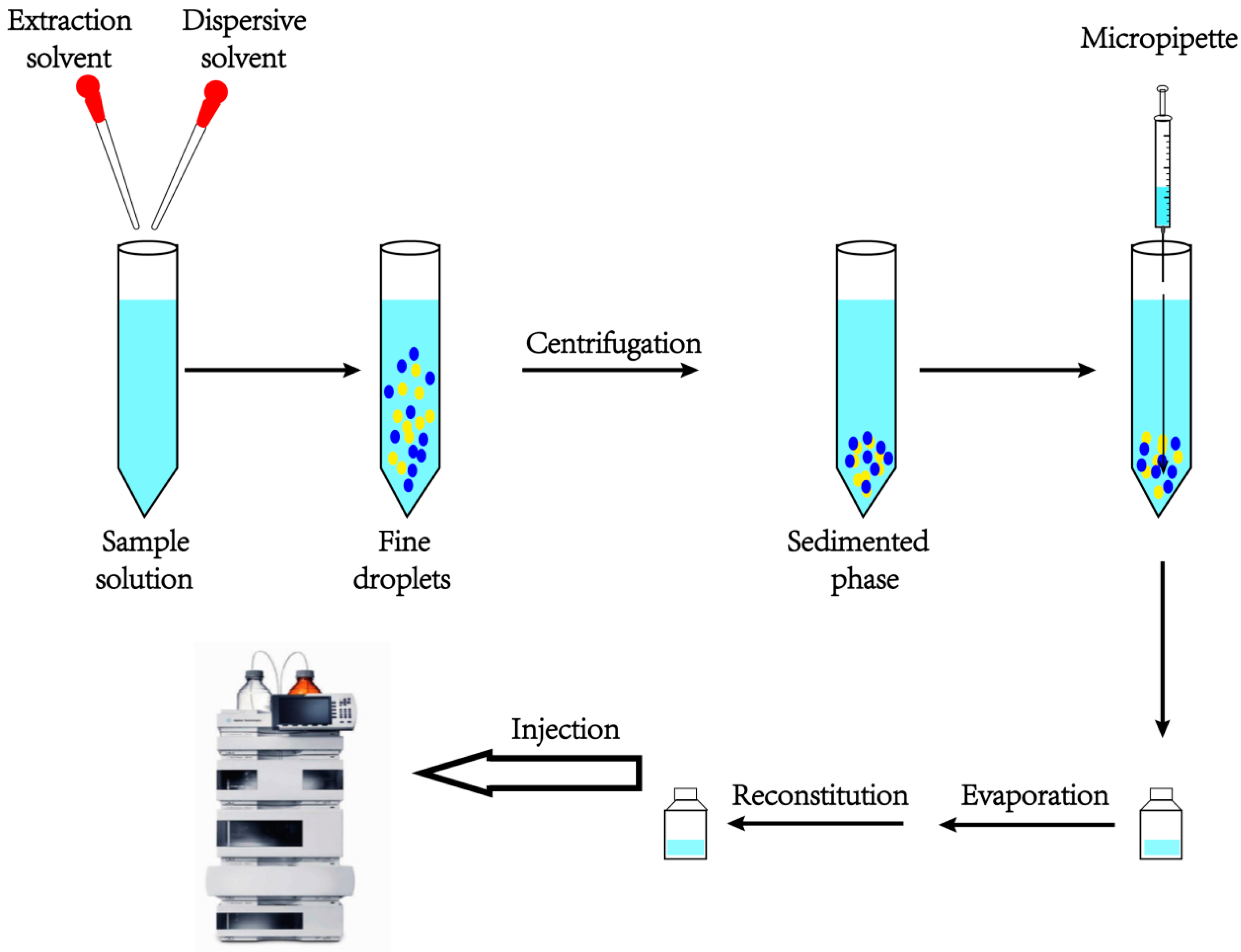
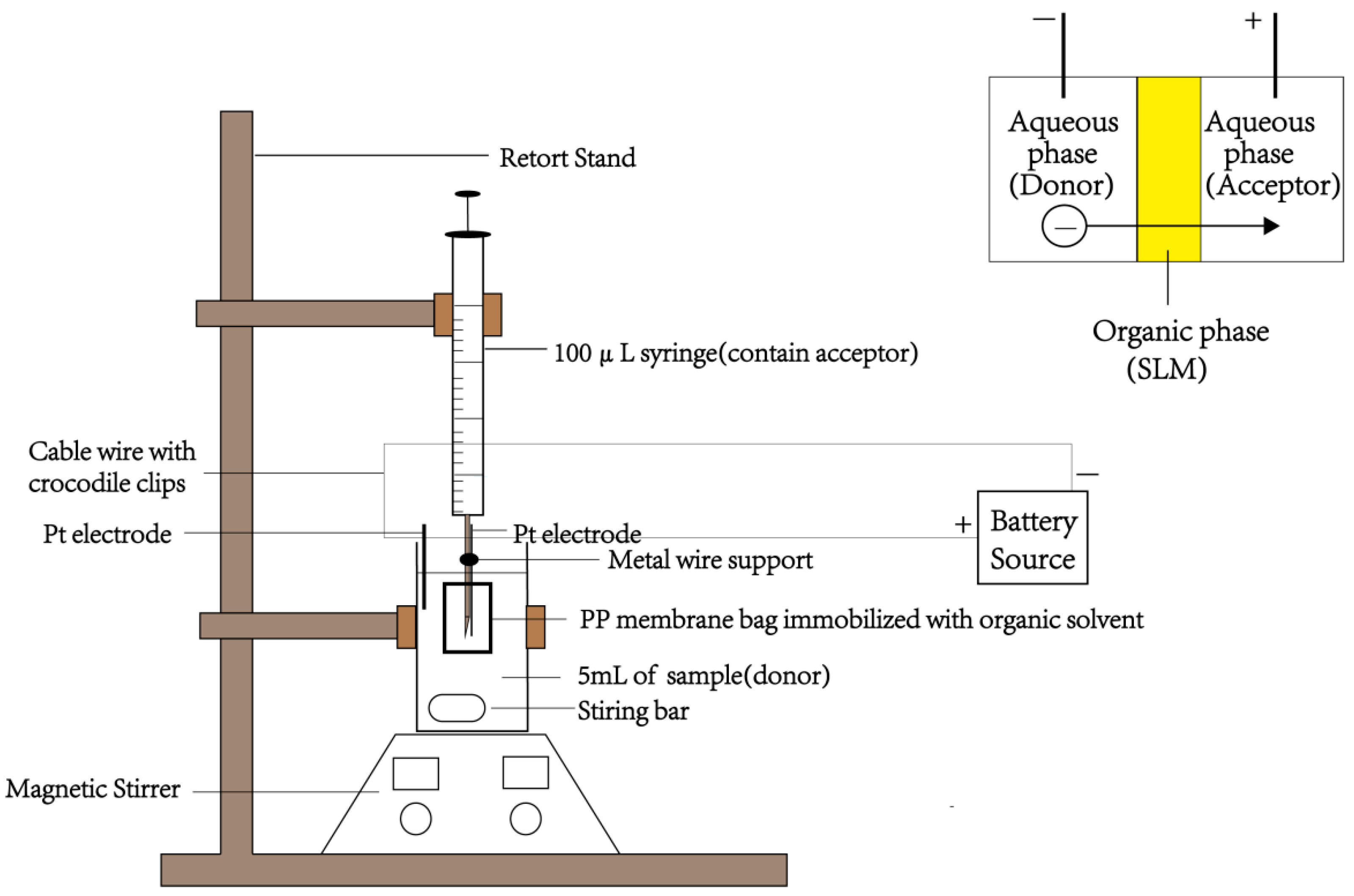
2.3. Liquid Phase Microextraction
2.4. SPE plus SPME
2.5. Summary
3. Analytical Methods
3.1. Spectroscopic Methods
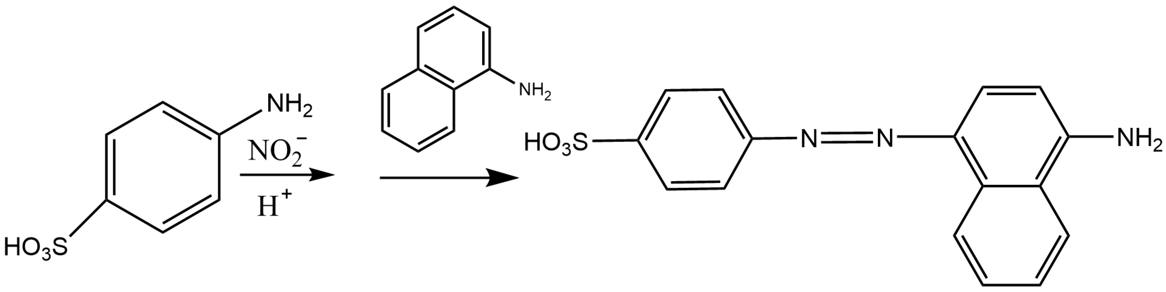
3.1.1. Spectrophotometry
3.1.2. Spectrofluorometry
3.1.3. Colorimetry
3.1.4. Chemiluminescence
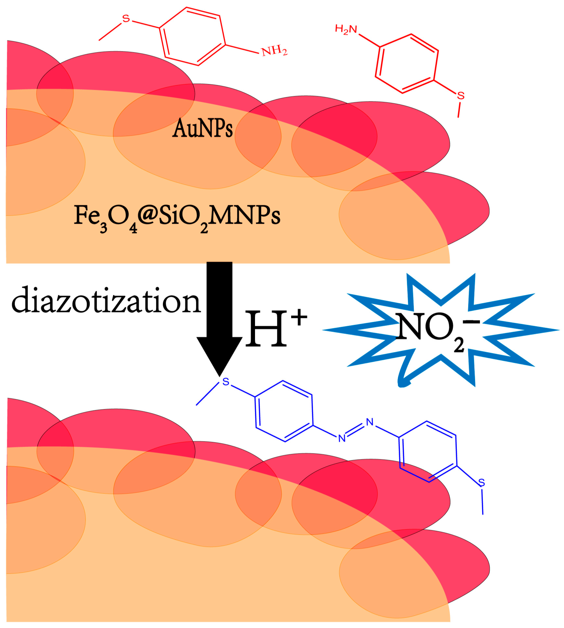
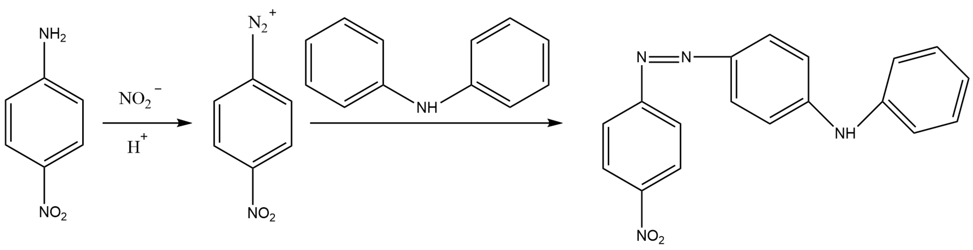
3.1.5. Raman Spectroscopy
3.2. HPLC Methods
3.3. HPLC-MS
3.4. Ion Chromatography
3.5. Capillary Electrophoresis
3.6. Paper-Based Analytical Devices
3.7. Electrochemical Sensors
3.8. GC-MS
3.9. Summary
4. Conclusions
Author Contributions
Funding
Institutional Review Board Statement
Informed Consent Statement
Data Availability Statement
Conflicts of Interest
Sample Availability
Abbreviations
| AHNDMS | 4-amino-5-hydroxynaphthalene-2,7-disulphonic acid monosodium salt |
| 4-ATP | 4-aminothiophenol |
| ATPE | aqueous two-phase extraction |
| BGE | back ground electrolyte |
| BIAMPA | batch injection analysis with multiple-pulse amperometric |
| BPS | bathophenanthroline disulfonic acid |
| BSA-Au NCs | bovine serum albumin stabilized gold nanoclusters |
| CIA | capillary ion analysis |
| CME | chemically modified electrode |
| CNT | carbon nanotube |
| CPE | cloud point extraction |
| CE | capillary electrophoresis |
| CZE | capillary zone electrophoresis |
| DAD | diode array detection |
| DLLME | dispersive liquid–liquid microextraction |
| DESs | deep eutectic solvents |
| DAN | 2,3-diaminonaphthalene |
| ES | electrokinetic stacking |
| EF | enrichment factor |
| ER | extraction recovery |
| EOF | electroosmotic flow |
| FD | fluorescence detection |
| FIA | flow injection analysis |
| GC-MS | gas chromatography-mass spectrometry |
| GC-NCI-MS | gas chromatography-negative chemical ionization-mass spectrometry |
| GQDs | graphene quantum dots |
| HP-β-CD | hydroxypropyl-β-cyclo-dextrin |
| HPLC | high performance liquid chromatography |
| HS-SDME | headspace Single drop liquid phase microextraction |
| IC | ion chromatography |
| ILs | ionic liquids |
| LLE | liquid–liquid extraction |
| LPME | liquid phase microextraction |
| LC-MS/MS | liquid chromatography-tandem mass spectrometry |
| LOD | limit of detection |
| LOQ | limit of quantification |
| ME-EC | microchip electrophoresis method with electrochemical detection |
| MNPS | magnetic nanoparticles |
| NAT | 2,3-naphthotriazole |
| NCs | nanoclusters |
| NIR-CDs | near-infrared carbon dots |
| NO2− | nitrite |
| NO3− | nitrate |
| NO | nitric oxide |
| PADS | paper-based analytical devices |
| PDA | photo-diode array |
| PDMS | polydimethylsiloxane |
| PLA | polylactic acid |
| SIA | sequential injection analysis |
| SPE | solid phase extraction |
| SERS | surface enhancement Raman spectroscopy |
| TMB | 3,3′,5,5′-tetramethylbenzidine |
| µPADs | microfluidic paper-based analytical devices |
| UPLC | ultra performance liquid chromatography |
| [Ru(npy)([9]aneS3)(CO)](ClO4) | (Ru = Ruthenium; npy = 2-(1-naphthyl)pyridine, [9]aneS3 = 1,4,7-trithiacyclononane) |
References
- Thuy, N.T.D.; Wang, X.; Zhao, G.; Liang, T.; Zou, Z. A Co3O4 Nanoparticle-Modified Screen-Printed Electrode Sensor for the Detection of Nitrate Ions in Aquaponic Systems. Sensors 2022, 22, 9730. [Google Scholar] [CrossRef]
- Hikin, L.J.; Ho, J.; Morley, S.R.; Ahluwalia, A.; Smith, P.R. Sodium nitrite poisoning: A series of 20 fatalities in which post-mortem blood nitrite and nitrate concentrations are reported. Forensic Sci. Int. 2023, 345, 111610. [Google Scholar] [CrossRef]
- Singh, P.; Singh, M.K.; Beg, Y.R.; Nishad, G.R. A review on spectroscopic methods for determination of nitrite and nitrate in environmental samples. Talanta 2019, 191, 364–381. [Google Scholar] [CrossRef]
- Wang, Q.H.; Yu, L.J.; Liu, Y.; Lin, L.; Lu, R.G.; Zhu, J.P.; He, L.; Lu, Z.L. Methods for the detection and determination of nitrite and nitrate: A review. Talanta 2017, 165, 709–720. [Google Scholar] [CrossRef]
- Calabrese, V.; Cornelius, C.; Rizzarelli, E.; Owen, J.B.; Dinkova-Kostova, A.T.; Butterfield, D.A. Nitric oxide in cell survival: A janus molecule. Antioxid. Redox Signal. 2009, 11, 2717–2739. [Google Scholar] [CrossRef]
- Basu, S.; Ricart, K.; Gladwin, M.T.; Patel, R.P.; Kim-Shapiro, D.B. Tri-iodide and vanadium chloride based chemiluminescent methods for quantification of nitrogen oxides. Nitric Oxide 2022, 121, 11–19. [Google Scholar] [CrossRef]
- Moorcroft, M.J.; Davis, J.; Compton, R.G. Detection and determination of nitrate and nitrite: A review. Talanta 2001, 54, 785–803. [Google Scholar] [CrossRef]
- Tsikas, D. Methods of quantitative analysis of the nitric oxide metabolites nitrite and nitrate in human biological fluids. Free Radic. Res. 2005, 39, 797–815. [Google Scholar] [CrossRef]
- Tsikas, D. Analysis of nitrite and nitrate in biological fluids by assays based on the Griess reaction: Appraisal of the Griess reaction in the L-arginine/nitric oxide area of research. J. Chromatogr. B Anal. Technol. Biomed. Life Sci. 2007, 851, 51–70. [Google Scholar] [CrossRef]
- Wu, A.; Duan, T.; Tang, D.; Zheng, Z.; Zhu, J.; Wang, R.; He, B.; Cheng, H.; Feng, L.; Zhu, Q. Review the application of chromatography in the analysis of nitric oxide-derived nitrite and nitrate ions in biological fluids. Curr. Anal. Chem. 2014, 10, 609–621. [Google Scholar] [CrossRef]
- Wierzbicka, E. Novel methods of nitrate and nitrite determination—A review. J. Elem. 2020, 25, 97–106. [Google Scholar] [CrossRef]
- Nagaraja, P.; Al-Tayar, N.G.; Shivakumar, A.; Shrestha, A.K.; Gowda, A.K. A simple and sensitive spectrophotometric method for the determination of trace amounts of nitrite in environmental and biological samples using 4-amino-5-hydroxynaphthalene-2,7-disulphonic acid monosodium salt. Spectrochim. Acta Part A Mol. Biomol. Spectrosc. 2010, 75, 1411–1416. [Google Scholar] [CrossRef]
- Guo, Y.X.; Zhang, Q.F.; Shangguang, X.; Zhen, G. Spectrofluorimetric determination of trace nitrite with o-phenylenediamine enhanced by hydroxypropyl-β-cyclodextrin. Spectrochim. Acta Part A Mol. Biomol. Spectrosc. 2013, 101, 107–111. [Google Scholar] [CrossRef]
- Tsikas, D. GC-MS Analysis of biological nitrate and nitrite using pentafluorobenzyl bromide in aqueous acetone: A dual role of carbonate/bicarbonate as an enhancer and inhibitor of derivatization. Molecules 2021, 26, 7003. [Google Scholar] [CrossRef]
- Hanff, E.; Lützow, M.; Kayacelebi, A.A.; Finkel, A.; Maassen, M.; Yanchev, G.R.; Haghikia, A.; Bavendiek, U.; Buck, A.; Lücke, T.; et al. Simultaneous GC-ECNICI-MS measurement of nitrite, nitrate and creatinine in human urine and plasma in clinical settings. J. Chromatogr. B Anal. Technol. Biomed. Life Sci. 2017, 1047, 207–214. [Google Scholar] [CrossRef]
- Schwarz, A.; Modun, D.; Heusser, K.; Tank, J.; Gutzki, F.M.; Mitschke, A.; Jordan, J.; Tsikas, D. Stable-isotope dilution GC-MS approach for nitrite quantification in human whole blood, erythrocytes, and plasma using pentafluorobenzyl bromide derivatization: Nitrite distribution in human blood. J. Chromatogr. B Anal. Technol. Biomed. Life Sci. 2011, 879, 1485–1495. [Google Scholar] [CrossRef]
- Liu, G.; Liu, J.; Wang, L. Determination of nitrite in urine by 2,3-diaminonaphthalene derivatization-GC-MS. Chin. J. Pubilc Health 2016, 32, 566–568. [Google Scholar]
- Zhao, J.; Wang, J.; Yang, Y.; Lu, Y. The determination of nitrate and nitrite in human urine and blood by high-performance liquid chromatography and cloud-point extraction. J. Chromatogr. Sci. 2015, 53, 1169–1177. [Google Scholar] [CrossRef]
- Zhao, J.; Lu, Y.; Fan, C.; Wang, J.; Yang, Y. Development of a cloud point extraction and spectrophotometry-based microplate method for the determination of nitrite in human urine and blood. Spectrochim. Acta Part A Mol. Biomol. Spectrosc. 2015, 136, 802–807. [Google Scholar] [CrossRef]
- Pourreza, N.; Reza Fat’hi, M.; Hatami, A. Indirect cloud point extraction and spectrophotometric determination of nitrite in water and meat products. Microchem. J. 2012, 104, 22–25. [Google Scholar] [CrossRef]
- Zuloaga, O.; Olivares, M.; Navarro, P.; Vallejo, A.; Prieto, A. Dispersive liquid-liquid microextraction: Trends in the analysis of biological samples. Bioanalysis 2015, 7, 2211–2225. [Google Scholar] [CrossRef]
- Madasamy, T.; Pandiaraj, M.; Balamurugan, M.; Bhargava, K.; Sethy, N.K.; Karunakaran, C. Copper, zinc superoxide dismutase and nitrate reductase coimmobilized bienzymatic biosensor for the simultaneous determination of nitrite and nitrate. Biosens. Bioelectron. 2014, 52, 209–215. [Google Scholar] [CrossRef]
- Senra-Ferreiro, S.; Pena-Pereira, F.; Lavilla, I.; Bendicho, C. Griess micro-assay for the determination of nitrite by combining fibre optics-based cuvette less UV-vis micro-spectrophotometry with liquid-phase microextraction. Anal. Chim. Acta 2010, 668, 195–200. [Google Scholar] [CrossRef]
- He, L.; Zhang, K.; Wang, C.; Luo, X.; Zhang, S. Effective indirect enrichment and determination of nitrite ion in water and biological samples using ionic liquid-dispersive liquid-liquid microextraction combined with high-performance liquid chromatography. J. Chromatogr. A 2011, 1218, 3595–3600. [Google Scholar] [CrossRef]
- Zhang, K.; Li, S.; Liu, C.; Wang, Q.; Wang, Y.; Fan, J. A hydrophobic deep eutectic solvent-based vortex-assisted dispersive liquid-liquid microextraction combined with HPLC for the determination of nitrite in water and biological samples. J. Sep. Sci. 2019, 42, 574–581. [Google Scholar] [CrossRef]
- Hu, C.-W.; Chang, Y.-J.; Yen, C.-C.; Chen, J.-L.; Muthukumaran, R.B.; Chao, M.-R. 15N-labelled nitrite/nitrate tracer analysis by LC-MS/MS: Urinary and fecal excretion of nitrite/nitrate following oral administration to mice. Free Radic. Biol. Med. 2019, 143, 193–202. [Google Scholar] [CrossRef]
- Yang, R.; Lin, Y.; Yang, J.; He, L.; Tian, Y.; Hou, X.; Zheng, C. Headspace solid-phase microextraction following chemical vapor generation for ultrasensitive, matrix effect-free detection of nitrite by microplasma optical emission spectrometry. Anal. Chem. 2021, 93, 6972–6979. [Google Scholar] [CrossRef]
- Liu, C.; Qiu, X.; Zhao, J.; Yang, Y. Aqueous two-phase extraction and spectrofluorimetric determination of nitrite with o-phenylenediamine enhanced by hydroxypropyl-β-cyclodextrin in urine samples. Anal. Methods 2014, 6, 463. [Google Scholar] [CrossRef]
- Tan, T.Y.; Basheer, C.; Ng, K.P.; Lee, H.K. Electro membrane extraction of biological anions with ion chromatographic analysis. Anal. Chim. Acta 2012, 739, 31–36. [Google Scholar] [CrossRef]
- Hanff, E.; Böhmer, A.; Jordan, J.; Tsikas, D. Stable-isotope dilution LC-MS/MS measurement of nitrite in human plasma after its conversion to S-nitrosoglutathione. J. Chromatogr. B Anal. Technol. Biomed. Life Sci. 2014, 970, 44–52. [Google Scholar] [CrossRef]
- Croitoru, M.D. Nitrite and nitrate can be accurately measured in samples of vegetal and animal origin using an HPLC-UV/VIS technique. J. Chromatogr. B Anal. Technol. Biomed. Life Sci. 2012, 911, 154–161. [Google Scholar] [CrossRef] [PubMed]
- Yan, H.; Zhuo, X.; Shen, B.; Xiang, P.; Shen, M. Determination of nitrite in whole blood by high-performance liquid chromatography with electrochemical detection and a case of nitrite poisoning. J. Forensic Sci. 2016, 61, 254–258. [Google Scholar] [CrossRef] [PubMed]
- Wang, X.; Masschelein, E.; Hespel, P.; Adams, E.; Van Schepdael, A. Simultaneous determination of nitrite and nitrate in human plasma by on-capillary preconcentration with field-amplified sample stacking. Electrophoresis 2012, 33, 402–405. [Google Scholar] [CrossRef] [PubMed]
- Wang, X.; Adams, E.; Van Schepdael, A. A fast and sensitive method for the determination of nitrite in human plasma by capillary electrophoresis with fluorescence detection. Talanta 2012, 97, 142–144. [Google Scholar] [CrossRef]
- Pandurangappa, M.; Venkataramanappa, Y. Nitrite/nitrate speciation through arsenomolybdenum blue complex at trace level: Application to biological and environmental samples. Anal. Methods 2011, 3, 1838–1844. [Google Scholar] [CrossRef]
- Piknova, B.; Schechter, A.N. Measurement of nitrite in blood samples using the ferricyanide-based hemoglobin oxidation assay. Methods Mol. Biol. 2011, 704, 39–56. [Google Scholar]
- Vahid, S.; Dashti-Khavidaki, S.; Sormaghi, M.S.; Ahmadi, F.; Amini, M. A new pre-column derivatization method for determination of nitrite and nitrate in human plasma by HPLC. J. Liq. Chromatogr. Relat. Technol. 2012, 35, 805–818. [Google Scholar] [CrossRef]
- Zhan, S.; Shao, Q.; Liu, L.; Fan, X. A simple and accurate method to determine nitrite and nitrate in serum based on high-performance liquid chromatography with fluorescence detection. Biomed. Chromatogr. 2013, 27, 1547–1553. [Google Scholar]
- Badiee, H.; Zanjanchi, M.A.; Zamani, A.; Fashi, A. Solvent stir bar microextraction technique with three-hollow fiber configuration for trace determination of nitrite in river water samples. Environ. Sci. Pollut. Res. Int. 2019, 26, 32967–32976. [Google Scholar] [CrossRef]
- Zhang, K.; Kujawski, D.; Spurrell, C.; Wang, B.; Crittenden, J.C. Screening ionic liquids for efficiently extracting perfluoroalkyl chemicals (PFACs) from wastewater. J. Environ. Sci. 2023, 127, 866–874. [Google Scholar] [CrossRef]
- Xiao, Z.; Liu, M.; Bi, W.; Chen, D.D.Y. Ionic liquid as hydrogen bond acceptor in the extraction of nutritional natural products. Food Chem. 2023, 412, 135589. [Google Scholar] [CrossRef] [PubMed]
- Zhou, L.; Wu, T.; Yu, C.; Liu, S.; Pan, C. Ionic liquid-dispersive micro-extraction and detection by high performance liquid chromatography-mass spectrometry for antifouling biocides in water. Molecules 2023, 28, 1263. [Google Scholar] [CrossRef] [PubMed]
- Song, J.; Liu, S.; Zhao, N.; Zhao, L. A new fluorescent probe based on metallic deep eutectic solvent for visual detection of nitrite and pH in food and water environment. Food Chem. 2023, 398, 133935. [Google Scholar] [CrossRef] [PubMed]
- Lo, H.S.; Lo, K.W.; Yeung, C.F.; Wong, C.Y. Rapid visual and spectrophotometric nitrite detection by cyclometalated ruthenium complex. Anal. Chim. Acta 2017, 990, 135–140. [Google Scholar] [CrossRef]
- Ghasemi, A.; Zahediasl, S. Preanalytical and analytical considerations for measuring nitric oxide metabolites in serum or plasma using the Griess method. Clin. Lab. 2012, 58, 615–624. [Google Scholar] [PubMed]
- Unnikrishnan, B.; Wei, S.C.; Chiu, W.J.; Cang, J.; Hsu, P.H.; Huang, C.C. Nitrite ion-induced fluorescence quenching of luminescent BSA-Au(25) nanoclusters: Mechanism and application. Analyst 2014, 139, 2221–2228. [Google Scholar] [CrossRef]
- Jin, L.; Wang, Y.; Liu, F.; Yu, S.; Gao, Y.; Zhang, J. The determination of nitrite by a graphene quantum dot fluorescence quenching method without sample pretreatment. Luminescence 2018, 33, 289–296. [Google Scholar] [CrossRef]
- Ning, G.; Mu, P.; Li, B.; Liu, J.; Xiao, Q.; Huang, S. Fluorine and nitrogen co-doped near-infrared carbon dots for fluorescence “on-off-on” determination of nitrite. Microchim. Acta 2022, 189, 230. [Google Scholar] [CrossRef]
- Di Fenza, R.; Yu, B.; Carroll, R.W.; Berra, L. Chemiluminescence-based assays for detection of nitric oxide and its derivatives from autoxidation and nitrosated compounds. J. Vis. Exp. 2022, 180, e63107. [Google Scholar] [CrossRef]
- Piknova, B.; Park, J.W.; Cassel, K.S.; Gilliard, C.N.; Schechter, A.N. Measuring nitrite and nitrate, metabolites in the nitric oxide pathway, in biological materials using the chemiluminescence method. J. Vis. Exp. 2016, 118, 54879. [Google Scholar]
- Siu, V.S.; Lu, M.; Hsieh, K.Y.; Raines, K.; Asaad, Y.A.; Patel, K.; Afzali-Ardakani, A.; Wen, B.; Budd, R. Toward a quantitative colorimeter for point-of-care nitrite detection. ACS Omega 2022, 7, 11126–11134. [Google Scholar] [CrossRef] [PubMed]
- Moazeni, M.; Gholipour, S.; Mahaki, B.; Ebrahimi, A. Short-Term Impact of Two Kinds of Vegetables to Exogenous Total Nitrate and Nitrite Intake: Is Antibacterial Mouthwash Influential? Int. J. Prev. Med. 2021, 12, 168. [Google Scholar] [PubMed]
- Xiao, N.; Yu, C. Rapid-response and highly sensitive noncross-linking colorimetric nitrite sensor using 4-aminothiophenol modified gold nanorods. Anal. Chem. 2010, 82, 3659–3663. [Google Scholar] [CrossRef]
- Xia, Q.; Mao, Y.; Wu, J.; Shu, T.; Yi, T. Two-component organogel for visually detecting nitrite anion. J. Mater. Chem. C 2014, 10, 1854–1861. [Google Scholar] [CrossRef]
- Zhang, J.; Yang, C.; Chen, C.; Yang, X. Determination of nitrite and glucose in water and human urine with light-up chromogenic response based on the expeditious oxidation of 3,3’,5,5’-tetramethylbenzidine by peroxynitrous acid. Analyst 2013, 138, 2398–2404. [Google Scholar] [CrossRef]
- Zhang, K.; Hu, Y.; Li, G. Diazotization-coupling reaction-based selective determination of nitrite in complex samples using shell-isolated nanoparticle-enhanced Raman spectroscopy. Talanta 2013, 116, 712–718. [Google Scholar] [CrossRef]
- Chen, J.; Pang, S.; He, L.; Nugen, S.R. Highly sensitive and selective detection of nitrite ions using Fe3O4@SiO2/Au magnetic nanoparticles by surface-enhanced Raman spectroscopy. Biosens. Bioelectron. 2016, 85, 726–733. [Google Scholar] [CrossRef] [PubMed]
- Ata, Ş.; Akyüz, M.; Dinç, E. Chemometric approach to the optimisation of LC-FL and GC-MS methods for the determination of nitrite and nitrate in some biological, food and environmental samples. Int. J. Environ. Anal. Chem. 2016, 96, 636–652. [Google Scholar] [CrossRef]
- Tatarczak-Michalewska, M.; Flieger, J.; Kawka, J.; Płaziński, W.; Flieger, W.; Blicharska, E.; Majerek, D. HPLC-DAD Determination of nitrite and nitrate in human saliva utilizing a phosphatidylcholine column. Molecules 2019, 24, 1754. [Google Scholar] [CrossRef]
- Burhan, A.; Vyas, B. Simultaneous determination of nitrite and nitrate by ultra-performance liquid chromatography in rat plasma. Int. J. Pharm. Pharm. Sci. 2016, 8, 294–296. [Google Scholar]
- Wu, A.; Duan, T.; Tang, D.; Xu, Y.; Feng, L.; Zheng, Z.; Zhu, J.; Wang, R.; Zhu, Q. Determination of nitric oxide-derived nitrite and nitrate in biological samples by HPLC coupled to nitrite oxidation. Chromatographia 2013, 76, 1649–1655. [Google Scholar] [CrossRef] [PubMed]
- Bramanti, E.; Angeli, V.; Mester, Z.; Pompella, A.; Paolicchi, A.; D’Ulivo, A. Determination of S-nitrosoglutathione in plasma: Comparison of two methods. Talanta 2010, 81, 1295–1299. [Google Scholar] [CrossRef] [PubMed]
- Tsikas, D.; Schmidt, M.; Böhmer, A.; Zoerner, A.A.; Gutzki, F.M.; Jordan, J. UPLC-MS/MS measurement of S-nitrosoglutathione (GSNO) in human plasma solves the S-nitrosothiol concentration enigma. J. Chromatogr. B Anal. Technol. Biomed. Life Sci. 2013, 927, 147–157. [Google Scholar] [CrossRef] [PubMed]
- Kim, M.; Kim, S.; Yang, W.; Sim, J. Determination of nitrite and nitrate in postmortem whole blood samples of 10 sodium nitrite poisoning cases: The importance of nitrate in determining nitrite poisoning. Forensic Sci. Int. 2022, 335, 111279. [Google Scholar] [CrossRef] [PubMed]
- Zhang, Y.; Yang, L.; Tian, X.; Guo, Y.; Tang, W.; Yu, A.; Zhang, W.; Sun, B.; Zhang, S. Determination of trace nitrites and nitrates in human urine and plasma by field amplified sample stacking open tubular capillary electrochromatography in a nanolatex coated capillary. J. Anal. Chem. 2015, 70, 885–891. [Google Scholar] [CrossRef]
- Taus, F.; Pigaiani, N.; Bortolotti, F.; Mazzoleni, G.; Brevi, M.; Tagliaro, F.; Gottardo, R. Direct and specific analysis of nitrite and nitrate in biological and non-biological samples by capillary ion analysis for the rapid identification of fatal intoxications with sodium nitrite. Forensic Sci. Int. 2021, 325, 110855. [Google Scholar] [CrossRef]
- Siegel, J.M.; Schilly, K.M.; Wijesinghe, M.B.; Caruso, G.; Fresta, C.G.; Lunte, S.M. Optimization of a microchip electrophoresis method with electrochemical detection for the determination of nitrite in macrophage cells as an indicator of nitric oxide production. Anal. Methods 2019, 11, 148–156. [Google Scholar] [CrossRef]
- Gong, M.M.; Sinton, D. Turning the page: Advancing paper-based microfluidics for broad diagnostic application. Chem. Rev. 2017, 117, 8447–8480. [Google Scholar] [CrossRef]
- Yang, Y.; Noviana, E.; Nguyen, M.P.; Geiss, B.J.; Dandy, D.S.; Henry, C.S. Paper-based microfluidic devices: Emerging themes and applications. Anal. Chem. 2017, 89, 71–91. [Google Scholar] [CrossRef]
- Noiphung, J.; Nguyen, M.P.; Punyadeera, C.; Wan, Y.; Laiwattanapaisal, W.; Henry, C.S. Development of paper-based analytical devices for minimizing the viscosity effect in human saliva. Theranostics 2018, 8, 3797–3807. [Google Scholar] [CrossRef]
- Zhang, X.X.; Song, Y.Z.; Fang, F. Sensitive paper-based analytical device for fast colorimetric detection of nitrite with smartphone. Anal. Bioanal. Chem. 2018, 410, 2665–2669. [Google Scholar] [CrossRef] [PubMed]
- Evans, E.; Moreira Gabriel, E.F.; Benavidez, T.E.; Tomazelli Coltro, W.K.; Garcia, C.D. Modification of microfluidic paper-based devices with silica nanoparticles. Analyst 2014, 139, 5560–5567. [Google Scholar] [CrossRef] [PubMed]
- Yetisen, A.K.; Akram, M.S.; Lowe, C.R. Paper-based microfluidic point-of-care diagnostic devices. Lab. Chip. 2013, 13, 2210–2251. [Google Scholar] [CrossRef] [PubMed]
- Ferreira, F.T.S.M.; Mesquita, R.B.R.; Rangel, A.O.S.S. Novel microfluidic paper-based analytical devices (μPADs) for the determination of nitrate and nitrite in human saliva. Talanta 2020, 219, 121183. [Google Scholar] [CrossRef] [PubMed]
- Bhakta, S.A.; Borba, R.; Taba, M.J.; Garcia, C.D.; Carrilho, E. Determination of nitrite in saliva using microfluidic paper-based analytical devices. Anal. Chim. Acta 2014, 809, 117–122. [Google Scholar] [CrossRef]
- De Oliveira, R.A.G.; Camargo, F.; Pesquero, N.C.; Faria, R.C. A simple method to produce 2D and 3D microfluidic paper-based analytical devices for clinical analysis. Anal. Chim. Acta 2017, 957, 40–46. [Google Scholar] [CrossRef]
- Ferreira, F.T.S.M.; Mesquita, R.B.R.; Rangel, A.O.S.S. Design and functionalization of a µPAD for the enzymatic determination of nitrate in urine. Molecules 2021, 26, 6355. [Google Scholar] [CrossRef]
- Gaikwad, R.; Thangaraj, P.R.; Sen, A.K. Microfluidics-based rapid measurement of nitrite in human blood plasma. Analyst 2022, 147, 3370–3382. [Google Scholar] [CrossRef]
- Martinez, A.W.; Phillips, S.T.; Whitesides, G.M.; Carrilho, E. Diagnostics for the developing world: Microfluidic paper-based analytical devices. Anal. Chem. 2010, 82, 3–10. [Google Scholar] [CrossRef]
- Rajesh, S.; Kanugula, A.K.; Bhargava, K.; Ilavazhagan, G.; Kotamraju, S.; Karunakaran, C. Simultaneous electrochemical determination of superoxide anion radical and nitrite using Cu, ZnSOD immobilized on carbon nanotube in polypyrrole matrix. Biosens. Bioelectron. 2010, 26, 689–695. [Google Scholar] [CrossRef]
- Zhang, Y.; Yuan, R.; Chai, Y.; Li, W.; Zhong, X.; Zhong, H. Simultaneous voltammetric determination for DA, AA and NO2− based on graphene/poly-cyclodextrin/MWCNTs nanocomposite platform. Biosens. Bioelectron. 2011, 26, 3977–3980. [Google Scholar] [CrossRef] [PubMed]
- Wang, C.; Yuan, R.; Chai, Y.; Zhang, Y.; Hu, F.; Zhang, M. Au-nanoclusters incorporated 3-amino-5-mercapto-1,2,4-triazole film modified electrode for the simultaneous determination of ascorbic acid, dopamine, uric acid and nitrite. Biosens. Bioelectron. 2011, 30, 315–319. [Google Scholar] [CrossRef] [PubMed]
- Zhang, W.; Yuan, R.; Chai, Y.; Zhang, Y.; Chen, S. A simple strategy based on lanthanum–multiwalled carbon nanotube nanocomposites for simultaneous determination of ascorbic acid, dopamine, uric acid and nitrite. Sens. Actuators B Chem. Sens. Actuators B 2012, 166–167, 601–607. [Google Scholar] [CrossRef]
- Wang, C.; Yuan, R.; Chai, Y.; Chen, S.; Zhang, Y.; Hu, F.; Zhang, M. Non-covalent iron(III)-porphyrin functionalized multi-walled carbon nanotubes for the simultaneous determination of ascorbic acid, dopamine, uric acid and Nitrite. Electrochim. Acta 2012, 62, 109–115. [Google Scholar] [CrossRef]
- Yang, Y.; Li, W. CTAB functionalized graphene oxide/multiwalled carbon nanotube composite modifified electrode for the simultaneous determination of ascorbic acid, dopamine, uric acid and nitrite. Biosens. Bioelectron. 2014, 56, 300–306. [Google Scholar] [CrossRef] [PubMed]
- Balamurugan, M.; Madasamy, T.; Pandiaraj, M.; Bhargava, K.; Sethy, N.K.; Karunakaran, C. Electrochemical assay for the determination of nitric oxide metabolites using copper(II) chlorophyllin modified screen printed electrodes. Anal. Biochem. 2015, 478, 121–127. [Google Scholar] [CrossRef]
- Riahifar, V.; Haghnazari, N.; Keshavarzi, F.; Nasri, F. Design a high sensitive electrochemical sensor based on immobilized cysteine on Fe3O4@Au core-shell nanoparticles and reduced graphene oxide nanocomposite for nitrite monitoring. Microchem. J. 2021, 166, 106217. [Google Scholar] [CrossRef]
- Afshar, M.G.; Crespo, G.A.; Dorokhin, D.; Néel, B.; Bakker, E. Thin layer coulometry of nitrite with ion-selective membranes. Electroanalysis 2015, 27, 609–615. [Google Scholar] [CrossRef]
- Caetano, L.P.; Lima, A.P.; Tormin, T.F.; Richter, E.M.; Espindola, F.S.; Botelho, F.V.; Munoz, R.A.A. Carbon-nanotube modified screen-printed electrode for the simultaneous determination of nitrite and uric acid in biological fluids using batch-injection amperometric detection. Electroanalysis 2018, 30, 1870–1879. [Google Scholar] [CrossRef]
- Gross, A.J.; Holmes, S.; Dale, S.E.; Smallwood, M.J.; Green, S.J.; Winlove, C.P.; Benjamin, N.; Winyard, P.G.; Marken, F. Nitrite/nitrate detection in serum based on dual-plate generator-collector currents in a microtrench. Talanta 2015, 131, 228–235. [Google Scholar] [CrossRef]
- Cardoso, R.M.; Silva, P.R.L.; Lima, A.P.; Rocha, D.P.; Oliveira, T.C.; do Prado, T.M.; Fava, E.L.; Fatibello-Filho, O.; Richter, E.M.; Munoz, R.A.A. 3D-Printed graphene/polylactic acid electrode for bioanalysis: Biosensing of glucose and simultaneous determination of uric acid and nitrite in biological fluids. Sens. Actuators B Chem. 2020, 307, 127621. [Google Scholar] [CrossRef]
- Tsikas, D. Pentafluorobenzyl bromide—A versatile derivatization agent in chromatography and mass spectrometry: I. Analysis of inorganic anions and organophosphates. J. Chromatogr. B Anal. Technol. Biomed. Life Sci. 2017, 1043, 187–201. [Google Scholar] [CrossRef] [PubMed]
- Tsikas, D.; Suchy, M.T.; Mitschke, A.; Beckmann, B.; Gutzki, F.M. Measurement of nitrite in urine by gas chromatography-mass spectrometry. Methods Mol. Biol. 2012, 844, 277–293. [Google Scholar] [PubMed]
- Tsikas, D.; Schwarz, A.; Stichtenoth, D.O. Simultaneous measurement of [15N]nitrate and [15N]nitrite enrichment and concentration in urine by gas chromatography mass spectrometry as pentaflfluorobenzyl derivatives. Anal. Chem. 2010, 82, 2585–2587. [Google Scholar] [CrossRef]
- Tsikas, D. GC-MS Studies on Nitric Oxide Autoxidation and S-Nitrosothiol Hydrolysis to Nitrite in pH-Neutral Aqueous Buffers: Definite Results Using 15N and 18O Isotopes. Molecules 2023, 28, 4281. [Google Scholar] [CrossRef]
- Houben, E.; Hamer, H.M.; Luypaerts, A.; De Preter, V.; Evenepoel, P.; Rutgeerts, P.; Verbeke, K. Quantifification of 15N-nitrate in urine with gas chromatography combustion isotope ratio mass spectrometry to estimate endogenous NO production. Anal. Chem. 2010, 82, 601–607. [Google Scholar] [CrossRef]


| Pretreatment Methods | Derivatization Reagents | Analytical Technique | Analyte | Matrix | Recoveries | Precision | Ref. |
|---|---|---|---|---|---|---|---|
| Centrifugation and dilution | AHNDMS | Spectrophotometry | nitrite | saliva | 98% | <0.82% | [12] |
| Centrifugation/Filtration | griess | Spectrofluorimetry | nitrite | water, sausage, soil | 95–108% | <2.88% | [13] |
| Centrifugation/LLE | PFB-Br | GC-MS | nitrite nitrate | blood | 92–99% | <16.1% | [16] |
| Protein precipitation | PFB-Br | GC-MS | nitrite nitrate | erythrocytes, plasma | 95–113% | 0.2–16.2% | [16] |
| Centrifugation/ ultrafiltration | glutathione | LC-MS/MS | nitrite | plasma | 92–108% | <4.4% | [30] |
| Protein precipitation | griess | HPLC-UV | nitrite nitrate | plasma | n.d. | n.d. | [31] |
| Protein precipitation | / | IC-ED | nitrite | blood | 56–72% | <14.9% | [32] |
| Protein precipitation | NO | CE- fluorescence | nitrite nitrate | plasma | 92–113% | <2.6% | [33] |
| Protein precipitation | DAN | CE- fluorescence | nitrite | plasma | 85–112% | <1.1% | [34] |
| Protein precipitation | arsenate | Spectrophotometry | nitrite nitrate | blood | 99–101% | <2.1% | [35] |
| Protein precipitation/ATPE | OPE | Spectrofluorimetry | nitrite | urine | 93–105% | <1.7% | [28] |
| Protein precipitation | I3 | Chemiluminescence | nitrite | blood | n.d. | n.d. | [36] |
| Dilution/online SPE | DAN | LC-MS/MS | nitrite nitrate | urine | 99–112% | <7.4% | [26] |
| Soak, vortex, ultrasonic, centrifugation/online SPE | DAN | LC-MS/MS | nitrite nitrate | feces | 99–112% | <7.4% | [26] |
| LLE | PFB-Br | GC-MS | nitrite nitrate | urine | n.d. | n.d. | [14] |
| LLE | PFB-Br | GC-MS | nitrite nitrate | urine | 91–113% | 0.92–19% | [15] |
| LLE | DAN | GC-MS | nitrite | urine | 90–113% | <5.16% | [17] |
| LLE | griess | HPLC-UV | nitrite nitrate | human plasma | 98–102% | <8.77% | [37] |
| CPE | griess | HPLC-DAD | nitrite nitrate | plasma, urine | 90–98% | <4.19% | [18] |
| CPE | griess | spectrophotometry | nitrite | urine, blood | 92–101% | n.d. | [19] |
| CPE | griess | spectrophotometry | nitrite | meat, water | 91–103% | <3.4% | [20] |
| HS-SDME | griess | pectrophotometry | Nitrite | water | 98–137% | <10.6% | [23] |
| IL-DLLME | griess | HPLC-UV | nitrite | water and saliva | 96–107% | 4.1% | [24] |
| VA-DLLME | griess | HPLC-UV | nitrite | saliva | 90–115% | <4.6% | [25] |
| EME | / | IC-CD | nitrite | amniotic fluids | n.d. | <11% | [29] |
| SPME | sodium cyclamate | μPD-OES | nitrite | simulated gastric content, serum | 96–103% | 4.1% | [27] |
| LLE | LPME | SPE | SPME | Simple Sample Treatment |
|---|---|---|---|---|
| Evolution | ||||
| CPE, ATPE | HS-SDME, DLLME, EME | On-line SPE | HS-SPME | Protein precipitation, dilute and shoot |
| Advantages | ||||
| Being reliable | Good purification effect | Less reagent consumption than LLE | High enrichment factor | Low cost |
| Wide extraction range | High enrichment and extraction efficiency | No emulsification occurs during treatment | Each extraction head can be used more than 50 times | Time-saving |
| Simple equipment and easy to operate | Less organic solvent at the level of a microliter | Simplified sample handing process | Different adsorption fibers meet a variety of application requirements | Easy accessibility |
| Mild extraction conditions | Easy to automate with a low cost | High selectivity and reproducibility due to the use of highly efficient and selective adsorbents | Used with automatic sampler to ensure the accuracy of the experiment | Appropriate for simple and clean samples |
| Disadvantages | ||||
| Easily emulsifying and poor selectivity | The extraction efficiency is susceptible | Higher cost than LLE | Carry-over problems caused by repeated analysis with the same fiber | Limited ability to handle complex samples |
| Large amount of organic solvent | Limited potential for automation and efficiency |
| Methods | Matrix | Reagents | λmax (nm) | Linearity Range | Detection Limit (ng/mL) | Reaction Time (min) | Ref. |
|---|---|---|---|---|---|---|---|
| spectrophotometry | saliva samples | AHNDMS | 560 | 0.1 μg/mL–1.6 μg/mL | 7.5 | 2 | [12] |
| spectrophotometry | urine, blood | griess | 490 | 10 ng/mL–400 ng/mL | 2.5 | n.d. | [19] |
| spectrophotometry | blood samples | arsenomolybdneum blue | 840 | 2 μg/mL–10 μg/mL | 3 | 30 | [37] |
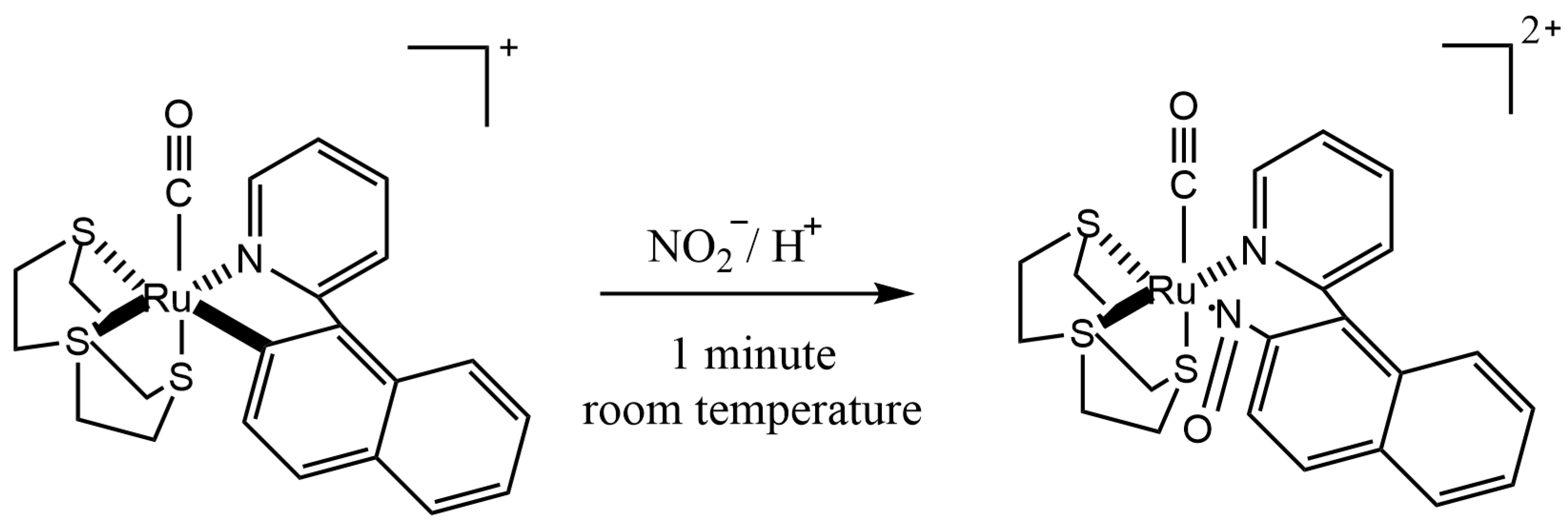
| spectrophotometry | urine | [Ru(npy)([9]aneS3)(CO)](ClO4) | 483 | 1 µM–840 µM | 26.91 | 1 | [44] |
| spectrofluorometry | urine | BSA-Au NCs | 660 | 1 μM–100 μM | 3.45 | 60 | [46] |
| spectrofluorometry | urine | GQDs | 480 | 0.025 μg/mL–0.09 μg/mL | 0.373 | 5 | [47] |
| spectrofluorometry | urine | NIR-CDs | 675 | 1 μM–50 μM | 3.864 | n.d. | [48] |
| spectrofluorometry | urine | hydroxypropyl-β-cyclodextrin | 568 | 5 ng/mL–1000 ng/mL | 1.5 | n.d. | [28] |
| colorimetry | artificial urine | griess | n.d. | 0.78 μM–200 μM | 110.4 | 1 | [51] |
| colorimetry | urine | TMB | 452 | 0.5 μM–30 μM | 6.9 | 1 | [55] |
| Matrix | Analytes | Column | Mobile Phase | Detector | Preparation | Linear Range | LOD | LOQ | Ref. |
|---|---|---|---|---|---|---|---|---|---|
| blood urine | nitrite nitrate | C18 column (150 mm × 4.6 mm, 5 mm) | A: acetonitrile B: methanol C: 1%tetrabutylammonium hydroxide. gradient elution | UV | decolorization and protein precipitation, CPE | 10–1000 ng/mL (nitrite) 0.1–10 μg/mL (nitrate) | 1 ng/mL for nitrite; 0.1 μg/mL for nitrate | n.d. | [18] |
| saliva | nitrite | VP-ODS column (150 mm × 4.6 mm, 5 μm) | methanol/water (90:10, v/v) isocratic elution | UV | IL-DLLME | 0.4–500.0 μg/L | 0.05 μg/L | 0.4 μg/L | [24] |
| urine and saliva | nitrite | C18 column (250 mm × 4.6 mm, 5 μm) | 95% methanol and 5% water isocratic elution | UV | vortex-assisted dispersive liquid–liquid microextraction | 1–300 μg/L | 0.2 μg/L | 1 μg/L | [25] |
| rat serum | nitrite nitrate | XBridge C18 (2.1 mm × 50 mm, 3.5 µm) | 15% (v/v) acetonitrile in 20 mM sodium phosphate buffer (pH 10) isocratic elution | FD | centrifugation | 0.02–2.00 μM for nitrite: 0.3125–20 μM for nitrate | 0.003 μM for nitrite 0.083 mM for nitrate | 0.009 μM for nitrite; 0.250 μM for nitrate | [38] |
| human plasma | nitrite nitrate | 120 CN (25 cm × 0.46 cm, 5 μm) | methanol and water (57.5:42.5, V:V) isocratic elution | UV | pre-column derivatization of nitrite anion using the Griess; nitrate with vanadium chloride (III) | 0.1–50 μM (nitrite) 1–500 μM (nitrate) | n.d. | 0.1 μM for nitrite | [37] |
| serum | nitrite nitrate | POLAR-RP column (250 mm × 4.6 mm, 4 µm) | A: acetonitrile B: double distilled–deionised water consisted of 0.1% trifluoroacetic acid gradient elution | FD | derivatization then LLE | 1–5000 ng/L (nitrite) 1–100 μg/L (nitrate) | 0.13 ng/L for nitrite; 0.19 ng/L for nitrate | 0.43 ng/L for nitrite; 0.58 ng/L for nitrate | [58] |
| human saliva | nitrite nitrate | Phosphatidylcholine Column (4.6 mm × 150 mm, 10 µm) | 20 mM NaCl isocratic elution | DAD | dilution and centrifugation | 8.98–18.52 µg/mL (nitrate) 3.50–5.34 µg/mL (nitrate) | 4.56 ng/mL for nitrate; 4.21 ng/mL for nitrite | 15.21ng/mL for nitrate; 14.03 ng/mL for nitrite | [59] |
| rabbit blood | nitrite nitrate | X Bridge C18 (2.1 mm × 50 mm, 2.5 μm) | A: tetrabutylammonium hydroxide 5 mM brought to pH 2.5 with sulfuric acid B: acetonitrile C: methanol gradient elution | UV | pre-column derivatization of nitrite anion using the Griess | 6–400 μg/L (nitrite) 0.2–200 μg/mL (nitrate) | 0.06 μg/mL for nitrate | 2 ng/mL for nitrite and 200 ng/mL for nitrate | [31] |
| rat plasma | nitrite nitrate | An Acquity UPLC® BEH C18 column (2.1 mm × 50 mm, 1.7 μm) | A: tetrabutylammonium hydroxide (12 mM), potassium dihydrogen phosphate (pH 7.0; 10 mM) B: consisted of tetrabutylammonium hydroxide (2.8 mM), methanol (30% v/v), potassium dihydrogen phosphate (pH 5.5; 100 mM). gradient elution | PDA | using filter plate to deprotein | 4–500 μM (nitrite) 6–400 μM (nitrate) | n.d. | 4 μM for nitrite; 6 μM for nitrate | [60] |
| rat plasma/urine | nitrite nitrate | C18 (250 mm × 4.6 mm, 5 µm) | methanol–water (containing 0.60 mM of phosphate salt and 2.5 mM TBAP) (2:98, V:V) isocratic elution | PDA | deproteinization with acetonitrile | 1–800 μM for nitrate and nitrite | 0.075 μM for nitrate | 0.25 μM for nitrate and nitrite | [61] |
| Matrix | Analytes | Paper Material | Fabrication Technique | Derivatization | Analytical Range | LOD | LOQ | Ref. |
|---|---|---|---|---|---|---|---|---|
| artificial saliva | nitrite | Whatman filter paper | a wax printing | Griess | 0.1–2.4 mg/dL | n.d. | n.d. | [70] |
| saliva | nitrite | glass fiber | electrokinetically stacking | Griess | 0.075–1.0 μg/mL | 73 ng/mL | n.d. | [71] |
| saliva | nitrite | Whatman filter paper | the laminating pouches were passed through the laminator | Griess | 5–250 μM | 0.05 μM | 0.17 μM | [74] |
| saliva | nitrate | Whatman filter paper | the laminating pouches were passed through the laminator | Griess | 0.2–1.2 mM | 0.08 mM | 0.27 mM | [74] |
| saliva | nitrite | cellulose filter paper | a wax printing | Griess | 1–100 μM | 10 μM | n.d. | [75] |
| urine sample | nitrite | cellulose filter paper | an inexpensive home cutter printer and plastic adhesives | Griess | 5–100 μM | 2.34 μM | n.d. | [76] |
| blood serum | nitrite | cellulose filter paper | an inexpensive home cutter printer and plastic adhesives | Griess | 5–600 μM | 4.35 μM | n.d. | [76] |
| human urine samples | nitrite | Whatman filter paper | the laminating pouches were passed through the laminator | Griess | 0.14–1.0 mM | 0.04 mM | 0.14 mM | [77] |
| blood plasma | nitrite | silicon substrate | an acoustics-based plasma separation device | Griess | 0–20 µM | 60 nM | n.d. | [78] |
| Electrode | Modification | Method | Analytes | Linearity Range | LOD (nM) | Precision | Recovery | Ref. |
|---|---|---|---|---|---|---|---|---|
| Pt | NaR–SOD1–CNT–PPy–Pt | CV | nitrite | 100 nM–1 mM | 50 nM | 3.57% | n.d. | [22] |
| Pt | NaR–SOD1–CNT–PPy–Pt | CV | nitrate | 500 nM–10 mM | 200 nM | 2.98% | n.d. | [22] |
| Pt | SOD1–CNT–PPy–Pt | CV | nitrite | 0.5–2000 μM | 0.5 ± 0.025 μM | n.d. | n.d. | [80] |
| GCE | CDP–GS–MWCNTs | CV | nitrite | 5 µM–6.75 mM | 1.65 μM | <3.7% | 95.0% and 106.6% | [81] |
| GCE | nano-Au/p-TA | DPV | nitrite | 15.9–277.0 µM | 0.89 μM | <2.3% | 91.0% and 109.0% | [82] |
| GCE | La–MWCNTs | CA | nitrite | 0.40–0.71 mM | 103 nM | <4.2% | 101.6% and 101.7% | [83] |
| GCE | Fe(III)P/MWCNTs | CV | nitrite | 1.00 μM–1.6 mM | 0.50 μM | <4.5% | 98.0% and 110.0% | [84] |
| GCE | CTAB-GO/MWNT | DPV | nitrite | 5.0–800 μM | 1.5 µm | 2.50% | 99% and 105% | [85] |
| ZnO-screen printed electrodes | CuCP | CV | nitrite | 100 nM–1 mM | 100 nM | n.d. | n.d. | [86] |
| GCE | Fe3O4@Au@Cys/rGO | CV | nitrite | 0.03–344 and 344–2215 µM | 8 nm | <3.26% | 98.5–104% | [87] |
| Ag/AgCl wire | A cobalt(II) tert-butyl salophen compound | CA | nitrite | 20–100 µM | 10 µm | <1% | n.d. | [88] |
| SPCE | MWCNT | BIA-MPA | nitrite | 1–40 µM | 0.3 µM | <1.3% | 77–121% | [89] |
| gold-gold microtrench electrode | silver | CA | nitrate | 200–1400 μM | 24 μM | <6.9% | 108% | [90] |
| 3D-printed electrode | graphene/PLA | BIA-MPA | nitrite | 0.5–250 µM | 0.03 µML | 1.10% | 70 and 110%. | [91] |
Disclaimer/Publisher’s Note: The statements, opinions and data contained in all publications are solely those of the individual author(s) and contributor(s) and not of MDPI and/or the editor(s). MDPI and/or the editor(s) disclaim responsibility for any injury to people or property resulting from any ideas, methods, instructions or products referred to in the content. |
© 2023 by the authors. Licensee MDPI, Basel, Switzerland. This article is an open access article distributed under the terms and conditions of the Creative Commons Attribution (CC BY) license (https://creativecommons.org/licenses/by/4.0/).
Share and Cite
Liu, G.; Guo, H.; Zhao, W.; Yan, H.; Zhang, E.; Gao, L. Advancements in Preprocessing and Analysis of Nitrite and Nitrate since 2010 in Biological Samples: A Review. Molecules 2023, 28, 7122. https://doi.org/10.3390/molecules28207122
Liu G, Guo H, Zhao W, Yan H, Zhang E, Gao L. Advancements in Preprocessing and Analysis of Nitrite and Nitrate since 2010 in Biological Samples: A Review. Molecules. 2023; 28(20):7122. https://doi.org/10.3390/molecules28207122
Chicago/Turabian StyleLiu, Guojie, Honghui Guo, Wanlin Zhao, Hongmu Yan, Enze Zhang, and Lina Gao. 2023. "Advancements in Preprocessing and Analysis of Nitrite and Nitrate since 2010 in Biological Samples: A Review" Molecules 28, no. 20: 7122. https://doi.org/10.3390/molecules28207122
APA StyleLiu, G., Guo, H., Zhao, W., Yan, H., Zhang, E., & Gao, L. (2023). Advancements in Preprocessing and Analysis of Nitrite and Nitrate since 2010 in Biological Samples: A Review. Molecules, 28(20), 7122. https://doi.org/10.3390/molecules28207122