
Biological Properties and Biomedical Applications of Pectin and Pectin-Based Composites: A Review


Abstract
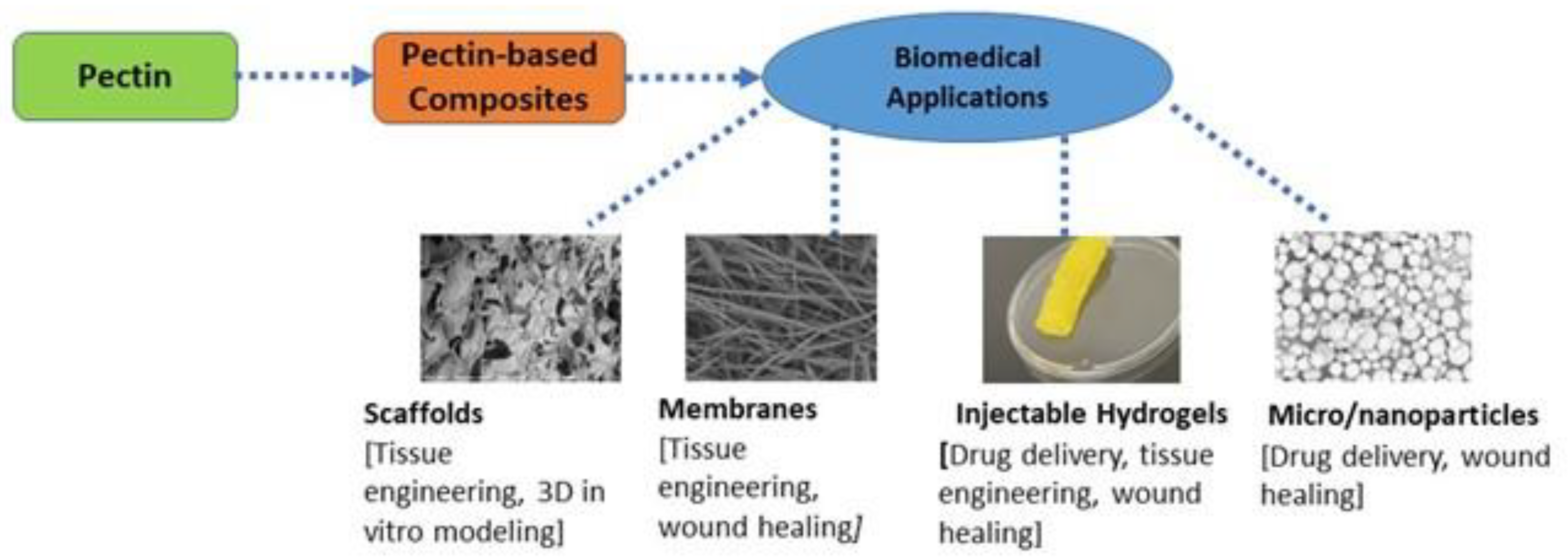
:1. Introduction
2. Properties of Pectic Polysaccharides
2.1. Immunoregulatory Activity
2.2. Anti-Inflammatory Activity
2.3. Antibacterial Activity
2.4. Anticancer Activity of Pectin and Pectin-Based Composites
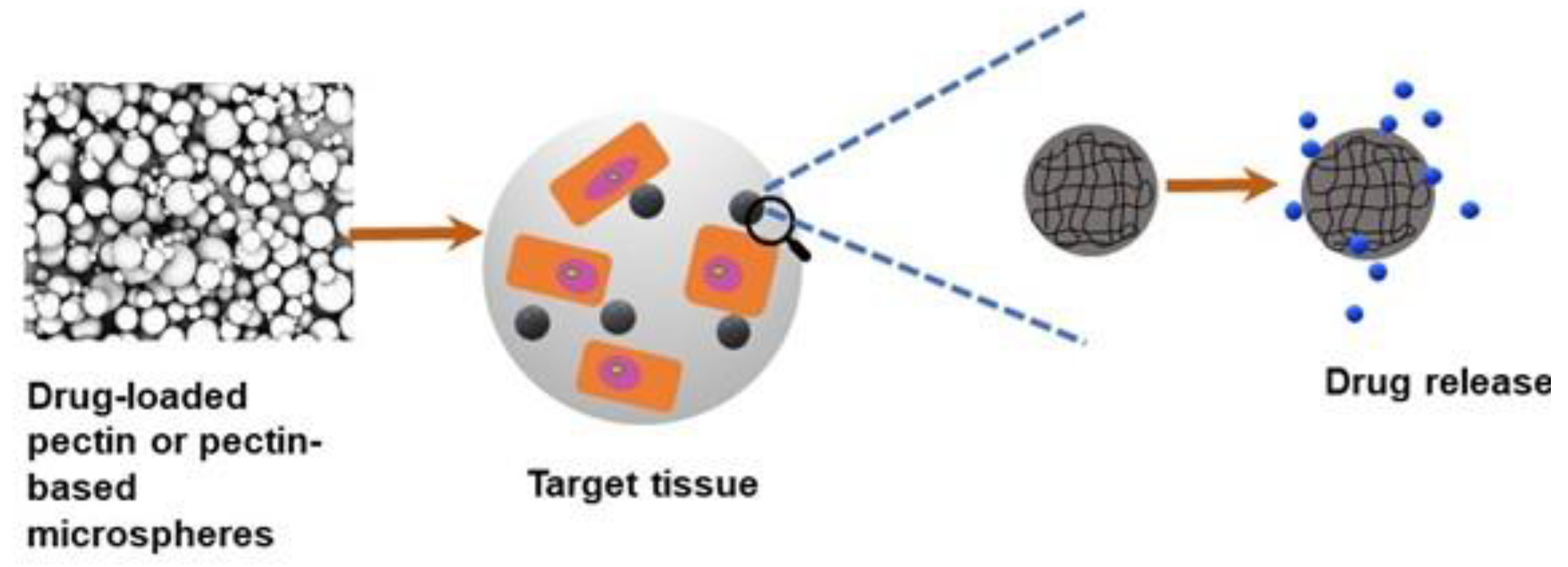
3. Pectin for Drug Delivery Applications
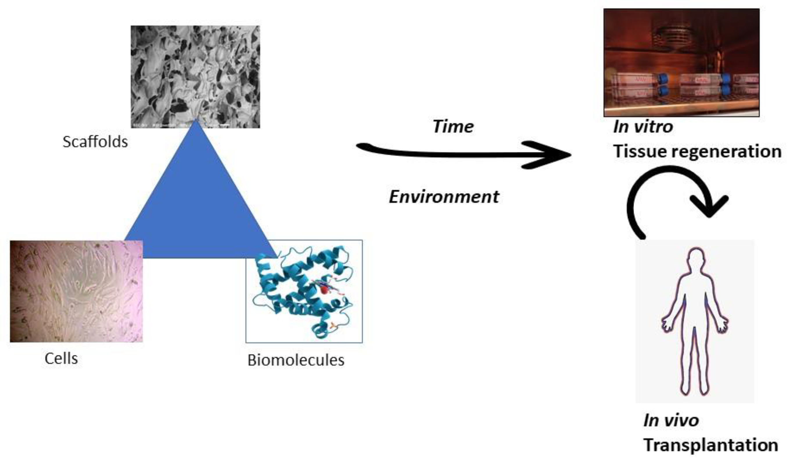
4. Pectin for Tissue Engineering Applications
5. Conclusions and Future Prospects
Funding
Data Availability Statement
Acknowledgments
Conflicts of Interest
References
- Minzanova, S.T.; Mironov, V.F.; Arkhipova, D.M.; Khabibullina, A.V.; Mironova, L.G.; Zakirova, Y.M.; Milyukov, V.A. Biological Activity and Pharmacological Application of Pectic Polysaccharides: A Review. Polymers 2018, 10, 1407. [Google Scholar] [CrossRef] [PubMed]
- Willats, W.G.; McCartney, L.; Mackie, W.; Knox, J.P. Pectin: Cell biology and prospects for functional analysis. Plant. Mol. Biol. 2001, 47, 9–27. [Google Scholar] [CrossRef] [PubMed]
- Liu, Y.; Dong, L.; Li, Y.; Chen, Q.; Wang, L.; Farag, M.A.; Liu, L.; Zhan, S.; Wu, Z.; Liu, L. Soy protein isolate-citrus pectin composite hydrogels induced by TGase and ultrasonic treatment: Potential targeted delivery system for probiotics. Food Hydrocoll. 2023, 143, 108901. [Google Scholar] [CrossRef]
- Liu, Y.; Weng, P.; Liu, Y.; Wu, Z.; Wang, L.; Liu, L. Citrus pectin research advances: Derived as a biomaterial in the construction and applications of micro/nano-delivery systems. Food Hydrocoll. 2022, 133, 107910. [Google Scholar] [CrossRef]
- Coimbra, P.; Ferreira, P.; de Sousa, H.C.; Batista, P.; Rodrigues, M.A.; Correia, I.J.; Gil, M.H. Preparation and chemical and biological characterization of a pectin/chitosan polyelectrolyte complex scaffold for possible bone tissue engineering applications. Int. J. Biol. Macromol. 2011, 48, 112–118. [Google Scholar] [CrossRef] [PubMed]
- Neufeld, L.; Bianco-Peled, H. Pectin-chitosan physical hydrogels as potential drug delivery vehicles. Int. J. Biol. Macromol. 2017, 101, 852–861. [Google Scholar] [CrossRef] [PubMed]
- Pereira, R.F.; Barrias, C.C.; Bártolo, P.J.; Granja, P.L. Cell-instructive pectin hydrogels crosslinked via thiol-norbornene photo-click chemistry for skin tissue engineering. Acta Biomater. 2018, 66, 282–293. [Google Scholar] [CrossRef] [PubMed]
- Khotimchenko, Y.; Khozhaenko, E.; Kovalev, V.; Khotimchenko, M. Cerium binding activity of pectins isolated from the seagrasses Zostera marina and Phyllospadix iwatensis. Mar. Drugs 2012, 10, 834–848. [Google Scholar] [CrossRef]
- Pérez, S.; Mazeau, K.; Hervé du Penhoat, C. The three-dimensional structures of the pectic polysaccharides. Plant Physiol. Biochem. 2000, 38, 37–55. [Google Scholar] [CrossRef]
- Marisol, O.-V.; Emmanuel, A.-H.; Irasema, V.-A.; Miguel ÁngelMarisol, M.-T. Plant Cell Wall Polymers: Function, Structure and Biological Activity of Their Derivatives. In Polymerization; Ailton De Souza, G., Ed.; IntechOpen: Rijeka, Croatia, 2012; p. 4. [Google Scholar]
- Cuijpers, V.M.; Walboomers, X.F.; Jansen, J.A. Scanning electron microscopy stereoimaging for three-dimensional visualization and analysis of cells in tissue-engineered constructs: Technical note. Tissue Eng. Part. C Methods 2011, 17, 663–668. [Google Scholar] [CrossRef]
- O’Brien, F.J. Biomaterials & scaffolds for tissue engineering. Mater. Today 2011, 14, 88–95. [Google Scholar]
- Kumar, P.T.; Ramya, C.; Jayakumar, R.; Nair, S.K.; Lakshmanan, V.K. Drug delivery and tissue engineering applications of biocompatible pectin–chitin/nano CaCO3 composite scaffolds. Colloids Surf. B Biointerfaces 2013, 106, 109–116. [Google Scholar] [CrossRef] [PubMed]
- Salman, H.; Bergman, M.; Djaldetti, M.; Orlin, J.; Bessler, H. Citrus pectin affects cytokine production by human peripheral blood mononuclear cells. Biomed. Pharmacother. 2008, 62, 579–582. [Google Scholar] [CrossRef] [PubMed]
- Popov, S.V.; Markov, P.A.; Popova, G.Y.; Nikitina, I.R.; Efimova, L.; Ovodov, Y.S. Anti-inflammatory activity of low and high methoxylated citrus pectins. Biomed. Prev. Nutr. 2013, 3, 59–63. [Google Scholar] [CrossRef]
- Boehler, R.M.; Graham, J.G.; Shea, L.D. Tissue engineering tools for modulation of the immune response. Biotechniques 2011, 51, 239–240, 242, 244. [Google Scholar] [CrossRef] [PubMed]
- Daguet, D.; Pinheiro, I.; Verhelst, A.; Possemiers, S.; Marzorati, M. Arabinogalactan and fructooligosaccharides improve the gut barrier function in distinct areas of the colon in the Simulator of the Human Intestinal Microbial Ecosystem. J. Funct. Foods 2016, 20, 369–379. [Google Scholar] [CrossRef]
- Vogt, L.M.; Sahasrabudhe, N.M.; Ramasamy, U.; Meyer, D.; Pullens, G.; Faas, M.M.; Venema, K.; Schols, H.A.; De Vos, P. The impact of lemon pectin characteristics on TLR activation and T84 intestinal epithelial cell barrier function. J. Funct. Foods 2016, 22, 398–407. [Google Scholar] [CrossRef]
- Ho, G.T.; Zou, Y.F.; Aslaksen, T.H.; Wangensteen, H.; Barsett, H. Structural characterization of bioactive pectic polysaccharides from elderflowers (Sambuci flos). Carbohydr. Polym. 2016, 135, 128–137. [Google Scholar] [CrossRef]
- Kapoor, S.; Dharmesh, S.M. Pectic Oligosaccharide from tomato exhibiting anticancer potential on a gastric cancer cell line: Structure-function relationship. Carbohydr. Polym. 2017, 160, 52–61. [Google Scholar] [CrossRef]
- Liu, Z.; Dang, J.; Wang, Q.; Yu, M.; Jiang, L.; Mei, L.; Shao, Y.; Tao, Y. Optimization of polysaccharides from Lycium ruthenicum fruit using RSM and its antioxidant activity. Int. J. Biol. Macromol. 2013, 61, 127–134. [Google Scholar] [CrossRef]
- Peng, Q.; Xu, Q.; Yin, H.; Huang, L.; Du, Y. Characterization of an immunologically active pectin from the fruits of Lycium ruthenicum. Int. J. Biol. Macromol. 2014, 64, 69–75. [Google Scholar] [CrossRef] [PubMed]
- Leivas, C.L.; Nascimento, L.F.; Barros, W.M.; Santos, A.R.; Iacomini, M.; Cordeiro, L.M. Substituted galacturonan from starfruit: Chemical structure and antinociceptive and anti-inflammatory effects. Int. J. Biol. Macromol. 2016, 84, 295–300. [Google Scholar] [CrossRef] [PubMed]
- Luan, F.; Peng, L.; Lei, Z.; Jia, X.; Zou, J.; Yang, Y.; He, X.; Zeng, N. Traditional Uses, Phytochemical Constituents and Pharmacological Properties of Averrhoa carambola L.: A Review. Front. Pharmacol. 2021, 12, 699899. [Google Scholar] [CrossRef] [PubMed]
- Oueslati, S.; Ksouri, R.; Falleh, H.; Pichette, A.; Abdelly, C.; Legault, J. Phenolic content, antioxidant, anti-inflammatory and anticancer activities of the edible halophyte Suaeda fruticosa Forssk. Food Chem. 2012, 132, 943–947. [Google Scholar] [CrossRef]
- Mzoughi, Z.; Abdelhamid, A.; Rihouey, C.; Le Cerf, D.; Bouraoui, A.; Majdoub, H. Optimized extraction of pectin-like polysaccharide from Suaeda fruticosa leaves: Characterization, antioxidant, anti-inflammatory and analgesic activities. Carbohydr. Polym. 2018, 185, 127–137. [Google Scholar] [CrossRef] [PubMed]
- Sherry, C.L.; Kim, S.S.; Dilger, R.N.; Bauer, L.L.; Moon, M.L.; Tapping, R.I.; Fahey, G.C., Jr.; Tappenden, K.A.; Freund, G.G. Sickness behavior induced by endotoxin can be mitigated by the dietary soluble fiber, pectin, through up-regulation of IL-4 and Th2 polarization. Brain Behav. Immun. 2010, 24, 631–640. [Google Scholar] [CrossRef] [PubMed]
- Do Nascimento, G.E.; Winnischofer, S.M.B.; Ramirez, M.I.; Iacomini, M.; Cordeiro, L.M.C. The influence of sweet pepper pectin structural characteristics on cytokine secretion by THP-1 macrophages. Food Res. Int. 2017, 102, 588–594. [Google Scholar] [CrossRef]
- Pedrosa, L.F.; Raz, A.; Fabi, J.P. The Complex Biological Effects of Pectin: Galectin-3 Targeting as Potential Human Health Improvement? Biomolecules 2022, 12, 289. [Google Scholar] [CrossRef]
- Zhang, W.; Zhao, X.J.; Jiang, Y.; Zhou, Z. Citrus pectin derived silver nanoparticles and their antibacterial activity. Inorg. Nano-Met. Chem. 2017, 47, 15–20. [Google Scholar] [CrossRef]
- Gupta, V.K.; Pathania, D.; Asif, M.; Sharma, G. Liquid phase synthesis of pectin–cadmium sulfide nanocomposite and its photocatalytic and antibacterial activity. J. Mol. Liq. 2014, 196, 107–112. [Google Scholar] [CrossRef]
- Pathania, D.; Sharma, G.; Thakur, R. Pectin @ zirconium (IV) silicophosphate nanocomposite ion exchanger: Photo catalysis, heavy metal separation and antibacterial activity. Chem. Eng. J. 2015, 267, 235–244. [Google Scholar] [CrossRef]
- Hassan, E.A.; Abou Elseoud, W.S.; Abo-Elfadl, M.T.; Hassan, M.L. New pectin derivatives with antimicrobial and emulsification properties via complexation with metal-terpyridines. Carbohydr. Polym. 2021, 268, 118230. [Google Scholar] [CrossRef] [PubMed]
- Supreetha, R.; Bindya, S.; Deepika, P.; Vinusha, H.; Hema, B. Characterization and biological activities of synthesized citrus pectin-MgO nanocomposite. Results Chem. 2021, 3, 100156. [Google Scholar] [CrossRef]
- Zhang, T.; Zhou, P.; Zhan, Y.; Shi, X.; Lin, J.; Du, Y.; Li, X.; Deng, H. Pectin/lysozyme bilayers layer-by-layer deposited cellulose nanofibrous mats for antibacterial application. Carbohydr. Polym. 2015, 117, 687–693. [Google Scholar] [CrossRef] [PubMed]
- Guerra-Rosas, M.I.; Morales-Castro, J.; Cubero-Márquez, M.; Salvia-Trujillo, L.; Martín-Belloso, O. Antimicrobial activity of nanoemulsions containing essential oils and high methoxyl pectin during long-term storage. Food Control 2017, 77, 131–138. [Google Scholar] [CrossRef]
- Nisar, T.; Yang, X.; Alim, A.; Iqbal, M.; Wang, Z.; Guo, Y. Physicochemical responses and microbiological changes of bream (Megalobrama ambycephala) to pectin-based coatings enriched with clove essential oil during refrigeration. Int. J. Biol. Macromol. 2019, 124, 1156–1166. [Google Scholar] [CrossRef] [PubMed]
- Chandel, V.; Biswas, D.; Roy, S.; Vaidya, D.; Verma, A.; Gupta, A. Current Advancements in Pectin: Extraction, Properties and Multifunctional Applications. Foods 2022, 11, 2683. [Google Scholar] [CrossRef]
- Cheng, H.; Zhang, Z.; Leng, J.; Liu, D.; Hao, M.; Gao, X.; Tai, G.; Zhou, Y. The inhibitory effects and mechanisms of rhamnogalacturonan I pectin from potato on HT-29 colon cancer cell proliferation and cell cycle progression. Int. J. Food Sci. Nutr. 2013, 64, 36–43. [Google Scholar] [CrossRef]
- Donadio, J.L.S.; d Prado, S.B.R.; Rogero, M.M.; Fabi, J.P. Effects of pectins on colorectal cancer: Targeting hallmarks as a support for future clinical trials. Food Funct. 2022, 13, 11438–11454. [Google Scholar] [CrossRef]
- Maxwell, E.G.; Colquhoun, I.J.; Chau, H.K.; Hotchkiss, A.T.; Waldron, K.W.; Morris, V.J.; Belshaw, N.J. Modified sugar beet pectin induces apoptosis of colon cancer cells via an interaction with the neutral sugar side-chains. Carbohydr. Polym. 2016, 136, 923–929. [Google Scholar] [CrossRef]
- Ogutu, F.O.; Mu, T.-H.; Sun, H.; Zhang, M. Ultrasonic Modified Sweet Potato Pectin Induces Apoptosis like Cell Death in Colon Cancer (HT-29) Cell Line. Nutr. Cancer 2018, 70, 136–145. [Google Scholar] [CrossRef] [PubMed]
- Delphi, L.; Sepehri, H. Apple pectin: A natural source for cancer suppression in 4T1 breast cancer cells in vitro and express p53 in mouse bearing 4T1 cancer tumors, in vivo. Biomed. Pharmacother. 2016, 84, 637–644. [Google Scholar] [CrossRef] [PubMed]
- Leclere, L.; Fransolet, M.; Cote, F.; Cambier, P.; Arnould, T.; Cutsem, P.V.; Michiels, C. Heat-Modified Citrus Pectin Induces Apoptosis-Like Cell Death and Autophagy in HepG2 and A549 Cancer Cells. PLoS ONE 2015, 10, e0115831. [Google Scholar] [CrossRef] [PubMed]
- Prado, S.B.; Ferreira, G.F.; Harazono, Y.; Shiga, T.M.; Raz, A.; Carpita, N.C.; Fabi, J.P. Ripening-induced chemical modifications of papaya pectin inhibit cancer cell proliferation. Sci. Rep. 2017, 7, 16564. [Google Scholar] [CrossRef] [PubMed]
- Bai, F.; Diao, J.; Wang, Y.; Sun, S.; Zhang, H.; Liu, Y.; Wang, Y.; Cao, J. A New Water-Soluble Nanomicelle Formed through Self-Assembly of Pectin–Curcumin Conjugates: Preparation, Characterization, and Anticancer Activity Evaluation. J. Agric. Food Chem. 2017, 65, 6840–6847. [Google Scholar] [CrossRef] [PubMed]
- Chen, J.; Mei, M.; Xu, Y.; Shi, S.; Wang, S.; Wang, H. Versatile functionalization of pectic conjugate: From design to biomedical applications. Carbohydr. Polym. 2023, 306, 120605. [Google Scholar] [CrossRef] [PubMed]
- Chen, W.; Gou, Y.; Li, W.; Zhang, P.; Chen, J.; Wu, H.; Hu, F.; Cheng, W. Activation of Intrinsic Apoptotic Signaling Pathway in A549 Cell by a Pectin Polysaccharide Isolated from Codonopsis pilosula and Its Selenized Derivative. J. Carbohydr. Chem. 2015, 34, 475–489. [Google Scholar] [CrossRef]
- Gaikwad, D.; Shewale, R.; Patil, V.; Mali, D.; Gaikwad, U.; Jadhav, N. Enhancement in in vitro anti-angiogenesis activity and cytotoxicity in lung cancer cell by pectin-PVP based curcumin particulates. Int. J. Biol. Macromol. 2017, 104, 656–664. [Google Scholar] [CrossRef]
- Ogbonna, C.; Kavaz, D. Development of novel silver-apple pectin nanocomposite beads for antioxidant, antimicrobial and anticancer studies. Biologia 2022, 77, 879–891. [Google Scholar] [CrossRef]
- Suganya, K.S.U.; Govindaraju, K.; Kumar, V.G.; Karthick, V.; Parthasarathy, K. Pectin mediated gold nanoparticles induce apoptosis in mammary adenocarcinoma cell lines. Int. J. Biol. Macromol. 2016, 93, 1030–1040. [Google Scholar] [CrossRef]
- El-Batal, A.I.; Mosalam, F.M.; Ghorab, M.; Hanora, A.; Elbarbary, A.M. Antimicrobial, antioxidant and anticancer activities of zinc nanoparticles prepared by natural polysaccharides and gamma radiation. Int. J. Biol. Macromol. 2018, 107, 2298–2311. [Google Scholar] [CrossRef] [PubMed]
- Dziadek, M.; Dziadek, K.; Salagierski, S.; Drozdowska, M.; Serafim, A.; Stancu, I.; Szatkowski, P.; Kopec, A.; Rajzer, I.; Douglas, T.E.; et al. Newly crosslinked chitosan- and chitosan-pectin-based hydrogels with high antioxidant and potential anticancer activity. Carbohydr. Polym. 2022, 290, 119486. [Google Scholar] [CrossRef] [PubMed]
- Zhang, Y.; Dong, L.; Liu, L.; Wu, Z.; Pan, D.; Liu, L. Recent Advances of Stimuli-Responsive Polysaccharide Hydrogels in Delivery Systems: A Review. J. Agric. Food Chem. 2022, 70, 6300–6316. [Google Scholar] [CrossRef] [PubMed]
- An, H.; Yang, Y.; Zhou, Z.; Bo, Y.; Wang, Y.; He, Y.; Wang, D.; Qin, J. Pectin-based injectable and biodegradable self-healing hydrogels for enhanced synergistic anticancer therapy. Acta Biomater. 2021, 131, 149–161. [Google Scholar] [CrossRef] [PubMed]
- Munarin, F.; Guerreiro, S.G.; Grellier, M.A.; Tanzi, M.C.; Barbosa, M.A.; Petrini, P.; Granja, P.L. Pectin-based injectable biomaterials for bone tissue engineering. Biomacromolecules 2011, 12, 568–577. [Google Scholar] [CrossRef] [PubMed]
- Zaitseva, O.; Khudyakov, A.; Sergushkina, M.; Solomina, O.; Polezhaeva, T. Pectins as a universal medicine. Fitoterapia 2020, 146, 104676. [Google Scholar] [CrossRef] [PubMed]
- O’Neill, M.A.; Ishii, T.; Albersheim, P.; Darvill, A.G. Rhamnogalacturonan II: Structure and function of a borate crosslinked cell wall pectic polysaccharide. Annu. Rev. Plant Biol. 2004, 55, 109–139. [Google Scholar] [CrossRef]
- Zhao, Z.Y.; Liang, L.; Fan, X.; Yu, Z.; Hotchkiss, A.T.; Wilk, B.J.; Eliaz, I. The role of modified citrus pectin as an effective chelator of lead in children hospitalized with toxic lead levels. Altern. Ther. Health Med. 2008, 14, 34–38. [Google Scholar]
- Eswaramma, S.; Reddy, N.S.; Rao, K.K. Phosphate crosslinked pectin based dual responsive hydrogel networks and nanocomposites: Development, swelling dynamics, and drug release characteristics. Int. J. Biol. Macromol. 2017, 103, 1162–1172. [Google Scholar] [CrossRef]
- Rambhia, K.J.; Ma, P.X. Controlled drug release for tissue engineering. J. Control. Release 2015, 219, 119–128. [Google Scholar] [CrossRef]
- Bombaldi de Souza, F.C.; Bombaldi de Souza, R.F.; Drouin, B.; Mantovani, D.; Moraes, Â.M. Comparative study on complexes formed by chitosan and different polyanions: Potential of chitosan-pectin biomaterials as scaffolds in tissue engineering. Int. J. Biol. Macromol. 2019, 132, 178–189. [Google Scholar] [CrossRef] [PubMed]
- Lapomarda, A.A.; De Acutis, A.; Chiesa, I.; Fortunato, G.M.; Montemurro, F.; De Maria, C.; Belmonte, M.M.; Gottardi, R.; Vozzi, G. Pectin-GPTMS-Based Biomaterial: Towards a Sustainable Bioprinting of 3D scaffolds for Tissue Engineering Application. Biomacromolecules 2020, 21, 319–327. [Google Scholar] [CrossRef] [PubMed]
- Mubarok, W.; Elvitigala, K.C.M.L.; Kotani, T.; Sakai, S. Visible light photocrosslinking of sugar beet pectin for 3D bioprinting applications. Carbohydr. Polym. 2023, 316, 121026. [Google Scholar] [CrossRef] [PubMed]
- Akshata, C.R.; Harichandran, G.; Murugan, E. Effect of pectin on the crystallization of strontium substituted HA for bone reconstruction application. Colloids Surf. B Biointerfaces 2023, 226, 113312. [Google Scholar] [CrossRef] [PubMed]
- Guzmán-Chávez, M.L.; Claudio-Rizo, J.A.; Caldera-Villalobos, M.; Cabrera-Munguía, D.A.; Becerra-Rodríguez, J.J.; Rodríguez-Fuentes, N. Novel bioactive collagen-polyurethane-pectin scaffolds for potential application in bone regenerative medicine. Appl. Surf. Sci. Adv. 2022, 11, 100317. [Google Scholar] [CrossRef]
- Hu, Z.; Cheng, J.; Xu, S.; Cheng, X.; Zhao, J.; Kenny Low, Z.W.; Chee, P.L.; Lu, Z.; Zheng, L.; Kai, D. PVA/pectin composite hydrogels inducing osteogenesis for bone regeneration. Mater. Today Bio 2022, 16, 100431. [Google Scholar] [CrossRef] [PubMed]
- Suliman, S.; Mieszkowska, A.; Folkert, J.; Rana, N.; Mohamed-Ahmed, S.; Fuoco, T.; Finne-Wistrand, A.; Dirscherl, K.; Jørgensen, B.; Mustafa, K.; et al. Immune-instructive copolymer scaffolds using plant-derived nanoparticles to promote bone regeneration. Inflamm. Regen. 2022, 42, 12. [Google Scholar] [CrossRef]


| Pectin Source | Uses and Mechanism of Action | Reference |
|---|---|---|
| i. Lemon Pectin | The physical-chemical characteristics of lemon pectin, for example, the degree of methyl esterification and the extent of polymerization, influence the immunostimulatory properties. It is significantly essential to utilize pectins to improve immune response. | [17,18] |
| ii. Sumbuci floss or elderflower | Used to heal various diseases linked with the immune system, for example, influenza, chill, or pyrexia. Extracts from S. nigra flowers have stimulation effects on macrophages. In vitro studies reported that the biological activity of rhamnogalacturonan I (RG-1) comprising polysaccharides of elderflowers contributes to higher immunomodulation activity and enhanced macrophage-stimulating effects. | [1,19] |
| iii. Tomato Pectin | Pectic oligosaccharides in sour raw tomatoes demonstrated potential as an anticancer on a gastric cancer cell line in vitro. | [20] |
| iv. Lycium ruthenium | Polysaccharides in L. ruthenium suppressed proinflammatory cytokines in lipopolysaccharide-stimulated macrophages and exhibited antifatigue, antioxidation, and hypoglycemic activity. | [21,22] |
| Pectin Source | Mechanism of Action | Reference |
|---|---|---|
| i. Star fruit (Averrhoa carambola L.) | In vivo, the study reported that the polysaccharides from starfruit exhibited antinociceptive and anti-inflammatory properties and were beneficial for controlling inflammatory pain. | [23,24] |
| ii. Suaeda fruiticosa (L.) Forssk | Polysaccharides, phenolic compounds, and bioactive flavonoids from S. fruticose, comprising free radical scavenging and lipid peroxidation, function as an anti-inflammatory agent and analgesic or antioxidant. | [25,26] |
| iii. Citrus pectin | An in vivo study demonstrated that low methyl-esterified pectin from citrus fruits inhibited systemic and local inflammation, whereas a high degree of esterification inhibited intestinal inflammation. | [15,27] |
| iv. Sweet pepper fruits | Both native and modified pectin possessed the inherent activity to control THP-1 macrophages. Due to the availability of lipopolysaccharides, anti-inflammatory properties occur by inhibiting proinflammatory and promoting anti-inflammatory cytokines. | [28,29] |
| Pectin-Based System | Mechanism of Action | Reference |
|---|---|---|
| i. Citrus pectin-coated Ag nanoparticles (NPs) | Citrus pectin-coated Ag NPs exhibited great antibacterial activities toward Gram-negative E. coli and Gram-positive S. Aureus. | [30] |
| ii. Pectin–cadmium sulfide nanocomposite (Pc/CSNC); pectin–zirconium (IV) silicophosphate nanocomposite (Pc/ZSPNC) | Pc/CSNC exhibited a significant effect of antibacterial activity against E. coli. PC/ZSPNC showed substantial antibacterial activity towards E. coli and S. aureus. | [31,32] |
| iii. Citrus pectin–MgO nanocomposites | Pectin–MgO showed significant antibacterial activity against clinical pathogens lactobacillus and Bacillus subtills. | [33] |
| iv. Pectin/lysozymes layer-by-layer nanofibrous mats | Pectin/lysosome nanofibrous mats exhibited significant antibacterial effects against E. coli and S. aureus. | [34] |
| v. Essential oils (EOs)/Pectin nanoemulsion | EOs/Pectin nanoemulsion exhibited antibacterial activity towards E. coli and L. innocua populations. | [35,36] |
| Pectin Source or Pectin-Based System | Target Cancer Cell Line | Mechanism of Action | Reference |
|---|---|---|---|
| Pectin from potato | Human colon cancer HT-29 cells | Rhamnogalacturonan (RG)-I domain-rich potato pectin showed the inhibitory effect of HT-29 cell proliferation in vitro. | [38,39] |
| Pectin from sugar beet | Human colon cancer cell lines (HT-29 and DLD-1) | An in vitro study reported that the pectin from sugar beet exhibited antiproliferative activity toward colon cancer cells—alkali-treated sugar beet pectin extract induced apoptosis. | [40] |
| Pectin from sweet potato | Human colon cancer HT-29 cells | Sweet potato pectin modified by ultrasonication inhibited HT-29 cell proliferation and induced apoptosis in vitro. | [41] |
| Pectin from apple | Breast cancer cells 4T1 | Pectic acid from apple pectin inhibited 4T1 breast cancer cell growth, reduced cell attachment, and induced apoptosis in vitro. In vivo, results exhibited that pectic acid inhibited tumor progression and increased apoptosis cell number. | [42] |
| Citrus pectin | Liver hepatocellular carcinoma cells HepG2 and adenocarcinoma human alveolar basal epithelial cells A549 | Citrus pectin (heat-modified) induced classical apoptosis and indicated the activation of autophagy in both HepG2 and A549 cancer cell lines. | [43] |
| Pectin from papaya | Colon cancer cell, prostate cancer cell | Papaya pectin extracted from intermediate ripening phases significantly decreased cell viability and induced necroptosis in cancer cell lines in vitro. | [44] |
| Pectin–curcumin | Breast and hepatic cervical cancer cells | The pectin–curcumin complex had better inhibitory activity against cancer cells than only curcumin due to the increased stability and solubility of the composites. | [45,46] |
| Pectic polysaccharide/Selenium | Adenocarcinomas human alveolar basal epithelial cells | Pectic polysaccharides/selenium showed a higher inhibiting capacity for cell migration and initiated cell apoptosis than the original pectin polysaccharides. | [47] |
| Pectin-polyvinyl pyrrolidone–curcumin | Lung cancer cells A549 | Pectin–polyvinyl pyrrolidone–curcumin particulates showed increased anti-tumor effects than curcumin alone. | [48] |
| Pectin/silver (Ag) nanocomposites | Epithelial human breast cancer cell line MDA-MB-231 | Pectin/Ag nanocomposites showed a significantly high inhibitory effect on breast cancer cell proliferation. | [49] |
| Pectin/gold nanoparticles | Mammary adenocarcinoma | Pectin/gold nanoparticles induced apoptosis and decreased the viability of the cancer cells. | [50] |
| Citrus pectin/Zn nanoparticles | Ehrlich ascites carcinoma and human colon adenocarcinoma | The citrus pectin/Zn nanoparticles showed anticancer properties by influencing cancer cell cytotoxicity. | [51] |
| Pectin/chitosan | Human colon cancer HT-29 cells | Pectin/chitosan composites exhibited antiproliferative effects on cancer cells but no cytotoxic effects on normal cells. | [52] |
| Pectin aldehyde/ poly(N-isopropyl acrylamide-stat-acyl hydrazide) P(NIPAM-stat-AH) | Colon carcinoma cells CT26 | In vivo, the study revealed that the self-healing and injectable composites had the potential for anticancer therapy. | [53] |
| Pectin Systems | Method | Application | References |
|---|---|---|---|
| Low-methoxyl citrus pectin | UV photocrosslinking with peptide crosslinkers (cell-degradable) and adhesive ligands (integrin-specific); lyophilization | Skin tissue engineering | [7] |
| Sugar beet pectin (SBP) crosslinked by visible light | Applying 405 nm visible light in the presence of tris(bipyridine)ruthenium (II) chloride hexahydrate and sodium persulfate, rapid hydrogenation of SBP was obtained; 3D hydrogel constructs were obtained using 3D bioprinting | Promising for liver and other soft tissue engineering | [64] |
| Citrus peel’s pectin crosslinked with (3glycidyloxypropyl) trimethoxysilane (GPTMS) | Freeze-drying or 3D bioprinting | Various tissue regeneration | [63] |
| Pectin/chitin/nano CaCO3 | Lyophilization | Bone regeneration | [13] |
| Pectin/chitosan | Freeze-drying | Bone tissue engineering | [5] |
| Pectin/strontium/hydroxyapatite | Solution-based chemical technique | Bone regeneration | [65] |
| Collagen/polyurethane/pectin | Semi-interpenetration process | Bone regeneration | [66] |
| Pectin/PVA | Freezing–thawing | Bone regeneration | [67] |
| Poly(L-lactide-co-ε-caprolactone) (PLCA)/pectin | Scaffolds functionalized with pectin | In vitro and in vivo bone regeneration | [68] |
Disclaimer/Publisher’s Note: The statements, opinions and data contained in all publications are solely those of the individual author(s) and contributor(s) and not of MDPI and/or the editor(s). MDPI and/or the editor(s) disclaim responsibility for any injury to people or property resulting from any ideas, methods, instructions or products referred to in the content. |
© 2023 by the author. Licensee MDPI, Basel, Switzerland. This article is an open access article distributed under the terms and conditions of the Creative Commons Attribution (CC BY) license (https://creativecommons.org/licenses/by/4.0/).
Share and Cite
Sultana, N. Biological Properties and Biomedical Applications of Pectin and Pectin-Based Composites: A Review. Molecules 2023, 28, 7974. https://doi.org/10.3390/molecules28247974
Sultana N. Biological Properties and Biomedical Applications of Pectin and Pectin-Based Composites: A Review. Molecules. 2023; 28(24):7974. https://doi.org/10.3390/molecules28247974
Chicago/Turabian StyleSultana, Naznin. 2023. "Biological Properties and Biomedical Applications of Pectin and Pectin-Based Composites: A Review" Molecules 28, no. 24: 7974. https://doi.org/10.3390/molecules28247974