Article

Hybrid Composite of Sn(IV)-Porphyrin and Mesoporous Structure for Enhanced Visible Light Photocatalytic Degradation of Organic Dyes

Department of Chemistry and Bioscience, Kumoh National Institute of Technology, Gumi 39177, Republic of Korea
*
Author to whom correspondence should be addressed.
Molecules 2023, 28(4), 1886; https://doi.org/10.3390/molecules28041886
Submission received: 10 January 2023 / Revised: 14 February 2023 / Accepted: 15 February 2023 / Published: 16 February 2023
(This article belongs to the Special Issue Hybrid Organic-Inorganic Systems for Catalytic Applications)

Abstract

:
Two hybrid composites (SnP@MCM−41 and SnP@SiO2) were fabricated by chemical adsorption of (trans-dihydroxo)(5,10,15,20-tetraphenylporphyrinato)tin(IV) (SnP) on mesoporous structured Mobil Composition of Matter No. 41 (MCM−41) and SiO2 nanoparticles. These materials were characterized by Fourier-transform infrared spectroscopy, ultraviolet–visible spectroscopy, fluorescence spectroscopy, transmission electron microscopy, and field-emission scanning electron microscopy techniques. The incorporation of SnP into MCM−41 and SiO2 supports efficient photocatalytic degradation of the anionic erioglaucine, cationic rhodamine B, and neutral m-cresol purple dyes under visible light irradiation in an aqueous solution. The performances of degradation of these dyes by these photocatalysts under visible light irradiation varied from 87 to 95%. The pseudo-first-order degradation rate constant of organic dyes for SnP@MCM−41 was higher than those of SnP@SiO2 and SnP. These visible light photocatalysts showed remarkable stability and reliable reusability.

Graphical Abstract

1. Introduction

The global environment suffers from water pollution due to increased industrial and agricultural activities, which causes serious problems for aquatic life [1,2]. Every year, large amounts of hazardous substances such as dyes, pesticides, pigments, herbicides, phenols, biphenyls, and nitro and amino compounds are discharged into water bodies, impairing the drinking quality of water. In this regard, scientific efforts to remove these toxic substances from wastewater are increasing [3,4]. Numerous physicochemical methods including adsorption [5], filtration [6], precipitation [7], and advanced oxidation processes (AOPs) [8,9] have been employed to remove these pollutants. AOP is the most suitable technology because it is simple, has a low cost and high degradation rate, and degrades the entire pollutant to nontoxic H2O and CO2 without generation of other secondary pollutants. Appropriate photocatalysts in the AOPs absorb visible light from the sun and generate reactive oxygen species in situ, which promote the degradation of chemicals in water. A large number of photocatalysts have been used to analyze the degradation of organic pollutants in water. Porphyrinoids (free-base porphyrins and metalloporphyrins) largely absorb visible light and can be used as visible-light-activated photocatalysts [10,11]. However, the use of porphyrinoids in homogeneous catalysis is limited by their aggregation in solution, small surface area, poor reusability, and difficulty in postseparation of the catalyst from the reaction mixture [12]. In addition, it is necessary to develop new photocatalysts to overcome main challenges such as the reduced bandgap, increased solar absorption, fast electron–hole recombination, and smooth recovery from the reaction medium. To this end, porphyrin-based hybrid composite materials have been promising photocatalysts for the degradation of organic pollutants in water and have attracted considerable attention in the applied chemistry community over the past decades [13,14,15,16,17]. In this method, porphyrinoids are combined with one or more components, leading to the formation of nonaggregated nanomaterials with controlled surface morphologies. These materials exhibit remarkable features compared to starting compounds, including a large surface area, high physiochemical stability, and morphology-dependent physical properties.
In this context, we have been interested in hybrid nanocomposite materials comprising metalloporphyrins and mesoporous supports. In this study, a mesoporous Mobil Composition of Matter No. 41 (MCM−41), which consists of a hexagonally well-ordered structure with large pore size and surface area, was used. The numerous silanol groups on the cavity surface attract other functional groups for chemical or physical adsorption [18,19,20]. Commercial SiO2 nanoparticles were used for comparative studies. We selected Sn(IV)-porphyrins as a source of porphynoid compounds. Sn(IV)-porphyrin was selected over others owing to its unique optical properties and peculiar octahedral geometry. Owing to the oxophilic nature of the Sn(IV) center, Sn(IV)-porphyrin can readily form stable six-coordinated complexes with two oxyanions of alkoxides or carboxylates at the trans position. This advantageous tuning capability of Sn(IV)-porphyrins enables creation of coordination complexes [21,22,23,24,25], multiporphyrin arrays [26,27,28], and nanostructures [29,30,31], which exhibit characteristic functions. In particular, supramolecular nanostructures based on Sn(IV)-porphyrins have been intensively studied for the application of photocatalysis of AOPs for water remediation [32,33,34,35,36,37,38,39,40]. Therefore, the combination of Sn(IV)-porphyrins with the mesoporous MCM−41 not only enables morphology fabrication of the surface but also enhances the photocatalytic degradation efficiencies of organic pollutants by manifold compared to the parent Sn(IV)-porphyrins (Scheme 1). As model organic pollutants, anionic erioglaucine (EG) [41], cationic rhodamine B (RhB) [42], and neutral m-cresol purple (MCP) [43] dyes were chosen to investigate photocatalytic degradation. These pollutants are potentially carcinogenic even at very low concentrations, non-biodegradable, and long-lasting in aqueous solutions. These water-soluble dyes have been widely used in the textile, paper-printing, and leather industries and are discharged in significant amounts to the environment. This results in undesirable water pollution, and thus it is required to remove these dyes from wastewater.

2. Results and Discussion

2.1. Fabrication and Characterization

Hybrid composites (SnP@MCM−41 or SnP@SiO2) composed of Sn(IV)-porphyrin and support materials were fabricated through the reaction of SnP with silanol groups of mesoporous materials. Condensation of the axial hydroxo ligand of Sn(IV)-porphyrin with a silanol group of the silica surface forms Sn−O−Si chemical bonds in the fabrication of the hybrid composites, as shown in Scheme 1. In a typical procedure, SnP was reacted with MCM-41 or SiO2 in dichloromethane at room temperature for 24 h, and then the solid was filtered to obtain a hybrid composite of SnP@MCM-41 or SnP@SiO2. The amount of adsorbed Sn(IV)-porphyrin (SnP) was estimated based on the tin content of the hybrid composite, SnP@MCM−41 or SnP@SiO2, measured by ICP analysis. The chemically adsorbed amounts of SnP in SnP@MCM−41 and SnP@SiO2 were estimated to be 0.094 and 0.091 mmol/g, respectively.
The surface modification was characterized by zeta potentials determined by the degree of electrostatic repulsion of the surface. As shown in Table S1, the average zeta potentials of SiO2 and MCM-41, and SnP are −23.80 and −30.66 mV, respectively. On the other hand, the average zeta potentials of SnP@SiO2 and SnP@MCM−41 are −17.56 and −27.53 mV, respectively. The positively increased zeta potentials for SnP@SiO2 and SnP@MCM−41 compared to those for bare SiO2 and MCM−41 show the strong binding of SnP with SiO2 and MCM−41, which indicates the formation of desired hybrid composites.
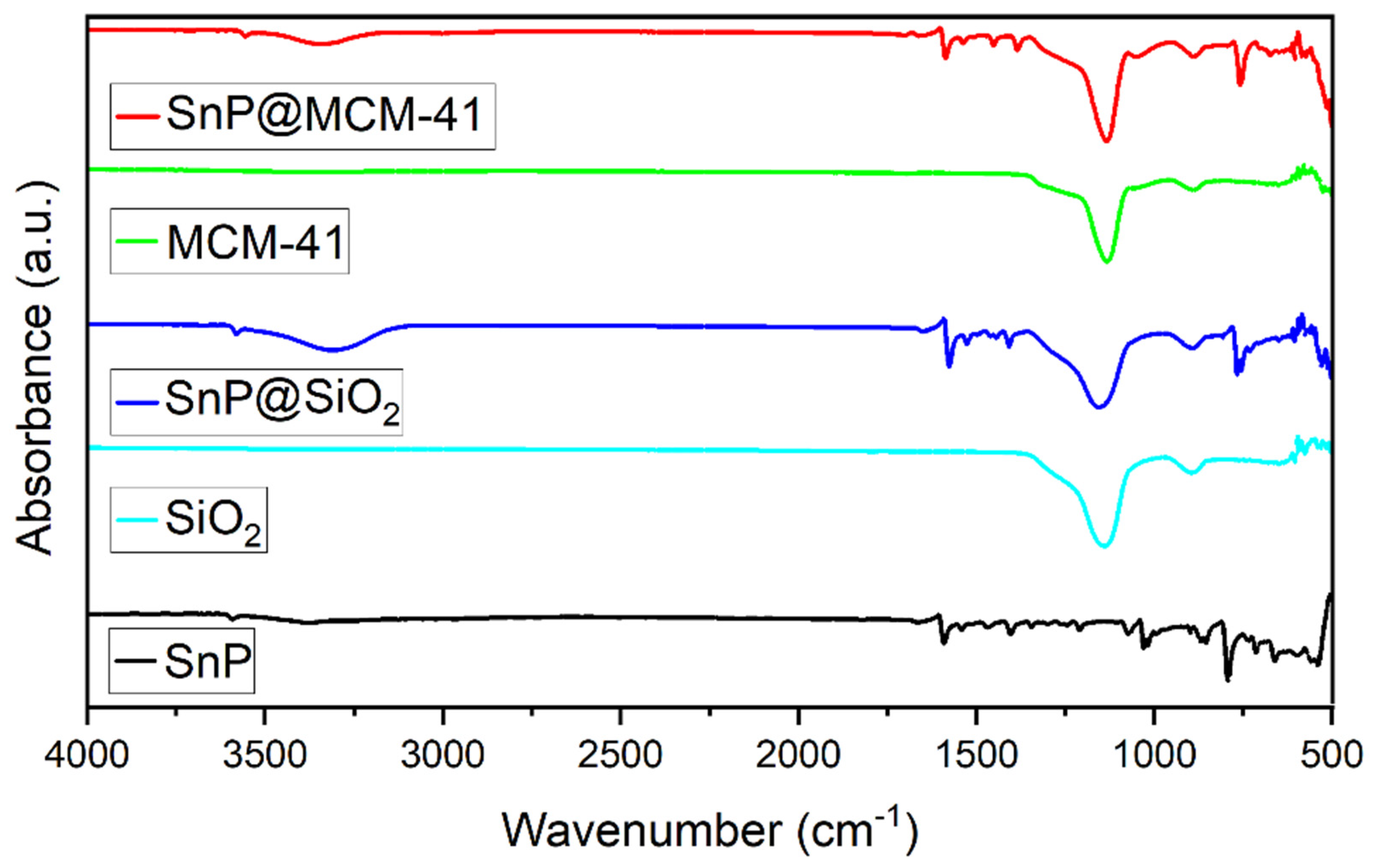
FTIR spectra of SnP@MCM−41 and SnP@SiO2 were compared to those of SnP, SiO2, and MCM−41, as shown in Figure 1. In the FTIR spectrum of SnP, the absorption peaks at 1021 and 796 cm−1 belong to the bending vibration of N−H and out-of-plane bending vibration of C−H in the benzene ring, respectively. The peaks at 3381, 1590, and 1409 cm−1 are attributed to the stretching vibrations of N−H, C=C, and C−N in the pyrrole ring, respectively. The peaks at 3600 cm−1 are assigned to the stretching vibrations of the OH signal of the axial hydroxo ligand present in the Sn(IV)-porphyrin. The absorption peaks at 1052 and 805 cm−1 are characteristic peaks of Si−O−Si stretching, while the peaks at 470 and 950 cm−1 are assigned to the bending of Si−O−Si and Si−OH for MCM−41, respectively. On the other hand, the peaks at 1063 and 803 cm−1 are characteristic peaks of Si−O−Si stretching, while those at 472 and 949 cm−1 are assigned to the bending of Si−O−Si and Si−OH for SiO2, respectively. The appearance of the peaks at 3430 and 1630 cm−1 could be attributed to adsorbed water and silanol groups of both SiO2 and MCM−41. The FTIR spectrum of SnP@MCM−41 shows the characteristic band of Si−O−Si stretching at 1055 cm−1. On the other hand, the absorption peak at 1070 cm−1 is assigned to the Si−O−Si stretching of SnP@SiO2. All other peaks are unchanged or slightly changed from those of the starting components. These observations indicate the attachment of SnP to MCM−41 and SiO2 to form SnP@MCM−41 and SnP@SiO2, respectively.
The structural patterns of SnP, SnP@MCM−41, and SnP@SiO2 were examined by X-ray diffractometer (Figure S1). It is evident from Figure S1 that the peaks are broad, and the XRD patterns are very similar for a typical amorphous solid. SnP shows two peaks centered at 9.8° and 17.8°. On the other hand, SnP@MCM−41 and SnP@SiO2 exhibit main peaks at 21.9° and 21.3°, respectively.
Solid-state UV–vis spectra reflect the light absorption properties of SnP, SiO2, MCM−41, SnP@MCM−41, and SnP@SiO2 (Figure 2). SnP exhibits strong light absorption at 429 nm belonging to the Soret band and two weak absorptions at 569 and 607 nm corresponding to the Q−bands. In contrast, SiO2 and MCM−41 did not exhibit strong light absorbance in the visible region. Compared to SnP, SnP@MCM−41 exhibits strong light absorption at 423 nm attributed to the Soret band and three weak bands at 519, 557, and 597 nm attributed to the Q−bands. Similarly, SnP@SiO2 exhibits strong light absorption at 425 nm attributed to the Soret band and three weak bands at 519, 558, and 598 nm attributed to the Q−bands. This observation implies that the Soret band, as well as the Q−bands of SnP, were blue-shifted, which confirms the strong attachment of SnP in both hybrid composites, SnP@MCM−41 and SnP@SiO2. The bandgap energy (Eg) calculated by the Tauc plot method is 2.53 eV for SnP@MCM−41, which is lower than that of SnP (2.85 eV). Similarly, the bandgap energy of SnP@SiO2 (2.64 eV) is lower than that of SnP (Table S2). Therefore, the enhanced light absorptions and narrower bandgaps of SnP@MCM−41 and SnP@SiO2 can effectively improve solar energy utilization, generating more photo-generated carriers to participate in the photocatalytic degradation reaction.
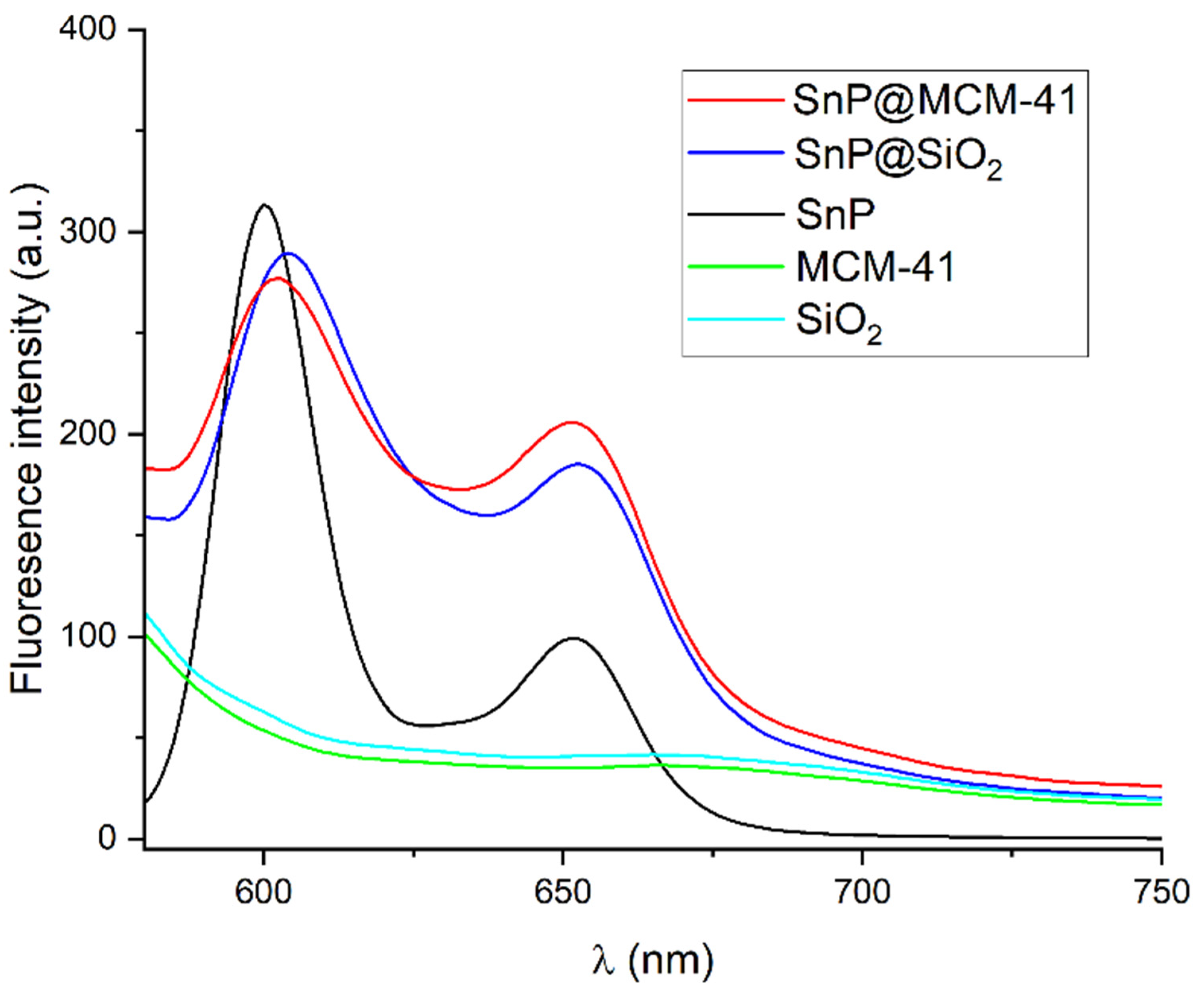
As the separation efficiency of photo-generated carriers has an important role in the photocatalytic process, its behavior was investigated by fluorescence spectroscopy. As shown in Figure 3, SnP emits fluorescence at 600 and 652 nm under excitation light with a wavelength of 560 nm, whereas SiO2 and MCM−41 do not exhibit fluorescence emission. Both hybrid composites similarly exhibited a set of emission bands, at 602 and 652 nm for SnP@MCM−41 and at 603 and 653 nm for SnP@SiO2. The emission peak-to-peak ratio of SnP was changed for SnP@MCM−41 and SnP@SiO2, which suggests strong adhesion.
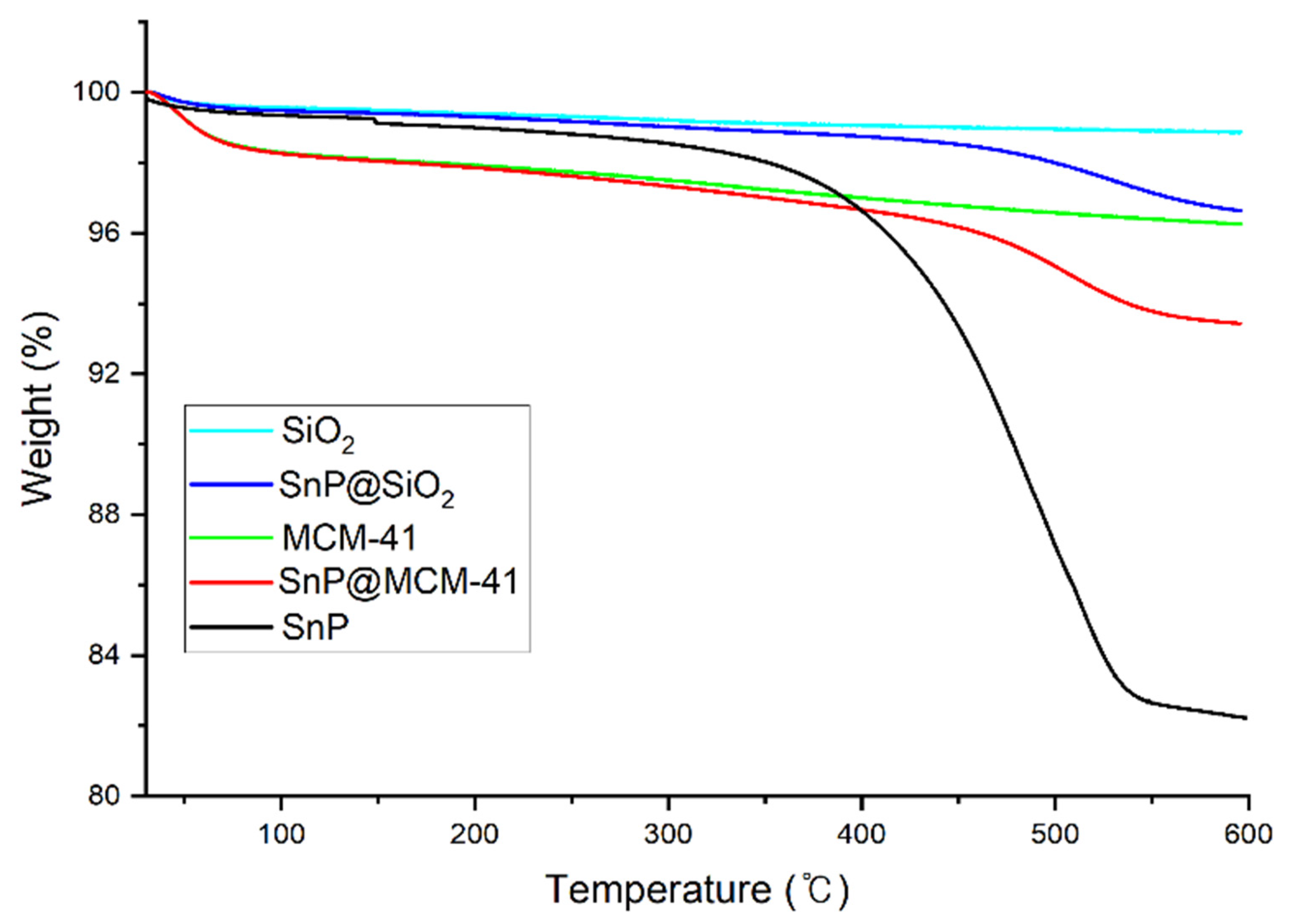
TGA curves for the materials are presented in Figure 4. SnP exhibits large weight loss starting at 350 °C. For SnP@MCM−41, the first change in the curve between 60 and 90 °C is assigned to the removal of physically adsorbed water on the surface, corresponding to a weight loss of approximately 2%. Additional weight loss was observed around 510 °C, attributed to the elimination of surface-bound organic groups of SnP. On the other hand, SnP@SiO2 experienced apparent weight loss starting at 525 °C, where the surface-bound organic groups of SnP were removed. The TGA observations indicate that SnP@MCM−41 and SnP@SiO2 are thermally stable up to 550 °C.
The structural and morphological development of the fabricated SnP@MCM−41 and SnP@SiO2 was investigated by FE−SEM. The morphologies of SnP@MCM−41 and SnP@SiO2 along with those of the starting materials are presented in Figure 5 and Figure S2. The FE−SEM images show that SnP@MCM−41 has an average diameter of approximately 100 nm with a narrow size distribution (Figure 5d). Compared to MCM−41, SnP@MCM−41 has an irregular surface, possibly due to the chemical adsorption of SnP on the surface of MCM−41. On the other hand, the sphere-shaped SnP@SiO2 has an average diameter of approximately 70 nm. Notably, the morphology of SnP does not exhibit a characteristic shape and size. EDS mapping images of SnP@SiO2 and SnP@MCM−41 (Figures S3 and S4) confirm the homogeneous distribution of elements (C, N, O, Si, and Sn).
The mesoporous structure of SnP@MCM−41 was further confirmed by TEM. The TEM image of SnP@MCM−41 (Figure 6) shows regular arrays of cylindrical mesopores forming a one-dimensional pore system in contrast to SnP@SiO2. The mesoporous channel of MCM−41 was thus expected to facilitate the efficient transport of electrons and holes photo-generated from SnP. The hydrodynamic particle sizes of SnP, SiO2, MCM−41, SnP@MCM−41, and SnP@SiO2 in a suspension were measured by DLS. The average particle size in each aqueous solution had a narrow distribution with diameters of 40, 45, 70, 125, and 100 nm for SnP, SiO2, MCM−41, SnP@MCM−41, and SnP@SiO2, respectively (Figure S5).
The nitrogen sorption isotherms for determination of the permanent porosities of MCM−41 and SnP@MCM−41 are presented in Table S3. The BET surface area and average pore size of SnP@MCM−41 are 648.7 m2/g and 1.53 nm, respectively, suitable for catalytic applications due to the large available surface. The decreases in the surface area and pore size from the bare MCM−41 to SnP@MCM−41 provide evidence for the incorporation of SnP within the channels of mesoporous frameworks. Therefore, MCM−41 was successfully coupled with SnP to form the SnP@MCM−41 mesoporous hybrid composite that maintained high thermal stability and a large surface area.

2.2. Photocatalytic Degradation of Organic Dyes

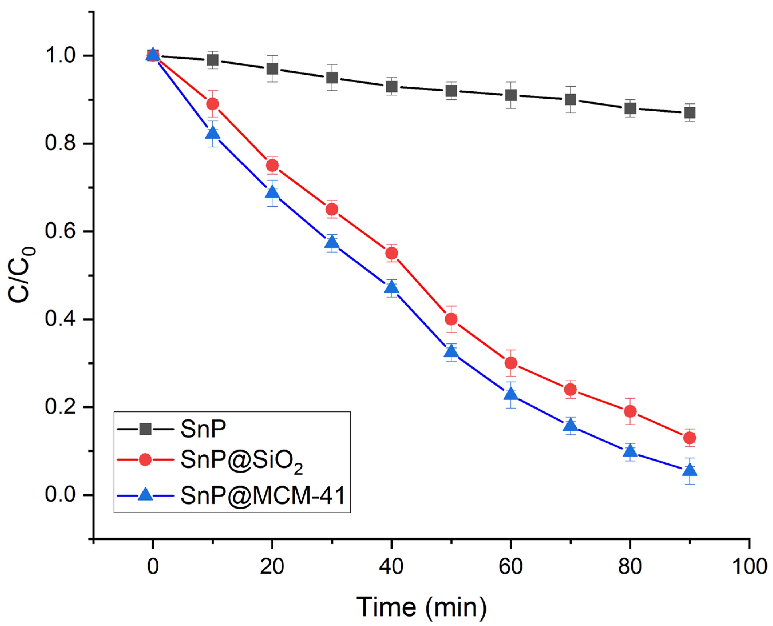
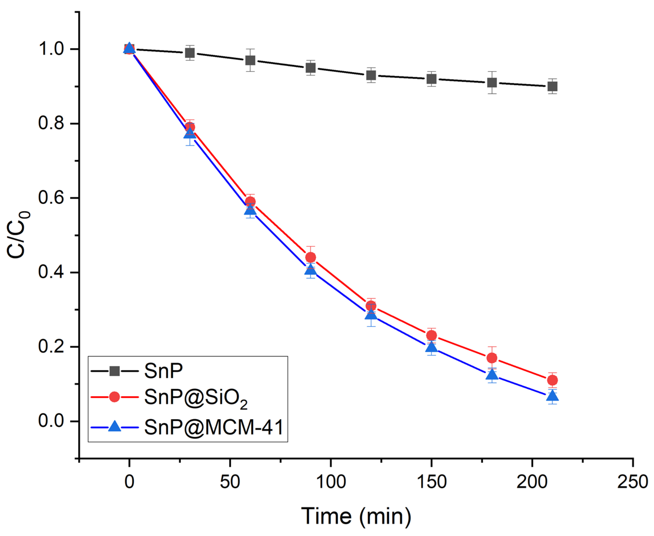
The photocatalytic performances of SnP@MCM−41 and SnP@SiO2 were investigated by the degradation of the organic dyes under visible light irradiation in aqueous solutions. For this purpose, anionic EG, cationic RhB, and neutral MCP were selected as target pollutant dyes for photocatalytic degradation. After approximately 30 min to reach the adsorption–desorption equilibrium, 5, 18, and 25% of EG was adsorbed by SnP, SnP@SiO2, and SnP@MCM−41, respectively (Figure S6). This indicates that the large surface area and mesoporous framework of SnP@MCM−41 can promote mass diffusion and enhance adsorption. Time-dependent absorption spectra of EG in the presence of SnP@MCM−41 under visible light irradiation are shown in Figure S7. Negligible decay of the EG dye was observed in the absence of either visible light or photocatalyst SnP@MCM−41 (Figure S8), which implies that visible light is essential for the degradation of the EG dye in addition to photocatalysts. In Figure S7, the absorbance of the EG dye at 629 nm decreases with the increase in the visible light irradiation time. Figure 7 shows that all prepared catalysts exhibit significant progress in the photodegradation of EG in an aqueous solution.
The degradation of the EG dye in the presence of photocatalysts can be described by its degradation efficiency, (C0 − C)/C0, where C0 is the initial concentration of EG and C is the concentration at time t. The observed degradation rate of EG is 13% for SnP, 87% for SnP@SiO2, and 95% for SnP@MCM−41 within 90 min of irradiation of visible light (Figure 7). Therefore, the photocatalyst SnP@MCM−41 exhibits better performance toward the degradation of the EG dye than SnP and SnP@SiO2. To further elucidate the reaction kinetics for the decay of the EG dye, we used the pseudo-first-order concept, expressed by ln(C0/C) = kt, which is generally used for a photocatalytic degradation experiment if the initial concentration of the dye is low, where k is the pseudo-first-order degradation rate constant. Based on the data plotted in Figure 7, the reaction kinetics of EG dye degradation are presented in Figure S9. The first-order rate constant for the degradation of the EG dye is 0.002 min−1 for SnP, 0.022 min−1 for SnP@SiO2, and 0.029 min−1 for SnP@MCM−41.
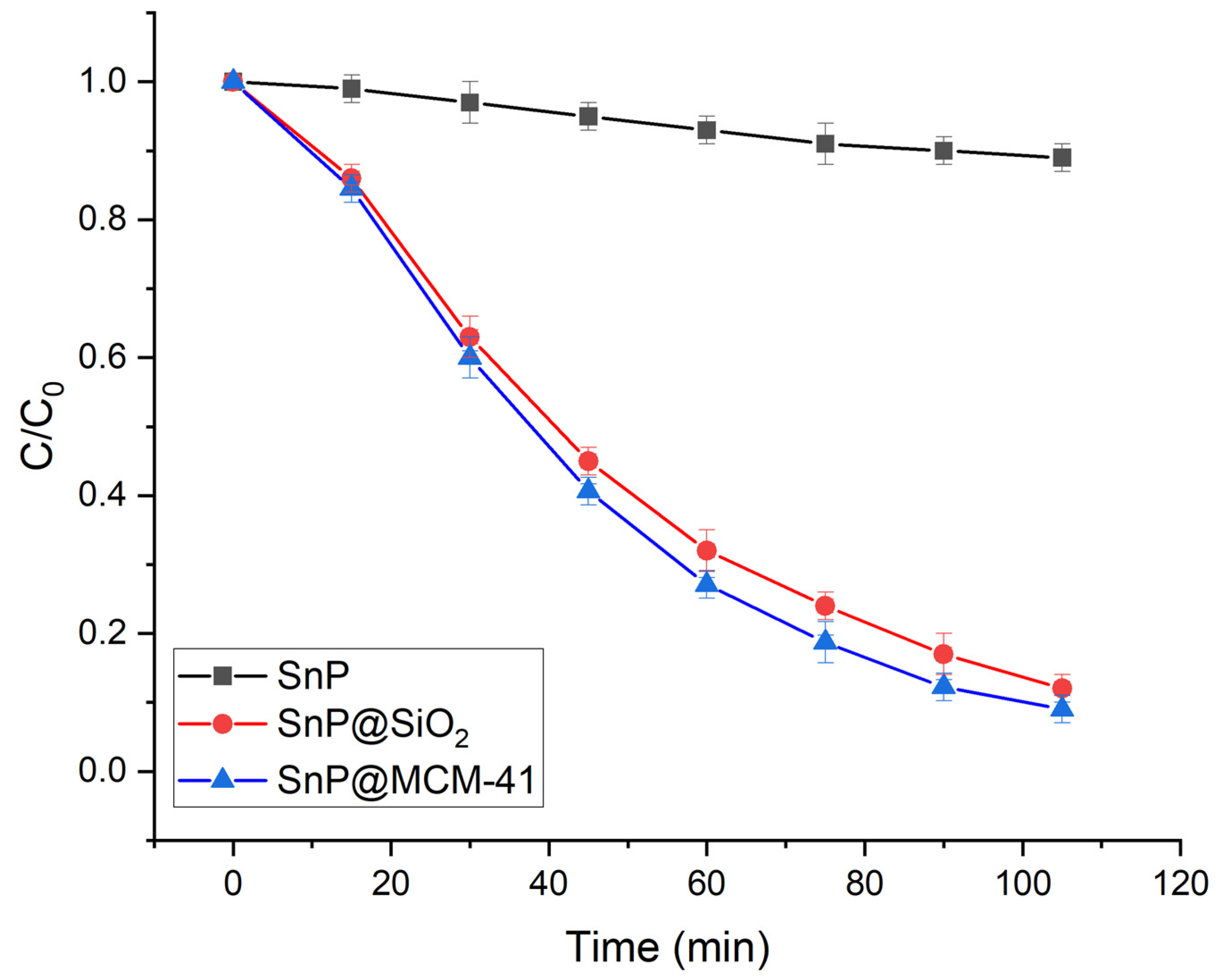
The photodegradation performances of SnP@MCM−41 and SnP@SiO2 were further analyzed toward diverse targets (cationic RhB and neutral MCP dyes) under visible light irradiation. Time-dependent absorption spectra of RhB in the presence of SnP@MCM−41 under visible light irradiation are shown in Figure S10. As shown in Figure 8 and Figure S11, the pseudo-first-order rate constant for the degradation of RhB is 0.0006 min−1 for SnP, 0.011 min−1 for SnP@SiO2, and 0.013 min−1 for SnP@MCM−41.
On the other hand, time-dependent absorption spectra of MCP in the presence of the photocatalyst SnP@MCM−41 under visible light irradiation are shown in Figure S12. The pseudo-first-order rate constant for the degradation of MCP is 0.001 min−1 for SnP, 0.021 min−1 for SnP@SiO2, and 0.024 min−1 for SnP@MCM−41 (Figure 9 and Figure S13).
The reusability of the photocatalysts SnP@MCM−41 and SnP@SiO2 is crucial for practical application, which was evaluated by recycling tests on SnP@MCM−41 toward EG dye degradation (Figure S14). Even after 10 consecutive cycles, SnP@MCM−41 still maintains a high performance for the degradation of the EG dye with a reduction of only 8%, which indicates that the photocatalyst SnP@MCM−41 has remarkable stability. After the degradation reaction, the structure of SnP@MCM−41 was additionally analyzed to confirm the stability of this photocatalyst. As shown in Figures S15 and S16, the FTIR spectrum and FE−SEM image of the used SnP@MCM−41 closely resembles that of the as-prepared state, which indicates that the mesoporous framework of this photocatalyst was intact during the photocatalytic reaction. Moreover, the process for the recovery of these photocatalysts, SnP@MCM−41 and SnP@SiO2, from the reaction mixture was very simple through successive filtration, washing, and drying procedures.
We optimized the reaction conditions in terms of the EG dye/photocatalyst ratio, temperature, and pH of the solution. Photodegradation experiments were carried out at different temperatures to evaluate the effect of temperature on the degradation of the EG dye by the photocatalyst SnP@MCM−41. With the increase in temperature, the degradation efficiency increases up to 50 °C (Figure S17). The pH of the aqueous EG dye solution apparently affected the degradation rate of the EG dye, as shown in Figure S18. The rate of degradation increases from pH = 2 to pH = 8, and then decreases to pH = 12. To evaluate the effect of the dye/photocatalyst ratio on the degradation of the EG dye, various concentrations of the EG solution (5, 10, 15, 20, 25, 30, 35, and 40 mg L−1) were applied with a constant amount (20 mg) of the photocatalyst SnP@MCM−41. The degradation rate decreases with the increase in the concentration of the EG dye. Most of the EG dye is degraded at concentrations of 5−25 mg L−1, while approximately 60% of the dye is degraded even at 40 mg L−1 (Figure S19).
Among all photocatalysts, SnP@MCM−41 shows the highest photodegradation rate, which can remove about 95% of EG dye within 90 min. To determine the effect of the amount of SnP adsorbed onto SnP@MCM−41 for photocatalytic activity, a series of SnP@MCM−41 samples varying only in a molar amount of SnP with respect to MCM−41 per gram (MCM−41/g) were used for the measurement of the EG dye degradation rate (Figure S20). The photodegradation rate of EG for composites SnP@MCM−41 (0.01 mmol/g to 0.2 mmol/g) was higher than pure SnP. The rate also increased when the molar amount of SnP compared to MCM−41/g increased in the composite, reaching the maximum of 0.1 mmol/g. After that, the rates decreased from 0.15 mmol/g to 0.2 mmol/g. When the molar amount of SnP to MCM−41/g is lower than 0.05 mmol/g, the host photocatalyst SnP has low content in the composite to reduce the amount of photogenerated carriers, resulting in relatively low catalytic efficiency. When the molar amount ratio is higher than 0.1 mmol/g, SnP significantly agglomerates on the MCM−41 surface, reducing reactive sites, suppressing photogenerated charge separation, and gradually reducing degradation efficiency of EG dye. This suggests the synergistic effect between SnP and MCM−41 is responsible for the noticeably enhanced photocatalytic activity of the composite SnP@MCM−41.
We previously reported a possible mechanism for the photodegradation of organic dyes by porphyrin-based nanomaterials in an aqueous solution under visible light irradiation [38,39]. Usually, the mechanism includes five steps. In step 1, a photocatalyst in an aqueous solution absorbs light upon exposure to visible light irradiation along with the EG dye. Valence-band electrons are promoted to the conduction band after crossing the bandgap. This assists in the generation of electron–hole (e/h+) pairs at the surface of the photocatalyst. These photogenerated holes (h+) react with H2O to form a highly reactive hydroxyl radical (OH) (step 2). On the other hand, the excited electron reacts with the dissolved O2 to generate highly reactive superoxide radical anions (O2−•) in step 3. These photogenerated, highly reactive superoxide radical anions and hydroxyl radicals react with the EG dye and degrade it into small molecules and finally to CO2 and H2O (steps 4 and 5). The photocatalytic degradation mechanism of 4-chlorophenol and acid orange 7 by SnP-based photocatalysts was similarly discussed [14].
The mechanism consists of five steps for a porphyrin-based porous framework P:
P + P ( e + h+)
H2O + h+OH + H+
O2 + 2e → O2−•
OH + EG → degraded products
O2−• + EG → degraded products
Radical trapping experiments were carried out to detect the photogenerated reactive species during the photocatalytic degradation of the EG dye [44,45]. We used ethylenediaminetetraacetic acid disodium (Na2EDTA) to capture holes, tert-butanol (tBuOH) to capture hydroxyl radicals (OH), and para-benzoquinone (p-BQ) for superoxide radical anions (O2−•) during the photodegradation experiment of the EG dye in the presence of SnP@MCM−41. Figure S8 confirms that the EG dye degradation rate is critically affected in the presence of tBuOH as well as p-BQ. Superoxide radicals (O2−•) are a major reactive species compared to hydroxyl radicals (OH) responsible for the catalytic degradation of the EG dye in an aqueous solution. However, the degradation of the EG dye was not affected by the presence of Na2EDTA or photogenerated holes.
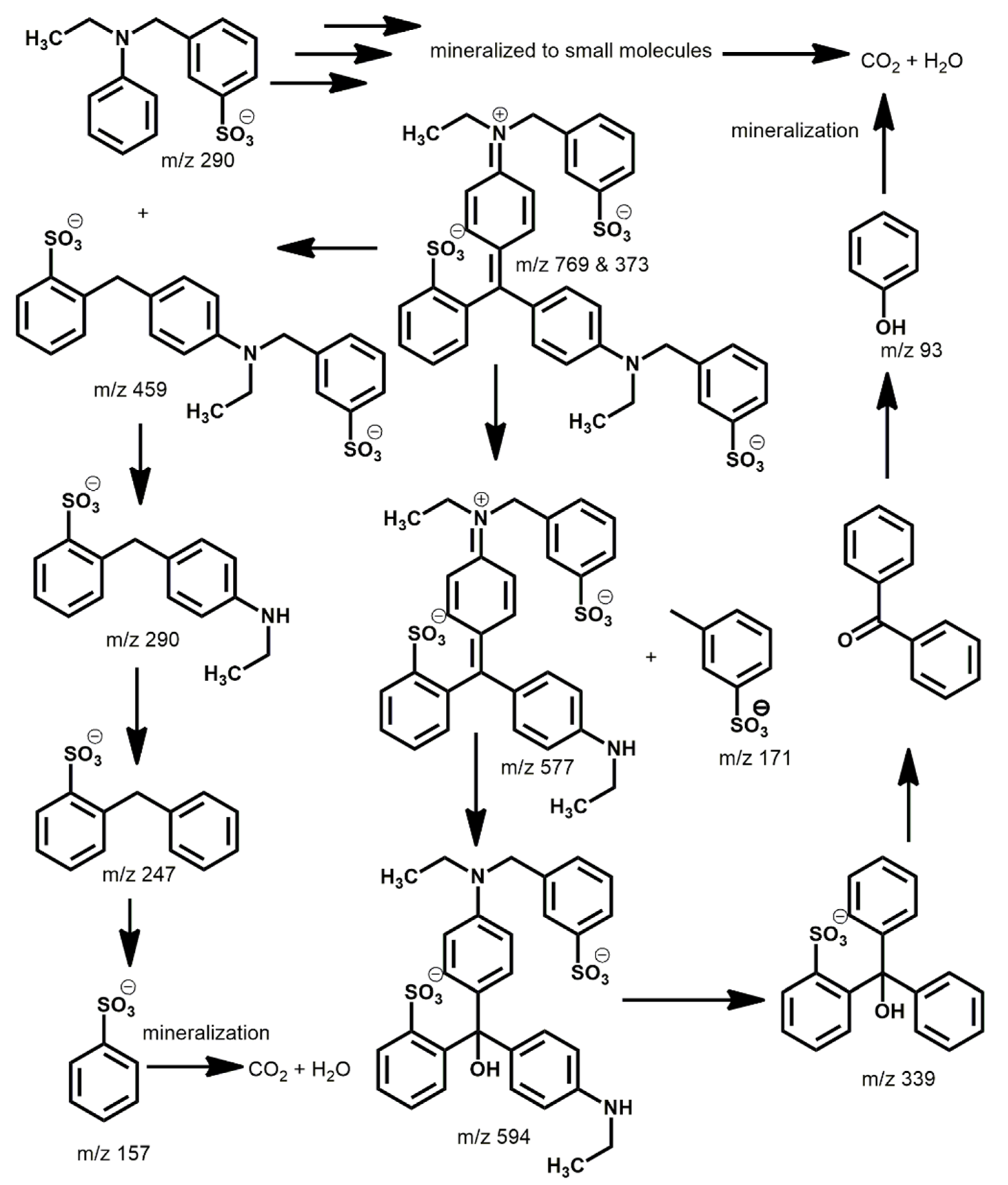
We also analyzed the degradation products of the EG dye after visible light irradiation in the presence of the photocatalyst SnP@MCM−41. The reaction mixture was analyzed by electrospray ionization–mass spectrometry (ESI-MS) after 45 min for each photodegradation experiment (Figure S21). New peaks in the mass spectra confirmed the degradation of the EG dye to new small molecules [46]. Based on the mass spectra in Figure S16, possible intermediates for the degradation of the EG dye are shown in Figure 10. Initially, the base peak (m/z = 769 for [EG−Na]) and another peak (m/z = 373 for [EG−2Na]) belong to the anionic EG dye. The EG dye can be fragmented in two different manners. The first is through cleavage of the tertiary amine bond and formation of two low-molecular-weight fragments with a secondary amine (m/z = 577) and 3-methyl-benzenesulfonic acid (m/z = 171). This secondary amine can further undergo hydrolysis and generate a tertiary alcohol derivative (m/z = 594). Again, successive oxidation of tertiary alcohol leads to formation of lower-molecular-weight fragments (m/z = 339 and 93). In the second process, the cleavage of the C–C double bond leads to formation of two lower-molecular-weight amine fragments (m/z = 459 and 290). These low-molecular-weight amines can then undergo successive hydrolysis that leads to formation of benzenesulfonic acid (m/z = 157). Finally, these lower-molecular-weight intermediate molecules were further fragmented and mineralized into CO2 and H2O. The total organic carbon (TOC) value was calculated to estimate the removal of the EG dye by photocatalysts [47]. The TOC removal percentage attained using SnP@MCM−41 was only 75%.
Finally, the degradation performances of organic dyes represented by the pseudo-first-order rate constants for SnP@MCM−41 and SnP@SiO2 were compared to those of selected photocatalysts reported in the literature. As listed in Table 1, SnP@MCM−41 and SnP@SiO2 under visible light irradiation exhibit higher than or comparable degradation performances for EG, MCP, and RhB to those of most reported photocatalysts.

3. Materials and Methods

MCM−41 and SiO2 nanoparticles were purchased from Sigma–Aldrich. All other chemicals were purchased from TCI and used without further purification unless otherwise specified. (trans-Dihydroxo)(5,10,15,20-tetraphenylporphyrinato)tin(IV) (SnP) was prepared similarly according to the previously reported procedure [59]. CH2Cl2 and pyrrole were distilled from a solution over calcium hydride. Fourier-transform infrared (FTIR) spectra were acquired using a Shimadzu FTIR−8400S spectrophotometer (Shimadzu, Tokyo, Japan). Steady-state ultraviolet–visible (UV–vis) spectra were recorded using a Shimadzu UV-3600 spectrophotometer (Shimadzu, Tokyo, Japan). Fluorescence spectra were recorded using a Shimadzu RF-5301PC fluorescence spectrophotometer (Shimadzu, Tokyo, Japan). Powder X-ray diffraction (PXRD) patterns were obtained on a Bruker AXS D8 Advance powder X-ray diffractometer (Bruker, Billerica, MA, USA). A thermogravimetric analysis (TGA) was performed using an Auto-TGA Q500 instrument (TA Instruments, New Castle, DE, USA). The Brunauer−Emmett−Teller (BET) surface area was determined using an analyzer (BELSORP-mini volumetric adsorption equipment) through N2 adsorption isotherms at 77 K. The surface area and pore size were estimated using an Autosorb-iQ and Quadrasorb SI. Field-emission scanning electron microscopy (FE−SEM) and transmission electron microscopy (TEM) images were acquired using a MAIA III (TESCAN, Brno, Czech Republic) and JEOL/JEM 2100, respectively. Zeta potentials were measured with an Otsuka Electronics ELSZ−2. Dynamic light scattering (DLS) experiments and inductively coupled plasma (ICP) analysis were performed using a NanoBrook 90Plus DLS size analyzer (Brookhaven, NY, USA) and ICP−Spectrociros CCD instrument, respectively.

3.1. Fabrication of SnP@MCM−41

Dried MCM−41 (3.0 g) was added into a solution of SnP (0.23 g) dissolved in CH2Cl2 (100 mL, ~3.0 mM) and stirred for 24 h at room temperature. The solids were filtered and washed three times successively with acetone, CH2Cl2, and N,N−dimethylformamide. Afterward, the solids were dried in a vacuum oven for 4 h at 80 °C, which yielded a powder of SnP@MCM−41 (2.864 g).

3.2. Fabrication of SnP@SiO2

Using dried SiO2 (3.0 g), a powder of SnP@SiO2 (3.003 g) was obtained by the same procedure.

3.3. Photocatalytic Degradation Experiment

The photocatalytic efficiencies of these hybrid composites were investigated by photodegrading EG, RhB, and MCP dyes in each aqueous solution. In a typical procedure, 20 mg of the photocatalyst was added to 250 mL of an aqueous solution of EG (20 mg L−1, distilled water with pH of 7.0) with stirring at 298 K. The reaction mixture was allowed to stand in dark for 30 min to reach the adsorption−desorption equilibrium. Afterward, the irradiation process was started using a 150 W xenon arc lamp with a UV cut-off filter (ABET Technologies, Old Gate Lane, Milford, CT, USA) at 298 K. After irradiation with visible light, a 3 mL suspension was collected at regular intervals. The photocatalyst was separated from the solution by centrifugation and collected by filtration with filter paper. The concentration of EG was determined by measuring the absorbance at 629 nm using a UV–vis spectrophotometer. Similarly, an aqueous solution of RhB (40 mg L−1) dye was prepared, and its concentration was determined by assessing the absorbance at 553 nm. For the MCP dye, we used an aqueous solution of MCP (65 mg L−1) and determined its concentration at 435 nm.

4. Conclusions

Hybrid composites of SnP@MCM−41 and SnP@SiO2 were fabricated by the reaction of (trans-dihydroxo)(5,10,15,20-tetraphenylporphyrinato)tin(IV) (SnP) with the mesostructured MCM−41 and nanoparticle SiO2, respectively. The formation of hybrid composites was readily achieved through condensation between the hydroxo ligand of SnP and silanol groups of MCM−41 or SiO2. These hybrid composites were fully characterized by FTIR spectroscopy, UV–vis spectroscopy, fluorescence spectroscopy, TEM, and FE−SEM techniques. The incorporated SnP onto the mesoporous structure provided efficient photocatalytic degradation for the anionic EG, cationic RhB, and neutral MCP dyes under visible light irradiation in an aqueous solution. The pseudo-first-order degradation rate constant of organic dyes for SnP@MCM−41 was higher than those of SnP@SiO2 and SnP. These visible light photocatalysts showed remarkable stability and reliable reusability. Our report on visible light photocatalysts provides a valuable contribution for the treatment of dyed wastewater and encourages the development of highly effective hybrid composites composed of porous materials and visible light photocatalysts for applications in environmental remediation.

Supplementary Materials

The following supporting information can be downloaded at: https://www.mdpi.com/article/10.3390/molecules28041886/s1, Table S1: Zeta potentials and mobilities of SiO2, SnP@SiO2, MCM−41, SnP@MCM−41, and SnP; Table S2: Band-gap energies (Eg) calculated by the Tauc plot method; Table S3: Porosities of MCM-41 and SnP@MCM−41; Figure S1: XRD patterns of SnP, SnP@SiO2, and SnP@MCM−41; Figure S2: High-resolution FE−SEM images of (a) SiO2, (b) SnP@SiO2, (c) MCM−41, (d) SnP@MCM−41, and (e) SnP; Figure S3: EDS mapping of SnP@SiO2; Figure S4: EDS mapping of SnP@MCM−41; Figure S5: Hydrodynamic particle size distributions of SnP, SiO2, MCM−41, SnP@MCM−41, and SnP@SiO2 obtained by the DLS method; Figure S6: Adsorptivities of SnP, SnP@MCM−41, and SnP@SiO2 for the EG dye; Figure S7: Time-dependent absorption spectra of EG in the presence of SnP@MCM−41 under visible light irradiation; Figure S8: Various external effects on the degradation of the EG dye in the presence of SnP@MCM−41 under visible light irradiation. ([Na2EDTA]0 = [p-BQ]0 = [tBuOH]0 = 2 mM, pH = 7.0, T = 298 K); Figure S9: Kinetics for the photocatalytic degradation of EG under visible light irradiation by the photocatalysts SnP, SnP@MCM−41, and SnP@SiO2; Figure S10: Time-dependent absorption spectra of the RhB dye in the presence of SnP@MCM−41 under visible light irradiation; Figure S11: Kinetics for the photocatalytic degradation of RhB under visible light irradiation by the photocatalysts SnP, SnP@MCM−41, and SnP@SiO2; Figure S12: Time-dependent absorption spectra of the MCP dye in the presence of SnP@MCM−41 under visible light irradiation; Figure S13: Kinetics for the photocatalytic degradation of MCP under visible light irradiation by the photocatalysts SnP, SnP@MCM−41, and SnP@SiO2; Figure S14: Typical catalytic cycle (up to 10 cycles) for the photocatalyst SnP@MCM−41 for the degradation of the EG dye; Figure S15: Comparison of FT-IR spectra of SnP@MCM−41 before and after the degradation of the EG dye; Figure S16: Comparison of FE−SEM images of SnP@MCM−41 before and after the degradation of EG dye; Figure S17: Effect of the temperature on the degradation of the EG dye by SnP@MCM−41; Figure S18: Effect of the pH of the solution of the EG dye for photodegradation by SnP@MCM−41; Figure S19: Effect of the concentration of the EG dye for photodegradation by SnP@MCM−41 (20 mg) within 90 min of visible light irradiation; Figure S20: Effect of the amount of SnP adsorbed onto SnP@MCM−41 composite for photocatalytic degradation of EG dye, where X = mmol of SnP per gram of MCM−41; Figure S21: ESI-MS spectrum (negative ion mode) of the reaction mixture of EG in the presence of SnP@MCM−41 after 45 min of visible light irradiation.

Author Contributions

Conceptualization, data curation, formal analysis, investigation, methodology, software, validation, visualization and writing—original draft, N.K.S.; data curation, formal analysis, investigation, methodology, B.-H.P.; conceptualization, funding acquisition, project administration, resources, supervision, validation, and writing—review and editing, H.-J.K. All authors have read and agreed to the published version of the manuscript.

Funding

This work was supported by the National Research Foundation of Korea (NRF) (grant No. 2022R1F1A1074420) funded by the Korean government (MSIT).

Institutional Review Board Statement

Not applicable.

Informed Consent Statement

Not applicable.

Data Availability Statement

Not applicable.

Conflicts of Interest

The authors declare no conflict of interest.

References

  1. Schwarzenbach, R.P.; Egli, T.; Hofstetter, T.; Von Gunten, U.; Wehrli, B. Global Water Pollution and Human Health. Annu. Rev. Environ. Resour. 2010, 35, 109–136. [Google Scholar] [CrossRef]
  2. Naidu, R.; Biswas, B.; Willett, I.R.; Cribb, J.; Kumar Singh, B.; Paul Nathanail, C.; Coulon, F.; Semple, K.T.; Jones, K.C.; Barclay, A.; et al. Chemical Pollution: A Growing Peril and Potential Catastrophic Risk to Humanity. Environ. Int. 2021, 156, 106616. [Google Scholar] [CrossRef] [PubMed]
  3. Wang, Q.; Li, H.; Yu, X.; Jia, Y.; Chang, Y.; Gao, S. Morphology regulated Bi2WO6 nanoparticles on TiO2 nanotubes by solvothermal Sb3+ doping as effective photocatalysts for wastewater treatment. Electrochim. Acta 2020, 330, 135167. [Google Scholar] [CrossRef]
  4. Parvulescu, V.I.; Epron, F.; Garcia, H.; Granger, P. Recent Progress and Prospects in Catalytic Water Treatment. Chem. Rev. 2022, 122, 2981–3121. [Google Scholar] [CrossRef] [PubMed]
  5. Ain, Q.U.; Rasheed, U.; Yaseen, M.; Zhang, H.; Tong, Z. Superior dye degradation and adsorption capability of polydopamine modified Fe3O4-pillared bentonite composite. J. Hazard. Mater. 2020, 397, 122758. [Google Scholar] [CrossRef] [PubMed]
  6. Glugoski, L.P.; de Jesus Cubas, P.; Fujiwara, S.T. Reactive Black 5 dye degradation using filters of smuggled cigarette modified with Fe3+. Environ. Sci. Pollut. Res. 2017, 24, 6143–6150. [Google Scholar] [CrossRef]
  7. Balcha, A.; Yadav, O.P.; Dey, T. Photocatalytic degradation of methylene blue dye by zinc oxide nanoparticles obtained from precipitation and sol-gel methods. Environ. Sci. Pollut. Res. 2016, 23, 25485–25493. [Google Scholar] [CrossRef]
  8. Garrido-Cardenas, J.A.; Esteban-García, B.; Agüera, A.; Sánchez-Pérez, J.A.; Manzano-Agugliaro, F. Wastewater Treatment by Advanced Oxidation Process and Their Worldwide Research Trends. Int. J. Environ. Res. Public Health 2020, 17, 170. [Google Scholar] [CrossRef] [Green Version]
  9. Dewil, R.; Mantzavinos, D.; Poulios, I.; Rodrigo, M.A. New perspectives for Advanced Oxidation Processes. J. Environ. Manag. 2017, 195, 93–99. [Google Scholar] [CrossRef] [Green Version]
  10. O’Neill, J.S.; Kearney, L.; Brandon, M.P.; Pryce, M.T. Design Components of Porphyrin-Based Photocatalytic Hydrogen Evolution Systems: A Review. Coord. Chem. Rev. 2022, 467, 214599. [Google Scholar] [CrossRef]
  11. Mota, H.P.; Quadrado, R.F.N.; Iglesias, B.A.; Fajardo, A.R. Enhanced Photocatalytic Degradation of Organic Pollutants Mediated by Zn(II)-Porphyrin/Poly(Acrylic Acid) Hybrid Microparticles. Appl. Catal. B Environ. 2020, 277, 119208. [Google Scholar] [CrossRef]
  12. Li, Y.; Wang, L.; Gao, Y.; Yang, W.; Li, Y.; Guo, C. Porous metalloporphyrinic nanospheres constructed from metal 5,10,15,20-tetraksi(4′-ethynylphenyl)porphyrin for efficient catalytic degradation of organic dyes. RSC Adv. 2018, 8, 7330–7339. [Google Scholar] [CrossRef] [PubMed] [Green Version]
  13. Jang, J.H.; Jeon, K.-S.; Oh, S.; Kim, H.-J.; Asahi, T.; Masuhara, H.; Yoon, M. Synthesis of Sn-Porphyrin-Intercalated Trititanate Nanofibers:  Optoelectronic Properties and Photocatalytic Activities. Chem. Mater. 2007, 19, 1984–1991. [Google Scholar] [CrossRef]
  14. Kim, W.; Park, J.; Jo, H.J.; Kim, H.-J.; Choi, W. Visible Light Photocatalysts Based on Homogeneous and Heterogenized Tin Porphyrins. J. Phys. Chem. C 2008, 112, 491–499. [Google Scholar] [CrossRef]
  15. Guo, X.; Li, Y.-Y.; Shen, D.-H.; Song, Y.-Y.; Wang, X.; Liu, Z.-G. Immobilization of cobalt porphyrin on CeO2@SiO2 core-shell nanoparticles as a novel catalyst for selective oxidation of diphenylmethane. J. Mol. Catal. A Chem. 2013, 367, 7–11. [Google Scholar] [CrossRef]
  16. Yoo, H.-Y.; Yan, S.; Ra, J.W.; Jeon, D.; Goh, B.; Kim, T.-Y.; Mackeyev, Y.; Ahn, Y.-Y.; Kim, H.-J.; Wilson, L.J.; et al. Tin porphyrin immobilization significantly enhances visible-light-photosensitized degradation of Microcystins: Mechanistic implications. Appl. Catal. B Environ. 2016, 199, 33–44. [Google Scholar] [CrossRef]
  17. Wang, J.; Zhong, Y.; Wang, X.; Yang, W.; Bai, F.; Zhang, B.; Alarid, L.; Bian, K.; Fan, H. pH-Dependent Assembly of Porphyrin-Silica Nanocomposites and Their Application in Targeted Photodynamic Therapy. Nano Lett. 2017, 17, 6916–6921. [Google Scholar] [CrossRef]
  18. Kresge, C.T.; Leonowicz, M.E.; Roth, W.J.; Vartuli, J.C.; Beck, J.S. Ordered mesoporous molecular sieves synthesized by a liquid-crystal template mechanism. Nature 1992, 359, 710–712. [Google Scholar] [CrossRef]
  19. Martínez-Edo, G.; Balmori, A.; Pontón, I.; Martí del Rio, A.; Sánchez-García, D. Functionalized Ordered Mesoporous Silicas (MCM-41): Synthesis and Applications in Catalysis. Catalysts 2018, 8, 617. [Google Scholar] [CrossRef] [Green Version]
  20. Costa, J.A.S.; Jesus, R.A.; Santos, D.; Manoa, J.F.; Romao, L.P.C.; Paranhos, C.M. Recent progresses in the adsorption of organic, inorganic, and gas compounds by MCM-41-based mesoporous materials. Microporous Mesoporous Mater. 2020, 291, 109698. [Google Scholar] [CrossRef]
  21. Kim, H.J.; Park, K.-M.; Ahn, T.K.; Kim, S.K.; Kim, K.S.; Kim, D.; Kim, H.-J. Novel fullerene–porphyrin–fullerene triad linked by metal axial coordination: Synthesis, X-ray crystal structure, and spectroscopic characterizations of trans-bis([60]fullerenoacetato)tin(iv) porphyrin. Chem. Commun. 2004, 2594–2595. [Google Scholar] [CrossRef]
  22. Kim, H.J.; Jeon, W.S.; Lim, J.H.; Hong, C.S.; Kim, H.-J. Synthesis, X-ray crystal structure, and electrochemistry of trans-bis(ferrocenecarboxylato)(tetraphenylporphyrinato)tin(IV). Polyhedron 2007, 26, 2517–2522. [Google Scholar] [CrossRef]
  23. Kim, H.J.; Jang, J.H.; Choi, H.; Lee, T.; Ko, J.; Yoon, M.; Kim, H.-J. Photoregulated Fluorescence Switching in Axially Coordinated Tin(IV) Porphyrinic Dithienylethene. Inorg. Chem. 2008, 47, 2411–2415. [Google Scholar] [CrossRef]
  24. Kim, M.K.; Shee, N.K.; Lee, J.; Yoon, M.; Kim, H.-J. Photoinduced Electron Transfer upon Supramolecular Complexation of (Porphyrinato) Sn-Viologen with Cucurbit[7]uril. Photochem. Photobiol. Sci. 2019, 18, 1996–2002. [Google Scholar] [CrossRef]
  25. Shee, N.K.; Kim, M.K.; Kim, H.-J. Fluorescent chemosensing for aromatic compounds by supramolecular complex composed of tin(IV) porphyrin, viologen, and cucurbit[8]uril. Chem. Commun. 2019, 55, 10575–10578. [Google Scholar] [CrossRef]
  26. Kim, H.-J.; Bampos, N.; Sanders, J.K.M. Assembly of Dynamic Heterometallic Oligoporphyrins Using Cooperative Zinc-Nitrogen, Ruthenium-Nitrogen, and Tin-Oxygen Coordination. J. Am. Chem. Soc. 1999, 121, 8120–8121. [Google Scholar] [CrossRef]
  27. Indelli, M.T.; Chiorboli, C.; Ghirotti, M.; Orlandi, M.; Scandola, F.; Kim, H.J.; Kim, H.-J. Photoinduced Electron Transfer in Ruthenium(II)/Tin(IV) Multiporphyrin Arrays. J. Phys. Chem. B 2010, 114, 14273–14282. [Google Scholar] [CrossRef] [PubMed]
  28. Kim, H.J.; Shee, N.K.; Park, K.-M.; Kim, H.-J. Assembly and X-ray crystal structures of heterometallic multiporphyrins with complementary coordination between ruthenium (II) and tin (IV) porphyrins. Inorg. Chim. Acta 2019, 488, 1–7. [Google Scholar] [CrossRef]
  29. Kim, H.-J.; Jo, H.J.; Kim, J.; Kim, S.-Y.; Kim, D.; Kim, K. Supramolecular self-assembly of tin(IV) porphyrin channels stabilizing single-file chains of water molecules. CrystEngComm 2005, 7, 417–420. [Google Scholar] [CrossRef] [Green Version]
  30. Li, C.; Park, K.-M.; Kim, H.-J. Ionic assembled hybrid nanoparticle consisting of tin(IV) porphyrin cations and polyoxomolybdate anions, and photocatalytic hydrogen production by its visible light sensitization. Inorg. Chem. Commun. 2015, 60, 8–11. [Google Scholar] [CrossRef]
  31. Shee, N.K.; Lee, C.-J.; Kim, H.-J. Octahedral Tin(IV) Porphyrin-Based Porous Square-Grid Coordination Networks Exhibiting Selective Uptake of CO2 over N2. Bull. Korean Chem. Soc. 2022, 43, 103–109. [Google Scholar] [CrossRef]
  32. Kim, S.-H.; Kim, H.-J. Photocatalytic Hydrogen Production by the Sensitization of Sn(IV)-Porphyrin Embedded in a Nafion Matrix Coated on TiO2. Molecules 2022, 27, 3770. [Google Scholar] [CrossRef] [PubMed]
  33. Shee, N.K.; Kim, M.K.; Kim, H.-J. Supramolecular Porphyrin Nanostructures Based on Coordination-Driven Self-Assembly and Their Visible Light Catalytic Degradation of Methylene Blue Dye. Nanomaterials 2020, 10, 2314. [Google Scholar] [CrossRef] [PubMed]
  34. Shee, N.K.; Kim, H.-J. Self-Assembled Nanomaterials Based on Complementary Sn(IV) and Zn(II)-Porphyrins, and Their Photocatalytic Degradation for Rhodamine B Dye. Molecules 2021, 26, 3598. [Google Scholar] [CrossRef]
  35. Shee, N.K.; Kim, H.-J. Three Isomeric Zn(II)-Sn(IV)-Zn(II) Porphyrin-Triad-Based Supramolecular Nanoarchitectures for the Morphology-Dependent Photocatalytic Degradation of Methyl Orange. ACS Omega 2022, 7, 9775–9784. [Google Scholar] [CrossRef]
  36. Shee, N.K.; Kim, H.-J. Morphology-controlled self-assembled nanostructures of complementary metalloporphyrin triads through intermolecular coordination tuning and their photocatalytic degradation for Orange II. Inorg. Chem. Front. 2022, 9, 5538–5548. [Google Scholar] [CrossRef]
  37. Shee, N.K.; Kim, H.-J. Coordination framework materials fabricated by the self-assembly of Sn(IV) porphyrins with Ag(I) ions for the photocatalytic degradation of organic dyes in wastewater. Inorg. Chem. Front. 2022, 9, 1270–1280. [Google Scholar] [CrossRef]
  38. Shee, N.K.; Kim, H.-J. Sn(IV) Porphyrin-Based Ionic Self-Assembled Nanostructures and Their Application in Visible Light Photo-Degradation of Malachite Green. Catalysts 2022, 12, 799. [Google Scholar] [CrossRef]
  39. Shee, N.K.; Kim, H.-J. Sn(IV)-Porphyrin-Based Nanostructures Featuring Pd(II)-Mediated Supramolecular Arrays and Their Photocatalytic Degradation of Acid Orange 7 Dye. Int. J. Mol. Sci. 2022, 23, 13702. [Google Scholar] [CrossRef]
  40. Shee, N.K.; Kim, H.-J. Supramolecular squares of Sn(IV)porphyrins with Re(I)-corners for the fabrication of self-assembled nanostructures performing photocatalytic degradation of Eriochrome Black T dye. Inorg. Chem. Front. 2022, 10, 174–183. [Google Scholar] [CrossRef]
  41. Jank, M.; Köser, H.; Lücking, F.; Martienssen, M.; Wittchen, S. Decolorization and Degradation of Erioglaucine (Acid Blue 9) Dye in Wastewater. Environ. Technol. 1998, 19, 741–747. [Google Scholar] [CrossRef]
  42. Larowska, D.; O’Brien, J.M.; Senge, M.O.; Burdzinski, G.; Marciniak, B.; Lewandowska-Andralojc, A. Graphene Oxide Functionalized with Cationic Porphyrins as Materials for the Photodegradation of Rhodamine B. J. Phys. Chem. C 2020, 124, 15769–15780. [Google Scholar] [CrossRef] [PubMed]
  43. Khezrianjoo, S.; Revanasiddappa, H.D. Effect of operational parameters and kinetic study on the photocatalytic degradation of m-cresol purple using irradiated ZnO in aqueous medium. Water Qual. Res. J. 2015, 51, 69–78. [Google Scholar] [CrossRef] [Green Version]
  44. Liu, D.; Song, J.; Chung, J.S.; Hur, S.H.; Choi, W.M. ZnO/Boron Nitride Quantum Dots Nanocomposites for the Enhanced Photocatalytic Degradation of Methylene Blue and Methyl Orange. Molecules 2022, 27, 6833. [Google Scholar] [CrossRef] [PubMed]
  45. Rojas-García, E.; García-Martínez, D.C.; López-Medina, R.; Rubio-Marcos, F.; Castañeda-Ramírez, A.A.; Maubert-Franco, A.M. Photocatalytic Degradation of Dyes Using Titania Nanoparticles Supported in Metal-Organic Materials Based on Iron. Molecules 2022, 27, 7078. [Google Scholar] [CrossRef] [PubMed]
  46. Borhade, A.V.; Tope, D.R.; Dabhade, G.B. Removal of erioglaucine dye from aqueous medium using ecofriendly synthesized ZnMnO3 photocatalyst. J. Surf. Sci. Nanotech. 2017, 15, 74–80. [Google Scholar] [CrossRef] [Green Version]
  47. Wang, Z.; Xia, L.; Chen, J.; Ji, L.; Zhou, Y.; Wang, Y.; Cai, L.; Guo, J.; Song, W. Fine Characterization of Natural SiO2-Doped Catalyst Derived from Mussel Shell with Potential Photocatalytic Performance for Organic Dyes. Catalysts 2020, 10, 1130. [Google Scholar] [CrossRef]
  48. Daneshvar, N.; Salari, D.; Niaei, A.; Khataee, A. Photocatalytic degradation of the herbicide erioglaucine in the presence of nanosized titanium dioxide: Comparison and modeling of reaction kinetics. J. Environ. Sci. Health Part B 2006, 41, 1273–1290. [Google Scholar] [CrossRef]
  49. Azar, F.; Yang, H.-B.; Venault, L.; Faure, S. Degradation of erioglaucine dye under γ-irradiation. Procedia Chem. 2012, 7, 647–653. [Google Scholar] [CrossRef] [Green Version]
  50. Jain, R.; Sikarwar, S. Photodestruction and COD removal of toxic dye erioglaucine by TiO2-UV process: Influence of operational parameters. Int. J. Phys. Sci. 2008, 3, 299–305. [Google Scholar]
  51. Shenoy, S.; Jang, E.; Park, T.J.; Gopinath, C.S.; Sridharan, K. Cadmium sulfide nanostructures: Influence of morphology on the photocatalytic degradation of erioglaucine and hydrogen generation. Appl. Surf. Sci. 2019, 483, 696–705. [Google Scholar] [CrossRef]
  52. Khezrianjoo, S.; Revanasiddappa, H.D. Electrochemical oxidation of m-cresol purple dye in aqueous media. Water Qual. Res. J. Can. 2015, 50, 305–313. [Google Scholar] [CrossRef]
  53. Chatterjee, S.; Tyagi, A.K.; Ayyub, P. Efficient Photocatalytic Degradation of Rhodamine B Dye by Aligned Arrays of Self Assembled Hydrogen Titanate Nanotubes. J. Nanomater. 2014, 5, 1–7. [Google Scholar] [CrossRef]
  54. Sundararajan, M.; Sailaja, V.; Kennedy, L.J.; Vijaya, J.J. Photocatalytic degradation of Rhodamine B under visible light using nanostructured zinc doped cobalt ferrite: Kinetics and mechanism. Ceram. Int. 2017, 43, 540–548. [Google Scholar] [CrossRef]
  55. Zhao, G.; Zou, J.; Li, C.; Yu, J.; Jiang, X.; Zheng, Y.; Hu, W.; Jiao, F. Enhanced photocatalytic degradation of rhodamine B, methylene blue and 4-nitrophenol under visible light irradiation using TiO2/MgZnAl layered double hydroxide. J. Mater. Sci. Mater. Electron. 2018, 29, 7002–7014. [Google Scholar] [CrossRef]
  56. Xue, Y.; Wu, Z.; He, X.; Yang, X.; Chen, X.; Gao, Z. Constructing a Z-scheme Heterojunction of Egg-Like Core@shell CdS@TiO2 Photocatalyst via a Facile Reflux Method for Enhanced Photocatalytic Performance. Nanomaterials 2019, 9, 222. [Google Scholar] [CrossRef] [Green Version]
  57. Luna-Flores, A.; Morales, M.A.; Agustín-Serrano, R.; Portillo, R.; Luna-López, J.A.; Pérez-Sánchez, G.F.; Luz, A.D.H.-d.l.; Tepale, N. Improvement of the Photocatalytic Activity of ZnO/Burkeite Heterostructure Prepared by Combustion Method. Catalysts 2019, 9, 817. [Google Scholar] [CrossRef] [Green Version]
  58. Liu, Z.; Song, Y.; Wang, Q.; Jia, Y.; Tan, X.; Du, X.; Gao, S. Solvothermal fabrication and construction of highly photoelectrocatalytic TiO2 NTs/Bi2MoO6 heterojunction based on titanium mesh. J. Colloid Interface Sci. 2019, 556, 92–101. [Google Scholar] [CrossRef]
  59. Jo, H.J.; Jung, S.H.; Kim, H.-J. Synthesis and Hydrogen-Bonded Supramolecular Assembly of Trans-Dihydroxotin (IV) Tetrapyridylporphyrin Complexes. Bull. Korean Chem. Soc. 2004, 25, 1869–1873. [Google Scholar]
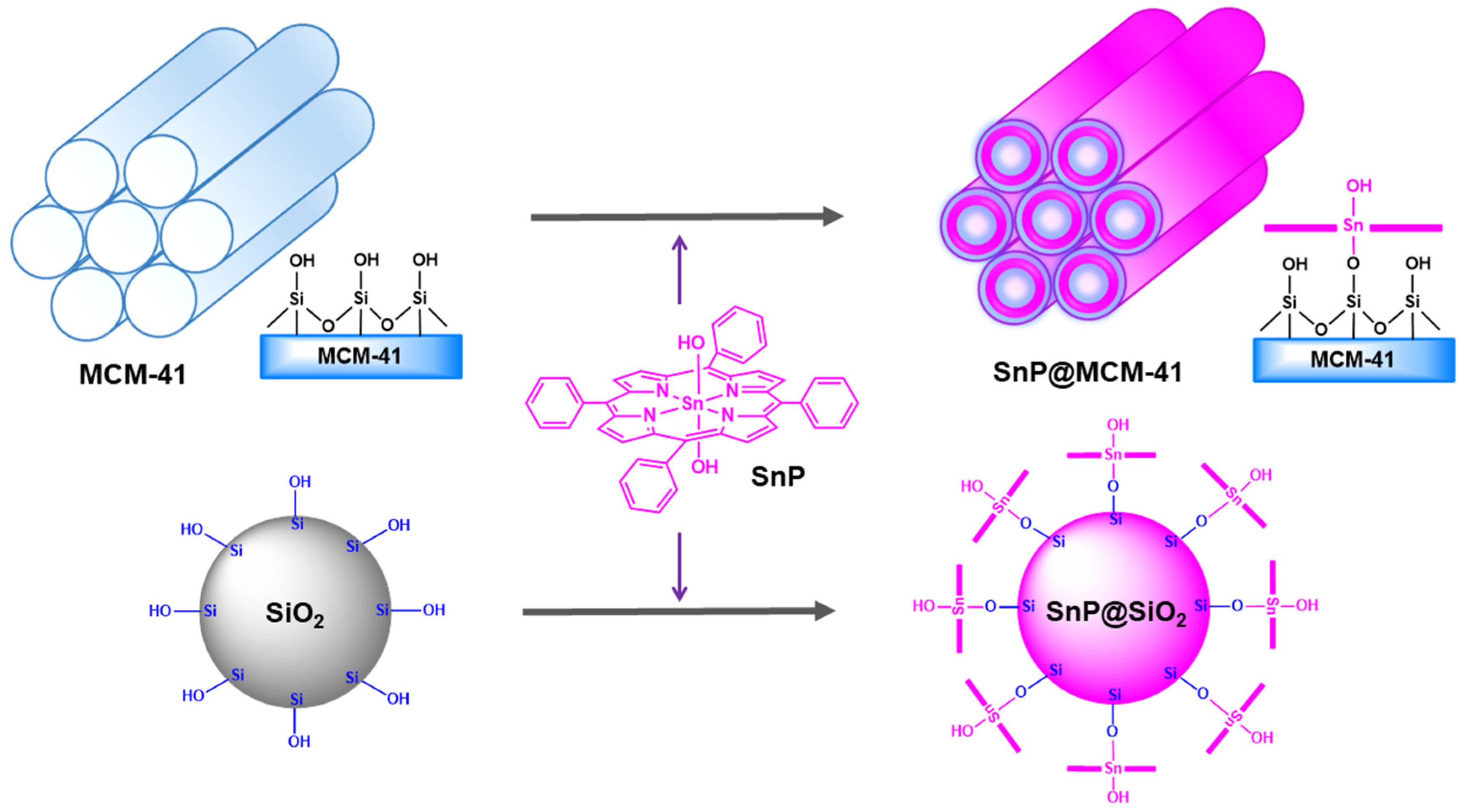
Scheme 1. Fabrication of the hybrid composite SnP@MCM−41 and SnP@SiO2.
Molecules 28 01886 sch001
Figure 1. FTIR spectra of SnP, SiO2, SnP@SiO2, MCM−41, and SnP@MCM−41.
Molecules 28 01886 g001
Figure 2. Solid-state UV–vis absorption spectra of SnP, SiO2, MCM−41, SnP@MCM−41, and SnP@SiO2. A Nujol paste was used for sample preparation. The photos represent both hybrid composite materials.
Molecules 28 01886 g002
Figure 3. Fluorescence spectra of SnP, SiO2, MCM−41, SnP@MCM−41, and SnP@SiO2 in a Nujol paste (λex = 560 nm).
Molecules 28 01886 g003
Figure 4. TGA curves of SnP, SiO2, MCM−41, SnP@MCM−41, and SnP@SiO2.
Molecules 28 01886 g004
Figure 5. FE−SEM images of (a) SiO2, (b) SnP@SiO2, (c) MCM−41, (d) SnP@MCM−41, and (e) SnP.
Molecules 28 01886 g005
Figure 6. TEM images of (a) SnP@SiO2 and (b) SnP@MCM−41. The inset represents an enlarged image of regular arrays of cylindrical mesopores.
Molecules 28 01886 g006
Figure 7. Photocatalytic degradation of the EG dye in an aqueous solution (pH = 7, T = 298 K) under visible light irradiation in the presence of SnP, SnP@MCM−41, and SnP@SiO2.
Molecules 28 01886 g007
Figure 8. Photocatalytic degradation of the RhB dye in an aqueous solution (pH = 7, T = 298 K) under visible light irradiation.
Molecules 28 01886 g008
Figure 9. Photocatalytic degradation of the MCP dye in an aqueous solution (pH = 7, T = 298 K) under visible light irradiation.
Molecules 28 01886 g009
Figure 10. Possible intermediates for the degradation of the EG dye in the presence of the photocatalyst SnP@MCM−41.
Molecules 28 01886 g010
Table 1. Performances of degradation for EG, MCP, and RhB by various photocatalysts.
PhotocatalystDyeRate Constant (min−1)References
TiO2 (P−25)EG0.0473[48]
γ−IrradiationEG0.0018[49]
TiO2 (UV)EG0.025[50]
CdS (one-dimensional)EG0.008[51]
SnPEG0.002This study
SnP@SiO2EG0.022This study
SnP@MCM−41EG0.029This study
Electrochemical-oxidationMCP0.32[52]
ZnOMCP0.022[53]
SnPMCP0.001This study
SnP@SiO2MCP0.021This study
SnP@MCM−41MCP0.024This study
TiO2 (P−25)RhB0.001[53]
Co0.6Zn0.4Fe2O4RhB0.015[54]
TiO2/MgZnAlRhB0.005[55]
CdSRhB0.003[56]
ZnORhB0.009[57]
TiO2 NTs/Bi2MoO6RhB0.0077[58]
SnPRhB0.0006This study
SnP@SiO2RhB0.011This study
SnP@MCM−41RhB0.013This study
Disclaimer/Publisher’s Note: The statements, opinions and data contained in all publications are solely those of the individual author(s) and contributor(s) and not of MDPI and/or the editor(s). MDPI and/or the editor(s) disclaim responsibility for any injury to people or property resulting from any ideas, methods, instructions or products referred to in the content.