

New NSAID Conjugates as Potent and Selective COX-2 Inhibitors: Synthesis, Molecular Modeling and Biological Investigation
 ,
,  , , , ,
Abstract
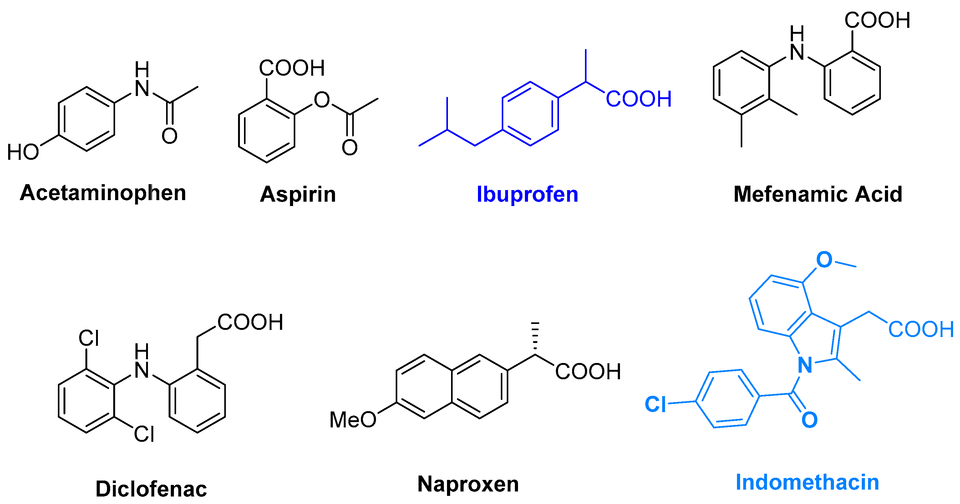
:1. Introduction
2. Results and Discussion
2.1. Chemistry
2.2. Biological Studies
2.2.1. Anti-Inflammatory Properties
2.2.2. Analgesic Properties
Peripheral Analgesic
Central Analgesic
2.2.3. Ulcerogenic Liability
2.2.4. Toxicological Bioassay
2.2.5. Inhibitory Properties of COX-1 and COX-2
2.2.6. Evaluation of NO Production of LPS-Induced RAW264.7
2.2.7. Evaluation of mRNA Levels of Inflammatory Cytokines in LPS-Induced RAW264.7 Cells
2.3. Molecular Modeling Studies
2.3.1. Docking Studies
2.3.2. D-QSAR Studies
2.3.3. Anti-Inflammatory QSAR Model
2.3.4. Peripheral Analgesic QSAR Model
2.3.5. Central Analgesic QSAR Model
3. Experimental Section
3.1. Chemistry
3.2. Preparation of Prop-2-yn-1-yl 2-(4-isobutylphenyl) Propanoate
3.3. Preparation of Prop-2-yn-1-yl 2-(1-(4-chlorobenzoyl)-5-methoxy-2-methyl-1H-indol-3-yl) Acetate
3.4. General Method for Preparation of and
3.4.1. (1-(2-Chlorophenyl)-1H-1,2,3-triazol-4-yl)methyl 2-(4-isobutylphenyl)propanoate (5a)
3.4.2. (1-(4-Chlorophenyl)-1H-1,2,3-triazol-4-yl)methyl 2-(4-isobutylphenyl)propanoate (5b)
3.4.3. (1-(4-Fluorophenyl)-1H-1,2,3-triazol-4-yl)methyl 2-(4-isobutylphenyl)propanoate (5c)
3.4.4. (1-(p-Tolyl)-1H-1,2,3-triazol-4-yl)methyl 2-(4-isobutylphenyl)propanoate (5d)
3.4.5. (1-(2-Methoxyphenyl)-1H-1,2,3-triazol-4-yl)methyl 2-(4-isobutylphenyl)propanoate (5e)
3.4.6. (1-(4-Methoxyphenyl)-1H-1,2,3-triazol-4-yl)methyl 2-(4-isobutylphenyl)propanoate (5f)
3.4.7. (1-(4-Nitrophenyl)-1H-1,2,3-triazol-4-yl)methyl 2-(4-isobutylphenyl)propanoate (5g)
3.4.8. (1-(2-Chlorophenyl)-1H-1,2,3-triazol-4-yl)methyl 2-(1-(4-chlorobenzoyl)-5-methoxy-2-methyl-1H-indol-3-yl)acetate (8a)
3.4.9. (1-(4-Chlorophenyl)-1H-1,2,3-triazol-4-yl)methyl 2-(1-(4-chlorobenzoyl)-5-methoxy-2-methyl-1H-indol-3-yl)acetate (8b)
3.4.10. (1-(4-Fluorophenyl)-1H-1,2,3-triazol-4-yl)methyl 2-(1-(4-chlorobenzoyl)-5-methoxy-2-methyl-1H-indol-3-yl)acetate (8c)
3.4.11. (1-(p-Tolyl)-1H-1,2,3-triazol-4-yl)methyl 2-(1-(4-chlorobenzoyl)-5-methoxy-2-methyl-1H-indol-3-yl)acetate (8d)
3.4.12. (1-(2-Methoxyphenyl)-1H-1,2,3-triazol-4-yl)methyl 2-(1-(4-chlorobenzoyl)-5-methoxy-2-methyl-1H-indol-3-yl)acetate (8e)
3.4.13. (1-(4-Methoxyphenyl)-1H-1,2,3-triazol-4-yl)methyl 2-(1-(4-chlorobenzoyl)-5-methoxy-2-methyl-1H-indol-3-yl)acetate (8f)
3.4.14. (1-(4-Nitrophenyl)-1H-1,2,3-triazol-4-yl)methyl 2-(1-(4-chlorobenzoyl)-5-methoxy-2-methyl-1H-indol-3-yl)acetate (8g)
3.5. Biological and Computational Studies
4. Conclusions
Supplementary Materials
Author Contributions
Funding
Institutional Review Board Statement
Informed Consent Statement
Data Availability Statement
Acknowledgments
Conflicts of Interest
Sample Availability
References
- Ricciotti, E.; FitzGerald, G.A. Prostaglandins and inflammation. Arterioscler. Thromb. Vasc. Biol. 2011, 31, 986–1000. [Google Scholar] [CrossRef] [PubMed]
- Kurumbail, R.G.; Stevens, A.M.; Gierse, J.K.; McDonald, J.J.; Stegeman, R.A.; Pak, J.Y.; Gildehaus, D.; Iyashiro, J.M.; Penning, T.D.; Seibert, K.; et al. Structural basis for selective inhibition of cyclooxygenase-2 by anti-inflammatory agents. Nature 1996, 384, 644–648. [Google Scholar] [CrossRef]
- Smith, W.L.; DeWitt, D.L.; Garavito, R.M. Cyclooxygenases: Structural, Cellular, and Molecular Biology. Annu. Rev. Biochem. 2000, 69, 145–182. [Google Scholar] [CrossRef] [PubMed] [Green Version]
- Mitchell, J.A.; Warner, T. Cyclo-oxygenase-2: Pharmacology, physiology, biochemistry and relevance to NSAID therapy. Br. J. Pharmacol. 1999, 128, 1121–1132. [Google Scholar] [CrossRef] [PubMed]
- Wang, D.; DuBois, R.N. The Role of Anti-Inflammatory Drugs in Colorectal Cancer. Annu. Rev. Med. 2013, 64, 131–144. [Google Scholar] [CrossRef] [PubMed]
- Méric, J.-B.; Rottey, S.; Olaussen, K.; Soria, J.-C.; Khayat, D.; Rixe, O.; Spano, J.-P. Cyclooxygenase-2 as a target for anticancer drug development. Crit. Rev. Oncol. Hematol. 2006, 59, 51–64. [Google Scholar] [CrossRef]
- Teismann, P.; Tieu, K.; Choi, D.-K.; Wu, D.-C.; Naini, A.; Hunot, S.; Vila, M.; Jackson-Lewis, V.; Przedborski, S. Cyclooxygenase-2 is instrumental in Parkinson’s disease neurodegeneration. Proc. Natl. Acad. Sci. USA 2003, 100, 5473–5478. [Google Scholar] [CrossRef] [Green Version]
- Sorokin, A. Cyclooxygenase-2: Potential Role in Regulation of Drug Efflux and Multidrug Resistance Phenotype. Curr. Pharm. Des. 2004, 10, 647–657. [Google Scholar] [CrossRef]
- Davis, A.; Robson, J. The dangers of NSAIDs: Look both ways. Br. J. Gen. Pr. 2016, 66, 172–173. [Google Scholar] [CrossRef]
- Luong, C.; Miller, A.B.; Barnett, J.W.; Chow, J.; Ramesha, C.; Browner, M.F. Flexibility of the NSAID binding site in the structure of human cyclooxygenase-2. Nat. Struct. Biol. 1996, 3, 927–933. [Google Scholar] [CrossRef]
- Dogné, J.-M.; Supuran, C.T.; Pratico, D. Adverse Cardiovascular Effects of the Coxibs. J. Med. Chem. 2005, 48, 2251–2257. [Google Scholar] [CrossRef] [PubMed]
- Nalamachu, S.; Wortmann, R. Role of Indomethacin in Acute Pain and Inflammation Management: A Review of the Literature. Postgrad. Med. 2014, 126, 92–97. [Google Scholar] [CrossRef] [PubMed]
- Varrassi, G.; Pergolizzi, J.V.; Dowling, P.; Paladini, A. Ibuprofen Safety at the Golden Anniversary: Are all NSAIDs the Same? A Narrative Review. Adv. Ther. 2020, 37, 61–82. [Google Scholar] [CrossRef] [PubMed] [Green Version]
- Panda, S.S.; Girgis, A.S.; Honkanadavar, H.H.; George, R.F.; Srour, A.M. Synthesis of new ibuprofen hybrid conjugates as potential anti-inflammatory and analgesic agents. Futur. Med. Chem. 2020, 12, 1369–1386. [Google Scholar] [CrossRef]
- Panda, S.S.; Girgis, A.S.; Thomas, S.J.; Capito, J.E.; George, R.F.; Salman, A.; El-Manawaty, M.A.; Samir, A. Synthesis, pharmacological profile and 2D-QSAR studies of curcumin-amino acid conjugates as potential drug candidates. Eur. J. Med. Chem. 2020, 196, 112293. [Google Scholar] [CrossRef]
- Girgis, A.S.; Tala, S.R.; Oliferenko, P.V.; Oliferenko, A.A.; Katritzky, A.R. Computer-assisted rational design, synthesis, and bioassay of non-steroidal anti-inflammatory agents. Eur. J. Med. Chem. 2012, 50, 1–8. [Google Scholar] [CrossRef]
- Sadiq, A.; Mahnashi, M.H.; Alyami, B.A.; Alqahtani, Y.S.; Alqarni, A.O.; Rashid, U. Tailoring the substitution pattern of Pyrrolidine-2,5-dione for discovery of new structural template for dual COX/LOX inhibition. Bioorganic Chem. 2021, 112, 104969. [Google Scholar] [CrossRef]
- Laali, K.K.; Zwarycz, A.T.; Beck, N.; Borosky, G.L.; Nukaya, M.; Kennedy, G.D. Curcumin Conjugates of Non-steroidal An-ti-Inflammatory Drugs: Synthesis, Structures, Anti-proliferative Assays, Computational Docking, and Inflammatory Response. ChemistryOpen 2020, 9, 822–834. [Google Scholar] [CrossRef]
- Korczak, M.; Roszkowski, P.; Granica, S.; Piwowarski, J.P. Conjugates of urolithin A with NSAIDs, their stability, cytotoxicity, and antiinflammatory potential. Sci. Rep. 2022, 12, 11676. [Google Scholar] [CrossRef]
- Naaz, F.; Pallavi, M.P.; Shafi, S.; Mulakayala, N.; Yar, M.S.; Kumar, H.S. 1,2,3-triazole tethered Indole-3-glyoxamide derivatives as multiple inhibitors of 5-LOX, COX-2 & tubulin: Their anti-proliferative & anti-inflammatory activity. Bioorganic Chem. 2018, 81, 1–20. [Google Scholar] [CrossRef]
- Bozorov, K.; Zhao, J.; Aisa, H.A. 1,2,3-Triazole-containing hybrids as leads in medicinal chemistry: A recent overview. Bioorganic Med. Chem. 2019, 27, 3511–3531. [Google Scholar] [CrossRef] [PubMed]
- Zhang, B. Comprehensive review on the anti-bacterial activity of 1,2,3-triazole hybrids. Eur. J. Med. Chem. 2019, 168, 357–372. [Google Scholar] [CrossRef] [PubMed]
- Kim, D.-K.; Kim, J.; Park, H.-J. Synthesis and biological evaluation of novel 2-pyridinyl-[1,2,3]triazoles as inhibitors of transforming growth factor β1 type 1 receptor. Bioorganic Med. Chem. Lett. 2004, 14, 2401–2405. [Google Scholar] [CrossRef]
- Whiting, M.; Tripp, J.C.; Lin, Y.C.; Lindstrom, W.; Olson, A.J.; Elder, J.H.; Sharpless, K.B.; Fokin, V.V. Rapid discovery and structure-activity profiling of novel inhibitors of human immunodeficiency virus type 1 protease enabled by the cop-per(I)-catalyzed synthesis of 1,2,3-triazoles and their further functionalization. J. Med. Chem. 2006, 49, 7697–7710. [Google Scholar] [CrossRef]
- Cheng, Z.-Y.; Li, W.-J.; He, F.; Zhou, J.-M.; Zhu, X.-F. Synthesis and biological evaluation of 4-aryl-5-cyano-2H-1,2,3-triazoles as inhibitor of HER2 tyrosine kinase. Bioorganic Med. Chem. 2007, 15, 1533–1538. [Google Scholar] [CrossRef]
- Da Silva, F.D.C.; de Souza, M.C.B.; Frugulhetti, I.I.; Castro, H.C.; Souza, S.L.D.O.; Souza, T.; Rodrigues, D.Q.; Souza, A.M.; Abreu, P.A.; Passamani, F. Synthesis, HIV-RT inhibitory activity and SAR of 1-benzyl-1H-1,2,3-triazole derivatives of carbohydrates. Eur. J. Med. Chem. 2009, 44, 373–383. [Google Scholar] [CrossRef]
- Faidallah, H.M.; Girgis, A.S.; Tiwari, A.D.; Honkanadavar, H.H.; Thomas, S.J.; Samir, A.; Kalmouch, A.; Alamry, K.A.; Khan, K.A.; Ibrahim, T.S.; et al. Synthesis, antibacterial properties and 2D-QSAR studies of quinolone-triazole conjugates. Eur. J. Med. Chem. 2018, 143, 1524–1534. [Google Scholar] [CrossRef]
- Seliem, I.A.; Panda, S.S.; Girgis, A.S.; Moatasim, Y.; Kandeil, A.; Mostafa, A.; Ali, M.A.; Nossier, E.S.; Rasslan, F.; Srour, A.M.; et al. New quinoline-triazole conjugates: Synthesis, and antiviral properties against SARS-CoV-2. Bioorganic Chem. 2021, 114, 105117. [Google Scholar] [CrossRef]
- BioVision Incorporated. COX-1/2 (Human) Inhibitor Screening Assay Kits (Fluorometric, Catalog # K547-100 and K548-100); BioVision Incorporated: Milpitas, CA, USA; Available online: www.biovision.com (accessed on 10 January 2023).
- Wang, W.; Liu, P.; Hao, C.; Wu, L.; Wan, W.; Mao, X. Neoagaro-oligosaccharide monomers inhibit inflammation in LPS-stimulated macrophages through suppression of MAPK and NF-κB pathways. Sci. Rep. 2017, 7, srep44252. [Google Scholar] [CrossRef] [Green Version]
- Friesner, R.A.; Banks, J.L.; Murphy, R.B.; Halgren, T.A.; Klicic, J.J.; Mainz, D.T.; Repasky, M.P.; Knoll, E.H.; Shelley, M.; Perry, J.K.; et al. Glide: A New Approach for Rapid, Accurate Docking and Scoring. 1. Method and Assessment of Docking Accuracy. J. Med. Chem. 2004, 47, 1739–1749. [Google Scholar] [CrossRef]
- Friesner, R.A.; Murphy, R.B.; Repasky, M.P.; Frye, L.L.; Greenwood, J.R.; Halgren, T.A.; Sanschagrin, P.C.; Mainz, D.T. Extra Precision Glide: Docking and Scoring Incorporating a Model of Hydrophobic Enclosure for Protein−Ligand Complexes. J. Med. Chem. 2006, 49, 6177–6196. [Google Scholar] [CrossRef] [Green Version]
- Sidhu, R.S.; Lee, J.Y.; Yuan, C.; Smith, W.L. Comparison of Cyclooxygenase-1 Crystal Structures: Cross-Talk between Monomers Comprising Cyclooxygenase-1 Homodimers. Biochemistry 2010, 49, 7069–7079. [Google Scholar] [CrossRef] [Green Version]
- Dvorakova, M.; Langhansova, L.; Temml, V.; Pavicic, A.; Vanek, T.; Landa, P. Synthesis, Inhibitory Activity, and In Silico Modeling of Selective COX-1 Inhibitors with a Quinazoline Core. ACS Med. Chem. Lett. 2021, 12, 610–616. [Google Scholar] [CrossRef] [PubMed]
- Rouzer, C.A.; Marnett, L.J. Structural and Chemical Biology of the Interaction of Cyclooxygenase with Substrates and Non-Steroidal Anti-Inflammatory Drugs. Chem. Rev. 2020, 120, 7592–7641. [Google Scholar] [CrossRef] [PubMed]
- Lou, Y.; Zhu, J. Carboxylic Acid Nonsteroidal Anti-Inflammatory Drugs (NSAIDs). In Bioactive Carboxylic Compound Classes; Lamberth, C., Dinges, J., Eds.; Wiley-VCH Verlag GmbH & Co. KGaA: Weinheim, Germany, 2016; pp. 221–236. [Google Scholar] [CrossRef]
- Blobaum, A.L.; Marnett, L.J. Molecular Determinants for the Selective Inhibition of Cyclooxygenase-2 by Lumiracoxib. J. Biol. Chem. 2007, 282, 16379–16390. [Google Scholar] [CrossRef] [PubMed] [Green Version]
- Genheden, S.; Ryde, U. The MM/PBSA and MM/GBSA methods to estimate ligand-binding affinities. Expert Opin. Drug Discov. 2015, 10, 449–461. [Google Scholar] [CrossRef] [PubMed]
- Dong, L.; Anderson, A.J.; Malkowski, M.G. Arg-513 and Leu-531 Are Key Residues Governing Time-Dependent Inhibition of Cyclooxygenase-2 by Aspirin and Celebrex. Biochemistry 2019, 58, 3990–4002. [Google Scholar] [CrossRef]
- Baek, S.-H.; Hwang, S.; Park, T.; Kwon, Y.-J.; Cho, M.; Park, D. Evaluation of Selective COX-2 Inhibition and In Silico Study of Kuwanon Derivatives Isolated from Morus alba. Int. J. Mol. Sci. 2021, 22, 3659. [Google Scholar] [CrossRef]
- Girgis, A.S.; Panda, S.S.; Farag, I.S.A.; El-Shabiny, A.M.; Moustafa, A.M.; Ismail, N.S.M.; Pillai, G.G.; Panda, C.S.; Hall, C.D.; Katritzky, A.R. Synthesis, and QSAR analysis of anti-oncological active spiro-alkaloids. Org. Biomol. Chem. 2015, 13, 1741–1753. [Google Scholar] [CrossRef]
- Nofal, Z.M.; Srour, A.M.; El-Eraky, W.I.; Saleh, D.O.; Girgis, A.S. Rational design, synthesis and QSAR study of vasorelaxant active 3-pyridinecarbonitriles incorporating 1H-benzimidazol-2-yl function. Eur. J. Med. Chem. 2013, 63, 14–21. [Google Scholar] [CrossRef]
- Available online: http://www.codessa-pro.com/manuals/manual.htm (accessed on 30 July 2022).


| Entry | Compd. | Mean Edema Thickness “mm” (% Inhibition of Edema ± Standard Error) | % Potency a | ||||
|---|---|---|---|---|---|---|---|
| 1 h | 2 h | 3 h | 4 h | 24 h | |||
| 1 | Control | 0.863 ± 0.02 (0.00 ± 0.01) | 0.713 ± 0.03 (0.00 ± 0.02) | 0.723 ± 0.02 (0.00 ± 0.01) | 0.713 ± 0.01 (0.00 ± 0.03) | 0.920 ± 0.07 (0.00 ± 0.04) | --- |
| 2 | Indo | 0.510 ± 0.03 ** (40.9 ± 1.2) | 0.353 ± 0.05 ** (50.5 ± 2.1) | 0.137 ± 0.01 ** (81.1 ± 1.8) | 0.350 ± 0.04 ** (50.9 ± 1.1) | 0.810 ± 0.03 ** (12.0 ± 2.1) | 100 |
| 3 | Ibu | 0.480 ± 0.04 ** (44.4 ± 2.5) | 0.403 ± 0.09 ** (43.5 ± 2.9) | 0.153 ± 0.01 ** (78.8 ± 0.9) | 0.563 ± 0.02 * (21.0 ± 1.1) | 0.817 ± 0.07 * (11.2 ± 0.6) | 97.2 |
| 4 | 3 | 0.727 ± 0.05 * (15.8 ± 1.9) | 0.589 ± 0.01 * (17.4 ± 0.9) | 0.103 ± 0.04 ** (85.8 ± 1.1) | 0.473 ± 0.03 * (33.7 ± 1.2) | 0.920 ± 0.02 * (0.00 ± 0.3) | 105.8 |
| 5 | 5a | 0.607 ± 0.04 * (29.7 ± 1.1) | 0.490 ± 0.03 * (31.3 ± 0.8) | 0.033 ± 0.01 ** (95.4 ± 0.7) | 0.387 ± 0.06 ** (45.7 ± 1.4) | 0.713 ± 0.02 ** (22.5 ± 0.9) | 117.6 |
| 6 | 5b | 0.360 ± 0.01 ** (58.3 ± 0.7) | 0.217 ± 0.07 ** (69.6 ± 2.1) | 0.040 ± 0.01 ** (94.5 ± 0.4) | 0.237 ± 0.08 ** (66.8 ± 1.6) | 0.843 ± 0.09 ** (8.4 ± 1.2) | 116.5 |
| 7 | 5c | 0.863 ± 0.08 * (0.00 ± 1.0) | 0.540 ± 0.01 * (24.3 ± 1.4) | 0.527 ± 0.03 * (27.1 ± 0.7) | 0.530 ± 0.05 * (25.7 ± 0.9) | 0.920 ± 0.01 * (0.00 ± 0.2) | 33.4 |
| 8 | 5d | 0.257 ± 0.08 ** (70.2 ± 2.2) | 0.206 ± 0.04 ** (71.1 ± 1.5) | 0.173 ± 0.01 ** (76.1 ± 0.8) | 0.490 ± 0.07 ** (31.3 ± 1.1) | 0.920 ± 0.09 * (0.00 ± 1.7) | 93.8 |
| 9 | 5e | 0.197 ± 0.02 ** (77.2 ± 1.9) | 0.161 ± 0.08 ** (77.4 ± 2.2) | 0.083 ± 0.02 ** (88.5 ± 2.0) | 0.660 ± 0.09 * (7.4 ± 1.8) | 0.533 ± 0.01 * (42.1 ± 0.4) | 109.1 |
| 10 | 5f | 0.703 ± 0.09 * (18.5 ± 2.5) | 0.270 ± 0.05 * (62.1 ± 2.2) | 0.300 ± 0.07 * (58.5 ± 2.4) | 0.700 ± 0.10 * (1.8 ± 0.7) | 0.897 ± 0.08 * (2.5 ± 0.9) | 72.1 |
| 11 | 5g | 0.570 ± 0.05 ** (34.0 ± 1.8) | 0.230 ± 0.02 ** (67.7 ± 1.1) | 0.249 ± 0.07 ** (65.6 ± 2.9) | 0.373 ± 0.04 ** (47.7 ± 1.5) | 0.920 ± 0.02 * (0.00 ± 0.5) | 80.9 |
| 12 | 8a | 0.863 ± 0.07 * (0.00 ± 1.5) | 0.393 ± 0.08 * (44.9 ± 1.7) | 0.488 ± 0.07 * (32.5 ± 1.5) | 0.521 ± 0.03 * (26.9 ± 1.1) | 0.614 ± 0.09 * (33.3 ± 2.4) | 40.1 |
| 13 | 8c | 0.750 ± 0.08 * (13.1 ± 1.7) | 0.466 ± 0.02 * (34.6 ± 0.9) | 0.403 ± 0.01 ** (44.3 ± 0.6) | 0.340 ± 0.04 ** (52.3 ± 0.8) | 0.920 ± 0.04 * (0.00 ± 1.0) | 54.6 |
| 14 | 8d | 0.863 ± 0.07 * (0.00 ± 0.8) | 0.618 ± 0.09 * (13.3 ± 1.9) | 0.723 ± 0.04 * (0.00 ± 0.5) | 0.713 ± 0.03 * (0.00 ± 0.6) | 0.920 ± 0.02 * (0.00 ± 0.4) | --- |
| 15 | 8e | 0.863 ± 0.01 * (0.00 ± 0.2) | 0.667 ± 0.05 * (6.5 ± 1.0) | 0.536 ± 0.07 * (25.9 ± 1.3) | 0.683 ± 0.08 * (4.2 ± 1.9) | 0.920 ± 0.01 * (0.00 ± 0.3) | 31.8 |
| 16 | 8g | 0.690 ± 0.02 ** (20.0 ± 0.7) | 0.394 ± 0.08 ** (44.7 ± 1.7) | 0.456 ± 0.01 ** (36.9 ± 0.5) | 0.462 ± 0.04 ** (35.2 ± 0.9) | 0.920 ± 0.05 * (0.00 ± 1.2) | 45.5 |
| Entry | Compd. | Writing Reflex ± SE | % Inhibition/Protection | Potency a |
|---|---|---|---|---|
| 1 | Control | 40 ± 3.2 | 0 | --- |
| 2 | Indo | 11.3 ± 1.6 ** | 71.8 | 100.0 |
| 3 | Ibu | 16.6 ± 0.7 ** | 58.5 | 81.5 |
| 4 | 3 | 11.3 ± 0.9 ** | 71.8 | 100.0 |
| 5 | 5a | 6.7 ± 0.7 ** | 83.3 | 116.0 |
| 6 | 5b | 5.0 ± 0.9 ** | 87.5 | 121.9 |
| 7 | 5c | 6.7 ± 0.5 ** | 83.3 | 116.0 |
| 8 | 5d | 7.0 ± 1.3 ** | 82.5 | 114.9 |
| 9 | 5e | 14.7 ± 1.6 ** | 63.3 | 88.2 |
| 10 | 5f | 8.0 ± 1.4 ** | 80.0 | 111.4 |
| 11 | 5g | 14.0 ± 1.9 ** | 65.0 | 90.5 |
| 12 | 7 | 4.3 ± 0.5 ** | 89.3 | 124.4 |
| 13 | 8a | 20.3 ± 1.8 * | 49.3 | 68.7 |
| 14 | 8b | 21.5 ± 1.5 * | 46.3 | 64.5 |
| 15 | 8c | 23.5 ± 1.4 * | 41.3 | 57.5 |
| 16 | 8d | 15.0 ± 1.6 * | 62.5 | 87.0 |
| 17 | 8e | 7.7 ± 1.3 ** | 80.8 | 112.5 |
| 18 | 8f | 13.3 ± 1.9 ** | 66.8 | 93.0 |
| 19 | 8g | 1.5 ± 0.4 ** | 96.3 | 134.1 |
| Entry | Compd. | Latency Period ± SE “Standard Error”, Second (% Protection) | Potency a | |||
|---|---|---|---|---|---|---|
| After 30 min. | After 60 min. | After 90 min. | After 120 min. | |||
| 1 | Control | 6.68 ± 1.0 (0.0) | 7.27 ± 1.4 (0.0) | 8.91 ± 0.9 (0.0) | 9.01 ± 1.1 (0.0) | --- |
| 2 | Indo | 12.50 ± 2.0 ** (87.1) | 13.15 ± 1.6 ** (80.9) | 14.07 ± 1.2 ** (57.9) | 14.10 ± 1.9 ** (56.5) | 100 |
| 3 | Ibu | 10.80 ± 0.8 ** (61.7) | 11.00 ± 1.3 ** (51.3) | 11.90 ± 1.5 ** (33.6) | 13.90 ±2.2 ** (54.3) | 96.1 |
| 4 | 3 | 10.37 ± 1.6 ** (55.2) | 10.78 ± 2.1 ** (48.3) | 10.62 ± 2.2 ** (19.2) | 10.35 ± 1.7 ** (14.9) | 26.4 |
| 5 | 5a | 11.97 ± 2.5 ** (79.2) | 12.47 ± 2.0 ** (71.5) | 11.98 ± 2.6 ** (34.5) | 13.92 ± 2.3 ** (54.5) | 96.5 |
| 6 | 5b | 12.30 ± 1.6 ** (84.1) | 10.27 ± 1.9 ** (41.3) | 11.14 ± 1.5 ** (25.0) | 11.45 ± 2.0 ** (27.1) | 48.0 |
| 7 | 5c | 14.17 ± 2.6 ** (112.1) | 15.0 ± 2.3 **(106.3) | 14.91 ± 2.5 ** (67.3) | 10.63 ± 1.8 ** (18.0) | 31.9 |
| 8 | 5d | 11.02 ± 1.4 ** (65.0) | 9.86 ± 0.7 * (35.6) | 10.69 ± 0.9 * (20.0) | 9.12 ± 1.1 * (1.2) | 2.1 |
| 9 | 5e | 12.61 ± 2.1 ** (88.8) | 10.53 ± 1.8 * (44.8) | 12.12 ± 1.7 * (36.03) | 11.53 ± 1.3 * (28.0) | 49.6 |
| 10 | 5f | 12.7 ± 0.9 * (90.1) | 11.40 ± 1.5 * (56.8) | 10.60 ± 1.9 * (19.0) | 9.03 ± 1.6 * (0.2) | 0.4 |
| 11 | 5g | 12.5 ± 2.1 ** (87.1) | 12.59 ± 1.5 ** (73.2) | 10.60 ± 2.6 ** (19.0) | 11.54 ± 2.2 ** (28.1) | 49.7 |
| 12 | 7 | 6.68 ± 2.0 * (0.0) | 9.54 ± 1.6 * (31.2) | 11.18 ± 1.5 * (25.5) | 10.78 ± 1.8 * (19.6) | 34.7 |
| 13 | 8a | 12.19 ± 1.9 ** (82.5) | 13.51 ± 1.7 ** (85.8) | 13.85 ± 1.4 ** (55.4) | 13.63 ± 1.3 ** (51.3) | 90.8 |
| 14 | 8b | 6.84 ± 1.4 * (2.4) | 8.31 ± 1.9 * (14.3) | 9.91 ± 1.7 * (11.2) | 9.88 ± 2.0 * (9.7) | 17.2 |
| 15 | 8c | 11.32 ± 0.8 * (69.5) | 9.39 ± 1.1 * (29.2) | 11.40 ± 1.6 * (27.9) | 13.34 ± 1.8 * (48.1) | 85.1 |
| 16 | 8d | 13.21 ± 2.5 ** (97.8) | 12.65 ± 2.0 ** (74.0) | 13.37 ± 1.6 ** (50.1) | 13.39 ± 1.2 ** (48.6) | 86.0 |
| 17 | 8e | 11.30 ± 1.1 ** (69.2) | 13.23 ± 1.6 ** (82.0) | 11.36 ± 2.5 ** (27.5) | 11.61 ± 2.2 ** (28.9) | 51.2 |
| 18 | 8f | 11.11 ± 1.6 ** (66.3) | 13.99 ± 1.9 ** (92.4) | 15.00 ± 2.5 ** (68.4) | 15.00 ± 2.6 ** (66.5) | 117.7 |
| 19 | 8g | 10.46 ± 2.4 ** (56.6) | 10.57 ± 2.7 ** (45.4) | 12.58 ± 2.0 ** (41.2) | 9.25 ± 2.3 * (2.7) | 4.8 |
| Entry | Compd. | Number of Animals with Ulcer | % Incidence of Ulcer Divided by 10 | Average of Ulcer Number | Average Severity of Ulcer | Ulcer Index |
|---|---|---|---|---|---|---|
| 1 | Control | 0/6 | 0 | 0 | 0 | 0 |
| 2 | Indo | 6/6 | 10 | 2 | 1.67 | 13.67 |
| 3 | Ibu | 2/6 | 3.33 | 0.33 | 0.67 | 4.33 |
| 4 | 3 | 2/6 | 3.33 | 0.33 | 0.67 | 4.33 |
| 5 | 5a | 0 | 0 | 0 | 0 | 0 |
| 6 | 5b | 0 | 0 | 0 | 0 | 0 |
| 7 | 5d | 0 | 0 | 0 | 0 | 0 |
| 8 | 5e | 0 | 0 | 0 | 0 | 0 |
| Entry | Compd. | IC50, μM ± SD | SI | |
|---|---|---|---|---|
| COX-1 | COX-2 | |||
| 1 | Indo | 0.354 ± 0.01 ** | 3.239 ± 0.09 ** | 0.109 |
| 2 | IBU | 13.16 ± 0.37 ** | 124.2 ± 3.53 * | 0.106 |
| 3 | 3 | 19.45 ± 0.55 ** | 8.599 ± 0.24 ** | 2.262 |
| 4 | 5a | 17.16 ± 0.41 ** | 0.743 ± 0.02 ** | 23.096 |
| 5 | 5b | 118.8 ± 3.4 ** | 12.35 ± 0.35 ** | 9.619 |
| 6 | 5d | 9.638 ± 0.27 ** | 4.467 ± 0.13 ** | 2.158 |
| 7 | 5e | 5.417 ± 0.15 ** | 13.99 ± 0.40 ** | 0.387 |
Disclaimer/Publisher’s Note: The statements, opinions and data contained in all publications are solely those of the individual author(s) and contributor(s) and not of MDPI and/or the editor(s). MDPI and/or the editor(s) disclaim responsibility for any injury to people or property resulting from any ideas, methods, instructions or products referred to in the content. |
© 2023 by the authors. Licensee MDPI, Basel, Switzerland. This article is an open access article distributed under the terms and conditions of the Creative Commons Attribution (CC BY) license (https://creativecommons.org/licenses/by/4.0/).
Share and Cite
Bokhtia, R.M.; Panda, S.S.; Girgis, A.S.; Samir, N.; Said, M.F.; Abdelnaser, A.; Nasr, S.; Bekheit, M.S.; Dawood, A.S.; Sharma, H.; et al. New NSAID Conjugates as Potent and Selective COX-2 Inhibitors: Synthesis, Molecular Modeling and Biological Investigation. Molecules 2023, 28, 1945. https://doi.org/10.3390/molecules28041945
Bokhtia RM, Panda SS, Girgis AS, Samir N, Said MF, Abdelnaser A, Nasr S, Bekheit MS, Dawood AS, Sharma H, et al. New NSAID Conjugates as Potent and Selective COX-2 Inhibitors: Synthesis, Molecular Modeling and Biological Investigation. Molecules. 2023; 28(4):1945. https://doi.org/10.3390/molecules28041945
Chicago/Turabian StyleBokhtia, Riham M., Siva S. Panda, Adel S. Girgis, Nermin Samir, Mona F. Said, Anwar Abdelnaser, Soad Nasr, Mohamed S. Bekheit, Abdelhameed S. Dawood, Horrick Sharma, and et al. 2023. "New NSAID Conjugates as Potent and Selective COX-2 Inhibitors: Synthesis, Molecular Modeling and Biological Investigation" Molecules 28, no. 4: 1945. https://doi.org/10.3390/molecules28041945
APA StyleBokhtia, R. M., Panda, S. S., Girgis, A. S., Samir, N., Said, M. F., Abdelnaser, A., Nasr, S., Bekheit, M. S., Dawood, A. S., Sharma, H., Wade, M., Sharma, S. K., & Ghanim, A. M. (2023). New NSAID Conjugates as Potent and Selective COX-2 Inhibitors: Synthesis, Molecular Modeling and Biological Investigation. Molecules, 28(4), 1945. https://doi.org/10.3390/molecules28041945