


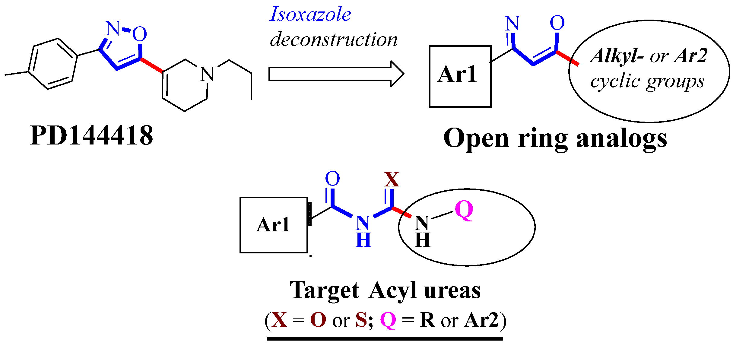
Design and Synthesis of New Acyl Urea Analogs as Potential σ1R Ligands

Abstract
:1. Introduction
2. Results and Discussion
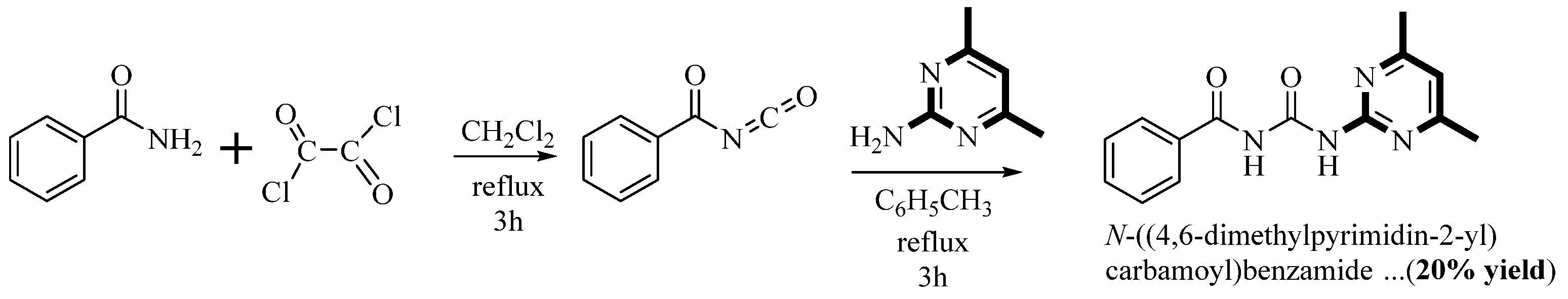
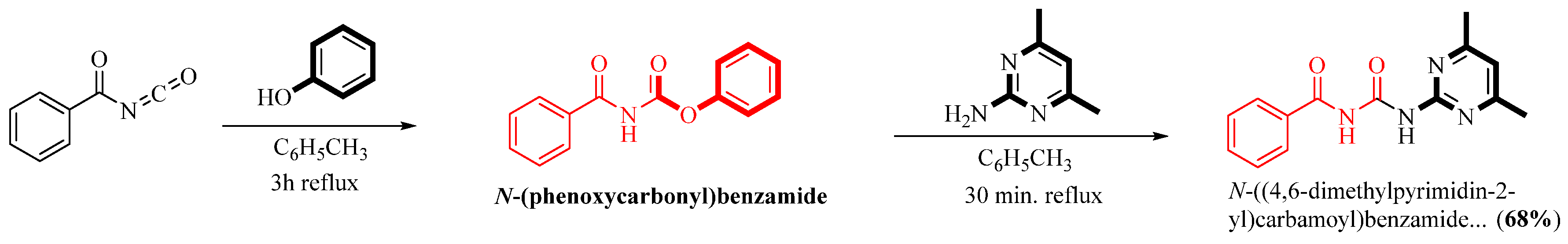
2.1. Chemistry
2.2. Drug-Likeness Predictions
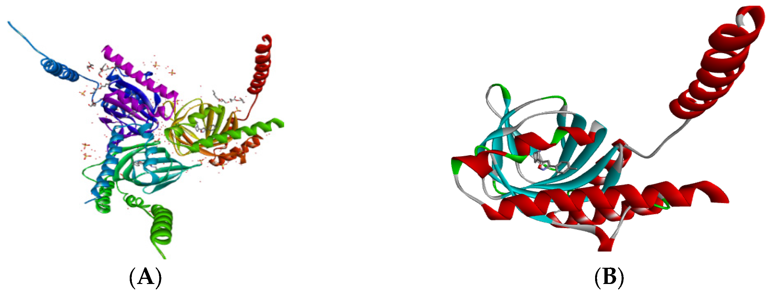
2.3. Molecular Docking
3. Experimental
3.1. Materials and General Methods
3.2. Synthetic Procedures and Compound Data
3.2.1. N-((5-Methylisoxazol-3-yl)carbamoyl)benzamide (1)
3.2.2. N-((1H-Benzo[D]imidazol-2-yl)carbamothioyl)benzamide (2)
3.2.3. N-((Pyridine-2-carbonothioyl)carbamoyl)benzamide (3)
3.2.4. N-((2-Methyl-1H-indol-5-yl)carbamoyl)benzamide (4)
3.2.5. N-Benzoyl-4-(pyrimidin-2-yl)piperazine-1-carboxamide (5)
3.2.6. N-Benzoyl-4-(pyridin-4-yl)piperazine-1-carboxamide (6)
3.2.7. N-Benzoyl-4-hydroxypiperazine-1-carboxamide (7)
3.2.8. N-Benzoyl-4-(p-tolyl)piperazine-1-carboxamide (8)
3.2.9. Tert-butyl 4-(3-benzoylureido)piperidine-1-carboxylate (9)
3.2.10. N-(Benzo[d]oxazol-2-ylcarbamoyl)benzamide (10)
3.2.11. N-(Benzoylcarbamoyl)-6-hydroxy-2,5,7,8-tetramethylchromane-2-carboxamide (11)
3.2.12. N-((4-Hydroxy-3-methoxyphenylcarbonothioyl)carbamoyl) benzamide (12)
3.2.13. N-((4,6-Dimethylpyrimidin-2-yl)carbamoyl)benzamide (13)
3.2.14. N-(Quinolin-3-ylcarbamoyl)benzamide (14)
3.2.15. N-(Isoquinolin-3-ylcarbamoyl)benzamide (15)
3.2.16. N-(Isoquinolin-3-ylcarbamothioyl)benzamide (16)
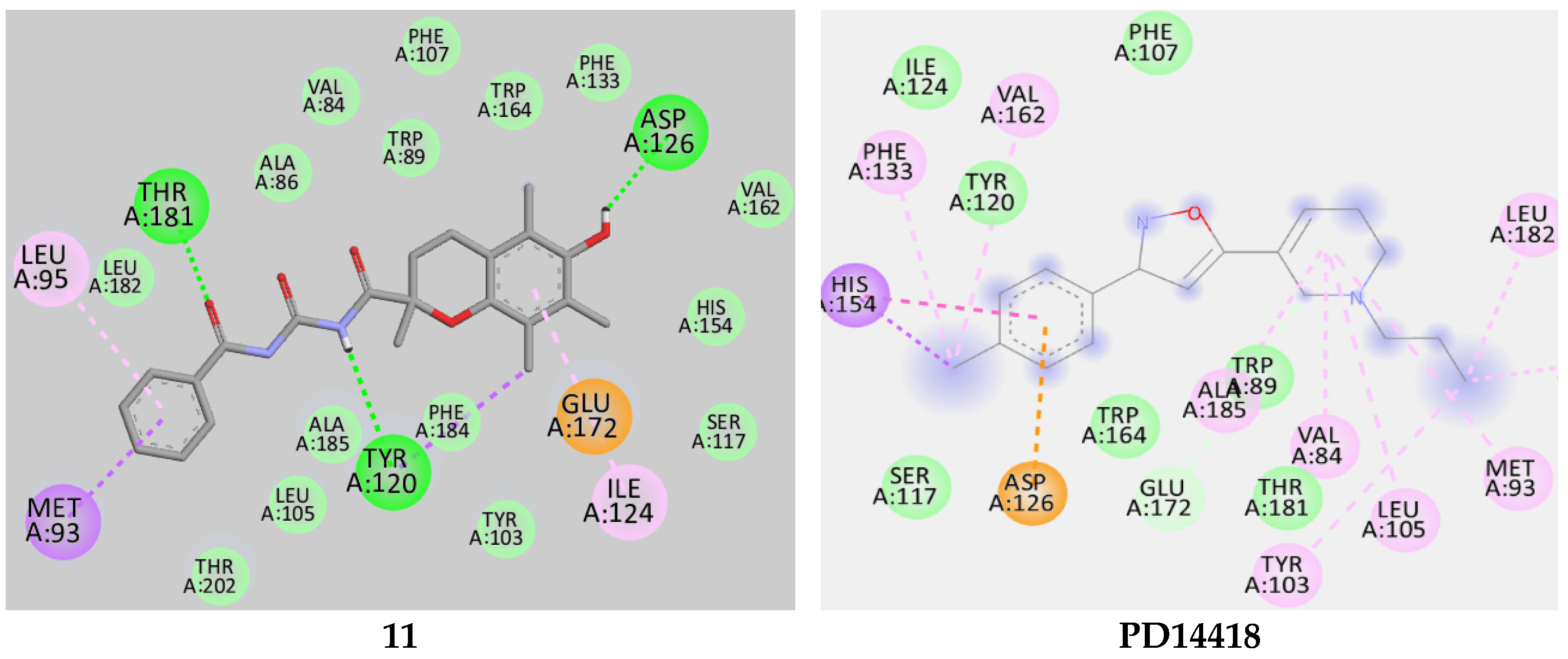
3.3. Molecular Docking
3.4. In Vitro σ1R Binding Assay
4. Conclusions
Supplementary Materials
Author Contributions
Funding
Institutional Review Board Statement
Informed Consent Statement
Data Availability Statement
Acknowledgments
Conflicts of Interest
References
- Fallica, A.N.; Pittalà, V.; Modica, M.N.; Salerno, L.; Romeo, G.; Marrazzo, A.; Helal, M.A.; Intagliata, S. Recent Advances in the Development of Sigma Receptor Ligands as Cytotoxic Agents: A medicinal Chemistry Perspective. J. Med. Chem. 2021, 64, 7926–7962. [Google Scholar] [CrossRef]
- Ghosh, A.K.; Brindisi, M. Urea derivatives in modern drug discovery and medicinal chemistry. J. Med. Chem. 2020, 63, 2751–2788. [Google Scholar] [CrossRef]
- Bialer, M.; Yagen, B.; Shimshoni, J.A. Acyl-Urea Derivatives and Uses Thereof. U.S. Patent US 8,846,903, 30 September 2014. [Google Scholar]
- Ujan, R.; Channar, P.A.; Ali Bahadur, A.; Abbas, Q.; Shah, M.; Rashid, S.G.; Iqbal, S.; Saeed, A.; Abd-Rabboh, H.S.M.; Raza, H.; et al. Synthesis, kinetics and biological assay of some novel aryl bis-thioureas: A potential drug candidates for Alzheimer’s disease. J. Mol. Struct. 2021, 1246, 131136. [Google Scholar] [CrossRef]
- Ryskamp, D.A.; Korban, S.; Zhemkov, V.; Kraskovskaya, N.; Bezprozvanny, I. Neuronal Sigma-1 Receptors: Signaling Functions and Protective Roles in Neurodegenerative Diseases. Front. Neurosci. 2019, 13, 862. [Google Scholar] [CrossRef] [Green Version]
- Nguyen, L.; Lucke-Wold, B.P.; Mookerjee, S.; Kaushal, N.; Matsumoto, R.R. Sigma-1 Receptors and Neurodegenerative Diseases: Towards a Hypothesis of Sigma-1 Receptors as Amplifiers of Neurodegeneration and Neuroprotection. Adv. Exp. Med. Biol. 2017, 964, 133–152. [Google Scholar]
- Maurice, T.; Su, T.-P. The Pharmacology of sigma-1 receptors. Pharmacol. Ther. 2009, 124, 195–206. [Google Scholar] [CrossRef] [Green Version]
- Vela, J.M. Repurposing sigma-1 receptor ligands for COVID-19 therapy? Front. Pharmacol. 2020, 11, 582310. [Google Scholar] [CrossRef]
- Lipinski, C.A.; Lombardo, F.; Dominy, B.W.; Feeney, P.J. Experimental and computational approaches to estimate solubility and permeability in drug discovery and development settings. Adv. Drug Deliv. Rev. 2012, 64, 4–17. [Google Scholar] [CrossRef]
- Lipinski, C.A. Lead- and drug-like compounds: The rule-of-five revolution. Drug Discov. Today Technol. 2004, 1, 337–341. [Google Scholar] [CrossRef]
- Topliss, J.G. Chance Correlations in Structure-Activity Studies Using Multiple Regression Analysis. J. Med. Chem. 1972, 15, 1006–1009. [Google Scholar] [CrossRef]
- Bhowmik, R.; Roy, S.; Sengupta, S.; Ravi, L. Computer aided drug design of florfenicol to target chloramphenicol acetyltransferase of vibriosis causing pathogens. J. Appl. Biol. Biotech. 2022, 10, 76–84. [Google Scholar]
- Molinspiration Cheminformatics. Available online: http://www.molinspiration.com/cgi-bin/properties (accessed on 25 January 2023).
- OSIRIS Property Explorer. Available online: https://www.organic-chemistry.org/prog/peo/ (accessed on 20 January 2023).
- Dallakyan, S.; Olson, A.J. Small-molecule library screening by docking with PyRx. Methods Mol. Biol. 2015, 1263, 243–250. [Google Scholar] [PubMed]
- Trott, O.; Olson, A.J. AutoDock Vina: Improving the speed and accuracy of docking with a new scoring function, efficient optimization, and multithreading. J. Comput. Chem. 2010, 31, 455–461. [Google Scholar] [CrossRef] [PubMed] [Green Version]
- Hurst, D.T.; Stacey, A.D.; Nethercleft, M.; Rahim, A.; Harnden, M.R. The synthesis of some pyrimidinyl and thiazolyl ureas and thioureas and some related compounds. Aust. J. Chem. 1988, 41, 1221–1229. [Google Scholar] [CrossRef]
- Ravn, J.; Ankersen, M.; Begtrup, M.; Lau, J.F. A novel solid-phase synthesis of di- and trisubstituted N-acyl ureas. Tetrahedron Lett. 2003, 44, 6931–6935. [Google Scholar] [CrossRef]
- Verbrugge, P.A.; De Waal, J.J. Process for Preparing Carbamates and Intermediates Therein. European Patent EP0387946A1, 19 September 1990. [Google Scholar]
- Voronkov, M.G.; Vlasova, N.N.; Grigor’eva, O.Y.; Belousova, L.I.; Vlasov, A.V. Acyl Iodides in Organic Synthesis. Reactions of Acetyl Iodide with Urea, Thiourea, and Their N,N′-Disubstituted Derivatives. Russ. J. Org. Chem. 2009, 45, 486–490. [Google Scholar] [CrossRef]
- Singh, A.K.; Chawla, R.; Yadav, L.D.S. In situ slow release of isocyanates: Synthesis and organocatalytic application of N-acylureas. Tetrahedron Lett. 2013, 54, 5099–5102. [Google Scholar] [CrossRef]
- Sun, C.; Zhang, X.; Huang, H.; Zhou, P. Synthesis and evaluation of a new series of substituted acyl(thio)urea and thiadiazolo [2,3-a] pyrimidine derivatives as potent inhibitors of influenza virus neuraminidase. Bioorg. Med. Chem. 2006, 14, 8574–8581. [Google Scholar] [CrossRef]
- Stokes, S.; Martin, N.G. A simple and efficient synthesis of N-benzoyl ureas. Tetrahedron Lett. 2012, 53, 4802–4804. [Google Scholar] [CrossRef]
- Hernandez, A.G.; Grooms, G.M.; El-Alfy, A.T.; Stec, J. Convenient one-pot two step synthesis of symmetrical and unsymmetrical diacyl ureas, acyl urea/carbamate/thiocarbamate derivatives, and related compounds. Synthesis 2017, 49, 2163–2176. [Google Scholar]
- Wang, H.; Zhai, Z.-W.; Shi, Y.-X.; Tan, C.-X.; Weng, J.-Q.; Han, L.; Li, B.-J.; Liu, X.-H. Novel trifluoromethylpyrazole acyl urea derivatives: Synthesis, crystal structure, fungicidal activity and docking study. J. Mol. Struct. 2018, 1171, 631–638. [Google Scholar] [CrossRef]
- Sroor, F.M.; Abdelmoniem, A.M.; Abdelhamid, I.A. Facile Synthesis, Structural Activity Relationship, Molecular Modeling and In vitro Biological Evaluation of New Urea Derivatives with Incorporated Isoxazole and Thiazole Moieties as Anticancer Agents. ChemistrySelect 2019, 4, 10113–10121. [Google Scholar] [CrossRef]
- Sroor, F.M.; Othman, A.M.; Tantawy, M.A.; Mahrous, K.F.; El-Naggar, M.E. Synthesis, antimicrobial, anti-cancer and in silico studies of new urea derivatives. Bioorg. Chem. 2021, 112, 104953. [Google Scholar] [CrossRef]
- Dovlatyan, V.V.; Eliazyan, K.A.; Kazaryan, E.A.; Pivazyan, V.P. N-pyrimidinyl-N-acylureas and thioureas. Zek. Hay. Gitut’yunneri Azg. Akad. 2004, 104, 119–122. [Google Scholar]
- Mohammadpoor-Baltork, I.; Sadeghi, M.M.; Esmayilpour, K. A Convenient and Inexpensive Method for Conversion of Thiocarbonyl Compounds to Their Oxo Derivatives Using Oxone Under Solvent-Free Conditions. Synth. Commun. 2003, 33, 953–959. [Google Scholar] [CrossRef]
- Roth, B. National Institute of Mental Health’s Psychoactive Drug Screening Program. In Assay Protocol Book; Contract # HHSN-271-2018-00023-C (NIMH PDSP); National Institute of Mental Health: Bethesda, MD, USA, 2018; p. 359. Available online: https://pdsp.unc.edu/pdspweb/content/PDSP%20Protocols%20II%202013-03-28.pdf (accessed on 28 February 2023).
- Veber, D.F.; Johnson, S.R.; Cheng, H.Y.; Smith, B.R.; Ward, K.W.; Kopple, K.D. Molecular properties that influence the oral bioavailability of drug candidates. J. Med. Chem. 2002, 45, 2615–2623. [Google Scholar] [CrossRef]
- Biovia, D.S.; Berman, H.M.; Westbrook, J.; Feng, Z.; Gilliland, G.; Bhat, T.N.; Richmond, T.J. Dassault systèmes BIOVIA, discovery studio visualizer, v. 17.2, San Diego: Dassault Systèmes, 2016. J. Chem. Phys. 2000, 10, 21–9991. [Google Scholar]
- Akunne, H.C.; Whetzel, S.Z.; Wiley, J.N.; Corbin, A.E.; Ninteman, F.W.; Tecle, H.; Pei, Y.; Pugsley, T.A.; Heffner, T.G. The pharmacology of the novel and selective sigma ligand, PD 144418. Neuropharmacology 1997, 36, 51–62. [Google Scholar] [CrossRef]
- Ropp, P.J.; Kaminsky, J.C.; Yablonski, S.; Durrant, J.D. Dimorphite-DL: An open-source program for enumerating the ionization states of drug-like small molecules. J. Cheminform. 2019, 11, 14. [Google Scholar] [CrossRef]
- O’Boyle, N.M.; Banck, M.; James, C.A.; Morley, C.; Vandermeersch, T.; Hutchinson, G.J. Open Babel: An open chemical toolbox. J. Cheminform. 2011, 3, 33. [Google Scholar] [CrossRef] [Green Version]

| Compds. | Amine | Acyl Urea a | % Yield b | MP (°C) |
|---|---|---|---|---|
| 1 |  | ** [25,26]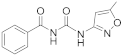 | 76 | 212–215 (Not reported) |
| 2 |  | 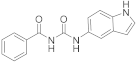 | 94 | 220–223 |
| 3 |  |  | 75 | 180–182 |
| 4 |  | 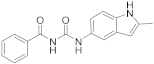 | 71 | 209–211 |
| 5 |  |  | 70 | 145–147 |
| 6 |  | 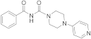 | 92 | 161–163 |
| 7 |  |  | 91 | 170–172 |
| 8 |  | 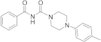 | 72 | 158–160 |
| 9 | 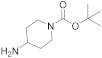 | 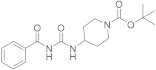 | 71 | 164–166 |
| 10 |  |  | 83 | 180–182 |
| 11 | 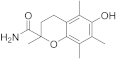 |  | 72 | 155–157 |
| 12 |  |  | 64 | 176–177 |
| 13 |  | ** [27]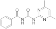 | 68 | 183–185 (Reported 184–186) |
| 14 |  |  | 94 | 233–236 |
| 15 |  |  | 94 | 237–238 |
| 16 |  | 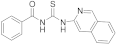 | 65 | 162–165 |
| Entry | MW | cLogP | TPSA | ON | OHNH | NRB | Drug-Likeness |
|---|---|---|---|---|---|---|---|
| 1 | 231.2 | 1.75 | 84.23 | 6 | 2 | 2 | 1.02 |
| 2 | 279.3 | 2.63 | 73.99 | 5 | 3 | 2 | −0.05 |
| 3 | 285.3 | 2.14 | 103.1 | 5 | 2 | 3 | 1.75 |
| 4 | 293.3 | 3.03 | 73.99 | 5 | 3 | 2 | 0.59 |
| 5 | 311.3 | 1.53 | 78.43 | 7 | 1 | 2 | 8.16 |
| 6 | 310.3 | 1.72 | 65.54 | 6 | 1 | 2 | 6.83 |
| 7 | 249.2 | 0.32 | 72.89 | 6 | 2 | 1 | 5.88 |
| 8 | 323.4 | 3.07 | 52.65 | 5 | 1 | 2 | 0.81 |
| 9 | 332.4 | 2.64 | 78.95 | 6 | 1 | 4 | −27.5 |
| 10 | 281.2 | 2.97 | 84.23 | 6 | 2 | 2 | 2.54 |
| 11 | 396.4 | 3.60 | 87.66 | 7 | 3 | 2 | 1.80 |
| 12 | 330.3 | 2.67 | 119.7 | 6 | 3 | 4 | 2.42 |
| 13 | 270.2 | 2.19 | 83.98 | 6 | 2 | 2 | 1.62 |
| 14 | 291.3 | 2.91 | 71.09 | 5 | 2 | 2 | 1.66 |
| 15 | 291.3 | 3.14 | 71.09 | 5 | 2 | 2 | 1.66 |
| 16 Ro5 | 307.3 ≤500 | 3.53 ≤5 | 86.11 | 4 ≤10 | 2 ≤5 | 4 | 0.73 |
| Entries | Binding Affinity | #H-Bond Interactions | Residues Involved in 5HK1 H-Bond Interactions |
|---|---|---|---|
| 1 | −10.0 | 2 | TYR 120, THR 181 |
| 2 | −9.6 | 1 | GLU 172 |
| 3 | −9.6 | 1 | GLU 172 |
| 4 | −10.4 | 1 | GLU 172 |
| 5 | −10.0 | 1 | TYR 120 |
| 6 | −9.6 | 1 | GLU 172 |
| 7 | −8.6 | 1 | GLU 172 |
| 8 | −9.7 | 1 | GLU 172 |
| 9 | −9.0 | 1 | GLU 172 |
| 10 | −10.6 | 3 | THR 181, TYR 108, GLU 172 |
| 11 | −9.5 | 3 | TYR 120, THR 181, ASP 126 |
| 12 | −9.9 | 2 | ASP 126, GLU 172 |
| 13 | −10.3 | 3 | GLU 213, SER 99, SER 33 |
| 14 | −10.6 | 1 | GLU 172 |
| 15 | −11.4 | 0 | - |
| 16 | −10.5 | 2 | GLU 172, TYR 103 |
| PD-14418 | −10.2 | 0 | - |
| Entries | Calculated σ1R Affinity (kcal/mol) | In Vitro σ1R Affinity (Ki, μM) |
|---|---|---|
| 1 | −10.0 | >10 |
| 2 | −9.6 | >10 |
| 3 | −9.6 | >10 |
| 4 | −10.4 | >10 |
| 5 | −10.0 | >10 |
| 6 | −9.6 | >10 |
| 7 | −8.6 | >10 |
| 8 | −9.7 | >10 |
| 9 | −9.0 | >10 |
| 10 | −10.6 | 2.18 |
| 11 | −9.5 | >10 |
| 12 | −9.9 | 9.54 |
| 13 | −10.3 | >10 |
| 14 | −10.6 | >10 |
| 15 | −11.4 | >10 |
| 16 | −10.5 | >10 |
| PD-14418 | −10.2 | * |
Disclaimer/Publisher’s Note: The statements, opinions and data contained in all publications are solely those of the individual author(s) and contributor(s) and not of MDPI and/or the editor(s). MDPI and/or the editor(s) disclaim responsibility for any injury to people or property resulting from any ideas, methods, instructions or products referred to in the content. |
© 2023 by the authors. Licensee MDPI, Basel, Switzerland. This article is an open access article distributed under the terms and conditions of the Creative Commons Attribution (CC BY) license (https://creativecommons.org/licenses/by/4.0/).
Share and Cite
Thapa, R.; Flores, R.; Cheng, K.H.; Mochona, B.; Sikazwe, D. Design and Synthesis of New Acyl Urea Analogs as Potential σ1R Ligands. Molecules 2023, 28, 2319. https://doi.org/10.3390/molecules28052319
Thapa R, Flores R, Cheng KH, Mochona B, Sikazwe D. Design and Synthesis of New Acyl Urea Analogs as Potential σ1R Ligands. Molecules. 2023; 28(5):2319. https://doi.org/10.3390/molecules28052319
Chicago/Turabian StyleThapa, Rajesh, Rafael Flores, Kwan H. Cheng, Bereket Mochona, and Donald Sikazwe. 2023. "Design and Synthesis of New Acyl Urea Analogs as Potential σ1R Ligands" Molecules 28, no. 5: 2319. https://doi.org/10.3390/molecules28052319
APA StyleThapa, R., Flores, R., Cheng, K. H., Mochona, B., & Sikazwe, D. (2023). Design and Synthesis of New Acyl Urea Analogs as Potential σ1R Ligands. Molecules, 28(5), 2319. https://doi.org/10.3390/molecules28052319