Article

Chemical Characterization of Trichoderma spp. Extracts with Antifungal Activity against Cocoa Pathogens

by
Ivan Chóez-Guaranda
1,
Fernando Espinoza-Lozano
1,
Dennys Reyes-Araujo
2,
Christian Romero
1,3,
Patricia Manzano
1,3,
Luis Galarza
1,3 and
Daynet Sosa
1,3,*
1
Centro de Investigaciones Biotecnológicas del Ecuador, ESPOL Polytechnic University, Escuela Superior Politécnica del Litoral, ESPOL, Guayaquil P.O. Box 091050, Ecuador
2
Departamento de Ciencias de la Vida y de la Agricultura, Universidad de las Fuerzas Armadas-ESPE, Sangolquí P.O. Box 171-5-231B, Ecuador
3
Facultad de Ciencias de la Vida, ESPOL Polytechnic University, Escuela Superior Politécnica del Litoral, ESPOL, Guayaquil P.O. Box 091050, Ecuador
*
Author to whom correspondence should be addressed.
Molecules 2023, 28(7), 3208; https://doi.org/10.3390/molecules28073208
Submission received: 18 January 2023 / Revised: 28 March 2023 / Accepted: 29 March 2023 / Published: 4 April 2023

Abstract

:
Ecuador is one of the major cocoa producers worldwide, but its productivity has lately been affected by diseases. Endophytic biocontrol agents have been used to minimize pathogenic effects; however, compounds produced by endophytes are minimally understood. This work presents the chemical characterization of the Trichoderma species extracts that proved inhibition against cocoa pathogens. Solid–liquid extraction was performed as a partitioning method using medium with the fungal mycelia of Trichoderma reesei (C2A), Trichoderma sp. (C3A), Trichoderma harzianum (C4A), and Trichoderma spirale (C10) in ethyl acetate individually. The extract of T. spirale (C10) exhibited the growth inhibition (32.97–47.02%) of Moniliophthora perniciosa at 10 µg/mL, while a slight stimulation of Moniliophthora roreri was shown by the extracts of T. reesei (C2A) and T. harzianum (C4A) at higher concentrations. The inhibitory activity could be related to alkaloids, lactones, quinones, flavonoids, triterpenes, and sterols, as indicated by chemical screening and antifungal compounds, such as widdrol, β-caryophyllene, tyrosol, butyl isobutyrate, sorbic acid, palmitic acid, palmitelaidic acid, linoleic acid, and oleic acid, which were identified by gas chromatography–mass spectrometry (GC-MS). The results showed that the extracts, particularly T. spirale (C10), have the potential as biocontrol agents against witches’ broom disease; however, further studies are needed to confirm their effectiveness.

Graphical Abstract

1. Introduction

Aging plantations, degraded soils, pests, and diseases have been described as the main causes that affect cocoa (Theobroma cacao) productivity worldwide, directly influencing cocoa farmers and the cocoa industry in the tropical regions of South East Asia and West Africa, as well as Central and South America [1]. In Ecuador, cocoa production is an important trade that supports the country’s economy and provides employment for thousands of people. Ecuador is the third-largest producer of cocoa in the world after the Ivory Coast and Ghana, and the biggest producer in Latin America, with an estimated production of over 365,000 metric tons in 2021 [2]. Nevertheless, the fungal diseases frosty pod rot (Moniliophthora roreri) and witches’ broom (Moniliophthora perniciosa, formerly Crinipellis perniciosa) have adversely affected cocoa plantations, causing significant economic losses due to the decrease in production yields [3]. The fungus M. roreri can grow in different environmental conditions causing different symptoms in a complete cycle of about 183 days. Sometimes, cocoa pods are seemingly healthy and continue normal growth but are internally infected and develop disease symptoms later in development. The typical symptoms are necrotic flecks on the surface of cacao pods, which can be observed in the 60–80 days old pods. Thereafter, a dark brown stain with an irregular border that entirely covers the pod, pod malformation, and internal damage characterized by a dark brown fine powder (spores) over the cocoa beans and pulp can be observed when the infection has been developing for more than 110 days [4]. M. perniciosa has multiple penetration modes, mainly through trichomes and stomata. It can cause infection in different plant organs, such as flowers, pods, and branches. The typical symptoms include abnormal shoots that display stem swellings, bud proliferation, and vegetative brooms, which inspired the name witches’ broom disease in cocoa [5]. The results of recent studies from the major cocoa-producing provinces of Ecuador showed that Moniliophthora species possess high morphological and genetic diversity [6,7,8,9]. For this reason, some alternatives such as cacao agroforestry systems [1] and biological controls using products based on endophytic organisms, including the combined application of natural or chemical products [10], have been used to reduce the impact of diseases, minimize the utilization of fungicides, and improve the yield of cocoa production.
In this context, Trichoderma comprises a diverse fungi group of more than 100 species, which have been identified using molecular techniques [11]. It is the most studied and marketed endophyte fungi, since it has been used as a potent biocontrol agent and plant growth promoter [12,13]. Its remarkable biological activity has demonstrated effectiveness against other fungi and plant pathogens, including bacteria, viruses, and nematodes, using antagonistic actions based on antibiosis, competition, and mycoparasitism [14,15]. Furthermore, the species of the Trichoderma genus can induce systemic resistance (ISR) in plants. ISR is a form of immunization in which host plants become resistant to pathogens. This resistance is induced by the application of natural compounds produced by Trichoderma, which activate the plant’s immune system, causing the production of defense proteins and other molecules that can fight off invading pathogens [16]. In fact, many secondary metabolites have been reported for different culture conditions, including volatile and non-volatile compounds during the in vitro interaction of different Trichoderma species with plants and other microorganisms [17,18,19] and the improvement in the production of target compounds using genetic engineering to increase metabolite production [20]. In particular, several studies from Mexico [21], Panama [22], Peru [23,24], and Brazil [25] have evidenced Trichoderma–Moniliophthora interactions. In Ecuador, the antagonism of Trichoderma species from different regions has shown inhibition against banana and cocoa pathogens [26,27]. Nevertheless, the responsible compounds of the biological activity have been rarely described.
Therefore, this study addresses the chemical characterization of four Ecuadorian Trichoderma spp. Extracts, which have been proven to inhibit the growth of the cocoa pathogens M. roreri and M. perniciosa, in order to determine potential specific applications that benefit cocoa cultivars.

2. Results and Discussion

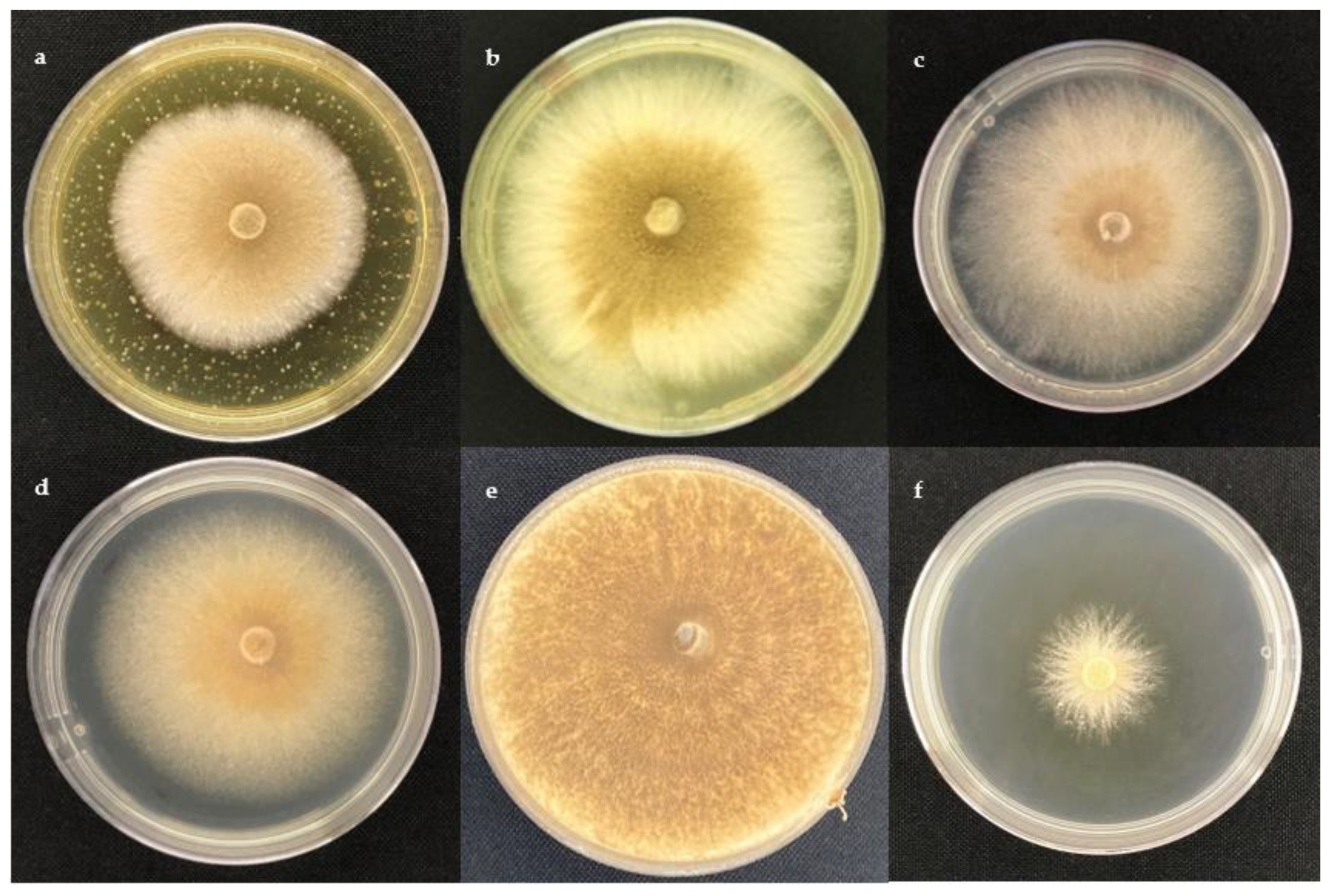
2.1. Antifungal Activity

The effect of ethyl acetate extracts on the assessed cocoa pathogens, M. perniciosa and M. roreri, is presented in Figure 1 and Figure 2, respectively.
The antifungal activity of metabolites produced by Trichoderma strains was expressed as the percentage of growth inhibition (PGI). The results showed that M. perniciosa was the most sensitive pathogen species (Figure 3). The maximum inhibition was observed for the extract of T. spirale (C10) at 10 µg/mL (47.02 ± 1.17%), followed by T. harzianum (C4A) at 10 µg/mL (39.04 ± 2.43%). In addition, the extract of T. sp. (C3A) presented the highest inhibition (36.07 ± 0.55%) at the minimum concentration of 1 µg/mL; however, no significant differences were detected among evaluated Trichoderma strains at this concentration. Even though the precise data on the T. spirale–M. perniciosa interaction are limited, higher inhibitory activity (75%) affecting mycelial growth and even spore germination of M. perniciosa has been reported for T. harzianum [28], and significant mycoparasitism levels against M. roreri have been achieved by T. spirale [29]. In addition, remarkably, antifungal activity against M. perniciosa and M. roreri has been found for foliar endophytic fungi [30] and endophytic Bacillus [31] isolated from cocoa cultivars. Additionally, T. spirale has been shown to inhibit the growth of Fusarium oxysporum during in vitro and greenhouse experiments, demonstrating the relevance of the metabolites produced by this strain, which also indicates plant protection against fungal disease [32].
On the other hand, the best PGI against M. roreri was observed for the extract of T. spirale (C10) at 1000 µg/mL (26.20 ± 2.06%) and 100 µg/mL (17.10 ± 1.23%). Higher PGI values (48%) have been reported for culture filtrates from Brazil [31] and comparable data (15.90–37.10%) for isolates from Mexico [20] against M. roreri. Differences among results could be attributed to the technique employed since the referenced studies used a dual-culture assay, through which the inoculum size, agar volume, well size, and other variables possibly affect the fungal interactions [17], isolates, culture filtrates, extracts, or compounds assessed. Contrasting results were shown for the extracts of T. reesei (C2A) (4.40%) and T. harzianum (C4A) (4.36%) at 1000 µg/mL. Additionally, the extracts of T. sp. (C3A) (0.49%) and T. spirale (C10) (1.45%) stimulated the growth of the pathogen at 100 and 1 µg/mL, respectively. The growth stimulation could be explained by the metabolites of the extracts, which, at higher concentrations, seem to favor the pathogen. Similar results have been reported for endophytic fungal isolates, demonstrating the growth promotion of Moniliphthora spp. Fungal endophytes produce several compounds, such as plant growth hormones (auxins, cytokinins, and gibberellins), which can also promote the growth of fungi [33]. Different findings have evidenced the growth restraint of M. roreri through T. harzianum (40.50–41.20%), T. ressei (20.30–25.80%) in Mexico [20], T. harzianum (75%) in Ecuador [27], and native Trichoderma strains (48.50–57.94%) in Peru [23]. Even so, M. roreri growth inhibition (25–97%) and growth stimulation (50–140%) ranges have been indicated by the fungicide flutolanil [32].
These results show the importance of identifying the metabolites secreted by Trichoderma species, since the extracts revealed similar properties as in the aforementioned commercial antifungal agent.

2.2. Chemical Screening

The content of dried ethyl acetate extracts was T. reesei (C2A) (12.60 g/L), T. sp. (C3A) (5.50 g/L), T. harzianum (C4A) (8.20 g/L), and T. spirale (C10) (5.30 g/L). In general, the chemical screening of Trichoderma spp. ethyl acetate extracts revealed the presence of alkaloids, lactones, quinones, reducing sugars, flavonoids, triterpenes, and sterols (Table 1). It should be noted that the extract of T. reesei (C2A) showed positive results for most of the secondary metabolites tests, followed by T. spirale (C10), T. harzianum (C4A), and T. sp. (C3A). Alkaloids were detected only in the extracts of T. reesei (C2A) and T. harzianum (C4A). The presence of flavonoids was shown by the extracts of T. reesei (C2A), T. sp. (C3A), and T. spirale (C10) strains, and only the extract of T. spirale (C10) indicated the occurrence of anthocyanidins. Nevertheless, resins, saponins, and amino acids were not found in any organic extracts. These results are consistent with preceding studies reporting the presence of alkaloids, flavonoids, and even phenols in T. aureoviride and T. harzianum ethyl acetate extracts [34].

2.3. GC-MS Metabolite Profile

The detected compounds in ethyl acetate extracts of the different Trichoderma spp. strains are presented in Table 2. The relative abundances were calculated according to the peak area percent of three replicates of each identified compound. In total, 63 metabolites were putatively identified, including terpenes, esters, fatty acids, and alcohols, among others. The extract of T. reesei (C2A) displayed 39 compounds, while the extracts of T. sp. (C3A), T. harzianum (C4A), and T. spirale (C10) showed 21, 20, and 15 compounds, respectively. All compounds appear in order of the elution of non-polar chromatographic column, and differences between extracts of Trichoderma strains were revealed (Figure 4).
The sesquiterpenes were mostly found in T. reesei (C2A) compared to other species in which free and esterified fatty acids predominated. Sesquiterpenes are produced mainly by different plant species, but T. virens has been reported as the producer of these compounds with remarkable antifungal properties [35]. Nerolidol has been described for T. reesei and has been proven mainly as an anti-inflammatory, antinociceptive, and antioxidant compound [36]. The 1,4-cadinadiene and widdrol have been indicated to have antiparasitic [37] and antifungal [38] properties, respectively. The β-caryophyllene has been identified in Trichoderma species [39] and described as an antibacterial, antifungal, antioxidant agent [40], as well as a plant growth promoter [41]. Mevalonolactone has demonstrated impressive antibacterial activities [42]. Sorbicillin was predominantly found in T. sp. (C3A), followed by T. reesei (C2A). This polyketide has been reported in several ascomycetes, including Trichoderma, and possesses antimicrobial activity [43]. Indeed, a recombinant T. reesei strain has recently been demonstrated to improve the production of sorbicillinoids [44], and sorbicillinoids isolated from the culture filtrate of T. ongibrachiatum have been revealed to have antifungal effects against Phytophthora infestans [45]. β-fenchyl alcohol was detected in T. harzianum (C4A), and it has been reported to be an antibacterial agent [46]. Palmitic acid has been described has having antifungal activity [47] and was found in extracts of T. reesei (C2A), T. sp. (C3A), and T. harzianum (C4A) strains. Butyl isobutyrate has been reported in the culture filtrates of T. asperellum with notable cytotoxic effects against fungal plant pathogens [48] and palmitelaidic acid has also been identified as a biomarker of T. virens against a cotton fungal disease caused by Rhizoctonia solani [49], both of which were detected in extracts of T. sp. (C3A), T. harzianum (C4A), and T. spirale (C10). Regarding the extract of T. spirale (C10), ethyl linoleate has been referred to as a plant growth regulator [50], sorbic acid has been reported as an antifungal compound that impedes spore germination and mycelial growth [51], and the chiral properties of 3-hydroxybutyric acid cause it to be a mediator in the synthesis of potential antibiotics [52].
Notably, five metabolites were common in all strains: phenylethyl alcohol, tyrosol, diethyl succinate, and linoleic and oleic acid. Phenolic compounds are produced by another Trichoderma species [53], and they have been utilized as quorum sensing molecules [54] and antifungal agents [55]. In particular, tyrosol has been isolated from T. harzianum and T. spirale [56]. Furthermore, the ester has been detected in fungal endophytes [57] and shows antimicrobial activity [58], whereas fatty acids have been confirmed to have antifungal effects [47]. In fact, it has been demonstrated that unsaturated fatty acids could inhibit mycelial growth and decrease the biomass production of M. perniciosa [59].
Overall, the potential inhibition of Trichoderma extracts could be related to the presence of different detected extracellular metabolites due to the aforementioned biological activities.

3. Materials and Methods

3.1. Standards and Chemicals

Potato dextrose agar (PDA) was purchased from Becton, Dickinson, and Company (Sparks, MD, USA). Gentamicin and N, O-Bis (trimethylsilyl), trifluoroacetamide (BSTFA), and dimethyl sulfoxide (DMSO) were obtained from Sigma-Aldrich (St. Louis, MO, USA). Saturated alkanes standard (C7–C40) was acquired from Supelco (Bellefonte, PA, USA). Ethyl acetate reagent grade was obtained from Fisher Scientific (Hampton, NH, USA).

3.2. Fungal Strains

T. reesei (C2A), T. sp. (C3A), T. harzianum (C4A), and T. spirale (C10) were provided by the Culture Collection of Microorganisms of Biotechnology Research Center of Ecuador (CCM-CIBE) [60]. These species were previously isolated from the rhizosphere of cocoa plants in Naranjal, Guayas, Ecuador. Monosporic cultures were identified by the sequencing of the internal transcribed spacer (ITS) region, as indicated by White et al. [61]. The DNA extraction and polymerase chain reaction (PCR) amplification of the universal region of fungi ITS1-5,8-ITS2 was carried out using the ITS1 (5′ TCC GTA GGT GAA CCT GCG G 3′) and ITS2 (5′TCC TCC GCT TAT TGA TAT GC 3′) primers. Subsequently, the PCR product was sequenced with the SANGER method, and the basic local alignment search tool (BLAST) was utilized for the processed sequences, from which the identities were obtained (Table 3). The fungal isolates were deposited at CCM-CIBE with (CCMCIBE-H1103), (CCMCIBE-H1104), (CCMCIBE-H1105), and (CCMCIBE-H1106) codes, respectively. In this work, the fungal spores of every strain already maintained in 10% (v/v) glycerol at −80 °C were grown individually in Petri dishes (90 mm) containing PDA culture medium with gentamicin (10 μg/mL) and incubated at 27 °C in darkness for 7 days. Then, each fungal strain was subcultured on 50 PDA plates to prepare a sufficient amount of mycelium. Trichoderma strains were incubated for 7 days to give the culture enough time to form robust mycelia, which are essential for the successful production of enzymes and metabolites [62].

3.3. Extraction Procedure

The extraction of secondary metabolites from Trichoderma strains followed Liu and Liu method with some modifications [62]. First, the solid medium containing fungal mycelia (SMF) of 50 inoculated PDA plates was cut into small pieces (approximately 10 × 10 mm) under aseptic conditions, transferred into a 2 L Erlenmeyer flask with 600 mL of ethyl acetate, shaken at 110 rpm for 1 h, and incubated at 25–28 °C for 24 h in darkness. Then, the organic phase was filtered, concentrated in a rotary evaporator under reduced pressure at 40 °C, and SMF extraction was repeated with recoveries every 24 h for 4 days. The extraction process was followed until the color faded, as recommended in protocols for recovering secondary metabolites [63]. Next, ethyl acetate extracts were placed together, concentrated, and dried for further analysis. This procedure was performed for each Trichoderma strain. The same method was used in the control treatments, except that culture media without fungi were used.

3.4. Antifungal Assay

Two cocoa crop pathogens preserved at CCM-CIBE were used: M. roreri (CCMCIBE-H815) and M. perniciosa (CCMCIBE-H1109). The poisoned culture medium technique was used as described by Guerrero-Rodriguez et al. [64]. The dried ethyl acetate extracts of Trichoderma strains were resuspended in pure dimethyl sulfoxide (DMSO) and sterilized using 0.22 μm syringe filters. After that, fungi strain disks (6 mm) were taken from 14-day-old cultures and individually transferred into Petri dishes (60 × 15 mm) containing PDA culture medium with different concentrations of Trichoderma extracts (1, 10, 100, and 1000 μg/mL), negative controls (DMSO and PDA), and positive controls using the commercial product Bankit® 25 SC Azoxystrobin (Syngenta, Mexico City, Mexico) at 10 μg/mL. The final concentration of DMSO in Petri dishes did not exceed 1% to avoid cytotoxicity [65]. Then, four replicates of cultures were incubated at 28 °C and radial growth was measured after 2 weeks. The radial data were processed from digital images using the Image J platform. Finally, the percentage of growth inhibition (PGI) was calculated using the equation PGI = ((R1 − R2)/R1) × 100, where R1 is the radial of control and R2 is the radial of the extract [66].

3.5. Chemical Screening

The chemical screening tests of ethyl acetate extracts were performed as reported elsewhere [67]. Certain aliquots of Trichoderma ethyl acetate extracts (200 µL) were taken separately for assessment using the Dragendorff, Mayer, Wagner, Baljet, Borntrager, Liebermann–Burchard, Resins, Fehling, Foam, Ninhydrin, Shinoda, Anthocyanidins, and Catechins qualitative tests.
For the alkaloid tests, the extract was evaporated and dissolved in 1% hydrochloric acid. Then, an aliquot of acid solution of the extract was mixed with three drops of Dragendorff reagent, three drops of Wagner reagent, sodium chloride, and three drops of Mayer reagent separately. The development of turbidity confirmed the existence of alkaloids.
In the Baljet test, the extract was evaporated and dissolved in 40 µL of 100% ethanol. After that, an aliquot of 40 µL of Baljet reagent was added to the ethanol solution. The development of the red precipitate indicated the occurrence of lactones.
In the Borntrager test, the extract was evaporated and dissolved in 100 µL of chloroform. Next, an aliquot of 100 µL of 5% sodium hydroxide was added. The development of pink coloration in the aqueous phase after shaking and phases separation revealed the presence of quinones.
In the Liebermann–Burchard test, the extract was evaporated and dissolved in 40 µL of chloroform. Then, an aliquot of 40 µL of acetic anhydride and three drops of 96% sulfuric acid were added to the organic solution. The development of a red-brick reddish-brown coloration indicated the occurrence of triterpenes, and the blue coloring, which turned blue-orange, indicated the occurrence of sterols.
In the resins test, the extract was evaporated and dissolved in 100 µL of 100% ethanol. Thereafter, an aliquot of 500 µL of distilled water was added to the ethanol solution. The development of precipitates showed the existence of resins.
In the Fehling test, the extract was evaporated and dissolved in 100 µL of distilled water. Next, an aliquot of 200 µL of Fehling reagent was added to the aqueous solution and heated in a water bath for 5 min. The development of red coloration or red precipitate indicated the presence of reducing sugars.
In the foam test, the extract was evaporated and dissolved in 100 µL of 100% ethanol. Then, an aliquot of 500 µL of distilled water was added to the ethanol solution and shaken vigorously for 5 min. The presence of foam (>2 mm in height) on the surface of the liquid after 2 min revealed the occurrence of saponins.
In the ninhydrin test, the extract was evaporated, dissolved in 200 µL of 2% ninhydrin aqueous solution, and heated in a water bath for 5 min. The development of purplish blue demonstrated the existence of amino acids.
For the flavonoids tests, the extract was evaporated and dissolved in 200 µL of 100% ethanol. After that, an aliquot of 100 µL of hydrochloric acid was added to the ethanol solution. In the Shinoda test, an aliquot of 100 µL of amyl alcohol and a piece of metallic magnesium was added to the acidified ethanol solution. The development of yellow-orange-red coloration after shaking and phases separation indicated the occurrence of flavonoids. In the anthocyanidins test, an aliquot of 100 µL of distilled water and 200 µL of amyl alcohol was added to the acidified ethanol solution, and the development of red-brown coloration after shaking and phases separation revealed the presence of anthocyanidins. Finally, in the catechins test, drops of ethyl acetate extract and 20% sodium carbonate were placed on filter paper. The development of carmelite-green coloration under ultraviolet light showed the presence of catechins.

3.6. Gas Chromatography–Mass Spectrometry (GC-MS) Analysis

The compound separation of ethyl acetate extracts was performed in a gas chromatography–mass spectrometer from Agilent Technologies (7890A GC system and 5975C inert XL MSD with a triple axis detector). A capillary column DB-5MS (30 m × 0.25 mm) with phenyl dimethylpolysiloxane for the stationary phase (0.25 micron film thickness) and helium as the carrier gas (1.2 mL/min). The extracts were diluted in a 1:10 ratio, and injection was carried out at 250 °C with the splitless mode in triplicate. The oven temperature was initially 70 °C for 2 min, then it was increased to 300 °C at 5 °C/min and was maintained at 300 °C for 6 min. The MSD transfer line was 300 °C, and the detector temperature was 230 °C with an electron ionization of 70 eV. Data compounds were collected with the full scan mode (40–600 amu) in the quadrupole mass analyzer. Finally, metabolite identification was achieved by matching the mass spectra of the samples with the data available in Wiley 9 and NIST 2011 libraries and by comparing the linear retention indices using a series of saturated n-alkanes (C7–C40).

3.7. Statistical Analysis

The normalization of PGI data was performed using the arcsin transformation (arcsin √x) [68]. Thereafter, the analysis of variance (ANOVA) and Tukey’s test (p < 0.05) were performed to compare the inhibition of different concentrations of Trichoderma spp. extracts against cocoa pathogens M. roreri and M. perniciosa.

4. Conclusions

This study revealed that extracts obtained from the Trichoderma species inhibited the in vitro growth of M. perniciosa better than M. roreri. The extract of T. spirale (C10) showed the maximum inhibition effect at 10 µg/mL, whereas T. reesei (C2A) and T. harzianum (C4A) displayed overgrowth at higher concentrations. Differences in secondary metabolites groups and metabolite profiles that depended on the Trichoderma species were found. The inhibitory activity could be attributed to the antifungal compounds detected, such as widdrol, β-caryophyllene, tyrosol, butyl isobutyrate, sorbic acid, palmitic acid, palmitelaidic acid, linoleic acid, and oleic acid. These findings suggest that these extracts could be used to develop prospective biocontrol agents against the witches’ broom disease, especially the extract of T. spirale (C10). However, further chemical and antagonistic studies are required for a complete characterization of the bioactivity revealed by the extracts.

Author Contributions

Conceptualization, D.S., L.G., C.R. and P.M.; methodology, I.C.-G., F.E.-L. and D.R.-A.; software, I.C.-G. and F.E.-L.; validation, I.C.-G. and F.E.-L.; formal analysis, I.C.-G. and F.E.-L.; investigation, I.C.-G., F.E.-L. and D.R.-A.; resources, D.S. and L.G.; data curation, I.C.-G. and F.E.-L.; writing—original draft preparation, I.C.-G.; writing—review and editing, I.C.-G., L.G. and D.S.; visualization, I.C.-G. and F.E.-L.; supervision, C.R. and P.M.; project administration, D.S. and L.G.; funding acquisition, D.S. All authors have read and agreed to the published version of the manuscript.

Funding

This study was supported by the Ecuadorian Network of Universities for Research and Postgraduate Studies (REDU) under Grant PREDU-2016-014 and was conducted under Scientific Research Authorization No. MAE-DNB-CM-2017-0076.

Institutional Review Board Statement

Not applicable.

Informed Consent Statement

Not applicable.

Data Availability Statement

All data can be found in the manuscript.

Acknowledgments

The authors extend their appreciation to the Culture Collection of Microorganisms of the Biotechnology Research Center of Ecuador (CCM-CIBE) for the preservation of microorganisms.

Conflicts of Interest

The authors declare no conflict of interest.

Sample Availability

Not available.

References

  1. Armengot, L.; Ferrari, L.; Milz, J.; Velásquez, F.; Hohmann, P.; Schneider, M. Cacao Agroforestry Systems Do Not Increase Pest and Disease Incidence Compared with Monocultures under Good Cultural Management Practices. Crop. Prot. 2020, 130, 105047. [Google Scholar] [CrossRef]
  2. International Cocoa Organization (ICCO) Production of Cocoa Beans. Available online: https://www.icco.org/statistics (accessed on 16 February 2023).
  3. Anzules-Toala, V.; Pazmiño-Bonilla, E.; Alvarado-Huamán, L.; Borjas-Ventura, R.; Castro-Cepero, V.; Julca-Otiniano, A. Control of Cacao (Theobroma cacao) Diseases in Santo Domingo de Los Tsachilas, Ecuador. Agron. Mesoam. 2022, 33, 45939. [Google Scholar] [CrossRef]
  4. Orea, M.D.D.; Romero-Cortes, T.; Lopez-Perez, P.A.; Perez Espana, V.H.; Ramirez-Lepe, M.; Cuervo-Parra, J.A. Current Status of Cocoa Frosty Pod Rot Caused by Moniliophthora roreri and a Phylogenetic Analysis. Plant Pathol. J. (Faisalabad) 2017, 16, 41–53. [Google Scholar] [CrossRef] [Green Version]
  5. Meraz-Pérez, I.M.; Carvalho, M.R.; Sena, K.F.; Soares, Y.J.B.; Junior, A.S.E.; Lopes, U.V.; dos Santos Filho, L.P.; Araújo, S.A.; Soares, V.L.F.; Pirovani, C.P.; et al. The Moniliophthora perniciosa—Cacao Pod Pathosystem: Structural and Activated Defense Strategies against Disease Establishment. Physiol. Mol. Plant Pathol. 2021, 115, 101656. [Google Scholar] [CrossRef]
  6. Maridueña-Zavala, M.G.; Feijoo, M.I.J.; Cevallos-Cevallos, J.M. Pathogenicity of Moniliophthora roreri Isolates from Selected Morphology Groups in Harvested Cacao Pods and in Vitro Sensitivity to Compost Tea. Bionatura 2021, 6, 1569–1574. [Google Scholar] [CrossRef]
  7. Maridueña-Zavala, M.G.; Villavicencio-Vásquez, M.E.; Cevallos-Cevallos, J.M.; Peralta, E.L. Molecular and Morphological Characterization of Moniliophthora roreri Isolates from Cacao in Ecuador. Can. J. Plant Pathol. 2016, 38, 460–469. [Google Scholar] [CrossRef]
  8. Maridueña-Zavala, M.G.; Freire-Peñaherrera, A.; Espinoza-Lozano, R.F.; Villavicencio-Vasquez, M.; Jimenez-Feijoo, M.; Cevallos-Cevallos, J.M. Genetic Characterization of Moniliophthora perniciosa from Ecuador and in Vitro Sensitivity to Compost Tea. Eur. J. Plant Pathol. 2019, 154, 943–959. [Google Scholar] [CrossRef]
  9. Espinoza-Lozano, F.; Amaya-Márquez, D.; Pinto, C.M.; Villavicencio-Vásquez, M.; Sosa Del Castillo, D.; Pérez-Martínez, S. Multiple Introductions of Moniliophthora roreri from the Amazon to the Pacific Region in Ecuador and Shared High Azoxystrobin Sensitivity. Agronomy 2022, 12, 1119. [Google Scholar] [CrossRef]
  10. ten Hoopen, G.M.; Krauss, U. Biological Control of Cacao Diseases. In Cacao Diseases: A History of Old Enemies and New Encounters; Bailey, B.A., Meinhardt, L.W., Eds.; Springer International Publishing: Cham, Switzerland, 2016; pp. 511–566. [Google Scholar]
  11. di Cologna, N.M.D.; Gómez-Mendoza, D.P.; Zanoelo, F.F.; Giannesi, G.C.; Guimarães, N.C.A.; Moreira, L.R.S.; Filho, E.X.F.; Ricart, C.A.O. Exploring Trichoderma and Aspergillus Secretomes: Proteomics Approaches for the Identification of Enzymes of Biotechnological Interest. Enzym. Microb Technol. 2018, 109, 1–10. [Google Scholar] [CrossRef]
  12. Adnan, M.; Islam, W.; Shabbir, A.; Khan, K.A.; Ghramh, H.A.; Huang, Z.; Chen, H.Y.H.; Lu, G. Plant Defense against Fungal Pathogens by Antagonistic Fungi with Trichoderma in Focus. Microb. Pathog. 2019, 129, 7–18. [Google Scholar] [CrossRef] [PubMed]
  13. Ferreira, F.V.; Musumeci, M.A. Trichoderma as Biological Control Agent: Scope and Prospects to Improve Efficacy. World J. Microbiol. Biotechnol. 2021, 37, 90. [Google Scholar] [CrossRef]
  14. Vinale, F.; Strakowska, J.; Mazzei, P.; Piccolo, A.; Marra, R.; Lombardi, N.; Manganiello, G.; Pascale, A.; Woo, S.L.; Lorito, M. Cremenolide, a New Antifungal, 10-Member Lactone from Trichoderma Cremeum with Plant Growth Promotion Activity. Nat. Prod. Res. 2016, 30, 2575–2581. [Google Scholar] [CrossRef] [PubMed] [Green Version]
  15. Guzmán-Guzmán, P.; Kumar, A.; Santos-Villalobos, S.D.L.; Parra-Cota, F.I.; Orozco-Mosqueda, M.D.C.; Fadiji, A.E.; Hyder, S.; Babalola, O.O.; Santoyo, G. Trichoderma Species: Our Best Fungal Allies in the Biocontrol of Plant Diseases—A Review. Plants 2023, 12, 432. [Google Scholar] [CrossRef] [PubMed]
  16. Salwan, R.; Sharma, A.; Kaur, R.; Sharma, R.; Sharma, V. The Riddles of Trichoderma Induced Plant Immunity. Biol. Control 2022, 174, 105037. [Google Scholar] [CrossRef]
  17. Verma, M.; Brar, S.K.; Tyagi, R.D.; Surampalli, R.Y.; Valéro, J.R. Antagonistic Fungi, Trichoderma Spp.: Panoply of Biological Control. Biochem. Eng. J. 2007, 37, 1–20. [Google Scholar] [CrossRef]
  18. Ahluwalia, V.; Kumar, J.; Rana, V.S.; Sati, O.P.; Walia, S. Comparative Evaluation of Two Trichoderma harzianum Strains for Major Secondary Metabolite Production and Antifungal Activity. Nat. Prod. Res. 2015, 29, 914–920. [Google Scholar] [CrossRef]
  19. Rukachaisirikul, V.; Chinpha, S.; Phongpaichit, S.; Saikhwan, N.; Sakayaroj, J.; Preedanon, S. Sesquiterpene and Monoterpene Derivatives from the Soil-Derived Fungus Trichoderma reesei PSU-SPSF013. Phytochem. Lett. 2019, 30, 124–129. [Google Scholar] [CrossRef]
  20. Dashtban, M.; Kepka, G.; Seiboth, B.; Qin, W. Xylitol Production by Genetically Engineered Trichoderma reesei Strains Using Barley Straw as Feedstock. Appl. Biochem. Biotechnol. 2013, 169, 554–569. [Google Scholar] [CrossRef]
  21. Reyes-Figueroa, O.; Ortiz-García, C.F.; Torres-de la Cruz, M.; Lagunes-Espinoza, L.D.; Valdovinos-Ponce, G. Trichoderma Del Agroecosistema Cacao Con Potencial de Biocontrol Sobre Moniliophthora roreri. Rev. Chapingo Ser. Cienc. For. Ambiente 2016, 22, 149–163. [Google Scholar] [CrossRef]
  22. Mejia, L.; Rojas, E.I.; Maynard, Z.; Van Bael, S.; Arnold, A.E.; Hebbar, P.; Samuels, G.J.; Robbins, N.; Herre, E.A. Endophytic Fungi as Biocontrol Agents of Theobroma cacao Pathogens. Biol. Control 2008, 46, 4–14. [Google Scholar] [CrossRef]
  23. Krauss, U.; Soberanis, W. Effect of Fertilization and Biocontrol Application Frequency on Cocoa Pod Diseases. Biol. Control 2002, 24, 82–89. [Google Scholar] [CrossRef]
  24. Leiva, S.; Oliva, M.; Hernández, E.; Chuquibala, B.; Rubio, K.; García, F.; de la Cruz, M.T. Assessment of the Potential of Trichoderma Spp. Strains Native to Bagua (Amazonas, Peru) in the Biocontrol of Frosty Pod Rot (Moniliophthora roreri). Agronomy 2020, 10, 1376. [Google Scholar] [CrossRef]
  25. Bastos, C.N. Isolate of Trichoderma Brevicompactum for the Control of Cocoa Witches Broom Disease: Preliminary Results. Agrotrópica 2012, 24, 21–26. [Google Scholar] [CrossRef]
  26. Gonzalez, M.F.; Magdama, F.; Galarza, L.; Sosa, D.; Romero, C. Evaluation of the Sensitivity and Synergistic Effect of Trichoderma reesei and Mancozeb to Inhibit under In Vitro Conditions the Growth of Fusarium oxysporum. Commun. Integr. Biol. 2020, 13, 160–169. [Google Scholar] [CrossRef] [PubMed]
  27. Galarza, L.; Akagi, Y.; Takao, K.; Kim, C.S.; Maekawa, N.; Itai, A.; Peralta, E.; Santos, E.; Kodama, M. Characterization of Trichoderma Species Isolated in Ecuador and Their Antagonistic Activities against Phytopathogenic Fungi from Ecuador and Japan. J. Gen. Plant Pathol. 2015, 81, 201–210. [Google Scholar] [CrossRef]
  28. Aneja, M.; Gianfagna, T.J.; Hebbar, P.K. Trichoderma harzianum Produces Nonanoic Acid, an Inhibitor of Spore Germination and Mycelial Growth of Two Cacao Pathogens. Physiol. Mol. Plant Pathol. 2005, 67, 304–307. [Google Scholar] [CrossRef]
  29. Evans, H.C.; Holmes, K.A.; Thomas, S.E. Endophytes and Mycoparasites Associated with an Indigenous Forest Tree, Theobroma Gileri, in Ecuador and a Preliminary Assessment of Their Potential as Biocontrol Agents of Cocoa Diseases. Mycol. Prog. 2003, 2, 149–160. [Google Scholar] [CrossRef]
  30. Villavicencio-Vásquez, M.; Espinoza-Lozano, R.F.; Pérez-Martínez, S.; Sosa, D.; Castillo, D. Hongos Endófitos Foliares Como Candidatos a Biocontroladores Contra Moniliophthora Spp. de Theobroma cacao (Malvaceae) En Ecuador. Acta Biol. Colomb. 2018, 23, 235–241. [Google Scholar] [CrossRef]
  31. Serrano, L.; Sosa Moreno, A.; Sosa Del Castillo, D.; Bonilla, J.; Romero, C.A.; Galarza, L.L.; Coronel–león, J.R. Biosurfactants Synthesized by Endophytic Bacillus Strains as Control of Moniliophthora perniciosa and Moniliophthora roreri. Sci. Agric. 2021, 78, e20200172. [Google Scholar] [CrossRef]
  32. Vargas Inciarte, L.; Fuenmayor Arrieta, Y.; Luzardo Méndez, M.; da Costa Jardin, M.; Vera, A.; Carmona, D.; Homen Pereira, M.; da Costa Jardin, P.; San Blas, E. Use of Different Trichoderma Species in Cherry Type Tomatoes (Solanum lycopersicun L.) Against Fusarium oxysporum Wilt in Tropical Greenhouses. Agron. Costarric. 2019, 43, 85–100. [Google Scholar] [CrossRef]
  33. Villavicencio, M.; Schuller, L.; Espinosa, F.; Noceda, C.; Sosa, D.; Pérez-Martínez, S. Foliar Endophytic Fungi of Theobroma cacao Stimulate More than Inhibit Moniliophthora Spp. Growth and Behave More as an Endophytes than Pathogens. AgriRxiv 2020, 2020, 1–33. [Google Scholar] [CrossRef]
  34. Moïse, N.A.; Severin, T.N.; Christelle, S.E.; Tibo, A.A.; Lambert, S.M.; Duplex, W.J. Efficacy of Trichoderma harzianum (Edtm) and Trichoderma Aureoviride (T4) as Potential Bio-Control Agent of Taro Leaf Blight Caused by Phytophthora Colocasiae. Int. J. Appl. Microbiol. Biotechnol. Res. 2018, 6, 115–126. [Google Scholar]
  35. Hu, Z.; Tao, Y.; Tao, X.; Su, Q.; Cai, J.; Qin, C.; Ding, W.; Li, C. Sesquiterpenes with Phytopathogenic Fungi Inhibitory Activities from Fungus Trichoderma Virens from Litchi Chinensis Sonn. J. Agric. Food Chem. 2019, 67, 10646–10652. [Google Scholar] [CrossRef] [PubMed]
  36. De Carvalho, R.B.F.; De Almeida, A.A.C.; Campelo, N.B.; Lellis, D.R.O.D.; Nunes, L.C.C. Nerolidol and Its Pharmacological Application in Treating Neurodegenerative Diseases: A Review. Recent Pat. Biotechnol. 2018, 12, 158–168. [Google Scholar] [CrossRef]
  37. Afoulous, S.; Ferhout, H.; Raoelison, E.G.; Valentin, A.; Moukarzel, B.; Couderc, F.; Bouajila, J. Chemical Composition and Anticancer, Antiinflammatory, Antioxidant and Antimalarial Activities of Leaves Essential Oil of Cedrelopsis Grevei. Food Chem. Toxicol. 2013, 56, 352–362. [Google Scholar] [CrossRef] [PubMed]
  38. Nuñez, Y.O.; Salabarria, I.S.; Collado, I.G.; Hernández-Galán, R. The Antifungal Activity of Widdrol and Its Biotransformation by Colletotrichum gloeosporioides (Penz.) Penz. & Sacc. and Botrytis cinerea Pers.: Fr. J. Agric. Food Chem. 2006, 54, 7517–7521. [Google Scholar] [CrossRef] [PubMed]
  39. Lee, S.; Yap, M.; Behringer, G.; Hung, R.; Bennett, J.W. Volatile Organic Compounds Emitted by Trichoderma Species Mediate Plant Growth. Fungal Biol. Biotechnol. 2016, 3, 7. [Google Scholar] [CrossRef] [Green Version]
  40. Dahham, S.; Tabana, Y.; Iqbal, M.; Ahamed, M.; Ezzat, M.; Majid, A.; Majid, A. The Anticancer, Antioxidant and Antimicrobial Properties of the Sesquiterpene β-Caryophyllene from the Essential Oil of Aquilaria Crassna. Molecules 2015, 20, 11808–11829. [Google Scholar] [CrossRef]
  41. Morath, S.U.; Hung, R.; Bennett, J.W. Fungal Volatile Organic Compounds: A Review with Emphasis on Their Biotechnological Potential. Fungal Biol. Rev. 2012, 26, 73–83. [Google Scholar] [CrossRef]
  42. Pinheiro, E.A.A.; Carvalho, J.M.; dos Santos, D.C.P.; Feitosa, A.O.; Marinho, P.S.B.; Guilhon, G.M.S.P.; Santos, L.S.; de Souza, A.L.D.; Marinho, A.M.R.; Pinheiro, E.A.A.; et al. Chemical Constituents of Aspergillus Sp. EJC08 Isolated as Endophyte from Bauhinia Guianensis and Their Antimicrobial Activity. Acad. Bras. Ciências 2013, 85, 1247–1253. [Google Scholar] [CrossRef] [PubMed] [Green Version]
  43. Meng, J.; Wang, X.; Xu, D.; Fu, X.; Zhang, X.; Lai, D.; Zhou, L.; Zhang, G. Sorbicillinoids from Fungi and Their Bioactivities. Molecules 2016, 21, 715. [Google Scholar] [CrossRef] [PubMed] [Green Version]
  44. Li, C.; Gu, R.; Lin, F.; Xiao, H. Sorbicillinoids Hyperproduction without Affecting the Cellulosic Enzyme Production in Trichoderma reesei JNTR5. Biotechnol. Biofuels Bioprod. 2022, 15, 85. [Google Scholar] [CrossRef]
  45. Ngo, M.; Nguyen, M.; Han, J.; Park, M.; Kim, H.; Choi, G. In Vitro and in Vivo Antifungal Activity of Sorbicillinoids Produced by Trichoderma longibrachiatum Men. J. Fungi 2021, 7, 428. [Google Scholar] [CrossRef] [PubMed]
  46. Kotan, R.; Kordali, S.; Cakir, A. Screening of Antibacterial Activities of Twenty-One Oxygenated Monoterpenes. Z. Nat. C 2007, 62, 507–513. [Google Scholar] [CrossRef]
  47. Pohl, C.; Kock, J.; Thibane, V. Antifungal Free Fatty Acids: A Review. Sci. Against Microb. Pathog. Commun. Curr. Res. Technol. Adv. 2011, 3, 61–71. [Google Scholar]
  48. Stracquadanio, C.; Quiles, J.M.; Meca, G.; Cacciola, S.O. Antifungal Activity of Bioactive Metabolites Produced by Trichoderma and Trichoderma in Liquid Medium. J. Fungi 2020, 6, 263. [Google Scholar] [CrossRef]
  49. Gajera, H.P.; Hirpara, D.G.; Savaliya, D.D.; Golakiya, B.A. Extracellular Metabolomics of Trichoderma Biocontroller for Antifungal Action to Restrain Rhizoctonia solani Kuhn in Cotton. Physiol. Mol. Plant Pathol. 2020, 112, 101547. [Google Scholar] [CrossRef]
  50. Uranga, C.C.; Beld, J.; Mrse, A.; Córdova-Guerrero, I.; Burkart, M.D.; Hernández-Martínez, R. Fatty Acid Esters Produced by Lasiodiplodia Theobromae Function as Growth Regulators in Tobacco Seedlings. Biochem. Biophys. Res. Commun. 2016, 472, 339–345. [Google Scholar] [CrossRef]
  51. Gregori, R.; Borsetti, F.; Neri, F.; Mari, M.; Bertolini, P. Effects of Potassium Sorbate on Postharvest Brown Rot of Stone Fruit. J. Food Prot. 2008, 71, 1626–1631. [Google Scholar] [CrossRef]
  52. Liu, Q.; Ouyang, S.P.; Chung, A.; Wu, Q.; Chen, G.Q. Microbial Production of R-3-Hydroxybutyric Acid by Recombinant E. Coli Harboring Genes of PhbA, PhbB, and TesB. Appl. Microbiol. Biotechnol. 2007, 76, 811–818. [Google Scholar] [CrossRef] [PubMed]
  53. Marra, R.; Nicoletti, R.; Pagano, E.; DellaGreca, M.; Salvatore, M.M.; Borrelli, F.; Lombardi, N.; Vinale, F.; Woo, S.L.; Andolfi, A. Inhibitory Effect of Trichodermanone C, a Sorbicillinoid Produced by Trichoderma citrinoviride Associated to the Green Alga Cladophora Sp., on Nitrite Production in LPS-Stimulated Macrophages. Nat. Prod. Res. 2019, 33, 3389–3397. [Google Scholar] [CrossRef]
  54. Wongsuk, T.; Pumeesat, P.; Luplertlop, N. Fungal Quorum Sensing Molecules: Role in Fungal Morphogenesis and Pathogenicity. J. Basic Microbiol. 2016, 56, 440–447. [Google Scholar] [CrossRef]
  55. Brilhante, R.S.N.; Caetano, P.; de Lima, R.A.C.; Marques, F.J.D.F.; Castelo-Branco, D.D.S.C.M.; de Melo, C.V.S.; Guedes, G.M.D.M.; de Oliveira, J.S.; de Camargo, Z.P.; Moreira, J.L.B.; et al. Terpinen-4-Ol, Tyrosol, and β-Lapachone as Potential Antifungals against Dimorphic Fungi. Braz. J. Microbiol. 2016, 47, 917–924. [Google Scholar] [CrossRef] [Green Version]
  56. Li, M.F.; Li, G.H.; Zhang, K.Q. Non-Volatile Metabolites from Trichoderma Spp. Metabolites 2019, 9, 58. [Google Scholar] [CrossRef] [PubMed] [Green Version]
  57. Pelo, S.P.; Adebo, O.A.; Green, E. Chemotaxonomic Profiling of Fungal Endophytes of Solanum Mauritianum (Alien Weed) Using Gas Chromatography High Resolution Time-of-Flight Mass Spectrometry (GC-HRTOF-MS). Metabolomics 2021, 17, 43. [Google Scholar] [CrossRef]
  58. Al-Marzoqi, A.H.; Hameed, I.H.; Idan, S.A. Analysis of Bioactive Chemical Components of Two Medicinal Plants (Coriandrum Sativum and Melia Azedarach) Leaves Using Gas Chromatography-Mass Spectrometry (GC-MS). Afr. J. Biotechnol. 2015, 14, 2812–2830. [Google Scholar] [CrossRef] [Green Version]
  59. Walters, D.; Raynor, L.; Mitchell, A.; Walker, R.; Walker, K. Antifungal Activities of Four Fatty Acids against Plant Pathogenic Fungi. Mycopathologia 2004, 157, 87–90. [Google Scholar] [CrossRef] [PubMed]
  60. Maridueña-Zavala, M.G.; Quevedo, A.; Aguaguiña, K.; Serrano, L.; Sosa, D. Colección de Cultivos Microbianos CIBE (CCM-CIBE): Una Colección Para La Investigación Microbial Culture Collection from CIBE (CCM-CIBE): A Collection for Research. Rev. Bionatura 2021, 6, 1664–1668. [Google Scholar] [CrossRef]
  61. White, T.J.; Bruns, T.; Lee, S.; Taylor, J. Amplification and Direct Sequencing of Fungal Ribosomal RNA Genes for Phylogenetics. In PCR Protocols: A Guide to Methods and Applications; Innis, M.A., Gelfand, D.H., Sninsky, J.J., White, T.J., Eds.; Academic Press: San Diego, CA, USA, 1990; pp. 315–322. [Google Scholar] [CrossRef]
  62. Liu, J.; Liu, G. Analysis of Secondary Metabolites from Plant Endophytic Fungi. In Plant Pathogenic Fungi and Oomycetes. Methods in Molecular Biology; Ma, W., Wolpert, T., Eds.; Humana Press: New York, NY, USA, 2018; Volume 1848, pp. 25–38. [Google Scholar]
  63. Kjer, J.; Debbab, A.; Aly, A.H.; Proksch, P. Methods for Isolation of Marine-Derived Endophytic Fungi and Their Bioactive Secondary Products. Nat. Protoc. 2010, 5, 479–490. [Google Scholar] [CrossRef]
  64. Guerrero-Rodríguez, E.; Solís-Gaona, S.; Hernández-Castillo, F.D.; Flores-Olivas, A.; Sandoval-López, V.; Jasso-Cantú, V. Actividad Biológica in Vitro de Extractos de Flourensia cernua D.C. en Patógenos de Postcosecha: Alternaria alternata (Fr.:Fr.) Keissl., Colletotrichum gloeosporioides (Penz.) Penz. y Sacc. y Penicillium digitatum (Pers.:Fr.) Sacc. Rev. Mex. Fitopatol. 2007, 25, 48–53. [Google Scholar]
  65. Jakšić, D.; Kocsubé, S.; Bencsik, O.; Kecskeméti, A.; Szekeres, A.; Jelić, D.; Kopjar, N.; Vágvölgyi, C.; Varga, J.; Šegvić Klarić, M. Fumonisin Production and Toxic Capacity in Airborne Black Aspergilli. Toxicol. Vitr. 2018, 53, 160–171. [Google Scholar] [CrossRef] [PubMed]
  66. Ezziyyani, M.; Pérez Sánchez, C.; Emilia Requena, M.; Rubio, L.; Emilia Candela, M.; Candela, C.M.E. Biocontrol Por Streptomyces Rochei-Ziyani-, de La Podredumbre Del Pimiento (Capsicum annuum L.) Causada Por Phytophthora Capsici. An. Biol. 2004, 26, 69–78. [Google Scholar]
  67. Tiwari, P.; Kumar, B.; Kaur, M.; Kaur, G.; Kaur, H. Phytochemical Screening and Extraction: A Review. Int. Pharm. Sci. 2011, 1, 98–106. [Google Scholar]
  68. Arispe Vazquez, J.L.; Sanchez Arizpe, A.; Galindo Cepeda, M.E.; Vazquez Badillo, M.E.; Oyervides Garcia, A.; Rodriguez Guerra, R. Antagonism of Trichoderma Spp. in Fungi Associated with Damage of Diatraea saccharalis Fabricius. (Lepidoptera: Crambidae) in Corn. Boletín Micológico 2019, 34, 17–24. [Google Scholar] [CrossRef]
Figure 1. Poisoned culture assay against M. perniciosa after 14 days of incubation. (a) T. spirale (C10) at 10 μg/mL, (b) T. harzianum (C4A) at 1000 μg/mL, (c) T. sp. (C3A) at 100 μg/mL, (d) negative control DMSO, (e) negative control PDA, and (f) positive control: azoxystrobin at 10 μg/mL.
Molecules 28 03208 g001
Figure 2. Poisoned culture assay against M. roreri after 14 days of incubation. (a) T. spirale (C10) at 1000 μg/mL, (b) T. harzianum (C4A) at 1000 μg/mL, (c) T. spirale (C10) at 1 μg/mL, (d) negative control DMSO, (e) negative control PDA, and (f) positive control: Azoxystrobin at 10 μg/mL.
Molecules 28 03208 g002
Figure 3. Percentage of growth inhibition (PGI) of Trichoderma spp. ethyl acetate extracts on cocoa pathogens (a) M. perniciosa and (b) M. roreri. Values are expressed as mean (n = 4) ± standard error. The same letter indicates values that are not significantly different among extracts at the assessed concentrations using Tukey’s test at (p < 0.05). C2A = T. reesei, C3A = T. sp., C4A = T. harzianum, C10 = T. spirale.
Molecules 28 03208 g003
Figure 4. Representative chromatographic profiles of Trichoderma spp. ethyl acetate extracts (a) T. reesei (C2A), (b) T. sp. (C3A), (c) T. harzianum (C4A), and (d) T. spirale (C10). The major peaks have been numbered, and the corresponding compound names are given in Table 2.
Molecules 28 03208 g004
Table 1. Chemical screening of Trichoderma spp. ethyl acetate extracts.
Secondary MetabolitesTestC2AC3AC4AC10
AlkaloidsDragendorff+
Mayer++
Wagner+++
LactonesBaljet++++++++
QuinonesBorntrager+++++
Triterpenes and sterolsLiebermann-Burchard++++
ResinsResins
Reducing sugarsFehling++++++++++++
SaponinsFoam
Amino acidsNinhydrin
FlavonoidsShinoda+
Anthocyanidins+
Catechins+++
C2A = T. reesei, C3A = T. sp., C4A = T. harzianum, C10 = T. spirale. (–) absence, (+) presence, (++) abundance, (+++) high abundance.
Table 2. Compounds identified in Trichoderma spp. ethyl acetate extracts.
PeakCompoundsPeak Area (%) (1),(2)Retention
Index
(Estimated) (3)
Retention
Index
(Reference) (4)
C2AC3AC4AC10
1Ethyl Valerate0.54 ± 0.060.95 ± 0.02--901.23884
23-Methylcyclohexanol0.80 ± 0.19---912.21969
3Butyl Isobutyrate-1.22 ± 0.040.76 ± 0.041.37 ± 0.01912.66920
4Ethyl 3-Hydroxybutyrate1.22 ± 0.11---931.88947
53-Hydroxybutyric acid---2.84 ± 0.48942.31938
61,1-Diethoxyacetone0.05 ± 0.01---996.06941
74-sec-Butoxy-2-butanone--0.07 ± 0.01-1018.54964
8Sorbic Acid---0.20 ± 0.021052.53990
9Phenylethyl Alcohol8.90 ± 1.065.42 ± 0.083.12 ± 0.130.83 ± 0.011104.391136
105,8-Decadien-2-one,5,9-dimethyl-,(E)--0.07 ± 0.01-1125.631204
114-Ethoxy-4-oxobutanoic Acid0.13 ± 0.03---1154.131141
12Diethyl Succinate0.11 ± 0.010.19 ± 0.040.11 ± 0.030.33 ± 0.001167.541151
13β-Fenchyl Alcohol--0.69 ± 0.02-1186.381138
145-Hydroxy-4,4,6-trimethyl-7-oxabicyclo [4.1.0]heptan-2-one--0.36 ± 0.03-1200.261298
154-Hydroxy-2,4,5-trimethyl-2,5-cyclohexadien-1-one---0.58 ± 0.031235.511246
161,3-Dioxolane-2-ethanethioic acid, 2-methyl-0.10 ± 0.03---1239.611224
17Mevalonolactone0.14 ± 0.02---1247.201156
182,5-Dimethylhydroquinone0.04 ± 0.020.06 ± 0.00--1364.401348
19Tyrosol1.80 ± 0.161.73 ± 0.021.34 ± 0.070.19 ± 0.001405.591356
201,4-Cadinadiene0.41 ± 0.02---1415.201440
21Neoclovene--0.13 ± 0.01-1437.691416
22Caryophyllene0.09 ± 0.01---1440.571494
237-Epi-cis-Sesquisabinene hydrate0.05 ± 0.02---1495.091523
24Nerolidol0.97 ± 0.070.11 ± 0.01--1547.131564
251-(3,3,6a-Trimethyl-1a,2,3,5,6a,6b-hexahydro-1H-6-oxa-cyclopropa[e]inden-5-yl)-ethanone---0.25 ± 0.021573.291508
26Spiro[4.5]dec-8-en-7-ol, 1,8-dimethyl-4-(1-methylethyl)-6.78 ± 0.801.12 ± 0.070.22 ± 0.05-1652.041630
27Widdrol0.55 ± 0.04---1664.671651
284-Acoren-3-one0.30 ± 0.01---1672.241614
297-Phenylheptan-1-ol---0.21 ± 0.001679.421633
306-Phenylhexanoic Acid-0.48 ± 0.08--1679.551647
31N-Methyl-N-[4-(1-pyrrolidinyl)-2-butynyl]-2-aminoacetamide0.09 ± 0.02---1684.371770
32Alpha-Bisabolol oxide B-0.32 ± 0.03--1690.131707
33Formic acid, 3,7,11-trimethyl-1,6,10-dodecatrien-3-yl ester0.09 ± 0.04---1690.351752
341-(hydroxymethyl)-2,5,5,8a-tetramethyldecahydro-2-naphthalenol1.70 ± 0.181.99 ± 0.02--1726.091825
35β-Santanol Acetate0.06 ± 0.010.16 ± 0.04--1795.411791
361,1,4,6-Tetramethyl-1a,2,3,4a,5,7,7a,7b-octahydrocyclopropa[e]azulene-4,5,6-triol0.07 ± 0.01---1805.001869
37Methyl 5,7-hexadecadiynoate--0.32 ± 0.01-1828.371913
38Palmitelaidic Acid-0.19 ± 0.030.53 ± 0.040.27 ± 0.031918.461976
39Artemisinin0.21 ± 0.05---1918.631903
40(S)-3-(4-Hydroxybenzyl)piperazine-2,5-dione0.06 ± 0.03---1932.112001
41Palmitic Acid4.67 ± 0.784.44 ± 0.045.93 ± 0.10-1942.181968
425α-Acetoxymethyl-4a,5,8,8α-tetrahydro-2,4aβ-dimethyl-1,4-naphthalindione1.41 ± 0.13---1963.711998
43Ethyl Palmitate0.10 ± 0.02---1975.091978
442,3-Dehydro-9-hydroxy-β-agarofuran0.26 ± 0.03---1979.632076
45Lactaropallidin0.36 ± 0.070.48 ± 0.02--2018.272003
46Methyl octadeca-6,9-diynoate--0.10 ± 0.00-2039.212112
47Hanphyllin-0.18 ± 0.01--2044.112085
48Linoleic acid5.00 ± 1.716.51 ± 0.1313.42 ± 0.151.70 ± 0.142109.952183
49Oleic acid9.76 ± 1.809.63 ± 0.1311.45 ± 0.137.57 ± 0.382116.352175
50Ethyl Linoleate---0.48 ± 0.202126.392193
51Cyclopropanecarboxylic acid, 2,6-di-t-butyl-4-methoxy-phenyl ester0.61 ± 0.041.32 ± 0.05--2134.172104
52Stearic acid1.58 ± 0.36---2139.502167
53Sorbicillin1.96 ± 0.084.14 ± 0.06--2164.422160
543-Ethyl-3-hydroxyandrostan-17-one--0.25 ± 0.08-2171.722251
55Acetyloxyparthenin---0.36 ± 0.072363.242284
566,8-dimethoxy-3-methyl-3-(3′-methylbut-2′-enyl)-1H-quinoline-2,4-dione--2.48 ± 0.45-2456.982397
57S-[(E)-1,3-Diphenylbut-2-enyl] N,N-dimethylcarbamothioate0.92 ± 0.02---2525.642436
584a,7a-Epoxy-5H-cyclopenta[a]cyclopropa[f]cycloundecen-4(1H)-one, 1a,6,7,10,11,11a-hexahydro-7,10,11-trihydroxy-1,1,3,6,9-pentamethyl-0.05 ± 0.010.23 ± 0.02--2534.542591
591,3,5,7,9,11,13,15,17,19,21,23-Cyclotetracosadodecaene0.16 ± 0.02---2619.262664
60Phorbol--0.40 ± 0.06-2793.652774
618,9-Benzodispiro[2.0.2.4]decane, 7-(3-methoxy-2-oxa-1-oxocyclopent-5-yl)-10-phenyl----0.61 ± 0.022952.572974
623-Hydroxyspirost-8-en-11-one--0.11 ± 0.04-3130.953044
637,8-Epoxylanostan-11-ol, 3-acetoxy-0.10 ± 0.01---3139.423145
(1) C2A = T. reesei, C3A = T. sp., C4A = T. harzianum, C10 = T. spirale. (2) Mean values (n = 3) ± standard deviation. (3) Estimated values in capillary column DB-5MS. (4) Reference values estimated in a non-polar capillary column.
Table 3. Summary of Trichoderma species identified by ITS region from the rhizosphere of cocoa plants.
StrainSourceLocationDNA RegionIdentityIdentify Code
C2ASoilGuayas, EcuadorITS1-5,8-ITS2Trichoderma reeseiCCMCIBE-H1103
C3ASoilGuayas, EcuadorITS1-5,8-ITS2Trichoderma sp.CCMCIBE-H1104
C4ASoilGuayas, EcuadorITS1-5,8-ITS2Trichoderma harzianumCCMCIBE-H1105
C10SoilGuayas, EcuadorITS1-5,8-ITS2Trichoderma spiraleCCMCIBE-H1106
Disclaimer/Publisher’s Note: The statements, opinions and data contained in all publications are solely those of the individual author(s) and contributor(s) and not of MDPI and/or the editor(s). MDPI and/or the editor(s) disclaim responsibility for any injury to people or property resulting from any ideas, methods, instructions or products referred to in the content.

