Article

Natural Deep Eutectic Solvents for the Extraction of Triterpene Saponins from Aralia elata var. mandshurica (Rupr. & Maxim.) J. Wen

by
Alyona A. Petrochenko
1,†,
Anastasia Orlova
2,†,
Nadezhda Frolova
3,
Evgeny B. Serebryakov
4,
Alena Soboleva
2,
Elena V. Flisyuk
1,
Andrej Frolov
2,* and
Alexander N. Shikov
1,*
1
Department of Technology of Pharmaceutical Formulations, St. Petersburg State Chemical Pharmaceutical University, 197376 Saint-Petersburg, Russia
2
Laboratory of Analytical Biochemistry and Biotechnology, K.A. Timiryazev Institute of Plant Physiology RAS, 127276 Moscow, Russia
3
Department of Plant Physiology and Biochemistry, St. Petersburg State University, 199034 Saint-Petersburg, Russia
4
Chemical Analysis and Materials Research Centre, St. Petersburg State University, 198504 Saint-Petersburg, Russia
*
Authors to whom correspondence should be addressed.
These authors contributed equally to the manuscript.
Molecules 2023, 28(8), 3614; https://doi.org/10.3390/molecules28083614
Submission received: 4 March 2023 / Revised: 6 April 2023 / Accepted: 13 April 2023 / Published: 21 April 2023

Abstract

:
The roots of the medicinal plant Aralia elata are rich in biologically active natural products, with triterpene saponins constituting one of their major groups. These metabolites can be efficiently extracted by methanol and ethanol. Due to their low toxicity, natural deep eutectic solvents (NADES) were recently proposed as promising alternative extractants for the isolation of natural products from medicinal plants. However, although NADES-based extraction protocols are becoming common in routine phytochemical work, their application in the isolation of triterpene saponins has not yet been addressed. Therefore, here, we address the potential of NADES in the extraction of triterpene saponins from the roots of A. elata. For this purpose, the previously reported recoveries of Araliacea triterpene saponins in extraction experiments with seven different acid-based NADES were addressed by a targeted LC-MS-based quantitative approach for, to the best of our knowledge, the first time. Thereby, 20 triterpene saponins were annotated by their exact mass and characteristic fragmentation patterns in the total root material, root bark and root core of A. elata by RP-UHPLC-ESI-QqTOF-MS, with 9 of them being identified in the roots of this plant for the first time. Triterpene saponins were successfully extracted from all tested NADES, with the highest efficiency (both in terms of the numbers and recoveries of individual analytes) achieved using a 1:1 mixture of choline chloride and malic acid, as well as a 1:3 mixture of choline chloride and lactic acid. Thereby, for 13 metabolites, NADES were more efficient extractants in comparison with water and ethanol. Our results indicate that new, efficient NADES-based extraction protocols, giving access to high recoveries of triterpene saponins, might be efficiently employed in laboratory practice. Thus, our data open the prospect of replacing alcohols with NADES in the extraction of A. elata roots.

Graphical Abstract

1. Introduction

Aralia elata var. mandshurica (Rupr. & Maxim.) J. Wen (1994) (syn. A. elata) [1] is a small, fast-growing tree or shrub that has shoots covered with thorns. It is also known as the “thorn tree” or “devil’s tree”. It is widely spread in Japan, eastern Siberia, South Korea and northeastern China [1,2]. The root and stem of A. elata, and especially their bark, are often used in traditional Chinese, Japanese and Korean medicine to treat rheumatic arthritis, soreness of the waist and knees, traumatic injury, lumps, abscesses, diabetes mellitus, epigastric pain, dysentery, insomnia and other inflammation-related disorders [2,3,4]. In the folk medicine of the Russian Far East region, Aralia roots were efficiently employed for the treatment of tonsillitis, flu, stomatitis and liver disorders. Aralia is the source of clinically proven adaptogens, i.e., extracts or individual natural products positively affecting the resilience and stress adaptability of organisms [5]. Indeed, multiple in vivo pharmacological studies have shown that tinctures and crude extracts prepared using the root of this plant demonstrated clear adaptogenic properties, manifested as enhanced stress tolerance [6] and pronounced gastroprotective [7,8], hepatoprotective [9,10], neuroprotective [11], hypolipidemic [12], antidiabetic [13], cardioprotective and antiarrhythmic [14] effects. In Russian clinical practice, the alcoholic tincture from the Aralia root is recommended for the treatment of arterial hypotension, asthenia and physical and mental fatigue [15].
To date, a total of approximately 200 compounds have been identified as biologically active constituents of Aralia extracts: terpenes, triterpene saponins, flavonoids, long-chain fatty acids and their esters, phenolic acids, coumarins, lignans, polyacetylenes, β-sitosterol, stigmasterol, adenosine, volatile oils and amino acids [2,16]. Thereby, triterpene saponins and terpenoids represent the key components isolated from A. elata [2]. These natural products underlie adaptogenic, hypoglycemic and hypertensive properties of A. elata isolates [9,12,15]. In the most efficient way, triterpene saponins can be extracted from the roots of this plant with water, alcohols (ethanol and methanol) and water–alcoholic mixtures [17,18,19,20]. Thus, the isolation of these biologically active metabolites on the industrial scale requires large volumes of organic solvents, which is associated with high costs, risk of environmental pollution and health concerns.
To reduce these negative effects, the use of natural deep eutectic solvents (NADES) was recently proposed as an alternative approach for the “green” extraction of natural products [21,22]. NADES represent the mixtures of hydrogen bond acceptors and hydrogen bond donors, which are typically known as commonly occurring natural constituents of the plant cell. The NADES most often considered are choline chloride, sugars, organic acids and amino acids [23]. The very important features of NADES are their low toxicity, high biodegradability and stability in mixtures, fairly low costs of components and well-established, straightforward synthetic procedures.
Among the commercially available NADES, acid-based solvents are the most representative group [23,24]. Such solvents have been successfully applied to the extraction of polar, semi-polar and non-polar compounds from crop and medicinal plants, fish-based products and seaweeds, e.g., iridoids [25], anthranoids and procyanidins [26,27,28], tanshinones [29], flavonoids [30,31], coumarins [32], tannins [33,34], curcumin [35], carotenoids, free fatty acids, polyunsaturated fatty acids [36], fucoxanthin [34], protein [37], alkaloids [38], terpenes [39], steroidal saponins [40] and triterpene saponins [41]. Remarkably, the latter group has still not yet been sufficiently addressed in the context of the application of NADES. Despite that NADES were successfully employed in the extraction of triterpene saponins, the systematic analysis of their efficiency in Aralia species is still missing. Moreover, the efficiency of this approach in application to their roots or individual parts is still mostly unknown. Therefore, to obtain a deeper insight into these aspects in the context of NADES extraction efficiency, in the present work, we systematically address the patterns of triterpene saponins in different parts of the A. elata root.

2. Results

2.1. Identification of Triterpene Saponins in A. elata Roots

At the first step, preliminary annotation relied on the data, acquired by reversed phase–ultra-high-performance liquid chromatography–electrospray ionization–quadrupole-time-of-flight–mass spectrometry (RP-UHPLC-ESI-QqTOF-MS) using a simulated sequential window acquisition of all theoretical mass spectra (SWATH) approach. For this, a comprehensive literature mining was accomplished and a representative list of triterpene saponins earlier annotated in A. elata roots was composed (Supplementary Information S1, Table S1). The core of this library relied on the work of Yu Xi et al., 2022 [2], which collected all known triterpene saponins, organized by different groups. Based on this list, a total of 104 metabolites could be annotated at the MS1 level in the samples of A. elata whole roots, bark and core. The exploration of the SWATH data revealed 25 hits, all of which could be assigned as prospective derivatives of caulophyllogenin, hederagenin and oleanolic and echinocystic acids (Table S2). However, the analysis of the MS1 data showed that annotated structures could potentially be not only the target saponins, but also the products of in-source fragmentation. This phenomenon is known in ESI-MS [42], and we addressed the probability of this scenario in detail.
One of the reasons for enhanced in-source fragmentation could be the design of the interface of the Shimadzu QqTOF mass spectrometer. Indeed, this phenomenon might be associated with not only heat-assisted desolvation (which is commonly employed to facilitate the ESI process), but also further droplet size degradation in a heated transfer capillary. Moreover, the instrument relied on Ar as a collision gas. Therefore, to reduce these effects and to verify our annotations, we employed another instrument—namely, Sciex TripleTOF 6600—for the acquisition of the MS/MS information. The interface of this instrument relies on curtain gas, which flows between the atmosphere pressure ionization (API) ion source and skimmer and further ion optics behind it [43]. Both of the curtain and collision gases were nitrogen, which resulted in lower energies of collision. With this mass spectrometer, we first performed two alternative experiments with a SWATH and DDA setup, in combination with a longer separation gradient, with the intention of obtaining fragmentation for all m/z, which could be considered as both prospective precursors and prospective in-source fragments in the data obtained using the Shimadzu instrument (listed in Table S3). This setup could present us an opportunity to address all unresolved issues by further targeted product ion scanning MS/MS experiments, if necessary.
All annotated triterpene saponins showed well-interpretable MS/MS spectra in the SWATH and DDA experiments, i.e., no targeted RP-UHPLC-QqTOF-MS/MS experiments were required. As was originally proposed, this analysis allowed the exclusion of most of the tentatively annotated species from the final list of the triterpene saponins discovered in the A. elata roots and the appearance of several new hits. The interpretation of the data was straightforward, although another QqTOF-MS instrument was used for the fragmentation experiments. Thus, only 20 metabolites of this class could be confirmed in the roots of A. elata based on their characteristic fragmentation patterns and the exclusion of false positive hits related to in-source fragmentation (Table S4). All these hits were in agreement with the spectral data acquired using the Shimadzu instrument, i.e., these analytes could be successfully re-annotated in the first dataset with both MS1 and MS2 information (Table 1). All finally annotated compounds were the derivatives of oleanolic acid (Figures S1–S14) and their patterns were in good agreement with the classical work of Yu Xi et al., 2022 [2]. Among the annotated compounds, a group of oleanolic acid O-glycosides of hexose and pentose was noted (compounds 1, 2, 8, 9, 11, 15, 19 and 20), along with several uronic acid derivatives (compounds 3, 4, 6, 7, 10, 12, 13, 1416 and 18). In addition, the presence of isomeric structures characterized by identical m/z but different tRs (e.g., compounds 1 vs. 15 vs. 20, 3 vs. 6, 7 vs. 13, 14 vs. 16 and 12 vs. 18) should be noted among the annotated compounds. Although most of the annotated compounds have previously been reported in A. elata roots [2], the metabolites 1, 3, 47, 13, 15 and 20 were identified in this organ for the first time. Thus, the earlier characterized metabolites 1, 4, 5, 6, 15 and 20 were found in leaves, compound 3 was found in steams and 7 and 13 were found in leaves and buds [2,20].

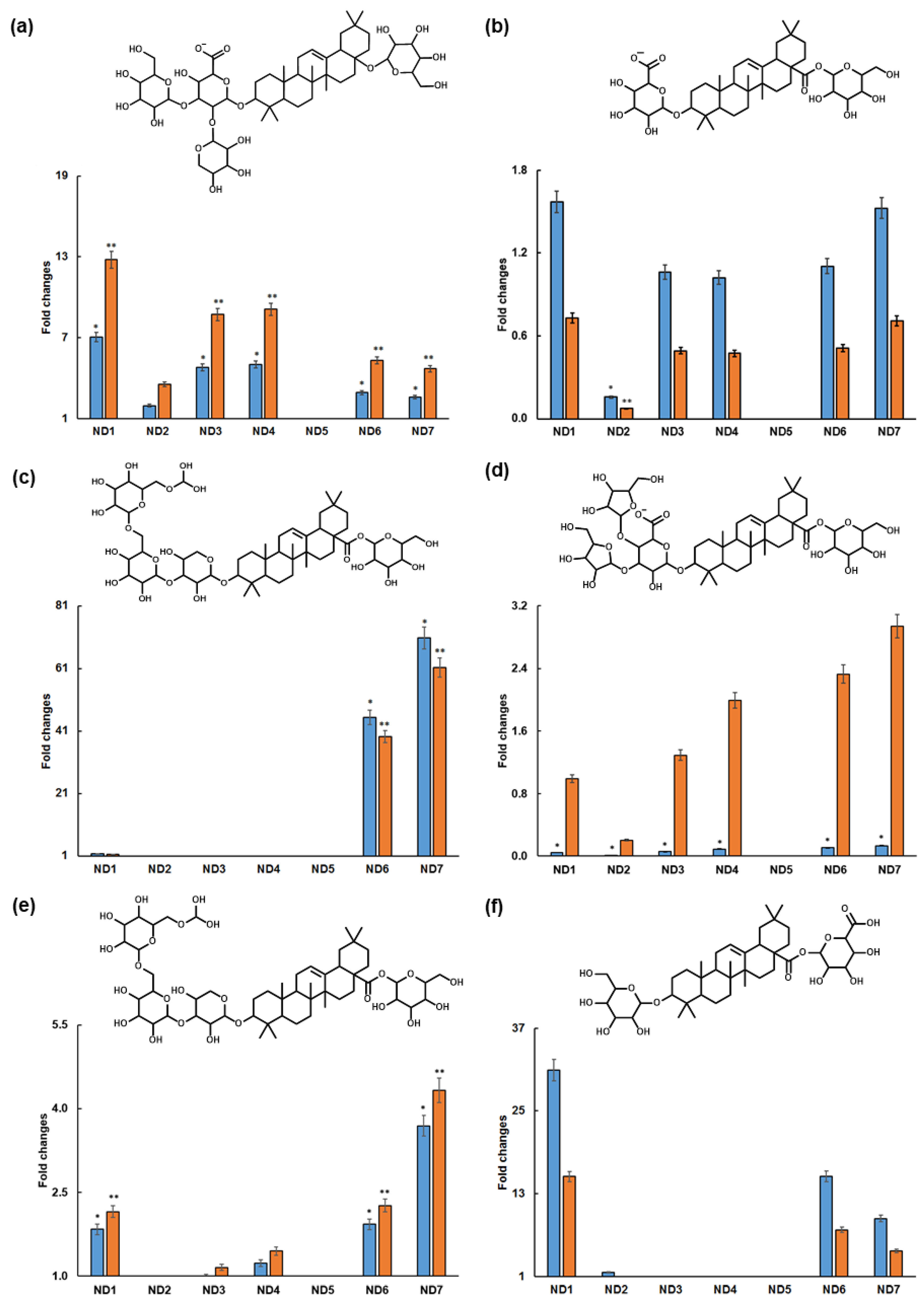
2.2. Extraction of Triterpene Saponins with NADES

The freshly prepared NADES, i.e., the mixtures of choline chloride, organic acids (malic acid, lactic acid and oxalic acid) and sugar alcohol (sorbitol) were employed for the extraction of A. elata roots. Thereby, choline chloride acted as hydrogen bond acceptors, whereas malic acid, lactic acid, oxalic acid and sorbitol were used as hydrogen bond donors [23,24]. While studying the mixture of sorbitol and malic acid, Dai and coworkers noted that sugars and organic acids can behave similarly to donors and acceptors of hydrogen bonds [23].
All the tested NADES proved to be applicable for the extraction of triterpene saponins from the roots of A. elata, although their efficiency as extractants differed. The maximal number of extracted metabolites is annotated by the ND1 (choline chloride–malic acid: 1:1) and ND3 (choline chloride–lactic acid: 1:3) extracts. However, although all twenty saponins could be identified both in the ND1 and ND3, the relative contents of the individual metabolites in the extract ND1 were higher than in the ND3. The typical chromatograms of the ND1 and ND3 extracts are presented in Figure 1 (for the whole tR range, please see Figure S15).
To address the relative recovery efficiencies of the individual triterpene saponins achieved with different NADES, we normalized them to the specific recoveries observed in the aqueous and ethanolic extracts. Thus, the triterpene recoveries obtained with all NADES were expressed as fold changes relative to those observed in the aqueous and ethanolic extracts of the A. elata roots and root parts (Figure 2 and Table S4). Thus, the fold change exceeding one indicated better recoveries obtained with NADES, i.e., better extraction efficiency was demonstrated by these extractants. As can be seen in Figure 2, some NADES were inefficient for some (ND2, ND3 and ND4) or all (ND5) metabolites addressed, as can be judged from the comparison of their recoveries with aqueous or ethanolic extracts.

3. Discussion

Interestingly, NADES proved to be more efficient solvents for the extraction of thirteen triterpene saponin metabolites than water or ethanol. The recoveries of 510, 1216, 18 and 19 (i.e., the relative contents of these compounds in corresponding extracts) were higher in NADES than in aqueous or ethanolic isolates. For example, NADES appeared to be more efficient than ethanol for the extraction of compound 10. The saponin 5 showed better recoveries using NADES than using water, while ethanol was a weaker extractant. The highest recoveries of compounds 6, 7, 1315 and 1819 were found when the 1:1 mixture of choline chloride and malic acid (ND1) was applied. On the other hand, compound 5 was better recovered using the 30% v/v aqueous 1:3 mixture of choline chloride and lactic acid (ND4), whereas compounds 810, 12 and 16 were better recovered using the 20% v/v aqueous 1:2 mixture of sorbitol and malic acid (ND7). Specifically, the relative recoveries of saponin 6 using the NADES extracts were higher than when using the ethanolic and aqueous extracts. Moreover, the recovery of compound 6 using ND1 was 13-fold higher than in the ethanolic extract and 9-fold higher than in the aqueous extract (Figure 2a). In contrast, the relative recoveries of compound 7 in NADES extracts appeared to be higher than in aqueous extracts, but not higher than ethanolic extracts, with the best effect observed using ND1 and ND7 (Figure 2b). The recoveries of saponin 9 using ND7 were 70-fold higher than the use of aqueous and 60-fold higher than the use of ethanolic extracts of A. elata (Figure 2c). The relative recoveries of compound 10 using NADES exceeded those when using ethanolic extracts, but not those when using the aqueous extracts (Figure 2d). This tendency was the clearest for ND7. The relative recoveries of compound 12 using NADES exceeded the contents of this compound recovered using ethanolic and aqueous extracts. This could be most clearly seen for ND7 (Figure 2e). On the other hand, the recoveries of triterpene saponin 13 were the highest using ND1 (Figure 2f). The pattern for compound 14 (araloside A isomer 1—Figure S16a) was similar to compound 9 (oleanolic acid-3-O-(methyldioxy-trihexopyranosyl-1-3-pentopyranosyl)-28-1-hexopyranosyl ester, Figure 2c); furthermore, the pattern for 18 (oleanolic acid-3-O-(hexosyl)-28-1-hexouronide ester isomer 2—Figure S16b) was similar to the pattern for the closely related structure of compound 12 (oleanolic acid-3-O-(hexosyl)-28-1-hexouronide ester isomer 1—Figure 2e). These similarities are explained by the similar structures of these compounds.
It is important to note that, despite the sufficient length of the LC gradient, some triterpene saponins co-eluted. Specifically, this was observed for compounds 1 and 2, 3 and 4, 17 and 18 and 6, 7, 8 and 9. In all cases, this chromatographic behavior can be explained by the similarities in the structures of their molecules (Figure 2).
Thus, the patterns of the NADES extraction activities, in respect to individual triterpene saponins, differed essentially (Figure 2), which can mostly be explained by the differences in analyte structures. Moreover, isomerism also impacts this phenomenon. For example, the recovery patterns of two isomers of calendulaglycosides C (Figure 2b,f) clearly differed from each other. It is also apparent that different types of NADES have different affinities for triterpene saponins of A. elata. Obviously, the structures of the individual constituents of NADES extracts, their ratio and their solvation degree directly affect the interactions between the molecules of solvents and target analytes [50,51].
In general, the efficiencies of NADES in the extraction of triterpene saponins from A. elata roots increased in the following order: water-free quaternary ammonium salt–dicarboxylic acid mixture (ND1) > 20% (v/v) aq. sugar–dicarboxylic acid (ND7) > 10% (v/v) aq. sugar–dicarboxylic acid (ND6) > 30% (v/v) aq. quaternary ammonium salt–monocarboxylic acid (ND4). Most likely, the observed high recoveries of triterpene saponins by NADES can be explained by close pH values of the NADES mixtures (2.26 for ND1 and 3.84 for ND4) to the pKa of oleanolic acid (4.74), the aglycon of all detected triterpene saponins.
Due to the fact that all of these mentioned above—ND3, 4, 6 and 7—showed much less viscosity in comparison to ND1 and ND5; the mixtures ND1, ND3 and ND7 were characterized with better mass transfer for triterpene natural products, which was most clearly seen for the mixtures based on malic acid or lactic acid. On the other hand, the solvent ND5 (based on the mixture of choline chloride and oxalic acid) showed weak extraction efficiency (Figure 2). Our results were in agreement with the studies of Lanjekar and coworkers, who demonstrated that choline chloride–lactic acid mixture (1:1 v/v) was efficient for the extraction of triterpene saponin glycyrrhizic acid from Glycyrrhiza glabra [41,52]. Furthermore, Suresh et al., 2022 showed that this NADES was also efficient in the extraction of steroidal saponins from Trillium govanianum [53]. Choline chloride–lactic acid mixture (1:2 v/v) proved to be efficient in the extraction of saponins from Acanthopanax senticosus [54]. Similar data for lactic acid-based NADES were obtained by Liu et al., 2023 for steroidal saponins from Polygonatum cyrtonema [55]. Increasing the relative contents of the aqueous component in NADES leads to better solvation of the polar analytes and, therefore, improved extractive capacity. The addition of water reduced viscosity. It promoted diffusion, while hydrogen bonds between the components weakened it [56]. It is noteworthy that for the extraction of kalopanax–Saponin F isomer 2 (6), calendulaglycoside C isomer 1 (7), calendulaglycoside C isomer 2 (13), araloside A isomer 1 (14), guaiacin B isomer 2 (15), oleanolic acid-3-O-(hexosyl)-28-1-hexouronide ester isomer 2 (18) and oleanolic acid-3-O-hexuronide-(1-3-pentafuranoside) (19), the most appropriate solvent was ND1 (choline chloride and malic acid in the ratio of 1:1). This solvent was also reported to be efficient for the extraction of ginsenoside Rb1 from Panax ginseng stems [57] and steroidal saponins from Dioscoreae nipponicae [40]. Other researchers remarked upon the efficiency of the acetylcholine chloride–malic acid–water mixture (ratio 1:2:2 v/v/v) for the extraction of triterpene saponins–madecassoside and asiaticoside from Centella asiatica [58].
It is important to note that the application of NADES not only gives access to a better efficiency of extraction, but also provides improved stabilities of the resulting extracts. On the other hand, the aqueous extracts are prone to microbial contamination and therefore have limited shelf life [24,34]. This might be underlined by the presence of additional hydroxyl and carboxyl groups in their structure of NADES, which are readily involved in the formation of hydrogen bonds with triterpene saponins [23,59].

4. Materials and Methods

4.1. Chemicals

Conventional solvents (purified water, ethanol 96%) were used for the comparison of efficiency of extraction. Unless stated otherwise, the materials were obtained from the following manufacturers: InnoGreenChem B.V. (Nijmegen, The Netherlands): lactic acid (80%); Merck KGaA (Darmstadt, Germany): acetonitrile (LC-MS grade), ammonium formate (99%, LC-MS grade), formic acid (98%, LC-MS grade), methanol (LC-MS grade) and water (LS-MS grade); NevaReactive (Saint Petersburg, Russia): choline chloride (≥99%) and oxalic acid (≥99%); sorbitol (≥99%) and malic acid (≥99%). Water was purified in-house with a water conditioning and purification system: GenPure Pro UV-TOC system (resistance 18 mΩ/cm, Thermo Fisher Scientific, Langenselbold, Germany).

4.2. Plant Material and Extraction Procedures

The roots of A. elata were collected in the Far East (Khabarovsk region) of Russia and the species identity was confirmed in the Department of Pharmacognosy of the Saint Petersburg State Chemical Pharmaceutical University (voucher of specimens MW0107420). All collected samples were dried without direct irradiation at room temperature. Whole roots (Y), isolated root bark (X) and root core (Z) were extracted using water, ethanol and different NADES. About 1.0 g of dried raw material (X, Y and Z) was extracted with 100 mL of hot boiling water with reflux for 2 h. Ethanol extracts were obtained by the extraction of 1.0 g of each part of raw material (X, Y and Z) with 100 mL of 96% (v/v) EtOH in Soxhlet. The resulting extracts were filtered, concentrated in a vacuum and freeze dried. Further, 10.0 mg of each lyophlisate was dissolved in 1 mL of purified water and centrifuged for 10 min at 10,000 rpm (4 °C), and the supernatant (180 µL) was analyzed by RP-UHPLC-QqTOF-MS.
All NADES were prepared by the heating and stirring method [23]. In detail, the mixtures of hydrogen bond donor and hydrogen bond acceptor with a certain amount of water (Table 2) were slightly heated (at the temperatures not exceeding 80 °C) with continuous gentle agitation before forming a clear transparent liquid (about 120 min) [23]. The composition of NADES (i.e., the composing solvents and their ratios) relied on the literature data [23,24] and our previous experiment.
Extraction using NADES was performed by maceration with continuous stirring. A total of 1.0 g of dried raw material and 40.0 g of NADES were transferred in flask. The mixture was stirred at 300 rpm for 60 min at 35 °C. The residue was filtered. Prior to metabolic profiling, NADES extracts were dissolved in water (in a ratio of 2:3) and placed in an ultrasonic bath for 15 min (35 kHz): 200 µL was centrifuged for 10 min at 10,000 rpm (4 °C). The supernatant (180 µL) was further analyzed using RP-UHPLC-QqTOF-MS.

4.3. Metabolite Analysis

The samples were analyzed using the randomization/standardization strategy by reversed phase–ultra-high-performance liquid chromatography–electrospray ionization–quadrupole-time-of-flight mass spectrometry (RP-UHPLC-ESI-QqTOF-MS) using a liquid chromatograph–mass spectrometer SHIMADZU LCMS-9030 System (Shimadzu Corporation, Kyoto, Japan) operated in negative ion mode. The data acquisition relied on the sequential window acquisition of all theoretical mass spectra (SWATH) mode. The chromatographic and mass spectrometric settings are specified in Supplementary Information S1 (Table S5). For the interpretation of the LC-MS data, Shimadzu LabSolution (Shimadzu Corporation, Kyoto, Japan), MSDial (version 3.12, free available via http://prime.psc.riken.jp/Metabolomics_Software/MS-DIAL/index2.html (accessed on 3 March 2023)), and manual mass spectra interpretation were used. The quantitation relied on the integration of the corresponding extracted ion chromatograms (XICs, m/z ± 0.025) at specific retention times (tR). The intensity matrix was generated after data processing in MSDial. Based on the intensities, the fold changes were calculated by the comparison of the amounts in the aqueous and ethanolic extracts. Data visualization was performed using Microsoft Excel 2016 (Supplementary Information S2 (Figure S17)).
For all features, which were annotated based on their [M-H] ions observed in the MS spectra (MS1 scans) of triterpene glycoside structures with a mass accuracy of better than 10 ppm but did not yield unambiguously interpretable fragmentation patterns in the first SWATH mode experiments (typically due to the uncertainty of the fragmentation patterns and simultaneous fragmentation of two or more intense m/z), additional SWATH and data-dependent acquisition (DDA) RP-UHPLC-MS/MS experiments were conducted using a TripleTOF 6600 mass spectrometer (AB Sciex, Darmstadt, Germany), using the LC conditions summarized in Supplementary Information S1 (Table S6). The nebulizer (GS1), drying (GS2) and curtain (CUR) gases were set to 60, 70 and 55 psig, while the ion spray voltage was set to −4500 V. The MS experiments were accomplished in the TOF-scan mode (the accumulation time was 50 or 75 ms in SWATH and DDA experiments, respectively) in the m/z range of 65–1250. The MS/MS conditions in the SWATH method were set as follows: each analysis was performed with 60 ms of accumulation time at a collision energy of −45 V with a collision energy spread (CES) of 35 V and a declustering potential (DP) of −35 V. The MS/MS conditions in the IDA method were set as follows: each analysis was performed with an accumulation of time 175 ms and a range of collision energy of −45 V with a collision energy spread (CES) of 35 V and a declustering potential (DP) of −35 V. Nitrogen was used as the CAD gas.

5. Conclusions

In this study, a total of twenty triterpene saponins (derivatives of oleanolic acid) were identified in the roots, root bark and root core of A. elata by RP-UHPLC-ESI-QqTOF-MS. Thereby, compounds 1, 3, 47, 13, 15 and 20 were identified in the roots of A. elata for, to the best of our knowledge, the first time. Furthermore, seven acid-based NADES were successfully applied for the extraction of triterpene saponins from the roots of A. elated for the first time. All of the tested NADES were able to extract triterpene saponins. The maximal number of triterpene saponins was identified using the ND1 (choline chloride with malic acid: 1:1) and ND3 (choline chloride with lactic acid: 1:3) extracts. Remarkably, NADES proved to be more efficient solvents than water or ethanol for the extraction of thirteen triterpene saponins. The relative recoveries of the compounds 510, 1216, 18 and 19 were higher using NADES than when using water or ethanol. For the extraction of compounds 610, 12, 15, 16, 18 and 19, the addressed NADES appeared to be more efficient than conventional solvents. Our data open the prospect of replacing aqueous and alcohols with non-toxic, “green” NADES for the extraction of A. elata.

Supplementary Materials

The following supporting information can be downloaded at: https://www.mdpi.com/article/10.3390/molecules28083614/s1. Supplementary Information S1—Table S1: the list of triterpene saponins predicted in different parts of A. elata based on literature mining. Table S2: triterpene saponins annotated in the extracts of A. elata roots by RP-HPLC-QqTOF-MS in untargeted SWATH experiments. Table S3: list of compounds confirmed by MS2 analysis in the roots of Aralia elata. Table S4: triterpene saponins annotated in the extracts of A. elata roots by RP-UHPLC-QqTOF-MS and MS/MS in SWATH and DDA experiments. Table S5: the conditions of RP-HPLC separation and the settings ESI-QqTOF-MS applied for the profiling of A. elata root semi-polar secondary metabolites. Table S6: the conditions of RP-UHPLC separation and the settings for ESI-QqTOF-MS applied for the SWATH and DDA MS/MS analysis of A. elata root semi-polar secondary metabolites. Figures—Figures S1–S14: spectral data of triterpene saponins annotated in the extracts of A. elata roots. Figure S15: full tR range in the chromatograms of ND1 and ND3 extracts of the whole roots of A. elata. Figure S16: structures and relative recoveries of 14 (araloside isomer 1, A) and 18 (oleanolic acid-3-O-(hexosyl)-28-1-hexouronide ester isomer 1, B), expressed as the difference (fold) in comparison to those observed in aqueous and ethanolic extracts. Supplementary Information S2—Table S7: results of untargeted metabolomic analysis of A. elata with MSDial software. Table S8: relative recoveries of triterpene saponins annotated in A. elata roots, extracted by conventional solvents and different types of NADES. Table S9: relative recoveries of individual triterpene saponins in aqueous, ethanolic and NADES extracts of A. elata whole roots, root core and bark. Figure S17: relative recovery diagrams of the triterpene saponins annotated in A. elata roots extracted with conventional solvents and different types of NADES. References [2,3,19,20,44,45,46,47,48,49,60,61,62,63,64,65,66,67,68,69,70,71,72,73,74,75,76,77,78,79,80,81,82,83,84,85,86,87] are cited in the supplementary materials.

Author Contributions

A.A.P. performed the literature mining and metabolite extraction, contributed to the quantitation and prepared the first manuscript draft; A.O. performed the interpretation of the mass spectra and conducted the structural annotation; N.F. performed the quantitative analysis and first interpretation of the extraction recovery data; E.B.S. and A.S. accomplished the UHPLC-MS and MS/MS analyses; E.V.F., A.F. and A.N.S. conceptualized the study, supervised the whole work and wrote the final draft. All authors have read and agreed to the published version of the manuscript.

Funding

Funding for this study was from the Russian Scientific Foundation: grant #21-76-10055 for the establishment of the LC-MS methodology and grant #21-74-30003 for the metabolite analysis and data interpretation.

Institutional Review Board Statement

Not applicable.

Informed Consent Statement

Not applicable.

Data Availability Statement

The data presented in this study are available on request from the corresponding authors.

Acknowledgments

We thank the Department of Pharmacognosy of the St. Petersburg State Chemical Pharmaceutical University and Centre for Molecular and Cell Technologies and Chemical Analysis and Materials Research Centre of Saint-Petersburg State University Research Park for technical support. The work was infrastructurally supported by the Ministry of Science and Higher Education of the Russian Federation (theme # 122042700043-9).

Conflicts of Interest

The authors declare no conflict of interest.

References

  1. Wen, J. New Taxa and Nomenclatural Changes in Aralia (Araliaceae). Novon 1994, 4, 400–403. [Google Scholar] [CrossRef]
  2. Xu, Y.; Liu, J.; Zeng, Y.; Jin, S.; Liu, W.; Li, Z.; Qin, X.; Bai, Y. Traditional Uses, Phytochemistry, Pharmacology, Toxicity and Quality Control of Medicinal Genus Aralia: A Review. J. Ethnopharmacol. 2022, 284, 114671. [Google Scholar] [CrossRef] [PubMed]
  3. Yoshikawa, M.; Yoshizumi, S.; Ueno, T.; Matsuda, H.; Murakami, T.; Yamahara, J.; Murakami, N. Medicinal Foodstuffs. I. Hypoglycemic Constituents from a Garnish Foodstuff “Taranome,” the Young Shoot of Aralia Elata SEEM.: Elatosides G, H, I, J, and K. Chem. Pharm. Bull. 1995, 43, 1878–1882. [Google Scholar] [CrossRef]
  4. Shikov, A.N.; Pozharitskaya, O.N.; Makarov, V.G. Aralia elata var. mandshurica (Rupr. & Maxim.) J. Wen: An overview of pharmacological studies. Phytomedicine 2016, 23, 1409–1421. [Google Scholar] [CrossRef]
  5. Panossian, A.G.; Efferth, T.; Shikov, A.N.; Pozharitskaya, O.N.; Kuchta, K.; Mukherjee, P.K.; Banerjee, S.; Heinrich, M.; Wu, W.; Guo, D.; et al. Evolution of the adaptogenic concept from traditional use to medical systems: Pharmacology of stress- and aging-related diseases. Med. Res. Rev. 2021, 41, 630–703. [Google Scholar] [CrossRef]
  6. Chernyak, D.M.; Titova, M.S. Anti-Stress Effect of the Far Eastern Plants. Pacific Med. J. 2014, 2, 28–30. [Google Scholar]
  7. Jung, C.; Lee, U. Pharmacological Studies on Root Bark Extract of Aralia elata—Antigastritic and Antiulcerative Effects in Rats. Yakhak Hoeji 1993, 37, 581–590. [Google Scholar]
  8. Hernandez, D.E.; Hancke, J.L.; Wikman, G. Evaluation of the Anti-Ulcer and Antisecretory Activity of Extracts of Aralia elata Root and Schizandra chinensis Fruit in the Rat. J. Ethnopharmacol. 1988, 23, 109–114. [Google Scholar] [CrossRef]
  9. Luo, Y.; Dong, X.; Yu, Y.; Sun, G.; Sun, X. Total Aralosides of Aralia elata (Miq) Seem (TASAES) Ameliorate Nonalcoholic Steatohepatitis by Modulating IRE1α-Mediated JNK and NF-ΚB Pathways in ApoE–/– Mice. J. Ethnopharmacol. 2015, 163, 241–250. [Google Scholar] [CrossRef]
  10. Saito, S.; Ebashi, J.; Sumita, S.; Furumoto, T.; Nagamura, Y.; Nishida, K.; Isiguro, I. Comparison of Cytoprotective Effects of Saponins Isolated from Leaves of Aralia elata SEEM. (Araliaceae) with Synthesized Bisdesmosides of Oleanoic Acid and Hederagenin on Carbon Tetrachloride-Induced Hepatic Injury. Chem. Pharm. Bull. 1993, 41, 1395–1401. [Google Scholar] [CrossRef]
  11. Ahumada, F.; Trincado, M.A.; Arellano, J.A.; Hancke, J.; Wikman, G. Effect of Certain Adaptogenic Plant Extracts on Drug-Induced Narcosis in Female and Male Mice. Phytother. Res. 1991, 5, 29–31. [Google Scholar] [CrossRef]
  12. Wojcicki, J.; Samochowiec, L.; Kadlubowska, D.; Lutomski, J. Studies on the Saponin Fraction from the Root of Aralia mandshurica Rupr. et Maxim. Part IV. Influence of the Saponosides on the Content of Lipids in Blood Serum and Liver in Experimental Hyperlipemia. Herba Pol. 1977, 23, 285–289. [Google Scholar]
  13. Liu, X.-H.; Li, X.-M.; Han, C.-C.; Fang, X.-F.; Ma, L. Effects of Combined Therapy with Glipizide and Aralia Root Bark Extract on Glycemic Control and Lipid Profiles in Patients with Type 2 Diabetes Mellitus: Effects of Aralia on Type 2 Diabetes Mellitus. J. Sci. Food Agric. 2015, 95, 739–744. [Google Scholar] [CrossRef] [PubMed]
  14. Wang, M.; Xu, X.; Xu, H.; Wen, F.; Zhang, X.; Sun, H.; Yao, F.; Sun, G.; Sun, X. Effect of the total saponins of Aralia elata (Miq) seem on cardiac contractile function and intracellular calcium cycling regulation. J. Ethnopharmacol. 2014, 155, 240–247. [Google Scholar] [CrossRef] [PubMed]
  15. State Register of Pharmaceutical Products for Medical Purposes. Available online: https://grls.rosminzdrav.ru/default.aspx (accessed on 9 November 2022).
  16. Lee, J.E.; Sim, S.J.; Jeong, W.; Choi, C.W.; Kim, N.; Park, Y.; Kim, M.-J.; Lee, D.; Hong, S.S. Diterpenoids and phenolic analogues from the roots of Aralia continentalis. J. Asian Nat. Prod. Res. 2021, 23, 371–378. [Google Scholar] [CrossRef]
  17. Sun, Y.; Li, B.; Lin, X.; Xue, J.; Wang, Z.; Zhang, H.; Jiang, H.; Wang, Q.; Kuang, H. Simultaneous Determination of Four Triterpene Saponins in Aralia elata Leaves by HPLC-ELSD Combined with hierarchical clustering analysis: Simultaneous determination of four triterpene saponins. Phytochem. Anal. 2017, 28, 202–209. [Google Scholar] [CrossRef]
  18. Xing, X.; Yan, M.; Zhang, X.; Yang, L.; Jiang, H. Quantitative analysis of triterpenes in different parts of Aralia elata (Miq.) seem using HPLC–ELSD and their inhibition of human umbilical vein endothelial cell ox-ldl-induced apoptosis. J. Liq. Chromatogr. Relat. Technol. 2017, 40, 984–990. [Google Scholar] [CrossRef]
  19. Zhang, Y.; Peng, Y.; Li, L.; Zhao, L.; Hu, Y.; Hu, C.; Song, S. Studies on cytotoxic triterpene saponins from the leaves of Aralia elata. Food Chem. 2013, 138, 208–213. [Google Scholar] [CrossRef]
  20. Han, F.; Liang, J.; Yang, B.-Y.; Kuang, H.-X.; Xia, Y.-G. Identification and comparison of triterpene saponins in Aralia elata leaves and buds by the energy-resolved MSAll technique on a liquid chromatography/quadrupole time-of-flight mass spectrometry. J. Pharm. Biomed. Anal. 2021, 203, 114176. [Google Scholar] [CrossRef]
  21. Choi, Y.H.; van Spronsen, J.; Dai, Y.; Verberne, M.; Hollmann, F.; Arends, I.W.C.E.; Witkamp, G.-J.; Verpoorte, R. Are natural deep eutectic solvents the missing link in understanding cellular metabolism and physiology? Plant Physiol. 2011, 156, 1701–1705. [Google Scholar] [CrossRef] [PubMed]
  22. Paiva, A.; Craveiro, R.; Aroso, I.; Martins, M.; Reis, R.L.; Duarte, A.R.C. Natural Deep Eutectic Solvents—Solvents for the 21st century. ACS Sustain. Chem. Eng. 2014, 2, 1063–1071. [Google Scholar] [CrossRef]
  23. Dai, Y.; van Spronsen, J.; Witkamp, G.-J.; Verpoorte, R.; Choi, Y.H. Natural Deep Eutectic Solvents as new potential media for green technology. Anal. Chim. Acta 2013, 766, 61–68. [Google Scholar] [CrossRef]
  24. Fuad, F.M.; Nadzir, M.M.; Harun, A. Hydrophilic Natural Deep Eutectic Solvent: A review on physicochemical properties and extractability of bioactive compounds. J. Mol. Liq. 2021, 339, 116923. [Google Scholar] [CrossRef]
  25. Ivanović, M.; Alañón, M.E.; Arráez-Román, D.; Segura-Carretero, A. Enhanced and Green Extraction of Bioactive Compounds from Lippia citriodora by Tailor-Made Natural Deep Eutectic Solvents. Food Res. Int. 2018, 111, 67–76. [Google Scholar] [CrossRef] [PubMed]
  26. Wu, Y.C.; Wu, P.; Li, Y.B.; Liu, T.C.; Zhang, L.; Zhou, Y.H. Natural Deep Eutectic Solvents as new green solvents to extract anthraquinones from Rheum palmatum L. RSC Adv. 2018, 8, 15069–15077. [Google Scholar] [CrossRef] [PubMed]
  27. Bosiljkov, T.; Dujmić, F.; Cvjetko Bubalo, M.; Hribar, J.; Vidrih, R.; Brnčić, M.; Zlatic, E.; Radojčić Redovniković, I.; Jokić, S. Natural Deep Eutectic Solvents and ultrasound-assisted extraction: Green approaches for extraction of wine lees anthocyanins. Food Bioprod. Process. 2017, 102, 195–203. [Google Scholar] [CrossRef]
  28. Alrugaibah, M.; Yagiz, Y.; Gu, L. Use Natural Deep Eutectic Solvents as efficient green reagents to extract procyanidins and anthocyanins from cranberry pomace and predictive modeling by RSM and artificial neural networking. Sep. Purif. Technol. 2021, 255, 117720. [Google Scholar] [CrossRef]
  29. He, X.; Yang, J.; Huang, Y.; Zhang, Y.; Wan, H.; Li, C. Green and efficient ultrasonic-assisted extraction of bioactive components from Salvia miltiorrhiza by Natural Deep Eutectic Solvents. Molecules 2019, 25, 140. [Google Scholar] [CrossRef]
  30. Torres-Vega, J.; Gómez-Alonso, S.; Pérez-Navarro, J.; Alarcón-Enos, J.; Pastene-Navarrete, E. Polyphenolic compounds extracted and purified from Buddleja globosa Hope (Buddlejaceae) leaves using Natural Deep Eutectic Solvents and centrifugal partition chromatography. Molecules 2021, 26, 2192. [Google Scholar] [CrossRef]
  31. Shang, X.; Dou, Y.; Zhang, Y.; Tan, J.-N.; Liu, X.; Zhang, Z. Tailor-made Natural Deep Eutectic Solvents for green extraction of isoflavones from Chickpea (Cicer arietinum L.) Sprouts. Ind. Crops Prod. 2019, 140, 111724. [Google Scholar] [CrossRef]
  32. Nadhira, A.; Febianli, D.; Fransisca, F.; Mun’Im, A.; Aryati, W.D. Natural Deep Eutectic Solvents ultrasound-assisted extraction (NADES-UAE) of trans-cinnamaldehyde and coumarin from cinnamon bark [Cinnamomum burmannii (Nees T. Nees) Blume]. JRP 2020, 24, 389–398. [Google Scholar] [CrossRef]
  33. Obluchinskaya, E.D.; Daurtseva, A.V.; Pozharitskaya, O.N.; Flisyuk, E.V.; Shikov, A.N. Natural Deep Eutectic Solvents as alternatives for extracting phlorotannins from brown algae. Pharm. Chem. J. 2019, 53, 243–247. [Google Scholar] [CrossRef]
  34. Obluchinskaya, E.D.; Pozharitskaya, O.N.; Zakharova, L.V.; Daurtseva, A.V.; Flisyuk, E.V.; Shikov, A.N. Efficacy of Natural Deep Eutectic Solvents for extraction of hydrophilic and lipophilic compounds from Fucus vesiculosus. Molecules 2021, 26, 4198. [Google Scholar] [CrossRef]
  35. Patil, S.S.; Pathak, A.; Rathod, V.K. Optimization and kinetic study of ultrasound Assisted Deep Eutectic Solvent based extraction: A greener route for extraction of curcuminoids from Curcuma longa. Ultrason. Sonochem. 2021, 70, 105267. [Google Scholar] [CrossRef] [PubMed]
  36. Wils, L.; Leman-Loubière, C.; Bellin, N.; Clément-Larosière, B.; Pinault, M.; Chevalier, S.; Enguehard-Gueiffier, C.; Bodet, C.; Boudesocque-Delaye, L. Natural Deep Eutectic Solvent formulations for spirulina: Preparation, intensification, and skin impact. Algal Res. 2021, 56, 102317. [Google Scholar] [CrossRef]
  37. Fang, X.; Li, Y.; Kua, Y.L.; Chew, Z.L.; Gan, S.; Tan, K.W.; Lee, T.Z.E.; Cheng, W.K.; Lau, H.L.N. Insights on the Potential of Natural Deep Eutectic Solvents (NADES) to Fine-Tune Durian Seed Gum for Use as Edible Food Coating. Food Hydrocoll. 2022, 132, 107861. [Google Scholar] [CrossRef]
  38. Li, Y.; Pan, Z.; Wang, B.; Yu, W.; Song, S.; Feng, H.; Zhao, W.; Zhang, J. Ultrasound-assisted extraction of bioactive alkaloids from Phellodendri amurensis cortex using Deep Eutectic Solvent aqueous solutions. New J. Chem. 2020, 44, 9172–9178. [Google Scholar] [CrossRef]
  39. Naseem, Z.; Zahid, M.; Hanif, M.; Shahid, M. Environmentally friendly extraction of bioactive compounds from Mentha arvensis using Deep Eutectic Solvent as green extraction media. Pol. J. Environ. Stud. 2020, 29, 3749–3757. [Google Scholar] [CrossRef]
  40. Yang, G.-Y.; Song, J.-N.; Chang, Y.-Q.; Wang, L.; Zheng, Y.-G.; Zhang, D.; Guo, L. Natural deep eutectic solvents for the extraction of bioactive steroidal saponins from Dioscoreae Nipponicae Rhizoma. Molecules 2021, 26, 2079. [Google Scholar] [CrossRef]
  41. Lanjekar, K.J.; Rathod, V.K. Green extraction of Glycyrrhizic acid from Glycyrrhiza glabra using choline chloride based Natural Deep Eutectic Solvents (NADESs). Process Biochem. 2021, 102, 22–32. [Google Scholar] [CrossRef]
  42. Xia, Y.-G.; Liang, J.; Li, G.-Y.; Yang, B.-Y.; Kuang, H.-X. Energy-Resolved Technique for Discovery and Identification of Malonyl-Triterpene Saponins in Caulophyllum robustum by UHPLC-Electrospray Fourier Transform Mass Spectrometry: Liquid Chromatography-Mass Spectrometry. J. Mass Spectrom. 2016, 51, 947–958. [Google Scholar] [CrossRef] [PubMed]
  43. Chernushevich, I.V.; Thomson, B.A. Collisional Cooling of Large Ions in Electrospray Mass Spectrometry. Anal. Chem. 2004, 76, 1754–1760. [Google Scholar] [CrossRef] [PubMed]
  44. Ahmad, V.U.; Perveen, S.; Bano, S. Guaiacin A and B from the Leaves of Guaiacum officinale. Planta Med. 1989, 55, 307–308. [Google Scholar] [CrossRef] [PubMed]
  45. Shao, C.-J.; Kasai, R.; Xu, J.-D.; Tanaka, O. Saponins from Roots of Kalopanax septemlobus (Thunb.) Koidz., Ciqiu: Structures of Kalopanax-Saponins C, D, E and F. Chem. Pharm. Bull. 1989, 37, 311–314. [Google Scholar] [CrossRef]
  46. Kasprzyk, Z.; Wojciechowski, Z. The Structure of Triterpenic Glycosides from the Flowers of Calendula officinalis L. Phytochemistry 1967, 6, 69–75. [Google Scholar] [CrossRef]
  47. Yen, P.H.; Cuc, N.T.; Huong, P.T.T.; Nhiem, N.X.; Hong Chuong, N.T.; Lien, G.T.K.; Huu Tai, B.; Tuyen, N.V.; Van Minh, C.; Van Kiem, P. Araliaarmoside: A New Triterpene Glycoside Isolated from the Leaves of Aralia armata. Nat. Prod. Commun. 2020, 15, 1–5. [Google Scholar] [CrossRef]
  48. Kochetkov, N.K.; Khorlin, A.J.; Vaskovsky, V.E. The Structures of Aralosides A and B. Tetrahedron Lett. 1962, 3, 713–716. [Google Scholar] [CrossRef]
  49. Song, S.; Nakamura, N.; Ma, C.; Hattori, M.; Xu, S. Four New Saponins from the Root Bark of Aralia elata. Chem. Pharm. Bull. 2000, 48, 838–842. [Google Scholar] [CrossRef]
  50. Ali, M.C.; Chen, J.; Zhang, H.; Li, Z.; Zhao, L.; Qiu, H. Effective Extraction of Flavonoids from Lycium barbarum L. Fruits by Deep Eutectic Solvents-Based Ultrasound-Assisted Extraction. Talanta 2019, 203, 16–22. [Google Scholar] [CrossRef]
  51. Maimulyanti, A.; Nurhidayati, I.; Mellisani, B.; Amelia Rachmawati Putri, F.; Puspita, F.; Restu Prihadi, A. Development of Natural Deep Eutectic Solvent (NADES) Based on Choline Chloride as a Green Solvent to Extract Phenolic Compound from Coffee Husk Waste. Arab. J. Chem. 2023, 16, 104634. [Google Scholar] [CrossRef]
  52. Lanjekar, K.J.; Rathod, V.K. Application of Ultrasound and Natural Deep Eutectic Solvent for the Extraction of Glycyrrhizic Acid from Glycyrrhiza glabra: Optimization and Kinetic Evaluation. Ind. Eng. Chem. Res. 2021, 60, 9532–9538. [Google Scholar] [CrossRef]
  53. Suresh, P.S.; Singh, P.P.; Anmol; Kapoor, S.; Padwad, Y.S.; Sharma, U. Lactic Acid-Based Deep Eutectic Solvent: An Efficient Green Media for the Selective Extraction of Steroidal Saponins from Trillium govanianum. Sep. Purif. Technol. 2022, 294, 121105. [Google Scholar] [CrossRef]
  54. Shi, X.; Yang, Y.; Ren, H.; Sun, S.; Mu, L.T.; Chen, X.; Wang, Y.; Zhang, Y.; Wang, L.H.; Sun, C. Identification of Multiple Components in Deep Eutectic Solvent Extract of Acanthopanax senticosus Root by Ultra-High-Performance Liquid Chromatography with Quadrupole Orbitrap Mass Spectrometry. Phytochem. Lett. 2020, 35, 175–185. [Google Scholar] [CrossRef]
  55. Liu, G.; Feng, S.; Sui, M.; Chen, B.; Sun, P. Extraction and Identification of Steroidal Saponins from Polygonatum cyrtonema Hua Using Natural Deep Eutectic Solvent-synergistic Quartz Sand Assisted Extraction Method. J. Sep. Sci. 2023, 2200823. [Google Scholar] [CrossRef] [PubMed]
  56. Dai, Y.; Witkamp, G.-J.; Verpoorte, R.; Choi, Y.H. Tailoring properties of Natural Deep Eutectic Solvents with water to facilitate their applications. Food Chem. 2015, 187, 14–19. [Google Scholar] [CrossRef]
  57. Liu, X.; Ahlgren, S.; Korthout, H.A.A.J.; Salomé-Abarca, L.F.; Bayona, L.M.; Verpoorte, R.; Choi, Y.H. Broad Range Chemical Profiling of Natural Deep Eutectic Solvent Extracts Using a High Performance Thin Layer Chromatography–Based Method. J. Chromatogr. A 2018, 1532, 198–207. [Google Scholar] [CrossRef]
  58. Phaisan, S.; Makkliang, F.; Putalun, W.; Sakamoto, S.; Yusakul, G. Development of a Colorless Centella asiatica (L.) Urb. Extract Using a Natural Deep Eutectic Solvent (NADES) and Microwave-Assisted Extraction (MAE) Optimized by Response Surface Methodology. RSC Adv. 2021, 11, 8741–8750. [Google Scholar] [CrossRef]
  59. Dai, Y.; Verpoorte, R.; Choi, Y.H. Natural Deep Eutectic Solvents Providing Enhanced Stability of Natural Colorants from Safflower (Carthamus tinctorius). Food Chem. 2014, 159, 116–121. [Google Scholar] [CrossRef]
  60. Wang, Q.-H.; Zhang, J.; Ma, X.; Ye, X.-Y.; Yang, B.-Y.; Xia, Y.-G.; Kuang, H.-X. A New Triterpenoid Saponin from the Leaves of Aralia Elata. Chin. J. Nat. Med. 2011, 9, 17–21. [Google Scholar] [CrossRef]
  61. Jiang, Y.T.; Xu, S.X.; Gu, X.H.; Ren, L.; Chen, Y.J.; Yao, X.S.; Miao, Z.C. Studies on the chemical constituents from Aralia elata. Yao Xue Xue Bao 1992, 27, 528–532. [Google Scholar]
  62. Lin, G.; Yang, J.-s. Studies on the Chemical Constituents of Aralia decaisneane II. Chin. Pharm. J. Beijing 2004, 39, 575–578. [Google Scholar]
  63. Miyase, T.; Shiokawa, K.-I.; Zhang, D.M.; Ueno, A. Araliasaponins I–XI, Triterpene Saponins from the Roots of Aralia decaisneana. Phytochemistry 1996, 41, 1411–1418. [Google Scholar] [CrossRef] [PubMed]
  64. Miyase, T.; Sutoh, N.; Zhang, D.M.; Ueno, A. Araliasaponins XII–XVIII, Triterpene Saponins from the Roots of Aralia chinensis. Phytochemistry 1996, 42, 1123–1130. [Google Scholar] [CrossRef] [PubMed]
  65. Gao, R.; Liao, M.; Huang, X.; Chen, Y.; Yang, G.; Li, J. Six New Triterpene Derivatives from Aralia chinensis var. dasyphylloides. Molecules 2016, 21, 1700. [Google Scholar] [CrossRef]
  66. Sun, W.J.; Zhang, D.K.; Sha, Z.F.; Zhang, H.L.; Zhang, X.L. Studies on the Saponins from the Root Bark of Aralia chinensis L. Acta Pharm. Sin. 1991, 26, 197–202. [Google Scholar]
  67. Yu, S.-S.; Yu, D.-Q.; Liang, X.-T. Triterpenoid Saponins from the Roots of Aralia spinifolia. J. Nat. Prod. 1994, 57, 978–982. [Google Scholar] [CrossRef]
  68. Yen, P.H.; Chuong, N.T.H.; Lien, G.T.K.; Cuc, N.T.; Nhiem, N.X.; Thanh, N.T.V.; Tai, B.H.; Seo, Y.; Namkung, W.; Park, S. Oleanane-Type Triterpene Saponins from Aralia armata Leaves and Their Cytotoxic Activity. Nat. Prod. Res. 2021, 36, 142–149. [Google Scholar] [CrossRef]
  69. Zaki, A.A.; Qiu, L. Machaerinic Acid 3-O-β-D-Glucuronopyranoside from Calendula officinalis. Nat. Prod. Res. 2020, 34, 2938–2944. [Google Scholar] [CrossRef]
  70. Liang, X.F.; Zhao, Y.Y.; Liu, X.Z.; Yang, X.J.; Fan, Y.; Guo, D.Y.; Song, X.M.; Song, B. Isolation and Identification of Chemical Constituents from Aralia Taibaiensis Cortex. Chin. J. Exp. Tradit. Med. Formulae 2018, 20, 56–61. [Google Scholar]
  71. Yoshikawa, M.; Harada, E.; Matsuda, H.; Murakami, T.; Yamahara, J.; Murakami, N. Elatosides A and B, Potent Inhibitors of Ethanol Absorption in Rats from the Bark of Aralia elata SEEM.: The Structure-Activity Relationships of Oleanolic Acid Oligoglycosides. Chem. Pharm. Bull. 1993, 41, 2069–2071. [Google Scholar] [CrossRef]
  72. Song, S.-J.; Nakamura, N.; Ma, C.-M.; Hattori, M.; Xu, S.-X. Five Saponins from the Root Bark of Aralia elata. Phytochemistry 2001, 56, 491–497. [Google Scholar] [CrossRef] [PubMed]
  73. Guo, M.; Zhang, L.; Liu, Z. Analysis of Saponins from Leaves of Aralia elata by Liquid Chromatography and Multi-Stage Tandem Mass Spectrometry. Anal. Sci. 2009, 25, 753–755. [Google Scholar] [CrossRef]
  74. Shuyu, C.; Xianggao, L.; Chongxi, Z. Chemical components of Aralia elata. J. Jilin Agric. Univ. 1992, 14, 29–32. [Google Scholar]
  75. Ma, Z.; Song, S.; Xu, S. Two new saponins from Aralia elata (Miq.) see. Chin. J. Med. Chem. 2004, 14, 47–48. [Google Scholar]
  76. Kuang, H.-X.; Sun, H.; Zhang, N.; Okada, Y.; Okuyama, T. Two New Saponins, Congmuyenosides A and B, from the Leaves of Aralia elata Collected in Heilongjiang, China. Chem. Pharm. Bull. 1996, 44, 2183–2185. [Google Scholar] [CrossRef] [PubMed]
  77. Kim, J.S.; Shim, S.H.; Chae, S.; Han, S.J.; Kang, S.S.; Son, K.H.; Chang, H.W.; Kim, H.P.; Bae, K. Saponins and Other Constituents from the Leaves of Aralia elata. Chem. Pharm. Bull. 2005, 53, 696–700. [Google Scholar] [CrossRef]
  78. Xi, F.-M.; Li, C.-T.; Han, J.; Yu, S.-S.; Wu, Z.-J.; Chen, W.-S. Thiophenes, Polyacetylenes and Terpenes from the Aerial Parts of Eclipata prostrata. Bioorg. Med. Chem. 2014, 22, 6515–6522. [Google Scholar] [CrossRef]
  79. Matsuo, Y.; Watanabe, K.; Mimaki, Y. Triterpene Glycosides from the Underground Parts of Caulophyllum thalictroides. J. Nat. Prod. 2009, 72, 1155–1160. [Google Scholar] [CrossRef]
  80. Nhiem, N.X.; Lim, H.Y.; Kiem, P.V.; Minh, C.V.; Thu, V.K.; Tai, B.H.; Quang, T.H.; Song, S.B.; Kim, Y.H. Oleanane-Type Triterpene Saponins from the Bark of Aralia elata and Their NF-ΚB Inhibition and PPAR Activation Signal Pathway. Bioorg. Med. Chem. Lett. 2011, 21, 6143–6147. [Google Scholar] [CrossRef]
  81. Tian, Y.; Zhang, X.; Liu, H.; Gong, D.; Li, X. Comparison of the Nutritional and Phytochemical Composition and Antioxidant Activities of Aralia elata (Miq.) Seem Fruits in Northeast China. Arab. J. Chem. 2021, 14, 103448. [Google Scholar] [CrossRef]
  82. Sakai, S.; Katsumata(Nee Ohtsuka), M.; Satoh, Y.; Nagasao, M.; Miyakoshi, M.; Ida, Y.; Shoji, J. Oleanolic Acid Saponins from Root Bark of Aralia elata. Phytochemistry 1994, 35, 1319–1324. [Google Scholar] [CrossRef] [PubMed]
  83. Kuljanabhagavad, T.; Thongphasuk, P.; Chamulitrat, W.; Wink, M. Triterpene Saponins from Chenopodium quinoa Willd. Phytochemistry 2008, 69, 1919–1926. [Google Scholar] [CrossRef] [PubMed]
  84. Waffo-Téguo, P.; Voutquenne, L.; Thoison, O.; Dumontet, V.; Nguyen, V.H.; Lavaud, C. Acetylated Glucuronide Triterpene Bidesmosidic Saponins from Symplocos glomerata. Phytochemistry 2004, 65, 741–750. [Google Scholar] [CrossRef]
  85. Yoshikawa, M.; Murakami, T.; Harada, E.; Murakami, N.; Yamahara, J.; Matsuda, H. Bioactive Saponins and Glycosides. VI. Elatosides A and B, Potent Inhibitors of Ethanol Absorption, from the Bark of Aralia elata SEEM. (Araliaceae): The Structure-Requirement in Oleanolic Acid Glucuronide-Saponins for the Inhibitory Activity. Chem. Pharm. Bull. 1996, 44, 1915–1922. [Google Scholar] [CrossRef] [PubMed]
  86. Tang, H.F.; Yi, Y.H.; Wang, Z.Z.; Jiang, Y.P.; Li, Y.Q. Oleanolic acid saponins from the root bark of Aralia taibaiensis. Yao Xue Xue Bao 1997, 32, 685–690. [Google Scholar] [PubMed]
  87. Miyase, T.; Kohsaka, H.; Ueno, A. Tragopogonosides A-I, Oleanane Saponins from Tragopogon pratensis. Phytochemistry 1992, 31, 2087–2091. [Google Scholar] [CrossRef]
Figure 1. Total ion chromatograms (TICs) of the whole root extracts of A. elata prepared with the NADES, with the mixture of choline chloride and malic acid being 1:1 (ND1: green) and the mixture of choline chloride and lactic acid being 1:3 (ND3: blue). The analytes are listed as in Table 1.
Molecules 28 03614 g001
Figure 2. Structures and relative recoveries of 6 (kalopanax–saponin F isomer 2, (a)), 7 (calendulaglycoside C isomer 1, (b)), 9 (oleanolic acid-3-O-(methyldioxy-trihexopyranosyl-1-3-pentopyranosyl)-28-1-hexopyranosyl ester, (c)), 10 (araloside (b,d)), 12 (oleanolic acid-3-O-(hexosyl)-28-1-hexouronide ester isomer 1, (e)) and 13 (calendulaglycoside C isomer 2, (f)) expressed as the ratio (fold) in comparison to those observed in aqueous (blue) and ethanolic (orange) extracts. The compounds are numbered as in Table 1. ND1—NADES based on the choline chloride–malic acid mixture (molar ratio 1:1). ND2—NADES with the molar ratio of choline chloride and malic acid of 1:2. ND3—NADES with the molar ratio of choline chloride and lactic acid of 1:3. ND4—NADES with the molar ratio of choline chloride and lactic acid of 1:3 + 30% (v/v) water. ND6—NADES with the molar ratio of sorbitol and malic acid of 1:1 + 10% (v/v) water. ND7—NADES with the molar ratio of sorbitol and malic acid of 1:2 + 20% (v/v) water. *—the difference is that the recovery of the compound is statistically significant compared to the recovery of the compound in water extract (p ≤ 0.05); **—the difference is that the recovery of the compound is statistically significant compared to the recovery of the compound in ethanol extract (p ≤ 0.05).
Molecules 28 03614 g002
Table 1. Targeted metabolites annotated in different parts of the root of A. elata var. mandshurica (Rupr. & Maxim.) J. Wen extracted using conventional and natural deep eutectic solvents by reversed phase–ultra-high-performance liquid chromatography–mass spectrometry in (RP-UHPLC-QqTOF-MS) in the negative ion mode.
No.tR
(min)
m/z
[M−H]
Observed
m/z
[M−H]
Calculated
Elemental Composition [M−H]MS2 Fragmentation Patterns-Product Ions, m/z (Rel. Intensity)Δm (ppm)AssignmentPlant PartSolventRef.Suppl. Spectra
13.8911.4993911.5010C47H75O17455.3507 (20), 617.4041 (15), 749.4472 (15), 911.4993 (100)1.9Guaiacin B isomer 1X,Y,Z 1W,E,ND1,2,3,4,6,7 2[44]Figure S1
23.81235.61071235.6066C59H95O27455.3522 (50), 617.4059 (15), 749.4492 (100), 911.5035 (10), 1235.6107 (10)−3.3Oleanolic acid-3-O-
(triglucopyranosyl-1-3-
arabinopyranosyl)-28-1-
glucopyranosyl
XW,E,ND1,2,3,4 Figure S2
YW,E,ND1,2,3,4,6,7
ZW,E,ND1,3,4,6,7
33.91087.53171087.5331C53H83O23455.3509 (10), 701.4265 (5), 925.4814 (15), 1087.5317 (100)1.3Kalopanax-Saponin F
isomer 1
XW,E, ND1,2,3,4,6,7[45]Figure S3
YAll
ZW,E,ND1,3,4,6,7
43.91117.53901117.5436C54H85O24455.3499 (3), 731.4347 (5), 955.4898 (10), 1117.5390 (100)4.1Calendulaglycoside AX,ZW,E,ND1,2,3,4,6,7[46]Figure S4
YAll
54.01249.58691249.5859C59H93O28455.3487 (5), 701.4254 (15), 925.4743 (7), 1057.5219 (80), 1087.5337 (50), 1153.5538 (100), 1249.5869 (70)−0.8AraliaarmosideX,Y,ZW,E,ND1,2,3,4,6,7[47]Figure S5
64.31087.53361087.5331C53H83O23455.3509 (10), 701.4265 (5), 925.4814 (15), 1087.55336 (100)−0.5Kalopanax-Saponin F
isomer 2
X,ZW,E,ND1,2,3,4,6,7[45]Figure S3
YAll
74.3955.4859955.4908C48H75O19455.3511 (5), 569.379 (5), 793.4317 (15), 955.4859 (100)5.1Calendulaglycoside C
isomer 1
X,Y,ZW,E,ND1,2,3,4,6,7[46]Figure S6
84.31073.55691073.5538C53H85O22455.3518 (50), 617.4052 (20), 749.4485 (30), 911.5022 (100), 1073.5569 (15)−2.9Oleanolic acid-3-O-
(diglucopyranosyl-1-3-
arabinopyranosyl)-28-1-
glucopyranosyl ester
XE,ND1,2,3,4,6,7 Figure S7
YE,ND1,2,3,6,7
ZND1,3,6,7
94.31119.56061119.5616C54H87O24455.3521 (5), 617.4057 (5), 749.4490 (7), 911.5019 (100), 1073.5551 (15), 1119.5606 (13)0.9Oleanolic acid-3-O-
(methyldioxy-trihexopyranosyl-1-3-
pentopyranosyl)-28-1-
hexopyranosyl ester
X,YW,E,ND1,2,3,4,6,7 Figure S8
ZW,E,ND1,3,4,6,7
104.41057.52491057.5225C52H81O22455.3517 (5), 701.4286 (7), 763.4309 (5), 895.4698 (7), 1057.5249 (100)−2.3Araloside BX,Y,ZW,E,ND1,2,3,4,6,7[48]Figure S9
114.51089.54931089.5487C53H85O23455.3501 (5), 719.4360 (10), 881.4901 (100), 1043.5437 (12), 1089.5493 (12)−0.6Araliasaponin IIIX,ZW,E,ND1,2,3,4,6,7[49]Figure S10
YW,E,ND1,3,4,6,7
124.6793.4312793.4380C42H65O14455.3471 (10), 631.3785 (15), 793.4312 (100)8.6Oleanolic acid-3-O-
(hexosyl)-28-1-hexouronide
ester isomer 1
X,ZW,E,ND1,2,3,4,6,7 Figure S11
YAll
135.1955.4899955.4908C48H75O19455.3511 (5), 569.379 (5), 793.4317 (15), 955.4899 (100)0.9Calendulaglycoside C isomer 2X,Y,ZW,E,ND1,2,3,4,6,7[46]Figure S6
145.3925.4805925.4802C47H73O18455.3508 (2), 569.3831 (5), 731.4366 (20), 925.4803 (100)0.3Araloside A isomer 1X,ZW,E,ND1,2,3,4,6,7[48]Figure S12
YAll
155.4911.5016911.5010C47H75O17455.3507 (20), 617.4041 (15), 749.4472 (15), 911.5016 (100)−0.7Guaiacin B isomer 2X,ZW,E,ND1,2,3,4,6,7[44]Figure S1
YAll
165.5925.4807925.4802C47H73O18455.3508 (2), 569.3831 (5), 731.4366 (20), 925.4803 (100)0.5Araloside A isomer 2X,ZW,E,ND1,2,3,4,6,7[48]Figure S12
YAll
175.6895.4696--455.3508 (5), 551.3731 (5), 895.4696 (100) Oleanolic acid unknown derivativesX,Y,ZW,E,ND1,2,3,4,6,7 Figure S13
185.6793.4360793.4380C42H65O14455.3471 (10), 631.3785 (15), 793.4312 (100)2.5Oleanolic acid-3-O-
(hexosyl)-28-1-hexouronide
ester isomer 2
X,ZW,E,ND1,2,3,4,6,7 Figure S11
YAll
195.7763.4260763.4274C41H63O13455.3502 (3), 631.3822 (5), 763.4260 (100)1.8Oleanolic acid 3-O-
hexuronide-(1-3-
pentafuranoside)
X,ZW,E,ND1,2,3,4,6,7 Figure S14
YAll
205.8911.4949911.5010C47H75O17455.3507 (20), 617.4041 (15), 749.4472 (15), 911.4949 (100)6.7Guaiacin B isomer 3X,Y,ZW,E,ND1,2,3,4,6,7[44]Figure S1
1 X—root bark. Y—whole root. Z—core of root. 2 W—water. E—ethanol. ND1—NADES with choline chloride and malic acid (molar ratio 1:1). ND2—NADES with the molar ratio of choline chloride and malic acid of 1:2. ND3—NADES with the molar ratio of choline chloride and lactic acid of 1:3. ND4—NADES with the molar ratio of choline chloride and lactic acid of 1:3 + 30% (v/v) water. ND6—NADES with the molar ratio of sorbitol and malic acid of 1:1 + 10% (v/v) water. ND7—NADES with the molar ratio of sorbitol and malic acid of 1:2 + 20% (v/v) water. All—metabolites were found in all extracts. The annotated metabolites (listed in Table 1) were quantified on the relative basis in the aqueous and ethanolic extracts of A. elata whole roots, bark and core. For this purpose, extracted ion chromatograms (XICs, m/z ± 0.02) were built for their [M-H] signals and integrated at characteristic tRs. The primary results of the bioinformatic analysis and peak integration are summarized in Supplementary Information S2 (Tables S7–S9). The analysis revealed the compounds 1, 2, 6 and 15 as being the most abundant in root bark, while metabolites 25, 712, 14, 1619 and 20 dominated in the core of roots.
Table 2. Composition of NADES [23,24].
CodeComponent 1Component 2Molar RatioAmount of Water (% (v/v))
ND1Choline chlorideMalic acid1:1-
ND2Choline chlorideMalic acid1:2-
ND3Choline chlorideLactic acid1:3-
ND4Choline chlorideLactic acid1:330
ND5Choline chlorideOxalic acid1:115
ND6SorbitolMalic acid1:110
ND7SorbitolMalic acid1:220
Disclaimer/Publisher’s Note: The statements, opinions and data contained in all publications are solely those of the individual author(s) and contributor(s) and not of MDPI and/or the editor(s). MDPI and/or the editor(s) disclaim responsibility for any injury to people or property resulting from any ideas, methods, instructions or products referred to in the content.

