Article

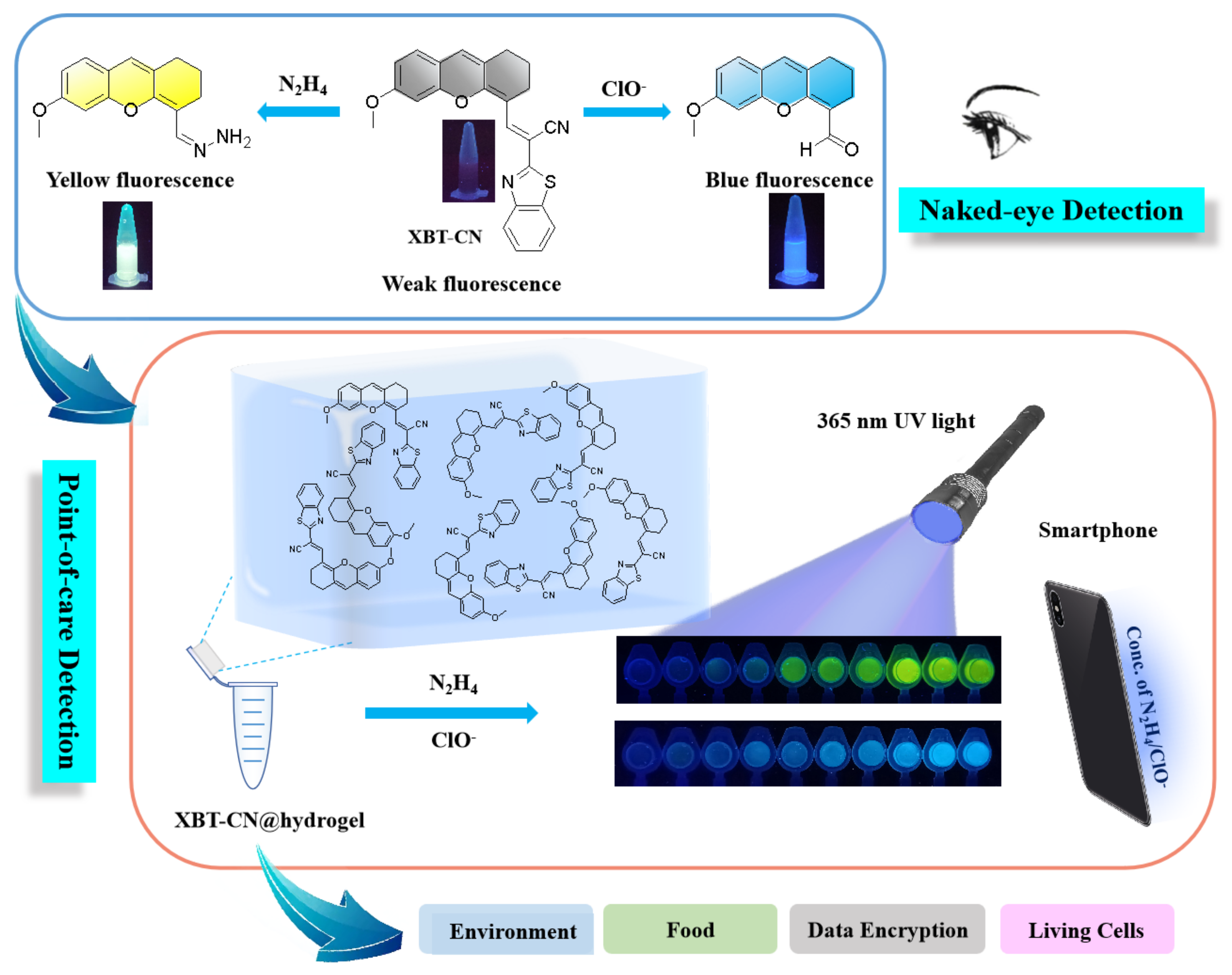
Point-of-Care and Dual-Response Detection of Hydrazine/Hypochlorite-Based on a Smart Hydrogel Sensor and Applications in Information Security and Bioimaging

1
College of Chemical and Pharmaceutical Engineering, Hebei University of Science and Technology, Shijiazhuang 050018, China
2
Hebei Lansheng Biotech Co., Ltd., Shijiazhuang 052260, China
*
Authors to whom correspondence should be addressed.
Molecules 2023, 28(9), 3896; https://doi.org/10.3390/molecules28093896
Submission received: 28 February 2023 / Revised: 18 April 2023 / Accepted: 21 April 2023 / Published: 5 May 2023

Abstract

:
A novel dual-response fluorescence probe (XBT-CN) was developed by using a fluorescence priming strategy for quantitative monitoring and visualization of hydrazine (N2H4) and hypochlorite (ClO). With the addition of N2H4/ClO, the cleavage reaction of C=C bond initiated by N2H4/ClO was transformed into corresponding hydrazone and aldehyde derivatives, inducing the probe XBT-CN appeared a fluorescence “off-on” response, which was verified by DFT calculation. HRMS spectra were also conducted to confirm the sensitive mechanism of XBT-CN to N2H4 and ClO. The probe XBT-CN had an obvious fluorescence response to N2H4 and ClO, which caused a significant color change in unprotected eyes. In addition, the detection limits of XBT-CN for N2H4 and ClO were 27 nM and 34 nM, respectively. Interference tests showed that other competitive analytes could hardly interfere with the detection of N2H4 and ClO in a complex environment. In order to realize the point-of-care detection of N2H4 and ClO, an XBT-CN@hydrogel test kit combined with a portable smartphone was developed. Furthermore, the portable test kit has been applied to the detection of N2H4 and ClO in a real-world environment and food samples, and a series of good results have been achieved. Attractively, we demonstrated that XBT-CN@hydrogel was successfully applied as an encryption ink in the field of information security. Finally, the probe can also be used to monitor and distinguish N2H4 and ClO in living cells, exhibiting excellent biocompatibility and low cytotoxicity.

1. Introduction

As a highly reactive alkali-reducing agent, hydrazine (N2H4) is extensively applied in chemical enterprises, agriculture, pharmaceuticals, missile, aerospace, and other various fields [1,2,3]. Although N2H4 is widely used, it seriously harms the health of human and can be absorbed by the body, causing serious damage to the organs and system, such as the kidneys, liver, lungs, central nervous system, and respiratory tract, even leading to death [4,5,6,7]. Moreover, due to its high carcinogenicity, N2H4 is included in the list of Class 2A carcinogens according to the preliminary list of carcinogens published by the World Health Organization (WHO) [8,9,10]. The low threshold limit (TLV) of N2H4 has been certified as 10 ppb by the US Environmental Protection Agency (EPA) [11,12]. Furthermore, hypochlorite (ClO) is an important member of reactive oxygen species (ROS) and plays a significant role in regulating many biological activities [13,14,15]. In living organisms, HOCl is produced endogenously by myeloperoxidase (MPO) which catalases the oxidation of chloride ions in the presence of hydrogen peroxide [16]. In addition, as a strong oxidant, ClO has been widely applied among people’s daily lives due to its high oxidizability and antibacterial activity, such as household bleach, drinking water disinfection, sewage purification, cooling water treatment, and bleach in paper and textile industries [17,18,19]. However, excessive endogenous ClO can lead to a series of lesions and diseases, including tissue damage, cardiovascular disease, inflammatory disease, neuronal degeneration, lung injury, atherosclerosis, and cancer [20,21,22]. Therefore, for the sake of human health and safety, it is indispensable to develop point-of-care analytical methods to selectively and sensitively detect N2H4 and ClO in the environment and food.
Traditional determination techniques of N2H4 and ClO include titration, capillary electrophoresis, electrochemical analysis, chemiluminescence, and chromatography [23,24,25,26,27,28,29,30]. However, these conventional methods have many shortcomings, such as time-consuming, complex sample processing, and great destructiveness to biological samples to be tested. Compared with the traditional methods, fluorescent probes have the advantages of easy realization, high sensitivity, good selectivity, no need for sample preparation, and are more attractive [31,32]. Up to now, various fluorescent probes of N2H4 [33,34,35] and ClO [36,37,38,39,40] have been reported. In order to give a correct view of N2H4/ClO probes developed in recent three years, a brief comparison with some recently developed N2H4/ClO fluorescent chemical sensors is also listed in Table S1.
Although some of these fluorescent probes exhibit powerful detection ability for N2H4 or ClO, few of them can identify N2H4 and ClO at the same time. However, N2H4 and ClO often exist in the same sample from the environment or food. If two different fluorescence probes are used for determination, it will cost a high quantity of human effort and resources to detect accurately. The dual-response fluorescence probe can detect two kinds of analytes with different fluorescence responses using only a single sensor, which can greatly improve testing efficiency and decrease product cost. The mechanisms of single sensors that can detect N2H4 and ClO simultaneously with two different outputs include two main kinds. One kind simultaneously introduces S=O and C=C bonds to the molecule of the fluorescence probe because N2H4 can induce C=C double bond cleavage and ClO can induced sulfur oxidation respectively. According to this, Li’s team synthesized a carbazole derivative probe for detecting N2H4 and ClO in 2020 [41]. Hu’s team designed a fluorescent sensor with two action sites for the separate detection of N2H4 and ClO in 2021 [42]. Xiao’s team also developed a colorimetric two-photon multianalyte sensor with a spirobifluorene motif for rapid detection of N2H4 and ClO in 2022 [43]. Further, another mechanism is that N2H4 and ClO could both trigger the same C=C bond cleavage generating two detecting products with different fluorescence responses. Based on this strategy, Shen’s team prepared a chemosensor detecting N2H4/ClO with different fluorescence responses by condensation of naphthoic aldehyde and phenobarbital acid in 2020 [44]. Mondal’s team designed a ratiometric fluorescent probe detecting N2H4 and ClO via a “dye-release” mechanism in 2022 [45]. There are only a few fluorescent probes mentioned above that can simultaneously detect N2H4 and ClO. In summary, it is still imperative to develop a fluorescent probe that can recognize and distinguish N2H4 and ClO at the same time.
In view of the above results, we designed and synthesized a novel dual-response chemical probe (XBT-CN), which is composed of anthracene aldehyde and benzothiazole acetonitrile by a condensation reaction, and it has varying fluorescence responses to various analytes. In one case, XBT-CN can react with N2H4 to form a yellow hydrazone derivative via a nucleophilic reaction to the C=C bond, with the fluorescence changing from subtle to luminous yellow. On the other hand, the C=C bond of XBT-CN can be cleaved via ClO oxidation to generate an aldehyde derivative, following the fluorescence changes from subtle to brilliant blue, which shows high sensitivity and selectivity. Therefore, XBT-CN can detect N2H4 and ClO in fluorescence emission channels. Moreover, we developed a portable XBT-CN@hydrogel test kit integrated with a color analysis on smartphones for point-of-care detection of N2H4 and ClO. Notably, the portable hydrogel kit was successfully applied to the quantitative detection of N2H4 and ClO in real samples from environment and food, providing a powerful tool for practical point-of-care detection in the environment and food safety. In addition, we also employ it as an encryption ink in the field of information security. Finally, XBT-CN has been practically applied to biological imaging of N2H4 and ClO in living cells (Figure 1).

2. Results and Discussion

2.1. Design and Synthesis of the Probe XBT-CN

XBT-CN probe was designed with xanthone as a fluorescent group and benzothiazole acetonitrile as a recognition unit. Xanthone is chosen in this paper because of its high emission efficiency and simple preparation process. Benzothiazole acetonitrile, as a typical recognition group of N2H4, is a good reaction site for recognizing N2H4 due to C=C bridging group and is introduced into xanthone derivative to detect N2H4 via a nucleophilic reaction to form a yellow hydrazone derivative. In addition, the strong electron withdrawing group of benzothiazole acetonitrile makes the C=C bond easier to be attacked by ClO, which leads to the cancellation of the conjugated system and the formation of aldehyde conjugated product of the oxidative nucleophilic reaction. The design strategy of XBT-CN reacts with N2H4 and ClO to produce two different products, which may lead to a fluorescence “on-off” strategy with different fluorescence signals. In Scheme 1, the synthesis process of probe XBT-CN is given. Firstly, at room temperature, cyclohexanone reacts with PBr3 in CH2Cl2 to give compound 1. Compound 2 was obtained by refluxing 2-hydroxy-4-methoxybenzaldehyde with compound 1 in DMF. Then, there was a condensation reaction occurred between compound 2 and 2-benzothiazole acetonitrile (3) in the presence of the catalytic amount of piperidine, and the probe XBT-CN was obtained as a dark blue solid. The probe XBT-CN and related intermediates were confirmed by 1H NMR, 13C NMR, and HRMS (Figures S1–S12).

2.2. UV Response of XBT-CN to N2H4 and ClO

The sensing properties of probe XBT-CN for N2H4 and ClO in CH3OH/PBS solution (v/v = 8:2, pH = 7.4, 10 mM) were determined for the first time by UV-Vis and fluorescence spectra. As can be seen from Figure 2a, the UV-Vis spectra of XBT-CN (10 μM) have two absorption peaks at 312 nm and 523 nm, respectively. With the addition of N2H4 (0–100 μM), the maximum absorption peaks at 312 nm and 523 nm decreased, and the peak at 384 nm increased gradually. Simultaneously, the modena color of the XBT-CN solution gradually changes to yellow under sunlight, which can be observed by the naked eye (Figure 2a, inset). As shown in Figure 2c, we also investigated the UV-Vis absorption of ClO by XBT-CN. After ClO (0–100 μM) was added, the absorption peaks of XBT-CN at 312 nm and 523 nm gradually decreased with the peak at 389 nm increased, and the color of the XBT-CN solution changed from a modena color to colorless under sunlight, which was beneficial to naked-eye monitoring of ClO (Figure 2c, inset). Furthermore, we also studied the color changes of XBT-CN solution with the addition of different concentrations of N2H4 and ClO under sunlight (Figure 2e,f), and these results significantly show that the probe XBT-CN can provide visual detection of N2H4 and ClO by naked eyes.

2.3. Fluorescence Response of XBT-CN to N2H4 and ClO

The fluorescence response of XBT-CN (10 μM) in mixed solution (CH3OH/PBS, v/v = 8/2, pH = 7.4) with or without N2H4/ClO was also investigated. As shown in Figure 3a, XBT-CN had a weak fluorescence intensity at 470 nm. After adding different concentrations of N2H4 (0–100 μM), the fluorescence intensity at 470 nm increased gradually until it reached the maximum at 100 μM. This change made the fluorescence of XBT-CN change from quenched to luminous yellow, which was witnessed by the naked eye under a 365 nm lamp (Figure 3e). In addition, in the range of N2H4 concentration from 0 to 100 μM, the emission intensity has a good linear relationship with N2H4 concentration (R2 = 0.9978), and the LOD is 27 nM (Figure 3b). In addition, the responses of XBT-CN and ClO were also studied. As exhibited in Figure 3c, when different concentrations of ClO (0–100 μM) were added, the fluorescence intensity of XBT-CN increased at 490 nm, which inducing the fluorescence of XBT-CN changed from quenched to brilliant blue. This change was also observed under a 365 nm lamp (Figure 3f). In addition, in the range of 0–100 μM, the relationship curve between fluorescence intensity and ClO concentration (R2 = 0.9977) shows a good linear relationship, and the LOD is as low as 34 nM (Figure 3d). These results indicate that XBT-CN has high sensitivity in fluorescent detection of N2H4 and ClO, providing a practical strategy for environmental safety assessment and biological sample detection.

2.4. Selectivity and Anti-Interference Performance of XBT-CN

In order to evaluate the selectivity of XBT-CN to N2H4 and ClO, the fluorescence performance of the probe in the presence of all kinds of interference was recorded. As shown in Figure 4a, the selectivity of probe XBT-CN to N2H4 was judged by using various analytes, such as anions, metal ions and amine. Except for N2H4, other analytes cannot cause obvious enhancement of fluorescence intensity. In addition, in the system where N2H4 coexists with other analytes (Figure 4b), the fluorescence response of XBT-CN to N2H4 was almost undisturbed. On the contrary, N2H4 significantly increased the fluorescence intensity of XBT-CN in CH3OH/PBS solution (70 times). We also obtained similar results, that is, XBT-CN showed high selectivity to ClO. As described in Figure 4c,d, various analytes, such as anions, metal ions and other oxidants, did not lead to apparent enhancement of fluorescence intensity except for ClO. These results demonstrate that XBT-CN has good specificity for the detection of N2H4 and ClO in actual environment and complex physical environment.

2.5. Effect of pH and Response Time

In the practical application of probes, pH value is an important influencing factor. The effect of pH on the fluorescence response of XBT-CN in the presence and absence of N2H4 and ClO was studied. As shown in Figure S13, in the range of pH 1.0–14.0, the fluorescence intensity of the probe itself has little change. This result suggests that the properties of XBT-CN are stable in a wide pH range and can be applied to detect the complex samples. Afterward, we recorded the fluorescence changes of XBT-CN treated with N2H4 or ClO. When the probe was incubated with N2H4 or ClO, the fluorescence intensity changed obviously in the range of pH 1.0–7.0, and reached the maximum in the range of pH 7.0–8.0. The fluorescence intensity under strong acidity in the pH range of 1 to 5 is weak, which may be the reason for the protonation of the analyte. Another reason may due to hydrazine’s strong alkalinity mainly existing in the form of [N2H5]+ under acidic conditions and loss of nucleophilicity. However, with the increase in pH value in the range of 9 to 14, the fluorescence intensity decreased obviously. This phenomenon may due to the nucleophilic attack of OH on XBT-CN under strong alkaline conditions, which disturb the nucleophilic attack on XBT-CN by N2H4 or ClO. The results show that the probe XBT-CN has a good response to N2H4 and ClO in the range of pH 7.0–8.0, and can be hopeful for the detection of N2H4 and ClO in biological system.
With the above promising results in hand, the time-dependent fluorescence responses of XBT-CN toward N2H4 and ClO were further studied in CH3OH/PBS (v/v = 8:2, pH = 7.4) at room temperature. As revealed in Figure S14, there were almost no fluorescence intensity at both 470 nm and 490 nm in the solution when without N2H4 or ClO. However, with the addition of N2H4, the fluorescence intensity of XBT-CN at 470 nm promptly enhanced and reached a plateau at 20 s. Meanwhile, the fluorescence intensity of XBT-CN at 490 nm increased gradually in the presence of ClO and became saturated at 15 s. These results strongly suggest that the rapid response probe XBT-CN can realized the real-time detection of N2H4 and ClO. Further kinetic studies also showed that the calculated pseudo-first-order rate constant (k’) for detecting of N2H4 and ClO was 0.3656 min−1 and 0.3374 min−1, respectively (Figures S16 and S17). All the above results demonstrated that XBT-CN is a promising fluorescent probe, and could detect N2H4 and ClO in quantitative and an expressed speed by the fluorescence spectrometry method.

2.6. Sensing Mechanism Study of XBT-CN to N2H4 and ClO

For purpose of determine the reaction mechanism, we investigated the reaction of XBT-CN with N2H4 and ClO on the basis of HRMS analysis. HRMS spectra of XBT-CN in CH3OH, N2H4, and ClO for 30 min were recorded respectively. As shown in Figure S15A, XBT-CN (ε = 3.39 × 10³ M−1 cm−1, Φ = 0.002) itself showed a peak at m/z 398.1163. When XBT-CN (10 μM) is treated with N2H4 (100 μM), the original peak at m/z 398.1163 disappeared and a significant peak is observed at m/z 256.1286, which is consistent with the value estimated with response product XBT-CN-N2H4 (expected [M + H]+ at 256.1212, Figure S16B). This spectral change shows that a hydrazone derivative XBT-CN-N2H4 is formed through the nucleophilic addition reaction between the probe and N2H4, and the reaction mechanism is illustrated in Scheme 2. Furthermore, we also analyzed the reaction product XBT-CN-ClO-adduct (Figure S16C). The product was separated and studied by HRMS. The peak at 242.1017 m/z is consistent with the expected formylation product (expected [M + H]+ 242.0943). The reasonable mechanism between XBT-CN and ClO is also described in Scheme 1. The C=C bond of XBT-CN was first attacked by ClO, and then cleaved by ClO oxidation to form an aldehyde derivative (XBT-CN-ClO). Thus it can be seen, these results prove that the products produced by the reaction of probe XBT-CN with N2H4 and ClO are expected XBT-CN-N2H4 (ε = 7.12 × 10⁴ M−1 cm−1, Φ = 0.46) and XBT-CN-ClO (ε = 9.74 × 10⁴ M−1 cm−1, Φ = 0.39), respectively.
The proposed sensing process was further inspected via HPLC analysis. As is shown in Figure 5a, the probe XBT-CN displayed a main peak with retention times (tR) of 14.47 min. After the incubation of XBT-CN (10 μM) with N2H4 (10 μM), a new peak at 4.51 min appeared, which was corresponded to that of XBT-CN-N2H4. Incubating XBT-CN (10 μM) with high concentration of N2H4 (100 μM) leads to a chromatographic peak identical to that of synthetic XBT-CN-N2H4 standard, indicating the complete conversion of XBT-CN to XBT-CN-N2H4. In addition, when XBT-CN (10 μM) reacted with ClO (10 μM), a new peak at 4.51 min appeared, which was consistent with the peak of XBT-CN-ClO (Figure 5b). Furhtermore, XBT-CN (10 μM) reacting with high concentration of ClO (100 μM) leads to a chromatographic peak corresponding to that of synthetic XBT-CN-ClO standard, indicating the complete conversion of XBT-CN to XBT-CN-ClO. These HPLC results evidently confirmed that the sensing mechanism which is consistent with the HRMS results.
The interaction mechanism of XBT-CN with N2H4 and ClO was further confirmed by DFT calculations. The molecule structures of XBT-CN and the response products to N2H4 and ClO were calculated by density functional theory at B3LYP/6-31 G(d) level using Gaussian 09 program. As shown in Figure 6, when reacted with N2H4, the optimized configurations of XBT-CN and XBT-CN-N2H4 are acquired. Their energies of HOMO and LUMO orbits are also calculated. Furthermore, the energy gap of HOMO-LUMO orbit of XBT-CN was 1.5728 eV. After reacting with N2H4 and ClO, the energy gaps of HOMO-LUMO orbit of XBT-CN-N2H4 and XBT-CN-ClO were 1.9241 eV and 2.1852 eV respectively, both higher than that of XBT-CN. This result may be because of the generation of C=N and C=O bond from the nucleophilic and oxidation reaction of C=C induced by N2H4 and ClO, respectively. The above calculation results are completely in agreement with the spectral analysis, further demonstrating the proposed sensing mechanism of the probe XBT-CN.

2.7. Point-of-Care Detection of N2H4/ClO by Hydrogel Test Kit with Smartphone

To achieve the point-of-care detection of N2H4 and ClO, a portable XBT-CN@hydrogel test kit was fabricated by placing agarose loaded with XBT-CN in a lid of centrifuge tube. The detection procedure for N2H4 and ClO by the portable test kit was displayed in Figure 7a. Moreover, the smartphone can photograph pictures of the fluorescence images and then exhibit values of R (red), G (green), and B (blue) to respond to different samples promptly. As could be seen from Figure 7b, with the addition of different concentrations of N2H4 or ClO, the fluorescence colors of the hydrogel test kit displayed a range of changes under UV light. After that, the color-analysis smartphone app analyzed the colors and immediately output the corresponding values of RGB. Interestingly, the RGB values was interconnected with the concentration of N2H4 or ClO. In the N2H4 titration experiment, there was a good linear relationship between (R+G)/B value and N2H4 concentration (R2 = 0.9961) in the range of 0–40 μM, and the LOD was calculated to be 19 nM (Figure 7c). Similarly, in the titration experiment of ClO, there was also a good linear relationship existed between (R+G+B)/B value and ClO concentration (R2 = 0.9878) within the range of 0–30 μM, and the LOD was calculated to be 28 nM. The LODs are lower than previous fluorescence methods based on bulky instruments, revealing the excellent detection sensitivity of this portable test kit. Thus it can be seen, the XBT-CN@hydrogel test kit combined smartphone holds a great potential to be a portable tool for point-of-care detection of N2H4 and ClO.

2.8. Application in Real Samples

Inspired by the above results, N2H4 and ClO in actual samples were detected by the XBT-CN@hydrogel test kit to demonstrate their practicability. We mainly focus on real samples in the environment (such as water and soil) and food samples. Environmental samples include soil (from cropland, wetlands, and sandland) and water (from tap water, lake water, and river water). Food samples include rice, flour, vegetables, and drinks. All the above species are might under the pressure of N2H4 or ClO risks. Firstly, the test solution of the actual sample was prepared, and then exogenous N2H4 and ClO with different concentrations (5.0 μM and 10.0 μM) were added. Then the sample solution was dropped into the hydrogel that was placed on the lid of the centrifuge tube and placed upside down for 15 min. Finally, the fluorescent color photos of the lids were photographed and input into the smartphone. According to the above linear regression equation, the levels of N2H4 and ClO in different samples were obtained (Figure 8). Obviously, the XBT-CN@hydrogel test kit could quantitatively detect N2H4 and ClO in real samples.
In order to further evaluate the practical application ability of the XBT-CN@hydrogel test kit in various practical samples, different concentrations of N2H4 and ClO in water, soil, and food were further determined by the probe XBT-CN in two methods. As shown in Tables S2–S7, the detection results are consistent with the actual addition of N2H4 or ClO in each sample by either the spectroscopic method or hydrogel test kit. Furthermore, the results showed that the recoveries ranged from 97% to 106%. The experimental results prove that XBT-CN can be used for the determination of N2H4 and ClO in various real samples and that the spectroscopic method and the hydrogel test kit have high accuracy and reliability for the quantitative determination of N2H4 and ClO in the environment and food.

2.9. Analysis of ClO in MPO/H2O2/Cl System

In order to evaluate the ability to detect ClO generated enzymatically employing MPO, the control experiment was conducted, as shown in Figure S18. After the addition of XBT-CN to the MPO/H2O2/Cl system, a rapid fluorescence intensity response was observed. The fluorescence growth for XBT-CN lasted for 5 min and then became steady. As depicted in Figure S18, the formation of product XBT-CN-ClO occurred only when MPO, NaCl, and H2O2 were present in the reaction mixture. The addition of catalase, which is responsible for the decomposition of hydrogen peroxide to water and oxygen, prevented the formation of ClO, and thus oxidation of XBT-CN to XBT-CN-ClO. The absence of any component necessary for the enzymatic production of HOCl resulted in the lack of formation of XBT-CN-ClO. It was also proved that the XBT-CN probe is able to detect MPO activity in real time.

2.10. Role as Encryption Ink in Data Security

Encouraged by the strong fluorescence visual response of XBT-CN@hydrogel to N2H4/ClO accompanied by its excellent stability, liquidity, and injectability, we found that XBT-CN@hydrogel can be used as encryption ink in the field of information security. As shown in Figure 9, the number “0611” was written on a thin glass slide with XBT-CN@hydrogel. In the beginning, the numbers were invisible with weak fluorescence under UV light. However, the numbers can be illuminated immediately and send out luminous yellow fluorescence visualization in 3 s by spraying a N2H4 solution. Similarly, once the NaClO solution was applied, the number “0611” displayed a strong blue fluorescence. In the absence and presence of N2H4/ClO, the XBT-CN@hydrogel always presented achromatous under sunlight. Therefore, XBT-CN@hydrogel has a broad prospect to be used as an encryption ink in the field of data security.

2.11. Fluorescence Imaging in Living Cells

The above experiments initially proved that XBT-CN could be used to detect N2H4 and ClO in a complex system, and the detection ability of the XBT-CN probe in living cells was further investigated. Firstly, the toxicity of XBT-CN to GL261 cells (mouse glioma cells) was evaluated by standard MTT assay. After being treated with different concentrations of XBT-CN for 24 h, the cell survival rate was over 94%, as shown in Figure S19, which indicated that XBT-CN had low cytotoxicity and excellent biocompatibility in living cells. Afterward, the imaging ability of the probe to N2H4 and ClO in living cells was evaluated by fluorescence microscope. GL261 cells were incubated with XBT-CN (20 μM) for 2 h, and intracellular red fluorescence was observed (Figure 10c). XBT-CN treated cells were then incubated with N2H4 (80 μM) or ClO (100 μM) for 2 h. As exhibited in Figure 10e, the red fluorescence at the red channel was weakened, and the green fluorescence at the green channel was obvious, which indicated that the C=C double bond of XBT-CN was transformed into hydrazone by N2H4, which caused the green fluorescence to increase. On the other hand, we also obtained a clear blue fluorescence signal in cells under a fluorescence microscope, indicating that ClO can completely oxidize the C=C double bond of XBT-CN into aldehyde and induce the fluorescence of living cells to change from red to blue (Figure 10g). These results indicate that XBT-CN possesses low cytotoxicity and excellent biocompatibility and further can be used as a reliable and powerful tool for the detection and visualization of N2H4 and ClO in living cells.

3. Materials and Methods

3.1. Materials and Instruments

All chemicals and solvents were purchased from commercial sources and used without further purification. Solvents were purified by standard procedures. The water was purified by a Millipore filtration system. All chemical reactions were detected by thin-layer chromatography under 254 nm (or 365 nm) UV lamp. 1H and 13C NMR spectra were recorded on a WIPM 400 spectrometer using tetramethylsilane (TMS) as the internal standard. High-resolution mass (HRMS) spectra were recorded on a Waters LCT Premier XE spectrometer using standard conditions (ESI, 70 eV). All absorption spectrums were measured on a UV-1800 UV visible spectrophotometer. All fluorescence spectra were measured on an FL-4500 fluorescence spectrometer. The pH measurements were taken on a METTLER TOLEDO FiveEasy Plus pH meter. The cell imaging experiments were taken under an Olympus IX71 inverted fluorescence microscope.

3.2. Synthesis

As illustrated in Scheme 1, the probe XBT-CN was synthesized through a process including four steps. The structures of all the related intermediates and probe were confirmed by 1H NMR, 13C NMR, and HRMS spectra (Figure S1–S12).

3.2.1. Synthesis of Compound 1

The mixture of DMF (4.5 mL, 60.0 mmol) and CH2Cl2 (30 mL) was kept at 0 °C. Then PBr3 (3.8 mL, 60.0 mmol) was slowly added into the mixture and stirred for 1.0 h at 0 °C. After that, cyclohexanone (1.7 mL, 20.0 mmol) was dropwise added into the mixture and stirred for 16 h at room temperature. After the reaction was completed, the mixture was slowly poured into ice water (100 mL), and NaHCO3 powder was slowly added to the mixture until the pH = 7. The solution was extracted with CH2Cl2, and the organic layer was dried with anhydrous Na2SO4 for 8 h. Finally, the solvent was evaporated to give 1 as a yellow oil (3.20 g, 85 % yield). 1H NMR(400 MHz, DMSO-d6) δ (TMS, ppm): 1.72–1.69 (m, 2H), 1.77–1.79(t, J = 4.0 Hz, 2H), 2.29–2.31(t, J = 4.0 Hz, 2H), 2.77 (m, 2H), and 10.04(s, 1H). 13C NMR (100 MHz, DMSO-d6) δ (TMS, ppm): 21.09, 24.26, 25.00, 38.83, 135.29, 143.59, 193.74. EI(+)-HRMS(m/z): [M]+ calcd. for C7H9BrO: 187.9837, found: 187.9831.

3.2.2. Synthesis of Compound 2

Compound 1 (0.47 g, 2.5 mmol) and 2-hydroxy-4-methoxybenzaldehyde (0.32 g, 2.0 mmol) were dissolved in DMF (10 mL), and Cs2CO3 (1.89 g, 6.0 mmol) were added to the mixture subsequently. The reaction mixture was stirred for 12 h at room temperature and then filtered and concentrated. The concentration was dissolved in CH2Cl2 (50 mL) and was washed with pure water (30 mL × 3). The organic layer was dried with anhydrous Na2SO4 for 8 h and then filtered. Afterward, the solvent was concentrated, and the obtained crude product was further purified by silica gel column chromatography (PE/EA, v/v = 15:1) to afford 2 as a yellow solid with a yield of 77% (0.37 g). 1H NMR (400 MHz, DMSO-d6) δ (TMS, ppm): 1.71–1.74 (t, J = 8.0 Hz, 2H), 2.44–2.47 (t, J = 8.0 Hz, 2H), 2.57 (m, 2H), 3.85 (s, 3H), 6.65–6.68 (q, 3H), 7.09–7.10 (d, J = 8.0 Hz, 1H), 10.32 (s, 1H). 13C NMR (100 MHz, DMSO-d6) δ (TMS, ppm): 20.42, 21.53, 29.91, 55.66, 100.50, 110.85, 112.59, 114.69, 126.59, 126.84, 127.46, 153.38, 160.77, 161.39, 187.60. ESI (+)-HRMS (m/z): [M+H]+ calcd. for C15H14O3: 242.0943, found: 242.1017.

3.2.3. Synthesis of Compound 3

The mixture of o-aminophenol (0.18 g, 1.0 mmol) and malononitrile (0.26 g, 1.2 mmol) in EtOH (10 mL) was refluxed for 9 h. Furthermore, the reaction solution was concentrated after the reaction was completed. Then the concentration was further purified by column chromatography (PE/EA, v/v = 20:1) to afford 3 as a white solid (0.15 g, 88 % yield). 1H NMR (400 MHz, CDCl3-d1) δ (TMS, ppm): 4.26 (s, 2H), 7.45–7.53 (m, 1H), 7.55–7.57 (t, J = 4.0 Hz, 1H), 7.92–7.93 (d, J = 4.0 Hz, 1H), 8.07–8.08 (d, J = 8.0 Hz, 1H). 13C NMR (100 MHz, CDCl3-d1) δ (TMS, ppm): 23.23, 114.89, 121.76, 123.44, 126.01, 126.75, 135.49, 152.88, 158.25. ESI (+)-HRMS (m/z): [M+H]+ calcd. for C9H6N2S: 174.0252, found: 174.0320.

3.2.4. Synthesis of Compound XBT-CN

Firstly, compound 2 (0.18 g, 1.0 mmol) and compound 3 (0.24 g, 1.2 mmol) were dissolved in 10 mL EtOH. Later, piperidine (0.1 mL) was dropped into the mixture, and the solution was refluxed for 12 h. Furthermore, the reaction solution was concentrated after the reaction was completed. Then the concentration was further purified by column chromatography (PE/EA, v/v = 30:1) to afford XBT-CN as a blue solid (0.21 g, 53 % yield). 1H NMR (400 MHz, DMSO-d6) δ (TMS, ppm): 1.28 (s, 1H), 1.86 (s, 2H), 2.58 (s, 2H), 3.03 (s, 2H), 3.88 (s, 3H), 6.66–6.78 (t, J = 4.0 Hz, 3H), 7.08–7.09 (d, J = 4.0 Hz, 1H), 7.35 (s, 1H), 7.48 (s, 1H), 7.85–7.86 (d, J = 4.0 Hz, 1H), 8.03–8.04 (d, J = 4.0 Hz, 1H), 8.58 (s, 1H). 13C NMR (100 MHz, DMSO-d6) δ (TMS, ppm): 20.89, 25.72, 29.47, 55.47, 55.77, 97.37, 100.71, 109.91, 111.28, 115.16, 118.83, 121.32, 122.80, 124.88, 126.42, 127.23, 13471, 140.46, 158.12, 156.42, 161.58, 166.00. ESI (+)-HRMS (m/z): [M+H]+ calcd. for C24H18N2O2S: 398.1089, found: 398.1163.

3.3. Spectroscopic Measurements

Stock solution of the probe XBT-CN (1 mM) were prepared in CH3OH. Stock solutions of N2H4, ClO, and other interfering analytes (10 mM) were all prepared in ultrapure water. All the fluorescence measurements were performed by scanning the spectra in the range of 200 nm and 700 nm and excited at the wavelength of 440 nm. The slit widths for both excitation and emission spectra were 5 nm. All fluorescence spectra were excited at 440 nm and acquired in CH3OH/PBS solution (v/v = 8:2, pH = 7.4, 10 mM) at room temperature.

3.4. Determination of the Fluorescence Quantum Yield

The fluorescence quantum yields of XBT-CN, XBT-CN-N2H4, and XBT-CN-ClO were determined with quinine sulfate (Φ = 0.54 in 0.1 M H2SO4) as a fluorescence standard. The quantum yields were calculated using the following equation:
Φr = Φs(FrAs/FsAr)(nr/ns)2
Φr and Φs denote the quantum yield of the samples and standard sample, respectively. F, A, and n denote the region of the emission band, absorbance, and refractive index of the solvent, respectively. Subscripts s and r refer to the standard and the unknown, respectively.

3.5. Selectivity Study

Selectivity experiments were conducted to investigate the specificity of the probe XBT-CN toward N2H4 and ClO over other interfering analytes such as cations (Mg2+, Na+, Zn2+, Hg2+, Ca2+, Cu2+), anions (Br, SO42−, NO2, CO32−, PO32−, SO42−, AcO, H2PO4), amino acids (cysteine, glutathione), amines (NEt3, NH3·H2O, NH2OH·HCl) and oxidizing substance (H2O2). Stock solutions of these interfering analytes (10 mM) were prepared in CH3OH or ultrapure water and then diluted with CH3OH/PBS solution (v/v = 8:2, pH = 7.4, 10 mM). The final concentration of interfering analytes was 100 μM while the concentration of N2H4 and ClO was 10 μM, respectively.

3.6. HPLC Analysis

HPLC analysis was carried out on a Waters 2695 Alliance system (Milford, MA, USA) equipped with a quaternary solvent delivery system, a column oven, an auto-sampler, and a photodiode array detector. The analytes were separated with a C18 column (Welchrom, 150 mm × 4.6 mm, 5.0 μm). The flow rate was 1.0 mL/min. The eluent components were water (A) and acetonitrile (B). The mobile phase gradient was as follows: the proportion of Phase B increased from 10 to 80% over 10 min at the flow rate of 1.0 mL/min. The detection wavelength was set at 365 nm.

3.7. Density Functional Theory

According to the basis set of B3LYP/6-31 G(d) [46,47,48,49], after responding to N2H4 and ClO, the geometrical configuration of the probe XBT-CN in the ground state and excited state were respectively calculated and optimized by density functional theory (DFT). Moreover, DFT can be applied to calculate the electron cloud distribution and energy gaps of the highest occupied molecular orbital (HOMO) and the lowest unoccupied molecular orbital (LUMO) state.

3.8. Preparation of the XBT-CN@Hydrogel Portable Test Kit

Firstly, agarose (0.25 g) was dissolved in 10 mL of ultrapure water and heated until it was completely dissolved, obtaining a hydrogel with an agarose content 2.5% (w/v). Secondly, 20 μL of XBT-CN (10 μM) was added to the hydrogel and stirred vigorously for 1 h. Then 200 μL above mixture was poured into the centrifuge tube lid and cooled at room temperature for the sake of using the tube as a portable XBT-CN@hydrogel test kit. Subsequently, N2H4 or ClO with different concentrations was added to the centrifuge tube. After that, the centrifuge tube was turned upside down for 15 min at room temperature to allow N2H4 or ClO diffuse into the hydrogel to react with XBT-CN completely. To observe the color change of the hydrogel, the centrifuge tube was turned upside down again, and the snap cap was opened. Fluorescence images of the hydrogel in the lid under a 365 UV light were recorded by smartphone and analyzed by a color-analysis app. Finally, the N2H4 or ClO concentration can be calculated according to the proportional relationship between them and RGB values.

3.9. Detection of N2H4 and ClO in Real Samples by Hydrogel Test Kit

For the purpose of verifying the practicability of the XBT-CN@hydrogel sensor combined with a smartphone, we selected several practical samples, such as water, soil, and food. Water samples were collected from the Hebei Technology University Campus (tap water, lake water), river water was collected from the Minxin River in Shijiazhuang, and industrial wastewater was collected from a pharmaceutical enterprise in Shijiazhuang. Both soil samples from cropland, wetland, and sandland were collected in Shijiazhuang. Food samples (rice, flour, beer, and cabbage) were purchased from the Beiguo supermarket in Shijiazhuang. All the real samples were pre-treated before the measurements. Water samples were standing placed for 12 h and then filtered. Each kind of soil sample (1.0 g) was added into pure water (5 mL), stirred for 12 h, and then filtered. Food samples like solid food other than drinks were firstly digested (refer to the reported pretreatment procedures) [50,51,52]. The solid food samples (1.0 g each) were added into 0.5% NaOH solution (m/m, 10 mL), stirred overnight at room temperature, and kept refluxed for 5 h. Then the mixture was cooled to room temperature and filtered. Afterward, the HCl solution (1.0 mol/L) was dropped into the filtrate to adjust the pH to 7.0–8.0. In addition, the samples of drinks were directly adjusted to pH 7.0–8.0 by HCl solution. The procedures and conditions used for the determination of N2H4 and ClO in real samples were the same as the above Section 3.6.

4. Conclusions

A novel dual-response fluorescence probe XBT-CN has been successfully developed for the detection of N2H4 and ClO simultaneously. In the nucleophilic reaction of C=C bond in XBT-CN, N2H4 induced the probe to produce a hydrazone structure with an obvious fluorescence change from quenched to luminous yellow. Meanwhile, It has been shown that ClO can oxidize C=C double bond to form an aldehyde derivative with obvious fluorescence change from quenched to brilliant blue. The probe XBT-CN can be used to quantitatively determine N2H4 and ClO with a rapid response time (within 20 s) and high sensitivity. The LOD of N2H4 and ClO is 27 nM and 34 nM, respectively. It has good selectivity in a complex physiological environment. It is worth noting that we have fabricated a portable hydrogel test kit integrated with dual-emission XBT-CN for point-of-care detection of N2H4 and ClO in environmental and food samples. Attractively, XBT-CN@hydrogel can also be employed as an encryption ink for information security applications. In addition, the results of living cell imaging showed that XBT-CN had low cytotoxicity and excellent biocompatibility and could recognize and visualize N2H4 and ClO in living cells. Overall, the neoteric dual-response fluorescence probe XBT-CN successfully realizes the simultaneous and point-of-care detection of N2H4 and ClO in various fields, such as environment, food, information security, and biological imaging.

Supplementary Materials

The following supporting information can be downloaded at: https://www.mdpi.com/article/10.3390/molecules28093896/s1. Table S1: Comparison of fluorescent probes for N2H4 and ClO; Figure S1: 1H NMR spectrum of compound 1 in DMSOd6; Figure S2: 13C NMR spectrum of probe 1 in DMSO-d6; Figure S3: HRMS spectrum of compound 1 in CH3OH; Figure S4: 1H NMR spectrum of compound 2 in DMSO-d6; Figure S5: 13C NMR spectrum of probe 2 in DMSO-d6; Figure S6: HRMS spectrum of compound 2 in CH3OH; Figure S7: 1H NMR spectrum of compound 3 in CDCl3-d1; Figure S8: 13C NMR spectrum of probe 3 in CDCl3-d1; Figure S9: HRMS spectrum of compound 3 in CH3OH; Figure S10: 1H NMR spectrum of probe XBT-CN in DMSO-d6; Figure S11: 13C NMR spectrum of probe XBT-CN in DMSO-d6; Figure S12: HRMS spectrum of probe XBT-CN in CH3OH; Figure S13: Effect of pH on the fluorescence intensity of XBT-CN; Figure S14: Time dependent fluorescence spectra of XBT-CN with added N2H4 and ClO; Figure S15: HRMS spectrum of XBT-CN upon addition of N2H4 and ClO; Figures S16 and S17: Pseudo-first-order kinetic plot; Tables S2–S7: Determination of N2H4 and ClO in real samples.; Figure S18: Time-dependent of fluorescence changes of XBT-CN in MPO/H2O2/Cl; Figure S19: Viability of GL261 cells were treated with various concentrations of XBT-CN [31,34,36,37,39,42,43,44,53,54,55].

Author Contributions

M.D.: Conceptualization, Synthesis, Characterization, Data curation, Funding acquisition, Writing-Original Draft. Y.Z.: Project Administration, Design, Funding Acquisition Writing-Review and Editing. Z.X.: Validation, Formal analysis, Discussion. Z.D.: Validation, Discussion. S.Z.: Discussion. H.D.: Discussion. H.Z.: Discussion. All authors have read and agreed to the published version of the manuscript.

Funding

This research was financially supported by grants from the Natural Science Foundation of Hebei (No. H2022208006 and No. H2020208008) and the Scientific Research Foundation of Hebei University of Science and Technology (No. 1181386).

Institutional Review Board Statement

Not applicable.

Informed Consent Statement

Not applicable.

Data Availability Statement

The data that support the findings of this study are available from the corresponding author upon reasonable request.

Conflicts of Interest

The authors declare no conflict of interest.

References

  1. Serov, A.; Kwak, C. Direct hydrazine fuel cells: A review. Appl. Catal. B Environ. 2010, 98, 1–9. [Google Scholar] [CrossRef]
  2. Ramachandraih, C.T.; Subramanyam, N.; Bar, K.J.; Baker, G.; Yeragani, V.K. Antidepressants: From MAOIs to SSRIs and more. Indian J. Psychiatr. 2011, 53, 180–182. [Google Scholar] [CrossRef]
  3. Nguyen, H.N.; Chenoweth, J.A.; Bebarta, V.S.; Albertson, T.E.; Nowadly, C.D. The toxicity, pathophysiology, and treatment of acute hydrazine propellant exposure: A systematic review. Mil. Med. 2021, 186, 319–326. [Google Scholar] [CrossRef] [PubMed]
  4. Chen, H.Y.; Albertson, T.E.; Olson, K.R. Treatment of drug-induced seizures. Br. J. Clin. Pharmacol. 2016, 81, 412–419. [Google Scholar] [CrossRef] [PubMed]
  5. Sinha, B.K.; Mason, R.P. Biotransformation of hydrazine dervatives in the mechanism of toxicity. J. Drug Metabol. Toxicol. 2014, 5, 1000168. [Google Scholar] [CrossRef]
  6. Finkelstein, D.A.; Imbeault, R.; Garbarino, S.; Rou, L.; Guay, D. Trends in catalysis and catalyst cost effectiveness for N2H4 fuel cells and sensors: A rotating disk electrode (RDE) study. J. Phys. Chem. C 2016, 12, 4717–4738. [Google Scholar] [CrossRef]
  7. Qin, H.Y.; Liu, Z.X.; Yin, W.X.; Zhu, J.K.; Li, Z.P. Effects of hydrazine addition on gas evolution and performance of the direct borohydride fuel cell. J. Power Sources 2008, 185, 895–898. [Google Scholar] [CrossRef]
  8. Nobari, N.; Behboudnia, M.; Maleki, R. Palladium-free electroless deposition of pure copper film on glass substrate using hydrazine as reducing agent. Appl. Surf. Sci. 2016, 385, 9–17. [Google Scholar] [CrossRef]
  9. Niemeier, J.K.; Kjell, D.P. Hydrazine and aqueous hydrazine solutions: Evaluating safety in chemical processes. Org. Process Res. Dev. 2013, 17, 1580–1590. [Google Scholar] [CrossRef]
  10. Ioannidou, H.A.; Koutentis, P.A. The conversion of Isothiazoles into pyrazoles using hydrazine. Tetrahedron 2009, 65, 7023–7037. [Google Scholar] [CrossRef]
  11. Aigner, B.A.; Darsow, U.; Grosber, M.; Ring, J.; Plőtz, S.G. Multiple basal cell carcinomas after long-term exposure to hydrazine: Case report and review of the literature. Dermatology 2010, 221, 300–302. [Google Scholar] [CrossRef]
  12. Vivekanandan, P.; Gobianand, K.; Priya, S.; Vijayalakshmi, P.; Karthikeyan, S. Protective effect of picroliv against hydrazine-induced hyperlipidemia and hepatic steatosis in rats. Drug Chem. Toxicol. 2007, 30, 241–252. [Google Scholar] [CrossRef]
  13. Liu, K.; Huang, S.J.; Li, T.R.; Sun, J.; Fan, L.; Wang, X.F.; Li, H.X.; Li, Y.J.; Zhang, W.; Yang, Z.Y. A benzocoumarin-based fluorescent probe for highly specific ultra-sensitive fast detecting endogenous/exogenous hypochlorous acid and its applications. J. Photoch. Photobiol. A Chem. 2022, 427, 113843. [Google Scholar] [CrossRef]
  14. Nguyen, K.H.; Hao, Y.Q.; Zeng, K.; Fan, S.N.; Li, F.; Yuan, S.K.; Ding, X.J.; Xu, M.T.; Liu, Y.N. A benzothiazole-based fluorescent probe for hypochlorous acid detection and imaging in living cells. Spectrochim. Acta 2018, 199, 189–193. [Google Scholar] [CrossRef]
  15. Zhu, B.C.; Wu, L.; Zhang, M.; Wang, Y.W.; Liu, C.Y.; Wang, Z.K.; Duan, Q.X.; Jia, P. A highly specific and ultrasensitive near-infrared fluorescent probe for imaging basal hypochlorite in the mitochondria of living cells. Biosens. Bioelectron. 2018, 107, 218–223. [Google Scholar] [CrossRef] [PubMed]
  16. Swierczyńska, M.; Słowinski, D.; Grzelakowska, A.; Szala, M.; Romanski, J.; Pierzchała, K.; Siarkiewicz, P.; Michalski, R.; Podsiadły, R. Selective, stoichiometric and fast-response fluorescent probe based on 7-nitrobenz-2-oxa-1,3-diazole fluorophore for hypochlorous acid detection. Dye. Pigment. 2021, 193, 109563. [Google Scholar] [CrossRef]
  17. Wu, W.L.; Zhao, Z.M.; Dai, X.; Su, L.; Zhao, B.X. A fast-response colorimetric and fluorescent probe for hypochlorite and its real applications in biological imaging. Sens. Actuator B Chem. 2016, 232, 390–395. [Google Scholar] [CrossRef]
  18. Zhao, X.J.; Jiang, Y.R.; Chen, Y.X.; Yang, B.Q.; Li, Y.T.; Liu, Z.H.; Liu, C. A new “off-on” NIR fluorescence probe for determination and bio-imaging of mitochondrial hypochlorite in living cells and zebrafish. Spectrochim. Acta 2019, 219, 509–516. [Google Scholar] [CrossRef]
  19. Yan, L.; Hu, C.; Li, J. A fluorescence turn-on probe for rapid monitoring of hypochlorite based on coumarin Schiff base. Anal. Bioanal. Chem. 2018, 410, 7457–7464. [Google Scholar] [CrossRef] [PubMed]
  20. Huo, F.J.; Zhang, J.J.; Yang, Y.T.; Chao, J.B.; Yin, C.X.; Zhang, Y.B.; Chen, T.G. A fluorescein based highly specific colorimetric and fluorescent probe for hypochlorites in aqueous solution and its application in tap water. Sens. Actuator B Chem. 2012, 166, 44–49. [Google Scholar] [CrossRef]
  21. Chan, C.M.; Zhang, W.N.; Xue, Z.L.; Fang, Y.Y.; Qiu, F.X.; Pan, J.M.; Tian, J.W. Near-infrared photoacoustic probe for reversible imaging of the ClO/GSH redox cycle in vivo. Anal. Chem. 2022, 94, 5918–5926. [Google Scholar] [CrossRef]
  22. Zhang, Y.R.; Liu, Y.; Feng, X.; Zhao, B.X. Recent progress in the development of fluorescent probes for the detection of hypochlorous acid. Sens. Actuator B Chem. 2017, 240, 18–36. [Google Scholar] [CrossRef]
  23. McAdam, K.; Kimpton, H.; Essen, S.; Davis, P.; Vas, C.; Wright, C.; Porter, A.; Rodu, B. Analysis of hydrazine in smokeless tobacco products by gas chromatography-mass spectrometry. Chem. Cent. J. 2015, 9, 13. [Google Scholar] [CrossRef]
  24. Karimi-Maleh, H.; Moazampour, M.; Ensafi, A.A.; Mallakpour, S.; Hatami, M. An electrochemical nanocomposite modified carbon paste electrode as a sensor for simultaneous determination of hydrazine and phenol in water and wastewater samples. Environ. Sci. Pollut. Res. Int. 2014, 21, 5879–5888. [Google Scholar] [CrossRef]
  25. Oh, J.A.; Shin, H.S. Simple determination of hydrazine in waste water by headspace solid-phase micro extraction and gas chromatography-tandem mass spectrometry after derivatization with trifluoro pentanedione. Anal. Chim. Acta 2017, 950, 57–63. [Google Scholar] [CrossRef]
  26. Sharma, S.; Ganeshan, S.K.; Pattnaik, P.K.; Kanungo, S.; Chappanda, K.N. Laser induced flexible graphene electrodes for electrochemical sensing of hydrazine. Mater. Lett. 2020, 262, 127150. [Google Scholar] [CrossRef]
  27. Tang, X.; Zhu, Z.; Wang, Y.; Han, J.; Ni, L.; Zhang, H.Q.; Li, J.; Mao, Y.L. A cyanobiphenyl based fluorescent probe for rapid and specific detection of hypochlorite and its bio-imaging applications. Sens. Actuator B Chem. 2018, 262, 57–63. [Google Scholar] [CrossRef]
  28. Jiang, Q.; Wang, Z.L.; Li, M.X.; Song, J.; Yang, Y.Q.; Xu, X.; Xu, H.J.; Wang, S.F. A novel nopinone-based fluorescent probe for colorimetric and ratiometric detection of hypochlorite and its applications in water samples and living cells. Analyst 2020, 145, 1033–1040. [Google Scholar] [CrossRef]
  29. Zhang, P.S.; Wang, H.; Hong, Y.X.; Yu, M.L.; Zeng, R.J.; Long, Y.F.; Chen, J. Selective visualization of endogenous hypochlorous acid in zebrafish during lipopolysaccharide-induced acute liver injury using a polymer micelles-based ratiometric fluorescent probe. Biosens. Bioelectron. 2018, 99, 318–324. [Google Scholar] [CrossRef]
  30. Ren, M.G.; Li, Z.H.; Deng, B.B.; Wang, L.; Lin, W.Y. Single fluorescent probe separately and continuously visualize H2S and HClO in lysosomes with different fluorescence signals. Anal. Chem. 2019, 91, 2932–2938. [Google Scholar] [CrossRef]
  31. Zhu, M.Q.; Zhao, Z.Y.; Huang, Y.; Fan, F.G.; Wang, F.; Li, W.L.; Wu, X.W.; Hua, R.M.; Wang, Y. Hydrazine exposure: A near-infrared ICT-based fluorescent probe and its application in bioimaging and sewage analysis. Sci. Total Environ. 2021, 759, 143102. [Google Scholar] [CrossRef]
  32. Tang, C.; Tong, H.J.; Liu, B.; Wang, X.N.; Jin, Y.S.; Tian, E.L.; Wang, F. Robust ERα-targeted near-infrared fluorescence probe for selective hydrazine imaging in breast cancer. Anal. Chem. 2022, 94, 14012–14020. [Google Scholar] [CrossRef]
  33. Wang, L.Y.; Xin, S.Q.; Xie, F.R.; Ran, X.G.; Tang, H.; Cao, D.R. A novel windmill-shaped AIE-active pyrrolopyrrole cyanine: Design, synthesis and efficient hydrazine detection. J. Mater. Chem. C 2022, 10, 14605–14615. [Google Scholar] [CrossRef]
  34. Li, D.H.; Liu, L.; Yang, H.G.; Ma, J.; Wang, H.L.; Pan, J.M. A novel dual-response triphenylamine-based fluorescence sensor for special detection of hydrazine in water. Mat. Sci. Eng. B 2022, 276, 115556. [Google Scholar] [CrossRef]
  35. Zhang, K.; Huang, Y.; Shen, Y.J.; Ma, S.; Chen, T.T. Combination of imine bond and samarium emitter enables turn-off fluorescence detection of hydrazine in vapor and water samples. Talanta 2021, 225, 122065. [Google Scholar] [CrossRef]
  36. Huang, X.H.; Luo, T.C.; Zhang, C.; Li, J.R.; Jia, Z.J.; Chen, X.L.; Hu, Y.J.; Huang, H. Dual-ratiometric fluorescence probe for viscosity and hypochlorite based on AIEgen with mitochondria-targeting ability. Talanta 2022, 241, 123235. [Google Scholar] [CrossRef]
  37. Xu, X.H.; Ding, H.C.; Zhang, Q.; Liu, G.; Pu, S.Z. A ratiometric fluorescent probe with an extremely large emission shift for detecting ClO and its application in test strips and cell imaging. Dye. Pigment. 2022, 207, 110776. [Google Scholar] [CrossRef]
  38. Zeng, C.H.; Xu, Z.Y.; Song, C.; Qin, T.Y.; Jia, T.H.; Zhao, C.; Wang, L.; Liu, B.; Peng, X.J. Naphthalene-based fluorescent probe for on-site detection of hydrazine in the environment. J. Hazard. Mater. 2023, 445, 130416. [Google Scholar] [CrossRef]
  39. Li, N.N.; Gao, Y.E.; Xu, X.Y.; Qiu, P.; Gao, Y.; Yan, M.; Zhang, Q.; Lin, W.Y.; Xing, Z.Y.; Zong, Z.A. Fluorescence probe with aggregation induced emission effect and application for colormetric detection of endogenous and exogenous hypochlorite. Dye. Pigment. 2023, 210, 110965. [Google Scholar] [CrossRef]
  40. Zhong, X.L.; Yang, Q.; Chen, Y.S.; Jiang, Y.L.; Dai, Z.H. Aggregation-induced fluorescence probe for hypochlorite imaging in mitochondria of living cells and zebrafish. J. Mater. Chem. B 2020, 8, 7375–7381. [Google Scholar] [CrossRef] [PubMed]
  41. Zhu, B.T.; Wu, X.L.; Rodrigues, J.; Hu, X.C.; Sheng, R.L.; Bao, G.M. A dual-analytes responsive fluorescent probe for discriminative detection of ClO and N2H4 in living cells. Spectrochim. Acta 2021, 246, 118953. [Google Scholar] [CrossRef]
  42. Wang, L.; Pan, Q.; Chen, Y.; Ou, Y.F.; Li, H.Y.; Li, B.W. A dual-response ratiometric fluorescent probe for hypochlorite and hydrazine detection and its imaging in living cells. Spectrochim. Acta 2020, 241, 118672. [Google Scholar] [CrossRef]
  43. Das, S.; Patra, L.; Das, P.P.; Ghoshal, K.; Gharami, S.; Walton, J.W.; Bhattacharyyae, M.; Mondal, T.K. A new ratiometric switch “two-way” detects hydrazine and hypochlorite via a “dye-release” mechanism with a PBMC bioimaging study. Phys. Chem. Chem. Phys. 2022, 24, 20941–20952. [Google Scholar] [CrossRef]
  44. He, X.J.; Deng, Z.A.; Xu, W.; Li, Y.H.; Xu, C.C.; Chen, H.; Shen, J.L. A novel dual-response chemosensor for bioimaging of exogenous/endogenous hypochlorite and hydrazine in living cells, pseudomonas aeruginosa and zebrafish. Sens. Actuator B Chem. 2020, 321, 128450. [Google Scholar] [CrossRef]
  45. Qiu, L.M.; Wan, J.Y.; Lu, Y.H.; Zhang, P.T.; Qin, D.S.; Yan, J.; Xiao, H.B. A dual-site colorimetric fluorescent probe for rapid detection of hydrazine/hypochlorite and its application in two-photon fluorescent bioimaging. Results Chem. 2022, 4, 100311. [Google Scholar] [CrossRef]
  46. Stephens, P.J.; Devlin, F.J.; Chabalowski, C.F.; Frisch, M.J. Ab initio calculation of vibrational absorption and circular dichroism spectra using density functional force fields. J. Phys. Chem. 1994, 98, 11623–11627. [Google Scholar] [CrossRef]
  47. Rassolov, V.A.; Ratner, M.A.; Pople, J.A.; Redfern, P.C.; Curtiss, L.A. 6-31G* basis set for third-row atoms. J. Comput. Chem. 2001, 22, 976–984. [Google Scholar] [CrossRef]
  48. Frisch, M.J.; Trucks, G.W.; Schlegel, H.B.; Scuseria, M.A.; Robb, J.R.; Cheesman, G.; Scalmani, V.; Petersson, G.A.; Nakatsuji, H.; Li, M.; et al. Gaussian 09, Revision C. 01; Gaussian, Inc.: Wallingford, CT, USA, 2009. [Google Scholar]
  49. Zhao, Y.; Truhlar, D.G. The M06 suite of density functionals for main group thermochemistry, thermochemical kinetics, noncovalent interactions, excited states, and transition elements: Two new functionals and systematic testing of four M06-class functionals and 12 other functionals. Theor. Chem. Acc. 2008, 120, 215–241. [Google Scholar] [CrossRef]
  50. Qi, Y.L.; Chen, J.; Zhang, B.; Li, H.; Li, D.D.; Wang, B.Z.; Yang, Y.; Zhu, H.L. A turn-on fluorescent sensor for selective detection of hydrazine and its application in Arabidopsis thaliana. Spectrochim. Acta 2020, 227, 117707. [Google Scholar] [CrossRef]
  51. Guo, Z.R.; Niu, Q.F.; Yang, Q.X.; Li, T.D.; Wei, T.; Yang, L.; Chen, J.B.; Qin, X.Y. New “naked-eye” colori/fluorimetric “turn-on” chemosensor: Ultrafast and ultrasensitive detection of hydrazine in similar to 100% aqueous solution and its bio-imaging in living cells. Anal. Chim. Acta 2020, 1123, 64–72. [Google Scholar] [CrossRef]
  52. Wang, J.L.; Wang, H.; Yang, S.X.; Tian, H.Y.; Liu, Y.G.; Hao, Y.F.; Zhang, J.; Sun, B.G. A fluorescent probe for sensitive detection of hydrazine and its application in red wine and water. Anal. Sci. 2018, 34, 329–333. [Google Scholar] [CrossRef]
  53. Mu, S.; Gao, H.; Li, C.; Li, S.S.; Wang, Y.Y.; Zhang, Y.; Ma, C.M.; Zhang, H.X.; Liu, X.Y. A dual-response fluorescent probe for detection and bioimaging of hydrazine and cyanide with different fluorescence signals. Talanta 2021, 221, 121606. [Google Scholar] [CrossRef] [PubMed]
  54. Chen, R.; Shi, G.J.; Wang, J.J.; Qin, H.F.; Zhang, Q.; Chen, S.J.; Wen, Y.H.; Guo, J.B.; Wang, K.P.; Hu, Z.Q. A highly-sensitive “turn on” probe based on coumarin β-diketone for hydrazine detection in PBS and living cells. Spectrochim. Acta 2021, 252, 119510. [Google Scholar] [CrossRef] [PubMed]
  55. Zhang, C.L.; Li, X.L.; Jiang, Y.H.; Zhang, Y.N.; Xie, Y.X.; Sun, Y.D.; Liu, C. A super large Stokes shift ratiometric fluorescent probe for highly selective sensing of ClO in bio-imaging and real water samples. Spectrochim. Acta 2022, 283, 121736. [Google Scholar] [CrossRef]
Figure 1. Illustration of the probe XBT-CN for detecting N2H4 and ClO and its practical application in real samples.
Molecules 28 03896 g001
Scheme 1. Synthesis of the probe XBT-CN.
Molecules 28 03896 sch001
Figure 2. UV−vis absorption spectra of XBT-CN (10 μM) with the addition of various concentrations of N2H4 (0−100 μM) (a) and ClO (0−100 μM) (c) in CH3OH/PBS solution (8/2, v/v, pH 7.4). The linear relationship between the absorption intensity ratio of XBT-CN (10 μM) and the concentrations of N2H4 (0−100 μM) (b) and ClO (0−100 μM) (d). Photographs of probe XBT-CN in the presence of increasing concentrations of N2H4 (0−100 μM) (e) and ClO (0−100 μM) (f) under a sunlight lamp.
Molecules 28 03896 g002
Figure 3. Fluorescence emission spectra of XBT-CN (10 μM) with addition of various concentrations of N2H4 (0−100 μM) (a) and ClO (0–100 μM) (c) in CH3OH/PBS solution (8/2, v/v, pH 7.4), λex = 440 nm, λem(N2H4) = 470 nm, λem(ClO) = 490 nm (Inset: the color fluorescence images of 10 μM XBT-CN in the absence and presence of 100 μM N2H4/ClO under illumination with a 365 nm UV lamp); The linear relationship between fluorescent intensity of XBT-CN (10 μM) and the concentrations of N2H4 (0−100 μM) (b) and ClO (0−100 μM) (d). Fluorescent photographs of probe XBT-CN in the presence of increasing concentrations of N2H4 (e) and ClO (f) under a 365 nm UV lamp.
Molecules 28 03896 g003
Figure 4. (a,c) Fluorescence performance of XBT-CN (10 μM) with the addition of various analytes (100 μM) in CH3OH/PBS solution (8/2, v/v, pH 7.4). (b,d) Competitive selectivity of XBT-CN (10 μM) to different analytes (100 μM) in CH3OH/PBS solution (8/2, v/v, pH 7.4). (e,f) The photographs of XBT-CN with the addition of various analytes in visible light and UV light. (1. XBT-CN, 2. Mg2+, 3. Na+, 4. Zn2+, 5. Hg2+, 6. Ca2+, 7 Cu2+, 8. PO32−, 9. N2H4·H2O, 10. CO32−, 11. SO42−, 12. ClO, 13. H2O2, 14. Cysteine, 15. Glutathione, 16. NEt3, 17. NH3·H2O, 18.NH2OH·HCl).
Molecules 28 03896 g004
Scheme 2. Proposed reaction scheme of sensor XBT-CN with N2H4 and ClO.
Molecules 28 03896 sch002
Figure 5. HPLC chromatograms of probe XBT-CN reacting with N2H4 (a) and ClO (b).
Molecules 28 03896 g005
Figure 6. Optimum structure, HOMO−LUMO energy gaps of the orbitals for XBT-CN before and after reaction with N2H4 and ClO were calculated. Calculations were performed with the density functional theory method [B3LYP/6−31 G(d)] using Gaussian 09 program.
Molecules 28 03896 g006
Figure 7. (a) Schematic diagram of XBT-CN@ agarose hydrogel sensing platform combined with a smartphone to detect N2H4 and ClO. (b) The linear relationship between B/(R+G) and concentrations of N2H4 (0–80 μM). The illustration is photo of XBT-CN@ agarose hydrogel at 365 nm to identified different concentrations of N2H4. (c) The linear relationship between B/(R+G) and concentrations of N2H4 (0–30 μM). (d) The linear relationship between (R+G+B)/B and concentration of ClO (0–80 μM). The illustration is photo of XBT-CN@ agarose hydrogel at 365 nm to identified different concentrations of ClO. (e) The linear relationship between (R+G+B)/B and concentration of ClO (0–30 μM).
Molecules 28 03896 g007
Figure 8. Schematic diagram of using the smartphone sensing platform to detect N2H4 and ClO in real samples.
Molecules 28 03896 g008
Figure 9. Naked-eye encryption and decryption of XBT-CN@hydrogel used as encryption ink on the glass plate observed in normal light and UV light, respectively, after contact with N2H4 and ClO.
Molecules 28 03896 g009
Figure 10. Fluorescence microscopy images of N2H4/ClO detection in GL261 cells using XBT-CN (20 μM). GL261 cells were incubated with XBT-CN for 2 h (ac), and then further cultured for 2 h in the presence of N2H4 (80 μM) (df) or ClO (100 μM) (gi) at blue channel (a,d,g); green channel (b,e,h); and red channel (c,f,i). The incubation temperature was 37 °C. Scale bar: 20 μm.
Molecules 28 03896 g010
Disclaimer/Publisher’s Note: The statements, opinions and data contained in all publications are solely those of the individual author(s) and contributor(s) and not of MDPI and/or the editor(s). MDPI and/or the editor(s) disclaim responsibility for any injury to people or property resulting from any ideas, methods, instructions or products referred to in the content.

