Review

Optimized DOX Drug Deliveries via Chitosan-Mediated Nanoparticles and Stimuli Responses in Cancer Chemotherapy: A Review

by
HafizMuhammad Imran
1,
Yixin Tang
1,
Siyuan Wang
1,
Xiuzhang Yan
1,
Chang Liu
1,
Lei Guo
1,
Erlei Wang
2,* and
Caina Xu
1,*
1
Department of Biochemistry, College of Basic Medical Sciences, Jilin University, Changchun 130021, China
2
College of Food Science and Engineering, Jilin University, Changchun 130062, China
*
Authors to whom correspondence should be addressed.
Molecules 2024, 29(1), 31; https://doi.org/10.3390/molecules29010031
Submission received: 15 October 2023 / Revised: 15 December 2023 / Accepted: 16 December 2023 / Published: 20 December 2023
(This article belongs to the Special Issue Recent Advances in Nanomaterial Synthesis)

Abstract

:
Chitosan nanoparticles (NPs) serve as useful multidrug delivery carriers in cancer chemotherapy. Chitosan has considerable potential in drug delivery systems (DDSs) for targeting tumor cells. Doxorubicin (DOX) has limited application due to its resistance and lack of specificity. Chitosan NPs have been used for DOX delivery because of their biocompatibility, biodegradability, drug encapsulation efficiency, and target specificity. In this review, various types of chitosan derivatives are discussed in DDSs to enhance the effectiveness of cancer treatments. Modified chitosan–DOX NP drug deliveries with other compounds also increase the penetration and efficiency of DOX against tumor cells. We also highlight the endogenous stimuli (pH, redox, enzyme) and exogenous stimuli (light, magnetic, ultrasound), and their positive effect on DOX drug delivery via chitosan NPs. Our study sheds light on the importance of chitosan NPs for DOX drug delivery in cancer treatment and may inspire the development of more effective approaches for cancer chemotherapy.

Graphical Abstract

1. Introduction

Chemotherapy has played an important role against cancer cells. However, cancer ranks as the top cause of death globally [1]. Mostly, chemotherapeutic drugs are unable to penetrate cancer cells due to intracellular efflux and the expression of p-glycoprotein [2]. However, numerous critical mechanisms are responsible for interactions between tumors and chemotherapeutic drugs. Correspondingly, alterations in protein structures due to genetic mutation could resist drugs binding with proteins [2]. Sometimes, drugs are unable to kill the cancer cells due to drug resistance during cancer genesis. Conventional chemotherapy has shown some severe side effects; for example, myeloid leukemia due to alkylating agents [3]. Many therapeutic agents and drug combinations with effective toxicity against tumor cells have been developed for chemotherapy. The main task in cancer treatment is to transport optimal quantities of drugs to the tumor positions without damaging normal cells [4].
Innovation in pharmaceutical nanotechnology has provided convenience in the field of medicine, particularly in the drug delivery area. Drug delivery through nanostructures is a great method for delivering therapeutic agents to the target site in a controlled environment for a desired period [5,6]. A large number of polymers have been developed for the controlled and targeted delivery of drugs to specific sites. These polymeric nanocomposites have exhibited numerous advantages in the field of biomedical applications [7,8]. Physiochemical properties, the surface potential, the size of nanocomposites, and their impact on cells are the main factors to consider when choosing effective delivery agents. These nanocomposites boost targeted drug absorption and protect against premature drug interactions with the healthy cells. These nanocarriers not only block premature drug interactions but also regulate the drug distribution profile and pharmacokinetics [9]. Researchers are directing their attention toward the characteristics and combinations of biopolymers with drugs in order to improve drug delivery for cancer treatment [10]. They have developed different therapeutic drugs and drug carriers for cancer treatment using nanotechnologies, such as NPs, nanocapsules, liposomes, dendrimers, nanocrystals, emulsions, and micelles [11]. NPs can deliver the drug to specific target sites, improve the solubility, and enhance the drug circulation and drug concentration around the cancer cells through the permeability and retention effect [12,13,14]. Chitosan NPs have gained substantial attention due to their outstanding biocompatibility, low toxicity, biodegradability, stimulus sensitivity, and cost-effectiveness [15].
Chitosan is a popular biopolymer because it contains active amino groups that allow for the attachment of numerous functional groups under mild reaction conditions. Furthermore, these amino groups are responsible for the cationic natures of chitosan as well as its water-soluble feature at low pHs [16]. Furthermore, chitosan’s major amine functional groups are involved in mucoadhesion, regulated drug administration, in situ gelation, permeability induction, efflux pump inhibition, and colon targeting [17]. Chitosan is a biomaterial that has antibacterial action, minimal immunogenicity, great biocompatibility, and biodegradability. The biodegradability of chitosan also plays an important role in DDS; chitosan can be degraded by chemical processes and enzyme catalysis. Enzyme catalysis depends on the degree of acetylation as well as accessibility of amino groups [16,18,19]. Chitosan nanocomposites, mostly with typical particle sizes smaller than 100 nm, are useful for biological applications because they are nontoxic, cost-effective, and sustainable [20]. Furthermore, because of their thermal and mechanical stability, chitosan nanocomposites may be produced in a variety of physical forms, such as films, powder, fiber, meshes, beads, membranes, porous frames, and hydrogels [21].
Because of its extensive formulation possibilities, chitosan is widely used in DDS. Furthermore, chitosan has important features, such as its hemostatic, anticarcinogenic, anticholestermic, fungistatic, and bacteriostatic properties [22,23]. Chitosan is used in a variety of forms, depending on the desired functionalities and manufacturing techniques, such as NPs, microspheres, capsules, hydrogels, conjugates, and more. Certain features of chitosan-mediated drug delivery systems, such as particle size, toxicity, thermal and chemical stability, and release kinetics, are largely dependent on the preparation procedures used [24]. Because chitosan NPs can be designed to target specific tissues, the cell-specific targeting of chitosan NPs is a promising approach for minimizing nonspecific interactions, increasing the local drug concentration, and lowering the toxicity and side effects associated with systemic administration [25]. Modifying NPs with peptides, antibodies, aptamers, or small molecules allows for targeted distribution. These targeting strategies not only enable the use of reduced therapeutic doses but also facilitate drug delivery to receptors present on cancer cells [26].
In the past, several therapeutic drugs have been used to treat cancer patients, among which doxorubicin (DOX) is a very effective drug against cancer cells [27]. DOX is capable of controlling various cancers, like gastric cancer, breast cancer, and bladder cancer [28,29]. However, clinical applications of DOX are limited due to toxicity in the cardiac, gastrointestinal part, and normal cells [30]. Moreover, the integration of DOX into the nucleus and cytoplasm is crucial to kill cancer cells [31,32]. Many studies have indicated the ability of nanocarriers to control DOX resistance and increase DOX accumulation to eliminate tumor cells. The codelivery of DOX with other anticancer agents also enhances the DOX capability and reduces the DOX resistance [33]. It has also been noted that the codelivery of DOX and tumor-suppressor genes increases the anticancer activity of DOX. Some internal and external stimulus responses also affect the anticancer activity of DOX. Using a biocompatible DDS is essential to cure cancer cells and enhance the therapeutic properties of DOX [34]. Nano-DDSs are developed for DOX delivery to reduce the cytotoxic effect through alteration in the rate of drug delivery and tissue distribution. For efficient DOX delivery, NPs are a good option to cross barriers and induce the DOX accumulation in the tumor microenvironment. NPs protect DOX against hydrolysis inactivation and enhance solubilization into cancer cells. These NPs also increase the retention time of DOX in tumor cells, which is beneficial in cancer treatment. For these purposes, chitosan NPs gained much attention. Many studies have been conducted on chitosan NPs, demonstrating their efficiency in drug conjugation and low toxicity to healthy cells [35]. These findings highlight the prominence of chitosan-based NPs for delivering DOX effectively.
Stimulus-sensitive drug delivery has gained consideration due to differences in the environment surrounding cancer cells [36,37]. Endogenous stimuli consist of redox, enzymes, pH, and temperature [38,39]. Exogenous stimuli consist of light, magnetic fields, and ultrasound [40,41]. Modified-chitosan-based NPs are considered one of the best drug delivery carriers among a variety of biomaterial nanocomposites due to their specific functional groups (amino and acetamido). This review describes the importance of chitosan NPs, as well as their properties, modifications, and the preparation for DDS in cancer chemotherapy. The mechanism of action of DOX and its delivery via chitosan NPs, along with different combinations, for the effective treatment against cancerous cells are explained. Various stimulus responses have also been discussed for DOX delivery to the tumor cells by using chitosan NPs. This study contributes to the comprehension of chitosan NPs in delivering DOX for enhanced chemotherapeutic strategies against cancer.

2. Biological Importance of Chitosan

Chitosan is a naturally occurring polysaccharide that has been used in wound healing [42], drug deliveries of chemotherapeutic agents [43,44], and the delivery of genes [45]. It is the second most abundant biopolymer and can be obtained through the hydrolysis of the N-acetyl glucosamine unit of chitin in alkaline conditions [46]. Chitosan is composed of deacetylated units, like glucosamine sugar, and acetylated units, like N-acetyl-D-glucosamine sugar, linked via the β 1 to 4 linkage (Figure 1). There are three main types of chitins, referred to as α-chitin, β-chitin, and γ-chitin, based on the arrangement of the polymer chains [46]: α-chitin, which showed antiparallel arrangements of polymer chains; β-chitin, which showed parallel arrangements of polymer chains; γ-chitin, which showed irregular arrangements of polymer chains. Generally, a pH fluctuation can degrade the chitosan NPs, while different modifications have increased the stability of chitosan [47]. The biodegradability of chitosan can be increased through the physical ionic gelation method by using polyanionic materials, such as tripolyphosphate (TPP) [48]. The molecular weight and the degree of deacetylation (DDA) are responsible for the physiochemical properties of chitosan, including the solubility and viscosity [49]. Chitosan biodegradability is inversely proportional to the molecular weight and DDA [50]. The capability of drug loading and releasing is directly dependent on the molecular weight of chitosan, and a high molecular weight results in a slow release compared to low-molecular chitosan [49]. Chitosan with high deacetylation exhibits optimum functioning and toxicity. The term “zeta potential” refers to the electrokinetic potential of charged particles in a fluid, and it is commonly measured to understand the nature and stability of particles in various environments, including living systems. Due to the protonation of the amino group in an acidic environment, chitosan acquires a positive charge [51]. Moreover, the ionic strength and pH of the solution are parameters responsible for the value of the zeta potential [52]. Surface chemistry and a high surface charge (positive or negative) are the two main factors for the delivery of drug-loaded particles. Previous studies have elaborated that the electrostatic forces between the negatively charged cancer cell membranes and the positively charged chitosan extend the retention time of chitosan at the target site and enhance the absorption capacity [53]. The positive charge of chitosan is also beneficial for cross-epithelial drug delivery, increasing paracellular permeability [54]. Moreover, the positive charge of chitosan can prevent the degradation of the therapeutic agent from passing through the lysosome via the proton sponge effect [55].

2.1. Clinical Medicinal Use

Chitosan and its derivatives are used in the therapeutic treatment of a variety of disorders, showing promising benefits in cholesterol reduction, immunomodulation, hemostasis, and the management of diseases such as cancer and diabetes [56]. Furthermore, they are suitable in avoiding oral diseases, such as periodontitis, mouth ulcers, and dental caries [57]. Furthermore, in DDS, these compounds act as outstanding release-controlling agents. Chitosan NPs behave as drug carriers in a variety of forms, including beads, capsules, bioadhesive gels, and films, and can be administered by oral, parenteral, transdermal, ocular, and other routes [58]. Chitosan-based NPs are of great interest to researchers due to their simplicity, stability, efficiency, and capability for targeted delivery.

2.2. Biomaterials

Chitosan and its derivatives can be used to produce a variety of medicinal biomaterials, including surgical sutures, medical membranes, and tissue-engineering scaffolds [59]. Notably, it is completely safe because it may be absorbed by the human body without the need for postoperative intervention. Chitosan is useful in tissue engineering for mending skin, cartilage, bone, liver tissue, and injured nerves [60]. For tissue engineering, several chitosan-based materials, such as porous scaffolds and gels, have been investigated [61]. Chitosan-based scaffolds, which act as temporary 3D frameworks, promote cell growth and guidance, resulting in the formation of desired tissues. The chitosan-based scaffolds disintegrate or merge with the tissue during culture. Chitosan has been shown to increase cell proliferation while reducing extralocal irritation. Furthermore, chitosan-based scaffolds may be used for gap filling as well as the regulated release of bioactive compounds (growth factors, nutrients).

2.3. Drug Delivery

Significant chitosan-based delivery systems for the mucosal delivery of peptides, proteins, polar medications, immunizations, and DNA have been described on a regular basis. So far, no major inflammatory or allergic responses have been reported in association with the implantation, injection, topical application, or ingestion of chitosan-based biomaterials in the human body [62]. Chitosan has the potential to be a drug control–release carrier. Several chitosan formulations (solutions, suspensions, gels, microemulsions, and powders) with mucoadhesive and penetration-enhancing capabilities have been proposed for the nose-to-brain delivery of drugs. Researchers have been encouraged to construct alternative nucleic acid delivery vectors due to the poor transfection efficacy of naked nucleic acids delivered in vitro and in vivo [63,64]. A chitosan-based delivery method (indirect method) was successful for nonviral gene therapy [65]. Chitosan and its derivatives have the ability to bind nucleic acids via electrostatic interactions and can be endocytosed into cells without dissolving the chitosan–DNA complex. Improved membrane adherence and the lysosomal escape of encapsulated DNA allow for efficient cell transfection [66].

3. Chemical Modification of Chitosan for Drug Delivery

The electrostatic interactions, molecular weight, and DDA affect the biological role of chitosan [67,68]. Numerous studies have focused on chemically altering the active groups of chitosan to enhance its application in immunotherapy. Various chemical approaches used for chitosan modification include N, O-substitution reactions, carboxymethylation, acylation, thiolation reactions, alkylation, quaternization, phosphorylation, sulfation, grafting, etc. For example, compared to chitosan, trimethyl chitosan (TMC) is easily soluble in alkaline and neutral solutions due to the presence of the protonated group (-N+(CH3)3) [69]. Such a positive charge favors drug delivery and enhances the mucoadhesion and diffusion capacity. As an immune potentiator, quaternized chitosan can encourage the production of proinflammatory cytokines through the activation of innate immune Toll-like receptors (TLRs) and also behaves as a delivery carrier to transport drugs and target antigen-presenting cells (APCs) [70]. The immune capability of quaternized chitosan is influenced by both the molecular weight and the degree of quaternization (DQ) [71]. Quaternized chitosan with a moderate DQ has an improved immunomodulatory effect, resulting in better outcomes. For example, quaternary ammonium derivatives of chitosan, such as N-(2-hydroxyl) propyl-3-trimethyl ammonium chitosan chlorides, are nontoxic, water-soluble, stable, and positively charged chitosan nanocomplexes in the physiological environment [72,73]. These nanocomposites, owing to their unique aspects, such as good permeability and mucoadhesion, have been considered potent nanocarriers for the delivery of therapeutic agents to cancer tissues [73]. Carboxymethyl chitosan (CMC) is synthesized by presenting carboxymethyl groups to the chitosan. It is an amphiphilic material due to the existence of carboxyl groups in its chemical structure [74]. CMC shows a pH dependency in water solubility, as it is insoluble at a pH from 3.5 to 6.5, but fully soluble except in this range [75]. CMC controls tumor growth and increases immunity in the living body by increasing the serum levels of IL-2 and tumor necrosis factor-α (TNF-α) [76]. Using CMC as a carrier with a vaccine can improve the uptake of dendritic cells, which is beneficial for the activation of the downstream immune system [77]. In addition, thiolated chitosan (TC) is obtained through the covalent linkage of thiol groups to chitosan [78,79]. TC exhibits adherence and good permeability, playing the role of a carrier by creating a mercaptan bond through transmembrane proteins that are efficiently internalized via cell endocytosis [80,81]. The thiol group empowers thiolated chitosan molecules to cross-link with each other, aiding in in situ gelation and providing mechanical stability to the carrier, thereby achieving effective drug release [82,83]. Glycated chitosan (GC) is a water-soluble complex produced by the reaction of galacturonic groups with chitosan [84]. GC has been used in phototherapy with TNF-α to treat tumors [85]. Several modified approaches of chitosan have been used for different objectives, which are valuable in immunotherapy, as shown in Table 1.

4. Methods for Chitosan NP Preparation for Drug Delivery

4.1. Emulsion Cross-Linking Method

The cross-linking method is commonly used for the synthesis of chitosan NPs. In this method, a water-in-oil emulsion is prepared by adding a chitosan solution in an oil phase. A suitable surfactant, such as span 80, is added to improve the stability of the aqueous droplets. After that, glutaraldehyde is mixed dropwise to cross-link with the NH2 groups of chitosan [98]. Then, the solution is filtered to obtain NPs, washed with alcohol, and dried [99]. In the cross-linking method, the concentration of chitosan and glutaraldehyde directly influences the physicochemical properties of the chitosan NPs, including the drug release, size, degradation rate, and zeta potential. For example, a higher concentration of chitosan with respect to glutaraldehyde may result in a denser network, thereby affecting the drug release from the chitosan NPs. Similarly, a different concentration of glutaraldehyde can lead to different degrees of cross-linking, influencing the size of the chitosan NPs. Chitosan-grafted polymers have been prepared by using a glutaraldehyde cross-linker with the carboxymethyl chitosan micelle surface [100]. In this method, stearic acid-grafted chitosan oligosaccharides (CSO-SAs) have been prepared for drug delivery. A chitosan solution was synthesized by adding hydrochloric acid and raising the temperature to 50 °C. After that, the chitosanase enzyme was added and the solution was filtered. CSO was mixed with the steric acid and subjected to sonication treatment to obtain the required product. After that, the drugs were introduced. Amphiphilic CSO-SA self-aggregated to produce nanosized materials. Different degrees of amino groups can change both the rate at which drugs are delivered and the solubility of the micelles. Similarly, glutaraldehyde cross-linking with the surface of NPs can enhance the capacity to control drug release [18].

4.2. Precipitation or Coacervation Method

Chitosan is insoluble in an alkaline pH. This physicochemical property of chitosan is utilized in this method. First, an acidic solution of chitosan (usually acetic acid) is prepared. This chitosan solution is propelled by using a compressed nozzle or spray technique into an alkaline environment solution, such as ethane diamine, NaOH–methanol, and sodium hydroxide, to prepare coacervate droplets. After that, the droplets are filtered and washed with distilled water. Mao and his colleagues synthesized chitosan–DNA NPs using a coacervation approach under optimum conditions. They synthesized chitosan–DNA NPs with the addition of chloroquine. Initially, the chitosan and DNA mixtures were heated at 50–55 °C independently and mixed quickly. Chloroquine was added and washed to obtain the desired product. According to this method, the DNA reacted with chitosan via electrostatic forces, and then a separation phase was performed to prepare the coacervates. Sodium sulfate was added to facilitate phase separation. The pKa value of the amino groups of chitosan was 6.5, which means that most amine groups were protonated at a pH of 5.5. In this way, the pH-stable NPs can be prepared without the need of cross-linking methods. Figure 2 illustrates the different preparation methods of the chitosan NPs under various environmental conditions to achieve the desired final product with different combinations.

4.3. Ionic Gelation Method

The ionic gelation method has gained substantial attention. According to this approach, chitosan NPs undergo reversible physical cross-linking with oppositely charged TPP through an electrostatic connection. To protonate the amine groups of chitosan, it was mixed with the aqueous acidic solution (acetic acid). Subsequently, this solution was added dropwise to the TPP solution with continuous stirring [71,101]. The surface of the chitosan NPs has amine groups that can be modified through various ligands. Wen Fan et al. prepared chitosan/TPP NPs using an ionic gelation approach. Initially, low-molecular-weight (LMW) chitosan was dissolved in acetic acid, with the acetic acid concentration being 0.4 times that of the chitosan. Afterward, the chitosan solution was mixed overnight on a magnetic stirrer and adjusted to a pH of 4.7–4.8 by adding sodium hydroxide solution. Subsequently, the entire solution was treated with a syringe filter to remove the insoluble particles. Additionally, TPP was mixed with distilled water and passed through a syringe filter. Next, the chitosan solution was heated to 60 °C and placed on a magnetic stirrer. This magnetic stirrer was then placed in a chest freezer at 2–4 °C and the TPP solution was added rapidly. The concentration of both TPP and chitosan can alter the particle size. Various parameters, such as the pH, concentration, and stirring speed, were carefully adjusted to synthesize the NPs with an enhanced drug-loading capability, which is beneficial for biomedical applications, such as the delivery of genes [102].

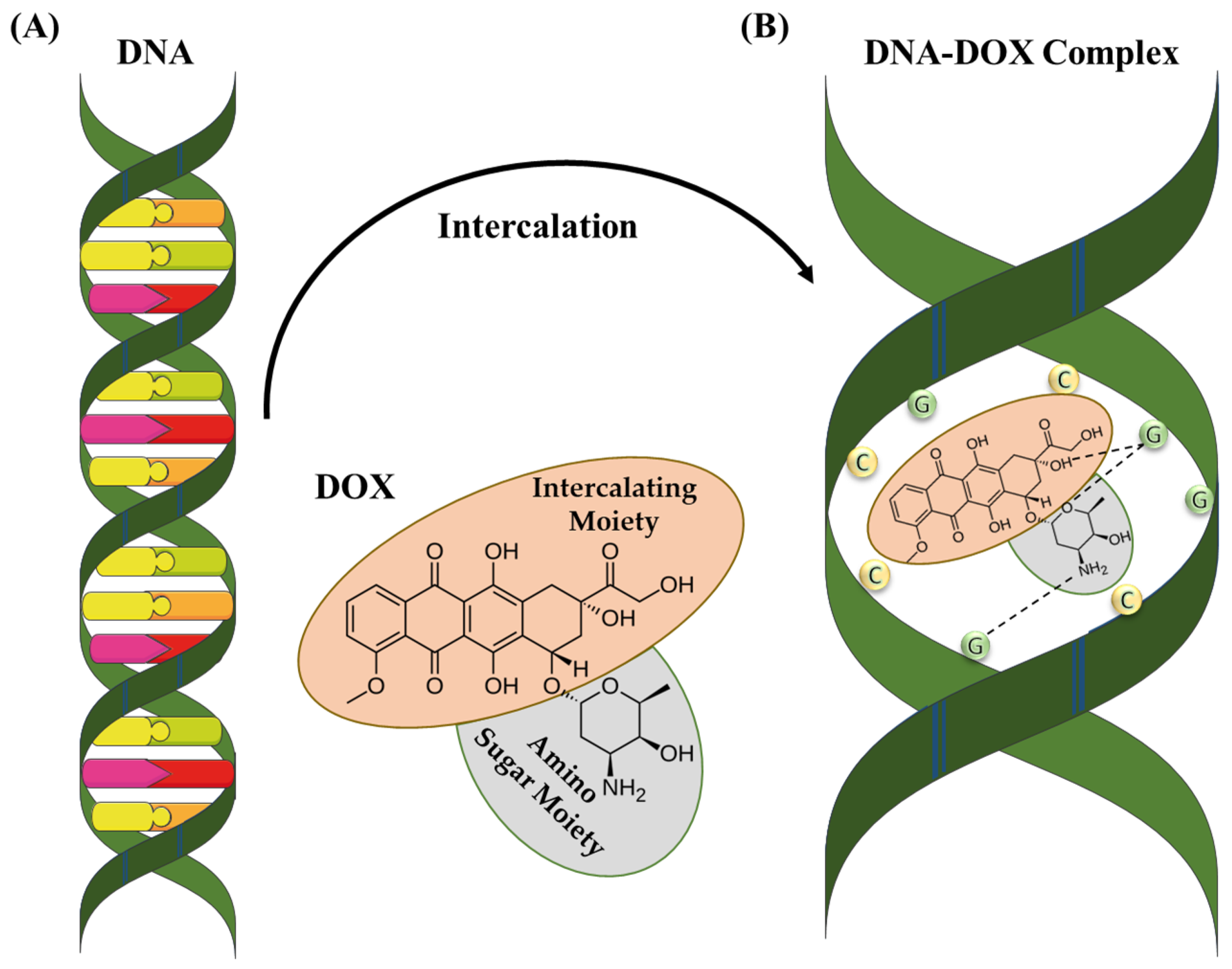
5. DOX Structure and Mechanism of Action

DOX is a chemotherapeutic drug that has been approved by the FDA for cancer treatment [103,104,105]. It is the most commonly used therapeutic drug due to its ability to control the rapid cell division of tumor cells [106], and has been utilized in cancer treatment for its chemotherapeutic abilities. DOX has a molecular weight of 543 g/mol and is crystalline as a solid [107]. This molecule comprises a polar sugar moiety and nonpolar intercalating moiety. It exhibits maximum absorption at a wavelength from 489 nm to 500 nm [108]. DOX and its derivatives can form bonds with plasma proteins for transportation toward tissues. Generally, DOX can enter both the nucleus and cytoplasm of the cells. Once inside the cell, it has a higher affinity for DNA in the nucleus due to its mechanism of action involving DNA intercalation and topoisomerase inhibition [109]. DOX intercalates with DNA or RNA base pairs and inhibits them by blocking topoisomerase II (Figure 3) [110]. This enzyme plays a crucial role in cell proliferation. During DNA replication, topoisomerase II cuts off the filaments. After cutting the filament, DOX intercalates with the DNA molecule to disturb the mechanism. DOX is not restricted to a specific phase of the cell cycle but it exhibits maximum toxicity in the S phase of the cell cycle.
DOX can enhance cell death by various targets, including reactive oxygen species formation and senescence induction [112]. Reactive oxygen species (ROS) are produced in aerobic organisms through various processes, including the electron transport chain (ETC), catabolic oxidases, and peroxisome metabolism [113,114]. However, an excessive ROS production can cause DNA damage due to radicals acting on bases and the sugar–phosphate backbone of DNA [115,116]. Unrepaired damage may result in cell-cycle arrest, senescence, and apoptosis [117,118]. DOX binds directly to cardiolipin on the inner mitochondrial membrane, resulting in ROS production [119]. Elevated ROS levels cause considerable damage to the mitochondrial structure, eventually leading to cell apoptosis. DOX-induced intrinsic and extrinsic apoptosis in cardiac cells is mediated by mitochondria-derived ROS and calcium. This is accomplished by a nuclear factor of the activated T-lymphocyte (NFAT)-mediated upregulation of the FAS antigen ligand (FASL) and the downregulation of the FLICE/caspase-8 inhibitory protein (FLIP) [120]. In investigations assessing the influence of DOX on death ligands in induced pluripotent stem cell-derived cardiomyocytes, the upregulation of death receptors, such as tumor necrosis factor receptor 1 (TNFR1), FAS, and death receptor 5 (DR5), was observed. This upregulation intensified apoptosis, particularly in conjunction with the TNF-related apoptosis-inducing ligand (TRAIL) [104]. The substantial cardiotoxicity and thrombocytopenia associated with DOX significantly limit its therapeutic applications [121].
Similarly, it is commonly accepted that the application of chemotherapeutic drugs to cancer cells can induce senescence. Cellular senescence can also be induced in normal cells as a response to chemotherapy. This side effect can be minimized by using specific targeted therapy approaches. Consequently, therapy-induced senescence (TIS) has become an attractive approach for combating cancer with reduced side effects. However, recent findings suggest that cancer cell senescence could have adverse consequences, as senescent cells tend to create a procancerogenic environment. In addressing this challenge, scientists have introduced a novel pharmacological category called senolytics, specifically designed to eliminate senescent cells [122]. Senescent cells exhibit distinctive features, including cell-cycle arrest, the expression of senescence-associated β-galactosidase, the formation of heterochromatin foci, telomere shortening, the hypermethylation of histone H3K9, and the secretion of various factors, such as chemokines and inflammatory molecules, including MMPs, IL-1, IL-6, and IL-8. This secretory pattern is recognized as the senescence-associated secretory phenotype (SASP) [123]. Additionally, disrupted Ca2+ uptake has been demonstrated to instigate apoptotic processes and facilitate necrotic cell death. Notably, DOX has been identified for its ability to perturb mitochondrial and cellular Ca2+ homeostasis, making it a potential target for therapy [124]. However, the medical application of DOX is limited due to its high toxicity. Mostly drug transporter and antiapoptotic aspects are assumed as the main reasons for DOX resistance. For example, P-glycoprotein (a membrane protein) is a very popular drug transporter that creates DOX resistance through a lack of accumulation in tumor cells [125]. P-glycoprotein has two pseudosymmetric halves in the structure. Every half consists of a nucleotide-binding domain (NBD) responsible for the binding and hydrolyzation of ATP, as well as a transmembrane domain (TMD) [126]. Conformational changes arise in the structure of the P-glycoprotein when the drug binds with it, in which the drug is bound to one site and released to another site of the P-glycoprotein [125]. These sites of P-glycoprotein can be targeted by NPs to inhibit its activity [127].

6. DOX–Chitosan-Mediated NPs for Drug Deliveries

6.1. Active and Passive Drug Delivery

Effective drug delivery to tumor cells via chitosan NPs is divided into two categories (active and passive) (Figure 4). In passive DOX delivery, chitosan NPs accumulate in tumor tissue through leaky or defective vessels using the permeability and retention (EPR) effect [128]. NPs carrying anticancer drugs can easily navigate through the blood vessels in the angiogenic tumor site. This characteristic leads to a higher concentration of these NPs in tumor tissue compared to natural anticancer drugs, a phenomenon referred to as the EPR effect [129]. Once a solid tumor achieves a specific size, the surrounding normal vasculature becomes inadequate to meet the increasing oxygen demands for tumor development. Subsequently, as tumor cells undergo cell death, they release growth factors that stimulate the formation of new blood vessels from nearby capillaries [130]. Angiogenesis denotes the rapid formation of novel and irregular blood vessels, characterized by a disrupted epithelium and the absence of the basal membrane typically present in normal vascular systems. Due to the vascularization needed by rapidly growing tumors, coupled with restricted lymphatic drainage, the resulting irregular vascular architecture gives rise to an amplified EPR effect [131]. However, passive targeted drug delivery exhibited lower therapeutic efficacy and systemic side effects [132].
The targeted delivery of chemotherapeutic drugs presents dual benefits. Firstly, precise delivery to the targeted site reduces the requirement of the dosage, enhancing the efficacy of the therapy method [133]. Secondly, by reducing the overall drug dosage, the manifestation of drug-induced adverse effect is either prevented or significantly minimized. Nanomedicine therapy influences the diverse active and passive targeting capabilities of NPs to deliver drugs to specific target site [134]. Because of these potentials, NPs are used as viable methods for overcoming the drawbacks of traditional cancer therapies, such as nonselective toxicity and drug resistance. Tumor-targeted drug delivery takes use of the differences between malignant and healthy tissues [135]. During tumor progression, the tumor microenvironment changes. Inadequate oxygen supply and glucose to lactate conversion, caused by increased metabolism and growth rates, lead to a fall in the pH of the tumor tissue. This change, in conjunction with hypoxia and glucose deprivation, increases angiogenesis, a mechanism critical for tumor proliferation, migration, and maintenance [136,137]. Many tumors have an overexpression of certain antigens, including on their surfaces, making them potential drug delivery targets. This approach is successful as long as the selected targets for a specific cancer cell type can be recognized with confidence and are not expressed in considerable amounts elsewhere in the body [122,123]. An investigation elaborated on the synthesis of the active targeted water-soluble delivery of DOX [138]. This research involved two biodegradable and biocompatible biopolymers, poly-γ-glutamic acid (PGA) and chitosan. The self-conjugation of these two polymers produced stable and negatively charged NPs with an 80-150 nm diameter. The targeting agent was folic acid and bonded with the NP surface and polyanion. In this study, the stability of NPs, the toxic effect, the active targeting effect, and the DOX release efficiency was examined in in vivo situations. The results showed that the DOX-loaded NPs induced the DOX delivery compared to free drugs without damaging the normal cells. Table 2 shows the DOX deliveries via chitosan NPs with different combinations of NPs for cancer treatment.
Figure 4. Chitosan-based NP drug delivery mechanisms. (A) Passive targeting mechanism: leaky tumor vessels release the DOX-loaded chitosan NPs at the cancer site via the EPR effect. (B) Active targeting mechanism: DOX-loaded chitosan NPs accumulate in cancerous cells via ligand-mediated endocytosis [139].
Molecules 29 00031 g004

6.2. Modified Chitosan–DOX Drug Deliveries

6.2.1. Amino Acid-Modified Chitosan NPs

Amino acids are crucial molecules for all living cells; essential and nonessential amino acids play a key role in cell growth and proliferation. In the tumor microenvironment, the rapid growth and proliferation of the tumor cells require more amino acids to synthesize protein [163]. Traditional drug delivery approaches have focused on “starving cancer cells to death” by blocking nutrient intake [164]. Due to the hydrophilic nature of amino acids, they cannot cross animal cell membranes [165]. The transfer of amino acids into the cell requires a specific transporter on the animal cell membrane [166]. Several amino acid transporters have been discovered to cross the animal cell membrane, categorized according to substrate specificity and coupling ions [166]. Consequently, amino acid transporters have been considered an emerging goal for cancer chemotherapy. In cancer cells, the blockage of amino acid transporters is more specific and avoids unwanted nontarget effects [164,165]. On the other hand, chitosan NPs modified with amino acids can serve various purposes. Amino acids have the potential to improve the transport of therapeutic drugs, stabilize the nanoparticles, and allow specific interactions with biological targets. This specificity offers an opportunity for targeted tumor therapy, such as application in boron neutron capture therapy (BNCT), positron emission tomography (PET), and chemotherapeutic DDS [167,168]. Nowadays, therapy with amino acids (TAAI) for cancer treatment has gained more attention. A new method has been designed using amino acids and polymers for cancer treatment [169]. DOX-loaded chitosan glutamic acid (CS-Ga–DOX) NPs were prepared through the ionic gelation method. CS-Ga–DOX NPs exhibited a positive zeta potential and spherical structure. At a pH of 5.5 and 7.4, DOX was continuously released with a mutual burst. CS-Ga–DOX NPs have great potential as a pH-responsive nanocarrier for anticancer chemotherapy.

6.2.2. Vitamin-Modified Chitosan NPs

Metastasis and stemness are the two main challenges in cancer treatment. Both have a solid connection with drug resistance and a low prognosis, finally leading to the failure of the cancer treatment. It has been described that cancer chemotherapy, particularly with the DOX in breast cancer treatment, can enhance metastasis and stemness. A combination therapy is an efficient method to suppress tumor cells with an enhanced synergistic effect. An advanced therapeutic system was developed by the combination of all-trans retinoic acid (ATRA) with DOX to resolve metastasis and stemness [170]. ATRA is a newly identified Pin1 (specific isomerase highly expressed within various tumor cells). It can effectively stop numerous cancerous pathways by successfully inhibiting and degrading Pin1. In this approach, researchers successfully synthesized a folic acid–chitosan-based polymer with both DOX (FA-CSOSA/DOX) and ATRA (FA-CSOSA/ATRA) NPs for cancer treatment. Due to the presence of FA, the uptake of these NPs was enhanced in cancer cells through folate receptors. This approach has a better synergistic effect as compared to DOX alone in the 4T1 cell line. In vivo, these NPs exhibited an 85.5% tumor inhibition, 2.5-fold higher than that of DOX–HCl alone. Another study was designed to synthesize vitamin E succinate–chitosan–histidine (VCH) multiprogram DOX carriers [171]. The π-π stacking bond between VCH and DOX was confirmed using a UV–vis spectrum. Drug release experimentations showed better pH sensitivity and sustained release results. DOX/VCH NPs were effectively taken up by HepG2 tumor cells, with the suppression rate reaching up to 56.27%. These NPs could combine with histidine and chitosan to attain pH sensitivity and P-gp inhibition, effectively suppressing the tumor cell growth and improving solubility. These multiprogram NPs show promise as an effective drug carrier in cancer chemotherapy without causing damage the healthy cells.

6.2.3. Antibody-Modified Chitosan NPs

Antibody–drug conjugates (ADCs) constitute a class of cancer cell-targeting drugs that have gained consideration. Monoclonal antibodies (mAbs) were synthesized to target antigens, and this approach was developed with the advent of hybridoma technology in 1975 [172]. Several mAbs have since gained approval, with herceptin being an example used in breast cancer treatment by targeting the HER2 receptor. However, mAbs alone are not enough for cancer treatment due to their low cytotoxicity in cancer cells [173]. Therefore, a new approach was designed to increase the cytotoxic effect in cancer cells. According to this strategy, mAbs were conjugated with biopolymers to enhance the drug efficiency. Similarly, chitosan NPs combined with two types of mAbs (anti-hMAM and anti-HER2) were synthesized for the treatment of breast cancer [141]. These PEGylated DOX-loaded CSNPs with mAbs exhibited enhanced cytotoxicity against MCF-7 cancer cells as compared to DOX-loaded CSNPs without mAbs. The synergetic therapy of immunogenic cell death (ICD) and the immune checkpoint blockade (ICB) has revealed extraordinary results against various types of cancers. For a safe and effective synergetic immunotherapy, researchers proposed all-in-one glycol chitosan NPs (CNPs) that administered anti-PD-L1 peptide (PP) and DOX to target tumor cells [146]. Briefly, a hydrophobic 5-cholanic acid was conjugated to the hydrophilic glycol chitosan backbones to create CNPs (Figure 5). Furthermore, the CNPs’ free amine groups were modified with tumor-targeting antibodies and peptides for targeted tumor drug delivery [174]. PP–CNPs thus avoided subcellular PD-L1 recycling, eventually abolishing the immunological escape mechanism in CT26 colon tumor. When the DOX–PP–CNPs were intravenously injected into mice with CT26 colon tumors, PP and DOX were successfully delivered to the tumor tissues via NP-derived passive and active targeting. This enhanced both lysosomal PD-L1 degradation and substantial ICD, ultimately leading to tumor regression through an antitumor immune response.

6.2.4. Hyaluronic Acid-Modified Chitosan NPs

Hyaluronic acid (HA) is a polysaccharide found in the extracellular matrix of connective tissues. Structurally, HA has repeating units of N-acetyl-D-glucosamine and D-glucuronic acid connected with the β-1,3 and β-1,4 glycosidic linkage [175]. HA has been extensively used in actively targeted deliveries due to the high attraction for CD44 receptors (overexpressed in stem cells of cancer). The active targeted anticancer drug delivery with HA can enhance the solubility and effectiveness. In this regard, catechol (Cat)-modified chitosan@HA NPs were synthesized to deliver the DOX [176]. The Cat moiety enabled the carriers with good adherence and a constant local distribution of DOX. The ionic gelation method was used to prepare Cat-NPs from Cat-functionalized chitosan and HA. These prepared NPs have a negative charge and spherical shape. As compared to unmodified NPs, these NPs showed better mucoadhesive properties in oral mucosal tissues. In this method, DOX was loaded onto the modified NPs with a high loading capacity of 250 μg/mg, and sustained release was achieved. DOX-loaded Cat-NPs (DOX-NPs) inhibited the expansion of the HN22 carcinoma cell line. DOX-NPs were taken up, accumulated, and caused apoptosis in cells more rapidly as compared to free DOX. The findings showed that the prepared Cat-NPs have great potential and could be used as a novel carrier for the local delivery of DOX to oral cancer cells. Another study explained the HA-modified chitosan NPs for DOX delivery for effective cancer chemotherapy. Magnetic NPs containing chitosan/HA complexed with κ-carrageenan were prepared by using the solution method (hydrothermal method) [177]. An MTT assay was performed to check the effect of these magnetic NPs on MCF-7 and MDA-MB-237 cells. These MNPs have a spherical shape and a 100–150 nm diameter with a 74.1% DOX encapsulation capacity. However, the drug encapsulation capacity was enhanced by increasing the κ-carrageenan amount. Subsequently, the pH-stimulus-responsive drug was released in a sustained manner without side effects.

6.2.5. PLGA- and PEG-Modified Chitosan NPs

PLGA and PEG are biocompatible and biodegradable synthetic polymers with the ability to increase stability and minimizing side effects. For example, pH-sensitive PEGylated chitosan NPs coated with PLGA were synthesized using the coassembly method to achieve effective DOX delivery for cancer chemotherapy [178]. The obtained DOX-loaded PEGylated chitosan/PLGA NPs (DOX–PCPNs) have a spherical structure, while the chitosan/PLGA formed a solid central core surrounded by hydrophilic PEG. DOX–PCPNs exhibited excellent stability and enhanced drug release in a serum-holding environment. Cytotoxic studies elaborated that the DOX–PCPNs were endocytosed, enhancing the DOX release in an in vitro environment and increasing DOX accumulation in cancer cells to improve antitumor efficiency. The hydrated shells of PEG protected against uptake by macrophage cells. In vivo results exhibited the ability of DOX–PCPNs to efficiently deliver DOX, reducing TRAMP-C1 tumor growth compared to free DOX. DOX–PCPNs exhibit great potential in drug delivery against tumor cells.

6.2.6. Genetic-Material-Modified Chitosan NPs

Sometimes, monotherapy against cancerous cells is insufficient for treatment. It has disadvantages, such as extreme toxicity in healthy cells and resistance [179,180,181]. Compared to conventional chemotherapy, gene therapy offers a safer route through nonviral vectors, but it exhibits limited efficiency and needs improvement [182,183]. Multidrug resistance (MDR) is also attributed to the malfunction of genes causing chromosomal changes in cancerous cells [184]. Many researchers have investigated gene delivery to treat cancerous cells with drug combinations [185]. The combined chemo- and gene therapy has provided an effective way for cancer treatment to overcome MDR due to its high synergetic effect [186,187,188]. Nucleic acids and genes require DDS because they are too large to enter the animal membrane. Moreover, gene therapy for cancer treatment is challenging due to the unstable and flexible nature of genes [179]. Here, we explored studies that highlight the use of chitosan-based NPs for the codelivery of DOX and genes in cancer treatment (Figure 6). Dendronized chitosan-based poly amidoamine deoxycholic acid (PAMAM-CS-DCA) NPs were synthesized [189,190,191]. These chitosan-based NPs exhibited low toxicity in healthy cells and a good gene transfection efficiency. Low doses of DOX increased the gene expression and synergetic ability, demonstrating great potential for the codelivery of genes and drugs. The overall results exhibited higher efficiency against tumor cells. Similarly, a double-walled microsphere was synthesized, and chitosan-based DNA nanocomplexes, including the p53 gene, were loaded for combined chemo- and gene therapy [192,193]. Another study described the chitosan-based NPs for the codelivery of DOX and siRNA. In this approach, carboxymethyl dextran (CMD) chitosan-based NPs were prepared to load the DOX and siRNA. The effect of these NPs on the epithelial–mesenchymal transition (EMT) gene expression and cell growth in HCT-116 cell lines was also explained [194]. In addition, another study elaborated the lung cancer therapy with HMGA2 (high-mobility group A2) suppressing small interfering RNA (siRNA) along with DOX by using chitosan-based NPs [195,196]. The findings showed that the codelivery of HMGA2, siRNA, and DOX through innovative CMDTMChiNPs was a novel therapeutic method with prodigious potential effectiveness for lung cancer treatment. The increased activity level and expression of P-glycoprotein were responsible for increasing the drug resistance [197,198]. To solve this, chitosan–g-D-α-tocopheryl polyethylene glycol (TPGS) NPs were produced by using evaporation techniques. TPGS NPs have the ability to control the P-glycoprotein activity and significantly decrease the ATP level, which is beneficial in preventing DOX efflux [199]. Chitosan–dextran–sulfate-coated PLGA–PVA (poly lactic-co-glycolic acid–polyvinyl alcohol) NPs for DOX distribution were used to test the effectiveness of the chitosan NPs by preventing DOX progression [200]. All the above-mentioned examples prove the effectiveness of codelivering DOX and genes using chitosan NPs as a carrier agent.

6.2.7. Immunotherapeutic-Modified Chitosan NPs

Cancer leads to a significant proliferation of abnormal cells and immunosuppressive cells that inhibit the immune response [202,203,204]. Reforming the immune environment of tumors can enhance the efficiency of the immune response against tumors. [205,206,207]. Recently, tumor immunotherapy has gained a lot of attention for cancer treatment [208,209]. However, the efficiency and toxicity effects of simple immunotherapy are minimal [210,211]. Cancerous cells are present at the core tumor mass, and the treatment efficiency is restricted [212]. Due to this condition, combination therapy is the most effective method of cancer treatment [213,214]. A facile approach was developed using CMC derivatives (M2pep-CMCS) targeting tumor-linked macrophages 2 (TAM2) and cyclodextrin derivative (R6RGD-CM βCD) with the tumor target [215]. DOX was loaded onto cyclodextrin-derivative NPs, and R848 was loaded onto CMC-derivative NPs, demonstrating good absorption. These NPs enormously increased the expression levels of cleaved Caspase 3, indicating enhanced cell apoptosis. Similarly, these NPs also altered the shape of tumor-linked macrophages. Overall, these materials are considered an effective delivery carrier in cancer treatment. Another study has investigated enhancing the immune response against cancer treatment, in which TMC-based NPs were synthesized and fabricated with DOX and interleukin-2 (rhIL-2) [216]. These NPs proficiently delivered the hydrophobic DOX and hydrophilic interleukin-2 to attain the combination therapy against the tumor microenvironment. DOX was attached to the TMC NPs with covalent bonds that led to the pH-sensitive release, while interleukin-2 bonded through electrostatic forces. The diameter of these NPs was about 200 nm, and a zeta potential > 20 mV was recorded. For the targeted delivery, folate modification was used to accumulate the drugs at the target site. The final results showed that the combinational therapy of interleukin-2 and DOX-based TMC NPs have the potential to kill the tumor cells and enhanced the immune response against cancer.

6.3. Combined Delivery with Other Anticancer Drugs

A large number of studies have focused on combining chemotherapeutic drugs and chemosensitizers due to the resistance of tumor cells to DOX alone. The combined effect of anticancer drugs may improve the tumor suppression efficiency and minimize side effects [217,218,219,220]. Overall, the codelivery of drugs with chitosan NPs has proven the efficiency of this method against various cancerous treatments by reducing the drug resistance without harming the noncancerous cells [221]. In this aspect, research was conducted on breast cancer treatment. The codelivery of DOX and Tanshinone IIA (TSIIA) (a bioactive compound isolated from Chinese herbs known as “Danshen”) was achieved using CMC chitosan-based hypoxia NPs for breast cancer treatment [222]. Hypoxia-responsive NPs are designed to respond to low-oxygen levels, a condition known as hypoxia, typically found in tumor microenvironments. These NPs showed an improvement in release efficiency and an increase in the cytotoxicity of DOX against the tumor microenvironment. The immunofluorescence staining of the tumor section confirmed that the combined nanoparticles exerted a synergistic antitumor effect by inhibiting tumor. Consequently, CMC chitosan-based NPs exhibited promising properties for drug delivery against breast cancer. Degradation of the delivery agent is crucial for DDS at specific target sites and to prevent the presence of the delivery agent’s byproduct in the body [223]. Cancerous cells produce glutathione (GSH) 7 to 10 times more than healthy cells, which is highly beneficial for disulfide bond degradation [224,225]. Thus, a disulfide-bond-based DDS proves to be very helpful for enhancing the stability of delivery agents and facilitating degradation at the targeted tumor site in cancer therapy [226,227,228]. To achieve this purpose, another approach was established, wherein redox-sensitive chitosan/stearic acid NPs (CSSA NPs) were synthesized for DOX and curcumin delivery in cancer treatment [229]. The degradable CSSA NPs had a size of about 200 nm and were synthesized based on disulfide cross-linking. The hydrophilic DOX and hydrophobic curcumin drugs were encapsulated onto the CSSA NPs. Codelivery of therapeutic drugs through this approach increased the efficiency of the cancer treatment. These NPs exhibited a low drug release in the normal cells, while approximately 98% of DOX and 96% of curcumin were released in the tumor cells under a GSH reducing environment. Consequently, this method has demonstrated enhanced encapsulation, release of dual drugs, and cytotoxicity for cancer treatment. Another method was established for CD44 receptor targeting, reducing multidrug resistance (MDR) and enhancing the drug release and cytotoxicity in tumor cells for breast cancer treatment [230]. The NPs consisted of three layers: a poly core, liposome, and chitosan, respectively. These NPs (Ch-MLNPs) were loaded with DOX, silybin, and paclitaxel. These three drugs were released at target-specific sites exploiting CD44 receptors in breast cancer cells. In vivo, studies showed the good efficiency of these three-layered NPs against breast cancer and lowered the MDR.

7. Stimuli-Sensitive Deliveries of Chitosan–DOX NPs

7.1. Endogenous Stimuli-Sensitive Drug Deliveries

7.1.1. pH-Sensitive Drug Deliveries

The cancerous cells have a pH of 6.5 (slightly acidic), which is lower than the physiological environment; drug release can be reduced due to that difference. The extracellular acidic environment is one of the most significant properties that differentiate healthy cells from cancerous cells [231]. For rapid progression, cancer cells choose aerobic glycolysis to continue the metabolic process, increasing the production of lactic acid and the accumulation of protons in the cell [231,232]. Nanocarrier derivatives can deliver the drug at a mildly acidic pH. The bond between the NPs and drug degrades (depending on the nature of bonds) in the mildly acidic pH of cancerous cells to release the drug at the target site [233]. Different bonds, like acetals, amine, oxime, ester, and amide, can be formed between the NPs and drugs [234]. The overcoming of the MDR, the effective release, and the accumulation of drugs are the necessary points to achieve. For this purpose, dual-pH-responsive chitosan NPs were invented to overcome the MDR tumor (MCF-7/ARD) in human breast cancer [235]. In this study, chitosan NPs were sensitized to extracellular tumors with a pH of 6.5, which participated in the surface charge reversal through the breakage of β-carboxylic amide, enhancing the cellular uptake efficiency. Furthermore, these chitosan-based NPs also exhibited responsiveness to an intracellular pH of 5.0 in the tumor microenvironment, with poor blood perfusion and limited oxygen supply. This pH fluctuation played a crucial role in inducing the protonation of the amino group within the acidic environment [236,237]. Cells assays verified that dual-pH-sensitive particles caused induced toxicity in the MDR tumor cells. Furthermore, the NPs could overcome tumor resistance by decreasing the intracellular levels of ATP and PARP-1, ultimately receiving a stronger antitumor efficiency. Moreover, the amphiphilic chitosan NPs showed a high efficiency against cancerous cells. Further research explored chitosan/polyvinylpyrrolidone/hematite (CS/PVP/α-Fe2O3) nanocarriers for the drug delivery of DOX. The NPs had a spherical shape and could load Fe2O3 on CS/PVP. The CS/PVP-based NPs provided pH-controlled drug delivery for tumor treatment and suppressed the breast cancer cells [238].
Nanocarriers with pH-sensitive capabilities are used for chemotherapy and drug delivery. Due to the nonspecificity of DDS, it affects normal cells as well. In a research study, hollow mesoporous silica NPs (HMSNGM-CS-FA) were produced for the combined delivery of DOX and pheophorbide (PA) [239]. These NPs exhibited efficient drug release capabilities based on the pH-sensitive swelling effect of the coating layer. This study demonstrated significant encapsulation and drug release capabilities, resulting in enhanced cytotoxicity in cancerous cells based on pH-sensitive NPs. Another feasible method was adopted to produce the pH-sensitive surface-charge reversal CMC-based DOX DDS using an organic solvent-free coprecipitation approach [240]. According to this approach, DOX was loaded in the core of PDPA fragments. These fragments were combined with the PEGylated CMC to form a shielding shell. Overall, results demonstrated the good drug-loading and releasing capacity, leading to tumor cells death. In addition, a chitosan-based polymeric drug was prepared for the DOX drug codelivery with siRNA for the tumor cell treatment. These polymeric nanocarriers were entered through hepatoma cells exploiting glycyrrhetinic acid-receptor-mediated endocytosis. The DOX releasing concentration was 90.2% out of the total drug-encapsulated concentration, and the siRNA releasing concentration was 81.3% (out of 50 μL of 100 μg/mL) after 10 h. This drug could suppress tumor cells by 88% (out of the total tumor size) through chemonucleic acid therapy (Figure 7) [241]. A high pH usually decreases the solubility of chitosan. Similarly, polyvinylpyrrolidone (PVP) was used, but alone it reduced the drug release. To control these problems, the conjugation of chitosan and PVP has been recommended to enhance the solubility of chitosan at a high pH level [238,242]. Another method was used to successfully fabricate iron (III) carboxylate metal–organic framework NPs coated with a glycyrrhetinic acid–chitosan conjugate (MIL-101/GA-CS), which behaved as a pH-responsive and target-specific agent to deliver DOX for hepatocellular carcinoma (HCC) therapy (Figure 7) [243]. These NPs have the advantages of a uniform size, drug encapsulation effectiveness, and pH-dependent targeted drug release. In vitro cytotoxic effects revealed that the NPs had excellent inhibitory effects on HepG2 cells due to the continual release of DOX, although these NPs had no significant toxicity in normal cells. As a result, MIL-101-DOX/GA-CS NPs have the potential to be used in therapy as a pH-responsive controlled DDS.

7.1.2. Redox-Sensitive Drug Deliveries

The redox imbalance represents another exclusive parameter within the tumor microenvironment that is accountable for increasing the rate of cancer proliferation [244]. The synthesis of ROS started by cancer cells, cancer-associated fibroblasts, and endothelial cells contributes to the progression of cancer cells. Further, glutathione (GSH) is an antioxidant and acts as a reducing agent for ROS and oxidative stress [245]. The combination of suppressor agents and chitosan has been used to produce nanocomposites for drug delivery improvement. Chitosan with oligosaccharide (CSO) and stearic acid (SA) can be utilized to synthesize glycolipids, specifically copolymers, and these demonstrate good capability in delivering drugs to the tumor microenvironment [246]. However, micelles synthesized from CSO–SA face a significant challenge of efficient drug delivery in vitro due to the slow degradation kinetics of the amide bond. To solve this problem, DOX could be coupled with the CSO–SA through the help of a disulfide bond. This method was beneficial in the production of CSO–SA-based nanocomposites (due to the high GSH level, the NPs were redox-sensitive) and could deliver DOX to eliminate the progression of breast cancer [247]. Another study has explored the redox-responsive chitosan NPs for DOX delivery. In this method, N, N′carbonyldiimidazole (CDI) catalysis generated amphiphilic low-molecular-weight chitosan–lipoic acid (LC-LA) conjugates with varying degrees of substitution (DS) of LA, which self-assembled into redox-sensitive micelles [248]. The diameter, zeta potential, biocompatibility, critical micelle concentration, and the redox-sensitive response of blank micelles were explored. According to the results, blank micelles with a low critical micelle concentration, nanosize, and positive zeta potential demonstrated excellent biocompatibility and redox-sensitive response. The DOX drug was loaded on these micelles for cancer treatment. The loading capability, drug-released behavior, antitumor efficiency, and cellular drug uptake were elaborated, indicating that DOX-loaded micelles have a good loading capability and show a redox-trigger response and solid antitumor efficiency against A549 cells. An increase in the DS of LA decreased the critical micelle concentration and cumulative release concentration of DOX while increasing the loading efficiency, antitumor capability, and cellular uptake of DOX-loaded micelles, which was caused by the increased contact of hydrophobic groups in the micelles with the DS of LA. In general, self-assembled LC-LA micelles with good biosecurity and redox-sensitive responsiveness capture favorable application diagnostics in DOX administration and provide an understanding on DOX’s cancer therapeutic impact.

7.1.3. Enzyme-Sensitive Drug Deliveries

Enzymes, such as protease, phospholipase, or glycosidase, play a crucial role in biological mechanisms, and abnormalities in the enzyme function can lead to a disorder like cancer [249]. Enzyme-dependent NPs have gained incredible consideration due to target specificity towards cells that overexpress enzymes [212]. The encapsulated drug in NPs is released at the target site due to the specific enzyme function [250]. Enzyme-responsive moieties are attached covalently or noncovalently with the polymers to obtain the desired NPs. However, covalently bonded NPs may disturb the function and target specificity of the enzymes due to suboptimal reactivity with the NPs [250]. On the other hand, noncovalently bonded NPs are highly specific to the target. A few studies have explained the enzyme-sensitive DOX delivery via chitosan NPs against a tumor environment. For example, hollow mesoporous silica spheres (HMSSs) were synthesized and linked with chitosan by the azo linkage (HMSS-N=N-CS) for enzyme–stimulus colon-specific drug delivery [251]. After that, DOX was loaded into the pores of HMSS-N=N-CS. These NPs showed stability, biocompatibility enhancement, and reduced protein adsorption on HMSSs. The final results revealed an increase in the cellular uptake of the drug after enzyme incubation.

7.2. Exogenous-Stimuli-Sensitive Drug Deliveries

7.2.1. Light/Photo-Sensitive Drug Deliveries

Photosensitizers have found applications in stimuli-sensitive systems, involving the chemical modification of chitosan to respond to stimuli, such as UV and light, in photodynamic therapy (PDT) for treating cancer cells [252]. Several methodologies have been proposed for utilizing chitosan-based NPs in light-dependent delivery. Light-sensitive NPs, characterized by a sphere-shaped structure with functional-group-rich surfaces exhibiting the enhanced EPR effect, were modified with polymers like poly (D, L-lactic-co-glycolic acid) PLGA and poly (ε-caprolactone) PCL. This modification enhances the biodegradability and biocompatibility of the NPs, allowing for intravenous injection [253]. These NPs exhibited a good encapsulation efficiency and cytotoxicity in the tumor microenvironment [254]. In another experiment, a light-dependent platform was developed consisting of DOX-loaded gold NPs for breast cancer. In addition to photothermal therapy, it was observed that DOX produced oxidative stress via ROS [255]. Another study was performed in which the photosensitizer chlorin e6 (Ce6) with DOX-encapsulated chitosan (CS)–tripolyphosphate (TPP) NPs were synthesized for cancer therapy [160]. The ionotropic gelation method was used to prepare these NPs, and their photophysical and morphological properties were studied. The Ce6 was loaded onto the NPs through the self-assembly of chitosan with TPP–DOX under an aqueous environment. The prepared NPs had an 80–120 nm diameter with a negative zeta potential of -6 mV. The absorption spectrum of Ce6-coated NPs was the same as free Ce6, suggesting that the Ce6 chromophore underwent no alterations as a result of the coating. These NPs exhibited strong photostability and singlet oxygen generation (SOG). The Ce6-fabricated and DOX-encapsulated NP size was about 90–130 nm, and the charge was about −30 mV. The results exhibited high-DOX-encapsulation capacity and pH-controlled release. Moreover, these NPs exhibited a high uptake of DOX drug under irradiation at near-infrared (NIR) ranges against MCF-7 cancerous cells. This study showed how NPs could be used to release DOX under photocontrol in a tumor microenvironment.

7.2.2. Magnetic-Sensitive Drug Deliveries

Magnetic-sensitive NPs can be synthesized using various methods, and their properties are often tailored for specific applications. One common approach involves incorporating magnetic materials into the nanoparticle structure. This technique relies on the magneto-sensitive moiety in the presence of a strong external magnetic field [2]. Magnetic flux can be changed for guiding the nanocarrier to deliver the drug to a specific target site. Several superparamagnetic iron oxide NPs (SPION) are prepared with moieties that can specifically bind at the desired position. The interaction between the external magnetic field and SPION raises the temperature at the tumor site, called the “magnetic thermal ablation” [2]. A gradient magnetic field can enhance the uptake of NPs into tumor cells and deliver the maximum drug at the target site. Chitosan-based magneto-sensitive NPs are capable of crossing the blood–brain barrier. Mostly, chitosan-containing magnetic NPs (MNPs) have a magnetic nucleus inside and a biodegradable outer shell. The inner magnetic core is responsible for transporting the NPs to the specific target site, and the outer biodegradable shell releases the drug. MNPs are highly active and readily oxidized in air, leading to a loss of magnetism. Chitosan preserves these MNPs from oxidation and reduces their toxicity. Chitosan also facilitates the binding of MNPs with various functional groups of the DOX drug moiety, resulting in less aggregation, a longer half-life, and enhanced stability. Fe3O4, ZnFe2O4, CoFe2O4, Fe2O3, and other magnetic particles are utilized in the synthesis of chitosan-based NPs. In this regard, recent research elaborated on the magnetic-sensitive chitosan-NP-based DOX delivery for cancer therapy. According to this study, pH-sensitive NPs were produced using chitosan and succinic anhydride (CSSA) for the targeted delivery of DOX to osteosarcoma cells [155]. First, they synthesized CS–folic acid (FA) conjugates by the amide linkage of chitosan with FA. Following that, CS-SA/CS-FA was produced by coating of Fe3O4 (MNPs) ferrofluid. Then, DOX molecules were placed onto the CS-SA/CS-FA NPs. The DOX release profiles at various pHs revealed that the DOX release was boosted in acidic environments. The MG-63 cells, which express folate receptors, demonstrated much better cellular absorption of the DOX-loaded CS-FA/CS-SA@MNPs than the lung cancer A549 cells. The cytotoxicity experiment revealed that these NPs exhibit cytocompatibility with MG-63 cells.

7.2.3. Ultrasound-Sensitive Drug Deliveries

Ultrasound-responsive NPs synthesized with chitosan have been extensively examined for their biosafety towards healthy cells. Ultrasound-triggered drug delivery has gained consideration as a noninvasive modality against cancerous cells. A study supports this noninvasive modality, in which a hydrophobic DOX drug was encapsulated onto the palmitoyl-modified glycol chitosan amphiphile (PmGCA) NPs [256]. The reappearance of the DOX fluorescence peak after 2 MHz under ultrasound exposure proved the drug release. In vivo, the results exhibited remarkably lower fluorescence in the liver and heart as compared to DOX alone. This approach is considered beneficial for DOX drug delivery for cancer treatment, with low side effects on the major organs. Similarly, other chitosan-derivative-based NPs were synthesized for the DOX delivery for efficient cancer treatment. O-carboxymethyl chitosan/perfluorohexane nanodroplets (O-CS NDs) encapsulated with DOX were tested in vitro [257]. O-CS NDs attained higher tumor cellular penetrations at an acidic pH, a good ultrasound imaging capability, and strong cytotoxicity. This research explained the improved cell interaction ability under ultrasound exposure and targeted DOX delivery against cancer cells.

7.3. Multisensitive Drug Deliveries

Multistimuli-sensitive systems are designed to respond to a combination of stimuli, providing a more sophisticated and controlled drug release profile. Such systems are often explored to enhance drug delivery precision and efficiency, especially in targeted therapy. [258,259,260]. In this regard, chitosan NPs with folate-covered dual-responsive mesoporous silica NPs (MSNs) actively targeted the tumor cells and provided efficient drug delivery [261]. These NPs were produced with a very economically cheap silica and sodium silicate source. When the DOX was loaded into the MSNs, it reacted with cystamine dihydrochloride, and then a folate conjugate was created to produce dual-stimulus-responsive NPs. DOX was released at the target from the MSNs under an acidic pH (a pH of 5.5, 10 mM GSH) and redox environment in vitro. These NPs showed 2.14-times-enhanced cytotoxicity in MCF-7 cells and 1.65-times-enhanced cytotoxicity in MDA-MB-231 cells as compared to DOX alone. Low stability, unpredictable drug release, and limited tumor penetration hinder the applicability of nanomaterial-based DDS. To solve this, pH- and enzyme-responsive shrinkable NPs were created [262]. LDC (a compound of laponite (LP), doxorubicin (DOX), and chito-oligosaccharides (COS)) was the main component of these double-responsive NPs. LDC NPs have a diameter of 100 nm, and DOX drugs can accumulate effectively in the tumor ecosystem under an in vivo environment. Lysozymes, found in the extracellular environment, facilitated the degradation of chitosan oligosaccharides (COSs) and resulted in the formation of smaller DOX-based nanoparticles. Additionally, they induced a size reduction in LDC nanoparticles, reducing their original size from 100 nm to 30 nm. This size reduction enhances the drug’s ability to penetrate deeply into tumor cells, as illustrated in Figure 8. Additionally, after entering the tumor tissues, the acidic and enzymatic cellular environment triggered a rapid release of DOX, leading to the quick death of cancer cells. LDC NPs often exhibited no significant cytotoxicity in the mice’s primary organs. The outcomes of this research exhibited that these LDC NPs possess efficient targeting capabilities for tumor tissues with controlled drug release. Another study supported the use of multiresponsive chitosan-based NPs for cancer therapy. In this method, triple-responsive (pH, redox, and ultrasound) hybrid NPs were produced for controlled and sustained drug delivery. These hybrid NPs were manufactured through a mesoporous-silica-coated magnetic core (Fe3O4@SiO2@mSiO2) and pH/redox-responsive polymer layer [263]. The pH/redox-responsive polymer (CS-LA) layer of the particles was created using chitosan (CS) and lipoic acid (LA). Then, Fe3O4@SiO2@mSiO2 NPs were fabricated using the CS-LA polymer. The results showed that the hybrid NPs efficiently delivered DOX under cellular pH differences. Additionally, the results of the dual pH/redox-responsive drug delivery demonstrated that the effective and controlled drug release profile was shown by the hybrid NPs at a pH of 5.5 in the MCF-7 cells. These hybrid triple-triggered NPs are well-suited for prolonged and regulated drug delivery in vivo.

8. Conclusions and Future Perspective

In comparison to traditional treatment methods, nanotechnology has emerged as a highly promising opportunity for cancer chemotherapy. To specifically target tumors, it is necessary to choose suitable and efficient targeting carriers [264]. Chitosan is the second most abundant natural polysaccharide, has recently gained more consideration, and extensive research has been conducted to demonstrate its drug delivery abilities and NP synthesis efficiencies [265,266,267]. Chitosan can be modified with different molecules for various purposes and requirements in drug delivery for cancer treatments [69,70]. Preclinical and clinical experiments have been conducted on chitosan due to its biocompatibility and cytotoxicity in cancerous cells. On the other hand, DOX is the most extensively used drug for cancer treatment. Although DOX has exhibited a toxic effect in the tumor environment, it has also damaged normal cells due to its nonspecificity. Moreover, DOX has also faced some resistance. To solve this problem, scientists have used chitosan NPs to overcome drug resistance and enhance the specificity of tumor cells without damaging healthy cells [140,144]. This review mainly focused on the importance of chitosan NPs and DOX deliveries using these NPs for cancer treatments. Chitosan NPs can be prepared and modified using different convenient methods. We have also discussed the basic mechanism of action of DOX and intercalation to DNA for tumor elimination. Chitosan NPs can be combined with other agents, enhancing their efficiency in drug delivery. Cancerous cells exhibit an acidic pH slightly lower than the normal physiological pH. Scientists have developed pH-sensitive chitosan NPs to deliver the DOX based on the pH difference [243,268,269,270]. Similarly, different stimuli-sensitive chitosan NPs have been developed, including redox-sensitive [248,271,272,273,274], enzyme-sensitive [275], light-sensitive [276], magnetic-sensitive [155,277], ultrasound-sensitive [256,278], and multistimuli-sensitive [261,262] chitosan NPs, to deliver DOX at target sites according to specific needs. These stimulus-responsive NPs have enhanced the DOX release at specific target sites. Chitosan possesses anticancer capabilities and produces a synergistic influence with DOX. Moreover, chitosan-based NPs also improve the internalization of DOX into tumor cells and help to increase the cytotoxicity in the tumor microenvironment. It also reduces the DOX resistance as well as the MDR.
Chitosan can be easily modified, making it a more valuable drug delivery agent for releasing drugs at the target site according to environmental needs. Lots of research exists on chitosan-based NPs for cancer treatments; further experiments can be applied in clinical trials. If chitosan NPs are used in clinical trials, they will become more efficient over time. Although chitosan NPs have good biocompatibility, they can still cause damage to some healthy cells. There is still a need to develop approaches for treating cancer patients with the complete benefits of chitosan NPs in clinical trials. DOX has severe toxicity in both cancerous and healthy cells. Chitosan NPs reduce the toxicity of DOX through target-specific delivery. However, we still need to develop unique methods to minimize the cytotoxic effects in normal cells. This goal can be achieved through combination therapy of DOX with other therapeutic molecules, immunotherapeutic agents, and genes to make it target-specific and achieve better synergetic effects with minimal damage to healthy cells.

Author Contributions

Conceptualization, C.X. and E.W.; Writing—original draft preparation, H.I., Writing—review and editing, Y.T., S.W., X.Y., C.L. and L.G. All authors have read and agreed to the published version of the manuscript.

Funding

The authors are thankful to the National Natural Science Foundation of China (52073278), the “Medical Science + X” Cross-innovation Team of the Norman Bethune Health Science of Jilin University (2022JBGS10), the Jilin Province Science and Technology Development Program (20230101045JC), the Education Department of Jilin Province (JJKH20231205KJ), the Health Commission of Jilin Province (2021JC036), and the Fundamental Research Funds for the Central Universities (2022-JCXK-09).

Institutional Review Board Statement

Not applicable.

Informed Consent Statement

Not applicable.

Data Availability Statement

Not applicable.

Conflicts of Interest

The authors declare no conflict of interest.

References

  1. Mukherjee, A.; Waters, A.K.; Kalyan, P.; Achrol, A.S.; Kesari, S.; Yenugonda, V.M. Lipid-Polymer Hybrid Nanoparticles as a Next-Generation Drug Delivery Platform: State of the Art, Emerging Technologies, and Perspectives. Int. J. Nanomed. 2019, 14, 1937–1952. [Google Scholar] [CrossRef] [PubMed]
  2. Alhodieb, F.S.; Barkat, M.A.; Barkat, H.A.; Ab Hadi, H.; Khan, M.I.; Ashfaq, F.; Rahman, M.A.; Hassan, M.Z. Chitosan-Modified Nanocarriers as Carriers for Anticancer Drug Delivery: Promises and Hurdles. Int. J. Biol. Macromol. 2022, 217, 457–469. [Google Scholar] [CrossRef] [PubMed]
  3. Hu, Q.; Luo, Y. Chitosan-based Nanocarriers for Encapsulation and Delivery of Curcumin: A Review. Int. J. Biol. Macromol. 2021, 179, 125–135. [Google Scholar] [CrossRef]
  4. Habibullah, M.M.; Mohan, S.; Syed, N.K.; Makeen, H.A.; Jamal, Q.M.S.; Alothaid, H.; Bantun, F.; Alhazmi, A.; Hakamy, A.; Kaabi, Y.A. Human Growth Hormone Fragment 176–191 Peptide Enhances the Toxicity of Doxorubicin-Loaded Chitosan Nanoparticles Against MCF-7 Breast Cancer Cells. Drug Des. Dev. Ther. 2022, 16, 1963–1974. [Google Scholar] [CrossRef] [PubMed]
  5. Jain, A.; Sharma, G.; Ghoshal, G.; Kesharwani, P.; Singh, B.; Shivhare, U.; Katare, O. Lycopene Loaded Whey Protein Isolate Nanoparticles: An Innovative Endeavor for Enhanced Bioavailability of Lycopene and Anti-Cancer Activity. Int. J. Pharm. 2018, 546, 97–105. [Google Scholar] [CrossRef] [PubMed]
  6. Sheikh, A.; Md, S.; Kesharwani, P. RGD Engineered Dendrimer Nanotherapeutic as an Emerging Targeted Approach in Cancer Therapy. J. Control. Release 2021, 340, 221–242. [Google Scholar] [CrossRef] [PubMed]
  7. Singh, V.; Kesharwani, P. Dendrimer as a Promising Nanocarrier for the Delivery of Doxorubicin as an Anticancer Therapeutics. J. Biomater. Sci. Polym. Ed. 2021, 32, 1882–1909. [Google Scholar] [CrossRef]
  8. Kumar, A.V.P.; Dubey, S.K.; Tiwari, S.; Puri, A.; Hejmady, S.; Gorain, B.; Kesharwani, P. Recent Advances in Nanoparticles Mediated Photothermal Therapy Induced Tumor Regression. Int. J. Pharm. 2021, 606, 120848. [Google Scholar] [CrossRef]
  9. Bisht, S.; Maitra, A. Dextran–doxorubicin/chitosan nanoparticles for solid tumor therapy. Wiley Interdiscip. Rev. Nanomed. Nanobiotechnol. 2009, 1, 415–425. [Google Scholar] [CrossRef]
  10. Cao, S.; Deng, Y.; Zhang, L.; Aleahmad, M. Chitosan Nanoparticles. As Biological Macromolecule-Based Drug Delivery Systems to Improve the Healing Potential of Artificial Neural Guidance Channels: A Review. Int. J. Biol. Macromol. 2022, 201, 569–579. [Google Scholar] [CrossRef]
  11. Li, J.; Zhu, L.; Kwok, H.F. Nanotechnology-Based Approaches Overcome Lung Cancer Drug Resistance through Diagnosis and Treatment. Drug Resist. Updates 2022, 66, 100904. [Google Scholar] [CrossRef] [PubMed]
  12. Taghipour-Sabzevar, V.; Sharifi, T.; Moghaddam, M.M. Polymeric Nanoparticles as Carrier for Targeted and Controlled Delivery of Anticancer Agents. Ther. Deliv. 2019, 10, 527–550. [Google Scholar] [CrossRef] [PubMed]
  13. Wang, G.; Li, R.; Parseh, B.; Du, G. Prospects and Challenges of Anticancer Agents’ Delivery via Chitosan-Based Drug Carriers to Combat Breast Cancer: A Review. Carbohydr. Polym. 2021, 268, 118192. [Google Scholar] [CrossRef] [PubMed]
  14. Sahne, F.; Mohammadi, M.; Najafpour, G.D. Single-Layer Assembly of Multifunctional Carboxymethylcellulose on Graphene Oxide Nanoparticles for Improving In Vivo Curcumin Delivery into Tumor Cells. ACS Biomater. Sci. Eng. 2019, 5, 2595–2609. [Google Scholar] [CrossRef]
  15. Dubey, S.K.; Bhatt, T.; Agrawal, M.; Saha, R.N.; Saraf, S.; Saraf, S.; Alexander, A. Application of Chitosan Modified Nanocarriers in Breast Cancer. Int. J. Biol. Macromol. 2022, 194, 521–538. [Google Scholar] [CrossRef] [PubMed]
  16. Dudhani, A.R.; Kosaraju, S.L. Bioadhesive Chitosan Nanoparticles: Preparation and characterization. Carbohydr. Polym. 2010, 81, 243–251. [Google Scholar] [CrossRef]
  17. Dongsar, T.T.; Dongsar, T.S.; Gupta, N.; Almalki, W.H.; Sahebkar, A.; Kesharwani, P. Emerging Potential of 5-Fluorouracil-Loaded Chitosan Nanoparticles in Cancer Therapy. J. Drug Deliv. Sci. Technol. 2023, 82, 104371. [Google Scholar] [CrossRef]
  18. Alizadeh, L.; Zarebkohan, A.; Salehi, R.; Ajjoolabady, A.; Rahmati-Yamchi, M. Chitosan-Based Nanotherapeutics for Ovarian Cancer Treatment. J. Drug Target. 2019, 27, 839–852. [Google Scholar] [CrossRef]
  19. Manna, S.; Seth, A.; Gupta, P.; Nandi, G.; Dutta, R.; Jana, S.; Jana, S. Chitosan Derivatives as Carriers for Drug Delivery and Biomedical Applications. ACS Biomater. Sci. Eng. 2023, 9, 2181–2202. [Google Scholar] [CrossRef]
  20. Harugade, A.; Sherje, A.P.; Pethe, A. Chitosan: A Review on Properties, Biological Activities and Recent Progress in Biomedical Applications. React. Funct. Polym. 2023, 191, 105634. [Google Scholar] [CrossRef]
  21. Gomte, S.S.; Agnihotri, T.G.; Khopade, S.; Jain, A. Exploring the Potential of pH-Sensitive Polymers in Targeted Drug Delivery. J. Biomater. Sci. 2023, 1–38. [Google Scholar] [CrossRef] [PubMed]
  22. Choukaife, H.; Seyam, S.; Alallam, B.; Doolaanea, A.A.; Alfatama, M. Current Advances in Chitosan Nanoparticles Based Oral Drug Delivery for Colorectal Cancer Treatment. Int. J. Nanomed. 2022, 17, 3933–3966. [Google Scholar] [CrossRef] [PubMed]
  23. Azadpour, A.; Hajrasouliha, S.; Khaleghi, S. Green Synthesized-Silver Nanoparticles Coated with Targeted Chitosan Nanoparticles for Smart Drug Delivery. J. Drug Deliv. Sci. Technol. 2022, 74, 103554. [Google Scholar] [CrossRef]
  24. Tian, B.; Hua, S.; Liu, J. Multi-Functional Chitosan-Based Nanoparticles for Drug Delivery: Recent Advanced Insight into Cancer Therapy. Carbohydr. Polym. 2023, 315, 120972. [Google Scholar] [CrossRef] [PubMed]
  25. Kumar, A.; Kumar, A. Chitosan-Based Drug Conjugated Nanocomposites: Advances and Innovation in Cancer Therapy. Regen. Eng. Transl. Med. 2023, 1–8. [Google Scholar] [CrossRef]
  26. Zaiki, Y.; Iskandar, A.; Wong, T.W. Functionalized Chitosan for Cancer Nano Drug Delivery. Biotechnol. Adv. 2023, 67, 108200. [Google Scholar] [CrossRef]
  27. Ibrahim, A.; Khalil, I.A.; Mahmoud, M.Y.; Bakr, A.F.; Ghoniem, M.G.; Al-Farraj, E.S.; El-Sherbiny, I.M. Layer-By-Layer Development of Chitosan/Alginate-Based Platelet-Mimicking Nanocapsules for Augmenting Doxorubicin Cytotoxicity Against Breast Cancer. Int. J. Biol. Macromol. 2023, 225, 503–517. [Google Scholar] [CrossRef]
  28. Rajendran, P.; Li, F.; Manu, K.A.; Shanmugam, M.K.; Loo, S.Y.; Kumar, A.P.; Sethi, G. γ-Tocotrienol is a Novel Inhibitor of Constitutive and Inducible STAT3 Signalling Pathway in Human Hepatocellular Carcinoma: Potential Role as an Antiproliferative, Pro-Apoptotic and Chemosensitizing Agent. Br. J. Pharmacol. 2011, 163, 283–298. [Google Scholar] [CrossRef]
  29. Rajendran, P.; Li, F.; Shanmugam, M.K.; Vali, S.; Abbasi, T.; Kapoor, S.; Ahn, K.S.; Kumar, A.P.; Sethi, G. Honokiol Inhibits Signal Transducer and Activator of Transcription-3 Signaling, Proliferation, and Survival of Hepatocellular Carcinoma Cells via the Protein Tyrosine Phosphatase SHP-1. J. Cell. Physiol. 2012, 227, 2184–2195. [Google Scholar] [CrossRef]
  30. Chen, L.; Zheng, J.; Du, J.; Yu, S.; Yang, Y.; Liu, X. Folic Acid-Conjugated Magnetic Ordered Mesoporous Carbon Nanospheres for Doxorubicin Targeting Delivery. Mater. Sci. Eng. C 2019, 104, 109939. [Google Scholar] [CrossRef]
  31. Amani, N.; Shokrzadeh, M.; Shaki, F. Clarithromycin Effectively Enhances Doxorubicin-Induced Cytotoxicity and Apoptosis in MCF7 Cells through Dysregulation of Autophagy. Adv. Med. Sci. 2020, 65, 235–243. [Google Scholar] [CrossRef] [PubMed]
  32. Rolle, F.; Bincoletto, V.; Gazzano, E.; Rolando, B.; Lollo, G.; Stella, B.; Riganti, C.; Arpicco, S. Coencapsulation of Disulfiram and Doxorubicin in Liposomes Strongly Reverses Multidrug Resistance in Breast Cancer Cells. Int. J. Pharm. 2020, 580, 119191. [Google Scholar] [CrossRef] [PubMed]
  33. Al Saqr, A.; Aldawsari, M.F.; Alrbyawi, H.; Poudel, I.; Annaji, M.; Mulabagal, V.; Ramani, M.V.; Gottumukkala, S.; Tiwari, A.K.; Dhanasekaran, M. Co-Delivery of Hispolon and Doxorubicin Liposomes Improves Efficacy against Melanoma Cells. AAPS PharmSciTech 2020, 21, 304. [Google Scholar] [CrossRef]
  34. Chandra, S.; Barick, K.; Bahadur, D. Oxide and Hybrid Nanostructures for Therapeutic Applications. Adv. Drug Deliv. Rev. 2011, 63, 1267–1281. [Google Scholar] [CrossRef]
  35. Gothwal, A.; Kesharwani, P.; Gupta, U.; Khan, I.; Cairul Iqbal Mohd Amin, M.; Banerjee, S.; Iyer, A.K. Dendrimers as an Effective Nanocarrier in Cardiovascular Disease. Curr. Pharm. Des. 2015, 21, 4519–4526. [Google Scholar] [CrossRef] [PubMed]
  36. Wei, M.; Gao, Y.; Li, X.; Serpe, M.J. Stimuli-responsive Polymers and Their Applications. Polym. Chem. 2017, 8, 127–143. [Google Scholar] [CrossRef]
  37. Gao, Y.; Wei, M.; Li, X.; Xu, W.; Ahiabu, A.; Perdiz, J.; Liu, Z.; Serpe, M.J. Stimuli-Responsive Polymers: Fundamental Considerations and Applications. Macromol. Res. 2017, 25, 513–527. [Google Scholar] [CrossRef]
  38. Chen, D.; Zhang, G.; Li, R.; Guan, M.; Wang, X.; Zou, T.; Zhang, Y.; Wang, C.; Shu, C.; Hong, H.; et al. Biodegradable, Hydrogen Peroxide, and Glutathione Dual Responsive Nanoparticles for Potential Programmable Paclitaxel Release. J. Am. Chem. Soc. 2018, 140, 7373–7376. [Google Scholar] [CrossRef]
  39. Kirsebom, H.; Galaev, I.Y.; Mattiasson, B. Stimuli-Responsive Polymers In the 21st Century: Elaborated Architecture to Achieve High Sensitivity, Fast Response, and Robust Behavior. J. Polym. Sci. Part B Polym. Phys. 2011, 49, 173–178. [Google Scholar] [CrossRef]
  40. Aguilar, M.R.; Elvira, C.; Gallardo, A.; Vazquez, B.; Román, J.S. Smart Polymers and Their Applications as Biomaterials. Top. Tissue Eng. 2007, 3, 1–27. [Google Scholar]
  41. Rajamanickam, R.; Baek, S.; Gwon, K.; Hwang, Y.; Shin, K.; Tae, G. Mechanical Stimuli Responsive and Highly Elastic Biopolymer/Nanoparticle Hybrid Microcapsules For Controlled Release. J. Mater. Chem. B 2016, 4, 4278–4286. [Google Scholar] [CrossRef] [PubMed]
  42. Ren, Y.; Huang, L.; Wang, Y.; Mei, L.; Fan, R.; He, M.; Wang, C.; Tong, A.; Chen, H.; Guo, G. Stereocomplexed Electrospun Nanofibers Containing Poly (Lactic Acid) Modified Quaternized Chitosan For Wound Healing. Carbohydr. Polym. 2020, 247, 116754. [Google Scholar] [CrossRef] [PubMed]
  43. Ali, M.S.; Metwally, A.A.; Fahmy, R.H.; Osman, R. Chitosan-Coated Nanodiamonds: Mucoadhesive Platform for Intravesical Delivery of Doxorubicin. Carbohydr. Polym. 2020, 245, 116528. [Google Scholar] [CrossRef] [PubMed]
  44. Mu, M.; Liang, X.; Chuan, D.; Zhao, S.; Yu, W.; Fan, R.; Tong, A.; Zhao, N.; Han, B.; Guo, G. Chitosan Coated pH-Responsive Metal-Polyphenol Delivery Platform for Melanoma Chemotherapy. Carbohydr. Polym. 2021, 264, 118000. [Google Scholar] [CrossRef] [PubMed]
  45. Chuan, D.; Jin, T.; Fan, R.; Zhou, L.; Guo, G. Chitosan for Gene Delivery: Methods for Improvement and Applications. Adv. Colloid Interface Sci. 2019, 268, 25–38. [Google Scholar] [CrossRef] [PubMed]
  46. Takeshita, S.; Zhao, S.; Malfait, W.J.; Koebel, M.M. Chemistry of Chitosan Aerogels: Three-Dimensional Pore Control for Tailored Applications. Angew. Chem. Int. Ed. 2021, 60, 9828–9851. [Google Scholar] [CrossRef] [PubMed]
  47. Karimi, K.; Mojtabavi, S.; Tehrany, P.M.; Nejad, M.M.; Rezaee, A.; Mohtashamian, S.; Hamedi, E.; Yousefi, F.; Salmani, F.; Zandieh, M.A.; et al. Chitosan-Based Nanoscale Delivery Systems in Hepatocellular Carcinoma: Versatile Bio-Platform with Theranostic Application. Int. J. Biol. Macromol. 2023, 242, 124935. [Google Scholar] [CrossRef]
  48. Saeed, R.M.; Dmour, I.; Taha, M.O. Stable Chitosan-Based Nanoparticles Using Polyphosphoric Acid or Hexametaphosphate for Tandem Ionotropic/Covalent Crosslinking and Subsequent Investigation as Novel Vehicles for Drug Delivery. Front. Bioeng. Biotechnol. 2020, 8, 4. [Google Scholar] [CrossRef]
  49. Mushtaq, A.; Li, L.; Grøndahl, L. Chitosan Nanomedicine in Cancer Therapy: Targeted Delivery and Cellular Uptake. Macromol. Biosci. 2021, 21, 2100005. [Google Scholar] [CrossRef]
  50. Kumirska, J.; Weinhold, M.X.; Thöming, J.; Stepnowski, P. Biomedical Activity of Chitin/Chitosan Based Materials-Influence of Physicochemical Properties Apart from Molecular Weight and Degree of N-Acetylation. Polymers 2011, 3, 1875–1901. [Google Scholar] [CrossRef]
  51. Skoglund, S.; Hedberg, J.; Yunda, E.; Godymchuk, A.; Blomberg, E.; Odnevall Wallinder, I. Difficulties and Flaws in Performing Accurate Determinations of Zeta Potentials of Metal Nanoparticles in Complex Solutions-Four Case Studies. PLoS ONE 2017, 12, e0181735. [Google Scholar] [CrossRef] [PubMed]
  52. Goy, R.C.; Morais, S.T.; Assis, O.B. Evaluation of the Antimicrobial Activity of Chitosan and Its Quaternized Derivative on E. Coli and S. Aureus Growth. Rev. Bras. Farmacogn. 2016, 26, 122–127. [Google Scholar] [CrossRef]
  53. Yu, S.; Xu, X.; Feng, J.; Liu, M.; Hu, K. Chitosan and Chitosan Coating Nanoparticles for the Treatment of Brain Disease. Int. J. Pharm. 2019, 560, 282–293. [Google Scholar] [CrossRef] [PubMed]
  54. Liang, X.; Mu, M.; Fan, R.; Zou, B.; Guo, G. Functionalized Chitosan as a Promising Platform for Cancer Immunotherapy: A Review. Carbohydr. Polym. 2022, 290, 119452. [Google Scholar] [CrossRef] [PubMed]
  55. Li, W.; Suarato, G.; Cathcart, J.M.; Sargunas, P.R.; Meng, Y. Design, Characterization, and Intracellular Trafficking of Biofunctionalized Chitosan Nanomicelles. Biointerphases 2020, 15, 061003. [Google Scholar] [CrossRef] [PubMed]
  56. Khalaf, E.M.; Abood, N.A.; Atta, R.Z.; Ramírez-Coronel, A.A.; Alazragi, R.; Parra, R.M.R.; Abed, O.H.; Abosaooda, M.; Jalil, A.T.; Mustafa, Y.F.; et al. Recent Progressions in Biomedical and Pharmaceutical Applications of Chitosan Nanoparticles: A Comprehensive Review. Int. J. Biol. Macromol. 2023, 231, 123354. [Google Scholar] [CrossRef] [PubMed]
  57. Frigaard, J.; Jensen, J.L.; Galtung, H.K.; Hiorth, M. The Potential of Chitosan in Nanomedicine: An Overview of the Cytotoxicity of Chitosan Based Nanoparticles. Front. Pharmacol. 2022, 13, 880377. [Google Scholar] [CrossRef] [PubMed]
  58. Kantak, M.N.; Bharate, S.S. Analysis of Clinical Trials on Biomaterial and Therapeutic Applications of Chitosan: A Review. Carbohydr. Polym. 2022, 278, 118999. [Google Scholar] [CrossRef]
  59. Abd-Allah, H.; Abdel-Aziz, R.T.; Nasr, M. Chitosan Nanoparticles Making Their Way to Clinical Practice: A Feasibility Study on Their Topical Use for Acne Treatment. Int. J. Biol. Macromol. 2020, 156, 262–270. [Google Scholar] [CrossRef]
  60. Xiang, W.; Cao, H.; Tao, H.; Jin, L.; Luo, Y.; Tao, F.; Jiang, T. Applications of Chitosan-Based Biomaterials: From Preparation to Spinal Cord Injury Neuroprosthetic Treatment. Int. J. Biol. Macromol. 2023, 230, 123447. [Google Scholar] [CrossRef]
  61. Doustdar, F.; Olad, A.; Ghorbani, M. Effect of Glutaraldehyde and Calcium Chloride as Different Crosslinking Agents on the Characteristics of Chitosan/Cellulose Nanocrystals Scaffold. Int. J. Biol. Macromol. 2022, 208, 912–924. [Google Scholar] [CrossRef] [PubMed]
  62. Liu, Z.; Wang, K.; Peng, X.; Zhang, L. Chitosan-Based Drug Delivery Systems: Current Strategic Design and Potential Application in Human Hard Tissue Repair. Eur. Polym. J. 2022, 166, 110979. [Google Scholar] [CrossRef]
  63. Tian, B.; Liu, J. Smart Stimuli-Responsive Chitosan Hydrogel for Drug Delivery: A Review. Int. J. Biol. Macromol. 2023, 235, 123902. [Google Scholar] [CrossRef] [PubMed]
  64. Hamedi, H.; Moradi, S.; Hudson, S.M.; Tonelli, A.E.; King, M.W. Chitosan Based Bioadhesives for Biomedical Applications: A Review. Carbohydr. Polym. 2022, 282, 119100. [Google Scholar] [CrossRef] [PubMed]
  65. Wang, H.; Qin, L.; Zhang, X.; Guan, J.; Mao, S. Mechanisms And Challenges of Nanocarriers as Non-Viral Vectors of Therapeutic Genes for Enhanced Pulmonary Delivery. J. Control. Release 2022, 352, 970–993. [Google Scholar] [CrossRef] [PubMed]
  66. Abdelhamid, H.N. Chitosan-Based Nanocarriers for Gene Delivery. Nanoeng. Biomater. 2022, 91–105. [Google Scholar]
  67. Zhao, J.; Li, J.; Jiang, Z.; Tong, R.; Duan, X.; Bai, L.; Shi, J. Chitosan, N, N, N-Trimethyl Chitosan (TMC) and 2-Hydroxypropyltrimethyl Ammonium Chloride Chitosan (HTCC): The Potential Immune Adjuvants and Nano Carriers. Int. J. Biol. Macromol. 2020, 154, 339–348. [Google Scholar] [CrossRef] [PubMed]
  68. Furlani, F.; Sacco, P.; Decleva, E.; Menegazzi, R.; Donati, I.; Paoletti, S.; Marsich, E. Chitosan Acetylation Degree Influences the Physical Properties of Polysaccharide Nanoparticles: Implication for the Innate Immune Cells Response. ACS Appl. Mater. Interfaces 2019, 11, 9794–9803. [Google Scholar] [CrossRef]
  69. Wu, M.; Long, Z.; Xiao, H.; Dong, C. Preparation of N, N, N-Trimethyl Chitosan via a Novel Approach Using Dimethyl Carbonate. Carbohydr. Polym. 2017, 169, 83–91. [Google Scholar] [CrossRef]
  70. Li, X.; Xing, R.; Xu, C.; Liu, S.; Qin, Y.; Li, K.; Yu, H.; Li, P. Immunostimulatory Effect of Chitosan and Quaternary Chitosan: A Review of Potential Vaccine Adjuvants. Carbohydr. Polym. 2021, 264, 118050. [Google Scholar] [CrossRef]
  71. Fan, Q.; Miao, C.; Huang, Y.; Yue, H.; Wu, A.; Wu, J.; Wu, J.; Ma, G. Hydroxypropyltrimethyl Ammonium Chloride Chitosan-Based Hydrogel as the Split H5N1 Mucosal Adjuvant: Structure-Activity Relationship. Carbohydr. Polym. 2021, 266, 118139. [Google Scholar] [CrossRef] [PubMed]
  72. Li, K.; Bian, S.; Zhen, W.; Li, H.; Zhao, L. Performance, Crystallization and Rheological Behavior of Poly (Lactic Acid)/N-(2-Hydroxyl) Propyl-3-Trimethyl Ammonium Chitosan Chloride Intercalated Vermiculite Grafted Poly (Acrylamide) Nanocomposites. React. Funct. Polym. 2021, 158, 104791. [Google Scholar] [CrossRef]
  73. Towongphaichayonte, P.; Yoksan, R. Water-Soluble Poly (Ethylene Glycol) Methyl Ether-Grafted Chitosan/Alginate Polyelectrolyte Complex Hydrogels. Int. J. Biol. Macromol. 2021, 179, 353–365. [Google Scholar] [CrossRef] [PubMed]
  74. Liu, Q.; Li, Y.; Yang, X.; Xing, S.; Qiao, C.; Wang, S.; Xu, C.; Li, T. O-Carboxymethyl Chitosan-Based pH-Responsive Amphiphilic Chitosan Derivatives: Characterization, Aggregation Behavior, and Application. Carbohydr. Polym. 2020, 237, 116112. [Google Scholar] [CrossRef] [PubMed]
  75. Vaghani, S.S.; Patel, M.M.; Satish, C. Synthesis and Characterization Of pH-Sensitive Hydrogel Composed of Carboxymethyl Chitosan for Colon Targeted Delivery of Ornidazole. Carbohydr. Res. 2012, 347, 76–82. [Google Scholar] [CrossRef]
  76. Fu, D.; Han, B.; Dong, W.; Yang, Z.; Lv, Y.; Liu, W. Effects of Carboxymethyl Chitosan on the Blood System of Rats. Biochem. Biophys. Res. Commun. 2011, 408, 110–114. [Google Scholar] [CrossRef] [PubMed]
  77. Liang, X.; Li, L.; Li, X.; He, T.; Gong, S.; Zhu, S.; Zhang, M.; Wu, Q.; Gong, C. A Spontaneous Multifunctional Hydrogel Vaccine Amplifies the Innate Immune Response to Launch a Powerful Antitumor Adaptive Immune Response. Theranostics 2021, 11, 6936. [Google Scholar] [CrossRef]
  78. Federer, C.; Kurpiers, M.; Bernkop-Schnürch, A. Thiolated Chitosans: A Multi-Talented Class of Polymers for Various Applications. Biomacromolecules 2020, 22, 24–56. [Google Scholar] [CrossRef]
  79. Kazemi, M.S.; Mohammadi, Z.; Amini, M.; Yousefi, M.; Tarighi, P.; Eftekhari, S.; Tehrani, M.R. Thiolated Chitosan-Lauric Acid as a New Chitosan Derivative: Synthesis, Characterization and Cytotoxicity. Int. J. Biol. Macromol. 2019, 136, 823–830. [Google Scholar] [CrossRef]
  80. Luo, Q.; Han, Q.; Wang, Y.; Zhang, H.; Fei, Z.; Wang, Y. The Thiolated Chitosan: Synthesis, Gelling and Antibacterial Capability. Int. J. Biol. Macromol. 2019, 139, 521–530. [Google Scholar] [CrossRef]
  81. Zhang, Y.; Zhou, S.; Deng, F.; Chen, X.; Wang, X.; Wang, Y.; Zhang, H.; Dai, W.; He, B.; Zhang, Q. The Function and Mechanism of Preactivated Thiomers in Triggering Epithelial Tight Junctions Opening. Eur. J. Pharm. Biopharm. 2018, 133, 188–199. [Google Scholar] [CrossRef] [PubMed]
  82. Moreno, M.; Pow, P.Y.; Tabitha, T.S.T.; Nirmal, S.; Larsson, A.; Radhakrishnan, K.; Nirmal, J.; Quah, S.T.; Geifman Shochat, S.; Agrawal, R. Modulating Release of Ranibizumab and Aflibercept from Thiolated Chitosan-Based Hydrogels for Potential Treatment of Ocular Neovascularization. Expert Opin. Drug Deliv. 2017, 14, 913–925. [Google Scholar] [CrossRef] [PubMed]
  83. Wu, S.W.; Liu, X.; Miller II, A.L.; Cheng, Y.S.; Yeh, M.L.; Lu, L. Strengthening Injectable Thermo-Sensitive Nipaam-G-Chitosan Hydrogels Using Chemical Cross-Linking of Disulfide Bonds as Scaffolds for Tissue Engineering. Carbohydr. Polym. 2018, 192, 308–316. [Google Scholar] [CrossRef] [PubMed]
  84. Zhou, F.; Song, S.; Chen, W.R.; Xing, D. Immunostimulatory Properties of Glycated Chitosan. J. X-ray Sci. Technol. 2011, 19, 285–292. [Google Scholar] [CrossRef] [PubMed]
  85. Chen, Z.; Zhuang, J.; Pang, J.; Liu, Z.; Zhang, P.; Deng, H.; Zhang, L.; Zhuang, B. Application of a Cationic Amylose Derivative Loaded with Single-Walled Carbon Nanotubes for Gene Delivery Therapy and Photothermal Therapy of Colorectal Cancer. J. Biomed. Mater. Res. Part A 2022, 110, 1052–1061. [Google Scholar] [CrossRef] [PubMed]
  86. Bhavsar, C.; Momin, M.; Gharat, S.; Omri, A. Functionalized and Graft Copolymers of Chitosan and its Pharmaceutical Applications. Expert Opin. Drug Deliv. 2017, 14, 1189–1204. [Google Scholar] [CrossRef] [PubMed]
  87. Najafabadi, A.H.; Abdouss, M.; Faghihi, S. Synthesis and Evaluation of PEG-O-Chitosan Nanoparticles for Delivery of Poor Water Soluble Drugs: Ibuprofen. Mater. Sci. Eng. 2014, 41, 91–99. [Google Scholar] [CrossRef]
  88. Gagliardi, A.; Giuliano, E.; Venkateswararao, E.; Fresta, M.; Bulotta, S.; Awasthi, V.; Cosco, D. Biodegradable Polymeric Nanoparticles for Drug Delivery to Solid Tumors. Front. Pharmacol. 2021, 12, 601626. [Google Scholar] [CrossRef]
  89. Ghaz-Jahanian, M.A.; Abbaspour-Aghdam, F.; Anarjan, N.; Berenjian, A.; Jafarizadeh-Malmiri, H. Application of Chitosan-Based Nanocarriers in Tumor-Targeted Drug Delivery. Mol. Biotechnol. 2015, 57, 201–218. [Google Scholar] [CrossRef]
  90. Corbet, C.; Ragelle, H.; Pourcelle, V.; Vanvarenberg, K.; Marchand-Brynaert, J.; Préat, V.; Feron, O. Delivery of siRNA Targeting Tumor Metabolism Using Non-Covalent Pegylated Chitosan Nanoparticles: Identification of an Optimal Combination of Ligand Structure, Linker and Grafting Method. J. Control. Release 2016, 223, 53–63. [Google Scholar] [CrossRef]
  91. Lee, S.J.; Koo, H.; Jeong, H.; Huh, M.S.; Choi, Y.; Jeong, S.Y.; Byun, Y.; Choi, K.; Kim, K.; Kwon, I.C. Comparative Study of Photosensitizer Loaded and Conjugated Glycol Chitosan Nanoparticles for Cancer Therapy. J. Control. Release 2011, 152, 21–29. [Google Scholar] [CrossRef] [PubMed]
  92. Anbinder, P.; Macchi, C.; Amalvy, J.; Somoza, A. Chitosan-Graft-Poly (N-Butyl Acrylate) Copolymer: Synthesis and Characterization of a Natural/Synthetic Hybrid Material. Carbohydr. Polym. 2016, 145, 86–94. [Google Scholar] [CrossRef] [PubMed]
  93. Banerjee, A.; Ray, S.K. Synthesis of Chitosan Grafted Polymethyl Methacrylate Nanopolymers and its Effect on Polyvinyl Chloride Membrane for Acetone Recovery by Pervaporation. Carbohydr. Polym. 2021, 258, 117704. [Google Scholar] [CrossRef] [PubMed]
  94. Liang, Y.; Wang, Y.; Wang, L.; Liang, Z.; Li, D.; Xu, X.; Chen, Y.; Yang, X.; Zhang, H.; Niu, H. Self-Crosslinkable Chitosan-Hyaluronic Acid Dialdehyde Nanoparticles for CD44-Targeted siRNA Delivery to Treat Bladder Cancer. Bioact. Mater. 2021, 6, 433–446. [Google Scholar] [CrossRef] [PubMed]
  95. Serrano-Sevilla, I.; Artiga, Á.; Mitchell, S.G.; De Matteis, L.; de la Fuente, J.M. Natural Polysaccharides for siRNA Delivery: Nanocarriers Based on Chitosan, Hyaluronic Acid, and Their Derivatives. Molecules 2019, 24, 2570. [Google Scholar] [CrossRef] [PubMed]
  96. AbouAitah, K.; Hassan, H.A.; Swiderska-Sroda, A.; Gohar, L.; Shaker, O.G.; Wojnarowicz, J.; Opalinska, A.; Smalc-Koziorowska, J.; Gierlotka, S.; Lojkowski, W. Targeted Nano-Drug Delivery of Colchicine against Colon Cancer Cells by Means of Mesoporous Silica Nanoparticles. Cancers 2020, 12, 144. [Google Scholar] [CrossRef] [PubMed]
  97. Shah, M.R.; Imran, M.; Ullah, S. Nanocarrier-Based Targeted Pulmonary Delivery: Novel Approaches For Effective Lung Cancer Treatment. In Nanocarriers for Cancer Diagnosis and Targeted Chemotherapy; Elsevier: Amsterdam, The Netherlands, 2019; pp. 129–161. [Google Scholar]
  98. Agnihotri, S.A.; Mallikarjuna, N.N.; Aminabhavi, T.M. Recent Advances on Chitosan-Based Micro-and Nanoparticles in Drug Delivery. J. Control. Release 2004, 100, 5–28. [Google Scholar] [CrossRef] [PubMed]
  99. Dash, M.; Chiellini, F.; Ottenbrite, R.M.; Chiellini, E. Chitosan-A Versatile Semi-Synthetic Polymer in Biomedical Applications. Prog. Polym. Sci. 2011, 36, 981–1014. [Google Scholar] [CrossRef]
  100. Naskar, S.; Kuotsu, K.; Sharma, S. Chitosan-Based Nanoparticles as Drug Delivery Systems: A Review on Two Decades of Research. J. Drug Target. 2019, 27, 379–393. [Google Scholar] [CrossRef]
  101. Zununi Vahed, S.; Fathi, N.; Samiei, M.; Maleki Dizaj, S.; Sharifi, S. Targeted Cancer Drug Delivery with Aptamer-Functionalized Polymeric Nanoparticles. J. Drug Target. 2019, 27, 292–299. [Google Scholar] [CrossRef]
  102. Fan, W.; Yan, W.; Xu, Z.; Ni, H. Formation Mechanism of Monodisperse, Low Molecular Weight Chitosan Nanoparticles by Ionic Gelation Technique. Colloids Surf. B Biointerfaces 2012, 90, 21–27. [Google Scholar] [CrossRef] [PubMed]
  103. Tacar, O.; Sriamornsak, P.; Dass, C.R. Doxorubicin: An Update on Anticancer Molecular Action, Toxicity and Novel Drug Delivery Systems. J. Pharm. Pharmacol. 2013, 65, 157–170. [Google Scholar] [CrossRef] [PubMed]
  104. Ashrafizadeh, M.; Zarrabi, A.; Hashemi, F.; Zabolian, A.; Saleki, H.; Bagherian, M.; Azami, N.; Bejandi, A.K.; Hushmandi, K.; Ang, H.L. Polychemotherapy with Curcumin and Doxorubicin via Biological Nanoplatforms: Enhancing Antitumor Activity. Pharmaceutics 2020, 12, 1084. [Google Scholar] [CrossRef] [PubMed]
  105. Mirzaei, S.; Zarrabi, A.; Hashemi, F.; Zabolian, A.; Saleki, H.; Azami, N.; Hamzehlou, S.; Farahani, M.V.; Hushmandi, K.; Ashrafizadeh, M. Nrf2 Signaling Pathway in Chemoprotection and Doxorubicin Resistance: Potential Application in Drug Discovery. Antioxidants 2021, 10, 349. [Google Scholar] [CrossRef] [PubMed]
  106. Teramoto, Y.; Tanaka, N.; Lee, S.H.; Endo, T. Pretreatment of Eucalyptus Wood Chips for Enzymatic Saccharification Using Combined Sulfuric Acid-Free Ethanol Cooking and Ball Milling. Biotechnol. Bioeng. 2008, 99, 75–85. [Google Scholar] [CrossRef] [PubMed]
  107. Cagel, M.; Grotz, E.; Bernabeu, E.; Moretton, M.A.; Chiappetta, D.A. Doxorubicin: Nanotechnological Overviews from Bench to Bedside. Drug Discov. Today 2017, 22, 270–281. [Google Scholar] [CrossRef] [PubMed]
  108. Yin, F.; Lin, L.; Zhan, S. Preparation and Properties of Cellulose Nanocrystals, Gelatin, Hyaluronic Acid Composite Hydrogel as Wound Dressing. J. Biomater. Sci. 2019, 30, 190–201. [Google Scholar] [CrossRef] [PubMed]
  109. Soltantabar, P.; Calubaquib, E.L.; Mostafavi, E.; Biewer, M.C.; Stefan, M.C. Enhancement of Loading Efficiency by Coloading of Doxorubicin and Quercetin in Thermoresponsive Polymeric Micelles. Biomacromolecules 2020, 21, 1427–1436. [Google Scholar] [CrossRef]
  110. Denel-Bobrowska, M.; Marczak, A. Structural Modifications in the Sugar Moiety as a Key to Improving the Anticancer Effectiveness of Doxorubicin. Life Sci. 2017, 178, 1–8. [Google Scholar] [CrossRef]
  111. D’Angelo, N.A.; Noronha, M.A.; Câmara, M.C.; Kurnik, I.S.; Feng, C.; Araujo, V.H.; Santos, J.H.; Feitosa, V.; Molino, J.V.; Rangel-Yagui, C.O.; et al. Doxorubicin Nanoformulations on Therapy Against Cancer: An Overview from the Last 10 Years. Biomater. Adv. 2022, 133, 112623. [Google Scholar] [CrossRef]
  112. Sritharan, S.; Sivalingam, N. A Comprehensive Review on Time-Tested Anticancer Drug Doxorubicin. Life Sci. 2021, 278, 119527. [Google Scholar] [CrossRef] [PubMed]
  113. Sarniak, A.; Lipińska, J.; Tytman, K.; Lipińska, S. Endogenous mechanisms of reactive oxygen species (ROS) generation. Adv. Hyg. Exp. Med. 2016, 70, 1150–1165. [Google Scholar] [CrossRef] [PubMed]
  114. Zorov, D.B.; Juhaszova, M.; Sollott, S.J. Mitochondrial reactive oxygen species (ROS) and ROS-induced ROS release. Physiol. Rev. 2014, 94, 909–950. [Google Scholar] [CrossRef] [PubMed]
  115. Cadet, J.; Douki, T.; Ravanat, J.L. Oxidatively Generated Base Damage to Cellular DNA. Free Radic. Biol. Med. 2010, 49, 9–21. [Google Scholar] [CrossRef]
  116. Cadet, J.; Davies, K.J.A. Oxidative DNA Damage & Repair: An Introduction. Free Radic. Biol. Med. 2017, 107, 2–12. [Google Scholar]
  117. Kuczler, M.D.; Olseen, A.M.; Pienta, K.J.; Amend, S.R. ROS-Induced Cell Cycle Arrest as a Mechanism of Resistance in Polyaneuploid Cancer Cells (PACCs). Prog. Biophys. Mol. Biol. 2021, 165, 3–7. [Google Scholar] [CrossRef]
  118. Benkafadar, N.; François, F.; Affortit, C.; Casas, F.; Ceccato, J.C.; Menardo, J.; Wang, J. ROS-Induced Activation of DNA Damage Responses Drives Senescence-Like State in Postmitotic Cochlear Cells: Implication for Hearing Preservation. Mol. Neurobiol. 2019, 56, 5950–5969. [Google Scholar] [CrossRef]
  119. Gilliam, L.A.; Moylan, J.S.; Patterson, E.W.; Smith, J.D.; Wilson, A.S.; Rabbani, Z.; Reid, M.B. Doxorubicin Acts via Mitochondrial ROS to Stimulate Catabolism in C2C12 Myotubes. Am. J. Physiol.-Cell Physiol. 2012, 302, 195–202. [Google Scholar] [CrossRef]
  120. Montalvo, R.N.; Doerr, V.; Min, K.; Szeto, H.H.; Smuder, A.J. Doxorubicin-Induced Oxidative Stress Differentially Regulates Proteolytic Signaling in Cardiac and Skeletal Muscle. Am. J. Physiol.-Regul. Integr. Comp. Physiol. 2020, 318, 227–233. [Google Scholar] [CrossRef]
  121. Zhang, S.; Liu, X.; Bawa-Khalfe, T.; Lu, L.S.; Lyu, Y.L.; Liu, L.F.; Yeh, E.T. Identification of the Molecular Basis of Doxorubicin-Induced Cardiotoxicity. Nat. Med. 2012, 18, 1639–1642. [Google Scholar] [CrossRef]
  122. Bojko, A.; Czarnecka-Herok, J.; Charzynska, A.; Dabrowski, M.; Sikora, E. Diversity of the Senescence Phenotype of Cancer Cells Treated with Chemotherapeutic Agents. Cells 2019, 8, 1501. [Google Scholar] [CrossRef] [PubMed]
  123. Hu, X.; Zhang, H. Doxorubicin-induced cancer Cell Senescence Shows a Time Delay Effect and is Inhibited by Epithelial-mesenchymal Transition (EMT). Int. Med. J. Exp. Clin. Res. 2019, 25, 3617. [Google Scholar] [CrossRef] [PubMed]
  124. Gorini, S.; De Angelis, A.; Berrino, L.; Malara, N.; Rosano, G.; Ferraro, E. Chemotherapeutic Drugs and Mitochondrial Dysfunction: Focus on Doxorubicin, Trastuzumab, and Sunitinib. Oxidative Med. Cell. Longev. 2018, 2018, 7582730. [Google Scholar] [CrossRef] [PubMed]
  125. Mirzaei, S.; Gholami, M.H.; Hashemi, F.; Zabolian, A.; Farahani, M.V.; Hushmandi, K.; Zarrabi, A.; Goldman, A.; Ashrafizadeh, M.; Orive, G. Advances in Understanding the Role Of P-Gp in Doxorubicin Resistance: Molecular Pathways, Therapeutic Strategies, and Prospects. Drug Discov. Today 2022, 27, 436–455. [Google Scholar] [CrossRef] [PubMed]
  126. Esser, L.; Zhou, F.; Pluchino, K.M.; Shiloach, J.; Ma, J.; Tang, W.K.; Gutierrez, C.; Zhang, A.; Shukla, S.; Madigan, J.P.; et al. Structures of the Multidrug Transporter P-Glycoprotein Reveal Asymmetric ATP Binding and the Mechanism of Polyspecificity. J. Biol. Chem. 2017, 292, 446–461. [Google Scholar] [CrossRef] [PubMed]
  127. Li, Y.; Tan, X.; Liu, X.; Liu, L.; Fang, Y.; Rao, R.; Ren, Y.; Yang, X.; Liu, W. Enhanced Anticancer Effect of Doxorubicin by TPGS-Coated Liposomes with Bcl-2 siRNA-Corona for Dual Suppression of Drug Resistance. Asian J. Pharm. Sci. 2020, 15, 646–660. [Google Scholar] [CrossRef] [PubMed]
  128. Torchilin, V. Tumor Delivery of Macromolecular Drugs Based on the EPR Effect. Adv. Drug Deliv. Rev. 2011, 63, 131–135. [Google Scholar] [CrossRef]
  129. Subhan, M.A.; Parveen, F.; Filipczak, N.; Yalamarty, S.S.K.; Torchilin, V.P. Approaches to Improve EPR-Based Drug Delivery for Cancer Therapy and Diagnosis. J. Pers. Med. 2023, 13, 389. [Google Scholar] [CrossRef]
  130. Rahim, M.A.; Jan, N.; Khan, S.; Shah, H.; Madni, A.; Khan, A.; Thu, H.E. Recent Advancements in Stimuli-Responsive Drug Delivery Platforms for Active and Passive Cancer Targeting. Cancers 2021, 13, 670. [Google Scholar] [CrossRef]
  131. Behera, A.; Padhi, S. Passive and Active Targeting Strategies for the Delivery of the Camptothecin Anticancer Drug: A Review. Environ. Chem. Lett. 2020, 18, 1557–1567. [Google Scholar] [CrossRef]
  132. Nie, C.; Zou, Y.; Liao, S.; Gao, Q.; Li, Q. Peptides As Carriers of Active Ingredients: A Review. Curr. Res. Food Sci. 2023, 7, 100592. [Google Scholar] [CrossRef] [PubMed]
  133. Pearce, A.K.; O’Reilly, R.K. Insights into Active Targeting of Nanoparticles in Drug Delivery: Advances in Clinical Studies and Design Considerations for Cancer Nanomedicine. Bioconjugate Chem. 2019, 30, 2300–2311. [Google Scholar] [CrossRef] [PubMed]
  134. Paunovska, K.; Loughrey, D.; Dahlman, J.E. Drug Delivery Systems for RNA Therapeutics. Nat. Rev. Genet. 2022, 23, 265–280. [Google Scholar] [CrossRef] [PubMed]
  135. Xu, M.; Li, S. Nano-Drug Delivery System Targeting Tumor Microenvironment: A Prospective Strategy for Melanoma Treatment. Cancer Lett. 2023, 574, 216397. [Google Scholar] [CrossRef] [PubMed]
  136. Hari, S.K.; Gauba, A.; Shrivastava, N.; Tripathi, R.M.; Jain, S.K.; Pandey, A.K. Polymeric Micelles And Cancer Therapy: An Ingenious Multimodal Tumor-Targeted Drug Delivery System. Drug Deliv. Transl. Res. 2023, 13, 135–163. [Google Scholar] [CrossRef] [PubMed]
  137. Zhang, J.; Wang, S.; Zhang, D.; He, X.; Wang, X.; Han, H.; Qin, Y. Nanoparticle-based drug delivery systems to enhance cancer immunotherapy in solid tumors. Front. Immunol. 2023, 14, 1230893. [Google Scholar] [CrossRef] [PubMed]
  138. Csikós, Z.; Kerekes, K.; Fazekas, E.; Kun, S.; Borbély, J. Biopolymer Based Nanosystem for Doxorubicin Targeted Delivery. Am. J. Cancer Res. 2017, 7, 715. [Google Scholar] [PubMed]
  139. Narmani, A.; Jafari, S.M. Chitosan-Based Nanodelivery Systems for Cancer Therapy: Recent Advances. Carbohydr. Polym. 2021, 272, 118464. [Google Scholar] [CrossRef]
  140. Soares, P.I.; Sousa, A.I.; Silva, J.C.; Ferreira, I.M.; Novo, C.M.; Borges, J.P. Chitosan-Based Nanoparticles as Drug Delivery Systems for Doxorubicin: Optimization and Modelling. Carbohydr. Polym. 2016, 147, 304–312. [Google Scholar] [CrossRef]
  141. Helmi, O.; Elshishiny, F.; Mamdouh, W. Targeted Doxorubicin Delivery and Release within Breast Cancer Environment Using Pegylated Chitosan Nanoparticles Labeled with Monoclonal Antibodies. Int. J. Biol. Macromol. 2021, 184, 325–338. [Google Scholar] [CrossRef]
  142. Wang, J.; Liu, J.; Lu, D.Q.; Chen, L.; Yang, R.; Liu, D.; Zhang, B. Diselenide-Crosslinked Carboxymethyl Chitosan Nanoparticles for Doxorubicin Delivery: Preparation and In Vivo Evaluation. Carbohydr. Polym. 2022, 292, 119699. [Google Scholar] [CrossRef]
  143. Xu, X.; Xue, Y.; Fang, Q.; Qiao, Z.; Liu, S.; Wang, X.; Tang, R. Hybrid Nanoparticles Based on Ortho Ester-Modified Pluronic L61 and Chitosan for Efficient Doxorubicin Delivery. Int. J. Biol. Macromol. 2021, 183, 1596–1606. [Google Scholar] [CrossRef]
  144. Zhang, H.; Xue, Q.; Zhou, Z.; He, N.; Li, S.; Zhao, C. Co-Delivery of Doxorubicin and Hydroxychloroquine via Chitosan/Alginate Nanoparticles for Blocking Autophagy and Enhancing Chemotherapy in Breast Cancer Therapy. Front. Pharmacol. 2023, 14, 1176232. [Google Scholar] [CrossRef] [PubMed]
  145. Ramnandan, D.; Mokhosi, S.; Daniels, A.; Singh, M. Chitosan, Polyethylene Glycol and Polyvinyl Alcohol Modified Mgfe2O4 Ferrite Magnetic Nanoparticles in Doxorubicin Delivery: A Comparative Study In Vitro. Molecules 2021, 26, 3893. [Google Scholar] [CrossRef] [PubMed]
  146. Song, S.; Shim, M.K.; Yang, S.; Lee, J.; Yun, W.S.; Cho, H.; Moon, Y.; Min, J.Y.; Han, E.H.; Yoon, H.Y. All-In-One Glycol Chitosan Nanoparticles for Co-Delivery of Doxorubicin and Anti-PD-L1 Peptide in Cancer Immunotherapy. Bioact. Mater. 2023, 28, 358–375. [Google Scholar] [CrossRef] [PubMed]
  147. Wang, X.; Zhen, X.; Wang, J.; Zhang, J.; Wu, W.; Jiang, X. Doxorubicin Delivery to 3D Multicellular Spheroids and Tumors Based on Boronic Acid-Rich Chitosan Nanoparticles. Biomaterials 2013, 34, 4667–4679. [Google Scholar] [CrossRef] [PubMed]
  148. Kong, F.; Tang, C.; Yin, C. Benzylguanidine and Galactose Double-Conjugated Chitosan Nanoparticles with Reduction Responsiveness for Targeted Delivery of Doxorubicin to CXCR 4 Positive Tumors. Bioconjugate Chem. 2020, 31, 2446–2455. [Google Scholar] [CrossRef]
  149. Shali, H.; Shabani, M.; Pourgholi, F.; Hajivalili, M.; Aghebati-Maleki, L.; Jadidi-Niaragh, F.; Baradaran, B.; Movassaghpour Akbari, A.A.; Younesi, V.; Yousefi, M. Co-Delivery of Insulin-Like Growth Factor 1 Receptor Specific siRNA and Doxorubicin Using Chitosan-Based Nanoparticles Enhanced Anticancer Efficacy in A549 Lung Cancer Cell Line. Artif. Cells Nanomed. Biotechnol. 2018, 46, 293–302. [Google Scholar] [CrossRef]
  150. Javid, A.; Ahmadian, S.; Saboury, A.A.; Kalantar, S.M.; Rezaei-Zarchi, S. Chitosan-Coated Superparamagnetic Iron Oxide Nanoparticles for Doxorubicin Delivery: Synthesis and Anticancer Effect against Human Ovarian Cancer Cells. Chem. Biol. Drug Des. 2013, 82, 296–306. [Google Scholar] [CrossRef]
  151. Deng, X.; Cao, M.; Zhang, J.; Hu, K.; Yin, Z.; Zhou, Z.; Xiao, X.; Yang, Y.; Sheng, W.; Wu, Y. Hyaluronic Acid-Chitosan Nanoparticles for Co-Delivery of Mir-34a and Doxorubicin in Therapy against Triple Negative Breast Cancer. Biomaterials 2014, 35, 4333–4344. [Google Scholar] [CrossRef]
  152. Mohammadi, Z.; Samadi, F.Y.; Rahmani, S.; Mohammadi, Z. Chitosan-Raloxifene Nanoparticle Containing Doxorubicin as a New Double-Effect Targeting Vehicle for Breast Cancer Therapy. DARU J. Pharm. Sci. 2020, 28, 433–442. [Google Scholar] [CrossRef] [PubMed]
  153. Siahmansouri, H.; Somi, M.H.; Babaloo, Z.; Baradaran, B.; Jadidi-Niaragh, F.; Atyabi, F.; Mohammadi, H.; Ahmadi, M.; Yousefi, M. Effects of HMGA2 siRNA and Doxorubicin Dual Delivery by Chitosan Nanoparticles on Cytotoxicity and Gene Expression of HT-29 Colorectal Cancer Cell Line. J. Pharm. Pharmacol. 2016, 68, 1119–1130. [Google Scholar] [CrossRef] [PubMed]
  154. Khdair, A.; Hamad, I.; Alkhatib, H.; Bustanji, Y.; Mohammad, M.; Tayem, R.; Aiedeh, K. Modified-Chitosan Nanoparticles: Novel Drug Delivery Systems Improve Oral Bioavailability of Doxorubicin. Eur. J. Pharm. Sci. 2016, 93, 38–44. [Google Scholar] [CrossRef] [PubMed]
  155. Amiryaghoubi, N.; Abdolahinia, E.D.; Nakhlband, A.; Aslzad, S.; Fathi, M.; Barar, J.; Omidi, Y. Smart Chitosan-Folate Hybrid Magnetic Nanoparticles for Targeted Delivery of Doxorubicin to Osteosarcoma Cells. Colloids Surf. B Biointerfaces 2022, 220, 112911. [Google Scholar] [CrossRef] [PubMed]
  156. Ye, B.L.; Zheng, R.; Ruan, X.J.; Zheng, Z.H.; Cai, H.J. Chitosan-Coated Doxorubicin Nano-Particles Drug Delivery System Inhibits Cell Growth of Liver Cancer via P53/PRC1 Pathway. Biochem. Biophys. Res. Commun. 2018, 495, 414–420. [Google Scholar] [CrossRef]
  157. Lohiya, G.; Katti, D.S. Carboxylated Chitosan-Mediated Improved Efficacy of Mesoporous Silica Nanoparticle-Based Targeted Drug Delivery System for Breast Cancer Therapy. Carbohydr. Polym. 2022, 277, 118822. [Google Scholar] [CrossRef]
  158. Xiong, W.; Li, L.; Wang, Y.; Yu, Y.; Wang, S.; Gao, Y.; Liang, Y.; Zhang, G.; Pan, W.; Yang, X. Design and Evaluation of a Novel Potential Carrier for a Hydrophilic Antitumor Drug: Auricularia Auricular Polysaccharide-Chitosan Nanoparticles as a Delivery System for Doxorubicin Hydrochloride. Int. J. Pharm. 2016, 511, 267–275. [Google Scholar] [CrossRef]
  159. Souto, G.D.; Farhane, Z.; Casey, A.; Efeoglu, E.; McIntyre, J.; Byrne, H.J. Evaluation of Cytotoxicity Profile and Intracellular Localisation of Doxorubicin-Loaded Chitosan Nanoparticles. Anal. Bioanal. Chem. 2016, 408, 5443–5455. [Google Scholar] [CrossRef]
  160. Bhatta, A.; Krishnamoorthy, G.; Marimuthu, N.; Dihingia, A.; Manna, P.; Biswal, H.T.; Das, M.; Krishnamoorthy, G. Chlorin e6 Decorated Doxorubicin Encapsulated Chitosan Nanoparticles for Photo-Controlled Cancer Drug Delivery. Int. J. Biol. Macromol. 2019, 136, 951–961. [Google Scholar] [CrossRef]
  161. Unsoy, G.; Khodadust, R.; Yalcin, S.; Mutlu, P.; Gunduz, U. Synthesis of Doxorubicin Loaded Magnetic Chitosan Nanoparticles for pH Responsive Targeted Drug Delivery. Eur. J. Pharm. Sci. 2014, 62, 243–250. [Google Scholar] [CrossRef]
  162. Anandhakumar, S.; Krishnamoorthy, G.; Ramkumar, K.; Raichur, A. Preparation of Collagen Peptide Functionalized Chitosan Nanoparticles by Ionic Gelation Method: An Effective Carrier System for Encapsulation and Release of Doxorubicin for Cancer Drug Delivery. Mater. Sci. Eng. C 2017, 70, 378–385. [Google Scholar] [CrossRef] [PubMed]
  163. Ganapathy, V.; Thangaraju, M.; Prasad, P.D. Nutrient Transporters in Cancer: Relevance to Warburg Hypothesis and Beyond. Pharmacol. Ther. 2009, 121, 29–40. [Google Scholar] [CrossRef] [PubMed]
  164. Bhutia, Y.D.; Babu, E.; Prasad, P.D.; Ganapathy, V. The Amino Acid Transporter SLC6A14 in Cancer and Its Potential Use in Chemotherapy. Asian J. Pharm. Sci. 2014, 9, 293–303. [Google Scholar] [CrossRef]
  165. Bhutia, Y.D.; Babu, E.; Ramachandran, S.; Ganapathy, V. Amino Acid Transporters in Cancer and Their Relevance to “Glutamine Addiction”: Novel Targets for the Design of a New Class of Anticancer Drugs. Cancer Res. 2015, 75, 1782–1788. [Google Scholar] [CrossRef] [PubMed]
  166. Bröer, S.; Fairweather, S.J. Amino Acid Transport Across the Mammalian Intestine. Compr. Physiol. 2018, 9, 343–373. [Google Scholar] [PubMed]
  167. Wei, L.; Tominaga, H.; Ohgaki, R.; Wiriyasermkul, P.; Hagiwara, K.; Okuda, S.; Kaira, K.; Oriuchi, N.; Nagamori, S.; Kanai, Y. Specific Transport of 3-fluoro-l-α-methyl-tyrosine by LAT 1 Explains Its Specificity to Malignant Tumors in Imaging. Cancer Sci. 2016, 107, 347–352. [Google Scholar] [CrossRef] [PubMed]
  168. Su, H.; Wang, Y.; Liu, S.; Wang, Y.; Liu, Q.; Liu, G.; Chen, Q. Emerging Transporter-Targeted Nanoparticulate Drug Delivery Systems. Acta Pharm. Sin. B 2019, 9, 49–58. [Google Scholar] [CrossRef]
  169. El-Ghaffar, A.; Ahmed, M.; Akl, M.A.A.; Kamel, A.M.; Hashem, M.S. Amino Acid Combined Chitosan Nanoparticles for Controlled Release of Doxorubicin Hydrochloride. Egypt. J. Chem. 2017, 60, 507–518. [Google Scholar] [CrossRef]
  170. Liu, Y.; Yu, F.; Dai, S.; Meng, T.; Zhu, Y.; Qiu, G.; Wen, L.; Zhou, X.; Yuan, H.; Hu, F. All-Trans Retinoic Acid and Doxorubicin Delivery by Folic Acid Modified Polymeric Picelles for the Modulation of Pin1-Mediated DOX-Induced Breast Cancer Stemness and Metastasis. Mol. Pharm. 2021, 18, 3966–3978. [Google Scholar] [CrossRef]
  171. Chen, X.; Guo, L.; Ma, S.; Sun, J.; Li, C.; Gu, Z.; Li, W.; Guo, L.; Wang, L.; Han, B.; et al. Construction of Multi-Program Responsive Vitamin E Succinate-Chitosan-Histidine Nanocarrier And Its Response Strategy in Tumor Therapy. Int. J. Biol. Macromol. 2023, 246, 125678. [Google Scholar] [CrossRef]
  172. Falini, B. Generation of the First Monoclonal Antibody Using Mouse Hybridomas. Haematologica 2022, 107, 2772. [Google Scholar] [CrossRef] [PubMed]
  173. Shefet-Carasso, L.; Benhar, I. Antibody-Targeted Drugs and Drug resistance—Challenges and Solutions. Drug Resist. Updates 2015, 18, 36–46. [Google Scholar] [CrossRef] [PubMed]
  174. Koo, H.; Huh, M.S.; Sun, I.C.; Yuk, S.H.; Choi, K.; Kim, K.; Kwon, I.C. In Vivo Targeted Delivery of Nanoparticles for Theranosis. Acc. Chem. Res. 2011, 44, 1018–1028. [Google Scholar] [CrossRef] [PubMed]
  175. Lei, C.; Liu, X.R.; Chen, Q.B.; Li, Y.; Zhou, J.L.; Zhou, L.Y.; Zou, T. Hyaluronic Acid and Albumin Based Nanoparticles for Drug Delivery. J. Control. Release 2021, 331, 416–433. [Google Scholar] [CrossRef] [PubMed]
  176. Pornpitchanarong, C.; Rojanarata, T.; Opanasopit, P.; Ngawhirunpat, T.; Patrojanasophon, P. Catechol-modified Chitosan/Hyaluronic Acid Nanoparticles as a New Avenue for Local Delivery of Doxorubicin to Oral Cancer Cells. Colloids Surf. B Biointerfaces 2020, 196, 111279. [Google Scholar] [CrossRef]
  177. Anbardan, M.A.; Alipour, S.; Mahdavinia, G.R.; Rezaei, P.F. Synthesis of Magnetic Chitosan/Hyaluronic Acid/κ-Carrageenan Nanocarriers for Drug Delivery. Int. J. Biol. Macromol. 2023, 253, 126805. [Google Scholar] [CrossRef]
  178. Huang, S.J.; Wang, T.H.; Chou, Y.H.; Wang, H.M.D.; Hsu, T.C.; Yow, J.L.; Tzang, B.S.; Chiang, W.H. Hybrid PEGylated Chitosan/PLGA Nanoparticles Designed as pH-Responsive Vehicles to Promote Intracellular Drug Delivery and Cancer Chemotherapy. Int. J. Biol. Macromol. 2022, 210, 565–578. [Google Scholar] [CrossRef]
  179. Zamarin, D.; Holmgaard, R.B.; Subudhi, S.K.; Park, J.S.; Mansour, M.; Palese, P.; Merghoub, T.; Wolchok, J.D.; Allison, J.P. Localized Oncolytic Virotherapy Overcomes Systemic Tumor Resistance to Immune Checkpoint Blockade Immunotherapy. Sci. Transl. Med. 2014, 6, 226–232. [Google Scholar] [CrossRef]
  180. Bar-Zeev, M.; Livney, Y.D.; Assaraf, Y.G. Targeted Nanomedicine for Cancer Therapeutics: Towards Precision Medicine Overcoming Drug Resistance. Drug Resist. Updates 2017, 31, 15–30. [Google Scholar] [CrossRef]
  181. Kopecka, J.; Trouillas, P.; Gašparović, A.Č.; Gazzano, E.; Assaraf, Y.G.; Riganti, C. Phospholipids and Cholesterol: Inducers of Cancer Multidrug Resistance and Therapeutic Targets. Drug Resist. Updates 2020, 49, 100670. [Google Scholar] [CrossRef]
  182. Hardee, C.; Arévalo-Soliz, L.; Hornstein, B.; Zechiedrich, L. Advances in Non-Viral DNA Vectors for Gene Therapy. Genes 2017, 8, 65. [Google Scholar] [CrossRef] [PubMed]
  183. Wang, H.; Jiang, Y.; Peng, H.; Chen, Y.; Zhu, P.; Huang, Y. Recent Progress in Microrna Delivery for Cancer Therapy by Non-Viral Synthetic Vectors. Adv. Drug Deliv. Rev. 2015, 81, 142–160. [Google Scholar] [CrossRef] [PubMed]
  184. McGranahan, N.; Swanton, C. Clonal Heterogeneity and Tumor Evolution: Past, Present, and the Future. Cell 2017, 168, 613–628. [Google Scholar] [CrossRef] [PubMed]
  185. Shen, J.; Yin, Q.; Chen, L.; Zhang, Z.; Li, Y. Co-Delivery of Paclitaxel and Survivin Shrna by Pluronic P85-Pei/Tpgs Complex Nanoparticles to Overcome Drug Resistance in Lung Cancer. Biomaterials 2012, 33, 8613–8624. [Google Scholar] [CrossRef]
  186. Song, Y.; Tang, C.; Yin, C. Enhanced Antitumor Efficacy of Arginine Modified Amphiphilic Nanoparticles Co-Delivering Doxorubicin and Isur-Pdna via the Multiple Synergistic Effect. Biomaterials 2018, 150, 1–13. [Google Scholar] [CrossRef] [PubMed]
  187. Uz, M.; Kalaga, M.; Pothuraju, R.; Ju, J.; Junker, W.M.; Batra, S.K.; Mallapragada, S.; Rachagani, S. Dual Delivery Nanoscale Device for Mir-345 and Gemcitabine Co-Delivery to Treat Pancreatic Cancer. J. Control. Release 2019, 294, 237–246. [Google Scholar] [CrossRef] [PubMed]
  188. Xu, B.; Xia, S.; Wang, F.; Jin, Q.; Yu, T.; He, L.; Chen, Y.; Liu, Y.; Li, S.; Tan, X. Polymeric Nanomedicine for Combined Gene/Chemotherapy Elicits Enhanced Tumor Suppression. Mol. Pharm. 2016, 13, 663–676. [Google Scholar] [CrossRef] [PubMed]
  189. Choi, C.; Nam, J.P.; Nah, J.W. Application of Chitosan and Chitosan Derivatives as Biomaterials. J. Ind. Eng. Chem. 2016, 33, 1–10. [Google Scholar] [CrossRef]
  190. Deng, J.; Zhou, Y.; Xu, B.; Mai, K.; Deng, Y.; Zhang, L.M. Dendronized Chitosan Derivative as a Biocompatible Gene Delivery Carrier. Biomacromolecules 2011, 12, 642–649. [Google Scholar] [CrossRef]
  191. Ahmadi, S.; Rabiee, N.; Fatahi, Y.; Bagherzadeh, M.; Gachpazan, M.; Baheiraei, N.; Nasseri, B.; Karimi, M.; Webster, T.J.; Hamblin, M.R. Controlled Gene Delivery Systems: Nanomaterials and Chemical Approaches. J. Biomed. Nanotechnol. 2020, 16, 553–582. [Google Scholar] [CrossRef]
  192. Xu, Q.; Leong, J.; Chua, Q.Y.; Chi, Y.T.; Chow, P.K.H.; Pack, D.W.; Wang, C.H. Combined Modality Doxorubicin-Based Chemotherapy and Chitosan-Mediated P53 Gene Therapy Using Double-Walled Microspheres for Treatment of Human Hepatocellular Carcinoma. Biomaterials 2013, 34, 5149–5162. [Google Scholar] [CrossRef]
  193. Ashrafizadeh, M.; Delfi, M.; Hashemi, F.; Zabolian, A.; Saleki, H.; Bagherian, M.; Azami, N.; Farahani, M.V.; Sharifzadeh, S.O.; Hamzehlou, S.; et al. Biomedical Application of Chitosan-Based Nanoscale Delivery Systems: Potential Usefulness in siRNA Delivery for Cancer Therapy. Carbohydr. Polym. 2021, 260, 117809. [Google Scholar] [CrossRef] [PubMed]
  194. Sadreddini, S.; Safaralizadeh, R.; Baradaran, B.; Aghebati-Maleki, L.; Hosseinpour-Feizi, M.A.; Shanehbandi, D.; Jadidi-Niaragh, F.; Sadreddini, S.; Kafil, H.S.; Younesi, V. Chitosan Nanoparticles as a Dual Drug/siRNA Delivery System for Treatment of Colorectal Cancer. Immunol. Lett. 2017, 181, 79–86. [Google Scholar] [CrossRef] [PubMed]
  195. Mittal, V. Epithelial Mesenchymal Transition in Aggressive Lung Cancers. Lung Cancer Pers. Med. Nov. Ther. Clin. Manag. 2016, 890, 37–56. [Google Scholar]
  196. Seifi-Najmi, M.; Hajivalili, M.; Safaralizadeh, R.; Sadreddini, S.; Esmaeili, S.; Razavi, R.; Ahmadi, M.; Mikaeili, H.; Baradaran, B.; Shams-Asenjan, K. siRNA/DOX Lodeded Chitosan Based Nanoparticles: Development, Characterization and In Vitro Evaluation on A549 Lung Cancer Cell Line. Cell. Mol. Biol. 2016, 62, 87–94. [Google Scholar] [PubMed]
  197. Mirzaei, S.; Zarrabi, A.; Hashemi, F.; Zabolian, A.; Saleki, H.; Ranjbar, A.; Saleh, S.H.S.; Bagherian, M.; Sharifzadeh, S.O.; Hushmandi, K.; et al. Regulation of Nuclear Factor-KappaB (NF-κB) signaling pathway by Non-Coding RNAs in Cancer: Inhibiting or Promoting Carcinogenesis? Cancer Lett. 2021, 509, 63–80. [Google Scholar] [CrossRef] [PubMed]
  198. Mirzaei, S.; Hushmandi, K.; Zabolian, A.; Saleki, H.; Torabi, S.M.R.; Ranjbar, A.; Saleh, S.H.S.; Sharifzadeh, H.O.; Khan, H.; Ashrafizadeh, M.; et al. Elucidating Role of Reactive Oxygen Species (ROS) in Cisplatin Chemotherapy: A Focus on Molecular Pathways and Possible Therapeutic Strategies. Molecules 2021, 26, 2382. [Google Scholar] [CrossRef]
  199. Guo, Y.; Chu, M.; Tan, S.; Zhao, S.; Liu, H.; Otieno, B.O.; Yang, X.; Xu, C.; Zhang, Z. Chitosan-g-TPGS Nanoparticles for Anticancer Drug Delivery and Overcoming Multidrug Resistance. Mol. Pharm. 2014, 11, 59–70. [Google Scholar] [CrossRef]
  200. Siddharth, S.; Nayak, A.; Nayak, D.; Bindhani, B.K.; Kundu, C.N. Chitosan-Dextran Sulfate Coated Doxorubicin Loaded PLGA-PVA-Nanoparticles Caused Apoptosis in Doxorubicin Resistance Breast Cancer Cells Through Induction of DNA Damage. Sci. Rep. 2017, 7, 2143. [Google Scholar] [CrossRef]
  201. Ashrafizadeh, M.; Hushmandi, K.; Mirzaei, S.; Bokaie, S.; Bigham, A.; Makvandi, P.; Rabiee, N.; Thakur, V.K.; Kumar, A.P.; Sharifi, E.; et al. Chitosan-Based Nanoscale Systems for Doxorubicin Delivery: Exploring Biomedical Application in Cancer Therapy. Bioeng. Transl. Med. 2023, 8, 10325. [Google Scholar] [CrossRef]
  202. Zürcher, A.; Knabben, L.; Janka, H.; Stute, P. Influence of the Levonorgestrel-Releasing Intrauterine System on the Risk of Breast Cancer: A Systematic Review. Arch. Gynecol. Obstet. 2023, 307, 1747–1761. [Google Scholar] [CrossRef] [PubMed]
  203. Akram, M.; Iqbal, M.; Daniyal, M.; Khan, A.U. Awareness and Current Knowledge of Breast Cancer. Biol. Res. 2017, 50, 33. [Google Scholar] [CrossRef] [PubMed]
  204. Chowdhury, P.; Ghosh, U.; Samanta, K.; Jaggi, M.; Chauhan, S.C.; Yallapu, M.M. Bioactive Nanotherapeutic Trends to Combat Triple Negative Breast Cancer. Bioact. Mater. 2021, 6, 3269–3287. [Google Scholar] [CrossRef] [PubMed]
  205. Yang, M.; Li, J.; Gu, P.; Fan, X. The Application of Nanoparticles in Cancer Immunotherapy: Targeting Tumor Microenvironment. Bioact. Mater. 2021, 6, 1973–1987. [Google Scholar] [CrossRef] [PubMed]
  206. Quail, D.F.; Joyce, J.A. Microenvironmental Regulation of Tumor Progression and Metastasis. Nat. Med. 2013, 19, 1423–1437. [Google Scholar] [CrossRef] [PubMed]
  207. Lei, X.; Lei, Y.; Li, J.K.; Du, W.X.; Li, R.G.; Yang, J.; Li, J.; Li, F.; Tan, H.B. Immune Cells within the Tumor Microenvironment: Biological Functions and Roles in Cancer Immunotherapy. Cancer Lett. 2020, 470, 126–133. [Google Scholar] [CrossRef]
  208. Koshy, S.T.; Mooney, D.J. Biomaterials for Enhancing Anti-Cancer Immunity. Curr. Opin. Biotechnol. 2016, 40, 1–8. [Google Scholar] [CrossRef] [PubMed]
  209. Mellman, I.; Coukos, G.; Dranoff, G. Cancer Immunotherapy Comes of Age. Nature 2011, 480, 480–489. [Google Scholar] [CrossRef]
  210. Shao, K.; Singha, S.; Clemente-Casares, X.; Tsai, S.; Yang, Y.; Santamaria, P. Nanoparticle-Based Immunotherapy for Cancer. ACS Nano 2015, 9, 16–30. [Google Scholar] [CrossRef]
  211. Hegde, P.S.; Chen, D.S. Top 10 Challenges in Cancer Immunotherapy. Immunity 2020, 52, 17–35. [Google Scholar] [CrossRef]
  212. Lee, S.Y.; Choi, H.K.; Lee, K.J.; Jung, J.Y.; Hur, G.Y.; Jung, K.H.; Kim, J.H.; Shin, C.; Shim, J.J.; In, K.H. The Immune Tolerance of Cancer is Mediated by IDO that is Inhibited by COX-2 Inhibitors Through Regulatory T Cells. J. Immunother. 2009, 32, 22–28. [Google Scholar] [CrossRef] [PubMed]
  213. Nam, J.; Son, S.; Park, K.S.; Zou, W.; Shea, L.D.; Moon, J.J. Cancer Nanomedicine for Combination Cancer Immunotherapy. Nat. Rev. Mater. 2019, 4, 398–414. [Google Scholar] [CrossRef]
  214. He, J.; Fu, L.H.; Qi, C.; Lin, J.; Huang, P. Metal Peroxides for Cancer Treatment. Bioact. Mater. 2021, 6, 2698–2710. [Google Scholar] [CrossRef]
  215. Chen, Z.; Wen, T.; Wang, X.; Yang, L.; Wang, Z.; Qin, Y.; Hu, Y.; Zhang, T.; Wang, D.; Liu, A. Co-Delivery of Immunochemotherapeutic by Classified Targeting Based on Chitosan and Cyclodextrin Derivatives. Int. J. Biol. Macromol. 2023, 226, 1396–1410. [Google Scholar] [CrossRef] [PubMed]
  216. Wu, J.; Tang, C.; Yin, C. Co-Delivery of Doxorubicin and Interleukin-2 via Chitosan Based Nanoparticles for Enhanced Antitumor Efficacy. Acta Biomater. 2017, 47, 81–90. [Google Scholar] [CrossRef] [PubMed]
  217. Jang, M.; Han, H.D.; Ahn, H.J. A RNA Nanotechnology Platform for a Simultaneous Two-In-One siRNA Delivery and its Application in Synergistic RNAi Therapy. Sci. Rep. 2016, 6, 32363. [Google Scholar] [CrossRef] [PubMed]
  218. Taratula, O.; Garbuzenko, O.B.; Chen, A.M.; Minko, T. Innovative Strategy for Treatment of Lung Cancer: Targeted Nanotechnology-Based Inhalation Co-Delivery of Anticancer Drugs and siRNA. J. Drug Target. 2011, 19, 900–914. [Google Scholar] [CrossRef]
  219. Yin, T.; Wang, L.; Yin, L.; Zhou, J.; Huo, M. Co-Delivery of Hydrophobic Paclitaxel and Hydrophilic AURKA Specific siRNA by Redox-Sensitive Micelles for Effective Treatment of Breast Cancer. Biomaterials 2015, 61, 10–25. [Google Scholar] [CrossRef]
  220. Greco, F.; Vicent, M.J. Combination Therapy: Opportunities and Challenges for Polymer-Drug Conjugates as Anticancer Nanomedicines. Adv. Drug Deliv. Rev. 2009, 61, 1203–1213. [Google Scholar] [CrossRef]
  221. Hossen, S.; Hossain, M.K.; Basher, M.; Mia, M.; Rahman, M.; Uddin, M.J. Smart Nanocarrier-Based Drug Delivery Systems for Cancer Therapy and Toxicity Studies: A Review. J. Adv. Res. 2019, 15, 1–18. [Google Scholar] [CrossRef]
  222. Lu, M.; Ma, L.; Li, J.; Li, J.; Tong, M.; Dai, F.; Song, F.; Zhang, X.; Qiu, T. Construction of Carboxymethyl Chitosan-Based Nanoparticles of Hypoxia Response for Co-Loading Doxorubicin and Tanshinone IIA. Int. J. Biol. Macromol. 2023, 244, 125362. [Google Scholar] [CrossRef] [PubMed]
  223. Agrawal, G.; Agrawal, R.; Pich, A. Dual Responsive Poly (N-Vinylcaprolactam) Based Degradable Microgels for Drug Delivery. Part. Part. Syst. Charact. 2017, 34, 1700132. [Google Scholar] [CrossRef]
  224. Agrawal, G.; Agrawal, R. Functional Microgels: Recent Advances in Their Biomedical Applications. Small 2018, 14, 1801724. [Google Scholar] [CrossRef] [PubMed]
  225. Sood, A.; Dev, A.; Mohanbhai, S.J.; Shrimali, N.; Kapasiya, M.; Kushwaha, A.C.; Roy Choudhury, S.; Guchhait, P.; Karmakar, S. Disulfide-Bridged Chitosan-Eudragit S-100 Nanoparticles for Colorectal Cancer. ACS Appl. Nano Mater. 2019, 2, 6409–6417. [Google Scholar] [CrossRef]
  226. Ling, X.; Chen, X.; Riddell, I.A.; Tao, W.; Wang, J.; Hollett, G.; Lippard, S.J.; Farokhzad, O.C.; Shi, J.; Wu, J. Glutathione-Scavenging Poly (Disulfide Amide) Nanoparticles for the Effective Delivery of Pt (IV) Prodrugs and Reversal of Cisplatin Resistance. Nano Lett. 2018, 18, 4618–4625. [Google Scholar] [CrossRef]
  227. Shen, Y.; Wang, J.; Li, Y.; Tian, Y.; Sun, H.; Ammar, O.; Tu, J.; Wang, B.; Sun, C. Co-Delivery of siRNA and Paclitaxel into Cancer Cells by Hyaluronic Acid Modified Redox-Sensitive Disulfide-Crosslinked PLGA-PEI Nanoparticles. RSC Adv. 2015, 5, 46464–46479. [Google Scholar] [CrossRef]
  228. Wu, J.; Zhao, L.; Xu, X.; Bertrand, N.; Choi, W.I.; Yameen, B.; Shi, J.; Shah, V.; Mulvale, M.; MacLean, J.L. Hydrophobic Cysteine Poly (Disulfide)-Based Redox-Hypersensitive Nanoparticle Platform for Cancer Theranostics. Angew. Chem. 2015, 127, 9350–9355. [Google Scholar] [CrossRef]
  229. Sood, A.; Gupta, A.; Bharadwaj, R.; Ranganath, P.; Silverman, N.; Agrawal, G. Biodegradable Disulfide Crosslinked Chitosan/Stearic Acid Nanoparticles for Dual Drug Delivery for Colorectal Cancer. Carbohydr. Polym. 2022, 294, 119833. [Google Scholar] [CrossRef]
  230. Lou, S.; Zhao, Z.; Dezort, M.; Lohneis, T.; Zhang, C. Multifunctional Nanosystem for Targeted and Controlled Delivery of Multiple Chemotherapeutic Agents for the Treatment of Drug-Resistant Breast Cancer. ACS Omega 2018, 3, 9210–9219. [Google Scholar] [CrossRef]
  231. Xiao, L.; Li, K.; Liu, B.; Tu, J.; Li, T.; Li, Y.T.; Zhang, G.J. A pH-Sensitive Field-Effect Transistor for Monitoring of Cancer Cell External Acid Environment. Talanta 2023, 252, 123764. [Google Scholar] [CrossRef]
  232. Boussadia, Z.; Zanetti, C.; Parolini, I. Role of microenvironmental acidity and tumor exosomes in cancer immunomodulation. Transl. Cancer Res. 2020, 9, 5775. [Google Scholar] [CrossRef] [PubMed]
  233. Makvandi, P.; Jamaledin, R.; Chen, G.; Baghbantaraghdari, Z.; Zare, E.N.; Di Natale, C.; Onesto, V.; Vecchione, R.; Lee, J.; Tay, F.R. Stimuli-Responsive Transdermal Microneedle Patches. Mater. Today 2021, 47, 206–222. [Google Scholar] [CrossRef] [PubMed]
  234. Zhuo, S.; Zhang, F.; Yu, J.; Zhang, X.; Yang, G.; Liu, X. pH-Sensitive Biomaterials for Drug Delivery. Molecules 2020, 25, 5649. [Google Scholar] [CrossRef] [PubMed]
  235. Chen, Q.; Jia, C.; Xu, Y.; Jiang, Z.; Hu, T.; Li, C.; Cheng, X. Dual-pH Responsive Chitosan Nanoparticles for Improving In Vivo Drugs Delivery and Chemoresistance in Breast Cancer. Carbohydr. Polym. 2022, 290, 119518. [Google Scholar] [CrossRef]
  236. Chauhan, D.S.; Mazumder, M.J.; Quraishi, M.; Ansari, K. Chitosan-Cinnamaldehyde Schiff Base: A Bioinspired Macromolecule as Corrosion Inhibitor for Oil and Gas Industry. Int. J. Biol. Macromol. 2020, 158, 127–138. [Google Scholar] [CrossRef]
  237. Wang, X.; He, L.; Wei, B.; Yan, G.; Wang, J.; Tang, R. Bromelain-Immobilized and Lactobionic Acid-Modified Chitosan Nanoparticles for Enhanced Drug Penetration in Tumor Tissues. Int. J. Biol. Macromol. 2018, 115, 129–142. [Google Scholar] [CrossRef]
  238. Gerami, S.E.; Pourmadadi, M.; Fatoorehchi, H.; Yazdian, F.; Rashedi, H.; Nigjeh, M.N. Preparation of pH-Sensitive Chitosan/Polyvinylpyrrolidone/A-Fe2O3 Nanocomposite For Drug Delivery Application: Emphasis On Ameliorating Restrictions. Int. J. Biol. Macromol. 2021, 173, 409–420. [Google Scholar] [CrossRef]
  239. Yan, T.; He, J.; Liu, R.; Liu, Z.; Cheng, J. Chitosan Capped pH-Responsive Hollow Mesoporous Silica Nanoparticles for Targeted Chemo-Photo Combination Therapy. Carbohydr. Polym. 2020, 231, 115706. [Google Scholar] [CrossRef]
  240. Xie, P.; Liu, P. pH-Responsive Surface Charge Reversal Carboxymethyl Chitosan-Based Drug Delivery System for pH and Reduction Dual-Responsive Triggered DOX Release. Carbohydr. Polym. 2020, 236, 116093. [Google Scholar] [CrossRef]
  241. Yan, T.; Zhu, S.; Hui, W.; He, J.; Liu, Z.; Cheng, J. Chitosan Based pH-Responsive Polymeric Prodrug Vector for Enhanced Tumor Targeted Co-Delivery of Doxorubicin and siRNA. Carbohydr. Polym. 2020, 250, 116781. [Google Scholar] [CrossRef]
  242. Wang, R.; Shou, D.; Lv, O.; Kong, Y.; Deng, L.; Shen, J. pH-Controlled Drug Delivery with Hybrid Aerogel of Chitosan, Carboxymethyl Cellulose and Graphene Oxide as the Carrier. Int. J. Biol. Macromol. 2017, 103, 248–253. [Google Scholar] [CrossRef] [PubMed]
  243. Cui, L.; Wang, X.; Liu, Z.; Li, Z.; Bai, Z.; Lin, K.; Yang, J.; Cui, Y.; Tian, F. Metal-Organic Framework Decorated with Glycyrrhetinic Acid Conjugated Chitosan as a pH-Responsive Nanocarrier for Targeted Drug Delivery. Int. J. Biol. Macromol. 2023, 240, 124370. [Google Scholar] [CrossRef] [PubMed]
  244. Mirhadi, E.; Mashreghi, M.; Maleki, M.F.; Alavizadeh, S.H.; Arabi, L.; Badiee, A.; Jaafari, M.R. Redox-Sensitive Nanoscale Drug Delivery Systems for Cancer Treatment. Int. J. Pharm. 2020, 589, 119882. [Google Scholar] [CrossRef]
  245. Ahmadi, S.; Rabiee, N.; Bagherzadeh, M.; Elmi, F.; Fatahi, Y.; Farjadian, F.; Baheiraei, N.; Nasseri, B.; Rabiee, M.; Dastjerd, N.T.; et al. Stimulus-Responsive Sequential Release Systems for Drug and Gene Delivery. Nano Today 2020, 34, 100914. [Google Scholar] [CrossRef] [PubMed]
  246. You, J.; Wang, Z.; Du, Y.; Yuan, H.; Zhang, P.; Zhou, J.; Liu, F.; Li, C.; Hu, F. Specific Tumor Delivery of Paclitaxel Using Glycolipid-Like Polymer Micelles Containing Gold Nanospheres. Biomaterials 2013, 34, 4510–4519. [Google Scholar] [CrossRef]
  247. Su, Y.; Hu, Y.; Du, Y.; Huang, X.; He, J.; You, J.; Yuan, H.; Hu, F. Redox-Responsive Polymer-Drug Conjugates Based on Doxorubicin and Chitosan Oligosaccharide-G-Stearic Acid for Cancer Therapy. Mol. Pharm. 2015, 12, 1193–1202. [Google Scholar] [CrossRef]
  248. Yuan, Y.; Wang, Z.; Su, S.; Mi, Y.; Li, Q.; Dong, F.; Tan, W.; Guo, Z. Redox-Sensitive Self-Assembled Micelles Based on Low Molecular Weight Chitosan-Lipoic Acid Conjugates for the Delivery of Doxorubicin: Effect of Substitution Degree of Lipoic Acid. Int. J. Biol. Macromol. 2023, 247, 125849. [Google Scholar] [CrossRef]
  249. Mura, S.; Nicolas, J.; Couvreur, P. Stimuli-Responsive Nanocarriers for Drug Delivery. Nat. Mater. 2013, 12, 991–1003. [Google Scholar] [CrossRef]
  250. Yang, Z.; Liang, G.; Xu, B. Enzymatic Hydrogelation of Small Molecules. Acc. Chem. Res. 2008, 41, 315–326. [Google Scholar] [CrossRef]
  251. Cai, D.; Han, C.; Liu, C.; Ma, X.; Qian, J.; Zhou, J.; Li, Y.; Sun, Y.; Zhang, C.; Zhu, W. Chitosan-capped Enzyme-Responsive Hollow Mesoporous Silica Nanoplatforms for Colon-specific Drug Delivery. Nanoscale Res. Lett. 2020, 15, 123. [Google Scholar] [CrossRef]
  252. Ding, Y.F.; Li, S.; Liang, L.; Huang, Q.; Yuwen, L.; Yang Wang, R.; Wang, L.H. Highly Biocompatible Chlorin e6-Loaded Chitosan Nanoparticles for Improved Photodynamic Cancer Therapy. ACS Appl. Mater. Interfaces 2018, 10, 9980–9987. [Google Scholar] [CrossRef] [PubMed]
  253. Marques, A.C.; Costa, P.J.; Velho, S.; Amaral, M.H. Stimuli-Responsive Hydrogels for Intratumoral Drug Delivery. Drug Discov. Today 2021, 26, 2397–2405. [Google Scholar] [CrossRef] [PubMed]
  254. Zhou, X.; Guo, L.; Shi, D.; Duan, S.; Li, J. Biocompatible Chitosan Nanobubbles for Ultrasound-Mediated Targeted Delivery of Doxorubicin. Nanoscale Res. Lett. 2019, 14, 24. [Google Scholar] [CrossRef] [PubMed]
  255. Sarkar, A.; Roy, S.; Sanpui, P.; Jaiswal, A. Plasmonic Gold Nanorattle Impregnated Chitosan Nanocarrier for Stimulus Responsive Theranostics. ACS Appl. Bio Mater. 2019, 2, 4812–4825. [Google Scholar] [CrossRef] [PubMed]
  256. Saeed, S.; Sarwar, U.; Yasinzai, M.; Raza, A. Glycol Chitosan Amphiphile Nanotheranostic System for Ultrasound-Mediated Localized Release and Biodistribution of Doxorubicin. J. Nanoparticle Res. 2023, 25, 194. [Google Scholar] [CrossRef]
  257. Meng, D.; Guo, L.; Shi, D.; Sun, X.; Shang, M.; Zhou, X.; Li, J. Charge-Conversion And Ultrasound-Responsive O-Carboxymethyl Chitosan Nanodroplets For Controlled Drug Delivery. Nanomedicine 2019, 14, 2549–2565. [Google Scholar] [CrossRef]
  258. Jiao, J.; Li, X.; Zhang, S.; Liu, J.; Di, D.; Zhang, Y.; Zhao, Q.; Wang, S. Redox and pH Dual-Responsive PEG And Chitosan-Conjugated Hollow Mesoporous Silica for Controlled Drug Release. Mater. Sci. Eng. 2016, 67, 26–33. [Google Scholar] [CrossRef]
  259. Yang, P.; Gai, S.; Lin, J. Functionalized Mesoporous Silica Materials for Controlled Drug Delivery. Chem. Soc. Rev. 2012, 41, 3679–3698. [Google Scholar] [CrossRef]
  260. Zhao, Q.; Liu, J.; Zhu, W.; Sun, C.; Di, D.; Zhang, Y.; Wang, P.; Wang, Z.; Wang, S. Dual-Stimuli Responsive Hyaluronic Acid-Conjugated Mesoporous Silica for Targeted Delivery to CD44-Overexpressing Cancer Cells. Acta Biomater. 2015, 23, 147–156. [Google Scholar] [CrossRef]
  261. Bhavsar, D.B.; Patel, V.; Sawant, K.K. Design and Characterization of Dual Responsive Mesoporous Silica Nanoparticles for Breast Cancer Targeted Therapy. Eur. J. Pharm. Sci. 2020, 152, 105428. [Google Scholar] [CrossRef]
  262. Zhong, G.; Wang, L.; Jin, H.; Li, X.; Zhou, D.; Wang, G.; Lian, R.; Xie, P.; Zhang, S.; Zheng, L.; et al. Tumor-Microenvironment Double-Responsive Shrinkable Nanoparticles Fabricated via Facile Assembly of Laponite with a Bioactive Oligosaccharide for Anticancer Therapy. J. Drug Deliv. Sci. Technol. 2023, 82, 104344. [Google Scholar] [CrossRef]
  263. Demirel, G.B.; Bayrak, Ş. Ultrasound/Redox/pH-Responsive Hybrid Nanoparticles for Triple-Triggered Drug Delivery. J. Drug Deliv. Sci. Technol. 2022, 71, 103267. [Google Scholar] [CrossRef]
  264. Raj, S.; Khurana, S.; Choudhari, R.; Kesari, K.K.; Kamal, M.A.; Garg, N.; Ruokolainen, J.; Das, B.C.; Kumar, D. Specific Targeting Cancer Cells with Nanoparticles and Drug Delivery in Cancer Therapy. Semin. Cancer Biol. 2021, 69, 166–177. [Google Scholar] [CrossRef] [PubMed]
  265. Geethakumari, D.; Sathyabhama, A.B.; Sathyan, K.R.; Mohandas, D.; Somasekharan, J.V.; Puthiyedathu, S.T. Folate Functionalized Chitosan Nanoparticles as Targeted Delivery Systems for Improved Anticancer Efficiency of Cytarabine in MCF-7 Human Breast Cancer Cell Lines. Int. J. Biol. Macromol. 2022, 199, 150–161. [Google Scholar] [CrossRef] [PubMed]
  266. Viswanadh, M.K.; Mehata, A.K.; Sharma, V.; Priya, V.; Varshney, N.; Mahto, S.K.; Muthu, M.S. Bioadhesive Chitosan Nanoparticles: Dual Targeting and Pharmacokinetic Aspects for Advanced Lung Cancer Treatment. Carbohydr. Polym. 2021, 274, 118617. [Google Scholar]
  267. Zhao, D.; Yu, S.; Sun, B.; Gao, S.; Guo, S.; Zhao, K. Biomedical Applications of Chitosan and its Derivative Nanoparticles. Polymers 2018, 10, 462. [Google Scholar] [CrossRef]
  268. Bhavsar, D.; Patel, V.; Sawant, K. Systematic Investigation of In Vitro and In Vivo Safety, Toxicity and Degradation of Mesoporous Silica Nanoparticles Synthesized Using Commercial Sodium Silicate. Microporous Mesoporous Mater. 2019, 284, 343–352. [Google Scholar] [CrossRef]
  269. Kim, H.; Kim, S.; Park, C.; Lee, H.; Park, H.J.; Kim, C. Glutathione-Induced Intracellular Release of Guests from Mesoporous Silica Nanocontainers with Cyclodextrin Gatekeepers. Adv. Mater. 2010, 22, 4280–4283. [Google Scholar] [CrossRef]
  270. Chen, W.; Zhong, P.; Meng, F.; Cheng, R.; Deng, C.; Feijen, J.; Zhong, Z. Redox And pH-Responsive Degradable Micelles for Dually Activated Intracellular Anticancer Drug Release. J. Control. Release 2013, 169, 171–179. [Google Scholar] [CrossRef]
  271. Conte, C.; Mastrotto, F.; Taresco, V.; Tchoryk, A.; Quaglia, F.; Stolnik, S.; Alexander, C. Enhanced Uptake in 2D-and 3D-Lung Cancer Cell Models of Redox Responsive Pegylated Nanoparticles with Sensitivity to Reducing Extra-and Intracellular Environments. J. Control. Release 2018, 277, 126–141. [Google Scholar] [CrossRef]
  272. Han, L.; Zhang, X.Y.; Wang, Y.L.; Li, X.; Yang, X.H.; Huang, M.; Hu, K.; Li, L.-H.; Wei, Y. Redox-Responsive Theranostic Nanoplatforms Based on Inorganic Nanomaterials. J. Control. Release 2017, 259, 40–52. [Google Scholar] [CrossRef] [PubMed]
  273. Wang, X.; Cai, X.; Hu, J.; Shao, N.; Wang, F.; Zhang, Q.; Xiao, J.; Cheng, Y. Glutathione-Triggered “Off–On” Release of Anticancer Drugs from Dendrimer-Encapsulated Gold Nanoparticles. J. Am. Chem. Soc. 2013, 135, 9805–9810. [Google Scholar] [CrossRef] [PubMed]
  274. Guo, R.; Li, L.L.; Zhao, W.H.; Chen, Y.X.; Wang, X.Z.; Fang, C.J.; Feng, W.; Zhang, T.L.; Ma, X.; Lu, M. The Intracellular Controlled Release from Bioresponsive Mesoporous Silica with Folate as Both Targeting and Capping Agent. Nanoscale 2012, 4, 3577–3583. [Google Scholar] [CrossRef]
  275. Park, C.; Kim, H.; Kim, S.; Kim, C. Enzyme Responsive Nanocontainers with Cyclodextrin Gatekeepers and Synergistic Effects in Release of Guests. J. Am. Chem. Soc. 2009, 131, 16614–16615. [Google Scholar] [CrossRef] [PubMed]
  276. Lu, D.Q.; Liu, D.; Liu, J.; Li, W.X.; Ai, Y.; Wang, J.; Guan, D. Facile Synthesis of Chitosan-Based Nanogels Through Photo-Crosslinking for Doxorubicin Delivery. Int. J. Biol. Macromol. 2022, 218, 335–345. [Google Scholar] [CrossRef]
  277. Kang, T.; Li, F.; Baik, S.; Shao, W.; Ling, D.; Hyeon, T. Surface Design of Magnetic Nanoparticles for Stimuli-Responsive Cancer Imaging and Therapy. Biomaterials 2017, 136, 98–114. [Google Scholar] [CrossRef]
  278. Yu, T.; Li, S.; Zhao, J.; Mason, T.J. Ultrasound: A Chemotherapy Sensitizer. Technol. Cancer Res. Treat. 2006, 5, 51–60. [Google Scholar] [CrossRef]
Figure 1. (A) N-acetyl-D-glucosamine unit, (B) glucosamine unit, and (C) β 1-4 linkage in the structure of chitosan.
Molecules 29 00031 g001
Figure 2. Several approaches (the emulsion cross-linking method, precipitation/coacervation method, and ionic gelation method) of chitosan NP preparation [18].
Molecules 29 00031 g002
Figure 3. Schematic explanation of the chemical structure of DOX and its binding capability to DNA molecules, and the inhibition of the DNA synthesis. (A) The structure of DNA, including the intercalating DOX molecule. (B) The binding of DOX into double-stranded DNA molecules, adapted from Ref. [111].
Molecules 29 00031 g003
Figure 5. DOX–PP–CNP-based immunotherapy for ICD and PD-L1 degradation. All-in-one NPs, anti-PD-L1 peptide-conjugated, and DOX-loaded glycol chitosan NPs (DOX–PP–CNPs) were created, and both passive and active tumor targeting allowed the DOX–PP–CNPs to aggregate in targeted tumor cells. Then, the DOX–PP–CNPs improved the PD-L1 multivalent binding on the surface of tumor cells, which internalized to favor the PD-L1 intracellular trafficking to lysosomes as an alternative to recycling endosomes. Reprinted with permission from Ref. [146]. Copyright 2023, Elsevier.
Molecules 29 00031 g005
Figure 6. Various chitosan-based NP approaches for the DOX drug codelivery with genes leads to a modification in gene expression and cell apoptosis in cancer treatment. Reprinted from Ref. [201]. Copyright 2023, AIChE.
Molecules 29 00031 g006
Figure 7. The controlled release of acid-sensitive micelles (GA-CS-PEI-HBA-DOX@siRNA) were produced. These micelles are excellent in delivering both DOX and siRNA to the tumor location, laying the groundwork for effective combined therapy. Adopted with permission from Ref. [241]. Copyright 2023, Elsevier.
Molecules 29 00031 g007
Figure 8. Schematic presentation of the production of LDC (a compound of laponite (LP), doxorubicin (DOX), and chito-oligosaccharides (COS)) NPs and their anticancer activity. Due to the presence of enzymes in the tumor microenvironment, LDC NP sizes become smaller, from 100 nm to 30 nm. After cellular uptake, the enzymatic and acidic pH environment enhances the DOX release. Reprinted with permission from Ref. [262]. Copyright 2023, Elsevier.
Molecules 29 00031 g008
Table 1. Different derivatives and modifications of chitosan, their functional groups, properties and applications.
Derivatives/ModificationsSpecific PropertiesBiomedical ApplicationsRef.
ChitosanBiocompatibility; Biodegradability; MucoadhesiveDrug delivery; Gene delivery; Wound healing[42,43,44,45]
Trimethyl chitosan (TMC)Soluble in alkaline solution; Positive chargedMucoadhesion drug delivery; Antibacterial applications[69,70]
PEG-conjugated chitosanImproved solubility; StabilityChemotherapeutics delivery; enhanced encapsulation efficiency[86,87,88]
Carboxymethyl chitosan (CMC)Increased solubility; pH sensitivity; Paracellular permeabilityMedically hemostatic; Conjugation with antibodies[74,75,89]
Thiolated chitosan (TC)Stability; PermeabilityWound healing; Effective drug release[78,79,80,81]
Quaternary ammonium derivativesWater soluble; Positively charged; Permeability; MucoadhesionTherapeutic drug carrier; Targeted delivery[72,73]
CS–drug conjugatesSensitivity; Stability; Prolonged circulation time Tumor site target; Macromolecule release at target site[90,91]
Poly butyl acrylate chitosanThermal stability; Structural integrityDrug delivery carrier[92,93]
Glycated chitosan (GC)Solubility; Nontoxic; HydrophilicCoating; Catalyst; Drug releasing[84,85]
Hyaluronic acid-conjugated chitosanIncreased stability in vivo; Prolonged circulation timeEnhance the antitumor ability; Increase drug accumulation in tumor cells[94,95]
Folic acid-conjugated chitosanStability; High affinity for folate receptorDrug uptake; Drug accumulation in colorectal cancer[96,97]
Table 2. Active and passive targeted drug deliveries by using chitosan NPs for DOX drug delivery in cancer treatments.
NP TypesCancer Type or Cell LineDrugsActive or PassiveRemarksRef.
Chitosan and O-HTCC (ammonium-quaternary derivative of chitosan) NPsKidney and osteosarcoma cancer/Vero and SaOs-2 cell linesDOXPassiveHigh encapsulation but low releasing capacity.[140]
PEGylated chitosan NPsBreast cancer/MCF-7 cell lineDOXActiveThree-times enhanced cytotoxicity.[141]
DSe-CMC (diselenide-cross-linked carboxymethylchitosan) NPsLiver cancer/HepG2
and H22 cell lines
DOXPassiveReleasing capacity enhanced below the acid and redox environment.[142]
L61-OE-CS
(acid-labile ortho-ester-modified pluronic and chitosan) NPs
Liver/HepG2 and
H22 cell lines
DOXPassiveDrug releasing rate was enhanced at an acidic pH.[143]
Chitosan/alginate NPsBreast cancer/MCF-7 and MDA-MB-231 cell linesDOX and HCQPassiveInhibited the autophagic degradation and enhanced
the drug delivery.
[144]
Chitosan–MgFe2O4 magnetic NPsBreast cancer SKBR-3 cell lineDOXPassiveAn 84.28% encapsulation efficiency and an 85.86% releasing capacity.[145]
PP-CS (anti-PD-L1 peptide and chitosan) NPsColon cancer/CT26 cell lineDOXActive Strong synergetic immunogenic response and induced tumor regression.[146]
CS-PAPBA (chitosan–poly(N-3-acrylamidophenylboronic acid) NPsLiver cancer/H22 cell lineDOXActiveEnhanced the deep penetration and accumulation in tumor cells. [147]
LGCC (lactobionic acid–guanidinobenzoic acid–cystamine bismethacrylamide-cross-linked chitosan-poly(methyl methacrylate))NPsBreast cancer/CXCR 4 cell
line
DOXActiveSignificant suppression of CXCR 4-positive hepatocarcinoma and breast cancer cells.[148]
Chitosan NPs/CMD (carboxymethyl dextran)Lung cancerA549 cell lineDOX and IGF-1R siRNAActiveSynergistic result of DOX cytotoxicity and apoptosis in cancerous cells.[149]
Chitosan–SPIO (superparamagnetic iron oxide) magnetic NPsOvarian cancer/A2780 and OVCAR-3 cell linesDOX PassiveHigh-tumor-growth inhibition after 96 h of exposure.[150]
HA (hyaluronic acid)-chitosan NPsBreast cancer/MDA-MB 231 cell lineDOX–miR-34aActiveCodelivery enhanced the efficiency and reduced the resistance and side effects.[151]
Chitosan–Raloxifene NPsBreast cancer/MCF-7 cell lineDOXActiveA 95% encapsulated and 60% DOX-release capacity; inhibited cell growth.[152]
Chitosan NPsColorectal cancer/HT-29 cell lineDOX and HMGA2–siRNAPassiveCombination was effective against tumor cells.[153]
Modified chitosan NPsMCF-7 and Caco-II cell lineDOXPassiveHigher loading ability and effectively eliminated tumors.[154]
CS-FA (chitosan–folic acid)/CS-SA-MNPs (succinic anhydride magnetic nanoparticles)Lung cancer/MG-63 and A549 cell linesDOXActiveNPs deliberated as an effective pH-dependent nano-DDS.[155]
FA (folic acid)–chitosan NPsLiver cancer/HepG2 cell lineDOXActiveInhibited the cell cycle at the G2/M phase.[156]
COOH–chitosan MSNs (mesoporous silica nanoparticle)Breast cancer/TNBC and HER2 cell linesDOXActiveEnhanced drug release; increased the efficiency of DDS. [157]
AAP-CS-NPs (Auricularia auricular polysaccharide–chitosan NPs)Breast cancer/MCF-7DOX –HClPassiveEnhanced cellular uptake compared to free DOX.[158]
Chitosan–TPP NPsLung cancer/A549 cell lineDOXPassiveEncapsulation efficiency of approximately 95% and a good cytotoxic effect.[159]
Ce6–chitosan–TPP NPsBreast cancer/MCF-7 cell lineDOXPassiveSignificant enhancement observed in the drug release rate.[160]
Chitosan magnetic NPsBreast cancer/MCF-7 cell lineDOXPassiveHigher drug released at a pH of 4.2 as compared to a pH of 5.[161]
CPCN NPs (collagen peptide chitosan nanoparticles)Cervical cancer/HeLa cell lineDOX–HClPassiveEnhanced drug release and increased apoptotic cell rate. [162]
Disclaimer/Publisher’s Note: The statements, opinions and data contained in all publications are solely those of the individual author(s) and contributor(s) and not of MDPI and/or the editor(s). MDPI and/or the editor(s) disclaim responsibility for any injury to people or property resulting from any ideas, methods, instructions or products referred to in the content.

