Article

Imidazole Carbamates as a Promising Alternative for Treating Trichomoniasis: In Vitro Effects on the Growth and Gene Expression of Trichomonas vaginalis

by
Víctor Martínez-Rosas
1,2,
Gabriel Navarrete-Vázquez
3,
Daniel Ortega-Cuellar
4,
Roberto Arreguin-Espinosa
5,
Verónica Pérez de la Cruz
6,
Ernesto Calderón-Jaimes
7,
Sergio Enríquez-Flores
8,
Carlos Wong-Baeza
9,
Isabel Baeza-Ramírez
9,
Laura Morales-Luna
1,10,
Montserrat Vázquez-Bautista
1,2,
Miriam Abigail Rojas-Alarcón
1,2,
Beatriz Hernández-Ochoa
7,* and
Saúl Gómez-Manzo
1,*
1
Laboratorio de Bioquímica Genética, Instituto Nacional de Pediatría, Secretaría de Salud, Mexico City 04530, Mexico
2
Programa de Posgrado en Biomedicina y Biotecnología Molecular, Escuela Nacional de Ciencias Biológicas, Instituto Politécnico Nacional, Mexico City 11340, Mexico
3
Facultad de Farmacia, Universidad Autónoma del Estado de Morelos, Av. Universidad 1001, Chamilpa, Cuernavaca 62209, Mexico
4
Laboratorio de Nutrición Experimental, Instituto Nacional de Pediatría, Secretaría de Salud, Mexico City 04530, Mexico
5
Departamento de Química de Biomacromoléculas, Instituto de Química, Universidad Nacional Autónoma de México, Mexico City 04510, Mexico
6
Neurobiochemistry and Behavior Laboratory, National Institute of Neurology and Neurosurgery “Manuel Velasco Suárez”, Mexico City 14269, Mexico
7
Laboratorio de Inmunoquímica, Hospital Infantil de México Federico Gómez, Secretaría de Salud, Mexico City 06720, Mexico
8
Laboratorio de Biomoléculas y Salud Infantil, Instituto Nacional de Pediatría, Secretaría de Salud, Mexico City 04530, Mexico
9
Laboratorio de Biomembranas, Departamento de Bioquímica, Escuela Nacional de Ciencias Biológicas, Instituto Politécnico Nacional, Mexico City 11350, Mexico
10
Posgrado en Ciencias Biológicas, Universidad Nacional Autónoma de México, Mexico City 04510, Mexico
*
Authors to whom correspondence should be addressed.
Molecules 2024, 29(11), 2585; https://doi.org/10.3390/molecules29112585
Submission received: 29 April 2024 / Revised: 21 May 2024 / Accepted: 27 May 2024 / Published: 31 May 2024
(This article belongs to the Special Issue The Design, Synthesis, and Biological Activity of New Drug Candidates)

Abstract

:
Metronidazole (MTZ) is the most common drug used against Trichomonas vaginalis (T. vaginalis) infections; however, treatment failures and high rates of recurrence of trichomoniasis have been reported, suggesting the presence of resistance in T. vaginalis to MTZ. Therefore, research into new therapeutic options against T. vaginalis infections has become increasingly urgent. This study investigated the trichomonacidal activity of a series of five imidazole carbamate compounds (AGR-1, AGR-2, AGR-3, AGR-4, and AGR-5) through in vitro susceptibility assays to determine the IC50 value of each compound. All five compounds demonstrated potent trichomonacidal activity, with IC50 values in the nanomolar range and AGR-2 being the most potent (IC50 400 nM). To gain insight into molecular events related to AGR-induced cell death in T. vaginalis, we analyzed the expression profiles of some metabolic genes in the trophozoites exposed to AGR compounds and MTZ. It was found that both AGR and MTZ compounds reduced the expression of the glycolytic genes (CK, PFK, TPI, and ENOL) and genes involved in metabolism (G6PD, TKT, TALDO, NADHOX, ACT, and TUB), suggesting that disturbing these key metabolic genes alters the survival of the T. vaginalis parasite and that they probably share a similar mechanism of action. Additionally, the compounds showed low cytotoxicity in the Caco-2 and HT29 cell lines, and the results of the ADMET analysis indicated that these compounds have pharmacokinetic properties similar to those of MTZ. The findings offer significant insights that can serve as a basis for future in vivo studies of the compounds as a potential new treatment against T. vaginalis.

Graphical Abstract

1. Introduction

Trichomoniasis, caused by the protozoan Trichomonas vaginalis (T. vaginalis), is the most prevalent non-viral sexually transmitted infection (STI) in the world, with an estimated 156 million new cases of T. vaginalis infection among people aged 15–49 years old globally in 2020 (73.7 million in females, 82.6 million in males) [1,2,3]. In women infected with T. vaginalis, the symptoms and signs are vaginal discharge, vulvar irritation and inflammation, bad odor, urethritis, and cervicitis, and in pregnant women, it may cause premature birth and low birth weight in newborns. On the contrary, men may remain asymptomatic or present with urethritis, prostatitis, and even, in some cases, infertility [3,4,5,6]. In addition to this, trichomoniasis can increase the risk of contracting or spreading other sexually transmitted infections by inducing genital inflammation and facilitating the acquisition or transmission of human immunodeficiency virus (HIV) to a sexual partner [7].
Trichomoniasis is a curable infection; the treatment for trichomoniasis relies primarily on the use of drugs of the 5-nitroimidazole family, the main member of which is metronidazole (MTZ), followed by tinidazole or secnidazole. MTZ, as a prodrug, exerts its trichomonacidal action after reducing the nitro radical via pyruvate ferredoxin oxidoreductase (PFOR), ferredoxin (Fdx), and nitroreductase (NTR) enzymes in the parasite [8]. However, side effects from the use of this drug, such as vomiting, diarrhea, abdominal pain, dizziness, a disulfiram-like reaction, and, in some cases, encephalopathy, have been reported, leading to discontinuation of the treatment in some patients [9,10]. In addition, failures in the treatment of trichomoniasis have been associated with the development of resistance to metronidazole in strains of T. vaginalis [11,12,13,14], a phenomenon called aerobic resistance [15], which is characterized by a decrease in the expressions of genes and proteins such as flavin reductase 1 (FR1), thioredoxin reductase (TrxR), thioredoxin peroxidase (TrxP), superoxide dismutase (SOD), and NADH oxidase [16,17,18], the activities of which are involved in oxygen elimination pathways and antioxidant defense mechanisms. On the other hand, laboratory-induced resistance has also been reported, which is characterized by a decrease in the expressions of genes and proteins involved in both pyruvate- and malate-dependent carbohydrate metabolism pathways, as well as a decrease in the expression of genes involved in the activation of 5-nitroimidazoles, such as PFOR, Fdx, malic enzyme/malate dehydrogenase, NADH dehydrogenase, and NTR [15,19,20].
Given that the drugs currently used to treat patients infected with T. vaginalis induce adverse side effects, and due to the increasing reports of resistant strains, it is imperative to explore new compounds with trichomonacidal activity that can be used as selective alternatives to treat trichomoniasis. However, the development of new drugs requires a significant investment. It is a long process, so an effective strategy to shorten the time required for the research and development of new drugs would be to transform or modify existing drugs. In this way, to improve the trichomonacidal activity of 5-nitroimidazole, Rocha-Garduño et al. [21] replaced the alcohol tail in MTZ with five different carbamates (AGR-1 to -5) so as to increase the lipophilicity of the compound and its activity. Following the enactment of this strategy, all compounds showed trichomonacidal activity against the GT3 strain of T. vaginalis, becoming more active than their parent drug; also, analyses of the molecular dynamics of the compounds and PFOR of T. vaginalis, as a potential target, revealed putative molecular interactions with key residues in the binding site of a protein implicated in the reduction of MTZ to the nitro radical, which is the reactive form of MTZ. These new compounds probably exert their trichomonacidal activity in a similar way to MTZ. However, new studies are required to elucidate the mechanisms of action of the AGR compounds. This work evaluated the trichomonacidal actions of five imidazole carbamates against another strain of T. vaginalis (Donne ATCC 30236) that is susceptible to MTZ, and their effects on the expression of genes involved in the metabolism of T. vaginalis, in order to determine the possible mechanism(s) by which these compounds affect the viability of the parasite.

2. Results and Discussion

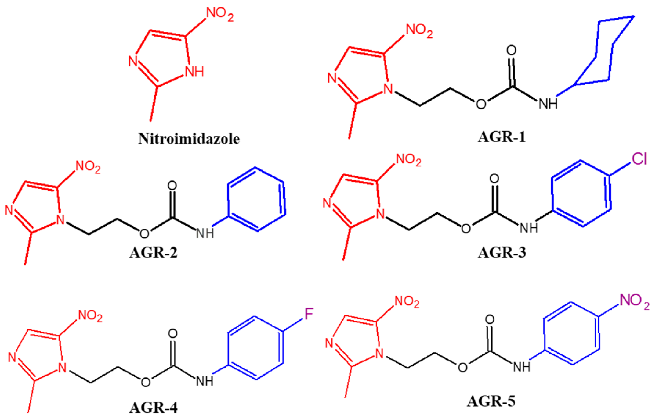
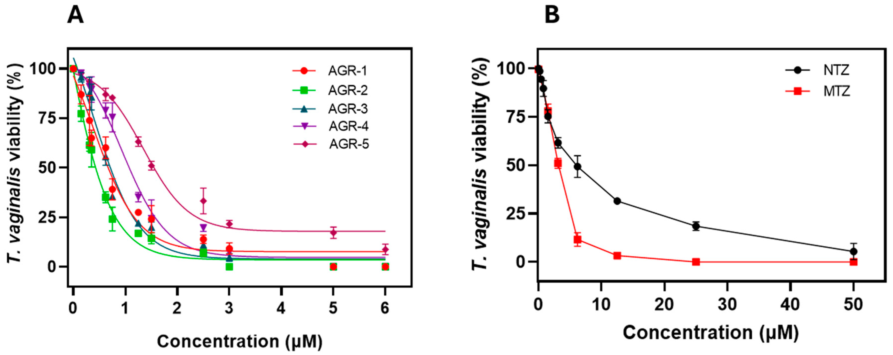
2.1. Selection of Compounds That Inhibit the Viability of Trichomonas vaginalis

Chemical compound libraries are advantageous for the discovery of new bioactive small molecules. We evaluated an in-house library comprising 55 compounds for their effects on the viability of T. vaginalis Donne ATCC 30236. The assays were carried out at a final concentration of 25 µM to identify the compounds that inhibited the viability of the trophozoites by more than 90% compared with no compound. Figure 1 shows the structures of the compounds that had trichomonacidal effects on the culture of T. vaginalis, where the viability of the trophozoites was completely eliminated (100%). These compounds were called AGR-1 to -5 and have previously been reported elsewhere [21]. In analyzing the structures of the compounds that decreased the viability of T. vaginalis, we observed that the compounds AGR-1 to -5 have a 5-nitroimidazole scaffold, similar to metronidazole (MTZ), an antiprotozoal drug, and a cycloalkyl/aryl carbamate tail.

2.2. Determination of IC50 Values

Previously, it was shown that the AGR compounds derived from 5-nitroimidazole can inhibit the viability of T. vaginalis Donne ATCC 30236 at 100% [21]; however, the IC50 was undetermined, and since this parameter is the most widely used to determine the drug’s efficacy, we sought to determine it in order to identify the most active compound. As shown in Figure 2, as the concentrations of the compounds increased, a decrease in the viability of T. vaginalis trophozoites was observed. The IC50 values for the compounds AGR-1, AGR-2, AGR-3, AGR-4, and AGR-5 were 0.67, 0.40, 0.64, 0.96, and 1.45 µM, respectively (Figure 2A). Interestingly, the compound with the most significant effect on the viability of the trophozoites was the compound AGR-2, followed by AGR-3, AGR-1, AGR-4, and AGR-5. In addition to this, we determined the IC50 values of the two antiparasitic drugs, namely metronidazole (MTZ) and nitazoxanide (NTZ) (commercial drugs used for standard therapies). Figure 2B shows a response in the effect when the concentration was increased, with an IC50 value of 3.1 µM for MTZ and 6.62 µM for NTZ. These results indicate that AGR compounds show a more significant trichomonacidal effect compared with already reported drugs, whereby enhancements of 8-fold and 16-fold were observed for the compound AGR-2 in relation to MTZ and NTZ, respectively. In contrast, AGR-1 and AGR-3 showed an enhancement of fivefold in relation to MTZ and an enhancement of 10-fold with respect to NTZ.
To understand the antiparasitic activities of these compounds as a function of time, we developed kinetic growth curves of T. vaginalis parasites in the absence and presence of these compounds (incubated at the IC50 concentration of each compound), and the numbers of trophozoites were counted after 96 h with trypan blue (0.4%) (1:1, v/v) using a Neubauer chamber and XTT assay tests. As seen in Figure 3, which show the kinetic growth curves of T. vaginalis in the absence of compounds (control group), we can observe four phases of progression: the lag phase (0–15 h), the logarithmic phase (15–40 h), the maximum stationary phase (48 h), and the death phase (after 48 h). However, when the T. vaginalis trophozoites were treated with each of the compounds, we observed that these compounds negatively affected the growth of T. vaginalis (Figure 3A). The same effect was observed with the commercial drugs MTZ and NTZ on the kinetic growth curves of T. vaginalis trophozoites (Figure 3B). Interestingly, all the compounds tested reduced the trophozoites’ proliferation in each of the phases of kinetic growth of T. vaginalis. Interestingly, the compounds AGR-1, AGR-2, and AGR-4 showed a more significant delay in the logarithmic and stationary phases. The results obtained for this assay demonstrated that after 24 h of incubation, the compounds reduced the proliferation of T. vaginalis trophozoite cells, as compared with cells without treatment.
From the assays to determine the IC50 value, it is evident that the series of five carbamate compounds showed potent trichomonacidal activity compared with the drugs MTZ and NTZ, showing a lower IC50 value. However, it was found that, among the series of compounds, AGR-2 showed the lowest value. But when the growth curve was examined in the presence of the compounds, the results revealed that the compound AGR-4 was the one that inhibited the growth of the parasite to a greater degree; this could be due to the mechanism of action of each of the compounds. Therefore, no compound was discarded for the subsequent expression tests.

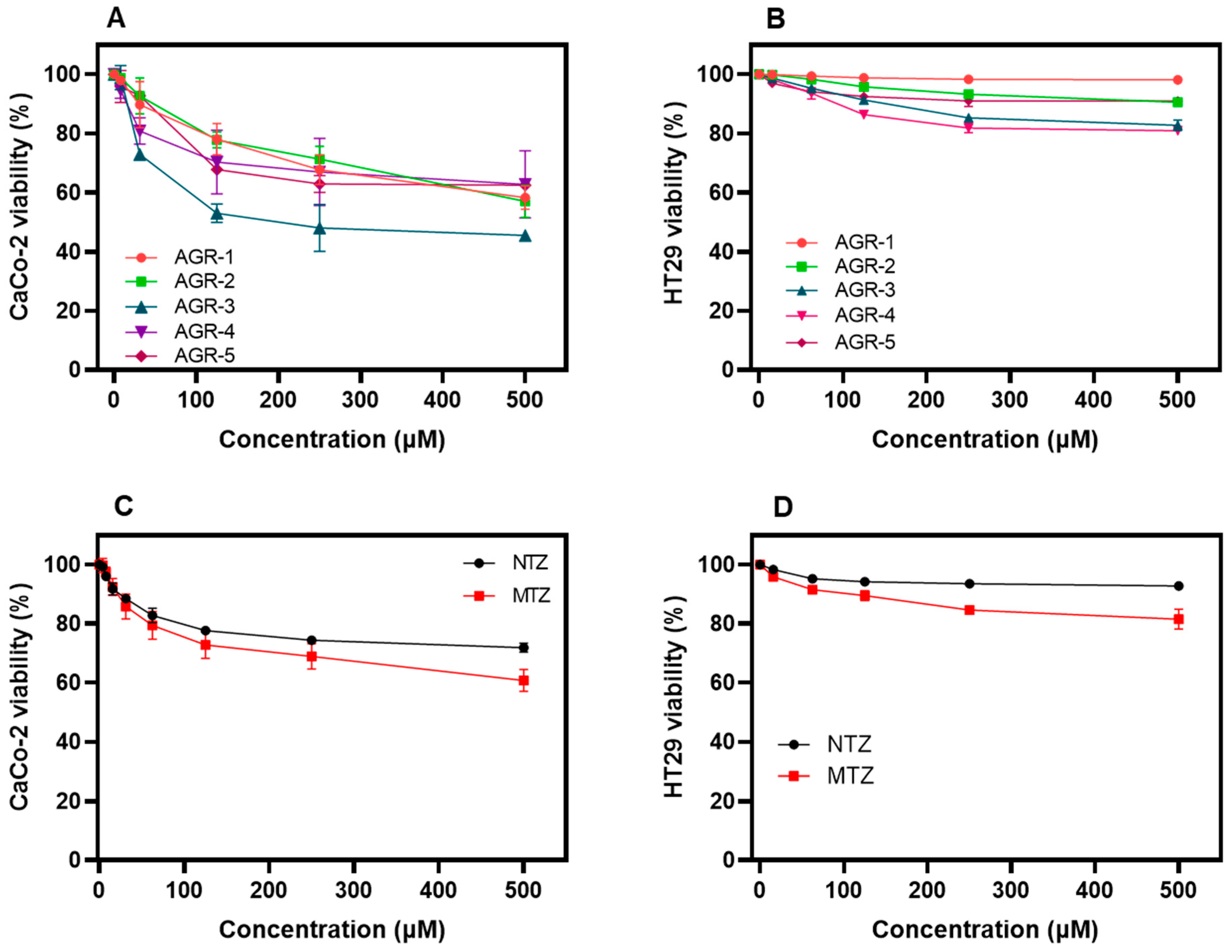
2.3. Evaluation of the Cytotoxicity of the AGR Compounds

The cytotoxic activities of the AGR compounds were evaluated in relation to the CaCo-2 and HT29 cells. As shown in Figure 4A, the data obtained from the XTT proliferation assay demonstrated that after 48 h of incubation at a concentration of 500 µM, the AGR compounds decreased the viability of CaCo-2 cells down to 40 (Figure 4A). Regarding HT29 cells, we determined an inhibition rate of around 20% at 500 µM in the presence of each one of the AGR compounds (Figure 4B).
From the concentration–response plots, the CC50 values (the concentration of the compound that reduces 50% of the cells’ viability) of each compound were obtained. The AGR compounds showed low toxicity in relation to the CaCo-2 eukaryotic cells, for which CC50 values from 510 µM to 650 µM were determined, while in HT29 eukaryotic cells, CC50 values from 532 µM to 730 µM were determined (Table 1). These results indicated that the AGR compounds offer very low median cytotoxic concentrations. In addition to this, we determined the CC50 values of the two commercial drugs used in standard therapies against trichomoniasis (MTZ and NTZ) in relation to CaCo-2 and HT29 eukaryotic cells. As seen in Table 1, the CC50 values for MTZ against CaCo-2 and HT29 eukaryotic cells were 19 and 265 µM, respectively, while the CC50 values for NTZ were 26.8 µM and >50 µM for CaCo-2 and HT29 eukaryotic cells, respectively. However, it is important to mention that the viability of T. vaginalis trophozoites at 100% was eliminated at 6 μM (Figure 2A). In contrast, at 10 μM of the compounds, the CaCo-2 and HT29 cell lines maintained 100% viability (Figure 4A,B).
On the other hand, the selectivity index (SI), which is the ratio of a compound’s CC50 value to its IC50 value, was calculated to assess the selective toxicity of the AGR compounds against T. vaginalis trophozoites (Table 1). A greater SI value indicates that the compounds are more selective, providing maximum trichomonacidal activity with minimal cell toxicity. Against CaCo-2 cells, we determined SI values ranging from 400 to >1000, while in HT29 cells, SI values ranging from 386 to >1000 were measured. Interestingly, the compounds AGR-1, AGR-2, and AGR-3 showed greater SI values in CaCo-2 and HT29 eukaryotic cells. In addition, we determined the SI values for MTZ and NTZ, which were 175 and 87.6 for CaCo-2 cells and 177 and 95.7 for HT29 cells, respectively. According to the selectivity index, the data shown in Table 1 indicate that both the compounds exhibited a higher cytotoxic selectivity against T. vaginalis, suggesting that they represent promising antiparasitic compounds and thus establishing a basis upon which to develop possible antitrichomonal drugs.

2.4. Predicted Pharmacokinetic Values of AGR Compounds

Computational pharmacology has been widely used as a tool to predict whether candidate drug compounds comply with the acceptable criteria for further evaluation. Therefore, prediction of the pharmacokinetic properties of the AGR compounds, such as the absorption, distribution, metabolism, excretion, and toxicity (ADMET) parameters, was performed using the platform ADMETlab 2.0’s online server (https://admetmesh.scbdd.com, accessed on 25 September 2023). As shown in Table 2, the calculations of the ADMET profiles demonstrated that all the AGR compounds presented high values of intestinal absorption and cross the blood–brain barrier (BBB) in the case of CNS protozooses. With Caco-2 as a predictive model of intestinal drug absorption, the absorption parameters indicated that all the AGR compounds had a high passive permeability, similar to MTZ. Regarding the evaluation of the distribution parameters, we determined that all the AGR compounds presented optimal distribution values (from 46 to 77%), wherein the optimal degree of plasma protein binding was less than 95%. In addition to this, we calculated the metabolic stability, and all compounds had low values as substrates of the main metabolizing enzymes in the body, particularly CYP1A2 and CYP2C19. The excretion parameters were predicted for all AGR compounds, suggesting satisfactory clearance values and a prolonged long half-life (>3 h). Finally, the toxicity parameters showed that the compounds had very low predicted hERG (human ether-a-go-go-related gene) channel blockage and could be considered non-cardiotoxic molecules, and they exhibited low acute oral toxicity in rat models (Table 2). Finally, it is worth mentioning that all the values of the ADMET parameters of the AGR compounds are in accordance with the parameters calculated for MTZ, a commercial drug used for standard therapies.

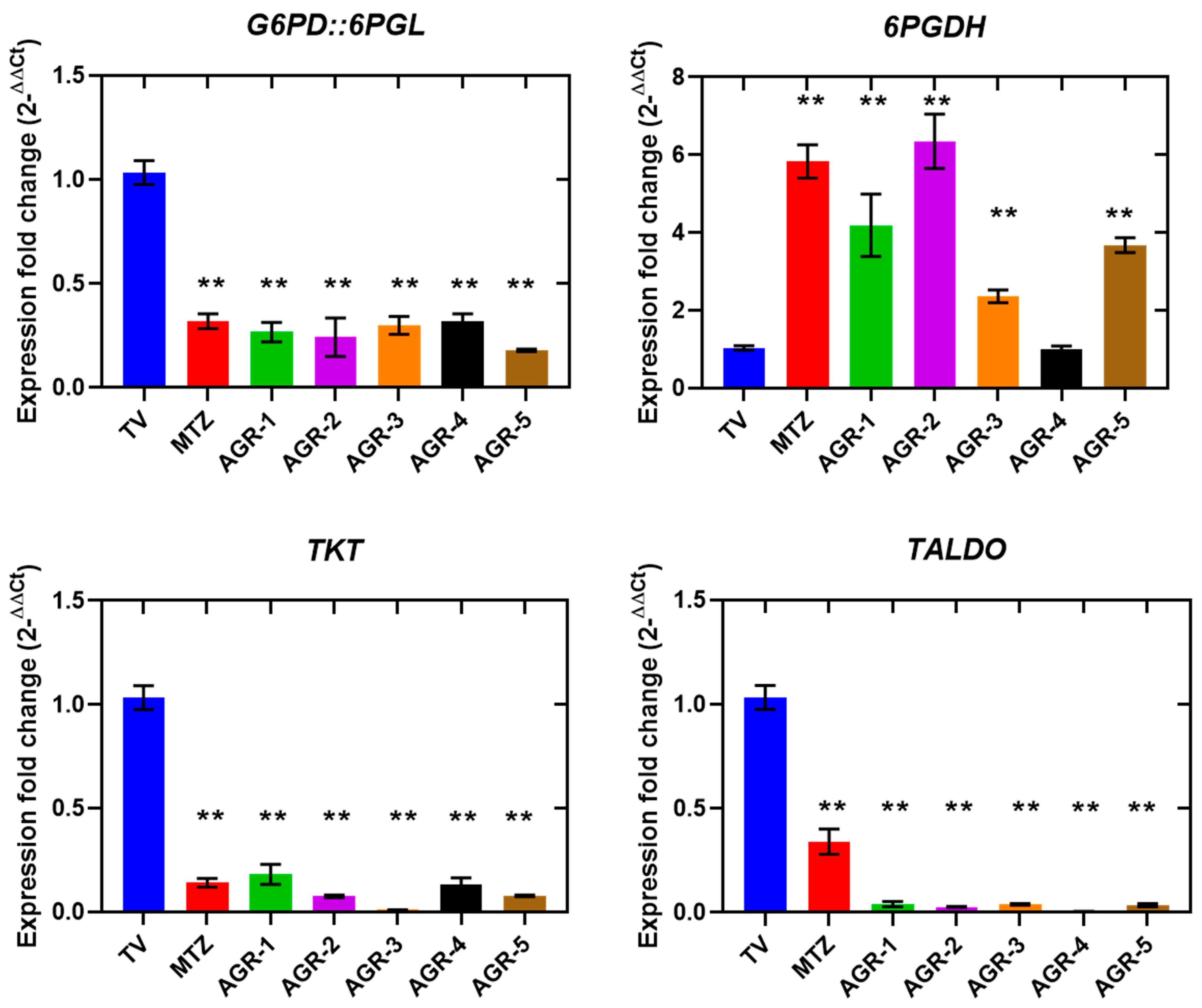
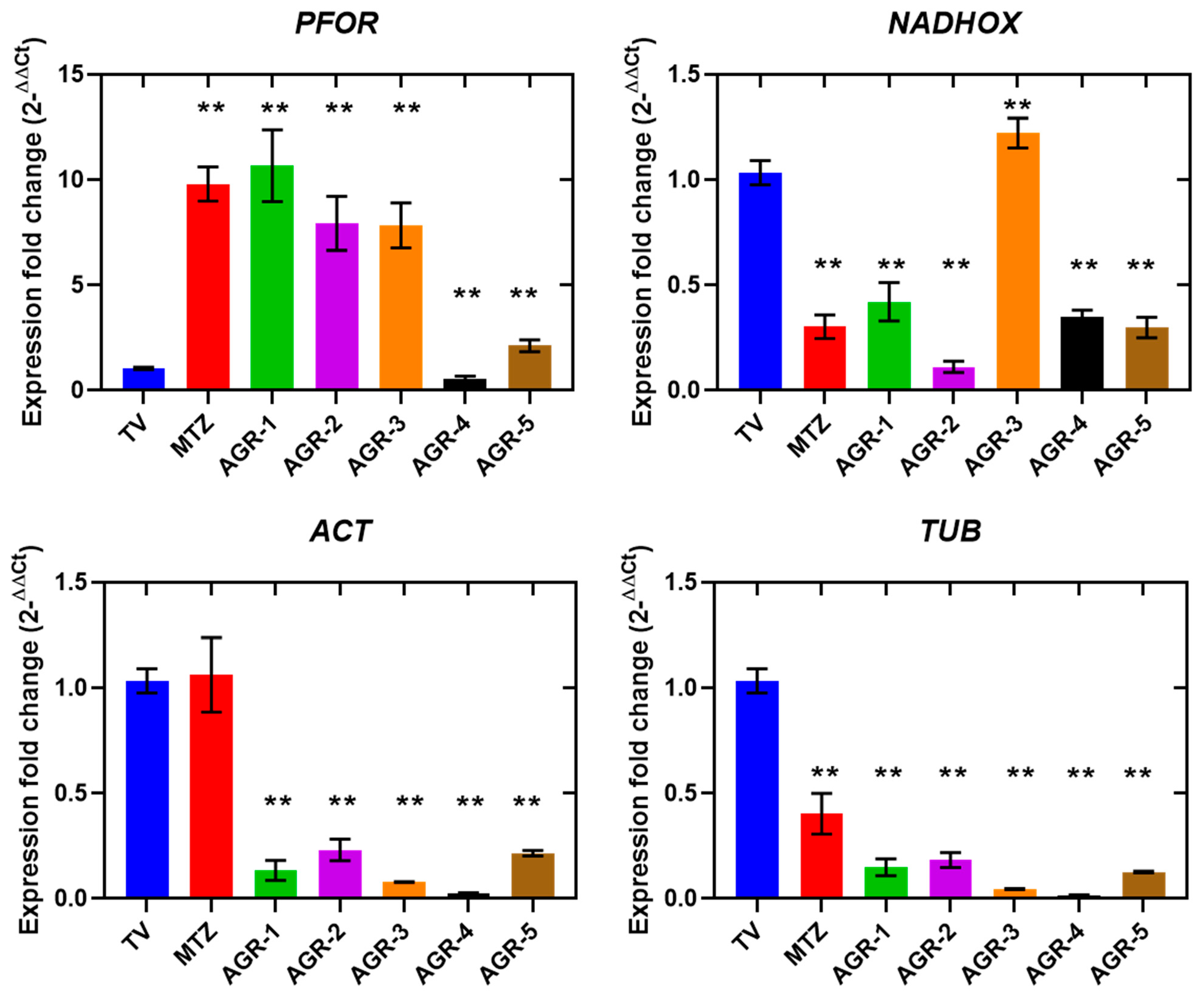
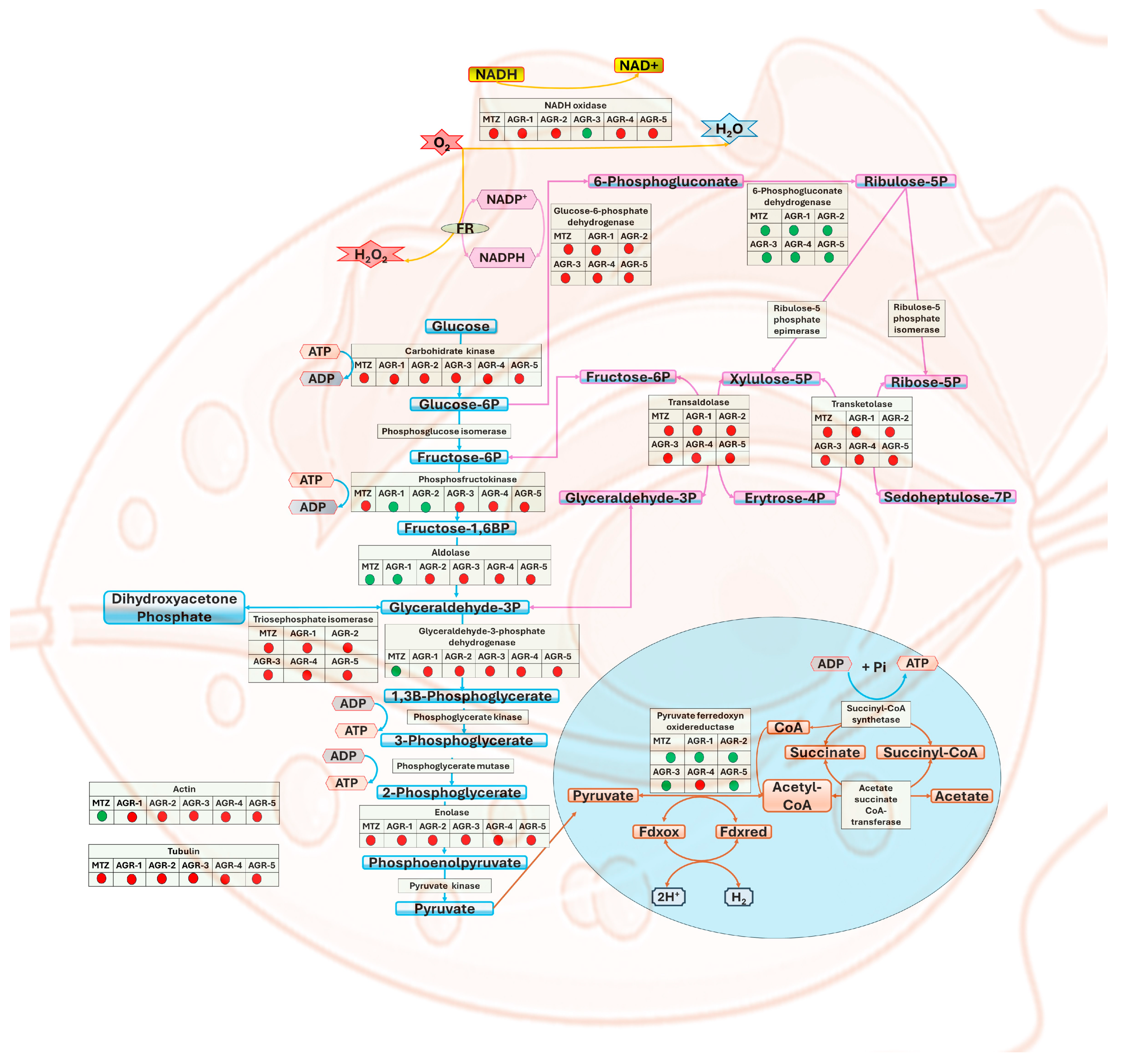
2.5. Determination of the Expression Levels of Genes Involved in the Metabolism of Trichomonas vaginalis

The main carbon and energy source for T. vaginalis is the fermentative carbohydrate metabolism [22,23]. We sought to determine whether the death of T. vaginalis is related to changes in its metabolic genes. A gene expression assay based on RT-qPCR was performed to evaluate the transcription levels of several genes in T. vaginalis trophozoites that were incubated for 24 h at the IC50 concentration for each compound. The results showed that the majority of the glycolytic genes that we assessed were downregulated. Specifically, the expression levels of the CK, TPI, and ENOL transcripts were significantly reduced after treatment with MTZ and all carbamate compounds (AGR-1 to -5), whereas AGR-2, -3, and -4 reduced ALDO and GAPDH, and, finally, AGR-3, -4, and -5 negatively affected PFK (Figure 5). Conversely, AGR-1 and AGR-2 increased PFK transcripts by 1.2- and 1.5-fold, respectively, and only ALDO was increased by AGR-1, similar to MTZ (Figure 5). On the other hand, trophozoites exposed to MTZ manifested overexpression of the ALDO and GAPDH genes, which were increased by 5.5- and 7-fold compared with the control. In summary, the results showed that each of the AGR compounds induced a different effect on the expression levels of glycolysis genes than the MTZ drug, which indicated that the mechanism of action could also be different from that described for the MTZ drug. The downregulation of CK, ALDO, TPI, GAPDH, and ENOL probably reduced the production of the corresponding proteins’ translation and consequently diminished glycolytic flux. As observed in a work where the impact of MTZ on the carbohydrate metabolism of Clostridioides difficile strains was evaluated, the amount of the enzymes glucose-6-phosphate isomerase, glyceraldehyde-3-phosphate dehydrogenase (GAPDH), aldolase, and enolase decreased in the presence of MTZ, while glycolysis enzymes involved in phosphorylation of the substrate, such as phosphoglycerate kinase, phosphoglycerate mutase, phosphofructokinase (PFK), and pyruvate kinase, were upregulated [24].
It is possible that reductions in glycolytic genes together with a diminished pentose phosphate pathway (PPP) may reduce energy and the redox balance, which consequently promotes the death of T. vaginalis. This suggests that exposure to the compounds alters the glycolytic pathway by which the energy required by T. vaginalis is obtained.
The expression profiles of the genes G6PD::6PGL, 6PGDH, TKT, and TALDO, the activities of which are involved in the PPP, were affected in trophozoites treated with both MTZ and the AGR compounds, which induced reductions in the transcripts of the G6PD::6PGL, TKT, and TALDO genes. However, the reduction in expression of the TKT and TALDO genes was more robust with the AGR compounds: the expression was practically fully inhibited (Figure 6). On the contrary, the expression of the 6GPDH gene increased in trophozoites treated with both MTZ and with the AGR compounds. The fundamental role of the PPP in T. vaginalis involves the production of NADPH and ribose-5-fosfato molecules, which are essential for maintaining the redox balance and synthesizing nucleic acids in the parasite [25]. Therefore, the results suggest that the AGR compounds and MTZ probably induced a deregulation in the redox balance, as well as a decrease in the precursors and proliferation of ribose-5-phosphate, which led to cell death. The PPP is a key metabolic route and, as such, it has been proposed as a strategic pathway for discovery of targets in drug design [26]. Furthermore, the G6PD:6PGL enzyme has been proposed as a potential pharmacological target for the design of drugs in parasites such as T. vaginalis, G. lamblia, and P. falciparum [27,28,29]. Due to the importance of the PPP in T. vaginalis, compounds that alter the expression of genes such as G6PD::6PGL are crucial therapeutic candidates, since, by negatively impacting expression, they also directly alter essential biological processes such as proliferation. In addition to the results of this work, it is also evident that AGR compounds also decrease the expression levels of genes in the oxidative phase of the PPP, such as TKT and ALDO, the proteins of which could be studied as therapeutic targets.
It has been shown that laboratory-generated T. vaginalis strains that are resistant to MTZ are associated with a downregulation of genes encoding hydrogenosomal enzymes that reduce MTZ, such as PFOR and Fd; however, clinically resistant T. vaginalis strains did not show a decrease in the transcription of PFOR or Fd [30,31,32]. In this work, we determined the expression profile of the PFOR gene in parasites in the presence or absence of the AGR compounds and MTZ, the activity of which is involved in hydrogenosomal metabolism. Parasites exposed to MTZ, AGR-1, AGR-2, AGR-3, and AGR-5 exhibited a positive regulation of PFOR, leading to overexpression by 9-, 11-, 7.5-, 7.5-, and 2.5-fold, respectively. Meanwhile, the compound AGR-4 induced a decrease of 1.5-fold in the expression of PFOR (Figure 7). T. vaginalis does not have mitochondria. Instead, it has an organelle called the hydrogenosome [33] (Figure 8); this organelle participates in the pyruvate metabolism and produces ATP and molecular hydrogen [34]. The pyruvate from degradation of glucose can be imported from the cytosol, and pyruvate is decarboxylated by the pyruvate:ferredoxin oxidoreductase (PFOR) enzyme in the hydrogenosome, generating CO2, acetyl-CoA and reduced ferredoxin (Fd). The CoA moiety of acetyl-CoA (from the PFOR reaction) is transferred to succinate by acetate:succinate CoAtransferase (ASCT), yielding acetate as an end product, and succinyl-CoA, yielding ATP through substrate-level phosphorylation [35]. To improve the quality of the therapy against T. vaginalis, it is important to develop compounds that interfere with key steps of the essential metabolic pathways, such as the production of energy. Thus, PFOR is a potential target for the design of drugs against T. vaginalis.
T. vaginalis is an anaerobic eukaryote whose central energy metabolism does not need O2, not even to survive and multiply. Even O2 becomes toxic for this parasite; it is generally believed that the inhibition of growth by O2 concentrations in anaerobes involves the inhibition of enzymes that contain FeS groups that are sensitive to oxygen, whose activity is involved in the central energy metabolism, such as PFOR [36,37]. To defend themselves against O2, T. vaginalis possesses a cytosolic NADH oxidase, which transfers four electrons from the oxidation of glucose directly to O2, producing water [38,39]. We determined the expression levels of NADH oxidase; the results revealed that MTZ increased the expression of the NADH oxidase gene, while four of the AGR compounds (AGR-1, -2, -4, and -5) decreased the expression of the NADH oxidase gene. This downregulation could have significant repercussions for improvements in O2 scavenging. These results suggest that AGR compounds manifest a redox imbalance (an increase in reactive oxygen species) in T. vaginalis, contrary to what was observed with MTZ, the effect of which was to increase the expression of NADH oxidase.
The expression profiles of the structural genes ACT and TUB were also determined in T. vaginalis after treatment with MTZ and the AGR compounds. The results revealed that MTZ did not induce changes in the expression of ACT, while it reduced the expression of TUB 2.5-fold compared with the control. In contrast, the trophozoites exposed to the AGR compounds showed strong decreases in the expressions of ACT and TUB compared with the corresponding trophozoites without treatment (Figure 7). Notably, the AGR compounds at the concentration tested almost completely inhibited the expression of the ACT and TUB genes. It has been described that the actin and tubulin proteins play a key role in morphogenesis, mitosis, and virulence in T. vaginalis [40,41]. Due to the importance of the microtubular cytoskeleton in T. vaginalis, compounds that alter the expression of genes such as TUB are crucial therapeutic components, since, by negatively impacting expression, they also directly alter essential biological processes, such as motility, cell adhesion, and division.

2.6. Limitations

Although all the previous results suggested that the five imidazole carbamate compounds of the AGR series showed potent trichomonacidal activity with better IC50 values than metronidazole, it is necessary to support further research and extend these observations of the AGR compounds, which should be evaluated on MTZ-resistant strains. Moreover, we observed that the values of the ADMET parameters of the AGR compounds were in accordance with the parameters calculated for MTZ; these compounds should be analyzed in an animal model of vaginal infection and, subsequently, the cytotoxicity, permeability, and the existence of these compounds in the active site of the vagina should be determined to detect the possible toxic effects of candidate drugs before administering them to humans in clinical trials.

3. Materials and Methods

3.1. Parasites and Cell Culture

The T. vaginalis Donne ATCC 30236 strain was acquired from the ATCC (American Type Culture Collection). It was cultured axenically in trypticase–yeast extract–maltose medium (TYM, at pH 6.0) supplemented with 10% sterile horse serum (previously inactivated at 56 °C for 30 min) and incubated at 37 °C [42]. The trophozoites were analyzed via motility, morphology, and trypan blue dye exclusion assays (0.4%) under an optical microscope with a magnification of 40×. The parasites were cultured for 24 h at 37 °C, corresponding to the logarithmic growth phase, and were used for anti-Trichomonas assays [43].

3.2. Selection of Compounds That Inhibit the Viability of Trichomonas vaginalis

Screening of an in-house library of 55 compounds with structural characteristics similar to antiparasitic drugs such as MTZ and NTZ was carried out. The compounds were designed and synthesized in the Medicinal Chemistry Laboratory of the Faculty of Pharmacy of the Autonomous University of the State of Morelos [21,44,45]. The purified compounds were dissolved in DMSO and incubated at a final concentration of 25 µM in the presence of 2.6 × 10⁵ trophozoites/mL in the TYM medium for 24 h at 37 °C. Thereafter, trypan blue dye exclusion and XTT assay tests were used for evaluations of cell viability. For the trypan blue dye exclusion assay, after incubation, trophozoites were counted with trypan blue (0.4%) (1:1, v/v) in a Neubauer chamber to determine the trophozoites’ motility, morphology, and viability. We also used the Cell Proliferation Kit II (XTT, Roche Diagnostics Deutschland GmbH, Mannheim, Germany); after 24 h of incubation; it analyzed the number of viable cells by the cleavage of tetrazolium salts added to the culture medium. All experiments were performed independently at least three times in triplicate, and the results were expressed as percentages of viable trophozoites compared with the negative control. We selected the compounds that manifested over 90% inhibition of the viability of T. vaginalis.

3.3. Determination of IC50 Values

The compounds that induced over 90% inhibition of the viability of T. vaginalis were identified as the most active compounds, and their 50% inhibitory concentrations (IC50) were determined. The anti-Trichomonas activity was evaluated in 1.5 mL tubes, where 2.6 × 10⁵ trophozoites/mL was treated with increasing concentrations (0–10 µM) of the selected compounds. The tubes were incubated at 37 °C for 24 h. Then the trypan blue dye exclusion and XTT assay tests were used for evaluations of cell viability, as previously described. Three controls were used for each compound: (1) a negative control containing only trophozoites, (2) a negative control using 0.06% DMSO, and (3) two positive controls containing MTZ and NTZ (Sigma-Aldrich, St. Louis, MO, USA). The IC50 values were calculated using GraphPad Prism 8.0 Inc. software, Version 8.0.2 (263) (San Diego, CA, USA).

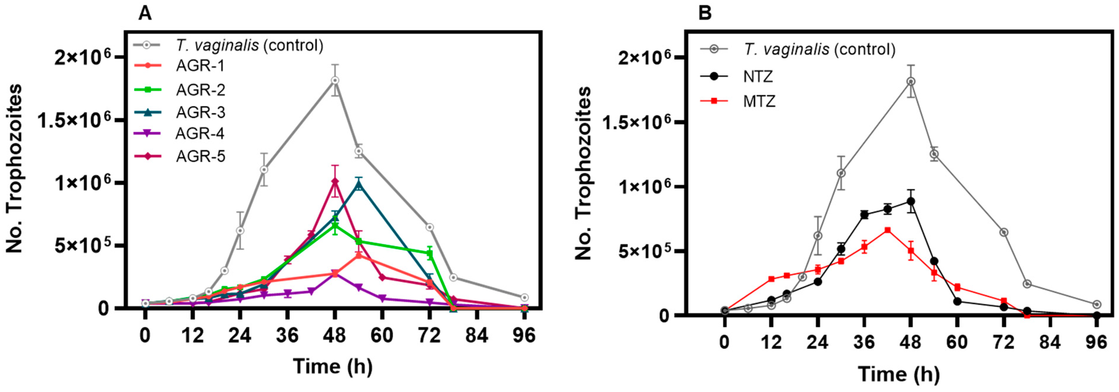
3.4. Effect of the Compounds on the Kinetic Growth Curve of Trichomonas vaginalis

To understand the antiparasitic activities of the compounds against T. vaginalis cultures, we produced a curve of the kinetic growth of T. vaginalis as a function of time. As previously described, T. vaginalis trophozoites were seeded in 1.5 mL tubes, and the compounds were added at the respective IC50 values; the tubes were incubated at 37 °C for 96 h. Trophozoites were observed via light microscopy, and their growth and viability were evaluated at 6, 12, 24, 48, 72, and 96 h. At the predetermined time points, the trypan blue dye exclusion (0.4%) (1:1, v/v) assay in a Neubauer chamber and XTT assay tests were performed for the evaluation of cell viability [46].

3.5. In Vitro Cytotoxicity Assay

The cytotoxicity of the active compounds against the Caco-2 and HT29 cell lines was evaluated. The Caco-2 and HT29 cell lines were provided by Luz Maria Rocha Ramirez from Hospital Infantil de México Federico Gómez, Secretaría de Salud. Cells were cultured in DMEM medium supplemented with 10% fetal bovine serum, 1% L-glutamine, and 1% penicillin/streptomycin at 37 °C in a 5% CO2 atmosphere. For the assay, cells were seeded in 96-well plates at a concentration of 2 × 10⁴ cells/well and incubated for 24 h [47]. After incubation, the cells were treated with increasing concentrations of each compound (0–500 µM) for 48 h at 37 °C in 5% CO2. After incubation, cell viability was assessed using the XTT assay. The results are expressed as a percentage of viable cells compared with untreated cells. The cytotoxic concentration 50 (CC50) was calculated using GraphPad Prism 7.0 software. As previously reported by Hernandez-Ochoa et al. [43], three controls were used in each assay: a negative control containing only trophozoites, a 0.06% DMSO control, and a positive control containing MTZ at 100 µM (Sigma-Aldrich, St. Louis, MO, USA). The assay was performed at least three times in triplicate. The selectivity index (SI) was calculated as SI = cells CC50/trophozoites IC50.

3.6. Primer Design

To evaluate the expression levels of the genes involved in the metabolism of T. vaginalis in the absence and presence of trichomonacidal compounds, the genetic expression levels of 15 genes were analyzed by means of real-time RT-qPCR. To this end, genes encoding enzymes involved in essential metabolic pathways for cell survival, such as glycolysis, the pentose phosphate pathway, structural proteins, and redox maintenance, were identified using the GenBank database (https://www.ncbi.nlm.nih.gov/genbank/ accessed on 15 January 2023) (Table 3).
The sequences for primer design were obtained from GenBank and then compared with reported sequences in the Trichomonas database (https://trichdb.org/trichdb/ accessed on 15 January 2023) (Table 4). The primer pairs were designed using mRNA sequences of the different genes, with lengths of 18–22 base pairs (bp) and a mean alignment temperature (Tm) of 60 ± 2 °C, and the formation of secondary structures and dimers was verified with the OligoEvaluatorTM online program from Sigma-Aldrich (http://www.oligoevaluator.com accessed on 15 January 2023). The oligonucleotides were designed to produce sizes of 80 to 150 bp, corresponding to each gene (Table 4).

3.7. RNA Extraction and Synthesis of First-Strand cDNA

The total RNA from trophozoites in the absence and presence of the compound was extracted using TRIzol® Reagent (Invitrogen, Carlsbad, CA, USA) according to the manufacturer’s instructions. The quality of RNA was assessed at 260/280 nm, and the integrity was assessed by 2.0% (w/v) agarose gel electrophoresis. For synthesis of the cDNA, samples were treated with 1 U of DNAase I enzyme (Thermo Fisher Scientific, Waltham, MA, USA). cDNA was synthesized using the primers Oligo (dT) 18 (Thermo Fisher Scientific) and RevertAid reverse transcriptase (Thermo Fisher Scientific). All synthesized cDNAs were quantified and stored at −70 °C until use.

3.8. Analysis of the Relative Gene Expression of T. vaginalis in the Presence of the Compounds

We evaluated the expression levels of genes involved in the metabolism of T. vaginalis trophozoites in the absence and presence of trichomonacidal compounds. The amplification of the specific PCR products and the specificity of the primers of the genes proposed in this study (Table 4) were determined using the Fast SYBR® Green Master Mix kit (Applied Biosystems, Foster City, CA, USA) in the StepOnePlusTM Real-Time PCR Systems platform (Life Technologies, Foster City, CA, USA), which was carried out according to the method previously reported by Gutiérrez-Cardona et al. [48].
The relative changes in the expression levels of the target genes (PK, CK, PFK, ALDO, TPI, GAPDH, ENOL, G6PD, 6PGDH, TKT, TALDO, PFOR, NADH, ACT, and TUB) were analyzed by the 2−ΔΔCt method, using the PK reference gene for normalization [48]. The curve was fitted with GraphPad Prism 8 software (GraphPad Software Inc., La Jolla, CA, USA), and statistical analysis was performed. ANOVA and the Tukey–Kramer test were used to evaluate the relative change in expression after normalization. Three replicates were included for each treatment, and all reactions were performed in triplicate.

4. Conclusions

The five imidazole carbamate compounds of the AGR series showed potent trichomonacidal activity with better IC50 values than metronidazole. The compound AGR-2 was the most potent, showing an IC50 of 400 nM. Regarding the expression profiles of metabolic genes in T. vaginalis, both MTZ and the five AGR compounds induced reductions in the expressions of the glycolytic genes CK, PFK, TPI, and ENOL, as well as the pentose phosphate genes G6PD, TKT, and TALDO; the redox maintenance gene NADHOX; and the structural genes ACT and TUB (Figure 8), suggesting that they probably share a similar mechanism of action. Finally, the compounds showed very low cytotoxicity in the Caco-2 and HT29 cell lines, and the ADMET parameters indicated that these compounds present pharmacokinetic properties similar to those of MTZ. The results are significant and can serve as a basis for future in vivo studies on the compounds to be used as new drugs against T. vaginalis.

Author Contributions

Conceptualization, S.G.-M. and B.H.-O.; methodology, software, validation, formal analysis, investigation, writing—original draft preparation, and writing—review and editing, V.M.-R., G.N.-V., D.O.-C., R.A.-E., V.P.d.l.C., E.C.-J., S.E.-F., C.W.-B., I.B.-R., L.M.-L., M.V.-B., M.A.R.-A., B.H.-O. and S.G.-M.; supervision, B.H.-O., G.N.-V., I.B.-R. and S.G.-M.; project administration, B.H.-O. and S.G.-M.; funding acquisition, S.G.-M. All authors have read and agreed to the published version of the manuscript.

Funding

This research was funded by the E022 Program, National Institute of Pediatrics, Mexico City, Mexico (Recursos Fiscales para la Investigación): INP 059/2021; program Hospital Infantil de Mexico Federico Gómez, Mexico City, Mexico (Fondos Federales): HIM/2019/036 SSA. 1595.

Institutional Review Board Statement

Not applicable.

Informed Consent Statement

Not applicable.

Data Availability Statement

Data are contained within the article.

Acknowledgments

V.M.-R., L.M.-L., M.V.-B. and M.A.R.-A. acknowledge the financial support from the CONACyT fellowship. We want to thank Pharmacy student Azriel Genaro Rocha for synthesizing the compound. In addition, we are grateful for the technical assistance of Maria Jose Gomez-Gonzalez and Ximena Gomez-Gonzalez. Finally, we thank Javier Gallegos Infante (Instituto de Fisiología Celular, UNAM) for assistance with the bibliographic materials.

Conflicts of Interest

The authors declare no conflicts of interest.

References

  1. Aquino, M.F.K.; Hinderfeld, A.S.; Simoes-Barbosa, A.  Trichomonas vaginalis. Trends Parasitol. 2020, 36, 646–647. [Google Scholar] [CrossRef]
  2. Kissinger, P. Trichomonas vaginalis: A review of epidemiologic, clinical and treatment issues. BMC Infect. Dis. 2015, 15, 307. [Google Scholar] [CrossRef]
  3. World Health Organization. Global Incidence and Prevalence of Selected Sexually Transmitted Infections-2008; World Health Organization: Geneva, Switzerland, 2012. [Google Scholar]
  4. Van Gerwen, O.T.; Muzny, C.A. Recent advances in the epidemiology, diagnosis, and management of Trichomonas vaginalis infection. F1000Research 2019, 8, 1666. [Google Scholar] [CrossRef]
  5. Leitsch, D. Recent advances in the Trichomonas vaginalis field. F1000Research 2016, 5, 162. [Google Scholar] [CrossRef]
  6. Bouchemal, K.; Bories, C.; Loiseau, P.M. Strategies for prevention and treatment of Trichomonas vaginalis infections. Clin. Microbiol. Rev. 2017, 30, 811–825. [Google Scholar] [CrossRef]
  7. Allsworth, J.E.; Ratner, J.A.; Peipert, J.F. Trichomoniasis and other sexually transmitted infections: Results from the 2001–2004 National Health and Nutrition Examination Surveys. Sex. Transm. Dis. 2009, 36, 738–744. [Google Scholar] [CrossRef]
  8. Upcroft, J.A.; Dunn, L.A.; Wright, J.M.; Benakli, K.; Upcroft, P.; Vanelle, P. 5-Nitroimidazole drugs effective against metronidazole-resistant Trichomonas vaginalis and Giardia duodenalis. Antimicrob. Agents. Chemother. 2006, 50, 344−347. [Google Scholar] [CrossRef]
  9. Sørensen, C.G.; Karlsson, W.K.; Amin, F.M.; Lindelof, M. Metronidazole-induced encephalopathy: A systematic review. J. Neurol. 2020, 267, 1–13. [Google Scholar] [CrossRef]
  10. Dunne, R.L.; Dunn, L.A.; Upcroft, P.; O’Donoghue, P.J.; Upcroft, J.A. Drug resistance in the sexually transmitted protozoan Trichomonas vaginalis. Cell Res. 2003, 13, 239–249. [Google Scholar] [CrossRef]
  11. Conrad, M.D.; Gorman, A.W.; Schillinger, J.A.; Fiori, P.L.; Arroyo, R.; Malla, N.; Dubey, M.L.; Gonzalez, J.; Blank, S.; Secor, W.E.; et al. Extensive genetic diversity, unique population structure and evidence of genetic exchange in the sexually transmitted parasite Trichomonas vaginalis. PLoS Negl. Trop. Dis. 2012, 6, e1573. [Google Scholar] [CrossRef]
  12. Workowski, K.A.; Bachmann, L.H.; Chan, P.A.; Johnston, C.M.; Muzny, C.A.; Park, I.; Reno, H.; Zenilman, J.M.; Bolan, G.A. Sexually Transmitted Infections Treatment Guidelines, 2021. MMWR Recomm. Rep. 2021, 70, 1–187. [Google Scholar] [CrossRef]
  13. Schmid, G.; Narcisi, E.; Mosure, D.; Secor, W.E.; Higgins, J.; Moreno, H. Prevalence of metronidazole-resistant Trichomonas vaginalis in a gynecology clinic. Int. J. Reprod. Med. 2001, 46, 545–549. [Google Scholar] [CrossRef]
  14. Kirkcaldy, R.D.; Augostini, P.; Asbel, L.E.; Bernstein, K.T.; Kerani, R.P.; Mettenbrink, C.J.; Pathela, P.; Schwebke, J.R.; Secor, W.E.; Workowski, K.A.; et al. Trichomonas vaginalis antimicrobial drug resistance in 6 US cities, STD Surveillance Network, 2009–2010. Emerg. Infect. Dis. 2012, 18, 939–943. [Google Scholar] [CrossRef]
  15. Kulda, J. Trichomonads, hydrogenosomes and drug resistance. Int. J. Parasitol. 1999, 29, 199–212. [Google Scholar] [CrossRef]
  16. Tachezy, J.; Kulda, J.; Tomkova, E. Aerobic resistance of Trichomonas vaginalis to metronidazole induced in vitro. Parasitology 1993, 106, 31–37. [Google Scholar] [CrossRef]
  17. Leitsch, D.; Janssen, B.D.; Kolarich, D.; Johnson, P.J.; Duchene, M. Trichomonas vaginalis flavin reductase 1 and its role in metronidazole resistance. Mol. Microbiol. 2014, 91, 198–208. [Google Scholar] [CrossRef]
  18. Leitsch, D.; Drinic, M.; Kolarich, D.; Duchene, M. Down-regulation of flavin reductase and alcohol dehydrogenase-1 (ADH1) in metronidazole-resistant isolates of Trichomonas vaginalis. Mol. Biochem. Parasitol. 2012, 183, 177–183. [Google Scholar] [CrossRef]
  19. Hrdý, I.; Cammack, R.; Stopka, P.; Kulda, J.; Tachezy, J. Alternative pathway of metronidazole activation in Trichomonas vaginalis hydrogenosomes. Antimicrob. Agents Chemother. 2005, 49, 5033–5036. [Google Scholar] [CrossRef]
  20. Cerkasovova, A.; Novak, J.; Cerkasov, J.; Kulda, J.; Tachezy, J. Metabolic properties of Trichomonas vaginalis resistant to metronidazole under anaerobic conditions. Acta Univ. Carol. Biol. 1988, 30, 505–512. [Google Scholar]
  21. Rocha-Garduno, G.; Hernandez-Martinez, N.A.; Colin-Lozano, B.; Estrada-Soto, S.; Hernandez-Nunez, E.; Prieto-Martinez, F.D.; Medina-Franco, J.L.; Chale-Dzul, J.B.; Moo-Puc, R.; Navarrete-Vazquez, G. Metronidazole and Secnidazole Carbamates: Synthesis, Antiprotozoal Activity, and Molecular Dynamics Studies. Molecules 2020, 25, 793. [Google Scholar] [CrossRef]
  22. Trussell, R.E.; Johnson, G. Physiology of pure culture of Trichomonas vaginalis: III. Fermentation of carbohydrates and related compounds. Proc. Soc. Exp. Biol. Med. 1941, 47, 176–178. [Google Scholar] [CrossRef]
  23. Müller, M.; Mentel, M.; van Hellemond, J.J.; Henze, K.; Woehle, C.; Gould, S.B.; Yu, R.Y.; van der Giezen, M.; Tielens, A.G.; Martin, W.F. Biochemistry and evolution of anaerobic energy metabolism in eukaryotes. Microbiol. Mol. Biol. Rev. 2012, 76, 444–495. [Google Scholar] [CrossRef] [PubMed]
  24. Doan, T.H.; Yen-Nicolaÿ, S.; Bernet-Camard, M.F.; Martin-Verstraete, I.; Péchiné, S. Impact of subinhibitory concentrations of metronidazole on proteome of Clostridioides difficile strains with different levels of susceptibility. PLoS ONE 2020, 15, e0241903. [Google Scholar] [CrossRef] [PubMed]
  25. Stover, N.A.; Dixon, T.A.; Cavalcanti, A.R. Multiple independent fusions of glucose-6-phosphate dehydrogenase with enzymes in the pentose phosphate pathway. PLoS ONE 2011, 6, e22269. [Google Scholar] [CrossRef] [PubMed]
  26. Morales-Luna, L.; Vázquez-Bautista, M.; Martínez-Rosas, V.; Rojas-Alarcón, M.A.; Ortega-Cuellar, D.; González-Valdez, A.; Pérez de la Cruz, V.; Arreguin-Espinosa, R.; Rodríguez-Bustamante, E.; Rodríguez-Flores, E.; et al. Fused Enzyme Glucose-6-Phosphate Dehydrogenase::6-Phosphogluconolactonase (G6PD::6PGL) as a Potential Drug Target in Giardia lamblia, Trichomonas vaginalis, and Plasmodium falciparum. Microorganisms 2024, 12, 112. [Google Scholar] [CrossRef] [PubMed]
  27. Allen, S.M.; Lim, E.E.; Jortzik, E.; Preuss, J.; Chua, H.H.; MacRae, J.I.; Rahlfs, S.; Haeussler, K.; Downton, M.T.; McConville, M.J.; et al. Plasmodium falciparum glucose-6-phosphate dehydrogenase 6-phosphogluconolactonase is a potential drug target. FEBS J. 2015, 282, 3808–3823. [Google Scholar] [CrossRef]
  28. Morales-Luna, L.; Hernández-Ochoa, B.; Martínez-Rosas, V.; Navarrete-Vázquez, G.; Ortega-Cuellar, D.; Rufino-González, Y.; González-Valdez, A.; Arreguin-Espinosa, R.; Franco-Vásquez, A.M.; de la Cruz, V.P.; et al. Giardia lamblia G6PD::6PGL Fused Protein Inhibitors Decrease Trophozoite Viability: A New Alternative against Giardiasis. Int. J. Mol. Sci. 2022, 23, 14358. [Google Scholar] [CrossRef] [PubMed]
  29. Martínez-Rosas, V.; Hernández-Ochoa, B.; Navarrete-Vázquez, G.; Martínez-Conde, C.; Gómez-Chávez, F.; Morales-Luna, L.; González-Valdez, A.; Arreguin-Espinosa, R.; Enríquez-Flores, S.; Pérez de la Cruz, V.; et al. Kinetic and Molecular Docking Studies to Determine the Effect of Inhibitors on the Activity and Structure of Fused G6PD::6PGL Protein from Trichomonas vaginalis. Molecules 2022, 27, 1174. [Google Scholar] [CrossRef]
  30. Mead, J.R.; Fernadez, M.; Romagnoli, P.A.; Secor, W.E. Use of Trichomonas vaginalis clinical isolates to evaluate correlation of gene expression and metronidazole resistance. J. Parasitol. 2006, 92, 196–199. [Google Scholar] [CrossRef]
  31. Wright, J.M.; Webb, R.I.; O’Donoghue, P.; Upcroft, P.; Upcroft, J.A. Hydrogenosomes of laboratory-induced metronidazole-resistant Trichomonas vaginalis lines are downsized while those from clinically metronidazole-resistant isolates are not. J. Eukaryot. Microbiol. 2010, 57, 171–176. [Google Scholar] [CrossRef]
  32. Wright, J.M.; Dunn, L.A.; Kazimierczuk, Z.; Burgess, A.G.; Krauer, K.G.; Upcroft, P.; Upcroft, J.A. Susceptibility in vitro of clinically metronidazole-resistant Trichomonas vaginalis to nitazoxanide, toyocamycin, and 2-fluoro-2′-deoxyadenosine. Parasitol. Res. 2010, 107, 847–853. [Google Scholar] [CrossRef] [PubMed]
  33. Hrdy, I.; Müller, M. Primary structure of the hydrogenosomal malic enzyme of Trichomonas vaginalis and its relationship to homologous enzymes. J. Eukaryot. Microbiol. 1995, 42, 593–603. [Google Scholar] [CrossRef] [PubMed]
  34. Müller, M. The hydrogenosome. J. Gen. Microbiol. 1993, 139, 2879–2889. [Google Scholar] [CrossRef] [PubMed]
  35. Hrdy, I.; Tachezy, J.; Muller, M. Metabolism of trichomonad hydrogenosomes. In Hydrogenosomes and Mitosomes: Mitochondria of Anaerobic Eukaryotes; Tachezy, J., Ed.; Springer: Berlin/Heidelberg, Germany, 2008; pp. 114–145. [Google Scholar]
  36. Chabrière, E.; Charon, M.H.; Volbeda, A.; Pieulle, L.; Hatchikian, E.C.; Fontecilla-Camps, J.C. Crystal structures of the key anaerobic enzyme pyruvate:ferredoxin oxidoreductase, free and in complex with pyruvate. Nat Struct Biol. 1999, 6, 182–190. [Google Scholar]
  37. Duwor, S.; Brites, D.; Mäser, P. Phylogenetic Analysis of Pyruvate-Ferredoxin Oxidoreductase, a Redox Enzyme Involved in the Pharmacological Activation of Nitro-Based Prodrugs in Bacteria and Protozoa. Biology 2024, 13, 178. [Google Scholar] [CrossRef] [PubMed]
  38. Müller, M. Biochemical cytology of trichomonad flagellates. I. Subcellular localization of hydrolases, dehydrogenases, and catalase in Tritrichomonas foetus. J. Cell Biol. 1973, 57, 453–474. [Google Scholar]
  39. Lamien-Meda, A.; Leitsch, D. Identification of the NADH-oxidase gene in Trichomonas vaginalis. Parasitol. Res. 2020, 119, 683–686. [Google Scholar] [CrossRef]
  40. Kusdian, G.; Woehle, C.; Martin, W.F.; Gould, S.B. The actin-based machinery of Trichomonas vaginalis mediates flagellate-amoeboid transition and migration across host tissue. Cell. Microbiol. 2013, 15, 1707–1721. [Google Scholar] [CrossRef] [PubMed]
  41. Lorenzo-Benito, S.; Rivera-Rivas, L.A.; Sánchez-Ayala, L.; Ortega-López, J.; Montes-Flores, O.; Talamás-Lara, D.; Arroyo, R. Omics Analyses of Trichomonas vaginalis Actin and Tubulin and Their Participation in Intercellular Interactions and Cytokinesis. Genes 2022, 13, 1067. [Google Scholar] [CrossRef]
  42. Diamond, L.S. The establishment of various trichomonads of animals and man in axenic cultures. J. Parasitol. 1957, 43, 488–490. [Google Scholar] [CrossRef]
  43. Hernández-Ochoa, B.; Martínez-Rosas, V.; Morales-Luna, L.; Calderón-Jaimes, E.; Rocha-Ramírez, L.M.; Ortega-Cuellar, D.; Rufino-González, Y.; González-Valdez, A.; Arreguin-Espinosa, R.; Enríquez-Flores, S.; et al. Pyridyl Methylsulfinyl Benzimidazole Derivatives as Promising Agents against Giardia lamblia and Trichomonas vaginalis. Molecules 2022, 27, 8902. [Google Scholar] [CrossRef]
  44. Nava-Zuazo, C.; Chávez-Silva, F.; Moo-Puc, R.; Chan-Bacab, M.J.; Ortega-Morales, B.O.; Moreno-Díaz, H.; Díaz-Coutiño, D.; Hernández-Núñez, E.; Navarrete-Vázquez, G. 2-Acylamino-5-nitro-1,3-thiazoles: Preparation and in vitro bioevaluation against four neglected protozoan parasites. Bioorg. Med. Chem. 2014, 22, 1626–1633. [Google Scholar] [CrossRef] [PubMed]
  45. Domínguez-Mendoza, E.A.; Galván-Ciprés, Y.; Martínez-Miranda, J.; Miranda-González, C.; Colín-Lozano, B.; Hernández-Núñez, E.; Hernández-Bolio, G.I.; Palomino-Hernández, O.; Navarrete-Vazquez, G. Design, Synthesis, and In Silico Multitarget Pharmacological Simulations of Acid Bioisosteres with a Validated In Vivo Antihyperglycemic Effect. Molecules 2021, 26, 799. [Google Scholar] [CrossRef] [PubMed]
  46. Alves, M.S.D.; Sena-Lopes, Â.; das Neves, R.N.; Casaril, A.M.; Domingues, M.; Birmann, P.T.; da Silva, E.T.; de Souza, M.V.N.; Savegnago, L.; Borsuk, S. In vitro and in silico trichomonacidal activity of 2,8-bis(trifluoromethyl) quinoline analogs against Trichomonas vaginalis. Parasitol. Res. 2022, 121, 2697–2711. [Google Scholar] [CrossRef] [PubMed]
  47. da Silva, C.C.; Pacheco, B.S.; das Neves, R.N.; Alves, M.S.D.; Sena-Lopes, A.; Moura, S.; Borsuk, S.; de Pereira, C.M.P. Antiparasitic activity of synthetic curcumin monocarbonyl analogues against Trichomonas vaginalis. Biomed. Pharmacother. 2019, 111, 367–377. [Google Scholar] [CrossRef]
  48. Gutiérrez-Cardona, J.Y.; Calderón-Jaimes, E.; Ortega-Cuellar, D.; Sánchez-Carrillo, A.; Castillo-Rodríguez, R.A.; Canseco-Ávila, L.M.; Rocha-Ramírez, L.M.; Martínez-Rosas, V.; Gómez-Manzo, S.; Hernández-Ochoa, B. Effect of Trichomonacide 6-Nitro-1H-benzimidazole Derivative Compounds on Expression Level of Metabolic Genes in Trichomonas vaginalis. Int. J. Mol. Sci. 2024, 25, 4568. [Google Scholar] [CrossRef]
Figure 1. Chemical structure of the compounds AGR-1 to -5 derived from 5-nitroimidazole. The AGR family has a basic structure formed from an imidazole ring with a nitro group in Position 5 (shown in red), and the constituents of the compounds AGR-1 to AGR-5 share a phenyl or cyclohexyl ring in the structure (shown in blue) linked by a carbamate group. These chemical structures have been generated with the ACD/ChemSketch.2.1 program.
Molecules 29 02585 g001
Figure 2. Determination of the IC50 values of AGR compounds on the culture of Trichomonas vaginalis. (A) Trichomonacidal activity of the AGR compounds and (B) the commercial drugs MTZ and NTZ in relation to the viability of T. vaginalis trophozoites. The trophozoites were incubated in the presence of the compounds for 24 h at 37 °C with different concentrations of each compound. Values represent the mean ± standard deviation of three independent experiments.
Molecules 29 02585 g002
Figure 3. Kinetic growth curves of Trichomonas vaginalis trophozoites treated with AGR compounds (A) and the commercial drugs MTZ and NTZ (B). The trophozoites were incubated at the IC50 values for each compound. The viability and number of trophozoites were monitored at 6, 12, 18, 24, 48, 72, and 96 h with trypan blue (0.4%) (1:1, v/v) and XTT assays. The values represent the mean ± standard deviation of three independent experiments, with p < 0.05 considered as statistically significant.
Molecules 29 02585 g003
Figure 4. Cytotoxic effects of AGR compounds and the commercial drugs MTZ and NTZ on Caco-2 and HT29 cells. Concentration–response curves for the viability of (A) Caco-2 and (B) HT29 cells treated with increasing concentrations of AGR compounds. Concentration–response curves for the viability of (C) Caco-2 and (D) HT29 cells treated with increasing concentrations of MTZ and NTZ. Cell viability was determined using trypan blue dye exclusion (0.4%) (1:1, v/v) in a Neubauer chamber, as well as an XTT proliferation assay. Values represent the mean ± standard deviation of three independent experiments.
Molecules 29 02585 g004
Figure 5. Relative expressions of glycolytic genes in Trichomonas vaginalis measured via RT-qPCR. Gene expression was compared among trophozoites of T. vaginalis without treatment, which were used as the negative control; trophozoites exposed to MTZ; and trophozoites exposed to the compounds AGR-1, AGR-2, AGR-3, AGR-4, and AGR-5, using PK as a reference gene. The asterisk indicates a significant difference (p < 0.05) in the expression. Values represent mean ± SD of three replicates.
Molecules 29 02585 g005
Figure 6. Relative expressions of pentose phosphate pathway genes in Trichomonas vaginalis measured via RT-qPCR. Comparison of the gene expression levels was carried out among trophozoites of T. vaginalis without treatment, used as the negative control; trophozoites exposed to MTZ; and trophozoites exposed to the compounds AGR-1, AGR-2, AGR-3, AGR-4 and AGR-5, using PK as a reference gene. The asterisk indicates a significant difference (p < 0.05) in the expression. Values indicate the mean ± SD of three replicates.
Molecules 29 02585 g006
Figure 7. Relative expressions of metabolic genes in Trichomonas vaginalis measured via RT-qPCR. Comparison of gene expressions carried out between trophozoites of T. vaginalis without treatment used as the negative control; trophozoites exposed to MTZ; and trophozoites exposed to the compounds AGR-1, AGR-2, AGR-3, AGR-4, and AGR-5, using PK as a reference gene. The asterisk indicates a significant difference (p < 0.05) in the expression. Bars indicate the mean ± SD values of three replicates.
Molecules 29 02585 g007
Figure 8. Differentially expressed genes in the trophozoites Trichomonas vaginalis exposed to MTZ and the compounds AGR-1, AGR-2, AGR-3, AGR-4, and AGR-5. Genes shown in red and green indicate downregulation and upregulation, respectively.
Molecules 29 02585 g008
Table 1. In vitro trichomonacidal activity and cytotoxicity of the AGR compounds.
CompoundIC50
(µM)
CaCo-2HT29
CC50
(µM)
SICC50
(µM)
SI
MTZ3.1545175550177
NTZ6.6258087.663495.7
AGR-10.67650970.0532794
AGR-20.40560>1000730>1000
AGR-30.64523817.1660>1000
AGR-40.96510531.2593617
AGR-51.45580400560386
The SI (selectivity index) corresponds to the ratio between the values of CC50 and IC50 for mammalian cells and trophozoites. CC50 represents the concentration of compounds that reduce the cells’ viability by 50%.
Table 2. Predicted pharmacokinetic values were calculated with ADMETLab 2.0 for AGR compounds.
ModelCompounds
AGR-1AGR-2AGR-3AGR-4AGR-5MTZ
AGastrointestinal absorption(+) High(+) High(+) High(+) High(+) High(+) High
Caco-2 permeability−4.67−4.5−4.46−4.59−4.66−4.71
Bioavailability (F)<30%<30%<30%<30%<30%<30%
DPlasma protein binding46%58%77%65%65%17%
BBB penetration0.820.6640.7950.620.290.83
Volume distribution0.809 L/kg0.831 L/kg0.859 L/kg0.878 L/kg0.84 L/kg0.83 L/kg
MCYP1A2 substrate(−) No(−) No(+) Yes(+) Yes(−) No(−) No
CYP2C19 substrate(−) No(+) Yes(+) Yes(+) Yes(−) No(+) Yes
CYP2C9 substrate(+) Yes(−) No(+) Yes(+) Yes(+) Yes(+) Yes
EClearance7.49 mL/min/kg9.47 mL/min/kg8.2 mL/min/kg9.44 mL/min/kg8.21 mL/min/kg6.29 mL/min/kg
Half-life (T½)>3 h>3 h>3 h>3 h>3 h>3 h
ThERG blockers0.210.220.470.370.680.04
Rat oral acute toxicity0.310.220.230.330.140.038
Absorption, A; distribution, D; metabolism, M; excretion, E; toxicity, T.
Table 3. Genes analyzed in this study.
Gene SymbolGene NameFunctionGenBank
CKCarbohydrate kinaseTransferase in glycolysisXM_001579622.1
PFKPhosphofructokinaseTransferase in glycolysisXM_001581728.2
ALDOAldolaseOxidoreductase in glycolysisXM_001315350.2
TPITriose phosphate isomeraseIsomerase in glycolysisXM_001320301.2
GAPDHGlyceraldehyde-3-phosphate dehydrogenaseOxidoreductase in glycolysisXM_001581066.2
ENOLEnolaseHydratase in glycolysisXM_001325471.2
PKPyruvate kinaseTransferase in glycolysisXM_001329865.2
G6PDGlucose-6-phosphate dehydrogenaseOxidoreductase in pentose phosphateXM_001321943.2
6PGDH6-phosphogluconate dehydrogenaseOxidoreductase in pentose phosphateXM_001323727.2
TKTTransketolase,Transferase in pentose phosphateXM_001326902.1
TALDOTransaldolaseTransferase in pentose phosphateXM_001330311.2
ACTActinCytoskeletal structural protein XM_001301716.2
TUBTubulinCytoskeletal structural proteinXM_001321203.2
PFORPyruvate-ferredoxin oxide reductaseOxidoreductase enzymeXM_001321286.2
NADHOXNADH oxidaseO2-Detoxifying enzymeXM_001315387.2
Table 4. Sequence of oligonucleotides used in this study.
GeneSequence 5′ → 3′Amplicon (bp)Tm (°C)
CKFw: 5′TACAACAGGAGCCGGAGATG 3′
Rv: 5′AGCAGCACAACCTCTCTTTG 3′
9760
PFKFw: 5′ TGCAGTTCTCTCTAGTGGCC 3′
Rv: 5′ CACGGAAGCCACCAGTAATG 3′
11660
ALDOFw: 5′ AAGTCACTCGGTCTCTGCAA 3′
Rv: 5′ TTGACGGAGGCTGTGATGAT 3′
12560
TPIFw: 5′ GGCAAGTGGGACGATGTTG 3′
Rv: 5′ TTAGCAGCAAGGATGTCACG 3′
12260
GAPDHFw: 5′ CCAAGTTGTCGCTATCCACG 3′
Rv: 5′ TGCTTAGCCTCATCGACTGT 3′
11460
ENOLFw: 5′ ACAGGTGTTGGTGAAGCTCT 3′
Rv: 5′ AGCACATTCCCTTGAGAGCT 3′
12460
PKFw: 5′ CCACAAGCAAACACTCGACA 3′
Rv: 5′ CTCCAACTTGCCAACACGAA 3′
10960
G6PDFw: 5′ ATTCTCACGTCTCCACCAGG 3′
Rv: 5′ GTCATCGTAGCCACCAGAGA 3′
10960
6PGDHFw: 5′ CGATGGTGGCAACTCTCACT 3′
Rv: 5′ CTCTTCACCGCCGGAGATAC 3′
12260
TKTFw: 5′ GGAGTAAGACTTGGCTGGGA 3′
Rv: 5′ CGTTCTGCACATTTCTCTGGT 3′
12560
TALDOFw: 5′ TCCTCAAGATTGTCCCAGGC 3′
Rv: 5′ TCTTGATTCCGGCTTCGTGA 3′
12360
ACTFw: 5′ GTCAAGCTTCTCACAGAGCG 3′
Rv: 5′ GGCCTTCTCCATTTCAGCAT 3′
12360
TUBFw: 5′ CTTCCGTGGCCGTATGTCAT 3′
Rv: 5′ GCAGATAGCGGACTTGACGT 3′
11560
PFORFw: 5′ CCAGATCACACCACTCGACT 3′
Rv: 5′ TTCCCAGTTCTTGCCCTCTT 3′
12160
NADHOXFw: 5′ ATTGGCTTGGCGTCCTTGAT 3′
Rv: 5′ TCGACGAGAACTGCACCTTC 3′
11860
Disclaimer/Publisher’s Note: The statements, opinions and data contained in all publications are solely those of the individual author(s) and contributor(s) and not of MDPI and/or the editor(s). MDPI and/or the editor(s) disclaim responsibility for any injury to people or property resulting from any ideas, methods, instructions or products referred to in the content.

