
Photochemical and Oxidative Degradation of Chamazulene Contained in Artemisia, Matricaria and Achillea Essential Oils and Setup of Protection Strategies


Abstract
:
1. Introduction
2. Results and Discussion
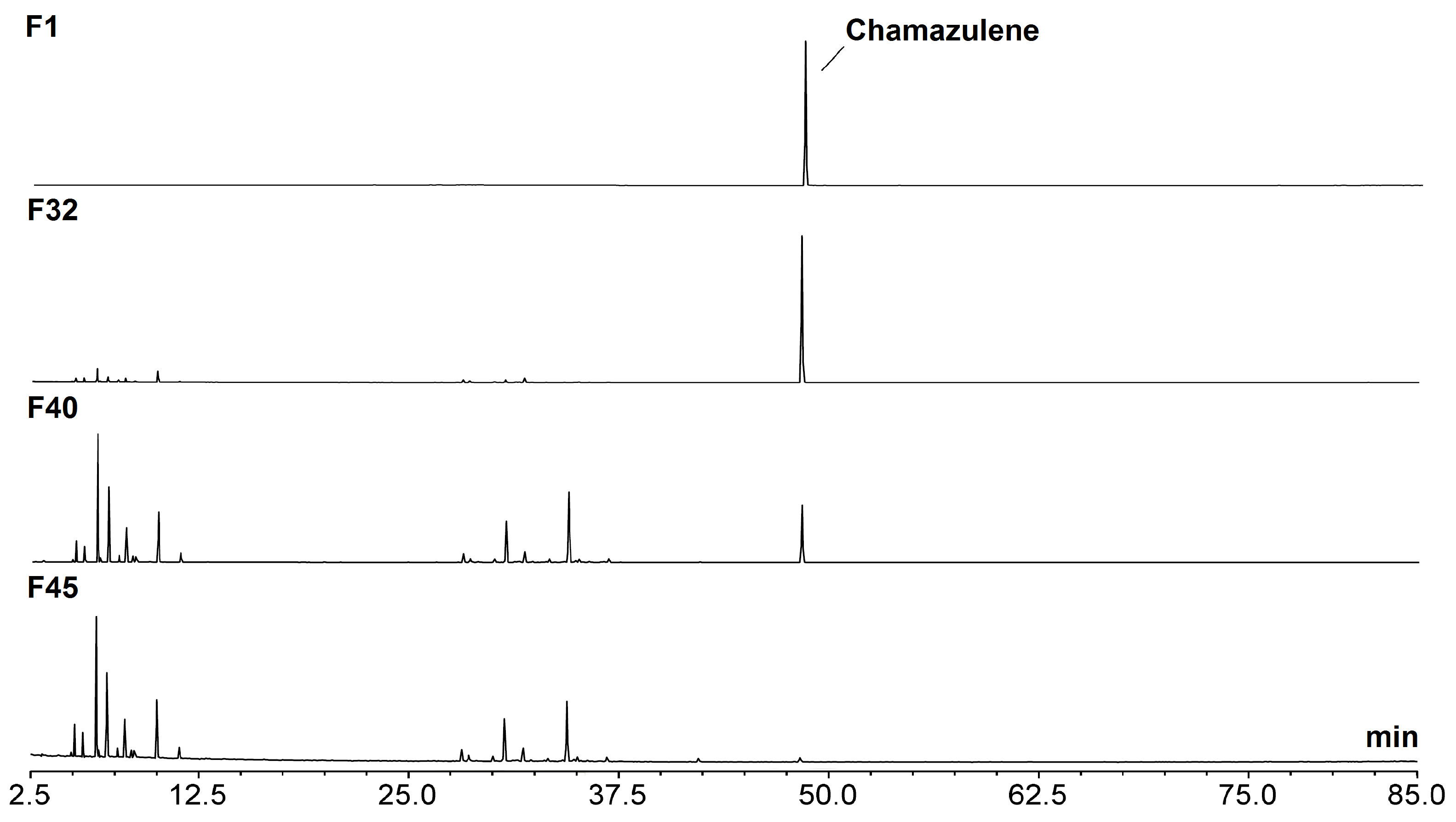
2.1. CA Isolation and Purification
2.2. Generation of Photo-Degradation Products of CA
2.3. Photostability Test
2.3.1. Effect of Solvent and Oxygen
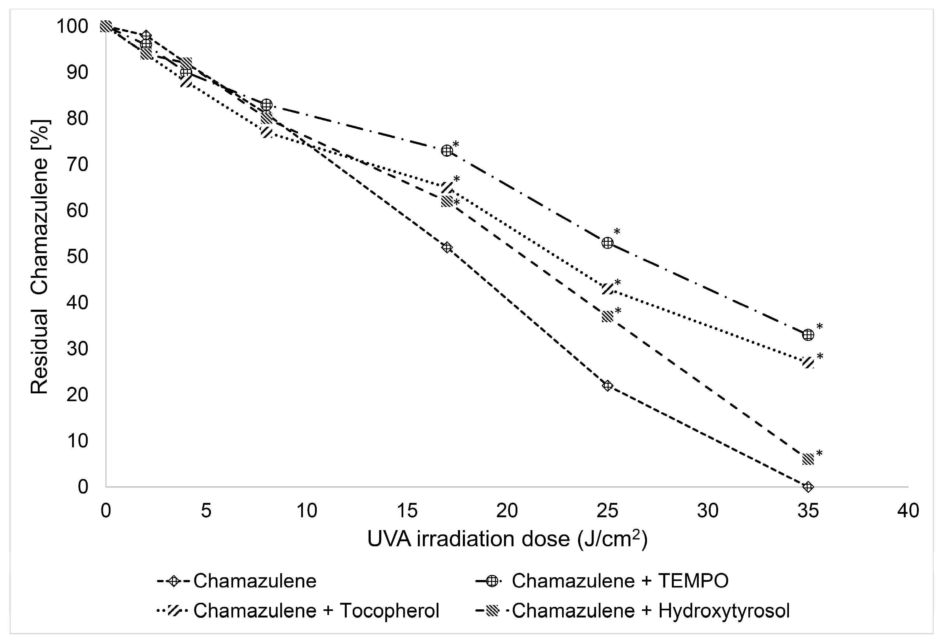
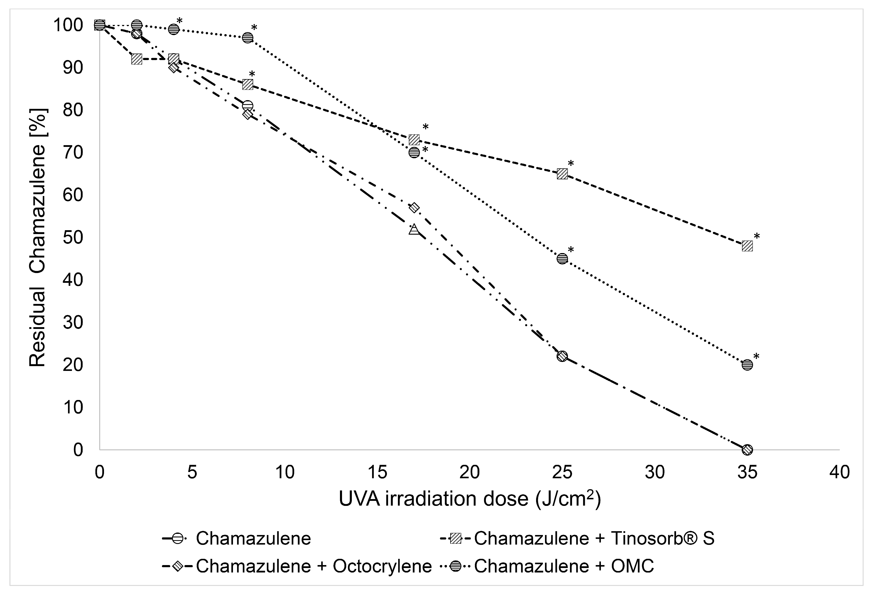
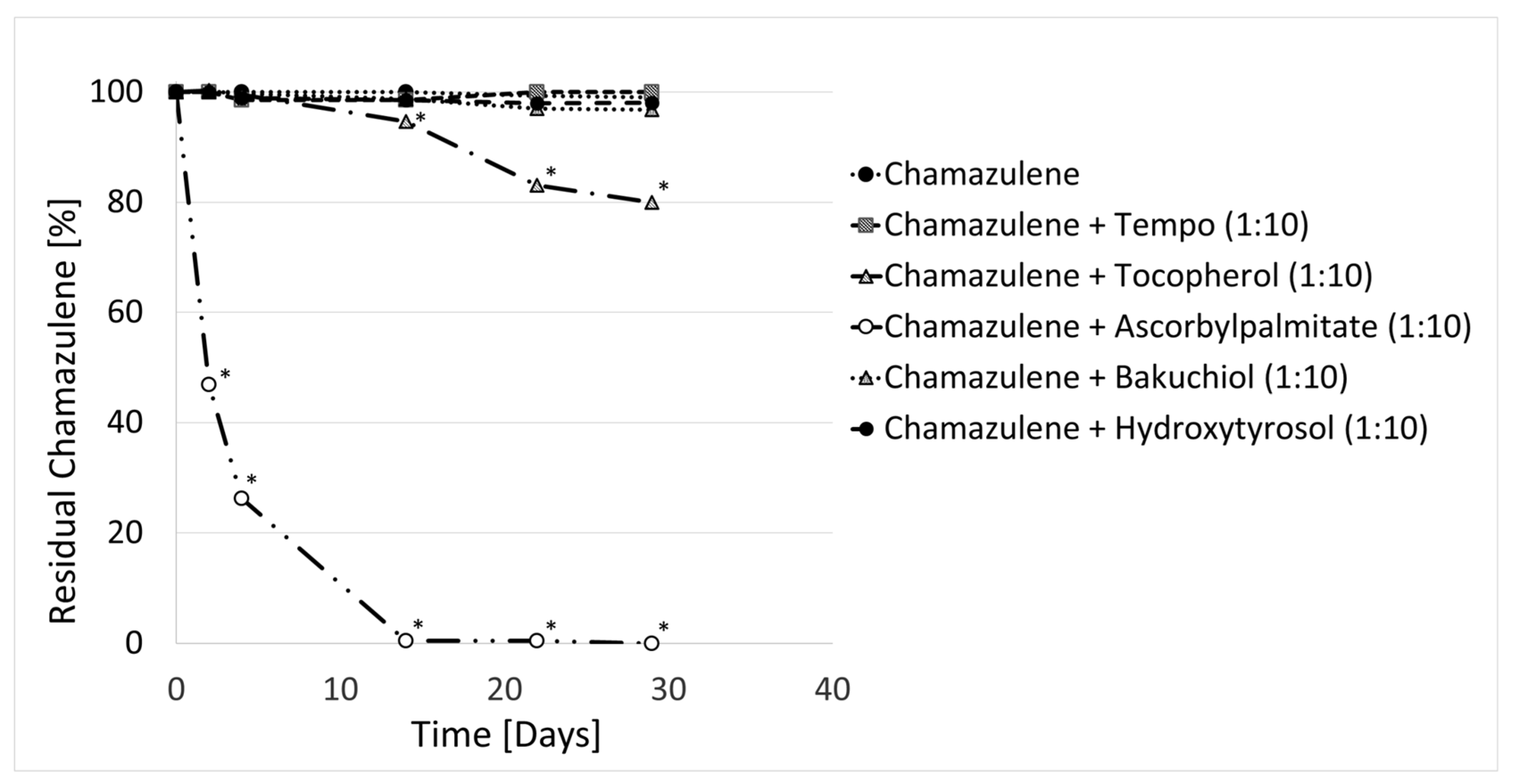
2.3.2. Effect of Antioxidants and Sunscreen
2.4. Thermal Stability of Chamazulene
3. Materials and Methods
3.1. Materials
3.2. Isolation of Chamazulene (CA)
3.3. Photolysis Experiments: Equipment and General Procedures
3.4. GC-MS Analysis
3.5. UPLC-ESI-MSn, ESI-MSn Analysis
3.6. LC-PDA Analysis
3.7. UV-Vis Spectrophotometry
3.8. Statistical Analysis
4. Conclusions
Supplementary Materials
Author Contributions
Funding
Institutional Review Board Statement
Informed Consent Statement
Data Availability Statement
Conflicts of Interest
References
- Russo, A.; Bruno, M.; Avola, R.; Cardile, V.; Rigano, D. Chamazulene-Rich Artemisia arborescens Essential Oils Affect the Cell Growth of Human Melanoma Cells. Plants 2020, 9, 1000. [Google Scholar] [CrossRef] [PubMed]
- Xu, Y.; Wang, Q.; Bao, W.; Pa, B. Antihyperlipidemic Effect, Identification and Isolation of the Lipophilic Components from Artemisia integrifolia. Molecules 2019, 24, 725. [Google Scholar] [CrossRef]
- Wang, Y.-F.; Zheng, Y.; Feng, Y.; Chen, H.; Dai, S.-X.; Wang, Y.; Xu, M. Comparative Analysis of Active Ingredients and Potential Bioactivities of Essential Oils from Artemisia argyi and A. verlotorum. Molecules 2023, 28, 3927. [Google Scholar] [CrossRef]
- De, J.; Lu, Y.; Ling, L.; Peng, N.; Zhong, Y. Essential Oil Composition and Bioactivities of Waldheimia glabra (Asteraceae) from Qinghai-Tibet Plateau. Molecules 2017, 22, 460. [Google Scholar] [CrossRef] [PubMed]
- Qasem, A.; Assaggaf, H.; Montesano, D.; Khalil, Z.; Al-Mijalli, S.H.; Baaboua, A.E.; El Omari, N.; El Menyiy, N.; Bakrim, S.; Sheikh, R.A.; et al. Determination of Chemical Compounds and Investigation of Biological Properties of Matricaria chamomilla Essential Oils, Honey, and Their Mixture. Molecules 2022, 27, 5850. [Google Scholar] [CrossRef] [PubMed]
- Mohammed, H.A. Phytochemical Analysis, Antioxidant Potential, and Cytotoxicity Evaluation of Traditionally Used Artemisia absinthium L. (Wormwood) Growing in the Central Region of Saudi Arabia. Plants 2022, 11, 1028. [Google Scholar] [CrossRef]
- Konarska, A.; Weryszko-Chmielewska, E.; Sulborska-Różycka, A.; Kiełtyka-Dadasiewicz, A.; Dmitruk, M.; Gorzel, M. Herb and Flowers of Achillea millefolium subsp. millefolium L.: Structure and Histochemistry of Secretory Tissues and Phytochemistry of Essential Oils. Molecules 2023, 28, 7791. [Google Scholar] [CrossRef]
- Ramadan, M.; Goeters, S.; Watzer, B.; Krause, E.; Lohmann, K.; Bauer, R.; Hempel, B.; Imming, P. Chamazulene carboxylic acid and matricin: A natural profen and its natural prodrug, identified through similarity to synthetic drug substances. J. Nat. Prod. 2006, 69, 1041–1045. [Google Scholar] [CrossRef]
- Sizova, N.V. Composition and antioxidant activity of essential oils containing azulene derivatives. Pharm. Chem. J. 2012, 46, 42–44. [Google Scholar] [CrossRef]
- Querio, G.; Antoniotti, S.; Foglietta, F.; Levi, R.; Bertea, C.M.; Canaparo, R.; Gallo, M.P. Chamazulene prevents ROS production in human dermal fibroblast and bovine aortic endothelial cells exposed to oxidative stress. Vascul. Pharmacol. 2018, 56, 103–105. [Google Scholar] [CrossRef]
- Capuzzo, A.; Occhipinti, A.; Maffei, M.E. Antioxidant and radical scavenging activities of chamazulene. Nat. Prod. Res. 2014, 28, 2321–2323. [Google Scholar] [CrossRef] [PubMed]
- Matera, R.; Lucchi, E.; Valgimigli, L. Plant Essential Oils as Healthy Functional Ingredients of Nutraceuticals and Diet Supplements: A Review. Molecules 2023, 28, 901. [Google Scholar] [CrossRef] [PubMed]
- Gabbanini, S.; Lucchi, E.; Carli, M.; Berlini, E.; Minghetti, A.; Valgimigli, L. In vitro evaluation of the permeation through reconstructed human epidermis of essentials oils from cosmetic formulations. J. Pharm. Biomed. Anal. 2009, 50, 370–376. [Google Scholar] [CrossRef] [PubMed]
- Zhou, Y.; He, L.; Wang, W.; Wei, G.; Ma, L.; Liu, H.; Yao, L. Artemisia sieversiana Ehrhart ex Willd. Essential Oil and Its Main Component, Chamazulene: Their Photoprotective Effect against UVB-Induced Cellular Damage and Potential as Novel Natural Sunscreen Additives. ACS Sustain. Chem. Eng. 2023, 11, 17675–17686. [Google Scholar] [CrossRef]
- Fiori, J.; Gotti, R.; Valgimigli, L.; Cavrini, V. Guaiazulene in health care products: Determination by GC-MS and HPLC-DAD and photostability test. J. Pharm. Biomed. Anal. 2008, 47, 710–715. [Google Scholar] [CrossRef] [PubMed]
- Valgimigli, L. Lipid Peroxidation and Antioxidant Protection. Biomolecules 2023, 13, 1291. [Google Scholar] [CrossRef] [PubMed]
- Fiori, J.; Gotti, R.; Albini, A.; Cavrini, V. Study on the photostability of guaiazulene by high-performance liquid chromatography/mass spectrometry and gas chromatography/mass spectrometry. Rapid Commun. Mass Spectrom. 2008, 22, 2698–2706. [Google Scholar] [CrossRef] [PubMed]
- Slon, E.; Slon, B.; Kowalczuk, D. Azulene and Its Derivatives as Potential Compounds in the Therapy of Dermatological and Anticancer Diseases: New Perspectives against the Backdrop of Current Research. Molecules 2024, 29, 2020. [Google Scholar] [CrossRef] [PubMed]
- Akram, W.; Tagde, P.; Ahmed, S.; Arora, S.; Emran, T.B.; Babalghith, A.O.; Sweilam, S.H. Simal-Gandara, J. Guaiazulene and related compounds: A review of current perspective on biomedical applications. Life Sci. 2023, 316, 121389. [Google Scholar] [CrossRef]
- Romeilah, R.M. Anticancer and Antioxidant Activities of Matricaria chamomilla L. and Marjorana hortensis Essential Oils. Res. J. Med. Med. Sci. 2009, 4, 332–339. [Google Scholar]
- Sashidhara, K.V.; Verma, R.S.; Ram, P. Essential oil composition of Matricaria recutita L. from the lower region of the Himalayas. Flavour Fragr. J. 2006, 21, 274–276. [Google Scholar] [CrossRef]
- Kazemi, M. Chemical Composition and Antimicrobial Activity of Essential Oil of Matricaria recutita. Int. J. Food Prop. 2015, 18, 1784–1792. [Google Scholar] [CrossRef]
- Farhoudi, R. Chemical Constituents and Antioxidant Properties of Matricaria recutita and Chamaemelum nobile Essential Oil Growing Wild in the South West of Iran. J. Essent. Oil Bear. Plants 2013, 16, 531–537. [Google Scholar] [CrossRef]
- Korenberg, C. The photo-ageing behaviour of selected watercolour paints under anoxic conditions. Br. Mus. Tech. Res. Bull. 2008, 2, 49–57. [Google Scholar]
- Arney, J.S.; Jacobs, A.J.; Newman, R. The Influence of Oxygen on the Fading of Organic Colorants. J. Am. Inst. Conserv. 1979, 18, 108–117. [Google Scholar] [CrossRef]
- Zielinski, Z.; Presseau, N.; Amorati, R.; Valgimigli, L.; Pratt, D.A. Redox Chemistry of Selenenic Acids and the Insight It Brings on Transition State Geometry in the Reactions of Peroxyl Radicals. J. Am. Chem. Soc. 2014, 136, 1570–1578. [Google Scholar] [CrossRef]
- Matsubara, Y.; Morita, M.; Takekuma, S.; Nakano, T.; Yamamoto, H.M.; Nozoe, T. Oxidation of 4,6,8-Trimethylazulene and Guaiazulene with Hydrogen Peroxide in Pyridine. Bull. Chem. Soc. Jpn. 1991, 64, 3497–3499. [Google Scholar] [CrossRef]
- Kaufmann, A.; Butcher, P.; Maden, K.; Walker, S.; Widmer, M. Using in silico fragmentation to improve routine residue screening in complex matrices. J. Am. Mass Spectrom. 2017, 28, 2705–2715. [Google Scholar] [CrossRef]
- Matsubara, Y.; Yamamoto, H.; Nozoe, T. Oxidation Products of Guaiazulene and Other Azulenic Hydrocarbons. Stud. Nat. Prod. Chem. 1994, 14, 313–354. [Google Scholar] [CrossRef]
- Chiang, H.-M.-; Yin, J.-J.; Xia, Q.; Zhao, Y.; Fu, P.P.; Wen, K.-C.; Yu, H. Photoirradiation of azulene and guaiazulene—Formation of reactive oxygen species and induction of lipid peroxidation. J. Photochem. Photobiol. A Chem. 2010, 211, 123–128. [Google Scholar] [CrossRef]
- Warren, J.J.; Tronic, T.A.; Mayer, J.M. Thermochemistry of Proton-Coupled Electron Transfer Reagents and its Implications. Chem. Rev. 2010, 110, 6961–7001. [Google Scholar] [CrossRef] [PubMed]
- Amorati, R.; Baschieri, A.; Morroni, G.; Gambino, R.; Valgimigli, L. Peroxyl Radical Reactions in Water Solution: A Gym for Proton-Coupled Electron-Transfer Theories. Chem. Eur. J. 2016, 22, 7924–7934. [Google Scholar] [CrossRef] [PubMed]
- Singha, A.; Kaishyop, J.; Khan, T.S.; Chowdhury, B. Visible-Light-Driven Toluene Oxidation to Benzaldehyde over WO3 Nanostructures. ACS Appl. Nano Mater. 2023, 6, 21818–21828. [Google Scholar] [CrossRef]
- Battin-Leclerc, F.; Warth, V.; Bounaceur, R.; Husson, B.; Herbinet, O.; Glaude, P.-A. The oxidation of large alkylbenzenes: An experimental and modeling study. Proc. Combust. Inst. 2015, 35, 349–356. [Google Scholar] [CrossRef]
- Zhao, J. Plant Troponoids: Chemistry, Biological Activity, and Biosynthesis. Curr. Med. Chem. 2007, 14, 2597–2621. [Google Scholar] [CrossRef] [PubMed]
- Fu, P.P.; Xia, Q.; Zhao, Y.; Wang, S.; Yu, H.; Chiang, H.-M. Phototoxicity of Herbal Plants and Herbal Products. J. Environ. Sci. Health C 2013, 31, 213–255. [Google Scholar] [CrossRef] [PubMed]
- Bolton, J.L.; Dunlap, T. Formation and Biological Targets of Quinones: Cytotoxic versus Cytoprotective Effects. Chem. Res. Toxicol. 2017, 30, 13–37. [Google Scholar] [CrossRef] [PubMed]
- ICH Q1B Guideline. Photostability Testing of New Drug Substances and Products. Available online: http://www.columbiapharma.com/reg_updates/international/ich/q1a.pdf (accessed on 1 May 2024).
- Caroprese, A.; Gabbanini, S.; Beltramini, C.; Lucchi, E.; Valgimigli, L. HS-SPME-GC-MS analysis of body odor to test the efficacy of foot deodorant formulations. Ski. Res. Technol. 2009, 15, 503–510. [Google Scholar] [CrossRef] [PubMed]
- Amorati, R.; Valgimigli, L.; Panzella, L.; Napolitano, A.; d’Ischia, M. 5-S-Lipoylhydroxytyrosol, a Multidefense Antioxidant Featuring a Solvent-Tunable Peroxyl Radical-Scavenging 3-Thio-1,2-dihydroxybenzene Motif. J. Org. Chem. 2013, 78, 9857–9864. [Google Scholar] [CrossRef]
- Cariola, A.; El Chami, M.; Granatieri, J.; Valgimigli, L. Anti-tyrosinase and antioxidant activity of meroterpene bakuchiol from Psoralea corylifolia (L.). Food Chem. 2023, 405, 134953. [Google Scholar] [CrossRef]
- Foti, M.C.; Ingold, K.U. Mechanism of Inhibition of lipid peroxidation by γ-terpinene, an unusual and potentially useful hydrocarbon antioxidant. J. Agric. Food Chem. 2003, 51, 2758–2765. [Google Scholar] [CrossRef] [PubMed]
- Guo, Y.; Baschieri, A.; Amorati, R.; Valgimigli, L. Synergic antioxidant activity of γ-terpinene with phenols and polyphenols enabled by hydroperoxyl radicals. Food Chem. 2021, 345, 128468. [Google Scholar] [CrossRef] [PubMed]
- Baschieri, A.; Valgimigli, L.; Gabbanini, S.; DiLabio, G.A.; Romero-Montalvo, E.; Amorati, R. Extremely Fast Hydrogen Atom Transfer between Nitroxides and HOO· Radicals and Implication for Catalytic Coantioxidant Systems. J. Am. Chem. Soc. 2018, 140, 10354–10362. [Google Scholar] [CrossRef] [PubMed]
- Valgimigli, L.; Lucarini, M.; Pedulli, G.F.; Ingold, K.U. Does β-Carotene Really Protect Vitamin E from Oxidation? J. Am. Chem. Soc. 1997, 119, 8095–8096. [Google Scholar] [CrossRef]
- Johansson, H.; Shanks, D.; Engman, L.; Amorati, R.; Pedulli, G.F.; Valgimigli, L. Long-lasting antioxidant protection: A regenerable BHA analogue. J. Org. Chem. 2010, 75, 7535–7541. [Google Scholar] [CrossRef] [PubMed]
- Pniewska, A.; Kalinowska-Lis, U. A Survey of UV Filters Used in Sunscreen Cosmetics. Appl. Sci. 2024, 14, 3302. [Google Scholar] [CrossRef]
- Nitulescu, G.; Lupuliasa, D.; Adam-Dima, I.; Nitulescu, G.M. Ultraviolet Filters for Cosmetic Applications. Cosmetics 2023, 10, 101. [Google Scholar] [CrossRef]
- Durand, L.; Habran, N.; Henschel, V.; Amighi, K. Encapsulation of ethylhexyl methoxycinnamate, a light-sensitive UV filter, in lipid nanoparticles. J. Mi-Croencapsulation 2010, 27, 714–725. [Google Scholar] [CrossRef] [PubMed]
- Dencausse, L.; Galland, A.; Clamou, J.L.; Basso, J. Validation of HPLC method for quantitative determination of Tinosorb S and three other sunscreens in a high protec-tion cosmetic product. Int. J. Cosmet. Sci. 2008, 30, 373–382. [Google Scholar] [CrossRef]
- Hüglin, D. Advanced UV Absorbers for the Protection of Human Skin. Chimia 2016, 70, 496–501. [Google Scholar] [CrossRef]
- Arimoto-Kobayashi, S.; Anma, N.; Yoshinaga, Y.; Douki, T.; Cadet, J.; Hayatsu, H. Oxidative damage and induced mutations in m13mp2 phage DNA exposed to N-nitrosopyrrolidine with UVA radiation. Mutagenesis 2000, 15, 473–477. [Google Scholar] [CrossRef] [PubMed]
- Yu, H.B.; Jin, W.; Ho, H.L.; Chan, K.C.; Chan, C.C.; Demokan, M.S.; Stewart, G.; Culshaw, B.; Liao, Y.B. Multiplexing of optical fiber gas sensors with a frequency-modulated continuous-wave technique. Appl. Opt. 2001, 40, 1011. [Google Scholar] [CrossRef] [PubMed]
- Wong, N.G.K.; Sereli, M.; Anstöter, C.S.; Dessent, C.E.H. Photochemical Degradation of the UV Filter Octyl Methoxy Cinnamate Probed via Laser-Interfaced Mass Spectrometry. Molecules 2022, 27, 8796. [Google Scholar] [CrossRef] [PubMed]
- Jentzsch, F.; Reich, M.; Kümmerer, K.; Olsson, O. Photolysis of mixtures of UV filters octocrylene and ethylhexyl methoxycinnamate leads to formation of mixed transformation products and different kinetics. Sci. Total Environ. 2019, 697, 134048. [Google Scholar] [CrossRef] [PubMed]
- Hanson, K.M.; Narayanan, S.; Nichols, V.M.; Bardeen, C.J. Photochemical degradation of the UV filter octyl methoxycinnamate in solution and in aggregates. Photochem. Photobiol. Sci. 2015, 14, 1607–1616. [Google Scholar] [CrossRef]
- Regulation (EC) No 1223/2009 of the European Parliament and of the Council of 30 November 2009 on Cosmetic Products. Available online: https://eur-lex.europa.eu/eli/reg/2009/1223/oj (accessed on 2 May 2024).
- Bowry, V.W.; Ingold, K.U. The Unexpected Role of Vitamin E (α-Tocopherol) in the Peroxidation of Human Low-Density Lipoprotein. Acc. Chem. Res. 1999, 32, 27–34. [Google Scholar] [CrossRef]


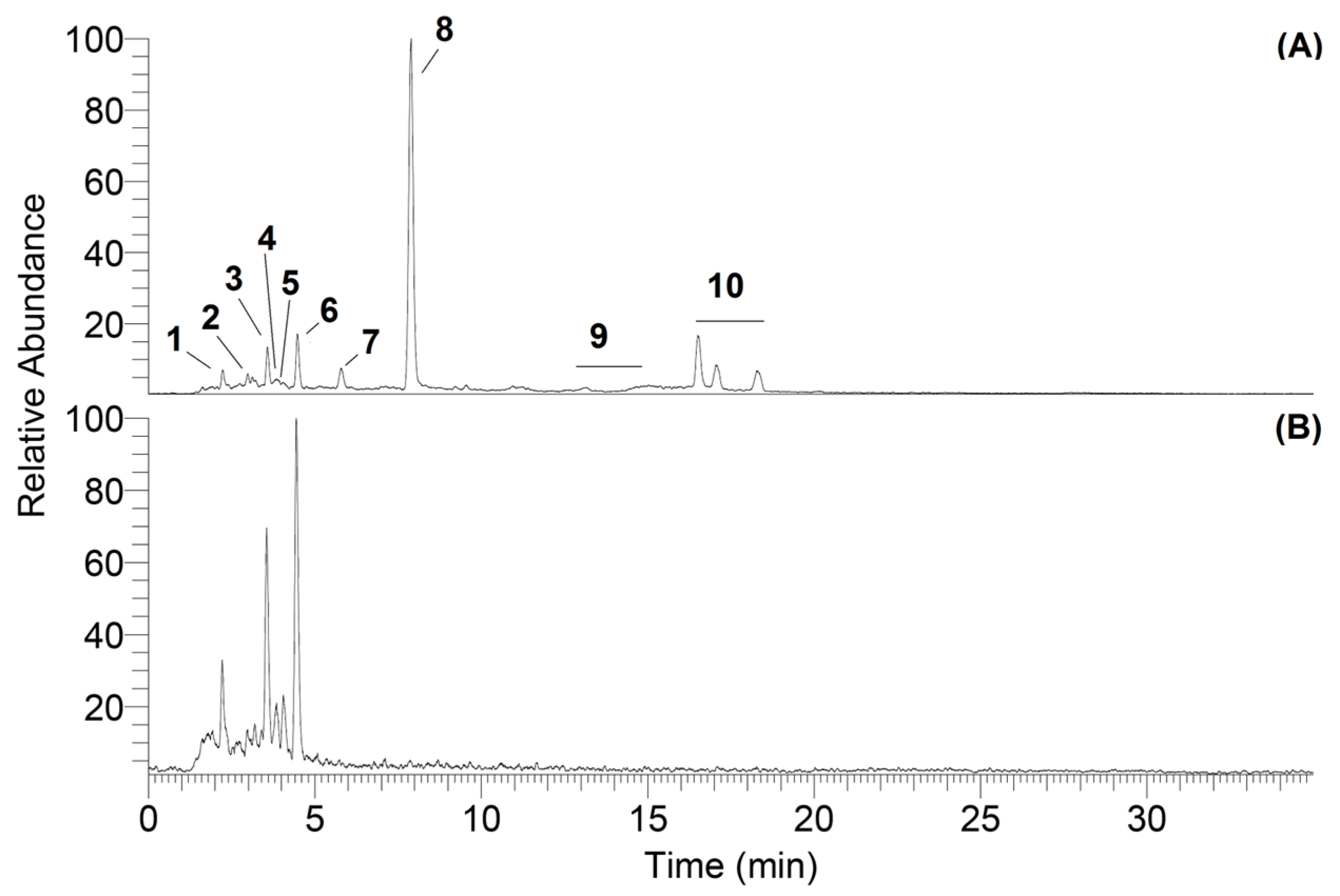
| Peak # | Compound | Retention Time (min) | Parent Peak m/z [M + H] + | Main Product Ion | RA (%) |
|---|---|---|---|---|---|
| 2 | Oxidized chamazulene | 2.88 | 183 | 169 [M + H − CH2]+ | 48 |
| 155 [M + H − CH2=CH2]+ | 38 | ||||
| 141 [M + H − C3H6]+ | 35 | ||||
| 3 | Chamazulene quinone | 3.61 | 215 | 197 [M + H − H2O]+ | 40 |
| 187 [M + H − CO]+ | 100 | ||||
| 173 [M + H − CH2CO]+ | 33 38 | ||||
| 159 [M + H − CH2=CH2 − CO]+ | |||||
| 4,5 | Chamazulene carbaldehyde | 3.75 | 199 | 184 [M + H − CH3]+ | 25 |
| 3.85 | 171 [M + H − CO]+ | 100 | |||
| 143 [M + H − CH2=CH2 − CO]+ | 78 | ||||
| 6 | Dihydrochamazulene | 4.44 | 187 | 172 [M + H − CH3]+ | 15 |
| 159 [M + H − C2H4]+ | 100 | ||||
| 131 [M + H − C4H8]+ | 22 | ||||
| 7 | Chamazulene benzenoid | 5.79 | 399 | 381 [M + H − H2O]+ | 100 |
| 371 [M + H − CO]+ | 57 | ||||
| 215 [M+H − Chamazulene]+ | 10 | ||||
| 199 [M+H − CH3Chamazulene] + | 63 | ||||
| 8 | Chamazulene | 7.89 | 185 | 169 [M + H − CH4]+ | 100 |
| 129 [M + H − CH2=CHCH2CH3]+ | 10 | ||||
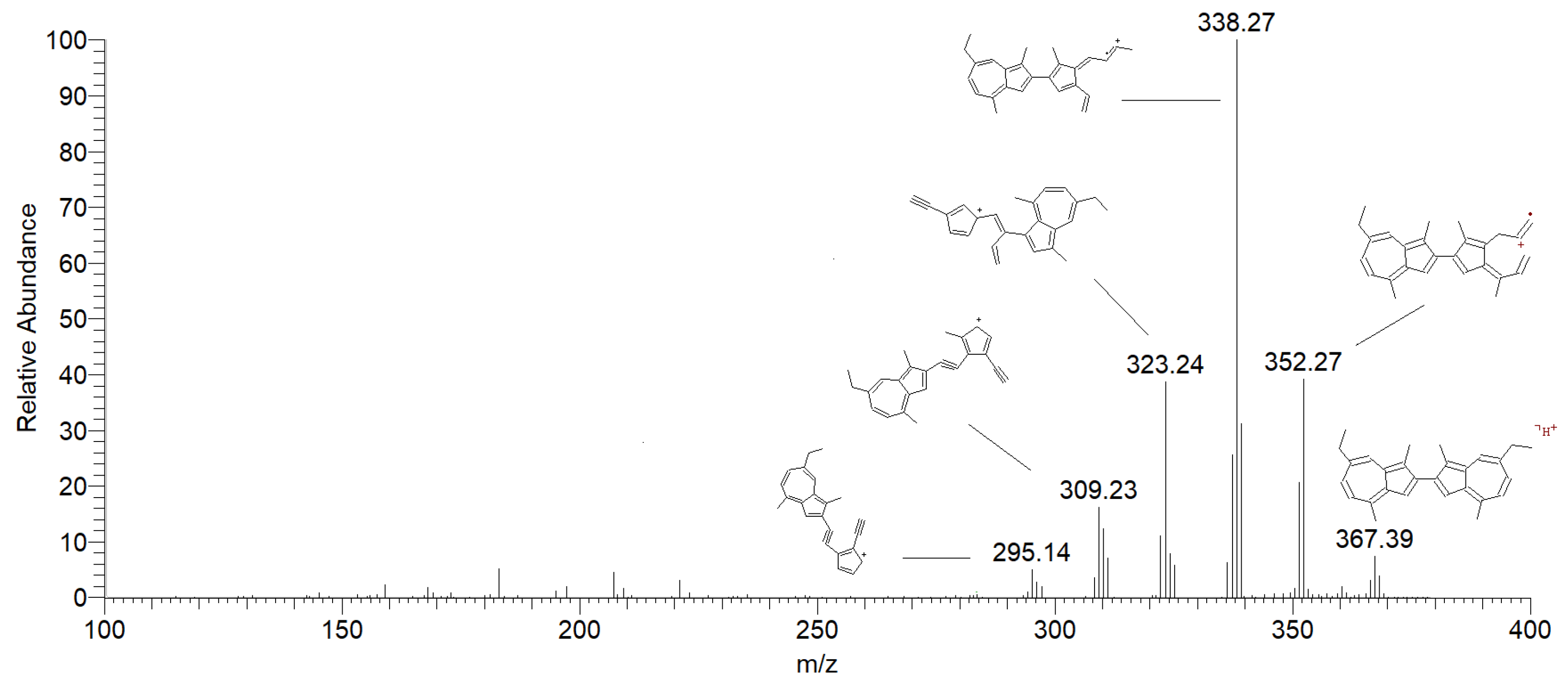
| 9 | Methylene dimers of chamazulene | 13.06 | 381 | 366 [M + H − CH3]+ | 38 |
| 363 [M + H − H2O]+ | 100 | ||||
| 353 [M + H − CH2=CH2]+ | 22 | ||||
| 14.90 | 337 [M + H − CH2=CHCH3]+ | 30 | |||
| 321 [M + H − C6H6]+ | 33 | ||||
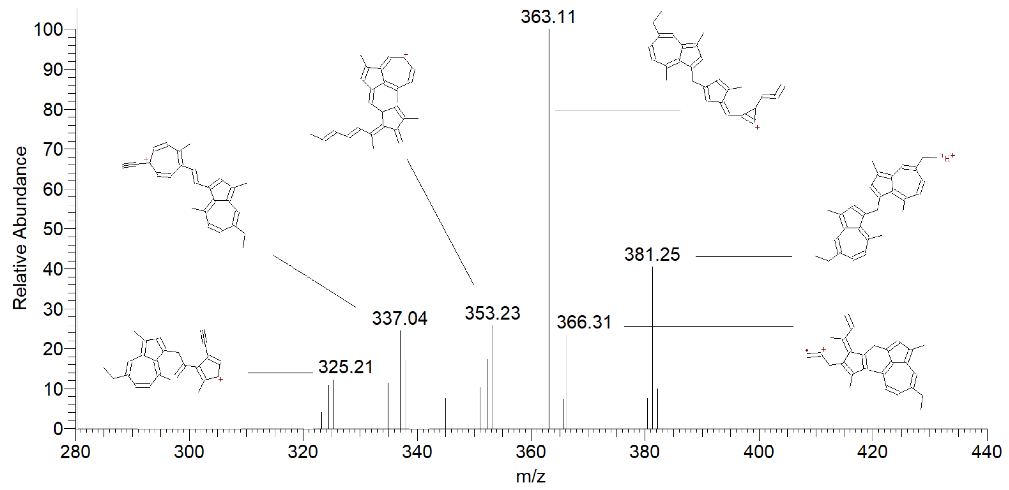
| 10 | Chamazulene dimers | 16.51 | 367 | 352 [M + H − CH3]+ | 60 |
| 17.05 | 338 [M + H − CH3CH2]+ | 100 | |||
| 18.28 | 309 [M + H − C4H10]+ | 25 |
| Type of UV | Wavelength [nm] | Radiance [mW cm−2 sr−1] | Irradiance [mW cm−2] | Energy Absorbed [J cm−2] (t = 15 min) | Energy Absorbed [J cm−2] (t = 4 h) |
|---|---|---|---|---|---|
| UVA | 320–400 | 35.2 | 2.4 | 2.2 | 34.6 |
| UVB | 290–320 | 7.5 | 0.5 | 0.5 | 8 |
| Air Saturated | |||
|---|---|---|---|
| HS 0 cm3 | HS 0.17 cm3 | HS 0.34 cm3 | |
| n-Hexane | y = −0.0551x + 99.638 R2 = 0.9368 | y = −0.0654x + 100.94 R2 = 0.9958 | y = −0.0736x + 101.04 R2 = 0.9692 |
| Methanol | y = −0.1107x + 101.17 R2 = 0.9659 | ||
| Acetonitrile | y = −0.3173x + 102.56 R2 = 0.9811 | ||
| N2 purged | |||
| HS 0 cm3 | |||
| n-Hexane | y = −0.0009x + 100.72 R2 = 0.0033 | ||
| Methanol | y = −0.0417x + 101.75 R2 = 0.7921 | ||
| Acetonitrile | y = −0.1146x + 103.17 R2 = 0.8943 | ||
| UV Irradiated Solutions of CA | Percentage of CA after UVA Irradiation | ||||||
|---|---|---|---|---|---|---|---|
| 0 J/cm2 | 2 J/cm2 | 4 J/cm2 | 8 J/cm2 | 17 J/cm2 | 25 J/cm2 | 35 J/cm2 | |
| CA | 100 | 98 ± 1 | 92 ± 2 | 81 ± 2 | 52 ± 3 | 22 ± 1 | 0 |
| MIXED WITH ANTIOXIDANTS | |||||||
| CA + TEMPO (1:10) | 100 | 96 ± 3 | 90 ± 2 | 83 ± 4 | 73 ± 3 * | 53 ± 1 * | 33 ± 1 * |
| CA + Tocopherol (1:10) | 100 | 94 ± 2 | 88 ± 2 | 77 ± 1 | 65 ± 2 * | 43 ± 1 * | 27 ± 1 * |
| CA + Ascorbyl palmitate (1:10) | 100 | 95 ± 2 | 94 ± 4 | 71 ± 3 | 49 ± 3 | 2 ± 1 * | 0 |
| CA + Bakuchiol (1:10) | 100 | 94 ± 3 | 91 ± 3 | 62 ± 3 * | 11 ± 3 * | 0 * | 0 |
| CA + Hydroxytyrosol (1:10) | 100 | 94 ± 3 | 92 ± 3 | 80 ± 2 | 62 ± 2 * | 37 ± 1 * | 6 ± 1 * |
| CA + Tocopherol + Bakuchiol (1:1:1) | 100 | 94 ± 1 | 92 ± 1 | 77 ± 3 | 41 ± 1 * | 10 ± 1 * | 0 |
| CA + Ascorbyl palmitate + Tocopherol (1:1:1) | 100 | 91 ± 2 | 80 ± 2 * | 68 ± 3 * | 17 ± 1 * | 0 * | 0 |
| CA + Tocopherol + Hydroxytyrosol (1:1:1) | 100 | 93 ± 2 | 81 ± 4 | 72 ± 3 * | 37 ± 2 * | 8 ± 3 * | 0 |
| CA + Tocopherol +TEMPO (1:1:1) | 100 | 96 ± 3 | 90 ± 2 | 76 ± 1 | 19 ± 1 * | 0 * | 0 |
| CA + Ascorbyl palmitate + TEMPO (1:1:1) | 100 | 96 ± 3 | 90 ± 2 | 75 ± 2 | 17 ± 1 * | 0 * | 0 |
| CA + γ-terpinene + Ascorbyl palmitate (1:5:1) | 100 | 94 ± 2 | 92 ± 3 | 76 ± 2 | 28 ± 4 * | 0 * | 0 |
| CA + γ-terpinene + Hydroxytyrosol (1:5:1) | 100 | 96 ± 2 | 90 ± 3 | 78 ± 1 | 38 ± 2 * | 0 * | 0 |
| CA + γ-terpinene + TEMPO (1:5:1) | 100 | 93 ± 1 | 93 ± 2 | 80 ± 2 | 57 ± 2 | 24 ± 1 | 6 ± 1 * |
| MIXED WITH UV FILTERS | |||||||
| CA + Tinosorb® S (5%) | 100 | 92 ± 2 | 92 ± 3 | 86 ± 2 * | 73 ± 4 * | 65 ± 2 * | 48 ± 1 * |
| CA + Octocrylene (5%) ¥ | 100 | 98 ± 3 | 90 ± 2 | 79 ± 3 | 57 ± 1 | 22 ± 1 | 0 |
| CA + Octyl methoxycinnamate (5%) | 100 | 100 ± 1 | 99 ± 3 | 97 ± 3 * | 70 ± 3 * | 45 ± 2 * | 20 ± 2 * |
| CA in the dark (negative control) | 100 | 100 ± 2 | 99 ± 2 * | 99 ± 2 * | 100 ± 1 * | 99 ± 3 * | 100 ± 2 * |
Disclaimer/Publisher’s Note: The statements, opinions and data contained in all publications are solely those of the individual author(s) and contributor(s) and not of MDPI and/or the editor(s). MDPI and/or the editor(s) disclaim responsibility for any injury to people or property resulting from any ideas, methods, instructions or products referred to in the content. |
© 2024 by the authors. Licensee MDPI, Basel, Switzerland. This article is an open access article distributed under the terms and conditions of the Creative Commons Attribution (CC BY) license (https://creativecommons.org/licenses/by/4.0/).
Share and Cite
Gabbanini, S.; Neba, J.N.; Matera, R.; Valgimigli, L. Photochemical and Oxidative Degradation of Chamazulene Contained in Artemisia, Matricaria and Achillea Essential Oils and Setup of Protection Strategies. Molecules 2024, 29, 2604. https://doi.org/10.3390/molecules29112604
Gabbanini S, Neba JN, Matera R, Valgimigli L. Photochemical and Oxidative Degradation of Chamazulene Contained in Artemisia, Matricaria and Achillea Essential Oils and Setup of Protection Strategies. Molecules. 2024; 29(11):2604. https://doi.org/10.3390/molecules29112604
Chicago/Turabian StyleGabbanini, Simone, Jerome Ngwa Neba, Riccardo Matera, and Luca Valgimigli. 2024. "Photochemical and Oxidative Degradation of Chamazulene Contained in Artemisia, Matricaria and Achillea Essential Oils and Setup of Protection Strategies" Molecules 29, no. 11: 2604. https://doi.org/10.3390/molecules29112604