
Overview on the Development of Electrochemical Immunosensors by the Signal Amplification of Enzyme- or Nanozyme-Based Catalysis Plus Redox Cycling

Abstract
:
1. Introduction
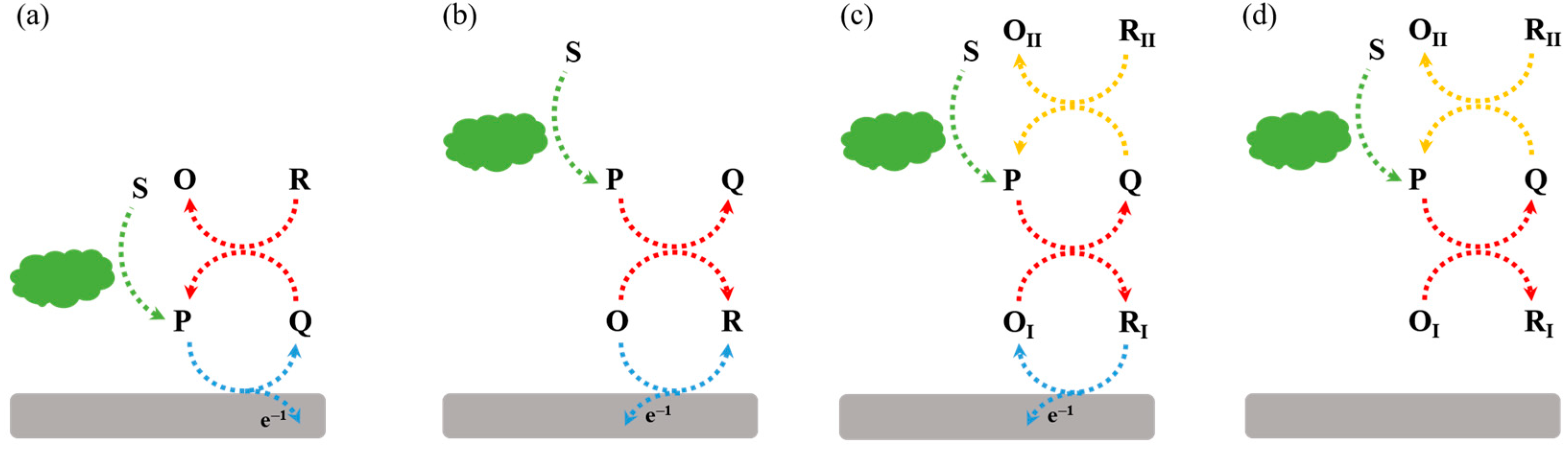
2. Types of Redox Cycling Reactions
2.1. Electrochemical–Enzymatic Redox Cycling
2.2. Electrochemical–Chemical Redox Cycling
2.3. Electrochemical–Electrochemical Redox Cycling
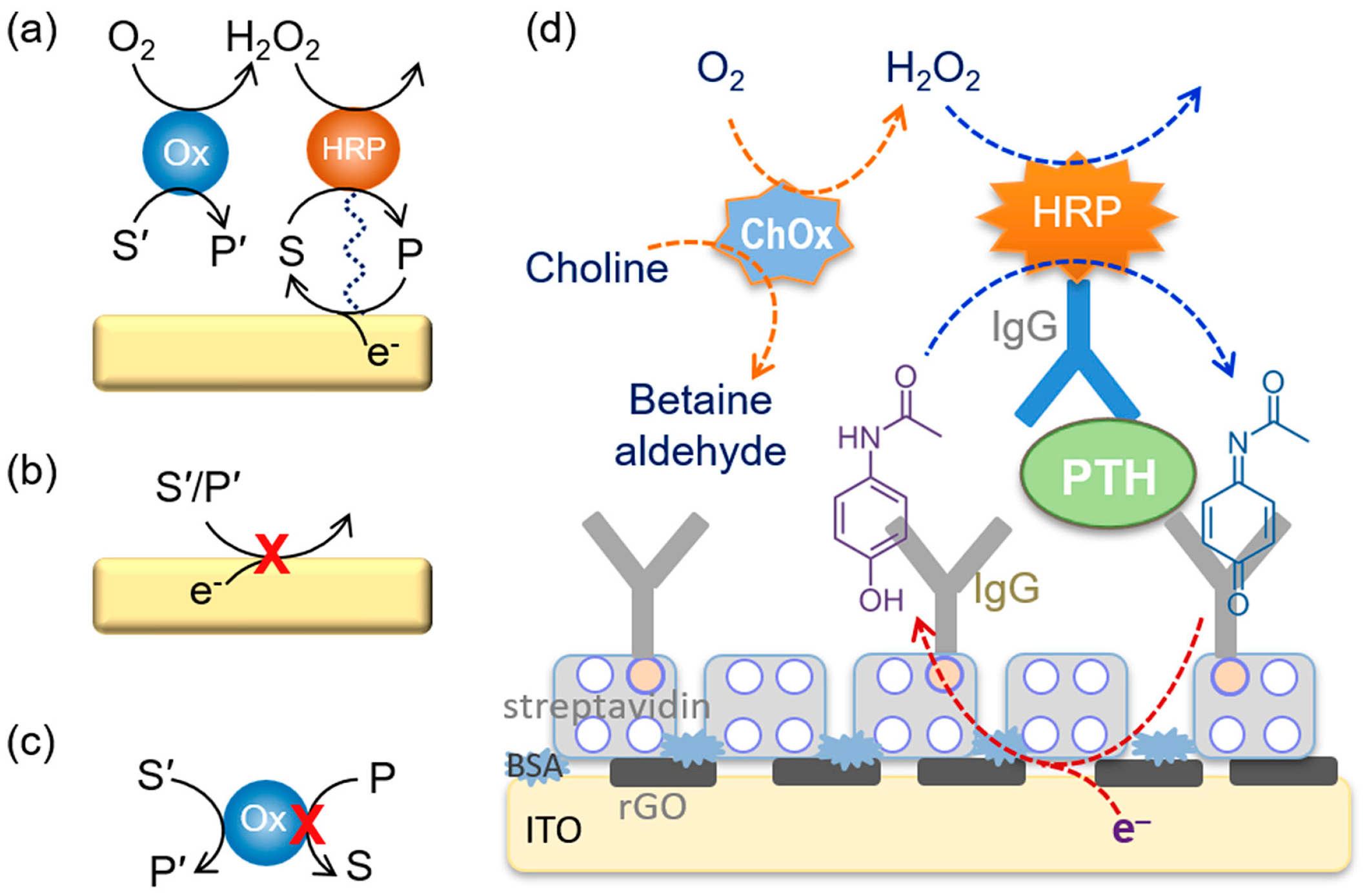
3. Oxidoreductases as the Signal Labels of Electrochemical Immunosensors
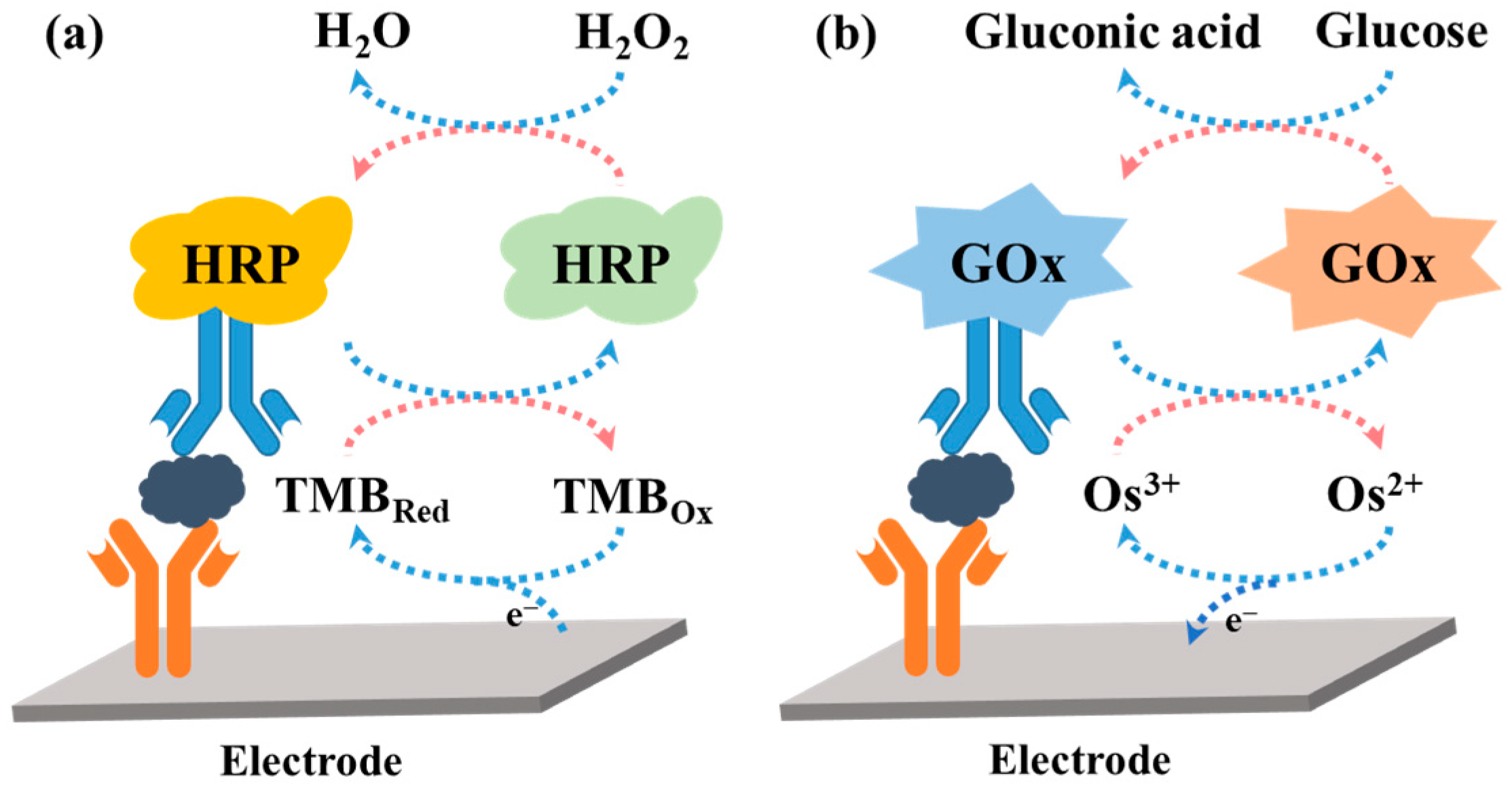
3.1. HRP
3.2. GOx
3.3. Tyrosinase
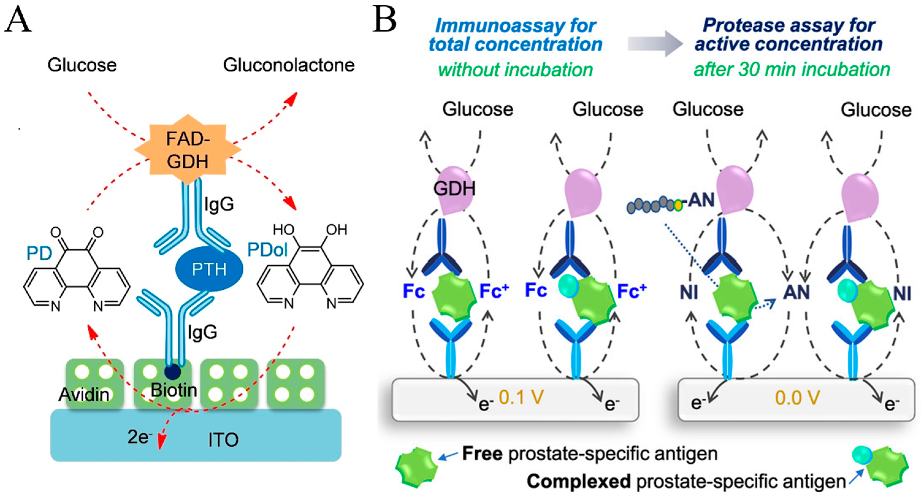
3.4. GDH
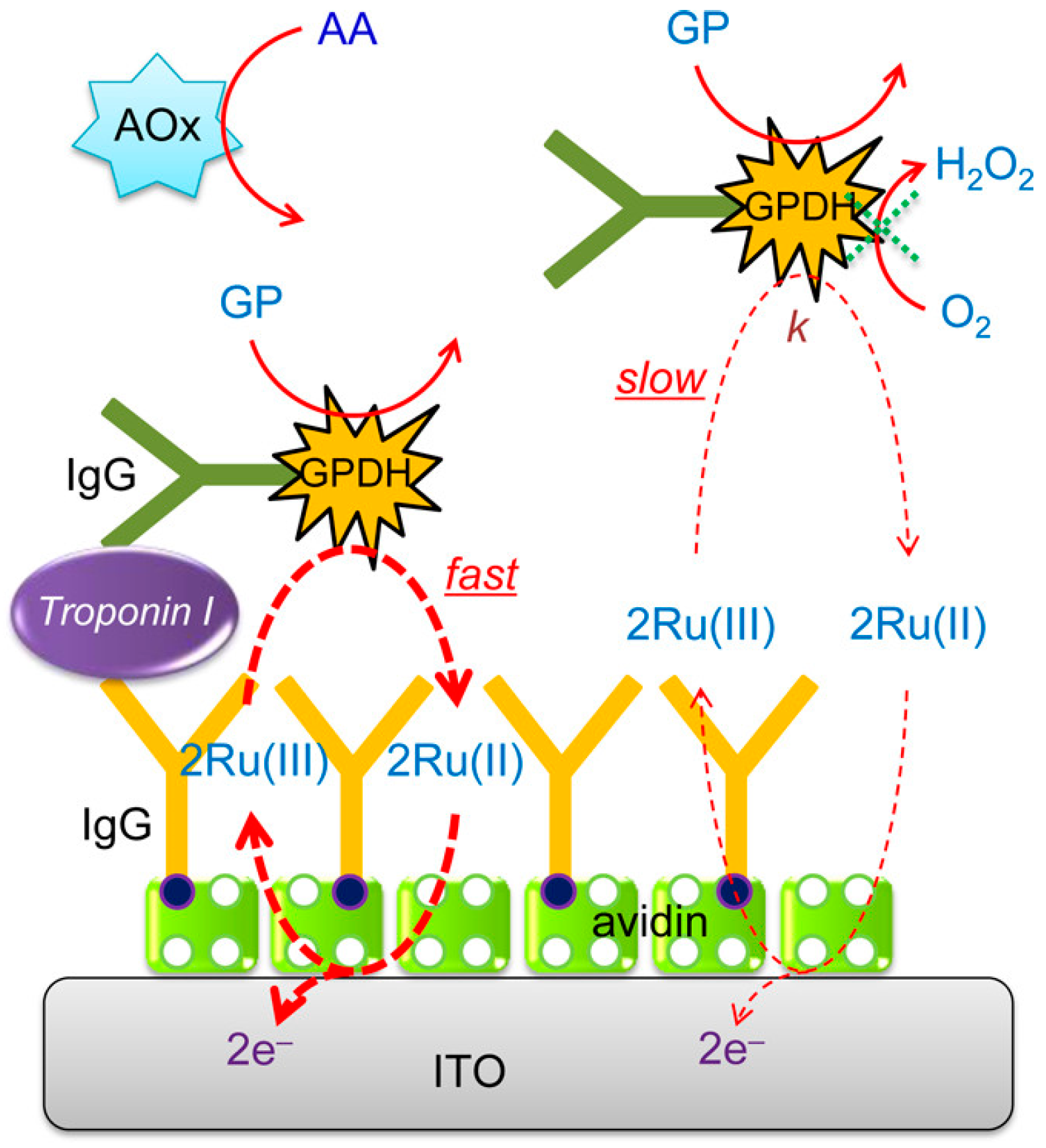
3.5. FAD-Dependent Glycerol-3-Phosphate Dehydrogenase (GPDH)
3.6. DT-Diaphorase (DT-D)
3.7. Nanocatalysts or Artificial Enzymes
4. Hydrolytic Enzymes as Signal Labels
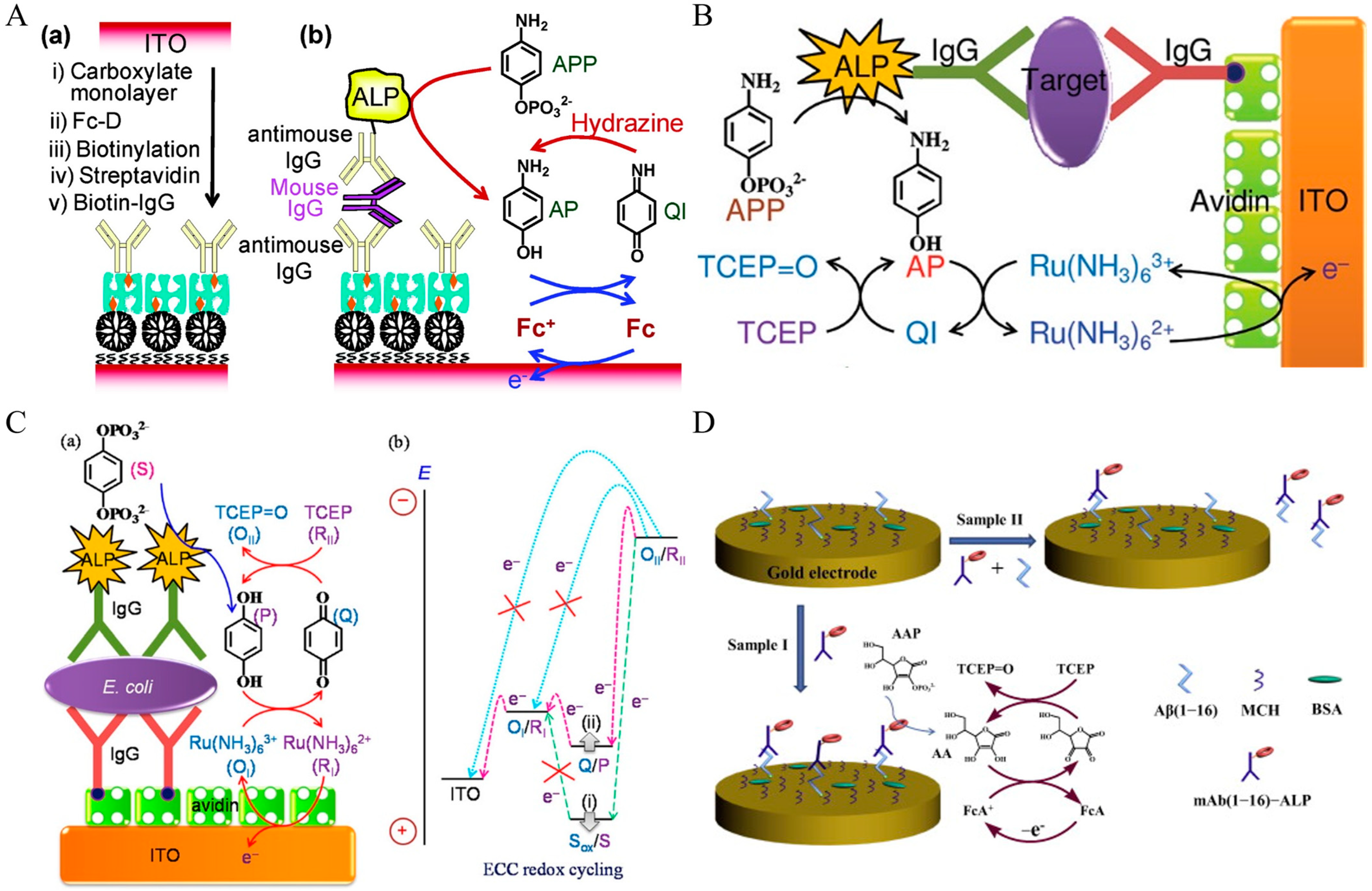
4.1. ALP-Based Redox Cycling
4.1.1. ALP-Based EC Redox Cycling
4.1.2. ALP-Based ECC Redox Cycling
4.1.3. ALP-Based Ag Biometallization
4.1.4. ALP-Based EN Redox Cycling
4.2. Protease
4.3. Others
5. Conclusions
Author Contributions
Funding
Institutional Review Board Statement
Informed Consent Statement
Data Availability Statement
Conflicts of Interest
References
- Felix, F.S.; Angnes, L. Electrochemical immunosensors-A powerful tool for analytical applications. Biosens. Bioelectron. 2018, 102, 470–478. [Google Scholar] [CrossRef] [PubMed]
- Ochoa-Ruiz, A.G.; Parra, G.; López-Espinoza, D.; Astudillo, P.; Galyamin, D.; Sabaté, N.; Esquivel, J.P.; Vallejo-Cardona, A.A. Electrochemical immunosensors: The evolution from ELISA to EμPADs. Electroanalysis 2022, 35, 2200053–2200067. [Google Scholar] [CrossRef]
- Wen, W.; Yan, X.; Zhu, C.; Du, D.; Lin, Y. Recent advances in electrochemical immunosensors. Anal. Chem. 2017, 89, 138–156. [Google Scholar] [CrossRef]
- Kokkinos, C.; Economou, A.; Prodromidis, M.I. Electrochemical immunosensors: Critical survey of different architectures and transduction strategies. TrAC-Trend. Anal. Chem. 2016, 79, 88–105. [Google Scholar] [CrossRef]
- Mollarasouli, F.; Kurbanoglu, S.; Ozkan, S.A. The role of electrochemical immunosensors in clinical analysis. Biosensors 2019, 9, 86. [Google Scholar] [CrossRef]
- Ju, H. Signal amplification for highly sensitive immunosensing. J. Anal. Test. 2017, 1, 7–24. [Google Scholar] [CrossRef]
- Tang, Z.; Ma, Z. Multiple functional strategies for amplifying sensitivity of amperometric immunoassay for tumor markers: A review. Biosens. Bioelectron. 2017, 98, 100–112. [Google Scholar] [CrossRef]
- Chen, C.; La, M.; Yi, X.; Huang, M.; Xia, N.; Zhou, Y. Progress in electrochemical immunosensors with alkaline phosphatase as the signal label. Biosensors 2023, 13, 855. [Google Scholar] [CrossRef]
- Zhang, Q. Application of hybridization chain reaction (HCR) in electrochemical analysis. Int. J. Electrochem. Sci. 2022, 17, 220227–220246. [Google Scholar] [CrossRef]
- Li, F.; Zhang, H.; Wang, Z.; Newbigging, A.M.; Reid, M.S.; Li, X.F.; Le, X.C. Aptamers facilitating amplified detection of biomolecules. Anal. Chem. 2015, 87, 274–292. [Google Scholar] [CrossRef]
- Liu, L.; Ma, X.; Chang, Y.; Guo, H.; Wang, W. Biosensors with boronic acid-based materials as the recognition elements and signal labels. Biosensors 2023, 13, 785. [Google Scholar] [CrossRef]
- Chang, Y.; Wang, Y.; Zhang, J.; Xing, Y.; Li, G.; Deng, D.; Liu, L. Overview on the design of magnetically assisted electrochemical biosensors. Biosensors 2022, 12, 954. [Google Scholar] [CrossRef]
- Chang, Y.; Lou, J.; Yang, L.; Liu, M.; Xia, N.; Liu, L. Design and application of electrochemical sensors with metal-organic frameworks as the electrode materials or signal tags. Nanomaterials 2022, 12, 3248. [Google Scholar] [CrossRef]
- Yu, C.-X.; Xiong, F.; Liu, L.-L. Electrochemical biosensors with silver nanoparticles as signal labels. Int. J. Electrochem. Sci. 2020, 15, 3869–3890. [Google Scholar] [CrossRef]
- Tang, J.; Tang, D. Non-enzymatic electrochemical immunoassay using noble metal nanoparticles: A review. Microchim. Acta 2015, 182, 2077–2089. [Google Scholar] [CrossRef]
- Kumar, A.; Purohit, B.; Maurya, P.K.; Pandey, L.M.; Chandra, P. Engineered nanomaterial assisted signal-amplification strategies for enhancing analytical performance of electrochemical biosensors. Electroanalysis 2019, 31, 1615–1629. [Google Scholar] [CrossRef]
- Nsabimana, A.; Lan, Y.; Du, F.; Wang, C.; Zhang, W.; Xu, G. Alkaline phosphatase-based electrochemical sensors for health applications. Anal. Methods 2019, 11, 1996–2006. [Google Scholar] [CrossRef]
- Li, X.-M.; Yang, X.-Y.; Zhang, S.-S. Electrochemical enzyme immunoassay using model labels. TrAC-Trend. Anal. Chem. 2008, 27, 543–553. [Google Scholar] [CrossRef]
- Shaban, S.M.; Byeok Jo, S.; Hafez, E.; Ho Cho, J.; Kim, D.-H. A comprehensive overview on alkaline phosphatase targeting and reporting assays. Coordin. Chem. Rev. 2022, 465, 214567–214604. [Google Scholar] [CrossRef]
- Scheller, F.W.; Bauer, C.G.; Makower, A.; Wollenberger, U.; Warsinke, A.; Bier, F.F. Coupling of immunoassays with enzymatic recycling electrodes. Anal. Lett. 2001, 34, 1233–1245. [Google Scholar] [CrossRef]
- Liu, G.; Wan, Y.; Gau, V.; Zhang, J.; Wang, L.; Song, S.; Fan, C. An enzyme-based E-DNA sensor for sequence-specific detection of femtomolar DNA targets. J. Am. Chem. Soc. 2008, 130, 6820–6825. [Google Scholar] [CrossRef]
- Torrente-Rodriguez, R.M.; Campuzano, S.; Montiel, V.R.; Montoya, J.J.; Pingarron, J.M. Sensitive electrochemical determination of miRNAs based on a sandwich assay onto magnetic microcarriers and hybridization chain reaction amplification. Biosens. Bioelectron. 2016, 86, 516–521. [Google Scholar] [CrossRef]
- Xu, D.; Huang, K.; Liu, Z.; Liu, Y.; Ma, L. Microfabricated disposable DNA sensors based on enzymatic amplification electrochemical detection. Electroanalysis 2001, 13, 882–887. [Google Scholar] [CrossRef]
- Wu, Y.; Chen, W.; Wang, C.; Xing, D. Assays for alkaline phosphatase that use L-ascorbic acid 2-phosphate as a substrate. Coordin. Chem. Rev. 2023, 495, 215370–215423. [Google Scholar] [CrossRef]
- Shao, Y.; Zhou, H.; Wu, Q.; Xiong, Y.; Wang, J.; Ding, Y. Recent advances in enzyme-enhanced immunosensors. Biotechnol. Adv. 2021, 53, 107867–107883. [Google Scholar] [CrossRef]
- Aizawa, M.; Morioka, A.; Suzuki, S.; Nagamura, Y. Enzyme immunosensor. III. Amperometric determination of human chorionic gonadotropin by membrane-bound antibody. Anal. Biochem. 1979, 94, 22–28. [Google Scholar] [CrossRef]
- Limoges, B.; Marchal, D.; Mavre, F.; Saveant, J.M.; Schollhorn, B. Theory and practice of enzyme bioaffinity electrodes. Direct electrochemical product detection. J. Am. Chem. Soc. 2008, 130, 7259–7275. [Google Scholar] [CrossRef]
- Lin, Y.; Zhou, Q.; Lin, Y.; Tang, D.; Niessner, R.; Knopp, D. Enzymatic hydrolysate-induced displacement reaction with multifunctional silica beads doped with horseradish peroxidase-thionine conjugate for ultrasensitive electrochemical immunoassay. Anal. Chem. 2015, 87, 8531–8540. [Google Scholar] [CrossRef]
- Cho, I.H.; Lee, J.; Kim, J.; Kang, M.S.; Paik, J.K.; Ku, S.; Cho, H.M.; Irudayaraj, J.; Kim, D.H. Current technologies of electrochemical immunosensors: Perspective on signal amplification. Sensors 2018, 18, 207. [Google Scholar] [CrossRef]
- Kim, J.; Park, M. Recent progress in electrochemical immunosensors. Biosensors 2021, 11, 360. [Google Scholar] [CrossRef]
- Ju, H. Functional nanomaterials and nanoprobes for amplified biosensing. Appl. Mater. Today 2018, 10, 51–71. [Google Scholar] [CrossRef]
- Wollenberger, U.; Schubert, F.; Pfeiffer, D.; Scheller, F.W. Enhancing biosensor performance using multienzyme systems. Trends Biotechnol. 1993, 11, 255–262. [Google Scholar] [CrossRef]
- Liu, L.; Xia, N.; Jiang, M.; Huang, N.; Guo, S.; Li, S.; Zhang, S. Electrochemical detection of amyloid-β oligomer with the signal amplification of alkaline phosphatase plus electrochemical–chemical–chemical redox cycling. J. Electroanal. Chem. 2015, 754, 40–45. [Google Scholar] [CrossRef]
- Liu, L.; Xia, N.; Liu, H.; Kang, X.; Liu, X.; Xue, C.; He, X. Highly sensitive and label-free electrochemical detection of microRNAs based on triple signal amplification of multifunctional gold nanoparticles, enzymes and redox-cycling reaction. Biosens. Bioelectron. 2014, 53, 399–405. [Google Scholar] [CrossRef]
- Guo, L.; Du, H.; Zhao, H.; Li, J. Amplified electrochemical response of phenol by oxygenation of tyrosinase coupling with electrochemical-chemical-chemical redox cycle. Electroanalysis 2019, 31, 1728–1735. [Google Scholar] [CrossRef]
- Liu, L.; Xia, N.; Meng, J.-J.; Zhou, B.-B.; Li, S.-J. An electrochemical aptasensor for sensitive and selective detection of dopamine based on signal amplification of electrochemical-chemical redox cycling. J. Electroanal. Chem. 2016, 775, 58–63. [Google Scholar] [CrossRef]
- Liu, L.; Gao, Y.; Liu, H.; Xia, N. An ultrasensitive electrochemical miRNAs sensor based on miRNAs-initiated cleavage of DNA by duplex-specific nuclease and signal amplification of enzyme plus redox cycling reaction. Sens. Actuat. B Chem. 2015, 208, 137–142. [Google Scholar] [CrossRef]
- Zhang, H.; Wu, S.; Xiao, H.J.; Wang, H.B.; Fang, L.; Cao, J.T. Chemical-chemical redox cycling for improving the sensitivity of the fluorescent assay: A proof-of-concept towards DNA methylation detection. Talanta 2023, 268, 125363–125371. [Google Scholar] [CrossRef]
- Zhang, H.; Wu, S.; Xing, Z.; Wang, H.B. ALP-assisted chemical redox cycling signal amplification for ultrasensitive fluorescence detection of DNA methylation. Analyst 2023, 148, 5753–5761. [Google Scholar] [CrossRef] [PubMed]
- Tang, J.; Liu, J.; Wang, F.; Yao, Y.; Hu, R. Colorimetric and photothermal dual-mode aptasensor with redox cycling amplification for the detection of ochratoxin A in corn samples. Food Chem. 2023, 439, 137968–137976. [Google Scholar] [CrossRef] [PubMed]
- Jiao, C.; Zhu, Y.; Ji, T.; Cai, X.; Wang, J. Yolk-shell structured nanoreactor Au@Co3O4/CeO2@mSiO2 with superior peroxidase-like activity as nanozyme for ultra-sensitive colorimetric biosensing. Talanta 2023, 260, 124571–124581. [Google Scholar] [CrossRef] [PubMed]
- Chen, Z.; Wang, H.; Zhang, Z.; Chen, L. Chemical redox-cycling for improving the sensitivity of colorimetric enzyme-linked immunosorbent assay. Anal. Chem. 2019, 91, 1254–1259. [Google Scholar] [CrossRef] [PubMed]
- Zhao, L.; Hu, Y.; Li, G.; Zou, S.; Ling, L. Chemical-chemical redox cycle signal amplification strategy combined with dual ratiometric immunoassay for surface-enhanced raman spectroscopic detection of cardiac troponin I. Anal. Chem. 2023, 95, 16677–16682. [Google Scholar] [CrossRef]
- Monteiro, T.; Almeida, M.G. Electrochemical enzyme biosensors revisited: Old solutions for new problems. Crit. Rev. Anal. Chem. 2019, 49, 44–66. [Google Scholar] [CrossRef] [PubMed]
- Kucherenko, I.S.; Soldatkin, O.O.; Kucherenko, D.Y.; Soldatkina, O.V.; Dzyadevych, S.V. Advances in nanomaterial application in enzyme-based electrochemical biosensors: A review. Nanoscale Adv. 2019, 1, 4560–4577. [Google Scholar] [CrossRef] [PubMed]
- Yang, H. Enzyme-based ultrasensitive electrochemical biosensors. Curr. Opin. Chem. Biol. 2012, 16, 422–428. [Google Scholar] [CrossRef] [PubMed]
- Chen, H.; Zhang, J.; Huang, R.; Wang, D.; Deng, D.; Zhang, Q.; Luo, L. The applications of electrochemical immunosensors in the detection of disease biomarkers: A review. Molecules 2023, 28, 3605. [Google Scholar] [CrossRef] [PubMed]
- Muñoz-San Martín, C.; Pedrero, M.; Manuel de Villena, F.J.; Garranzo-Asensio, M.; Rodríguez, N.; Domínguez, G.; Barderas, R.; Campuzano, S.; Pingarrón, J.M. Disposable amperometric immunosensor for the determination of the E-cadherin tumor suppressor protein in cancer cells and human tissues. Electroanalysis 2018, 31, 309–317. [Google Scholar] [CrossRef]
- Wang, M.; Xu, Z.; Chen, L.; Yin, H.; Ai, S. Electrochemical immunosensing platform for DNA methyltransferase activity analysis and inhibitor screening. Anal. Chem. 2012, 84, 9072–9078. [Google Scholar] [CrossRef] [PubMed]
- Volpe, G.; Draisci, R.; Palleschi, G.; Compagnone, D. 3,3′,5,5′-tetramethylbenzidine as electrochemical substrate for horseradish peroxidase based enzyme immunoassays. A comparative study. Analyst 1998, 123, 1303–1307. [Google Scholar] [CrossRef]
- He, Z.; Gao, N.; Jin, W. Determination of tumor marker CA125 by capillary electrophoretic enzyme immunoassay with electrochemical detection. Anal. Chim. Acta 2003, 497, 75–81. [Google Scholar] [CrossRef]
- Zhao, S.; Luong, J.H.T. An electrocatalytic approach for the measurement of chlorophenols. Anal. Chim. Acta 1996, 327, 235–242. [Google Scholar] [CrossRef]
- Xia, N.; Zhang, Y.; Chang, K.; Gai, X.; Jing, Y.; Li, S.; Liu, L.; Qu, G. Ferrocene-phenylalanine hydrogels for immobilization of acetylcholinesterase and detection of chlorpyrifos. J. Electroanal. Chem. 2015, 746, 68–74. [Google Scholar] [CrossRef]
- Ruiz-Valdepenas Montiel, V.; Campuzano, S.; Torrente-Rodriguez, R.M.; Reviejo, A.J.; Pingarron, J.M. Electrochemical magnetic beads-based immunosensing platform for the determination of α-lactalbumin in milk. Food Chem. 2016, 213, 595–601. [Google Scholar] [CrossRef] [PubMed]
- Ito, S.; Yamazaki, S.-i.; Kano, K.; Ikeda, T. Highly sensitive electrochemical detection of alkaline phosphatase. Anal. Chim. Acta 2000, 424, 57–63. [Google Scholar] [CrossRef]
- Limoges, B.; Marchal, D.; Mavre, F.; Saveant, J.M. Theory and practice of enzyme bioaffinity electrodes. Chemical, enzymatic, and electrochemical amplification of in situ product detection. J. Am. Chem. Soc. 2008, 130, 7276–7285. [Google Scholar] [CrossRef] [PubMed]
- Limoges, B.; Marchal, D.; Mavre, F.; Saveant, J.M. High amplification rates from the association of two enzymes confined within a nanometric layer immobilized on an electrode: Modeling and illustrating example. J. Am. Chem. Soc. 2006, 128, 6014–6015. [Google Scholar] [CrossRef] [PubMed]
- Park, S.; Seo, S.; Lee, N.S.; Yoon, Y.H.; Yang, H. Sensitive electrochemical immunosensor using a bienzymatic system consisting of β-galactosidase and glucose dehydrogenase. Analyst 2021, 146, 3880–3887. [Google Scholar] [CrossRef] [PubMed]
- Liao, Y.; Yuan, R.; Chai, Y.; Zhuo, Y.; Yuan, Y.; Bai, L.; Mao, L.; Yuan, S. In-situ produced ascorbic acid as coreactant for an ultrasensitive solid-state tris(2,2′-bipyridyl) ruthenium(II) electrochemiluminescence aptasensor. Biosens. Bioelectron. 2011, 26, 4815–4818. [Google Scholar] [CrossRef]
- Shuai, H.L.; Huang, K.J.; Chen, Y.X.; Fang, L.X.; Jia, M.P. Au nanoparticles/hollow molybdenum disulfide microcubes based biosensor for microRNA-21 detection coupled with duplex-specific nuclease and enzyme signal amplification. Biosens. Bioelectron. 2017, 89, 989–997. [Google Scholar] [CrossRef]
- Chen, Y.-X.; Huang, K.-J.; Lin, F.; Fang, L.-X. Ultrasensitive electrochemical sensing platform based on graphene wrapping SnO2 nanocorals and autonomous cascade DNA duplication strategy. Talanta 2017, 175, 168–176. [Google Scholar] [CrossRef]
- Jeong, J.; Das, J.; Choi, M.; Jo, J.; Aziz, M.A.; Yang, H. Arsenic(III) detection using electrochemical-chemical-chemical redox cycling at bare indium-tin oxide electrodes. Analyst 2014, 139, 5813–5817. [Google Scholar] [CrossRef]
- Wu, L.; Peng, M. An electrochemical DNA sensor for ultrasensitive detection of ARID1a targeting PD-1 checkpoint inhibitor immunological response. Anal. Methods 2019, 11, 2996–3005. [Google Scholar] [CrossRef]
- Dutta, G.; Lillehoj, P.B. An ultrasensitive enzyme-free electrochemical immunosensor based on redox cycling amplification using methylene blue. Analyst 2017, 142, 3492–3499. [Google Scholar] [CrossRef]
- da Silva Neves, M.M.P.; García, M.B.G.; Santos, D.H.; Fanjul-Bolado, P. Hydroquinone diphosphate/Ag+ as an enzymatic substrate for alkaline phosphatase catalyzed silver deposition. Electrochem. Commun. 2015, 60, 1–4. [Google Scholar] [CrossRef]
- Yi, Z.; Li, X.Y.; Gao, Q.; Tang, L.J.; Chu, X. Aptamer-aided target capturing with biocatalytic metal deposition: An electrochemical platform for sensitive detection of cancer cells. Analyst 2013, 138, 2032–2037. [Google Scholar] [CrossRef]
- Si, Y.; Sun, Z.; Zhang, N.; Qi, W.; Li, S.; Chen, L.; Wang, H. Ultrasensitive electroanalysis of low-level free microRNAs in blood by maximum signal amplification of catalytic silver deposition using alkaline phosphatase-incorporated gold nanoclusters. Anal. Chem. 2014, 86, 10406–10414. [Google Scholar] [CrossRef]
- Kishikawa, N.; Ohkubo, N.; Ohyama, K.; Nakashima, K.; Kuroda, N. Chemiluminescence assay for quinones based on generation of reactive oxygen species through the redox cycle of quinone. Anal. Bioanal. Chem. 2009, 393, 1337–1343. [Google Scholar] [CrossRef]
- Fukuda, M.; El-Maghrabey, M.H.; Kishikawa, N.; Ikemoto, K.; Kuroda, N. Ultrasensitive determination of pyrroloquinoline quinone in human plasma by HPLC with chemiluminescence detection using the redox cycle of quinone. J. Pharm. Biomed. Anal. 2017, 145, 814–820. [Google Scholar] [CrossRef]
- El-Maghrabey, M.; Sato, Y.; Kaladari, F.; Kishikawa, N.; Kuroda, N. Development of quinone linked immunosorbent assay (QuLISA) based on using Folin’s reagent as a non-enzymatic tag: Application to analysis of food allergens. Sens. Actuat. B Chem. 2022, 368, 132167–132176. [Google Scholar] [CrossRef]
- Kaladari, F.; El-Maghrabey, M.; Kishikawa, N.; Kuroda, N. Development of signal multiplication system for quinone linked immunosorbent assay (Multi-QuLISA) by using poly-L-lysine dendrigraft and 1,2-naphthoquinone-4-sulfonate as enzyme-free tag. Talanta 2023, 253, 123911–123919. [Google Scholar] [CrossRef]
- Kaladari, F.; Kishikawa, N.; Shimada, A.; El-Maghrabey, M.; Kuroda, N. Anthracycline-functionalized dextran as a new signal multiplication tagging approach for immunoassay. Biosensors 2023, 13, 340. [Google Scholar] [CrossRef]
- Niwa, O.; Xu, Y.; Halsall, H.B.; Heineman, W.R. Small-volume voltammetric detection of 4-aminophenol with interdigitated array electrodes and its application to electrochemical enzyme immunoassay. Anal. Chem. 1993, 65, 1559–1563. [Google Scholar] [CrossRef]
- Lee, G.Y.; Park, J.H.; Chang, Y.W.; Cho, S.; Kang, M.J.; Pyun, J.C. Redox cycling-based immunoassay for detection of carcinogenic embryonic antigen. Anal. Chim. Acta. 2017, 971, 33–39. [Google Scholar] [CrossRef]
- Lee, G.Y.; Park, J.H.; Chang, Y.W.; Cho, S.; Kang, M.J.; Pyun, J.C. Chronoamperometry-based redox cycling for application to immunoassays. ACS Sens. 2018, 3, 106–112. [Google Scholar] [CrossRef]
- Yamamoto, S.; Uno, S. Redox cycling realized in paper-based biochemical sensor for selective detection of reversible redox molecules without micro/nano fabrication process. Sensors 2018, 18, 730. [Google Scholar] [CrossRef]
- Bard, A.J.; Crayston, J.A.; Kittlesen, G.P.; Varco Shea, T.; Wrighton, M.S. Digital simulation of the measured electrochemical response of reversible redox couples at microelectrode arrays: Consequences arising from closely spaced ultramicroelectrodes. Anal. Chem. 1986, 58, 2321–2331. [Google Scholar] [CrossRef]
- Dotan, T.; Jog, A.; Kadan-Jamal, K.; Avni, A.; Shacham-Diamand, Y. In vivo plant bio-electrochemical sensor using redox cycling. Biosensors 2023, 13, 219. [Google Scholar] [CrossRef]
- Neugebauer, S.; Stoica, L.; Guschin, D.; Schuhmann, W. Redox-amplified biosensors based on selective modification of nanopore electrode structures with enzymes entrapped within electrodeposition paints. Microchim. Acta 2008, 163, 33–40. [Google Scholar] [CrossRef]
- Park, J.H.; Lee, G.Y.; Song, Z.; Bong, J.H.; Kim, H.R.; Kang, M.J.; Pyun, J.C. A vertically paired electrode for redox cycling and its application to immunoassays. Analyst 2023, 148, 1349–1361. [Google Scholar] [CrossRef]
- Butler, D.; Ebrahimi, A. Rapid and sensitive detection of viral particles by coupling redox cycling and electrophoretic enrichment. Biosens. Bioelectron. 2022, 208, 114198–114206. [Google Scholar] [CrossRef]
- Kang, M.; Mun, C.; Jung, H.S.; Ansah, I.B.; Kim, E.; Yang, H.; Payne, G.F.; Kim, D.H.; Park, S.G. Tethered molecular redox capacitors for nanoconfinement-assisted electrochemical signal amplification. Nanoscale 2020, 12, 3668–3676. [Google Scholar] [CrossRef]
- Şen, M.; Avcı, İ. A simple microfluidic redox cycling-based device for high-sensitive detection of dopamine released from PC12 cells. J. Electroanal. Chem. 2023, 939, 117473–117479. [Google Scholar] [CrossRef]
- Fan, H.; Wang, X.; Jiao, F.; Zhang, F.; Wang, Q.; He, P.; Fang, Y. Scanning electrochemical microscopy of DNA hybridization on DNA microarrays enhanced by HRP-modified SiO2 nanoparticles. Anal. Chem. 2013, 85, 6511–6517. [Google Scholar] [CrossRef]
- Wu, J.; Yan, Y.; Yan, F.; Ju, H. Electric field-driven strategy for multiplexed detection of protein biomarkers using a disposable reagentless electrochemical immunosensor array. Anal. Chem. 2008, 80, 6072–6077. [Google Scholar] [CrossRef]
- Valverde, A.; Povedano, E.; Montiel, V.R.; Yanez-Sedeno, P.; Garranzo-Asensio, M.; Barderas, R.; Campuzano, S.; Pingarron, J.M. Electrochemical immunosensor for IL-13 receptor α2 determination and discrimination of metastatic colon cancer cells. Biosens. Bioelectron. 2018, 117, 766–772. [Google Scholar] [CrossRef]
- He, Y.N.; Chen, H.Y.; Zheng, J.J.; Zhang, G.Y.; Chen, Z.L. Differential pulse voltammetric enzyme-linked immunoassay for the determination of helicobacter pylori specific immunoglobulin G (IgG) antibody. Talanta 1997, 44, 823–830. [Google Scholar] [CrossRef]
- Höfs, S.; Hülagü, D.; Bennet, F.; Carl, P.; Flemig, S.; Schmid, T.; Schenk, J.A.; Hodoroaba, V.D.; Schneider, R.J. Electrochemical immunomagnetic ochratoxin A sensing: Steps forward in the application of 3,3’,5,5’-tetramethylbenzidine in amperometric assays. ChemElectroChem 2021, 8, 2597–2606. [Google Scholar] [CrossRef]
- Santandreu, M.; Alegret, S.; Fàbregas, E. Determination of β-HCG using amperometric immunosensors based on a conducting immunocomposite. Anal. Chim. Acta 1999, 396, 181–188. [Google Scholar] [CrossRef]
- Zhang, Y.; Chen, S.; Ma, J.; Zhou, X.; Sun, X.; Jing, H.; Lin, M.; Zhou, C. Enzyme-catalyzed electrochemical aptasensor for ultrasensitive detection of soluble PD-L1 in breast cancer based on decorated covalent organic frameworks and carbon nanotubes. Anal. Chim. Acta. 2023, 1282, 341927–341935. [Google Scholar] [CrossRef]
- Eletxigerra, U.; Martinez-Perdiguero, J.; Merino, S.; Barderas, R.; Ruiz-Valdepeñas Montiel, V.; Villalonga, R.; Pingarrón, J.M.; Campuzano, S. Electrochemical magnetoimmunosensor for progesterone receptor determination. Application to the simultaneous detection of estrogen and progesterone breast-cancer related receptors in raw cell lysates. Electroanalysis 2015, 28, 1787–1794. [Google Scholar] [CrossRef]
- Valverde, A.; ben Hassine, A.; Serafín, V.; Muñoz-San Martín, C.; Pedrero, M.; Garranzo-Asensio, M.; Gamella, M.; Raouafi, N.; Barderas, R.; Yáñez-Sedeño, P.; et al. Dual amperometric immunosensor for improving cancer metastasis detection by the simultaneous determination of extracellular and soluble circulating fraction of emerging metastatic biomarkers. Electroanalysis 2019, 32, 706–714. [Google Scholar] [CrossRef]
- Torriero, A.A.; Salinas, E.; Raba, J.; Silber, J.J. Sensitive determination of ciprofloxacin and norfloxacin in biological fluids using an enzymatic rotating biosensor. Biosens. Bioelectron. 2006, 22, 109–115. [Google Scholar] [CrossRef]
- Zhang, Y.; Gao, Y.; Zhang, X.; Wang, H.; Xia, T.; Bian, C.; Liang, S.; Tang, X.; Wang, X. Electrochemical immunosensor for HBe antigen detection based on a signal amplification strategy: The co-catalysis of horseradish peroxidase and nanoporous gold. Sens. Actuat. B Chem. 2019, 284, 296–304. [Google Scholar] [CrossRef]
- Attar, A.; Cubillana-Aguilera, L.; Naranjo-Rodriguez, I.; de Cisneros, J.L.; Palacios-Santander, J.M.; Amine, A. Amperometric inhibition biosensors based on horseradish peroxidase and gold sononanoparticles immobilized onto different electrodes for cyanide measurements. Bioelectrochemistry 2015, 101, 84–91. [Google Scholar] [CrossRef]
- Tang, D.; Yuan, R.; Chai, Y. Electron-transfer mediator microbiosensor fabrication based on immobilizing HRP-labeled Au colloids on gold electrode surface by 11-mercaptoundecanoic acid monolayer. Electroanalysis 2006, 18, 259–266. [Google Scholar] [CrossRef]
- Wu, J.; Zhang, Z.; Fu, Z.; Ju, H. A disposable two-throughput electrochemical immunosensor chip for simultaneous multianalyte determination of tumor markers. Biosens. Bioelectron. 2007, 23, 114–120. [Google Scholar] [CrossRef]
- Sun, B.; Cai, J.; Li, W.; Gou, X.; Gou, Y.; Li, D.; Hu, F. A novel electrochemical immunosensor based on PG for early screening of depression markers-heat shock protein 70. Biosens. Bioelectron. 2018, 111, 34–40. [Google Scholar] [CrossRef]
- Ju, H.; Yan, G.; Chen, F.; Chen, H. Enzyme-linked immunoassay of α-1-fetoprotein in serum by differential pulse voltammetry. Electroanalysis 1999, 11, 124–128. [Google Scholar] [CrossRef]
- Zhang, S.; Zou, J.; Yu, F. Investigation of voltammetric enzyme-linked immunoassay based on a new system of HAP-H2O2-HRP. Talanta 2008, 76, 122–127. [Google Scholar] [CrossRef]
- Campas, M.; Marty, J.L. Highly sensitive amperometric immunosensors for microcystin detection in algae. Biosens. Bioelectron. 2007, 22, 1034–1040. [Google Scholar] [CrossRef]
- Ding, C.; Zhao, F.; Ren, R.; Lin, J.M. An electrochemical biosensor for α-fetoprotein based on carbon paste electrode constructed of room temperature ionic liquid and gold nanoparticles. Talanta 2009, 78, 1148–1154. [Google Scholar] [CrossRef]
- Doldan, X.; Fagundez, P.; Cayota, A.; Laiz, J.; Tosar, J.P. Electrochemical sandwich immunosensor for determination of exosomes based on surface marker-mediated signal amplification. Anal. Chem. 2016, 88, 10466–10473. [Google Scholar] [CrossRef]
- Haque, A.M.; Park, H.; Sung, D.; Jon, S.; Choi, S.Y.; Kim, K. An electrochemically reduced graphene oxide-based electrochemical immunosensing platform for ultrasensitive antigen detection. Anal. Chem. 2012, 84, 1871–1878. [Google Scholar] [CrossRef]
- Kang, H.J.; Aziz, M.â.A.; Jeon, B.; Jo, K.; Yang, H. Strategy for low background-current levels in the electrochemical biosensors using horse-radish peroxidase labels. Electroanalysis 2009, 21, 2647–2652. [Google Scholar] [CrossRef]
- Yan, K.; Haque, A.J.; Nandhakumar, P.; Bhatia, A.; Lee, N.S.; Yoon, Y.H.; Yang, H. Boosting electrochemical immunosensing performance by employing acetaminophen as a peroxidase substrate. Biosens. Bioelectron. 2020, 165, 112337–112343. [Google Scholar] [CrossRef]
- Tang, D.; Ren, J. In situ amplified electrochemical immunoassay for carcinoembryonic antigen using horseradish peroxidase-encapsulated nanogold hollow microspheres as labels. Anal. Chem. 2008, 80, 8064–8070. [Google Scholar] [CrossRef]
- Tang, D.; Yuan, R.; Chai, Y. Ultrasensitive electrochemical immunosensor for clinical immunoassay using thionine-doped magnetic gold nanospheres as labels and horseradish peroxidase as enhancer. Anal. Chem. 2008, 80, 1582–1588. [Google Scholar] [CrossRef]
- Hou, Y.H.; Wang, J.J.; Jiang, Y.Z.; Lv, C.; Xia, L.; Hong, S.L.; Lin, M.; Lin, Y.; Zhang, Z.L.; Pang, D.W. A colorimetric and electrochemical immunosensor for point-of-care detection of enterovirus 71. Biosens. Bioelectron. 2018, 99, 186–192. [Google Scholar] [CrossRef]
- Yu, X.; Munge, B.; Patel, V.; Jensen, G.; Bhirde, A.; Gong, J.D.; Kim, S.N.; Gillespie, J.; Gutkind, J.S.; Papadimitrakopoulos, F.; et al. Carbon nanotube amplification strategies for highly sensitive immunodetection of cancer biomarkers. J. Am. Chem. Soc. 2006, 128, 11199–11205. [Google Scholar] [CrossRef]
- Chen, Y.; Li, Y.; Deng, D.; He, H.; Yan, X.; Wang, Z.; Fan, C.; Luo, L. Effective immobilization of Au nanoparticles on TiO2 loaded graphene for a novel sandwich-type immunosensor. Biosens. Bioelectron. 2018, 102, 301–306. [Google Scholar] [CrossRef]
- Zhong, Z.; Wu, W.; Wang, D.; Wang, D.; Shan, J.; Qing, Y.; Zhang, Z. Nanogold-enwrapped graphene nanocomposites as trace labels for sensitivity enhancement of electrochemical immunosensors in clinical immunoassays: Carcinoembryonic antigen as a model. Biosens. Bioelectron. 2010, 25, 2379–2383. [Google Scholar] [CrossRef]
- Zhong, Z.; Li, M.; Xiang, D.; Dai, N.; Qing, Y.; Wang, D.; Tang, D. Signal amplification of electrochemical immunosensor for the detection of human serum IgG using double-codified nanosilica particles as labels. Biosens. Bioelectron. 2009, 24, 2246–2249. [Google Scholar] [CrossRef]
- Knopp, D.; Tang, D.; Niessner, R. Review: Bioanalytical applications of biomolecule-functionalized nanometer-sized doped silica particles. Anal. Chim. Acta 2009, 647, 14–30. [Google Scholar] [CrossRef]
- Arevalo, B.; Blazquez-Garcia, M.; Valverde, A.; Serafin, V.; Montero-Calle, A.; Solis-Fernandez, G.; Barderas, R.; Campuzano, S.; Yanez-Sedeno, P.; Pingarron, J.M. Binary MoS2 nanostructures as nanocarriers for amplification in multiplexed electrochemical immunosensing: Simultaneous determination of B cell activation factor and proliferation-induced signal immunity-related cytokines. Microchim. Acta 2022, 189, 143–157. [Google Scholar] [CrossRef]
- Tang, J.; Tang, D.; Niessner, R.; Chen, G.; Knopp, D. Magneto-controlled graphene immunosensing platform for simultaneous multiplexed electrochemical immunoassay using distinguishable signal tags. Anal. Chem. 2011, 83, 5407–5414. [Google Scholar] [CrossRef]
- Yan, K.; Nandhakumar, P.; Bhatia, A.; Lee, N.S.; Yoon, Y.H.; Yang, H. Electrochemical immunoassay based on choline oxidase-peroxidase enzymatic cascade. Biosens. Bioelectron. 2021, 171, 112727–112731. [Google Scholar] [CrossRef]
- Zhang, Y.; Tsitkov, S.; Hess, H. Proximity does not contribute to activity enhancement in the glucose oxidase-horseradish peroxidase cascade. Nat. Commun. 2016, 7, 13982–13990. [Google Scholar] [CrossRef]
- Alim, S.; Kafi, A.K.M.; Rajan, J.; Yusoff, M.M. Application of polymerized multiporous nanofiber of SnO2 for designing a bienzyme glucose biosensor based on HRP/GOx. Int. J. Biol. Macromol. 2019, 123, 1028–1034. [Google Scholar] [CrossRef] [PubMed]
- Dai, Z.; Bao, J.; Yang, X.; Ju, H. A bienzyme channeling glucose sensor with a wide concentration range based on co-entrapment of enzymes in SBA-15 mesopores. Biosens. Bioelectron. 2008, 23, 1070–1076. [Google Scholar] [CrossRef] [PubMed]
- Karyakin, A.A. Glucose biosensors for clinical and personal use. Electrochem. Commun. 2021, 125, 106973–106976. [Google Scholar] [CrossRef]
- Zhang, J.; Pearce, M.C.; Ting, B.P.; Ying, J.Y. Ultrasensitive electrochemical immunosensor employing glucose oxidase catalyzed deposition of gold nanoparticles for signal amplification. Biosens. Bioelectron. 2011, 27, 53–57. [Google Scholar] [CrossRef] [PubMed]
- Bright, H.J.; Appleby, M. The pH dependence of the individual steps in the glucose oxidase reaction. J. Biol. Chem. 1969, 244, 3625–3634. [Google Scholar] [CrossRef] [PubMed]
- Chen, L.; Gorski, W. Bioinorganic composites for enzyme electrodes. Anal. Chem. 2001, 73, 2862–2868. [Google Scholar] [CrossRef] [PubMed]
- Cass, A.E.; Davis, G.; Francis, G.D.; Hill, H.A.; Aston, W.J.; Higgins, I.J.; Plotkin, E.V.; Scott, L.D.; Turner, A.P. Ferrocene-mediated enzyme electrode for amperometric determination of glucose. Anal. Chem. 1984, 56, 667–671. [Google Scholar] [CrossRef] [PubMed]
- Gregg, B.A.; Heller, A. Cross-linked redox gels containing glucose oxidase for amperometric biosensor applications. Anal. Chem. 1990, 62, 258–263. [Google Scholar] [CrossRef] [PubMed]
- Lai, G.; Yan, F.; Ju, H. Dual signal amplification of glucose oxidase-functionalized nanocomposites as a trace label for ultrasensitive simultaneous multiplexed electrochemical detection of tumor markers. Anal. Chem. 2009, 81, 9730–9736. [Google Scholar] [CrossRef] [PubMed]
- Singh, A.; Park, S.; Yang, H. Glucose-oxidase label-based redox cycling for an incubation period-free electrochemical immunosensor. Anal. Chem. 2013, 85, 4863–4868. [Google Scholar] [CrossRef] [PubMed]
- Dutta, G.; Kim, S.; Park, S.; Yang, H. Washing-free heterogeneous immunosensor using proximity-dependent electron mediation between an enzyme label and an electrode. Anal. Chem. 2014, 86, 4589–4595. [Google Scholar] [CrossRef]
- Cosnier, S.; Popescu, I.C. Poly(amphiphilic pyrrole)-tyrosinase-peroxidase electrode for amplified flow injection-amperometric detection of phenol. Anal. Chim. Acta 1996, 319, 145–151. [Google Scholar] [CrossRef]
- Fu, Y.; Li, P.; Bu, L.; Wang, T.; Xie, Q.; Chen, J.; Yao, S. Exploiting metal-organic coordination polymers as highly efficient immobilization matrixes of enzymes for sensitive electrochemical biosensing. Anal. Chem. 2011, 83, 6511–6517. [Google Scholar] [CrossRef]
- Noh, S.; Yang, H. Sensitive phenol detection using tyrosinase-based phenol oxidation combined with redox cycling of catechol. Electroanalysis 2014, 26, 2727–2731. [Google Scholar] [CrossRef]
- Besombes, J.L.; Cosnier, S.; Labbe, P. Polyphenol oxidase-catechol: An electroenzymatic model system for characterizing the performance of matrices for biosensors. Talanta 1996, 43, 1615–1619. [Google Scholar] [CrossRef]
- Cosnier, S.; Fombon, J.-J.; Labbe, P.; Limosin, D. Development of a PPO-poly amphiphilic pyrrole electrode for on site monitoring of phenol in aqueous effluents. Sens. Actuat. B Chem. 1999, 59, 134–139. [Google Scholar] [CrossRef]
- Yan, K.; Wu, J.; Ji, W.; Wu, J.; Zhang, J. Integration of redox cycling in a photoelectrochemical sensing platform for tyrosinase activity evaluation. Electrochem. Commun. 2019, 108, 106555–106559. [Google Scholar] [CrossRef]
- Kwon, J.; Kang, H.Y.; Yang, H. Permeabilization-free β-galactosidase-induction-based electrochemical detection of Escherichia coli. Sens. Actuat. B Chem. 2021, 337, 129768–129774. [Google Scholar] [CrossRef]
- Chumyim, P.; Rijiravanich, P.; Somasundrum, M.; Surareungchai, W. Tyrosinase multilayer-functionalised carbon nanotubes as electrochemical labels: Application to immunoassay. BioNanoScience 2014, 4, 240–250. [Google Scholar] [CrossRef]
- Akanda, M.R.; Ju, H. An integrated redox cycling for electrochemical enzymatic signal enhancement. Anal. Chem. 2017, 89, 13480–13486. [Google Scholar] [CrossRef] [PubMed]
- Akanda, M.R.; Ju, H. A tyrosinase-responsive nonenzymatic redox cycling for amplified electrochemical immunosensing of protein. Anal. Chem. 2016, 88, 9856–9861. [Google Scholar] [CrossRef]
- Park, S.; Kwak, D.E.; Haque, A.J.; Lee, N.S.; Yoon, Y.H.; Yang, H. Phenolic tyrosinase substrate with a formal potential lower than that of phenol to obtain a sensitive electrochemical immunosensor. ACS Sens. 2022, 7, 790–796. [Google Scholar] [CrossRef]
- Shen, D.; Meyerhoff, M.E. Pyrroloquinoline quinone-doped polymeric nanospheres as sensitive tracer for binding assays. Anal. Chem. 2009, 81, 1564–1569. [Google Scholar] [CrossRef] [PubMed]
- Zimmerman, L.B.; Lee, K.D.; Meyerhoff, M.E. Visual detection of single-stranded target DNA using pyrroloquinoline-quinone-loaded liposomes as a tracer. Anal. Biochem. 2010, 401, 182–187. [Google Scholar] [CrossRef] [PubMed]
- Zimmerman, L.B.; Worley, B.V.; Palermo, E.F.; Brender, J.R.; Lee, K.D.; Kuroda, K.; Ramamoorthy, A.; Meyerhoff, M.E. Absorbance-based assay for membrane disruption by antimicrobial peptides and synthetic copolymers using pyrroloquinoline quinone-loaded liposomes. Anal. Biochem. 2011, 411, 194–199. [Google Scholar] [CrossRef] [PubMed]
- Ferri, S.; Kojima, K.; Sode, K. Review of glucose oxidases and glucose dehydrogenases: A bird’s eye view of glucose sensing enzymes. J. Diabetes Sci. Technol. 2011, 5, 1068–1076. [Google Scholar] [CrossRef] [PubMed]
- Flexer, V.; Mano, N. Wired pyrroloquinoline quinone soluble glucose dehydrogenase enzyme electrodes operating at unprecedented low redox potential. Anal. Chem. 2014, 86, 2465–2473. [Google Scholar] [CrossRef] [PubMed]
- Zayats, M.; Katz, E.; Baron, R.; Willner, I. Reconstitution of apo-glucose dehydrogenase on pyrroloquinoline quinone-functionalized Au nanoparticles yields an electrically contacted biocatalyst. J. Am. Chem. Soc. 2005, 127, 12400–12406. [Google Scholar] [CrossRef] [PubMed]
- Tatsumi, H.; Osaku, N. Sensitive electrochemical detection of the hydroxyl radical using enzyme-catalyzed redox cycling. Anal. Sci. 2011, 27, 1065–1067. [Google Scholar] [CrossRef] [PubMed]
- Durand, F.; Limoges, B.; Mano, N.; Mavre, F.; Miranda-Castro, R.; Saveant, J.M. Effect of substrate inhibition and cooperativity on the electrochemical responses of glucose dehydrogenase. Kinetic characterization of wild and mutant types. J. Am. Chem. Soc. 2011, 133, 12801–12809. [Google Scholar] [CrossRef] [PubMed]
- Zhang, L.; Miranda-Castro, R.; Stines-Chaumeil, C.; Mano, N.; Xu, G.; Mavre, F.; Limoges, B. Heterogeneous reconstitution of the PQQ-dependent glucose dehydrogenase immobilized on an electrode: A sensitive strategy for PQQ detection down to picomolar levels. Anal. Chem. 2014, 86, 2257–2267. [Google Scholar] [CrossRef]
- Jiaul Haque, A.M.; Kwon, J.; Kim, J.; Kim, G.; Lee, N.S.; Ho Yoon, Y.; Yang, H. Sensitive and low-background electrochemical immunosensor employing glucose dehydrogenase and 1,10-phenanthroline-5,6-dione. Electroanalysis 2021, 33, 1877–1885. [Google Scholar] [CrossRef]
- Park, S.; Shin, J.; Kwon, J.; Lee, W.; Kim, J.; Kim, G.; Joo, J.M.; Yang, H. Interference-free duplex detection of total and active enzyme concentrations at a single working electrode. ACS Sens. 2021, 6, 1305–1311. [Google Scholar] [CrossRef]
- Caduff, A.; Talary, M.S.; Zakharov, P. Cutaneous blood perfusion as a perturbing factor for noninvasive glucose monitoring. Diabetes Technol. Ther. 2010, 12, 1–9. [Google Scholar] [CrossRef] [PubMed]
- Dutta, G.; Park, S.; Singh, A.; Seo, J.; Kim, S.; Yang, H. Low-interference washing-free electrochemical immunosensor using glycerol-3-phosphate dehydrogenase as an enzyme label. Anal. Chem. 2015, 87, 3574–3578. [Google Scholar] [CrossRef] [PubMed]
- Haque, A.J.; Nandhakumar, P.; Kim, G.; Park, S.; Yu, B.; Lee, N.S.; Yoon, Y.H.; Jon, S.; Yang, H. Diaphorase-catalyzed formation of a formazan precipitate and its electrodissolution for sensitive affinity biosensors. Anal. Chem. 2020, 92, 3932–3939. [Google Scholar] [CrossRef] [PubMed]
- Ichzan, A.M.; Hwang, S.H.; Cho, H.; Fang, C.S.; Park, S.; Kim, G.; Kim, J.; Nandhakumar, P.; Yu, B.; Jon, S.; et al. Solid-phase recombinase polymerase amplification using an extremely low concentration of a solution primer for sensitive electrochemical detection of hepatitis B viral DNA. Biosens. Bioelectron. 2021, 179, 113065–113072. [Google Scholar] [CrossRef] [PubMed]
- Nandhakumar, P.; Lee, W.; Nam, S.; Bhatia, A.; Seo, J.; Kim, G.; Lee, N.S.; Yoon, Y.H.; Joo, J.M.; Yang, H. Di(thioether sulfonate)-substituted quinolinedione as a rapidly dissoluble and stable electron mediator and its application in sensitive biosensors. Adv. Healthcare Mater. 2022, 11, e2101819–e2101827. [Google Scholar] [CrossRef] [PubMed]
- Bhatia, A.; Na, H.S.; Nandhakumar, P.; Yu, B.; Jon, S.; Chung, J.; Yang, H. Electrochemical detection of interleukin-8 in human saliva using a polyenzyme label based on diaphorase and neutravidin. Sens. Actuat. B Chem. 2021, 326, 128979–128985. [Google Scholar] [CrossRef]
- Prayikaputri, P.U.; Park, S.; Kim, S.; Yoon, Y.H.; Kim, S.; Yang, H. Sensitive electrochemical immunosensor via amide hydrolysis by DT-diaphorase combined with five redox-cycling reactions. Biosens. Bioelectron. 2023, 224, 115058–115067. [Google Scholar] [CrossRef]
- Kang, C.; Kang, J.; Lee, N.S.; Yoon, Y.H.; Yang, H. DT-diaphorase as a bifunctional enzyme label that allows rapid enzymatic amplification and electrochemical redox cycling. Anal. Chem. 2017, 89, 7974–7980. [Google Scholar] [CrossRef]
- Bhatia, A.; Nandhakumar, P.; Kim, G.; Kim, J.; Lee, N.S.; Yoon, Y.H.; Yang, H. Ultrasensitive detection of parathyroid hormone through fast silver deposition induced by enzymatic nitroso reduction and redox cycling. ACS Sens. 2019, 4, 1641–1647. [Google Scholar] [CrossRef]
- Bhatia, A.; Nandhakumar, P.; Kim, G.; Lee, N.-S.; Yoon, Y.H.; Yang, H. Simple and fast Ag deposition method using a redox enzyme label and quinone substrate for the sensitive electrochemical detection of thyroid-stimulating hormone. Biosen. Bioelectron. 2022, 197, 113773–113780. [Google Scholar] [CrossRef] [PubMed]
- Ichzan, A.M.; Lee, S.; San Fang, C.; Nandhakumar, P.; Ha, H.; Joo, J.M.; Kim, K.S.; Yang, H. Use of a phosphatase-like DT-diaphorase label for the detection of outer membrane vesicles. Anal. Chem. 2019, 91, 4680–4686. [Google Scholar] [CrossRef] [PubMed]
- Nandhakumar, P.; Ichzan, A.M.; Lee, N.S.; Yoon, Y.H.; Ma, S.; Kim, S.; Yang, H. Carboxyl esterase-like activity of DT-diaphorase and its use for signal amplification. ACS Sens. 2019, 4, 2966–2973. [Google Scholar] [CrossRef] [PubMed]
- Rochelet-Dequaire, M.; Djellouli, N.; Limoges, B.; Brossier, P. Bienzymatic-based electrochemical DNA biosensors: A way to lower the detection limit of hybridization assays. Analyst 2009, 134, 349–353. [Google Scholar] [CrossRef] [PubMed]
- Kang, J.; Shin, J.; Yang, H. Rapid and sensitive detection of NADH and lactate dehydrogenase using thermostable DT-diaphorase immobilized on electrode. Electroanalysis 2018, 30, 1357–1362. [Google Scholar] [CrossRef]
- Haque, A.M.J.; Nandhakumar, P.; Yang, H. Specific and rapid glucose detection using NAD-dependent glucose dehydrogenase, diaphorase, and osmium complex. Electroanalysis 2019, 31, 876–882. [Google Scholar] [CrossRef]
- Campas, M.; de la Iglesia, P.; Le Berre, M.; Kane, M.; Diogene, J.; Marty, J.L. Enzymatic recycling-based amperometric immunosensor for the ultrasensitive detection of okadaic acid in shellfish. Biosens. Bioelectron. 2008, 24, 716–722. [Google Scholar] [CrossRef] [PubMed]
- Park, S.; Park, K.; Cho, H.; Kwon, J.; Kim, K.S.; Yang, H. Wash-free amperometric Escherichia coli detection via rapid and specific proteolytic cleavage by its outer membrane OmpT. Anal. Chem. 2022, 94, 4756–4762. [Google Scholar] [CrossRef] [PubMed]
- Zhang, X.; Lin, S.; Liu, S.; Tan, X.; Dai, Y.; Xia, F. Advances in organometallic/organic nanozymes and their applications. Coordin. Chem. Rev. 2021, 429, 213652–213670. [Google Scholar] [CrossRef]
- Liu, G.; Xia, N.; Tian, L.; Sun, Z.; Liu, L. Progress in the development of biosensors based on peptide-copper coordination interaction. Biosensors 2022, 12, 809. [Google Scholar] [CrossRef]
- Wei, H.; Wang, E. Nanomaterials with enzyme-like characteristics (nanozymes): Next-generation artificial enzymes. Chem. Soc. Rev. 2013, 42, 6060–6093. [Google Scholar] [CrossRef] [PubMed]
- Mahmudunnabi, R.G.; Farhana, F.Z.; Kashaninejad, N.; Firoz, S.H.; Shim, Y.B.; Shiddiky, M.J.A. Nanozyme-based electrochemical biosensors for disease biomarker detection. Analyst 2020, 145, 4398–4420. [Google Scholar] [CrossRef]
- Wang, X.; Dong, S.; Wei, H. Recent advances on nanozyme-based electrochemical biosensors. Electroanalysis 2022, 35, 38–49. [Google Scholar] [CrossRef]
- Zuccarello, L.; Barbosa, C.; Todorovic, S.; Silveira, C.M. Electrocatalysis by heme enzymes—Applications in biosensing. Catalysts 2021, 11, 218. [Google Scholar] [CrossRef]
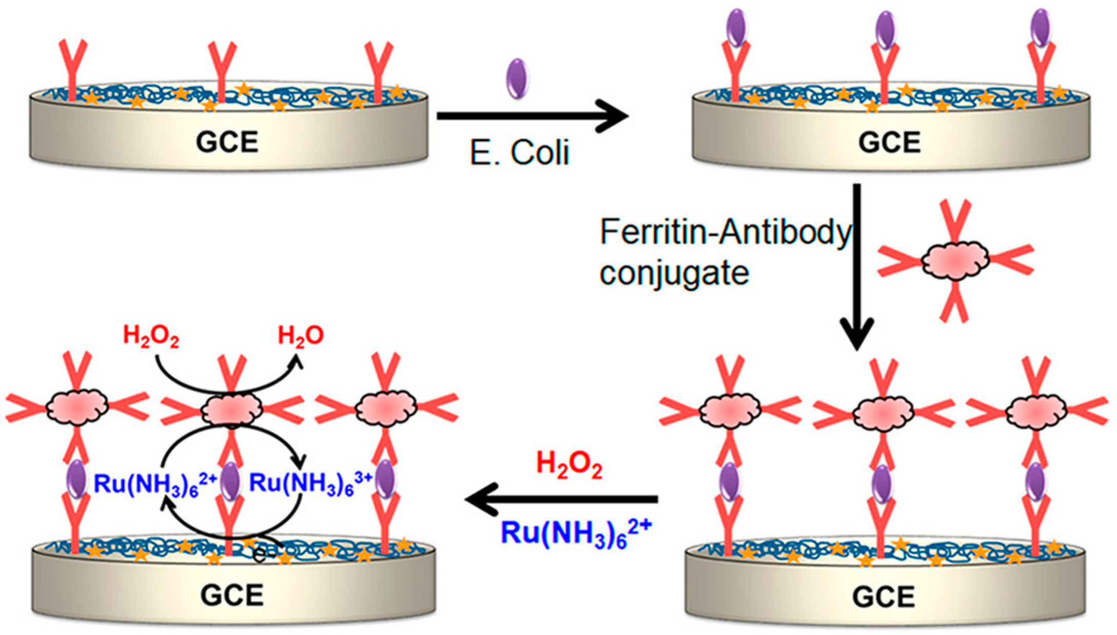
- Wang, C.; Liu, Q.; Huang, X.; Zhuang, J. Ferritin nanocages: A versatile platform for nanozyme design. J. Mater. Chem. B 2023, 11, 4153–4170. [Google Scholar] [CrossRef] [PubMed]
- Tang, Z.; Wu, H.; Zhang, Y.; Li, Z.; Lin, Y. Enzyme-mimic activity of ferric nano-core residing in ferritin and its biosensing applications. Anal. Chem. 2011, 83, 8611–8616. [Google Scholar] [CrossRef] [PubMed]
- Watt, G.D.; Jacobs, D.; Frankel, R.B. Redox reactivity of bacterial and mammalian ferritin: Is reductant entry into the ferritin interior a necessary step for iron release? Proc. Natl. Acad. Sci. USA 1988, 85, 7457–7461. [Google Scholar] [CrossRef] [PubMed]
- Akanda, M.R.; Ju, H. Ferritin-triggered redox cycling for highly sensitive electrochemical immunosensing of protein. Anal. Chem. 2018, 90, 8028–8034. [Google Scholar] [CrossRef] [PubMed]
- Hu, L.; Liu, X.; Cecconello, A.; Willner, I. Dual switchable CRET-induced luminescence of CdSe/ZnS quantum dots (QDs) by the hemin/G-quadruplex-bridged aggregation and deaggregation of two-sized QDs. Nano Lett. 2014, 14, 6030–6035. [Google Scholar] [CrossRef]
- Zhang, M.; Xu, S.; Minteer, S.D.; Baum, D.A. Investigation of a deoxyribozyme as a biofuel cell catalyst. J. Am. Chem. Soc. 2011, 133, 15890–15893. [Google Scholar] [CrossRef]
- Funabashi, H. Hemin/G-quadruplex complex as a signal generator for electrochemical assays of bioanalytes. Electrochemistry 2016, 84, 290–295. [Google Scholar] [CrossRef]
- Zhang, K.; Zhu, X.; Wang, J.; Xu, L.; Li, G. Strategy to fabricate an electrochemical aptasensor: Application to the assay of adenosine deaminase activity. Anal. Chem. 2010, 82, 3207–3211. [Google Scholar] [CrossRef] [PubMed]
- Yang, N.; Cao, Y.; Han, P.; Zhu, X.; Sun, L.; Li, G. Tools for investigation of the RNA endonuclease activity of mammalian Argonaute2 protein. Anal. Chem. 2012, 84, 2492–2497. [Google Scholar] [CrossRef] [PubMed]
- Liu, S.; Wang, C.; Zhang, C.; Wang, Y.; Tang, B. Label-free and ultrasensitive electrochemical detection of nucleic acids based on autocatalytic and exonuclease III-assisted target recycling strategy. Anal. Chem. 2013, 85, 2282–2288. [Google Scholar] [CrossRef] [PubMed]
- Pelossof, G.; Tel-Vered, R.; Elbaz, J.; Willner, I. Amplified biosensing using the horseradish peroxidase-mimicking DNAzyme as an electrocatalyst. Anal. Chem. 2010, 82, 4396–4402. [Google Scholar] [CrossRef] [PubMed]
- Pelossof, G.; Tel-Vered, R.; Willner, I. Amplified surface plasmon resonance and electrochemical detection of Pb2+ ions using the Pb2+-dependent DNAzyme and hemin/G-quadruplex as a label. Anal. Chem. 2012, 84, 3703–3709. [Google Scholar] [CrossRef] [PubMed]
- Tang, J.; Hou, L.; Tang, D.; Zhang, B.; Zhou, J.; Chen, G. Hemin/G-quadruplex-based DNAzyme concatamers as electrocatalysts and biolabels for amplified electrochemical immunosensing of IgG1. Chem. Commun. 2012, 48, 8180–8182. [Google Scholar] [CrossRef]
- Alizadeh, N.; Hallaj, R.; Salimi, A. Dual amplified electrochemical immunosensor for Hepatitis B virus surface antigen detection using hemin/G-quadruplex immobilized onto Fe3O4-AuNPs or (hemin-amino-rGO-Au) nanohybrid. Electroanalysis 2017, 30, 402–414. [Google Scholar] [CrossRef]
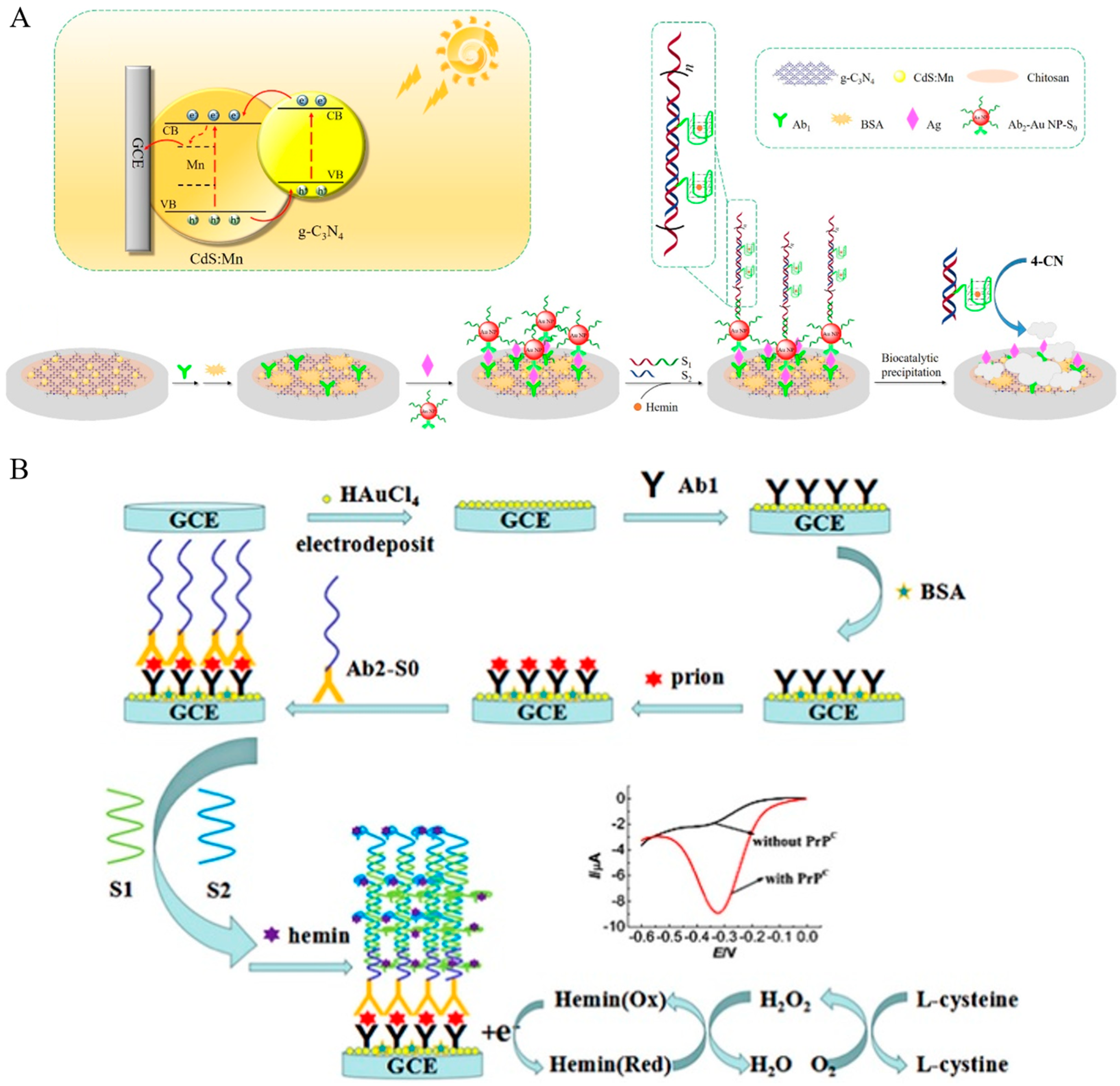
- Zhang, K.; Lv, S.; Lin, Z.; Tang, D. CdS:Mn quantum dot-functionalized g-C3N4 nanohybrids as signal-generation tags for photoelectrochemical immunoassay of prostate specific antigen coupling DNAzyme concatamer with enzymatic biocatalytic precipitation. Biosens. Bioelectron. 2017, 95, 34–40. [Google Scholar] [CrossRef]
- Golub, E.; Freeman, R.; Willner, I. A hemin/G-quadruplex acts as an NADH oxidase and NADH peroxidase mimicking DNAzyme. Angew. Chem. Int. Ed. 2011, 50, 11710–11714. [Google Scholar] [CrossRef]
- Golub, E.; Freeman, R.; Willner, I. Hemin/G-quadruplex-catalyzed aerobic oxidation of thiols to disulfides: Application of the process for the development of sensors and aptasensors and for probing acetylcholine esterase activity. Anal. Chem. 2013, 85, 12126–12133. [Google Scholar] [CrossRef]
- Li, X.; Li, J.; Zhu, C.; Zhang, X.; Chen, J. A new electrochemical immunoassay for prion protein based on hybridization chain reaction with hemin/G-quadruplex DNAzyme. Talanta 2018, 182, 292–298. [Google Scholar] [CrossRef]
- Polsky, R.; Gill, R.; Kaganovsky, L.; Willner, I. Nucleic acid-functionalized Pt nanoparticles: Catalytic labels for the amplified electrochemical detection of biomolecules. Anal. Chem. 2006, 78, 2268–2271. [Google Scholar] [CrossRef]
- Wu, D.; Ma, H.; Zhang, Y.; Jia, H.; Yan, T.; Wei, Q. Corallite-like magnetic Fe3O4@mno2@Pt nanocomposites as multiple signal amplifiers for the detection of carcinoembryonic antigen. ACS Appl. Mater. Interfaces 2015, 7, 18786–18793. [Google Scholar] [CrossRef] [PubMed]
- Peng, X.; Zhu, J.; Wu, Z.; Wen, W.; Zhang, X.; Chen, M.-M.; Wang, S. High-efficient Pt@COF nanospheres-based electrochemical-chemical-chemical redox cycling for ultrasensitive microRNAs biosensing. Sens. Actuat. B Chem. 2023, 392, 134074–134082. [Google Scholar] [CrossRef]
- Selvaraju, T.; Das, J.; Jo, K.; Kwon, K.; Huh, C.H.; Kim, T.K.; Yang, H. Nanocatalyst-based assay using DNA-conjugated Au nanoparticles for electrochemical DNA detection. Langmuir 2008, 24, 9883–9888. [Google Scholar] [CrossRef]
- Fang, C.S.; Oh, K.H.; Oh, A.; Lee, K.; Park, S.; Kim, S.; Park, J.K.; Yang, H. An ultrasensitive and incubation-free electrochemical immunosensor using a gold-nanocatalyst label mediating outer-sphere-reaction-philic and inner-sphere-reaction-philic species. Chem. Commun. 2016, 52, 5884–5887. [Google Scholar] [CrossRef]
- Wang, J.; Wang, X.; Wu, S.; Song, J.; Zhao, Y.; Ge, Y.; Meng, C. Fabrication of highly catalytic silver nanoclusters/graphene oxide nanocomposite as nanotag for sensitive electrochemical immunoassay. Anal. Chim. Acta 2016, 906, 80–88. [Google Scholar] [CrossRef] [PubMed]
- Tang, J.; Zhou, J.; Li, Q.; Tang, D.; Chen, G.; Yang, H. In situ amplified electronic signal for determination of low-abundance proteins coupling with nanocatalyst-based redox cycling. Chem. Commun. 2013, 49, 1530–1532. [Google Scholar] [CrossRef]
- Das, J.; Aziz, M.A.; Yang, H. A nanocatalyst-based assay for proteins: DNA-free ultrasensitive electrochemical detection using catalytic reduction of p-nitrophenol by gold-nanoparticle labels. J. Am. Chem. Soc. 2006, 128, 16022–16023. [Google Scholar] [CrossRef]
- Selvaraju, T.; Das, J.; Han, S.W.; Yang, H. Ultrasensitive electrochemical immunosensing using magnetic beads and gold nanocatalysts. Biosens. Bioelectron. 2008, 23, 932–9328. [Google Scholar] [CrossRef] [PubMed]
- Tang, J.; Tang, D.; Su, B.; Huang, J.; Qiu, B.; Chen, G. Enzyme-free electrochemical immunoassay with catalytic reduction of p-nitrophenol and recycling of p-aminophenol using gold nanoparticles-coated carbon nanotubes as nanocatalysts. Biosens. Bioelectron. 2011, 26, 3219–3226. [Google Scholar] [CrossRef]
- Nandhakumar, P.; Kim, B.; Lee, N.S.; Yoon, Y.H.; Lee, K.; Yang, H. Nitrosoreductase-like nanocatalyst for ultrasensitive and stable biosensing. Anal. Chem. 2018, 90, 807–813. [Google Scholar] [CrossRef] [PubMed]
- Nandhakumar, P.; Munoz San Martin, C.; Arevalo, B.; Ding, S.; Lunker, M.; Vargas, E.; Djassemi, O.; Campuzano, S.; Wang, J. Redox cycling amplified electrochemical lateral-flow immunoassay: Toward decentralized sensitive insulin detection. ACS Sens. 2023, 8, 3892–3901. [Google Scholar] [CrossRef] [PubMed]
- Masud, M.K.; Yadav, S.; Islam, M.N.; Nguyen, N.T.; Salomon, C.; Kline, R.; Alamri, H.R.; Alothman, Z.A.; Yamauchi, Y.; Hossain, M.S.A.; et al. Gold-loaded nanoporous ferric oxide nanocubes with peroxidase-mimicking activity for electrocatalytic and colorimetric detection of autoantibody. Anal. Chem. 2017, 89, 11005–11013. [Google Scholar] [CrossRef] [PubMed]
- Fan, H.; Guo, Z.; Gao, L.; Zhang, Y.; Fan, D.; Ji, G.; Du, B.; Wei, Q. Ultrasensitive electrochemical immunosensor for carbohydrate antigen 72-4 based on dual signal amplification strategy of nanoporous gold and polyaniline-Au asymmetric multicomponent nanoparticles. Biosens. Bioelectron. 2015, 64, 51–56. [Google Scholar] [CrossRef] [PubMed]
- Lee, Y.; Garcia, M.A.; Frey Huls, N.A.; Sun, S. Synthetic tuning of the catalytic properties of Au-Fe3O4 nanoparticles. Angew. Chem. Int. Ed. 2010, 49, 1271–1274. [Google Scholar] [CrossRef] [PubMed]
- Wu, D.; Fan, H.; Li, Y.; Zhang, Y.; Liang, H.; Wei, Q. Ultrasensitive electrochemical immunoassay for squamous cell carcinoma antigen using dumbbell-like Pt-Fe3O4 nanoparticles as signal amplification. Biosens. Bioelectron. 2013, 46, 91–96. [Google Scholar] [CrossRef] [PubMed]
- Yang, Z.; Chai, Y.; Yuan, R.; Zhuo, Y.; Li, Y.; Han, J.; Liao, N. Hollow platinum decorated Fe3O4 nanoparticles as peroxidase mimetic couple with glucose oxidase for pseudobienzyme electrochemical immunosensor. Sens. Actuat. B Chem. 2014, 193, 461–466. [Google Scholar] [CrossRef]
- Ma, H.; Li, Y.; Wang, Y.; Hu, L.; Zhang, Y.; Fan, D.; Yan, T.; Wei, Q. Cubic Cu2O nanoframes with a unique edge-truncated structure and a good electrocatalytic activity for immunosensor application. Biosens. Bioelectron. 2016, 78, 167–173. [Google Scholar] [CrossRef]
- Luo, Y.; Wang, Y.; Yan, H.; Wu, Y.; Zhu, C.; Du, D.; Lin, Y. SWCNTs@GQDs composites as nanocarriers for enzyme-free dual-signal amplification electrochemical immunoassay of cancer biomarker. Anal. Chim. Acta 2018, 1042, 44–51. [Google Scholar] [CrossRef] [PubMed]
- Martínez-García, G.; Agüí, L.; Yáñez-Sedeño, P.; Pingarrón, J.M. Multiplexed electrochemical immunosensing of obesity-related hormones at grafted graphene-modified electrodes. Electrochim. Acta 2016, 202, 209–215. [Google Scholar] [CrossRef]
- Xu, Y.; Halsall, H.B.; Heineman, W.R. Solid-phase electrochemical enzyme immunoassay with attomole detection limit by flow injection analysis. J. Pharm. Biomed. Anal. 1989, 7, 1301–1311. [Google Scholar] [CrossRef] [PubMed]
- Park, M.; Song, Y.; Kim, K.J.; Oh, S.J.; Ahn, J.K.; Park, H.; Shin, H.B.; Kwon, S.J. Electrochemical immunosensor for human IgE using ferrocene self-assembled monolayers modified ITO electrode. Biosensors 2020, 10, 38. [Google Scholar] [CrossRef] [PubMed]
- Tang, H.T.; Lunte, C.E.; Halsall, H.B.; Heineman, W.R. p-Aminophenyl phosphate: An improved substrate for electrochemical enzyme immnoassay. Anal. Chim. Acta 1988, 214, 187–195. [Google Scholar] [CrossRef]
- Heineman, W.R.; Halsall, B.; Xu, Y. Heterogeneous enzyme immunoassay of alpha-fetoprotein in maternal serum by flow-injection amperometric detection of 4-aminophenol. Clin. Chem. 1990, 36, 1941–1944. [Google Scholar]
- Walter, A.; Wu, J.; Flechsig, G.U.; Haake, D.A.; Wang, J. Redox cycling amplified electrochemical detection of DNA hybridization: Application to pathogen E. Coli bacterial RNA. Anal. Chim. Acta 2011, 689, 29–33. [Google Scholar] [CrossRef]
- Cheng, F.F.; Zhang, J.J.; He, T.T.; Shi, J.J.; Abdel-Halim, E.S.; Zhu, J.J. Bimetallic Pd-Pt supported graphene promoted enzymatic redox cycling for ultrasensitive electrochemical quantification of microRNA from cell lysates. Analyst 2014, 139, 3860–3865. [Google Scholar] [CrossRef]
- Yan, K.; Liu, Y.; Guan, Y.; Bhokisham, N.; Tsao, C.Y.; Kim, E.; Shi, X.W.; Wang, Q.; Bentley, W.E.; Payne, G.F. Catechol-chitosan redox capacitor for added amplification in electrochemical immunoanalysis. Colloids Surf. B 2018, 169, 470–477. [Google Scholar] [CrossRef]
- Akanda, M.R.; Aziz, M.A.; Jo, K.; Tamilavan, V.; Hyun, M.H.; Kim, S.; Yang, H. Optimization of phosphatase- and redox cycling-based immunosensors and its application to ultrasensitive detection of troponin I. Anal. Chem. 2011, 83, 3926–3933. [Google Scholar] [CrossRef]
- Wang, D.; Wang, Z.; Chen, J.; Kinchla, A.J.; Nugen, S.R. Rapid detection of salmonella using a redox cycling-based electrochemical method. Food Control 2016, 62, 81–88. [Google Scholar] [CrossRef]
- Seo, J.; Ha, H.; Park, S.; Haque, A.J.; Kim, S.; Joo, J.M.; Yang, H. Immunosensor employing stable, solid 1-amino-2-naphthyl phosphate and ammonia-borane toward ultrasensitive and simple point-of-care testing. ACS Sens. 2017, 2, 1240–1246. [Google Scholar] [CrossRef] [PubMed]
- Liao, X.J.; Xiao, H.J.; Cao, J.T.; Ren, S.W.; Liu, Y.M. A novel split-type photoelectrochemical immunosensor based on chemical redox cycling amplification for sensitive detection of cardiac troponin I. Talanta 2021, 233, 122564–122570. [Google Scholar] [CrossRef] [PubMed]
- Liu, X.; Cheng, H.; Zhao, Y.; Wang, Y.; Ge, L.; Huang, Y.; Li, F. Immobilization-free dual-aptamer-based photoelectrochemical platform for ultrasensitive exosome assay. Talanta 2024, 266, 125001–125009. [Google Scholar] [CrossRef] [PubMed]
- Zhao, Y.; Xiang, J.; Cheng, H.; Liu, X.; Li, F. Flexible photoelectrochemical biosensor for ultrasensitive microRNA detection based on concatenated multiplex signal amplification. Biosens. Bioelectron. 2021, 194, 113581–113588. [Google Scholar] [CrossRef] [PubMed]
- Qin, N.; Deng, L.; Wang, M.; Hun, X. Gold nanoparticles/Mo2C/MoO2-modified electrodes for nucleic acid detection through CRISPR/Cas12a photoelectrochemical assay. ACS Appl. Nano Mater. 2021, 4, 10701–10707. [Google Scholar] [CrossRef]
- Cao, J.T.; Wang, B.; Dong, Y.X.; Wang, Q.; Ren, S.W.; Liu, Y.M.; Zhao, W.W. Photogenerated hole-induced chemical redox cycling on Bi2S3/Bi2Sn2O7 heterojunction: Toward general amplified split-type photoelectrochemical immunoassay. ACS Sens. 2018, 3, 1087–1092. [Google Scholar] [CrossRef] [PubMed]
- Yi, W.; Cai, R.; Xiang, D.; Wang, Y.; Zhang, M.; Ma, Q.; Cui, Y.; Bian, X. A novel photoelectrochemical strategy based on an integrative photoactive heterojunction nanomaterial and a redox cycling amplification system for ultrasensitive determination of microRNA in cells. Biosens. Bioelectron. 2019, 143, 111614–111619. [Google Scholar] [CrossRef] [PubMed]
- Mi, Z.Z.; Hu, H.C.; Sun, J.J.; Wu, S.H. Heating promoted super sensitive electrochemical detection of p53 gene based on alkaline phosphatase and nicking endonuclease Nt.BstNBI-assisted target recycling amplification strategy at heated gold disk electrode. Anal. Chim. Acta 2023, 1275, 341583–341589. [Google Scholar] [CrossRef]
- Xia, N.; Ma, F.; Zhao, F.; He, Q.; Du, J.; Li, S.; Chen, J.; Liu, L. Comparing the performances of electrochemical sensors using p-aminophenol redox cycling by different reductants on gold electrodes modified with self-assembled monolayers. Electrochim. Acta 2013, 109, 348–354. [Google Scholar] [CrossRef]
- Van Rooijen, H.W.; Poppe, H. An electrochemical reactivation method for solid electrodes used in electrochemical detectors for high-performance liquid chromatography and flow injection analysis. Anal. Chim. Acta 1981, 130, 9–22. [Google Scholar] [CrossRef]
- Frederix, F.; Bonroy, K.; Laureyn, W.; Reekmans, G.; Campitelli, A.; Dehaen, W.; Maes, G. Enhanced performance of an affinity biosensor interface based on mixed self-assembled monolayers of thiols on gold. Langmuir 2003, 19, 4351–4357. [Google Scholar] [CrossRef]
- Beulen, M.W.J.; Kastenberg, M.I.; van Veggel, F.C.J.M.; Reinhoudt, D.N. Electrochemical stability of self-assembled monolayers on gold. Langmuir 1998, 14, 7463–7467. [Google Scholar] [CrossRef]
- Liu, L.; He, Q.; Zhao, F.; Xia, N.; Liu, H.; Li, S.; Liu, R.; Zhang, H. Competitive electrochemical immunoassay for detection of β-amyloid (1-42) and total β-amyloid peptides using p-aminophenol redox cycling. Biosens. Bioelectron. 2014, 51, 208–212. [Google Scholar] [CrossRef] [PubMed]
- Das, J.; Jo, K.; Lee, J.W.; Yang, H. Electrochemical immunosensor using p-aminophenol redox cycling by hydrazine combined with a low background current. Anal. Chem. 2007, 79, 2790–2796. [Google Scholar] [CrossRef] [PubMed]
- Nassef, H.M.; Radi, A.-E.; O’Sullivan, C.K. Electrocatalytic sensing of NADH on a glassy carbon electrode modified with electrografted o-aminophenol film. Electrochem. Commun. 2006, 8, 1719–1725. [Google Scholar] [CrossRef]
- Kato, D.; Iijima, S.; Kurita, R.; Sato, Y.; Jia, J.; Yabuki, S.; Mizutani, F.; Niwa, O. Electrochemically amplified detection for lipopolysaccharide using ferrocenylboronic acid. Biosens. Bioelectron. 2007, 22, 1527–1531. [Google Scholar] [CrossRef] [PubMed]
- Kwon, J.; Cho, E.M.; Nandhakumar, P.; Yang, S.I.; Yang, H. Rapid and sensitive detection of aspergillus niger using a single-mediator system combined with redox cycling. Anal. Chem. 2018, 90, 13491–13497. [Google Scholar] [CrossRef] [PubMed]
- Antiochia, R.; Lavagnini, I.; Pastore, P.; Magno, F. A comparison between the use of a redox mediator in solution and of surface modified electrodes in the electrocatalytic oxidation of nicotinamide adenine dinucleotide. Bioelectrochemistry 2004, 64, 157–163. [Google Scholar] [CrossRef]
- Kwon, S.J.; Yang, H.; Jo, K.; Kwak, J. An electrochemical immunosensor using p-aminophenol redox cycling by NADH on a self-assembled monolayer and ferrocene-modified Au electrodes. Analyst 2008, 133, 1599–1604. [Google Scholar] [CrossRef]
- Lykkesfeldt, J. Determination of ascorbic acid and dehydroascorbic acid in biological samples by high-performance liquid chromatography using subtraction methods: Reliable reduction with tris[2-carboxyethyl]phosphine hydrochloride. Anal. Biochem. 2000, 282, 89–93. [Google Scholar] [CrossRef] [PubMed]
- Bova, M.P.; Mattson, M.N.; Vasile, S.; Tam, D.; Holsinger, L.; Bremer, M.; Hui, T.; McMahon, G.; Rice, A.; Fukuto, J.M. The oxidative mechanism of action of ortho-quinone inhibitors of protein-tyrosine phosphatase α is mediated by hydrogen peroxide. Arch. Biochem. Biophys. 2004, 429, 30–41. [Google Scholar] [CrossRef] [PubMed]
- Akanda, M.R.; Choe, Y.L.; Yang, H. "Outer-sphere to inner-sphere" redox cycling for ultrasensitive immunosensors. Anal. Chem. 2012, 84, 1049–1055. [Google Scholar] [CrossRef] [PubMed]
- Akanda, M.R.; Tamilavan, V.; Park, S.; Jo, K.; Hyun, M.H.; Yang, H. Hydroquinone diphosphate as a phosphatase substrate in enzymatic amplification combined with electrochemical-chemical-chemical redox cycling for the detection of E. coli O157:H7. Anal. Chem. 2013, 85, 1631–1636. [Google Scholar] [CrossRef] [PubMed]
- Xia, N.; Liu, L.; Wu, R.; Liu, H.; Li, S.-J.; Hao, Y. Ascorbic acid-triggered electrochemical–chemical–chemical redox cycling for design of enzyme-amplified electrochemical biosensors on self-assembled monolayer-covered gold electrodes. J. Electroanal. Chem. 2014, 731, 78–83. [Google Scholar] [CrossRef]
- Bauer, C.G.; Eremenko, A.V.; Ehrentreich-Forster, E.; Bier, F.F.; Makower, A.; Halsall, H.B.; Heineman, W.R.; Scheller, F.W. Zeptomole-detecting biosensor for alkaline phosphatase in an electrochemical immunoassay for 2,4-dichlorophenoxyacetic acid. Anal. Chem. 1996, 68, 2453–2458. [Google Scholar] [CrossRef] [PubMed]
- Fang, C.S.; Kim, K.S.; Yu, B.; Jon, S.; Kim, M.S.; Yang, H. Ultrasensitive electrochemical detection of miRNA-21 using a zinc finger protein specific to DNA-RNA hybrids. Anal. Chem. 2017, 89, 2024–2031. [Google Scholar] [CrossRef]
- Liu, L.; Gao, Y.; Liu, H.; Du, J.; Xia, N. Electrochemical-chemical-chemical redox cycling triggered by thiocholine and hydroquinone with ferrocenecarboxylic acid as the redox mediator. Electrochim. Acta 2014, 139, 323–330. [Google Scholar] [CrossRef]
- Xia, N.; Zhang, Y.; Wei, X.; Huang, Y.; Liu, L. An electrochemical microRNAs biosensor with the signal amplification of alkaline phosphatase and electrochemical-chemical-chemical redox cycling. Anal. Chim. Acta 2015, 878, 95–101. [Google Scholar] [CrossRef]
- Zhang, J.; Qin, N.; Wang, M.; Hun, X. Double-redox cycling signal amplification coupling Mo2C-graphyne-AuNPs modified electrode based photoelectrochemical assay for Aβ1-40 oligomers. Sens. Actuat. B Chem. 2021, 326, 128947–128955. [Google Scholar] [CrossRef]
- Tan, X.; Yu, H.; Liang, B.; Han, M.; Ge, S.; Zhang, L.; Li, L.; Li, L.; Yu, J. A target-driven self-feedback paper-based photoelectrochemical sensing platform for ultrasensitive detection of ochratoxin A with an In2S3/WO3 heterojunction structure. Anal. Chem. 2022, 94, 1705–1712. [Google Scholar] [CrossRef] [PubMed]
- Li, X.; Wang, R.; Liu, L.; Hun, X. Ti3C2@WSe2 as photoelectractive materials coupling with recombinase polymerase amplification for nucleic acid detection. Anal. Chim. Acta 2022, 1214, 339961–339967. [Google Scholar] [CrossRef] [PubMed]
- Wang, B.; Xu, Y.T.; Lv, J.L.; Xue, T.Y.; Ren, S.W.; Cao, J.T.; Liu, Y.M.; Zhao, W.W. Ru(NH3)63+/Ru(NH3)62+-mediated redox cycling: Toward enhanced triple signal amplification for photoelectrochemical immunoassay. Anal. Chem. 2019, 91, 3768–3772. [Google Scholar] [CrossRef] [PubMed]
- Cao, J.T.; Lv, J.L.; Liao, X.J.; Ma, S.H.; Liu, Y.M. Photogenerated hole-induced chemical-chemical redox cycling strategy on a direct Z-scheme Bi2S3/Bi2MoO6 heterostructure photoelectrode: Toward an ultrasensitive photoelectrochemical immunoassay. Anal. Chem. 2021, 93, 9920–9926. [Google Scholar] [CrossRef] [PubMed]
- Cao, J.T.; Lv, J.L.; Liao, X.J.; Ma, S.H.; Liu, Y.M. A membraneless self-powered photoelectrochemical biosensor based on Bi2S3/BiPO4 heterojunction photoanode coupling with redox cycling signal amplification strategy. Biosens. Bioelectron. 2022, 195, 113651–113656. [Google Scholar] [CrossRef] [PubMed]
- Wang, B.; Mei, L.P.; Ma, Y.; Xu, Y.T.; Ren, S.W.; Cao, J.T.; Liu, Y.M.; Zhao, W.W. Photoelectrochemical-chemical-chemical redox cycling for advanced signal amplification: Proof-of-concept toward ultrasensitive photoelectrochemical bioanalysis. Anal. Chem. 2018, 90, 12347–12351. [Google Scholar] [CrossRef] [PubMed]
- Möller, R.; Csáki, A.; Köhler, J.M.; Fritzsche, W. Electrical classification of the concentration of bioconjugated metal colloids after surface adsorption and silver enhancement. Langmuir 2001, 17, 5426–5430. [Google Scholar] [CrossRef]
- Park, S.J.; Taton, T.A.; Mirkin, C.A. Array-based electrical detection of DNA with nanoparticle probes. Science 2002, 295, 1503–1506. [Google Scholar] [CrossRef]
- Wang, J.; Xu, D.; Kawde, A.N.; Polsky, R. Metal nanoparticle-based electrochemical stripping potentiometric detection of DNA hybridization. Anal. Chem. 2001, 73, 5576–5581. [Google Scholar] [CrossRef]
- Moreno-Hagelsieb, L. Sensitive DNA electrical detection based on interdigitated Al/Al2O3 microelectrodes. Sens. Actuat. B Chem. 2004, 98, 269–274. [Google Scholar] [CrossRef]
- Lv, J.L.; Wang, B.; Liao, X.J.; Ren, S.W.; Cao, J.T.; Liu, Y.M. Chemical-chemical redox cycling amplification strategy in a self-powered photoelectrochemical system: A proof of concept for signal amplified photocathodic immunoassay. Chem. Commun. 2021, 57, 1883–1886. [Google Scholar] [CrossRef] [PubMed]
- Huang, N.; Xu, E.; Xie, J.; Liu, Y.; Deng, Z.; Wang, J.; Liu, Z.; Tian, J.; Liu, Y.; Ye, Q. A sliver deposition signal-enhanced optical biomolecular detection device based on reduced graphene oxide. Talanta 2022, 249, 123691–123697. [Google Scholar] [CrossRef] [PubMed]
- Hwang, S.; Kim, E.; Kwak, J. Electrochemical detection of DNA hybridization using biometallization. Anal. Chem. 2005, 77, 579–584. [Google Scholar] [CrossRef] [PubMed]
- Moller, R.; Powell, R.D.; Hainfeld, J.F.; Fritzsche, W. Enzymatic control of metal deposition as key step for a low-background electrical detection for DNA chips. Nano Lett. 2005, 5, 1475–1482. [Google Scholar] [CrossRef] [PubMed]
- Fanjul-Bolado, P.; Hernandez-Santos, D.; Gonzalez-Garcia, M.B.; Costa-Garcia, A. Alkaline phosphatase-catalyzed silver deposition for electrochemical detection. Anal. Chem. 2007, 79, 5272–5277. [Google Scholar] [CrossRef]
- Jiaul Haque, A.M.; Kim, J.; Dutta, G.; Kim, S.; Yang, H. Redox cycling-amplified enzymatic Ag deposition and its application in the highly sensitive detection of creatine kinase-MB. Chem. Commun. 2015, 51, 14493–14496. [Google Scholar] [CrossRef] [PubMed]
- Li, Z.; Xu, Y.-T.; Hu, J.; Wang, T.; Liu, F.-Q.; Zhou, H.; Chen, G.-X.; Lin, P.; Zhao, W.-W.; Xu, J.-J.; et al. High-gain signal-on PEDOT:PSS organic photoelectrochemical transistor biosensing modulated by a MXene/MOFs/NiO schottky heterojunction. Sci. China Chem. 2022, 66, 578–585. [Google Scholar] [CrossRef]
- Escamilla-Gomez, V.; Campuzano, S.; Pedrero, M.; Pingarron, J.M. Immunosensor for the determination of Staphylococcus aureus using a tyrosinase-mercaptopropionic acid modified electrode as an amperometric transducer. Anal. Bioanal. Chem. 2008, 391, 837–845. [Google Scholar] [CrossRef] [PubMed]
- Carralero, V.; Gonzalez-Cortes, A.; Yanez-Sedeno, P.; Pingarron, J.M. Nanostructured progesterone immunosensor using a tyrosinase-colloidal gold-graphite-Teflon biosensor as amperometric transducer. Anal. Chim. Acta 2007, 596, 86–91. [Google Scholar] [CrossRef] [PubMed]
- Piao, Y.; Jin, Z.; Lee, D.; Lee, H.J.; Na, H.B.; Hyeon, T.; Oh, M.K.; Kim, J.; Kim, H.S. Sensitive and high-fidelity electrochemical immunoassay using carbon nanotubes coated with enzymes and magnetic nanoparticles. Biosens. Bioelectron. 2011, 26, 3192–3199. [Google Scholar] [CrossRef]
- Park, S.; Kim, G.; Seo, J.; Yang, H. Ultrasensitive protease sensors using selective affinity binding, selective proteolytic reaction, and proximity-dependent electrochemical reaction. Anal. Chem. 2016, 88, 11995–12000. [Google Scholar] [CrossRef] [PubMed]
- Shin, J.; Park, K.; Park, S.; Yang, H. Trypsin detection using electrochemical reduction-based redox cycling. Bull. Korean Chem. Soc. 2020, 42, 37–42. [Google Scholar] [CrossRef]
- Lee, Y.M.; Jeong, Y.; Kang, H.J.; Chung, S.J.; Chung, B.H. Cascade enzyme-linked immunosorbent assay (CELISA). Biosens. Bioelectron. 2009, 25, 332–337. [Google Scholar] [CrossRef] [PubMed]
- Park, S.; Kim, J.; Kim, S.; Kim, G.; Lee, N.S.; Yoon, Y.H.; Yang, H. Combined signal amplification using a propagating cascade reaction and a redox cycling reaction for sensitive thyroid-stimulating hormone detection. Anal. Chem. 2019, 91, 7894–7901. [Google Scholar] [CrossRef] [PubMed]
- Park, S.; Lee, H.; Yang, H. Sensitive affinity-based biosensor using the autocatalytic activation of trypsinogen mutant by trypsin with low self-activation. ACS Appl. Bio Mater. 2022, 5, 4516–4522. [Google Scholar] [CrossRef] [PubMed]
- Liu, L.; Deng, D.; Wang, Y.; Song, K.; Shang, Z.; Wang, Q.; Xia, N.; Zhang, B. A colorimetric strategy for assay of protease activity based on gold nanoparticle growth controlled by ascorbic acid and Cu(II)-coordinated peptide. Sens. Actuat. B Chem. 2018, 266, 246–254. [Google Scholar] [CrossRef]
- Deng, D.; Liu, L.; Bu, Y.; Liu, X.; Wang, X.; Zhang, B. Electrochemical sensing devices using ATCUN-Cu(II) complexes as electrocatalysts for water oxidation. Sens. Actuat. B Chem. 2018, 269, 189–194. [Google Scholar] [CrossRef]
- Xia, N.; Deng, D.; Yang, S.; Hao, Y.; Wang, L.; Liu, Y.; An, C.; Han, Q.; Liu, L. Electrochemical immunosensors with protease as the signal label for the generation of peptide-Cu(II) complexes as the electrocatalysts toward water oxidation. Sens. Actuat. B Chem. 2019, 291, 113–119. [Google Scholar] [CrossRef]
- Noh, S.; Choe, Y.; Tamilavan, V.; Hyun, M.H.; Kang, H.Y.; Yang, H. Facile electrochemical detection of Escherichia coli using redox cycling of the product generated by the intracellular β-D-galactosidase. Sens. Actuat. B Chem. 2015, 209, 951–956. [Google Scholar] [CrossRef]
- Adkins, J.A.; Boehle, K.; Friend, C.; Chamberlain, B.; Bisha, B.; Henry, C.S. Colorimetric and electrochemical bacteria detection using printed paper- and transparency-based analytic devices. Anal. Chem. 2017, 89, 3613–3621. [Google Scholar] [CrossRef]
- VanArsdale, E.; Tsao, C.Y.; Liu, Y.; Chen, C.Y.; Payne, G.F.; Bentley, W.E. Redox-based synthetic biology enables electrochemical detection of the herbicides dicamba and roundup via rewired Escherichia coli. ACS Sens. 2019, 4, 1180–1184. [Google Scholar] [CrossRef] [PubMed]
- Chen, I.J.; White, I.M. High-sensitivity electrochemical enzyme-linked assay on a microfluidic interdigitated microelectrode. Biosens. Bioelectron. 2011, 26, 4375–4381. [Google Scholar] [CrossRef] [PubMed]
- Zhou, Y.; Yin, H.; Li, X.; Li, Z.; Ai, S.; Lin, H. Electrochemical biosensor for protein kinase A activity assay based on gold nanoparticles-carbon nanospheres, phos-tag-biotin and β-galactosidase. Biosens. Bioelectron. 2016, 86, 508–515. [Google Scholar] [CrossRef] [PubMed]
- Li, Q.; Jin, J.; Lou, F.; Xiao, Y.; Zhu, J.; Zhang, S. Carbon nanomaterials-based electrochemical immunoassay with β-galactosidase as labels for carcinoembryonic antigen. Electroanalysis 2018, 30, 852–858. [Google Scholar] [CrossRef]
- Nistor, C.; Rose, A.; Wollenberger, U.; Pfeiffer, D.; Emneus, J. A glucose dehydrogenase biosensor as an additional signal amplification step in an enzyme-flow immunoassay. Analyst 2002, 127, 1076–1081. [Google Scholar] [CrossRef]
- Park, S.; Singh, A.; Kim, S.; Yang, H. Electroreduction-based electrochemical-enzymatic redox cycling for the detection of cancer antigen 15-3 using graphene oxide-modified indium-tin oxide electrodes. Anal. Chem. 2014, 86, 1560–1566. [Google Scholar] [CrossRef]


Disclaimer/Publisher’s Note: The statements, opinions and data contained in all publications are solely those of the individual author(s) and contributor(s) and not of MDPI and/or the editor(s). MDPI and/or the editor(s) disclaim responsibility for any injury to people or property resulting from any ideas, methods, instructions or products referred to in the content. |
© 2024 by the authors. Licensee MDPI, Basel, Switzerland. This article is an open access article distributed under the terms and conditions of the Creative Commons Attribution (CC BY) license (https://creativecommons.org/licenses/by/4.0/).
Share and Cite
Xia, N.; Gao, F.; Zhang, J.; Wang, J.; Huang, Y. Overview on the Development of Electrochemical Immunosensors by the Signal Amplification of Enzyme- or Nanozyme-Based Catalysis Plus Redox Cycling. Molecules 2024, 29, 2796. https://doi.org/10.3390/molecules29122796
Xia N, Gao F, Zhang J, Wang J, Huang Y. Overview on the Development of Electrochemical Immunosensors by the Signal Amplification of Enzyme- or Nanozyme-Based Catalysis Plus Redox Cycling. Molecules. 2024; 29(12):2796. https://doi.org/10.3390/molecules29122796
Chicago/Turabian StyleXia, Ning, Fengli Gao, Jiwen Zhang, Jiaqiang Wang, and Yaliang Huang. 2024. "Overview on the Development of Electrochemical Immunosensors by the Signal Amplification of Enzyme- or Nanozyme-Based Catalysis Plus Redox Cycling" Molecules 29, no. 12: 2796. https://doi.org/10.3390/molecules29122796