Article

Phytochemical Investigation of Carex praecox Schreb. and ACE-Inhibitory Activity of Oligomer Stilbenes of the Plant

by
Csilla Zsuzsanna Dávid
1,
Norbert Kúsz
1,
Orinamhe Godwin Agbadua
1,
Róbert Berkecz
2,
Annamária Kincses
1,3,
Gabriella Spengler
4,
Attila Hunyadi
1,3,
Judit Hohmann
1,3 and
Andrea Vasas
1,3,*
1
Department of Pharmacognosy, University of Szeged, 6720 Szeged, Hungary
2
Institute of Pharmaceutical Analysis, University of Szeged, 6720 Szeged, Hungary
3
HUN-REN-USZ Biologically Active Natural Products Research Group, University of Szeged, Eötvös u. 6, 6720 Szeged, Hungary
4
Department of Medical Microbiology, Albert Szent-Györgyi Health Center, Albert Szent-Györgyi Medical School, University of Szeged, 6725 Szeged, Hungary
*
Author to whom correspondence should be addressed.
Molecules 2024, 29(14), 3427; https://doi.org/10.3390/molecules29143427
Submission received: 20 June 2024 / Revised: 17 July 2024 / Accepted: 17 July 2024 / Published: 22 July 2024
(This article belongs to the Special Issue Bioactive Compounds from Natural Sources III)

Abstract

:
Phenolic compounds are the main special metabolites of Cyperaceae species from phytochemical, pharmacological, and chemotaxonomical points of view. The present study focused on the isolation, structure determination, and pharmacological investigation of constituents from Carex praecox. Twenty-six compounds, including lignans, stilbenes, flavonoids, megastigmanes, chromenes, and phenylpropanoids, were identified from the methanol extract of the plant. Five of these compounds, namely, carexines A–E, are previously undescribed natural products. All compounds were isolated for the first time from C. praecox. The ACE-inhibitory activity of seven stilbenoid compounds was tested, and (–)-hopeaphenol proved to be the most active (IC50 7.7 ± 0.9 μM). The enzyme–kinetic studies revealed a mixed-type inhibition; therefore, domain-specific studies were also conducted. The in silico docking of (–)-hopeaphenol to the ACE affirmed some favorable interactions. In addition, the antiproliferative and antibacterial effects of some compounds were also evaluated.

1. Introduction

Cardiovascular diseases (ischaemic heart disease and stroke) are still the leading causes of death responsible for approximately 27% of the total deaths worldwide [1,2,3]. The angiotensin I-converting enzyme (ACE), a zinc-dependent dipeptidyl carboxypeptidase, is one of the main targets in treating hypertension, heart failure, myocardial infarction, and other related diseases. The ACE is composed of two independent catalytic domains: The C-domain is mainly responsible for the conversion of angiotensin I to angiotensin II and, thus, regulates blood pressure and hydrolyzing bradykinine, while the N-domain hydrolyzes other peptides, including the hemoregulatory peptide, AcSDKP [4,5]. There are several available inhibitors of the ACE; however, most of them cause unpleasant adverse effects (e.g., dry cough, angioedema). Selective domain inhibitors might have potency in the treatment of hypertension without the undesirable adverse effects and in utilizing the different physiological effects of each ACE domain in clinical use [6].
The Carex genus, belonging to the family Cyperaceae (sedges), comprises approximately 2000 species that dominate wetlands, pastures, prairies, tundra, and the herb layer of temperate forests [7]. Sedges are rich sources of phenolic secondary metabolites, like stilbenes, flavonoids, and lignans, but other types of plant metabolites, such as coumarins, quinones, alkaloids, and terpenoids have also been isolated from species of this plant family [8,9,10,11]. However, up to now only a limited number of species (approx. 20) have been investigated from phytochemical and pharmacological points of view [12]. Previous studies revealed that Carex species are an abundant source of stilbene-type metabolites, among them monomers, dimers, trimers, and tetramers [12,13,14,15]. Cyperaceae stilbenes are mainly oligomers of piceatannol and resveratrol. A 1,2-diaryl-dihydro benzofuran skeleton with trans-oriented aryl rings is the most important framework in stilbene oligomers of this family, and it is considered to be biosynthesized by regio- and stereoselective pathways [16]. Stilbenes possess noteworthy biological activities and have been isolated from other heterogeneous and phylogenetically unrelated plant families, e.g., Dipterocarpaceae, Gnetaceae, Leguminosae, Polygonaceae, and Vitaceae, etc. [12,17]. Our study aimed to isolate and identify specialized metabolites, especially stilbenes, from Carex praecox. C. praecox Schreb. (early sedge, spring sedge) is a perennial, 8–30 cm plant native to Europe and western Asia and is commonly found in moist to wet habitats, forests, or mountain grasslands. There is no available information on the ethnomedicinal importance of C. praecox and, according to the literature, it has not been investigated either from phytochemical or pharmacological points of view.
We describe here the isolation and structure determination of carexins A–E (15) and the identification of 21 other compounds (626), among them lignans, flavonoids, stilbenes, and megastigmanes, as well as the ACE-inhibitory activity of the isolated stilbenes.

2. Results

2.1. Isolation and Structure Determination of the Compounds

Dried and ground C. praecox plant material was extracted with methanol at room temperature. After evaporation, the extract was dissolved in 50% aqueous methanol and subjected to solvent–solvent partitioning with n-hexane, chloroform, and ethyl acetate. Both the chloroform and the ethyl acetate phases of the plant were further purified by different chromatographic techniques, including column chromatography (CC), vacuum liquid chromatography (VLC), flash chromatography (FC), rotational planar chromatography (RCP), preparative thin-layer chromatography (TLC), and HPLC to afford altogether 26 compounds, among them 5 previously undescribed natural products (carexines A–E, 15) (Figure 1).
The structure elucidation of the isolated compounds was carried out by extensive spectroscopic analysis, applying 1D (1H and JMOD) and 2D (1H–1H COSY, HSQC, HMBC, and NOESY) NMR and HRMS measurements, and a comparison of the spectral data with the values in the literature.
Compound 1 was isolated as yellowish oil and exhibited a brownish-purple color on the TLC plate by spraying with vanillin sulfuric acid and then heating. The molecular formula was determined as C30H40O15 from molecular ion peaks [M + NH4]+ at m/z 658.2711 (calcd for C30H44NO15+ 658.2711) and [M + Na]+ m/z 663.2258 (calcd for C30H40O15Na+ 663.2264) in the HRESIMS. The 1H NMR spectrum displayed signals due to four aromatic protons [δH 6.74 (2H, s, H-2, H-6), 6.71 (2H, s, H-2′, H-6′)]; two methines [δH 2.44 (1H, m, H-8), 2.32 (1H, m, H-8′)]; two oxymethylenes [δH 3.96 (1H, dd, J = 10.1, 5.0 Hz, H-9a), 3.65 (1H, dd, J = 10.1, 5.0 Hz, H-9b), 3.71 (1H, dd, J = 11.6, 4.4 Hz, H-9′a), 3.59 (1H, dd, J = 11.6, 4.4 Hz, H-9′b)]; two oxymethines [δH 5.05 (1H, d, J = 8.4 Hz, H-7), 4.98 (1H, d, J = 8.5 Hz, H-7′)]; four methoxy groups [δH 3.83 (12H, s, CH3O-3, CH3O-5, CH3O-3′, CH3O-5′)]; one methyl group [δH 1.94 (3H), s]; and an anomeric proton [δH 4.25 (1H, d, J = 7.9 Hz, H-1″)] (Table 1). The 13C NMR spectrum showed signals due to a glucopyranosyl group [δC 64.6 (C-6″), 71.6 (C-4″), 75.1 (C-2″), 78.0 (C-3″), 75.3 (C-5″), 104.7 (C-1″)]; one methyl group (δC 20.6); and a carbonyl group (δC 172.7). The relatively large 3J1″–2″ value (7.8 Hz) of the anomeric proton of this glucopyranosyl group indicated a β-orientation.
From these data, the aglycone suggested a 2,5-diaryl-tetrahydrofuranoid-type lignan, and based on the comparison of its NMR data with the literature values, it was identified as icariol A2 [18]. HMBC spectrum determined the position of the β-d-glucopyranosyl group to be at C-9 by showing correlations between the anomeric proton (δH 4.25) and C-9 (δC 69.9) of the aglycone, as shown in Figure 2. This derivative is also known in nature [19]. Moreover, the HMBC between the methyl protons (δH 1.94) and methylene protons of H2-6″ (δH 4.33 and 4.15) with carbonyl carbon (δC 172.7) determined that the glucose moiety was acetylated at C-6″.
The NOESY correlations confirmed the position of the β-d-glucopyranosyl group to be at C-9 by showing correlations between H-1″ and H-9a and H-9b. The NOESY correlations were detected between H-7 and H-9a/9b and H-7′ and H-9′a/9′b, proving H-7 to be on the same side as 9-methylene, and H-7′ on the same side as 9′-methylene. This is in agreement with the coupling constants of H-7 (J7,8 = 8.4 Hz) and H-7′ (J7′,8′ = 8.5 Hz) in trans position with H-8 and H-8′, respectively. Thus, the structure of 1 was assigned as icariol A2 9-O-β-d-(6″-acetyl)-glucopyranoside, a new natural compound, and named carexine A (Figure 1).
The molecular formula of carexine B (2) was determined as C21H24O8 from a molecular ion peak at m/z 403.1443 [M − H] (calcd for C21H23O8 403.1441) in the HRESIMS. The 1H NMR spectrum of compound 2 displayed six olefin methine proton signals at δH 6.91 (1H, d, J = 1.8 Hz, H-2); 6.78 (1H, d, J = 8.1 Hz, H-5); 6.77 (1H, dd, J = 8.1, 1.8 Hz, H-6); 6.92 (1H, d, J = 1.8 Hz, H-2′); 6.80 (1H, d, J = 8.0 Hz, H-5′); and 6.78 (1H, dd, J = 8.0, 1.8 Hz, H-6′) due to two 1,3,4-trisubstituted benzene rings (Table 2). The proton signals at δH 5.07 (1H, d, J = 7.2 Hz, H-7) and 4.26 (1H, d, J = 10.4 Hz, H-7′) were interpreted as two oxygenated methines, and the proton signals at δH 3.63 (1H, t, J = 8.6, 7.6 Hz, H-9′a) and 3.56 (1H, t, J = 8.6 Hz, H-9′b) were due to an oxygenated methylene showing geminal (2J) coupling. Moreover, the proton signals at δH 3.84, 3.87, and 3.06 (3 × 3H, s) indicated the presence of three methoxy groups in the molecule. The remaining proton signals at δH 3.07 (1H, m, H-8) and 3.00 (1H, m, H-8′) were determined as two methines, suggesting the presence of a furanolignan skeleton. In the JMOD spectrum, 18 carbon signals, including a carboxyl group [δC 176.6 (C-9)] were observed, confirming 2 to be a lignan substituted with three methoxy (δC 2 × 56.4 and 56.0) and two hydroxyl groups. The partial structures of compound 2 were determined by a 1H–1H COSY experiment (Figure 3). Correlations were observed between δH 3.00 (H-8′), with the oxygenated methylene proton signals at δH 3.63 (H-9′a) and 3.56 (H-9′b), and the methine proton signals at δH 3.07 (H-8) and δH 4.26 (H-7′). Moreover, δH 3.07 (H-8) correlated with δH 5.07 (H-7). The location of the phenyl, methoxy, and carboxyl groups of compound 2 was determined by the HMBC experiment (Figure 3). HMBCs were observed between the methine proton signal H-7′, with the aromatic methine carbon signals at δC 121.5 (C-6′) and 111.4 (C-2′), methoxy carbon signal at δC 56.0 (OCH3-7′) and methine signal at δC 51.2 (C-8′). The methine proton signal H-8 showed a correlation with the carboxy carbonyl signal at δC 176.6 (C-9) and the quaternary carbon signal at δC 133.9 (C-1). Finally, diagnostic HMBCs were observed between H-7 and aromatic ring signals at δC 110.6 (C-2) and 119.7 (C-6). Therefore, the structure of compound 2 was determined to be 8-carboxyl-4,4′-dihydroxy-3,3′,7′ trimethoxy-7,9′-epoxy-8,8′-lignan, and named carexine B.
In the NOESY spectrum, the cross peaks between H-7/H-8, H-7/H-9′b, H-7/H-7′, and H-7′/H-9′b indicated the β-configuration of these protons, while the NOE correlation between H-9′a and H-8′ indicated the α-configuration of H-9′a and H-8′ (Figure 4).
The molecular formula and molecular weight of carexine C (3) were found to be the same as those of compound 2, based on the HRESIMS data. Only slight differences could be observed in the 1D and 2D NMR spectra (Table 2). The only difference between the two compounds was the cis orientation of H-8/H-8′, as shown by the NOESY correlation between H-8′ and H-8 and H-8′ and H-7 (Figure 4). Previously, a lignan (vibruresinol) with the same skeleton was reported from the stems of Viburnum erosum [20].
Carexine D (4) was obtained as a pale yellow amorphous powder, and its molecular formula was established as C18H16O6 from the HRESIMS peak at m/z 329.1018 [M + H]+ (calcd for C18H17O6, 329.1020). The 1H NMR spectrum of 4 exhibited six aromatic proton signals, among them an ABX system at δH 8.04 (1H, d, J = 8.9 Hz, H-5); 7.07 (1H, dd, J = 8.9, 2.4 Hz, H-6); 7.28 (1H, d, J = 2.4 Hz, H-8)]; two singlet protons at δH 7.33 (2H, s, H-2′, H-6′); and one methine proton [δH 6.78 (1H, s, H-3)]. Besides the aromatic protons, signals of three methoxy groups at δH 3.98 (3H, OCH3) and 3.97 (6H, 2 × OCH3) could be detected. In the JMOD spectrum, signals assigned to a 15-carbon-containing flavonoid skeleton were detected (Table 3). The aromatic singlet at δH 6.78 was assigned to H-3 as it showed an HMBC with C-10 (δC 118.2), C-2 (δC 166.1), and C-1′ (δC 123.0) (Figure 5). The three methoxy groups were assigned to be attached to C-7, C-3′, and C-5′, which were determined by HMBC cross-peaks from δH 3.98 to δC 166.4 (C-7), and δH 3.97 to δC 149.8 (C-3′ and C-5′). The connection of ring B to ring C was proved by the HMBC correlation detected between H-2′,6′, and C-2. Interestingly, ring A is substituted only with a methoxy group at C-7. Hence, compound 4 was determined as 4′-hydroxy-7,3′,5′-trimethoxyflavone, a new flavonoid named carexine D.
Carexine E (5) has the molecular formula C17H14O6 compatible with the protonated molecular peak at m/z 315.0865 [M + H]+ (calcd for C17H15O6+ 315.0869) in the HRESIMS. In the 1H NMR spectrum, five aromatic proton signals at δH 8.00 (1H, s, H-2); 7.13 (1H, d, J = 8.3 Hz, H-6′); 6.49 (1H, dd, J = 8.3, 2.5 Hz, H-5′); 6.48 (1H, br s, H-3′) and 6.41 (1H, s, H-8); one methoxy signal at δH 3.78 (3H, s, CH3O-4′); and one methyl signal (δH 2.07, s, CH3-11) were detected. The isoflavone nature of 5 was evident from the H-2 chemical shift of the singlet at δH 8.00 and from HMBCs detected between H-2 and C-9, C-4, and C-1′ and between H-6′ and C-3. Based on the 1H NMR and JMOD spectra, compound 5 is a penta-substituted isoflavone. The position of the substituents was determined by HMBC measurement, long-range correlations were observed between CH3-11 and C-5, C-6, and C-7 and between OCH3-4′ and C-4′, proving that the methyl group is joined at C-6, and the methoxy group is attached to C-4′. Therefore, compound 5 was determined as 5,7,2′-trihydroxy-4′-methoxy-6-methylisoflavone and named carexine E.
Besides the new compounds carexines A–E (15), 21 known compounds were also isolated from C. praecox. The structural characterization was performed through HRESIMS, 1D, and 2D NMR experiments and then by comparison of the 1H and 13C assignations with reported literature data. All compounds were isolated for the first time from this plant. The compounds were identified as two known lignans [4-ketopinoresinol 6) [21] and (+)-pinoresinol (7)] [22], three flavonoids [chrysosplenol F (8) [23], tricin (9) [24], quercetin (10) [25], the chalcone cilicicone b (11) [26], seven stilbenes [trans-resveratrol (12) [27], cis-ε-viniferin (13) [28], trans-ε-viniferin (14) [28], Z-miyabenol C (15) [29], (–)-hopeaphenol (16) [30], kobophenol A (17) [29], carexinol A (18) [15], two megastigmanes, namely, (S)-(+)-dehydrovomifoliol (19) [31] and 5α,6α-epoxy-3β-hydroxy-7-megastimen-9-one (20) [32], two chromones [7-hydroxychromone (21) [33] and 5,7-dihydroxychromone (22) [34], and other phenolic compounds [vanillin (23) [35], p-hydroxybenzaldehyde (24), piceol (25) [36], and vanillic acid (26) [35] (Figure 6). Previously unpublished 1H and 13C NMR data of compound 8 (chrysosplenol F) and 1H NMR data of 9 in methanol are given in the experimental section.
Based on the isolated compounds, the main constituents of C. praecox are stilbenoids. Among the stilbenoids, monomers [trans-resveratrol (12)], dimers [cis- and trans-ε-viniferin (13, 14)], trimers [Z-miyabenol C (15)], tetramers [(–)-hopeaphenol (16), kobophenol A (17), and carexinol A (18)] were identified. Compounds 12, 13, 15, 17, and 18 were isolated previously from other Cyperaceae species [12]. The lignans identified from C. praecox are tetrahydrofuranoid-type ones. The chromones are known as flavonoid degradation products.
According to our phytochemical results, C. praecox is a rich source of polyphenolic compounds. Polyphenols, including flavonoids, stilbenes, phenolic acids, lignans, and others, possess different health benefits [37]. They are secondary plant metabolites implicated in protection against pathogens and ultraviolet radiation and have allelopathic effects [38]. Due to their known antioxidant activity, they have been attributed a probable role in preventing various diseases associated with oxidative stress, such as cancer and cardiovascular and neurodegenerative diseases [39]. It is hypothesized that the increasing concentration of complex stilbenes often occurs in response to plant stresses (via unknown mechanisms) and potentially enhances antioxidant activity and antifungal capacities [40].
Megastigmanes are identified as phytotoxic compounds. Several compounds inhibited the germination of Lactuca sativa seeds [41], e.g., 5,7-dihydroxy chromone (22) inhibited the germination of velvetleaf seeds; therefore, it has allelopathic activity [42]. p-Cresol (25) also possessed an allelopathic effect [43]. Tricin (9) has been previously isolated from other Cyperaceae species, such as Cyperus exaltatus var. iwasakii [44], Rhynchospora corymbose [45], and Cyperus rotundus [46]. Tricin (9) exerts unique biological activities over other flavonoids, such as antileishmanial [47], and antihistaminic [48] effects, and has a protective effect against UV-B-irradiation-caused skin damage [49].
Hopeaphenol (16) was the first oligostilbene to be isolated in 1951 from Hopea odorata (Dipterocarpaceae) [50]. (–)-Hopeaphenol (16), was identified as a selective inhibitor of HIV transcription that targets, in part, PKC- and NF-κB-mediated HIV transcription and CDK9 activity in T cells, resulting in the inhibition of virus production in vitro and infectious virus replication in peripheral blood mononuclear cells (PBMCs) [51]. The compound also inhibited cellular entry of SARS-CoV-2 USA-WA1/2020, B.1.1.7, and B.1.351 variants [52].

2.2. Pharmacological Assays

The isolated stilbenes (1218) were subjected to different pharmacological studies. To investigate the potential cardioprotective effect of the isolated stilbenes, an ACE-inhibitory assay was performed, and the IC50 values of the compounds were determined. All the tested stilbenes (except resveratrol (12)) exerted notable activity at a concentration of 90 μM; among them, the tetramer (–)-hopeaphenol (16) was the most active, with an IC50 value of 7.7 ± 0.9 μM (Table 4).
For a better understanding of the actual interaction between (–)-hopeaphenol (16) and the ACE, a domain-specific assay was performed. The inhibitory activity of compound 16 compared to bradykinin-potentiating peptide b (BPPb) on the C- and N-domain of a rabbit lung ACE using the FRET substrates was investigated. Based on the results, (–)-hopeaphenol (16) inhibits the N-domain favorably (IC50 = 35.67 ± 2.3 μM), while it has a 10 times lower affinity for the C-domain (IC50 > 300 μM) (Table 5).
Since selective inhibition of the N-domain will result in the accumulation of AcSDKP, it might be promising for treating fibrosis without affecting blood pressure [53]. Furthermore, inhibition of the N-terminal ACE-active site may have important clinical applications in facilitating hematopoietic recovery after aggressive cancer chemotherapy by controlling the hematopoietic cycle and stem cell proliferation [54].

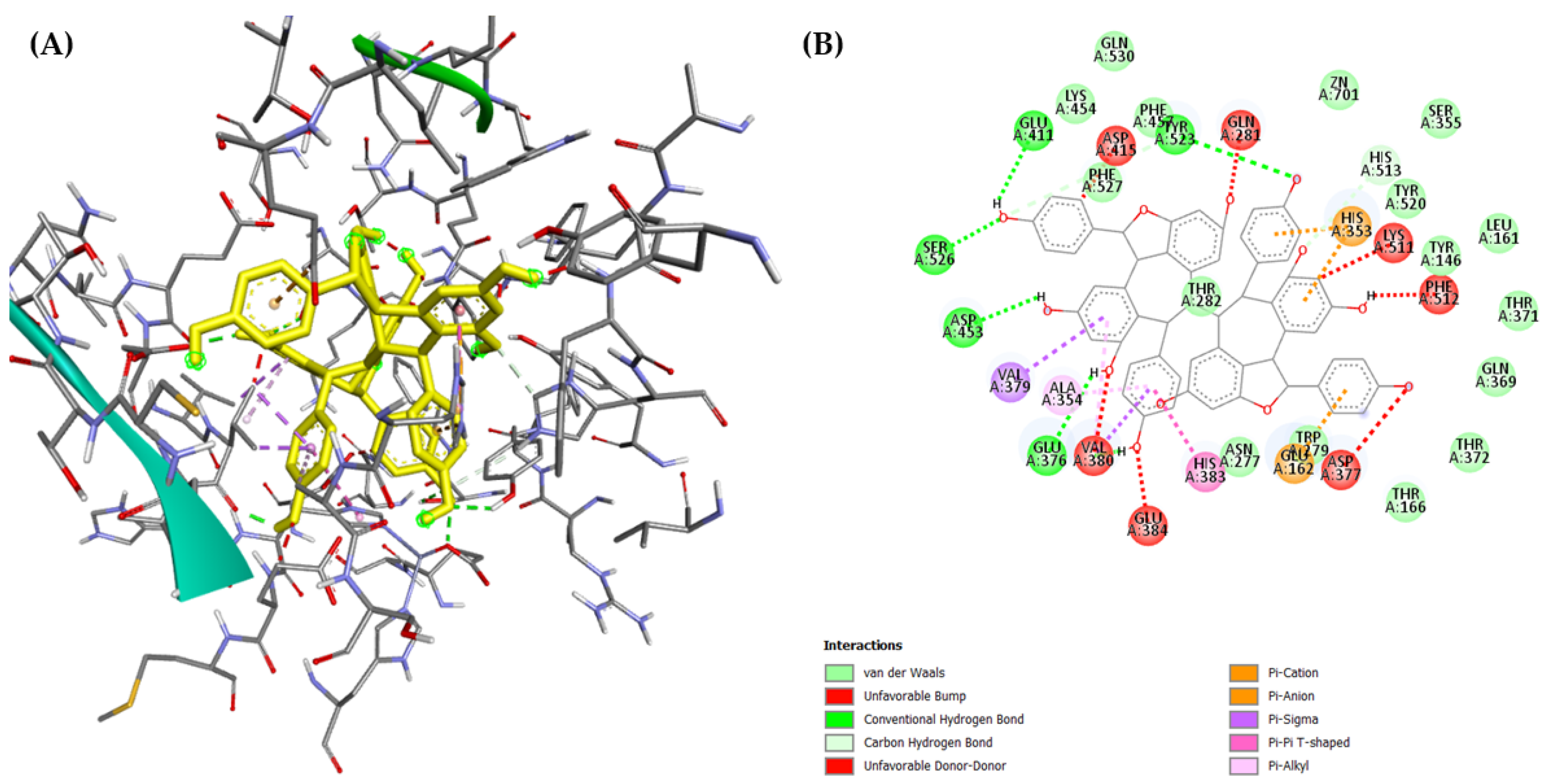
2.3. Molecular Docking

In silico docking was applied to characterize the binding behavior of (–)-hopeaphenol (16). The energy-minimized model of compound (16) was docked using AutoDock4 into both domains of the ACE crystal structure retrieved from the Protein Data Bank (PDB ID: 1O86 for the C-domain and 2C6N for the N-domain) to explain the chemical interactions between (–)-hopeaphenol (16) and both ACE active binding sites. This program uses the Lamarckian genetic algorithm (LGA) to generate a range of potential conformations from a starting ligand in an arbitrary conformation and then searches for favorable dockings at the protein-binding site [55]. The docking results revealed a network of hydrogen bonds and electrostatic and hydrophobic interactions between (–)-hopeaphenol (16) and the N-domain of the ACE with a strong binding energy (E = −9.83 kcal/mol). Importantly, interaction with residues Tyr369, Arg381, and Thr496 could be detected; these residues were previously connected to the N-domain selectivity [56]. The most important interactions are the π–π interactions between (–)-hopeaphenol (16) and Tyr369 and the carbon–hydrogen bond with Arg381 (Figure 7).
On the other hand, (–)-hopeaphenol (16) bound to the C-domain with a very poor affinity (binding energy: E = +41.42 kcal/mol). Several unfavorable interactions were identified as the reason for the low binding efficiency (Figure 8). This is well in line with the results of the in vitro domain-specific studies, suggesting that (–)-hopeaphenol (16) is an N-domain-selective ACE inhibitor.

2.4. Other Pharmacological Assays

The antiproliferative capacity of compounds 1218 was tested against human colon adenocarcinoma cells (Colo 205 sensitive and the resistant Colo 320/MDR-LRP expressing ABCB1). A thiazolyl blue tetrazolium bromide (MTT) assay was used for each compound to assess the concentration required for 50% inhibition of viability of the cell population (IC50), and cisplatin and doxorubicin were used as positive controls. Interestingly, only the monomer resveratrol (12) and the tetramer (–)-hopeaphenol (16) exerted notable antiproliferative activity against both cell lines with IC50 values comparable to those of the two positive controls, while the other stilbenes were found inactive (Table 6).
The antibacterial effect of the n-hexane, chloroform, and ethyl acetate fractions of the methanol extract of C. praecox was investigated against Bacillus subtilis, Escherichia coli, Klebsiella pneumoniae, Moraxella catarrhalis, MRSA, Pseudomonas aeruginosa, Staphylococcus aureus, S. epidermidis, and Streptococcus pyogenes using the disk diffusion method. The ethyl acetate fraction showed remarkable activity against S. epidermidis (inhibitory zone 18 mm), S. aureus (14 mm), MRSA (14 mm), M. catarrhalis (9 mm), and B. subtilis (12 mm), respectively. Therefore, the antibacterial effect of the isolated stilbenes (1218) was also investigated against these bacterial strains, but none of the compounds possessed remarkable antibacterial activity (data not indicated).

3. Materials and Methods

3.1. General Experimental Procedures

NMR spectra were recorded in methanol-d4, chloroform (CDCl3), and DMSO-d6 on a Bruker Avance DRX 500 spectrometer (Bruker, Ettlingen, Germany) at 500 MHz (1H) and 125 MHz (13C). The signals of the deuterated solvents were chosen as references. The chemical shift values (δ) were given in ppm, and the coupling constants (J) are in Hz. The two-dimensional (2D) experiments were conducted using standard TopSpin 3.6.1 Bruker software. Gradient-enhanced versions were applied in correlation spectroscopy (1H–1H COSY), nuclear Overhauser effect spectroscopy (NOESY), heteronuclear single quantum coherence spectroscopy (HSQC), and heteronuclear multiple bond correlation (HMBC) experiments. The high-resolution MS spectra were acquired with a Thermo Scientific Q-Exactive Plus Orbitrap mass spectrometer (Thermo Fisher Scientific, Waltham, MA, USA) equipped with an ESI ion source in positive ionization mode. The data were acquired and processed with MassLynx software 4.1 (SCN805). Optical rotation measurements were carried out by a Jasco-P2000 digital polarimeter (JASCO Corporation, Tokyo, Japan).
The normal and reversed-phase vacuum liquid chromatography (VLC) were carried out on a silica gel (Kieselgel 60 GF254, 15 μm, Merck, Darmstadt, Germany) and on a reversed-phase silica gel [RediSep C-18, 40–60 μm, Teledyne Isco, Lincoln, NE, USA]. The thin-layer chromatography was performed on Kieselgel 60 RP-18 F254 and Kieselgel 60 F254 (Merck, Darmstadt, Germany). The TLC plates were detected under a UV light at 254 nm and by spraying with a vanillin–sulfuric acid reagent, followed by heating. The flash chromatography (FC) was processed with a Combi Flash Rf+ Lumen instrument (Teledyne Isco) on a reversed-phase RediSep Rf HP Gold (50 g) column. Sephadex LH-20 (25–100 μm, Sigma-Aldrich, St. Louis, MO, USA) was used for gel filtration. The rotation planar chromatography (RPC) was carried out using a Chromatotron instrument (Model 8924, Harrison Research, T-Squared Technology, Inc., San Bruno, CA, USA). The high-performance liquid chromatographic (HPLC) separation was carried out on a Waters HPLC (Waters 600 controller, Waters 600 pump, and Waters 2998 photodiode array detector, Waters, Milford, MA, USA) and a Shimadzu HPLC (Shimadzu LC40, Shimadzu, Tokyo, Japan) using normal phase [LiChrospher Si 100 (250 × 4 mm, 5 μm, Merck)] and reversed-phase [Kinetex C18 (150 × 4.6 mm, 5 μm, 100 Å, Phenomenex, Torrance, CA, USA) and Kinetex Phenyl-hexyl] columns. The flow rate was 1 mL/min, and the injection volume was 10 μL. The data were acquired and processed with Empower 3 and LabSolutions software 5.111. All solvents used for CC were of at least analytical grade (VWR Ltd., Szeged, Hungary). Ultrapure water was prepared with a Milli-Q water purification system (Millipore, Molsheim, France).

3.2. Plant Material

The whole plants of Carex praecox Schreb. (1.8 kg of dried plant material) were collected during the flowering period in Besenyőtelek, Hungary (GPS coordinates: 47.691670, 20.438270) in July 2019 and were identified by László Bakacsy (Department of Plant Biology, University of Szeged, 6726 Szeged, Hungary). A voucher specimen (No. 899) was deposited in the Herbarium of the Department of Pharmacognosy, University of Szeged, Szeged, Hungary.

3.3. Extraction and Isolation
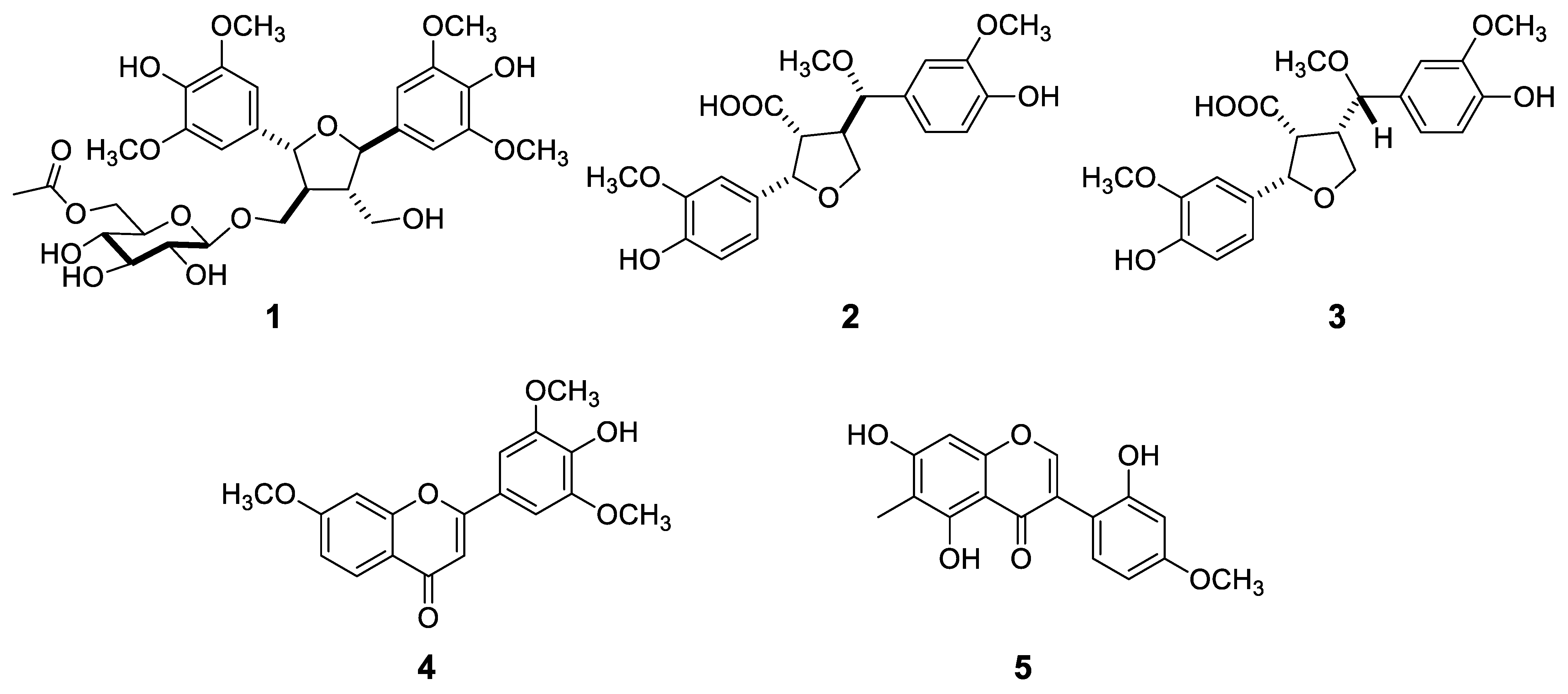
The dried and ground whole C. praecox plant (1.8 kg) was percolated with methanol (MeOH, 50 L) at room temperature. The methanolic extract was concentrated in vacuo (310 g), dissolved in 50% aqueous methanol (1500 mL), and subjected to solvent–solvent partition with n-hexane (10 × 500 mL), chloroform (CHCl3, 10 × 500 mL), and ethyl acetate (EtOAc, 10 × 500 mL).
The concentrated CHCl3-soluble fraction (11 g) was further separated by the normal phase (NP) vacuum liquid chromatography (VLC) applying a gradient solvent system of CHCl3–MeOH [from 100:0 to 8:2, and finally MeOH (500 mL/eluent)] resulting in 11 main fractions (C/A-K). Fraction C/B (354 mg) was further separated by a reversed-phase (RP) MPLC, affording 10 subfractions (C/B/1–10). From fraction C/B/1, compound 23 (1.4 mg) and from fraction C/B/7, compound 8 (3.8 mg) were isolated using an NP and RP preparative thin-layer chromatography (prep TLC), and by NP-HPLC. Fraction C/C (482 mg) was further purified by an RP flash chromatography (FC) to yield 12 subfractions (C/C/1–12). Fraction C/C/1 was chromatographed using RP-VLC, RP-prep TLC, and RP-HPLC methods to yield compounds 6 (3 mg), 7 (2.7 mg), 2 (1.6 mg), and 3 (1.5 mg), while subfraction C/C/4 was purified by an NP-prep TLC and afforded compounds 5 (18.7 mg) and 4 (2.8 mg). Fraction C/D (203 mg) was subjected to gel chromatography using Sephadex LH-20 gel as a stationary phase and a mixture of MeOH–CH2CL2 (1:1) as an eluent, which resulted in seven subfractions (C/D/1–7). Fraction C/D/4 was purified by RP-prep TLC and RP-HPLC, and compounds 19 (1.4 mg) and 20 (4.5 mg) were isolated, while fraction C/D/6 afforded compound 22 (4.6 mg). Fraction C/E (564 mg) was subjected to an RP-VLC, applying gradient elution with MeOH–H2O [from 1:9 to 9:1, and finally MeOH (300 mL/eluent)], resulting in 8 subfractions (C/E/1–8). From fraction C/E/6, compounds 24 (4.1 mg) and 25 (2.4 mg) were isolated through RP- and NP-VLC and an NP-HPLC. Fraction C/F (710 mg) was separated by an RP-MPLC, providing 14 subfractions (C/F/1–14). Using the RP-HPLC (gradient elution with acetonitrile–H2O), the purification of fraction C/F/2 afforded compound 21 (2.6 mg), while C/F/13 yielded compound 9 (4.0 mg).
The concentrated EtOAc-soluble fraction (10 g) was subjected to VLC on NP silica gel, with a gradient system of CHCl3–MeOH [from 98:2 to 7:3, and finally MeOH (800 mL/eluent)], to yield 11 main fractions (E/A-K). Fractions E/B (77 mg), E/D (339 mg), E/F (538 mg), E/G (2020 mg), and E/H (526 mg) were separated by gel chromatography on the Sephadex LH-20 stationary phase using MeOH–CH2Cl2 (1:1) as an eluent, yielding 4 (E/B/1–4), 8 (E/D/1–8), 9 (E/F/1–9), 8 (E/G/1–8), and 8 (E/H/1–8) subfractions, respectively. From fraction E/B/3, compound 26 (1.2 mg) was isolated via RP-HPLC. Fraction E/D/6 was purified via RP-prep TLC and RP-HPLC, and compounds 11 (12.9 mg) and 12 (1.2 mg) were obtained, while fraction E/D/8 proved to be pure for compound 10 (25.0 mg). From fraction E/F/2, compound 1 (8.8 mg) was obtained by RP-prep TLC (MeOH–H2O 6:4). Fraction E/F/7 was subjected to rotational planar chromatography (RPC) [gradient system of CHCl3–MeOH from 99:1 to 7:3, and finally MeOH (100 mL/eluent)] affording 5 subfractions. After purification by RP-HPLC (MeOH–H2O gradient system), fraction E/F/7/3 yielded compounds 13 (10.7 mg) and 14 (27.1 mg). From fraction E/F/7/4, compound 15 (5.9 mg) was isolated by RP-HPLC (MeOH–H2O gradient system). Fraction E/H/6 was further purified by RP-prep TLC, and RP-HPLC to obtain compounds 17 (16.4 mg) and 18 (3.6 mg). Fraction E/I (1631 mg) was subjected to RP-VLC, applying MeOH–H2O gradient elution [from 1:9 to 8:2, and finally MeOH (300 mL/eluent)] to afford 5 subfractions. After purification by RP-prep TLC (MeOH–H2O 1:1), fraction E/I/3 yielded compound 16 (4.7 mg).
  • Carexine A (1): yellowish oil; [α ] D 26 −6.0 (c 0.20, MeOH); 1H and 13C NMR data (CD3OD, see Table 1); HRESIMS m/z 658.2711 [M + NH4]+ (calcd for C30H44NO15 658.2711), 663.2258 [M + Na]+ (calcd for C30H40O15Na 663.2264).
  • Carexine B (2): white amorphous solid; [α ] D 26 −14.4 (c 0.05, MeOH); 1H and 13C NMR data (CD3OD, see Table 2); HRESIMS m/z 403.1443 [M − H] (calcd for C21H23 O8 403.1441).
  • Carexine C (3): pale yellow amorphous solid; [α ] D 26 +7.6 (c 0.05, MeOH); 1H and 13C NMR data (CD3OD, see Table 2); HRESIMS m/z 403.1443 [M −H] (calcd for C21H23 O8 403.1441).
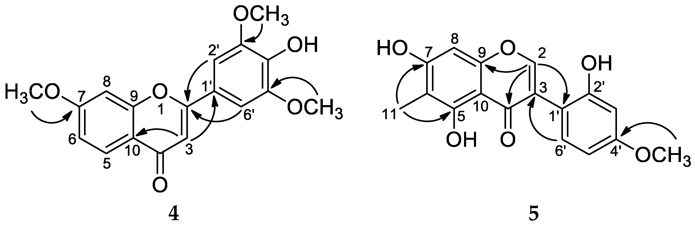
  • Carexine D (4): pale yellow amorphous solid; 1H and 13C NMR data (CD3OD, see Table 3); HRESIMS m/z 329.1018 [M + H]+ (calcd for C18H17O6 329.1020).
  • Carexine E (5): light brown amorphous solid; 1H and 13C NMR data (CD3OD, see Table 3); HRESIMS m/z 315.0865 [M + H]+ (calcd for C17H14O6 315.0869).
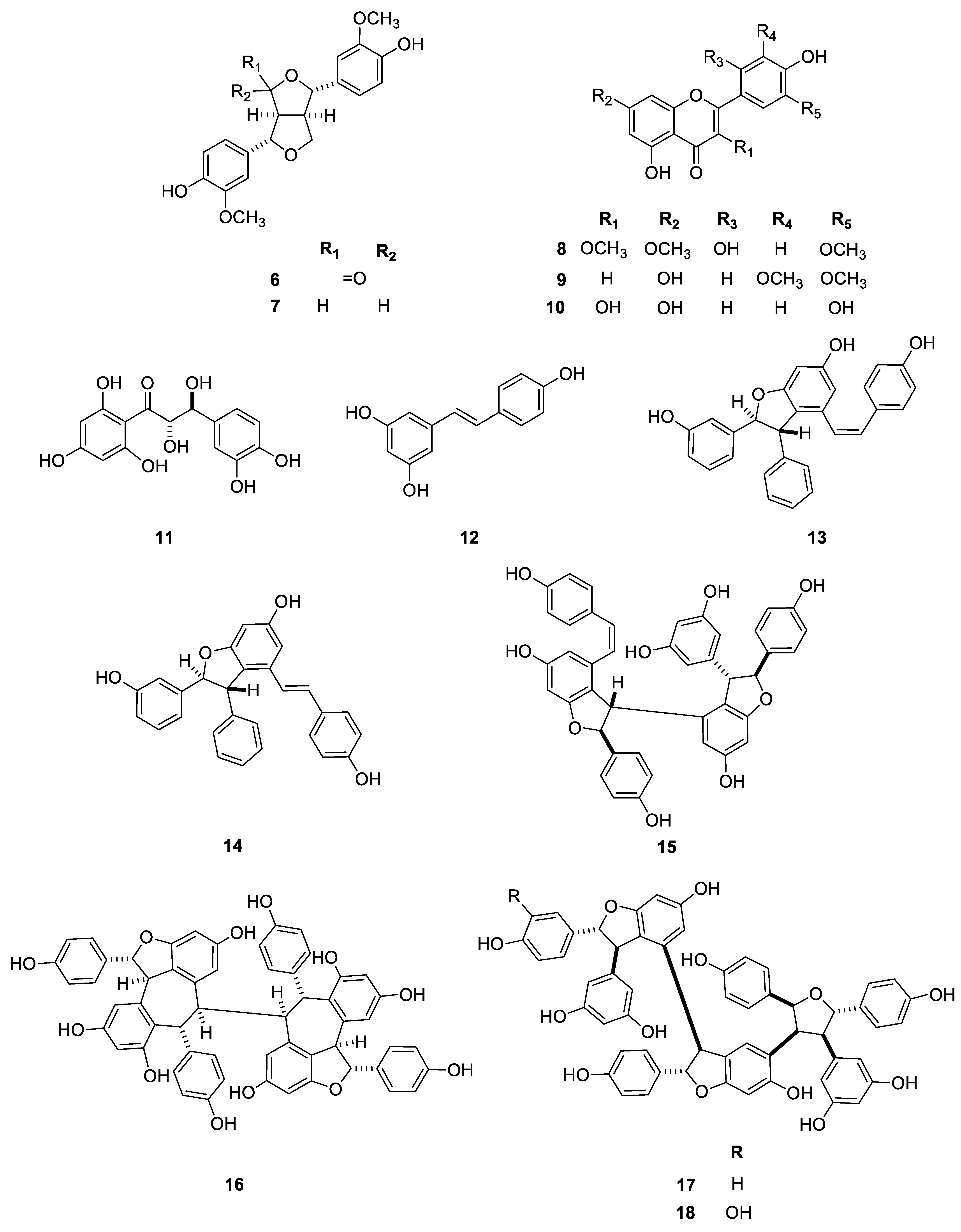
  • Compound 8 (chrysosplenol F): 1H NMR (CD3OD) δH 7.02 (1H, s, H-6′), 6.55 (1H, d, J = 2.2 Hz, H-8), 6.48 (1H, s, H-3′), 6.34 (1H, d, J = 2.2 Hz, H-6), 3.87 (3H, s, 7-OMe), 3.83 (3H, s, 5′-OMe), 3.74 (3H, s, 3-OMe); δC 180.0 (C-4), 167.3 (C-7), 162.9 (C-5), 159.2* (C-2), 159.0* (C-9), 152.3# (C-2′), 152.1# (C-4′), 142.8 (C-5′), 140.4 (C-3), 114.6 (C-6′), 109.1 (C-1′), 107.1 (C-10), 93.3 (C-8), 105.3 (C-3′), 99.0 (C-6), 56.5 (OMe-7), 57.3 (OMe-5′), 61.2 (OMe-3); *,# are interchangeable.
  • Compound 9 (tricin): 1H NMR (CD3OD) δH 7.25 (2H, s, H-2′,6′), 6.63 (1H, s, H-3), 6.45 (1H, d, J = 2.0 Hz, H-8), 6.2 (1H, d, J = 2.0 Hz, H-6), 3.95 (6H, s, 2 × OMe, 3′,5′).

3.4. Pharmacological Assays

3.4.1. ACE-Inhibitory Assay

Angiotensin-converting enzyme inhibition was determined using the Angiotensin-I Converting Enzyme (ACE) Activity Assay Kit (CS0002, Sigma-Aldrich, USA) with modification of the volume of enzyme and samples added to the wells. In summary, a 96-well black plate (655096, F-bottom, Grenier bio-one, Frickenhausen, Germany) containing 25 μL of samples diluted in methanol-assay buffer was filled with 25 μL of enzyme solution (each 25 μL contained 1.5 mU of the ACE: ACE from rabbit lung, A6778, Sigma-Aldrich, USA). The solution was incubated for 5 min at 37 °C with shaking, and then 50 μL of the substrate (a 100-fold dilution of the substrate provided) was added. A plate reader (BMG Lambtech GmbH, Ortenberg, Germany) was used to monitor the fluorescence for 5 min in kinetic mode at Ex/Em 290/450 nm as soon as the substrate was added. At the end of the measurement, the percentage inhibition by each compound was calculated as follows:
% inhibition = (values without samples − sample values)/(values without samples) × 100.
Dose–effect studies on the compounds 1218 were used to determine the concentration that inhibits 50% of the ACE. ACE-inhibitory kinetic studies were performed on compound 16, the most potent compound, to determine its inhibition mechanism. Similarly, 25 μL of enzyme was added to plate wells containing 25 μL of several concentrations of compound 16 (0–10 μM). Following a 5 min incubation period at 37 °C, 50 μL of the substrate Abz-Gly-Phe (NO2)-Pro (4003531, Bachem, Bubendorf, Switzerland) was added at varying concentrations (125–500 μM). The plate reader was then used to monitor the fluorescence in kinetic mode at extinction values of Ex/Em 290/450 nm. The Michaelis constant (Km) and maximal velocity (Vmax) of ACE were determined via Lineweaver–Burk plots, using the pharmacological and biochemistry transform and simple linear regression functions of the software, GraphPad Prism 8.0 (La Jolla, CA, USA).

3.4.2. Domain-Specific Studies

ACE domain-specific inhibition studies were performed based on previously reported methods (Carmona et al., 2006 [57]; Lunow et al., 2015 [58]). Fluorescence resonance energy transfer (FRET) substrates, Abz-SDK(Dnp)P-OH and Abz-LFK(Dnp)-OH were used for the N-domain and C-domain, respectively. The initial velocity of the reaction was determined using various concentrations (1–128 μM) of the FRET substrates. Briefly, 40 μL of assay buffer and 60 μL of the FRET substrate solutions were preincubated for 10 min at 37 °C, with the reaction started by adding 20 μL of diluted ACE solution (5 μL ACE + 15 μL 0.1 mol/L TRIS buffer) and fluorescence measured at λex/λem = 290/450 nm every minute at 37 °C for 30 min. The corresponding KM of the FRET substrates, determined using the Michaelis–Menten equation, was used as the substrate concentration of the FRET substrates used in percent inhibition studies. The inhibitory activity of 16 on both domains was determined as described above with the 40 μL solution containing the inhibitor (inhibitor in DMSO–Assay buffer, 1:9). The control samples, which correspond to 100% enzyme activity, were prepared by replacing the inhibitor solution with TRIS buffer. Dose–effect studies on 16 using the FRET substrates were used to determine the IC50 of this compound on both ACE domains. All experiments were performed in triplicates. The ACE-inhibitory activity was calculated using the following equation:
(%) = (Ab − Aa) − (Cb − Ca)/(Ab − Aa) × 100
where Aa is the absorbance of control wells at 0 min; Ab is the absorbance of control wells at 15 min; Ca is the absorbance of the inhibitor wells at 0 min; and Cb is the absorbance of inhibitor wells at 15 min.

3.4.3. Molecular Docking

The structure of the compound was drawn using a ChemDraw 12.0.2 software (ACD/LABS, Advanced Chemistry Development, Inc., Toronto, ON, Canada), and the energy of the compound (16) was minimized at the default mode, using a minimum RMS gradient of 0.010 in the software, Chem3D Pro 12.0 (ACD/LABS, Advanced Chemistry Development, Inc.). The energy-minimized compound was subsequently saved in PDB format before using it in the docking procedure. The X-ray crystallographic structures of the C- and N-domains of the human angiotensin I-converting enzyme complexed with lisinopril were obtained from the RCSB Protein Data Bank (PDB ID: 1O86 and 2C6N, respectively) [56,59]. PDB files for the enzyme and compounds were converted to the PDBQT format using the graphical user interface, AutoDock4 (The Scripps Research Institute, La Jolla, CA, USA) [60].
Before the docking analysis, water molecules and the lisinopril were eliminated from the 1O86 ACE protein model (C-domain) using AutoDock 4.2 (The Scripps Research Institute), while the zinc and chlorine atoms were retained in the ACE protein model, as these have been reported to be essential for the activity of ACE. By adding polar hydrogens, combining non-polar hydrogens, and adding Kollman charge to 1O86 using AutoDockTools, the final receptor for docking was created. A grid box (X: 43.817, Y: 38.308, and Z: 46.652, with 50 × 70 × 50 grid points of 0.375 Å spacing) created to include all active residues around the Zn(II) prosthetic group was used to calculate the zinc-centered map for ACE 1O86 [61].
The N-domain enzyme, 2C6N, was prepared using an identical method to the one used in preparing the C-domain enzyme, except that the protein’s water molecules and sugar moieties were eliminated along with the lisinopril. To encompass all active residues and Zn(II) heteroatom in the A chain of this domain [62], a grid box (X: −28.034, Y: −24.612, and Z: −33.992; number of grid points in the three dimensions [npts]: X: 70, Y: 70, and Z: 60; spacing: 0.375 Å) was defined.
The docking procedure was performed with 10 docking runs. The Lamarckian algorithm was used to dock ligands once the docking parameters were set to their default settings. The binding energies were obtained from the resulting DLG files, and visualization of the interactions was achieved via Biovia (Discovery Studio visualizer version 21.1.0.20298; Dassault Systèmes, Vélizy-Villacoublay, France) after conversion of the docked PDBQT files into PDB files using OpenBabel GUI software version 2.4.1 [63].

3.4.4. Cell Line Cultures

Two human colon adenocarcinoma cell lines, namely Colo 205 (ATCC-CCL-222) doxorubicin-sensitive parent cells and Colo 320/MDR-LRP (ATCC-CCL-220.1) doxorubicin-resistant cells expressing ABCB1, were purchased from LGC Promochem (Teddington, UK). The cells were cultured in an RPMI-1640 medium supplemented with 10% heat-inactivated fetal bovine serum (FBS), 2 mM of l-glutamine, 1 mM of sodium pyruvate, and 100 mM of HEPES. The cells were incubated at 37 °C, in a 5% CO2 and 95% air atmosphere.

3.4.5. Antiproliferative Assay

The antiproliferative effects of the compounds were tested in decreasing serial dilutions (2-fold dilutions starting from 100 μM) of human cancer cell lines (Colo 205, Colo 320) in 96-well flat-bottomed microtiter plates. First, the compounds were diluted in 100 μL of the medium, and then 6 × 10³ cells in 100 μL of RPMI medium were added to each well, excluding the medium control wells. The culture plates were incubated at 37 °C for 72 h. Following incubation, 20 μL of the MTT solution (from a 5 mg/mL stock solution) was added to each well. After incubation at 37 °C for 4 h, 100 μL of sodium dodecyl sulfate (SDS) solution (10% SDS in 0.01 M HCl) was added to each well, and the plates were further incubated at 37 °C overnight. Cell growth was determined by measuring the optical density at 540 nm (ref.: 630 nm) using a Multiscan EX ELISA reader (Thermo Labsystems, Cheshire, WA, USA). The concentration that decreased cell viability by 50% was expressed as the IC50 (μM) ± SD for each compound, derived using the log (inhibitor) vs. response nonlinear fitting model of GraphPad Prism Version 9.4.0.

3.4.6. Bacterial Strains and Culture Conditions for Antimicrobial Assays

The test microorganisms included the standard Gram-positive strains Staphylococcus aureus (ATCC 29213), methicillin-resistant S. aureus (MRSA) (ATCC 43300), S. epidermidis (ATCC 12228), Streptococcus pyogenes (ATCC 19615), and Bacillus subtilis (ATCC 6633). The standard Gram-negative strains were Escherichia coli (ATCC 35218), Klebsiella pneumoniae (ATCC 700603), Pseudomonas aeruginosa (ATCC 27853), and Moraxella catarrhalis (ATCC 25238). The bacterial cultures were grown on a standard Mueller–Hinton (MH) agar at 37 °C overnight in an aerobic environment.

3.4.7. Determination of Antibacterial Activity Using the Disk Diffusion Method

The disk diffusion method was employed to screen compounds for their antibacterial activities against standard bacterial strains to determine their inhibitory zones. Briefly, the samples were dissolved in DMSO at a concentration of 10 mM. Sterile filter paper disks [6 mm in diameter, Whatman antibiotic paper disk (Cytiva, Marlborough, MA, USA)] coated with 10 μL of the sample solutions were placed on top of the bacterial suspension (inoculum: 0.5 McFarland, 1.5 × 10⁸ CFU·mL−1). The disks containing antibiotics (ciprofloxacin and ampicillin) were used as positive controls, and the disks containing DMSO served as negative controls. Under aerobic conditions, the plates were incubated at 37 °C for 20 h. The diameters of the zones of inhibition caused by the compounds, including the disk, were measured in triplicate. For each of the three repetitions, an average zone of inhibition was calculated [64].

4. Conclusions

The phytochemical investigation of a Hungarian sedge, C. praecox, resulted in the isolation and identification of 26 compounds, among which there are 5 new natural compounds, carexines A–E (15). Considering the chemical characteristics of the isolated compounds, the main constituents of C. praecox are phenolic compounds, mainly stilbenes (1218). The ACE-inhibitory capacity of the isolated stilbenes was evaluated, the tetramer (–)-hopeaphenol (16) being the most potent inhibitor of the enzyme. The domain-specific assay revealed that hopeaphenol binds favorably to the N-domain of the ACE, which was also affirmed by the in silico docking studies. Selective inhibitors of the N-domain could be applied to treat tissue injury and fibrosis without affecting blood pressure and facilitate hematopoietic recovery after cancer chemotherapy.

Supplementary Materials

The following supporting information can be downloaded at: https://www.mdpi.com/article/10.3390/molecules29143427/s1, Figures S1–S73: 1D and 2D NMR, and HRESIMS spectra of compounds 15, 1H and JMOD spectra of 623, and 1H NMR spectra of 2426.

Author Contributions

Conceptualization, A.V.; methodology, A.V.; investigation, C.Z.D., N.K., R.B., A.K., G.S., and O.G.A.; resources, J.H.; data curation, A.V.; writing—original draft preparation, C.Z.D. and A.V.; writing—review and editing, J.H. and A.H.; supervision, A.V.; project administration, A.V.; funding acquisition, J.H. All authors have read and agreed to the published version of the manuscript.

Funding

This research was funded by the National Research, Development and Innovation Office, Hungary (NKFIH; K-135845 and K-134704), and the Ministry of Innovation and Technology of Hungary from NKFIH Fund, project no. TKP2021-EGA-32.

Institutional Review Board Statement

Not applicable.

Informed Consent Statement

Not applicable.

Data Availability Statement

The original contributions presented in the study are included in the article/Supplementary Materials, further inquiries can be directed to the corresponding author (A.V.).

Acknowledgments

Support from the New National Excellence Program of the Ministry of Human Capacities [UNKP-21-3 (C.Z.D.)] is gratefully acknowledged.

Conflicts of Interest

The authors declare no conflicts of interest. The funders had no role in the design of the study; in the collection, analyses, or interpretation of data; in the writing of the manuscript; or in the decision to publish the results.

References

  1. WHO: The Top 10 Causes of Death. Available online: https://www.who.int/news-room/fact-sheets/detail/the-top-10-causes-of-death (accessed on 29 February 2024).
  2. Curtin, S.C.; Tejada-Vera, B.; Bastian, B.A. Deaths: Leading causes for 2020. In National Vital Statistics Reports; National Center for Health Statistics: Hyattsville, MD, USA, 2023; Volume 72, No 13. [Google Scholar]
  3. OECD: Main Causes of Mortality. Available online: https://www.oecd-ilibrary.org/sites/a72a34af-en/index.html?itemId=/content/component/a72a34af-en (accessed on 29 February 2024).
  4. Bernstein, K.E.; Shen, X.Z.; Gonzalez-Villalobos, R.A.; Billet, S.; Okwan-Duodu, D.; Ong, F.S.; Fuchs, S. Different in vivo functions of the two catalytic domains of angiotensin-converting enzyme (ACE). Curr. Opin. Pharmacol. 2011, 11, 105–111. [Google Scholar] [CrossRef] [PubMed]
  5. Masuyer, G.; Schwager, S.; Sturrock, E.; Isaac, R.E.; Acharya, K.R. Molecular recognition and regulation of human angiotensin-I converting enzyme (ACE) activity by natural inhibitory peptides. Sci. Rep. 2012, 2, 717. [Google Scholar] [CrossRef] [PubMed]
  6. Alves-Lopes, R.; Montezano, A.C.; Neves, K.B.; Harvey, A.; Rios, F.J.; Skiba, D.S.; Arendse, L.B.; Guzik, T.J.; Graham, D.; Poglitsch, M.; et al. Selective inhibition of the C-domain of ACE (Angiotensin-Converting Enzyme) combined with inhibition of NEP (Neprilysin)—A potential new therapy for hypertension. Hypertension 2021, 78, 604–616. [Google Scholar] [CrossRef] [PubMed]
  7. Fiorentino, A.; D’Abrosca, B.; Pacifico, S.; Izzo, A.; Letizia, M.; Esposito, A.; Monaco, P. Potential allelopatic effects of stilbenoids and flavonoids from leaves of Carex distachya Desf. Biochem. Syst. Ecol. 2008, 36, 691–698. [Google Scholar] [CrossRef]
  8. Dhar, P.; Dhar, D.G.; Rawat, A.K.S.; Srivastava, S. Medicinal chemistry and biological potential of Cyperus rotundus Linn.: An overview to discover elite chemotype(s) for industrial use. Ind. Crops Prod. 2017, 108, 232–247. [Google Scholar] [CrossRef]
  9. Cho, N.; Valenciano, A.L.; Du, Y.; Clement, J.; Cassera, M.B.; Goetz, M.; Kingston, D.G. Antiplasmodial flavanones and a stilbene from Carpha glomerata. Bioorg. Med. Chem. Lett. 2018, 28, 3368–3371. [Google Scholar] [CrossRef] [PubMed]
  10. Nakajima, K.; Taguchi, H.; Endo, T.; Yosioka, I. The constituents of Scirpus fluviatilis (Torr). A. Gray. I. The structures of two new hydroxystilbene dimers, scirpusin A and B. Chem. Pharm. Bull. 1978, 26, 3050–3057. [Google Scholar] [CrossRef]
  11. Gamal, M.A.; Hani, K.M.K.; Sameh, E.S.; Sabrin, I.R.M. A Review: Compounds isolated from Cyperus species (Part I): Phenolics and nitrogenous. Int. J. Pharm. Phytochem. Res. 2015, 7, 51–67. [Google Scholar]
  12. Dávid, C.Z.; Hohmann, J.; Vasas, A. Chemistry and pharmacology of Cyperaceae stilbenoids: A review. Molecules 2021, 26, 2794. [Google Scholar] [CrossRef]
  13. Fiorentino, A.; D’Abrosca, B.; Pacifico, S.; Natale, A.; Monaco, P. Structures of bioactive carexanes from the roots of Carex distachya Desf. Phytochemistry 2006, 67, 971–977. [Google Scholar] [CrossRef]
  14. Kurihara, H.; Kawabata, J.; Ichikawa, S.; Mizutani, J. (–)-ε-Viniferin and related oligostilbenes from Carex pumila Thunb. (Cyperaceae). Agric. Biol. Chem. 1990, 54, 1097–1099. [Google Scholar] [CrossRef]
  15. Arraki, K.; Richard, T.; Badoc, A.; Pédrot, E.; Bisson, J.; Waffo-Téguo, P.; Mahjoub, A.; Mérillon, J.M.; Decendit, A. Isolation, characterization and quantification of stilbenes from some Carex species. Rec. Nat. Prod. 2013, 7, 281–291. [Google Scholar]
  16. Sotheeswaran, S.; Pasupathy, V. Distribution of resveratrol oligomers in plants. Phytochemistry 1993, 32, 1083–1092. [Google Scholar] [CrossRef]
  17. Teka, T.; Zhang, L.; Ge, X.; Li, Y.; Han, L.; Yan, X. Stilbenes: Source plants, chemistry, biosynthesis, pharmacology, application and problems related to their clinical application—A comprehensive review. Phytochemistry 2022, 197, 113128. [Google Scholar] [CrossRef] [PubMed]
  18. Matsushita, H.; Miyase, T.; Ueno, A. Lignan and terpene glycosides from Epimedium sagittatum. Phytochemistry 1991, 30, 2025–2027. [Google Scholar] [CrossRef]
  19. Yamauchi, H.; Kakuda, R.; Yaoita, Y.; Machida, K.; Kikuchi, M. Two new glycosides from the whole plant of Gleochoma hederacea L. Chem. Pharm. Bull. 2007, 55, 346–347. [Google Scholar] [CrossRef] [PubMed]
  20. In, S.J.; Seo, K.H.; Song, N.Y.; Lee, D.S.; Kim, Y.C.; Baek, N.I. Lignans and neolignans from the stems of Viburnum erosum and their neuroprotective and anti-inflammatory activity. Arch. Pharm. Res. 2015, 38, 26–34. [Google Scholar] [CrossRef] [PubMed]
  21. Moon, S.S.; Rahman, A.A.; Kim, J.Y.; Kee, S.H. Hanultarin, a cytotoxic lignan as an inhibitor of actin cytoskeleton polymerization from the seeds of Trichosanthes kirilowii. Bioorg. Med. Chem. 2008, 16, 7264–7269. [Google Scholar] [CrossRef]
  22. Xie, L.H.; Akao, T.; Hamasaki, K.; Deyama, T.; Hattori, M. Biotransformation of pinoresinol diglucoside to mammalian lignans by human intestinal microflora, and isolation of Enterococcus faecalis strain PDG-1 responsible for the transformation of (+)-pinoresinol to (+)-lariciresinol. Chem. Pharm. Bull. 2003, 51, 508–515. [Google Scholar] [CrossRef]
  23. Arisawa, M.; Hayashi, T.; Shimizu, M.; Morita, N. Isolation and cytotoxicity of two new flavonoids from Chrysosplenium grayanum and related flavonols. J. Nat. Prod. 1991, 54, 898–901. [Google Scholar] [CrossRef]
  24. Wang, Y.; Shen, J.Z.; Chan, Y.W.; Ho, W.S. Identification and growth inhibitory activity of the chemical constituents from Imperata cylindrica aerial part ethyl acetate extract. Molecules 2018, 23, 1807. [Google Scholar] [CrossRef] [PubMed]
  25. Sinha, R.; Gadhwal, M.K.; Joshi, U.J.; Srivastava, S.; Govil, G. Modifying effect of quercetin on model biomembranes: Studied by molecular dynamic simulation, DSC and NMR. Int. J. Curr. Pharm. Res. 2012, 4, 70–79. [Google Scholar]
  26. Ahmed, B.; Al-Howiriny, T.A. Two new hydroxy chalcone derivatives from Thymus cilicicus. Z. Naturforsch B 2007, 62, 121–124. [Google Scholar] [CrossRef]
  27. Guiso, M.; Marra, C.; Farina, A. A new efficient resveratrol synthesis. Tetrahedron Lett. 2002, 43, 597–598. [Google Scholar] [CrossRef]
  28. Kim, H.J.; Chang, E.J.; Bae, S.J.; Shim, S.M.; Park, H.D.; Rhee, C.H.; Park, J.H.; Choi, S.W. Cytotoxic and antimutagenic stilbenes from seeds of Paeonia lactiflora. Arch. Pharm. Res. 2002, 25, 293–299. [Google Scholar] [CrossRef]
  29. Kurihara, H.; Kawabata, J.; Ichikawa, S.; Mishima, M.; Mizutani, J. Oligostilbenes from Carex kobomugi. Phytochemistry 1991, 30, 649–653. [Google Scholar] [CrossRef]
  30. Aisha, F.; Din, L.B.; Yaacob, W.A. Resveratrol tetramer of hopeaphenol isolated from Shorea johorensis (Dipterocarpaceae). In Proceedings of the 2014 UKM FST Postgraduate Colloquium, Selangor, Malaysia, 9–11 April 2014; Volume 1614, pp. 302–308. [Google Scholar] [CrossRef]
  31. Häusler, M.; Montag, A. Isolation, identification and quantitative determination of the norisoprenoid (S)-(+)-dehydrovomifoliol in honey. Z. Lebensm. Unters. Forsch. 1989, 189, 113–115. [Google Scholar] [CrossRef]
  32. Yan, Z.H.; Han, Z.Z.; Hu, X.Q.; Liu, Q.X.; Zhang, W.D.; Liu, R.H.; Li, H.L. Chemical constituents of Euonymus alatus. Chem. Nat. Compd. 2013, 49, 340–342. [Google Scholar] [CrossRef]
  33. Kataoka, K.I.; Shiota, T.; Takeyasu, T.; Mochizuki, T.; Taneda, K.; Ota, M.; Tanabe, H.; Yamaguchi, H. Potent inhibitors of acyl-CoA:cholesterol acyltransferase. Structure-activity relationships of novel N-(4-oxo-8-chromanyl) amides. J. Med. Chem. 1995, 38, 3174–3186. [Google Scholar] [CrossRef]
  34. Pendse, R.; Rao, A.V.R.; Venkataraman, K. 5,7-Dihydroxychromone from Arachis hypogoea shells. Phytochemistry 1973, 12, 2033–2034. [Google Scholar] [CrossRef]
  35. Suresh, D.; Gurudutt, K.N.; Srinivasan, K. Degradation of bioactive spice compound: Curcumin during domestic cooking. Eur. Food Res. Technol. 2008, 228, 807–812. [Google Scholar] [CrossRef]
  36. Fishman, A.; Tao, Y.; Wood, T.K. Toluene 3-monooxygenase of Ralstonia pickettii PKO1 is a para-hydroxylating enzyme. J. Bacteriol. 2004, 186, 3117–3123. [Google Scholar] [CrossRef] [PubMed]
  37. Sies, H. Polyphenols and health: Update and perspectives. Arch. Biochem. Biophys. 2010, 501, 2–5. [Google Scholar] [CrossRef]
  38. Manach, C.; Scalbert, A.; Morand, C.; Rémésy, C.; Jiménez, L. Polyphenols: Food sources and bioavailability. Am. J. Clin. Nutr. 2004, 79, 727–747. [Google Scholar] [CrossRef] [PubMed]
  39. Rudrapal, M.; Khairnar, S.J.; Khan, J.; Dukhyil, A.B.; Ansari, M.A.; Alomary, M.N.; Alshabrmi, F.M.; Palai, S.; Kumar, P.; Devi, R. Dietary polyphenols and their role in oxidative stress-induced human diseases: Insights into protective effects, antioxidant potentials and mechanism(s) of action. Front. Pharmacol. 2022, 13, 806470. [Google Scholar] [CrossRef] [PubMed]
  40. Loupit, G.; Fonayet, J.V.; Lorensen, M.D.B.B.; Franc, C.; De Revel, G.; Janfelt, C.; Cookson, S.J. Tissue-specific stilbene accumulation is an early response to wounding/grafting as revealed by using spatial and temporal metabolomics. Plant Cell Environ. 2023, 46, 3871–3886. [Google Scholar] [CrossRef] [PubMed]
  41. DellaGreca, M.; Cutillo, F.; D’Abrosca, B.; Fiorentino, A.; Zarrelli, A. Isolation of seed germination and plant growth inhibitors from mediterranean plants: Their potential use as herbicides. In Natural Products for Pest Management; Rimando, A.M., Duke, S.O., Eds.; American Chemical Society: New York, NY, USA, 2006; Volume 927, pp. 24–36. [Google Scholar] [CrossRef]
  42. Spencer, G.F.; Tjarks, L.W. Germination inhibition by 5,7-dihydroxychromone, a flavonoid decomposition product. J. Plant Growth Regul. 1985, 4, 177–180. [Google Scholar] [CrossRef]
  43. Tena, C.; Santiago, A.R.; Osuna, D.; Sosa, T. Phytotoxic activity of p-cresol, 2-phenylethanol and 3-phenyl-1-propanol, phenolic compounds present in Cistus ladanifer L. Plants 2021, 10, 1136. [Google Scholar] [CrossRef] [PubMed]
  44. Kim, H.; Hwang, B.; Cho, S.; Kim, W.J.; Myung, S.C.; Choi, Y.H.; Kim, W.J.; Lee, S.; Moon, S.K. The ethanol extract of Cyperus exaltatus var. iwasakii exhibits cell cycle dysregulation, ERK1/2/p38 MAPK/AKT phosphorylation, and reduced MMP-9-mediated metastatic capacity in prostate cancer models in vitro and in vivo. Phytomedicine 2023, 114, 154794. [Google Scholar] [CrossRef]
  45. Pagning, A.; Tamokou, J.D.D.; Lateef, M.; Tapondjou, L.; Kuiate, J.R.; Ngnokam, D.; Ali, M. New triterpene and new flavone glucoside from Rhynchospora corymbosa (Cyperaceae) with their antimicrobial, tyrosinase and butyrylcholinesterase inhibitory activities. Phytochem. Lett. 2016, 16, 121–128. [Google Scholar] [CrossRef]
  46. Sayed, H.M.; Mohamed, M.H.; Farag, S.F.; Mohamed, G.A.; Proksch, P. A new steroid glycoside and furochromones from Cyperus rotundus L. Nat. Prod. Res. 2007, 21, 343–350. [Google Scholar] [CrossRef] [PubMed]
  47. Santos, A.L.; Yamamoto, E.S.; Passero, L.F.D.; Laurenti, M.D.; Martins, L.F.; Lima, M.L.; Uemi, M.; Soares, M.G.; Lago, J.H.G.; Tempone, A.G.; et al. Antileishmanial activity and immunomodulatory effects of tricin isolated from leaves of Casearia arborea (Salicaceae). Chem. Biodivers. 2017, 14, e1600458. [Google Scholar] [CrossRef] [PubMed]
  48. Kuwabara, H.; Mouri, K.; Otsuka, H.; Kasai, R.; Yamasaki, K. Tricin from a malagasy connaraceous plant with potent antihistaminic activity. J. Nat. Prod. 2003, 66, 1273–1275. [Google Scholar] [CrossRef] [PubMed]
  49. Moon, J.; Park, S.; Jhee, K.; Yang, S. Protection against UVB-induced wrinkle formation in SKH-1 hairless mice: Efficacy of tricin isolated from enzyme-treated Zizania latifolia extract. Molecules 2018, 23, 2254. [Google Scholar] [CrossRef] [PubMed]
  50. Coggon, P.; Janes, N.F.; King, F.E.; King, T.J.; Molyneux, R.J.; Morgan, J.W.W.; Sellars, K. Hopeaphenol, an extractive of the heartwood of Hopea odorata and Balanocarpus heimii. J. Chem. Soc. 1965, 406–409. [Google Scholar] [CrossRef]
  51. Tietjen, I.; Schonhofer, C.; Sciorillo, A.; Naidu, M.E.; Haq, Z.; Kannan, T.; Kossenkov, A.V.; Rivera-Ortiz, J.; Mounzer, K.; Hart, C.; et al. The natural stilbenoid (–)-hopeaphenol inhibits HIV transcription by targeting both PKC and NF-κB signaling and cyclin-dependent kinase 9. Antimicrob. Agents Chemother. 2023, 67, e01600-22. [Google Scholar] [CrossRef] [PubMed]
  52. Tietjen, I.; Cassel, J.; Register, E.T.; Zhou, X.Y.; Messick, T.E.; Keeney, F.; Lu, L.D.; Beattie, K.D.; Rali, T.; Tebas, P.; et al. The natural stilbenoid (–)-hopeaphenol inhibits cellular entry of SARS-CoV-2 USA-WA1/2020, B.1.1.7, and B.1.351 variants. Antimicrob. Agents Chemother. 2021, 65, e0077221. [Google Scholar] [CrossRef]
  53. Sharifi, N.; Khajeh, K.; Mahernia, S.; Balalaie, S.; Ataie, G.; Jahanbani, R.; Amanlou, M. Probing angiotensin converting enzyme (ACE) domain-dependent inhibition of onopordia, isolated from Onopordon acanthium L., using a continuous fluorescent assay. Pharm. Sci. 2018, 24, 31–37. [Google Scholar] [CrossRef]
  54. Dive, V.; Cotton, J.; Yiotakis, A.; Michaud, A.; Vassiliou, S.; Jiracek, J.; Vazeux, G.; Chauvet, M.T.; Cuniasse, P.; Corvol, P. RXP 407, a phosphinic peptide, is a potent inhibitor of angiotensin I converting enzyme able to differentiate between its two active sites. Proc. Natl. Acad. Sci. USA 1999, 96, 4330–4335. [Google Scholar] [CrossRef]
  55. Forli, S.; Huey, R.; Pique, M.E.; Sanner, M.F.; Goodsell, D.S.; Olson, A.J. Computational protein-ligand docking and virtual drug screening with the AutoDock suite. Nat. Protoc. 2016, 11, 905–919. [Google Scholar] [CrossRef]
  56. Corradi, H.R.; Schwager, S.L.; Nchinda, A.T.; Sturrock, E.D.; Acharya, K.R. Crystal structure of the N domain of human somatic angiotensin I-converting enzyme provides a structural basis for domain-specific inhibitor design. J. Mol. Biol. 2006, 357, 964–974. [Google Scholar] [CrossRef] [PubMed]
  57. Carmona, A.K.; Schwager, S.L.; Juliano, M.A.; Juliano, L.; Sturrock, E.D. A continuous fluorescence resonance energy transfer angiotensin I-converting enzyme assay. Nat. Protoc. 2006, 1, 1971–1976. [Google Scholar] [CrossRef]
  58. Lunow, D.; Kaiser, S.; Rückriemen, J.; Pohl, C.; Henle, T. Tryptophan-containing dipeptides are C-domain selective inhibitors of angiotensin converting enzyme. Food Chem. 2015, 166, 596–602. [Google Scholar] [CrossRef] [PubMed]
  59. Natesh, R.; Schwager, S.L.; Sturrock, E.D.; Acharya, K.R. Crystal structure of the human angiotensin-converting enzyme–lisinopril complex. Nature 2003, 421, 551–554. [Google Scholar] [CrossRef]
  60. Morris, G.M.; Huey, R.; Lindstrom, W.; Sanner, M.F.; Belew, R.K.; Goodsell, D.S.; Olson, A.J. AutoDock4 and AutoDockTools4: Automated docking with selective receptor flexibility. J. Comput. Chem. 2009, 30, 2785–2791. [Google Scholar] [CrossRef]
  61. Xie, D.; Du, L.; Lin, H.; Su, E.; Shen, Y.; Xie, J.; Wei, D. In vitro-in silico screening strategy and mechanism of angiotensin I-converting enzyme inhibitory peptides from α-lactalbumin. LWT 2022, 156, 112984. [Google Scholar] [CrossRef]
  62. Douglas, R.G.; Sharma, R.K.; Masuyer, G.; Lubbe, L.; Zamora, I.; Acharya, K.R.; Chibale, K.; Sturrock, E.D. Fragment-based design for the development of N-domain-selective angiotensin-1-converting enzyme inhibitors. Clin. Sci. 2014, 126, 305–313. [Google Scholar] [CrossRef]
  63. O’Boyle, N.M.; Banck, M.; James, C.A.; Morley, C.; Vandermeerch, T.; Hutchison, G.R. Open Babel: An open chemical toolbox. J. Cheminform. 2011, 3, 33. [Google Scholar] [CrossRef]
  64. Ghazal, T.S.A.; Schelz, Z.; Vidács, L.; Szemerédi, N.; Veres, K.; Spengler, G.; Hohmann, J. Antimicrobial, multidrug resistance reversal and biofilm formation inhibitory effect of Origanum majorana extracts, essential oil and monoterpenes. Plants 2022, 11, 1432. [Google Scholar] [CrossRef]
Figure 1. Structures of carexines A–E (15).
Molecules 29 03427 g001
Figure 2. Diagnostic 1H–1H COSY (Molecules 29 03427 i001) and HMBCs (H → C) of compound 1.
Molecules 29 03427 g002
Figure 3. Diagnostic 1H-1H COSY (Molecules 29 03427 i001
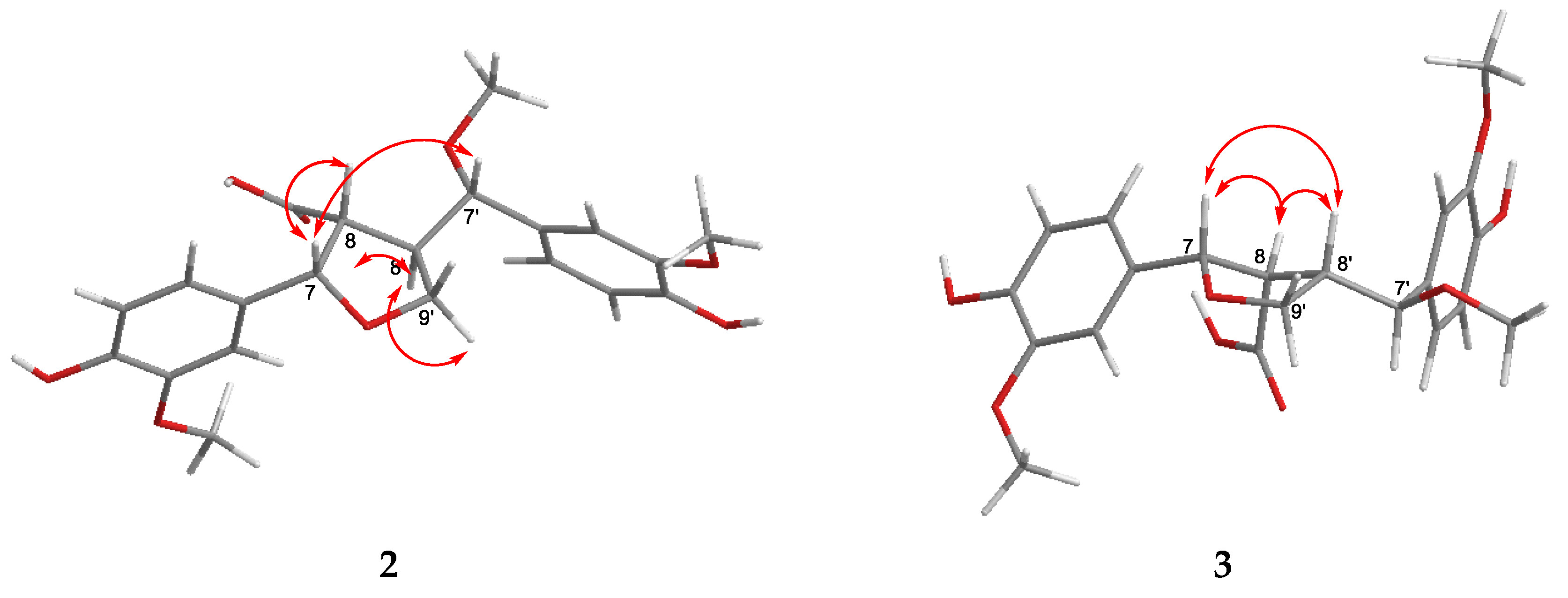
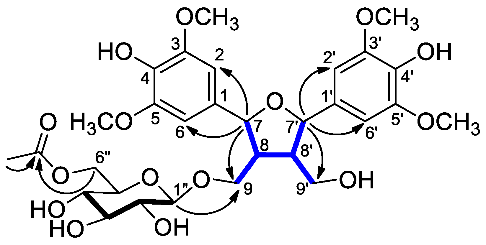
) and HMBCs (H → C) of compound 2.
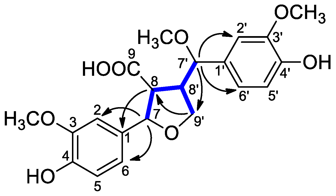
Molecules 29 03427 g003
Figure 4. Key NOESY correlations () of compounds 2 and 3.
Molecules 29 03427 g004
Figure 5. Diagnostic HMBCs (H → C) of compounds 4 and 5.
Molecules 29 03427 g005
Figure 6. Structures of other compounds (626) isolated from C. praecox.
Molecules 29 03427 g006aMolecules 29 03427 g006b
Figure 7. Binding of (–)-hopeaphenol (16) to the N-domain. (A): 3-D interactions of (–)-hopeaphenol (16) in yellow, with the active site amino acids. (B): 2-D interaction showing the type of bonding interactions with the active site residues.
Molecules 29 03427 g007
Figure 8. Binding of (–)-hopeaphenol (16) to the C-domain. (A): 3-D interactions of (–)-hopeaphenol (16) in yellow, with the active site amino acid residues. (B): 2-D interaction showing the type of bonding interactions, with several unfavorable interactions observed.
Molecules 29 03427 g008
Table 1. 1H (500 MHz) and 13C (125 MHz) NMR Data of Compound 1.
Position1H13CPosition1H13C
δH (ppm), J (Hz)δC, Type δH (ppm), J (Hz)δC, Type
1 134.3, C1′ 134.3, C
2,66.74 s104.9, CH2′,6′6.71 s104.9, CH
3 149.3, C3′ 149.3, C
4 136.1, * C4′ 136.2, * C
5 149.3, C5′ 149.3, C
75.05 d (8.4)84.6, CH7′4.98 d (8.5)84.3, CH
82.44 m 51.9, CH8′2.32 m54.5, CH
93.96 dd (10.1, 5.0),
3.65 dd (10.1, 5.0)
69.9, CH29′3.71 dd (11.6, 4.4),
3.59 dd (11.6, 4.4)
61.0, CH2
1″4.25 d (7.9)104.7, CH
2″3.16 br t (7.9)75.1, CH
3″3.30 m78.0, CH
4″3.25 m71.6, CH
5″3.41 m75.3, CH
6″4.33 dd (11.9, 2.1),
4.15 dd (11.9, 5.8)
64.6, CH2
3/3′-OCH33.83 s56.9, CH3
5/5′-OCH33.83 s56.9, CH3
Ac-CO 172.7, C
Ac-Me1.94 s20.6, CH3
* interchangeable signals.
Table 2. 1H (500 MHz) and 13C (125 MHz) NMR Data of Compounds 2 and 3.
23
Position1H13C1H13C
δH (ppm), J (Hz)δC, TypeδH (ppm), J (Hz)δC, Type
1-133.9, C-134.9, C
26.91 d (1.8)110.6, CH6.90 d (1.6)110.6, CH
3-149.1, C-148.9, C
4-147.9, * C-147.1, C
56.78 d (8.1)116.1, CH6.77 d (8.1)116.2, CH
66.77 dd (8.1, 1.8)119.7, CH6.78 dd (8.1, 1.6)119.6, CH
75.07 d (7.2)86.0, CH5.12 d (5.9)85.5, CH
83.07 m56.4, CH2.72 dd (8.4, 5.9)56.4, CH
9-176.6, C-176.8, C
1′-132.8, C-132.9, C
2′6.92 d (1.8)111.4, CH6.85 d (1.6)111.7, CH
3′-149.4, C-149.1, C
4′-147.4, * C-147.3, C
5′6.80 d (8.0)116.1, CH6.73 d (8.1)116.0, CH
6′6.78 dd (8.0, 1.8)121.5, CH6.76 dd (8.1, 1.6)121.0, CH
7′4.26 d (10.4)83.6, CH4.51 d (7.1)83.3, CH
8′3.00 m51.2, CH2.90 m50.3, CH
9′a (α)3.63 t (8.6, 7.6)71.4, CH24.15 d (7.9) 2H71.6, CH2
9′b (β)3.56 t (8.6)
3-OCH33.84 s56.4, CH33.80 s56.4, CH3
3′-OCH33.87 s56.4, CH33.84 s56.4, CH3
7′-OCH33.06 s56.0, CH33.15 s56.6, CH3
* interchangeable signals.
Table 3. 1H (500 MHz) and 13C (125 MHz) NMR Data of Compounds 4 and 5.
45
Position1H13C1H13C
δH (ppm), J (Hz)δC, TypeδH (ppm), J (Hz)δC, Type
2 166.1, C 8.00 s156.5, C
36.78 s106.1, CH 122.3, C
4 180.2, C 182.6, C
58.04 d (8.9)127.5, C 160.5, C
67.07 dd (8.9, 2.4)116.1, CH 109.1, C
7 166.4, C 164.0, C
87.28 d (2.4)101.7, CH6.41 s93.8, CH
9 159.7, C 157.5, C
10 118.2 105.9, C
11--2.07 s7.4, CH3
1′ 123.0, C 112.3, C
2′7.33 s105.5, CH 157.8, C
3′ 149.8, C6.48 br s 103.1, CH
4′ 141.2, CH 162.7, C
5′ 149.8, C6.49 dd (8.3, 2.5)106.6, CH
6′7.33 s105.5, CH27.13 d (8.3)133.2, CH
7-OCH33.98 s56.7, CH3
3′-OCH33.97 s57.2, CH3
4′-OCH3 3.78 s55.7, CH3
5′-OCH33.97 s57.2, CH3
Table 4. Results of the ACE-inhibitory assay.
CompoundInhibition at 90 μM (%) ± SDIC50 (μM) ± SD
1235.4 ± 3.9185.8 ± 12.8
1395.5 ± 2.818.0 ± 1.2
14106.7 ± 1.114.0 ± 0.7
1596.7 ± 6.315.2 ± 0.4
16102.8 ± 1.57.7 ± 0.9
1798.5 ± 1.214.8 ± 0.8
1898.7 ± 3.822.3 ± 0.9
Captopril80.8 ± 1.20.2 ± 0.1
Table 5. Domain-specific inhibitory capacity of (–)-hopeaphenol (16) compared to BPPb (substrate concentration [S] = KM calculated from initial velocity studies of both substrates; Abz-SDK(Dnp)P-OH; [S] = 72 μM and Abz-LFK(Dnp)-OH; [S] = 21 μM).
Inhibition (%) ± SD
N-DomainC-Domain
BPPb (200 nM)5.2 ± 0.283.20 ± 3.6
16 (10 μM)42.41 ± 0.818.17 ± 0.5
16 (50 μM)55.74 ± 3.223.39 ± 0.6
Table 6. Results of the antiproliferative assay (IC50 in μM ± SD).
Colo 205Colo 320
CompoundIC50 (μM) ± SDIC50 (μM) ± SD
1248.33 ± 1.5440.4 ± 1.84
13>100>100
14>100>100
15>100>100
161.59 ± 0.116.08 ± 0.24
17>100>100
18>100>100
DMSO>1%>1%
Cisplatin53.93 ± 3.9264.68 ± 3.56
Doxorubicin0.33 ± 0.030.67 ± 0.06
Disclaimer/Publisher’s Note: The statements, opinions and data contained in all publications are solely those of the individual author(s) and contributor(s) and not of MDPI and/or the editor(s). MDPI and/or the editor(s) disclaim responsibility for any injury to people or property resulting from any ideas, methods, instructions or products referred to in the content.

