Article

Temperature-Sensitive Template for Preparation of ZnO/CeO2 Composite Photocatalytic Materials and Its Catalytic Performance

1
School of Chemistry and Materials Science, Hubei Engineering University, Xiaogan 432000, China
2
School of Materials Science and Engineering, Hubei University, Wuhan 430062, China
3
School of Civil Engineering, Hubei Engineering University, Xiaogan 432000, China
*
Authors to whom correspondence should be addressed.
Molecules 2024, 29(15), 3589; https://doi.org/10.3390/molecules29153589 (registering DOI)
Submission received: 28 June 2024 / Revised: 25 July 2024 / Accepted: 27 July 2024 / Published: 30 July 2024
(This article belongs to the Special Issue Photocatalysis in the Control of Environmental Pollution)

Abstract

:
In this work, a series of thermosensitive ionic liquid functionalized polymers, PNx(IL)y, with controllable morphology and particle size were prepared by free radical polymerization. Then, using the polymer PN64(IL)8 with uniform morphology as a templating agent, the ZnO composite photocatalytic materials doped with rare earth metal Ce were prepared in combination with a microwave-assisted and templated hydrothermal reaction method. Series different Ce-doping amount photocatalytic materials ZnO-Ce-x‰ were characterized by XRD, SEM, TEM, XPS, and other methods. The results demonstrated that the templated materials PN64(IL)8 can prepare ZnO-Ce-2‰ with uniform petaloid ambulacra shape, good distribution of elements, and excellent photocatalytic performance. Photocatalytic degradation experiments of methyl orange (MO) showed that when the Ce-doping amount is only 2‰, the degradation rate of organic dyes can reach 96.5% by reacting the photocatalytic materials in water for 1 h. In addition, this kind of photocatalyst can be used for the degradation of high-concentration MO, as well as being easily recovered and effectively reused by simple filtration. Therefore, the structure of this kind of photocatalyst is controllable in the preparation process with an extremely low Ce-doping amount compared with current reports, and it has a good application prospect in the field of wastewater treatment technology.

1. Introduction

Organic dyes are one of the important components in textiles, optoelectronics, food, cosmetics, and other fields [1]. They are usually insoluble in water, and easily cause industrial organic wastewater, resulting in environmental pollution [2,3]. To solve this problem, researchers have been working hard to find effective ways to remove organic pollutants. Up to now, the organic dyes in industrial wastewater are mainly treated by chemical coagulation, precipitation, adsorption, and membrane filtration technologies [4,5]. However, these methods have the disadvantages of low efficiency and high cost.
As a semiconductor-based photocatalyst, ZnO nanoparticles have been widely reported for the removal of organic dyes, which have the advantages of low cost, good biocompatibility, simple preparation method, and can be reused many times [6,7,8]. Although it has been reported that ZnO can degrade Rhodamine B with a degradation rate of 90%, it can also be used as an effective photocatalytic material to decompose methylene orange, Nile blue, and other organic dyes [9,10,11]. However, the photocatalytic efficiency of pure ZnO is strongly influenced by the particle size, morphology, and specific surface area. In addition, ZnO nanoparticles are prone to agglomeration in an aqueous solution, resulting in a rapid decrease in surface area and hence catalytic efficiency. Therefore, it is urgent to modify ZnO nanoparticles to enhance their photocatalytic activity. However, ZnO has the problems of low light absorption efficiency, high e--h+ recombination rate, and photo corrosion. To solve these problems, researchers have done a lot of work to explore the modification of ZnO, including control morphology, semiconductor recombination, ion doping, precious metal deposition, and so on. In many studies, doped ZnO has shown better photocatalytic performance, and the dopant can be metal, such as Mn [12,13,14], Fe [15,16], Ni [17,18], Ag [19,20,21], Ce [22,23], or nonmetal C [24,25], and N [26,27,28]. Among all kinds of doping, Ce doping can significantly enhance ZnO carriers and thus improve photocatalytic activity. Despite the optical and electrical properties of Ce-doped ZnO in the literature, studies on the photocatalysis performance of ZnO/CeO2 nanocomposites remain relatively scarce. It has been revealed that ZnO/CeO2 nanocomposites possess exceptional properties suitable for diverse applications, including catalysis [29], biomedical [30], and antibacterial applications [31].
In preparing Ce-doped ZnO, the difficulty lies in how to obtain the material with good morphology and uniform loading. Based on our existing research foundation, considering the use of the template hydrothermal reaction method [32], we can prepare photocatalytic materials with excellent morphology and performance by using a suitable template. In our previous study, the temperature-sensitive ionic liquid functional polymer PNx(IL)y is a kind of water-soluble polymer that can self-assemble into nanoparticles with regular morphology and structure in water, and it has been reported to be applied in the field of catalysis [33]. Therefore, we can choose this kind of polymer, PNx(IL)y, combined with the template hydrothermal method to prepare excellent photocatalytic materials.
In this study, a series of temperature-sensitive ionic liquid functional polymers (PNx(IL)y) were prepared by RAFT polymerization. Using the polymer with regular morphology and uniform particle size as the template agent, the rare earth metal Ce-doped ZnO composite photocatalytic materials were prepared by microwave-assisted and template hydrothermal reaction methods. The series characterization shows that Ce is successfully doped into the ZnO structure and the morphology is good. Finally, it was applied to the degradation of organic dyes, and the catalytic performance, kinetic reaction performance, and recovery performance of the materials were investigated.

2. Results and Discussion

2.1. XRD of the Ce-Doped ZnO Photocatalysts

Figure 1 shows the XRD pattern of ZnO and different Ce amount of photocatalytic materials ZnO-Ce-x‰. It can be seen from the results that ZnO peaks appear at 2θ 31.72°, 34.34°, 36.21°, 47.65°, 56.55°, 62.79°, 66.51°, 67.81°, and 69.02°, respectively. The diffraction peaks correspond to (100), (002), (101), (102), (110), (103), (200), (112), and (201) crystal faces of hexagonal wurtzite [34]. The radius of Ce4+ (0.103 nm) was larger than that of Zn2+ (0.074 nm), so it was difficult for Ce4+ to enter the ZnO lattice by substituting Zn2+ (CeZn) or filling zinc vacancy (VZn) [35]. Relevant studies have shown that Ce4+ could be bonded to the surface of ZnO crystals by chemical bonding of Zn-O-Ce [36]. When using PN64(IL)8 as a template, the XRD pattern of the Ce-doped ZnO composite photocatalytic material showed the same characteristic peaks as that of the ZnO standard diagram. The same peaks indicate that Ce doping did not affect the typical structure of ZnO, and the crystal surface remains intact. With the increase in metal Ce content, the characteristic diffraction peak gradually showed at 28.56° and 32.97°. The new characteristic peak corresponds to the (111) crystal face of CeO2 of the cubic cerium mineral phase. In addition, the composite photocatalytic material did not change the characteristic structure of the original ZnO crystal after Ce doping. The (111) crystal face of CeO2 can observe the crystal face characteristics of Ce more in ZnO-Ce-10‰ and ZnO-Ce-14‰, which was marked by “*” at 28.56° and 32.97°.

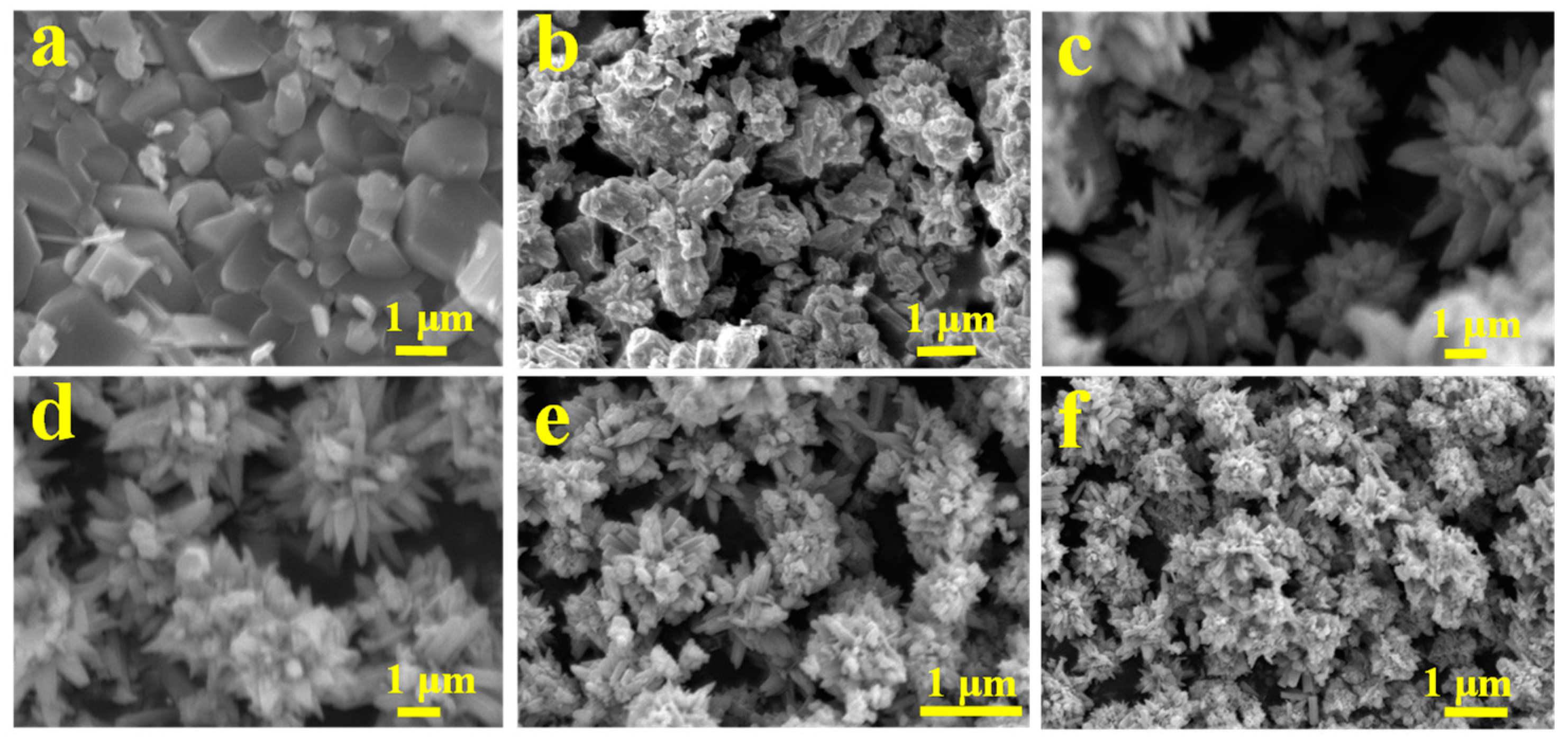
2.2. SEM of the Ce-Doped ZnO Photocatalysts

We investigated the effect of the templating agent PN64(IL)8 on the morphology of Ce-doped ZnO composite photocatalytic materials by SEM characterization. The results are shown in Figure 2. Without the templating agent PN64(IL)8, the pure ZnO prepared by the microwave-assisted combined hydrothermal method had no regular morphology, and showed sheet structures with different sizes and small specific surface area. After the addition of PN64(IL)8, ZnO grows into three-dimensional morphology material and the specific surface area increases. ZnO-Ce-2‰ photocatalytic materials have been prepared by adding Ce under the action of template agent PN64(IL)8. The prepared ZnO-Ce-2‰ showed a regular and uniform petal-like morphology, and the specific surface area of ZnO-Ce-2‰ was larger with a diameter of about 4 μm. When the doping amount was 6‰, ZnO-Ce-6‰ still showed a regular petal-like morphology. While further increasing the Ce amount, the obtained ZnO-Ce-10‰ and ZnO-Ce-14‰ materials showed petal-like imperfections. The imperfections were probably caused by the chemical bonding of metal Ce to form the Zn-O-Ce and ZnO crystal surfaces, and the petal-like surface grows CeO. Therefore, under the role of template agent PN64(IL)8 with the Ce-doping amount between 2‰ and 6‰, the petal-like materials with regular morphology and large specific surface area can be obtained.

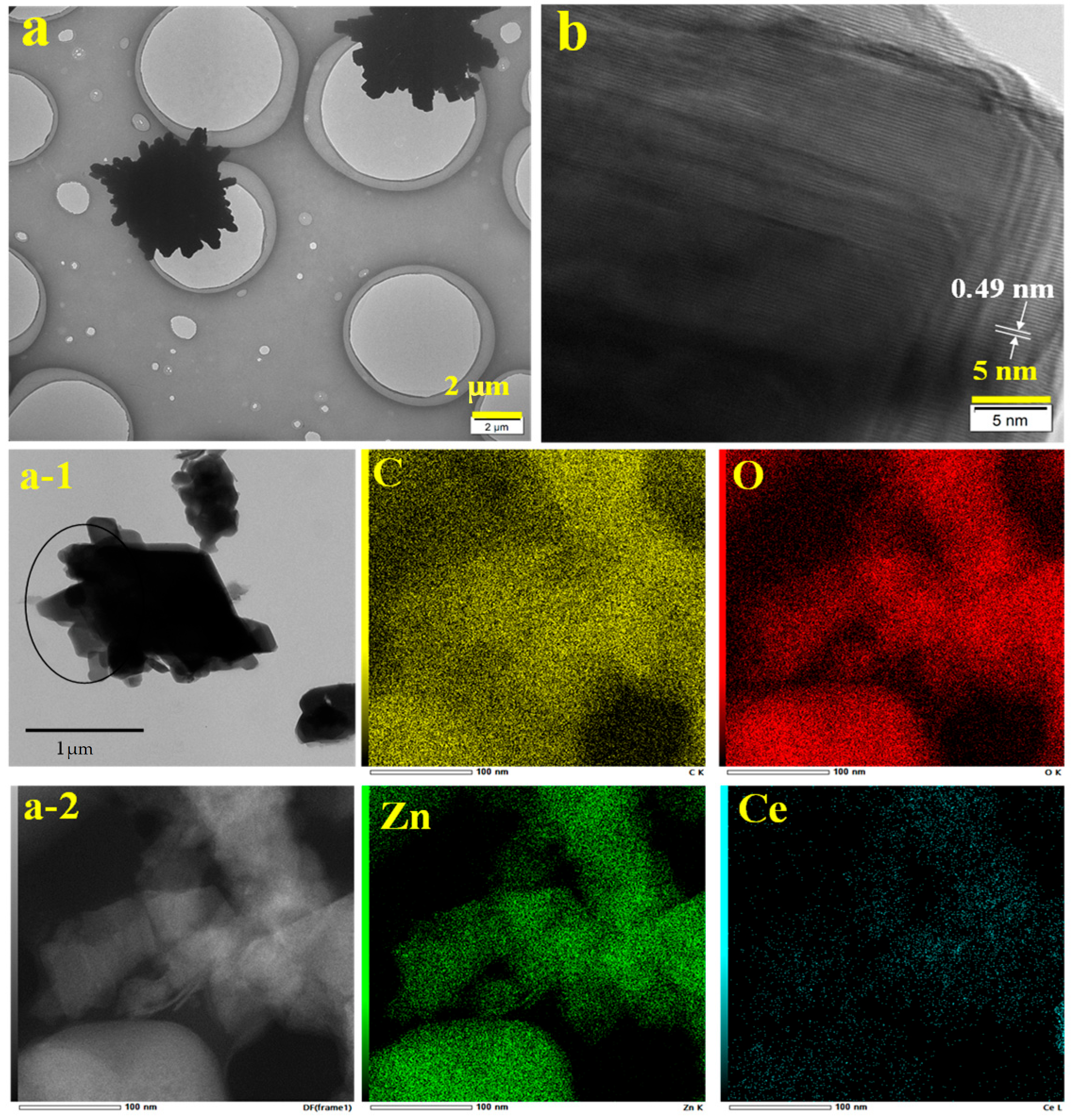
2.3. TEM and TEM Mapping of the ZnO-Ce-2‰ Photocatalyst

As shown in Figure 3, the morphological features of ZnO-Ce-2‰ were further analyzed by TEM and HRTEM. As the same with SEM, the TEM image also exhibits ZnO-Ce-2‰ with regular and uniform petal-like morphology, and the diameter was about 4 μm. In the HRTEM characterization, only one fringe structure with a lattice spacing of 0.49 nm was observed, which belongs to the lattice fringe of the wurtzite ZnO structure [37]. The lattice structure of Ce in the ZnO-Ce-2‰ structure was not observed, which may be due to the low doping amount and that Ce does not form a good crystal structure on the surface of the ZnO structure.
To further prove Ce was successfully doped into the ZnO structure, the ZnO-Ce-2‰ was characterized by TEM mapping. As Figure 3 shows, Zn and Ce were evenly distributed in the ZnO-Ce-2‰ photocatalyst by scanning the element content in the petal structure.

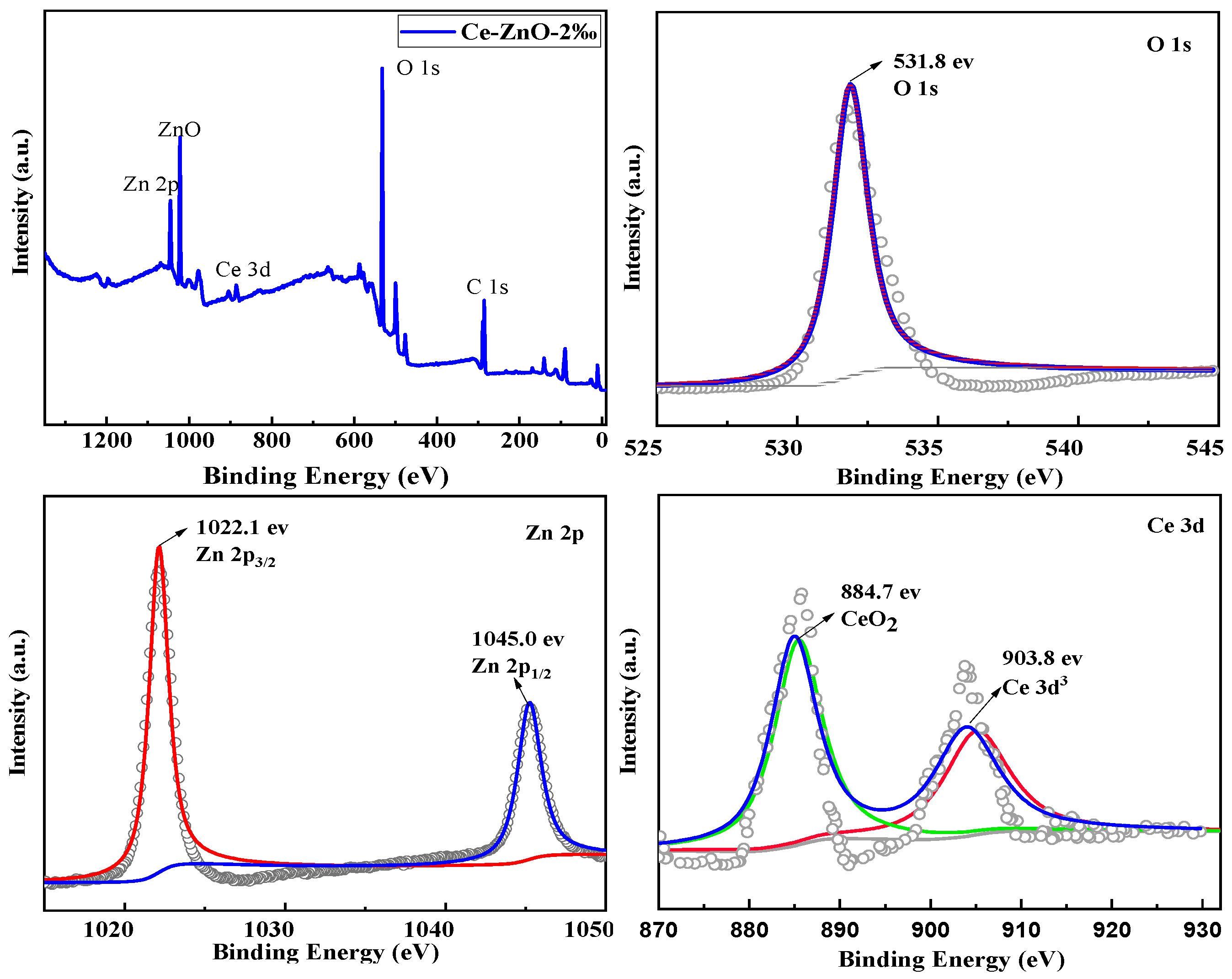
2.4. XPS of the ZnO-Ce-2‰ Photocatalyst

In order to investigate the chemical composition and valence states of the elements, the ZnO-Ce-2‰ photocatalytic material was tested by XPS. As shown in Figure 4, O, Zn, and Ce in ZnO-Ce-2‰ materials belong to ZnO and CeO2, respectively. The binding energy of the C 1s peak was used to calibrate the baseline. Figure 4 shows that the binding energy of Zn in ZnO-Ce-2‰ material is around 1022.1 eV and 1045.0 eV, and the high-resolution XPS spectra of Zn 2p were perfectly fitted, so Zn in the sample exists in the form of Zn2+ [38]. The Ce 3d photoelectron peak could be divided into two pairs of spin-orbit multiplets, with the peak values at 884.7 eV and 903.8 eV, confirming the existence of Ce4+ [39]. To better understand the content of Ce element, we performed XPS analysis on all materials of ZnO-Ce-6‰, ZnO-Ce-10‰, and ZnO-Ce-14‰ and provided the atomic content percentage in Supplementary Materials. As Raman spectra in Supplementary Materials, the E1(LO) band at 582.8 of ZnO-Ce-2‰ is a manifestation of the resonant enhancement of the LO mode due to the presence of Ce-doping defects. The systematic increase in the E 1 ( L O ) / E 2 high is in accord with an increasing defect concentration with the adding of Ce to form ZnO-Ce-2‰ ( E 1 ( L O ) / E 2 high = 0.2115) than ZnO ( E 1 ( L O ) / E 2 high = 0.0888) by calculation [40,41].

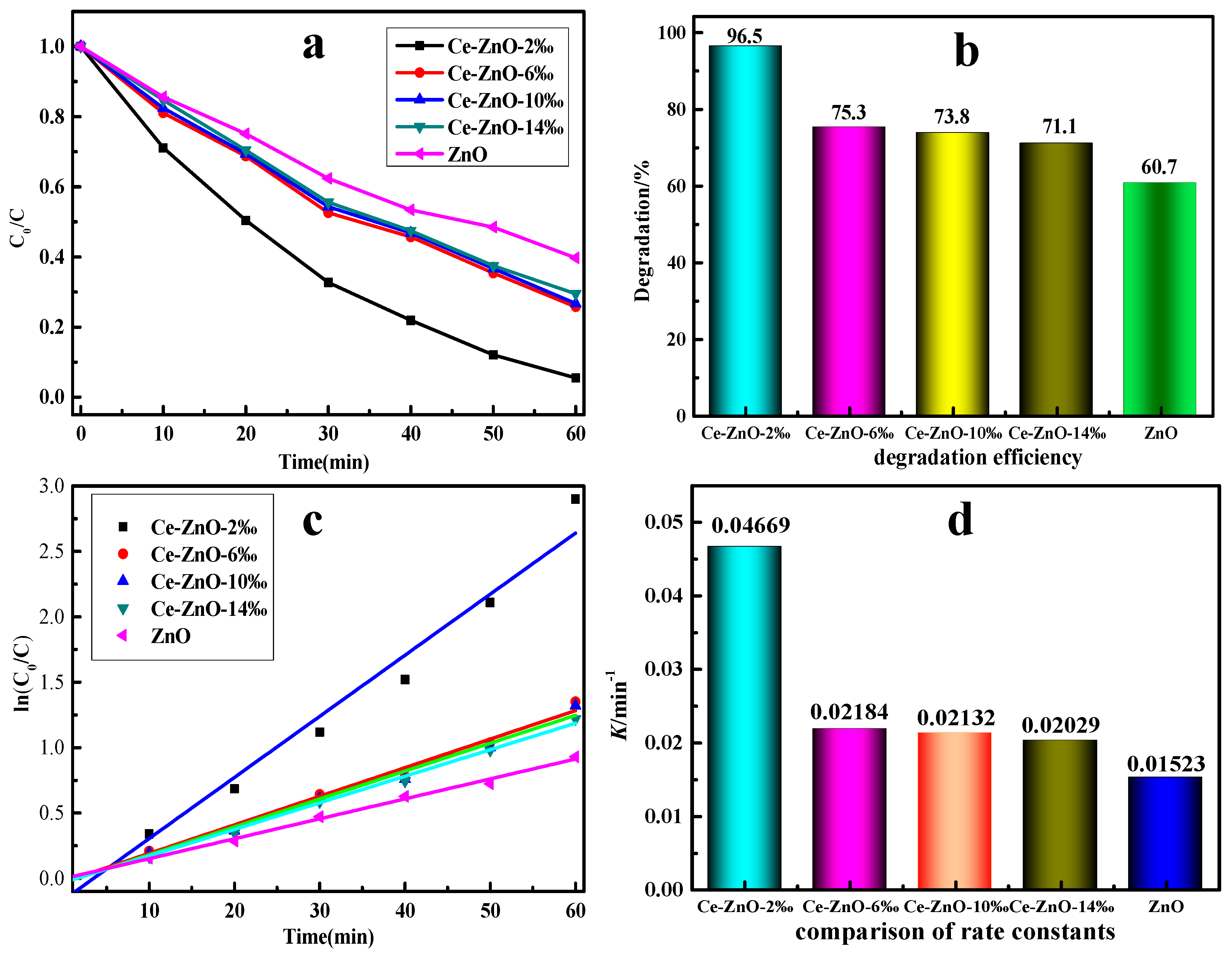
2.5. Catalytic Performance of Photocatalysts

Figure 5 shows the effect of photocatalytic material prepared in the presence of template agent PN64(IL)8 on the degradation of MO solution. It can be seen that the photodegradation efficiency of ZnO prepared by the template agent without Ce was only 60.7%, while the photodegradation efficiency of MO can be significantly improved after Ce doping. When PN64(IL)8 was used as a template agent, the efficiency of the ZnO-Ce-2‰ composite photocatalytic material can be greatly improved even if the doping concentration was only 2‰. The conversion rate could reach to 96.5% after 60 min of reaction. The catalytic efficiency of ZnO-Ce-6‰ decreased to 75.3%, and ZnO-Ce-10‰ maintained at 73.8% (Figure 5a,b). As the Ce content continued to increase, the degradation efficiency of the composite photocatalyst decreased. The decreased efficiency was mainly due to the excessive addition of Ce ions which destroy the hexagonal wurtzite ZnO structure, thus affecting the photodegradation efficiency. Therefore, under the condition of PN64(IL)8 as a template agent, the microwave-assisted hydrothermal method could be used to obtain the composite photocatalytic material. Additionally, excellent morphology and excellent photocatalytic efficiency were obtained when the doping amount was only 2‰ at low temperature (120 °C).
To further understand the kinetic characteristics of the reaction process, the degradation process was fitted linearly. It was found that the degradation was in accordance with the first reaction rate equation, and the linear correlation was good (Figure 5c). Among the reaction rate constants, the rate constant of the photocatalytic material ZnO-Ce-2‰ was the highest with 0.04669, about twice that of other photocatalytic materials. The highest catalytic efficiency of ZnO-Ce-2‰ was probably due to the best petal-like shape with a large specific surface area. The rate constant of pure ZnO photocatalytic materials was only 0.01523, which was lower than that of other Ce-doped photocatalytic materials (Figure 5d). The above kinetic results show that doping Ce for ZnO could significantly improve the degradation rate of MO [42,43].

2.6. Time-Dependent ZnO-Ce-2‰ for MO

The photocatalytic performance test model was conducted with ZnO-Ce-2‰ as the catalytic material and MO as the degradation material. Kinetic tests were conducted with samples taken every 10 min, and the results are shown in Figure 6. It can be seen from the kinetic experiment that the MO intensity at 464 nm decreases with time, and the degradation rate reaches the highest of 96.5% at 60 min. Moreover, the initial concentrations of MO were then investigated, and MO solutions with concentrations of 10 mg/L, 15 mg/L, 20 mg/L, 25 mg/L, and 30 mg/L were prepared. The results were shown in Supplementary Materials. Even at a higher concentration of 25 mg/L, the degradation rate is up to 75.3%. This work was higher than the general MO concentration reported in the current literature, and the catalytic degradation effect was much better [44,45,46,47].

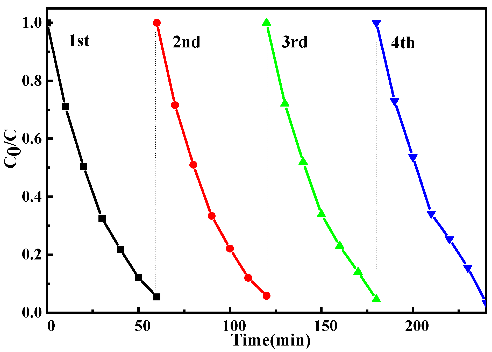
2.7. Reuse of ZnO-Ce-2‰ for MO

To test the industrial performance of the catalytic material, the recycling test was carried out. After the reaction, a centrifuge at 8000 rpm was used for 5 min, and the precipitate in the lower layer was washed and dried with deionized water for the next reuse. As shown in Figure 7, after 4 cycles, the degradation rate of MO was still at a high level of more than 90%. Therefore, according to the results of repeated use experiments, the photocatalytic material ZnO-Ce-2‰ prepared by using water-soluble temperature-sensitive material as a template and microwave-assisted hydrothermal method has good catalytic degradation performance of MO. What is more, ZnO-Ce-2‰ still exhibits good catalytic degradation performance when soaked in MO solution for a long time. The Ce-doped ZnO photocatalytic material prepared by this new method has a series of advantages in the field of wastewater treatment, such as low doping amount, less dosage, stable material properties, and has a good prospect for industrial application. To prove the stability of ZnO-Ce-2‰, we carried out the SEM characterization of the reuse for ZnO without Ce doping. The results showed that the SEM morphology of pure ZnO was changed after the UV photocatalytic reaction, indicating that ZnO suffers from photocorrosion. After 4 cycles of photocatalytic reaction, the morphology of ZnO-Ce-2‰ was characterized, and it was found that the SEM morphology had no change. After recovery, XRD and XPS remained unchanged, and the characteristic peaks were still present (see Supplementary Materials). The results show that the addition of Ce element can protect the morphology of zinc oxide and attenuate its photocorrosion [48].

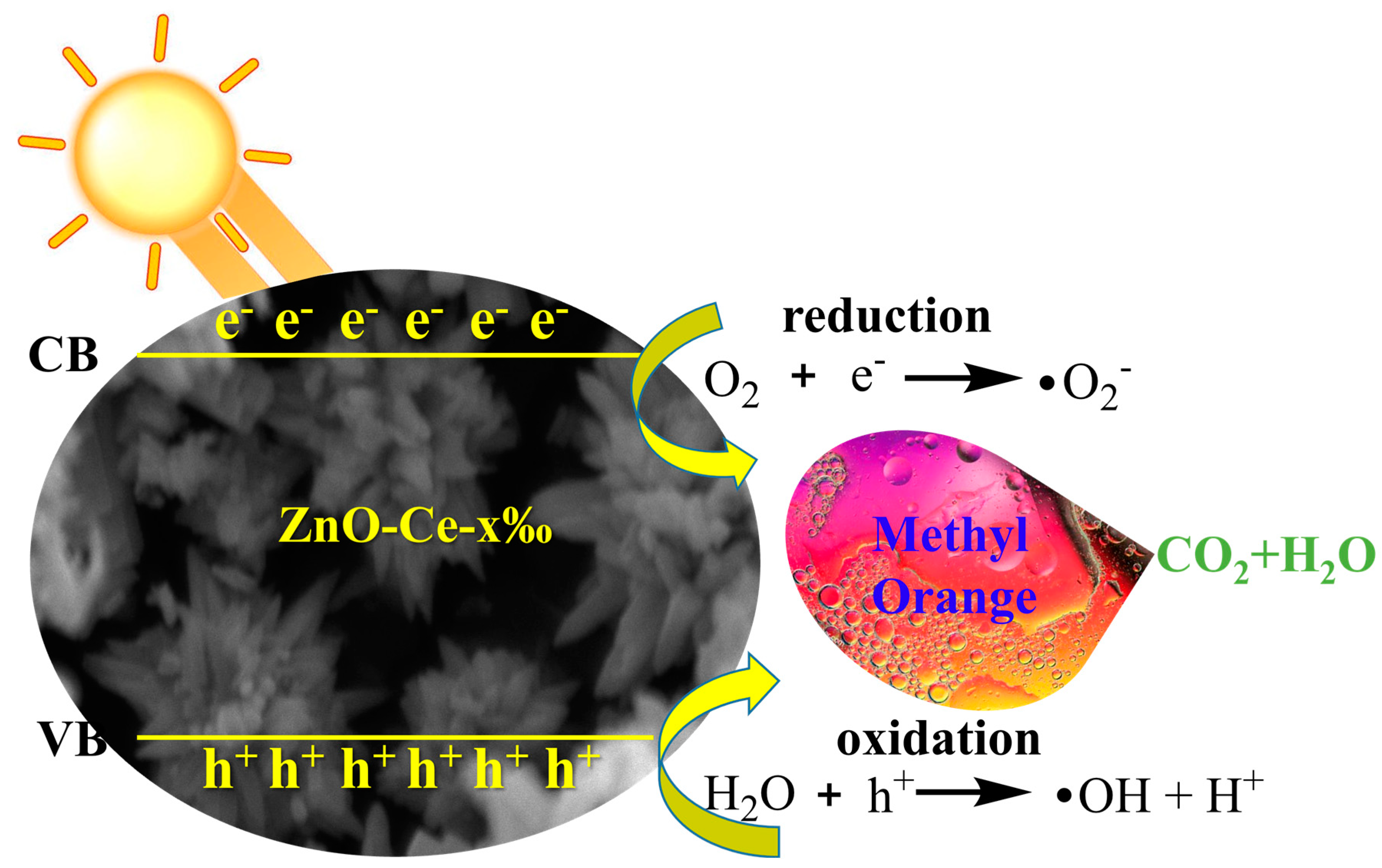
2.8. Mechanism of ZnO-Ce-2‰ for MO Degradation

The valence band of ZnO produced an excited e- due to the absorption of UV energy, which transitions to the ZnO conduction band and leaves a h+ in the valence band. After e- and h+ were excited to transfer to the ZnO crystal surface or the ZnO-Ce-x‰ crystal surface through ZnO, they can react with O2, H2O, and OH- in solution, respectively. Then, they generate photocatalytic •O2 free radicals and •OH free radicals [49,50]. The doped Ce3+ was finally attached to the surface of ZnO crystals in the form of Ce4+, and CeO2 had a strong oxygen absorption capacity, which increases the O2 content near the ZnO crystals. On the one hand, the consumption of photogenerated e- could effectively reduce the photogenerated e-h+ pair recombination in ZnO crystals. On the other hand, with the increase in •O2 and •OH active groups, the photocatalytic activity of the ZnO catalyst was improved (Figure 8). In addition, under high-concentration doping, the ZnO crystal structure will be slightly distorted, resulting in more VO, VZn, and other defects, which may become new e-h+ composite centers, resulting in reduced photocatalytic activity of ZnO. Therefore, ZnO-Ce-2‰ performed the highest catalytic degradation efficiency.

3. Materials and Methods

3.1. Materials

Zinc salt (ZnAc2·2H2O), sodium hydroxide, cerium chloride heptahydrate (CeCl3·7H2O), citric acid, N-isopropyl acrylamide (NIPAAm), vinyl imidazole, and methyl orange (MO) were purchased from Picard medical. Azobisisobutyronitrile was purchased from Shanghai Civic Chemical (Shanghai, China). Other commercially available chemicals were laboratory-grade reagents from local suppliers. All solvents were purified by standard procedure. Column chromatography silica gel and thin-layer silica gel were produced by Qingdao Ocean Chemical Silica Gel Factory (Qingdao, China). The synthesis of vinyl imidazole ionic liquid is referred to in the literature [33].

3.2. Analytical Methods

In gel permeation chromatography (GPC, Alltech, Lexington, KY, USA) analyses, THF was used as a solvent eluting at a flow of 1 mL/min with polystyrene standards as calibration, and the detection temperature is 40 °C with a column temperature of 30 °C through a Jordi GPC 10000 A column (300 mm × 7.8 mm) (Mansfield, MA, USA) equipped with an Alltech ELSD 800. FT-IR spectras were obtained using an AVATAR 370 Thermo Nicolet spectrophotometer (Nicolet, Green Bay, WI, USA). XRD patterns were recorded on a Bruker-D8 diffractometer with Cu Kα radiation (λ = 0.15406 nm) to evaluate the crystal structure of ZnO-Ce-x‰ samples (Bruker, Rheinstetten, Germany). The particle size and the morphology of the ZnO-Ce-x‰ were observed by SEM (Hitachi S3400N, Hitachi, Tokyo, Japan) and TEM (FEI Tecnai G20, FEI, Hillsboro, OR, USA). The XPS analysis was carried out on a Thermo Fisher Scientific K-Alpha spectrometer (Waltham, MA, USA) using monochromatic Al-Ka radiation at a detection angle of 30° (Invitrigen, Waltham, MA, USA). The photochemical reaction apparatus (GHX-IV, Shanghai, China) was used as a photocatalytic material for the degradation of MO. UV-Vis Agilent 8453 was used to test the absorbance of MO solution (Agilent, Santa Clara, CA, USA).

3.3. Preparation of Temperature-Sensitive Template PN64(IL)8

Vinylimidazole ionic liquid compounds were prepared by the 1:1 reaction of vinylimidazole with bromoethane as described in the literature [51,52]; 10 mmol of vinyl imidazole and 10 mmol of chloromethane were added to 20 mL ethanol, forming a colorless solution that was refluxed under nitrogen for 24 h. After the full reaction, the resulting mixture was purified by re-crystallization from ethanol, with ethyl acetate as an anti-solvent, and named IL. Then, 6.4 mmol of NIPAAm, 0.8 mmol of IL, 1/6 mmol of thiopropionic acid benzoic acid ester, and 1/30 mmol of azodiisobutyronitrile (AIBN) were dissolved in methanol and the solution was added to Schlenk tube. The reaction was carried out at 60 ℃ for 24 h under N2 protection. Then, concentrated reaction liquid was under a vacuum and the polymer was repeatedly precipitated with several times of excess ether to obtain a light yellow product. After vacuum drying at 30 °C, the fluffy light-yellow polymerization product PN64(IL)8 was obtained. PN64(IL)8: FT-IR (KBr): γmax/cm−1 3293, 3067, 2972, 2937, 2874, 1648, 1543, 1463, 1388, 1365, 1271, 1253, 1206, 1173, 1131, 1028, 924, 868, 850, 756, 709, 647, 587, 489 cm−1. GPC (THF): Mn = 9581, Mw = 10372, PDI = 1.08.

3.4. Preparation of Nanometer ZnO Materials Doped with Ce in Different Proportions

The different contents of Ce-doped ZnO photocatalysts were prepared by hydrothermal reactor method using PN64(IL)8 as a template (Table 1); 0.01 g PN64(IL)8 was dissolved in water, then ZnAc2·2H2O (0.998 mmol), citric acid (0.735 mmol), and CeCl3·7H2O (0.002 mmol) were added to the above solution under stirring circumstances until all reagents were completely dissolved. Microwaved for 0.5 h, and transferred to hydrothermal reactor at 120 °C for another 10 h. Cooled to room temperature, the obtained precipitate was centrifuged at 8000 r/min and washed at least three times with water to remove residual salts. Dried overnight in a vacuum drying oven at 50 °C, giving a white powder of ZnO-Ce-2‰. The references [53,54] show that interstitial incorporation is favored at low Ce concentrations, while substitution and interstitial substitution are comparable processes at high Ce concentrations. Based on economic principles, four different ratios of Ce-doped ZnO photocatalysts were prepared, and the doping concentrations of Ce were in mole percent: 0, 0.002, 0.006, 0.010, and 0.014 Ce-doped ZnO, respectively. The series photocatalysts were named ZnO-Ce-2‰, ZnO-Ce-6‰, ZnO-Ce-10‰, and ZnO-Ce-14‰, respectively (Table 1).

3.5. Photocatalytic Tests with Different Photocatalysts and Influence of Initial Concentration of MO

Next, 20 mg/L MO solution was accurately prepared, and 10 mg powder of different photocatalytic materials of ZnO-Ce-x‰ were weighed, respectively. ZnO-Ce-x‰ was added to 100 mL 20 mg/L MO solution, stirred, and dispersed for 30 min in the dark so that MO was completely pre-adsorbed on the catalyst surface; 10 mL of the solution was centrifuged at high speed and the supernatant solution was retained. The remaining solution was placed in the photocatalytic reaction apparatus, and the photodegradation experiment was carried out under the UV lamp at 500 W light with circulating water. The sample was taken at regular intervals and the supernatant was separated at high speed. UV spectrophotometer with the best detection wavelength (about 464 nm) was used to scan the 20 mg/L MO solution and measure the absorbance of the above two supernatants at the best detection wavelength. The degradation rate of the MO solution of the catalytic material was calculated by using the degradation rate formula. Then MO solutions of 10 mg/L, 15 mg/L, 20 mg/L, 25 mg/L, and 30 mg/L were prepared, and 10 mg of the catalytic material ZnO-Ce-2‰ powder was added to 100 mL of MO solution of different concentrations. All of the other steps were the same as above. The degradation rate of MO solutions was calculated to assess the influence of the initial concentration of MO. The following Formula (1) was used to calculate the degradation rate.
D = (A0 − At)/A0 × 100%
In the formula, D is the degradation rate; A0 is the initial absorbance value of MO solution; At is the photocatalytic time absorbance value of MO solution [55].
The formula used to calculate the reactivity values was the first-order kinetic reaction, which is represented by the mathematical expression as mentioned in Equation (2). By rearranging the above formula, we calculated the value of K (min−1), which shows the reactivity of a photocatalyst, or commonly known as the photodegradation constant [56].
ln ( C 0 C ) = K t
C0 is the initial concentration of MO solution (mg L−1), and Ct is the instant concentration of MO solution (mg L−1) at the time of the moment.

4. Conclusions

A novel series of polymers PNx(IL)y were prepared, and the polymer with uniform morphology PN64(IL)8 was chosen as the template agent. Series different Ce-doping amount photocatalytic materials ZnO-Ce-x‰ have been successfully designed and prepared. Then, ZnO-Ce-x‰ were sufficiently characterized by XRD, SEM, TEM, and XPS to determine the morphology. Characterization results proved ZnO-Ce-2‰ with a uniform petaloid ambulacra shape and good distribution of elements. This kind of ZnO-Ce-x‰ catalyst could exhibit good performance compared with ZnO without a template agent. Remarkably, the photocatalyst with a lower Ce amount for ZnO-Ce-2‰ acquired the highest catalyst efficiency. The degradation rate of organic dyes can reach 96.5% by reacting with the photocatalytic materials in water for 1 h. Moreover, the ZnO-Ce-x‰ catalysts could be facilely recovered from the MO solution system for four cycles by simple centrifuge. The controllable preparation, high activity, as well as outstanding reusability, pave a green and efficient way for wastewater treatment in a green system.

Supplementary Materials

The following supporting information can be downloaded at: https://www.mdpi.com/article/10.3390/molecules29153589/s1, Figure S1: Characterization of the template PN64(IL)8; Figure S2: XPS of ZnO-Ce-6‰, ZnO-Ce-10‰, ZnO-Ce-14‰ with the added of template PN64(IL)8; Figure S3: Raman spectra of ZnO (a), ZnO-Ce-2‰ (b), ZnO-Ce-6‰ (c), ZnO-Ce-10‰ (d), ZnO-Ce-14‰ (e); Figure S4: Influence of catalyst amount (a) and initial concentration of MO (b) in the presence of ZnO-Ce-2‰; Table S1: Atomic percentages of ZnO-Ce-x‰; Table S2: Raman modes of ZnO nanospindles in samples ZnO, ZnO-Ce-2‰, ZnO-Ce-6‰, ZnO-Ce-10‰, ZnO-Ce-14‰ [57].

Author Contributions

Y.Z.: Conceptualization, Project administration, Funding, Writing—original draft; Writing—review and editing; W.Y.: Data curation, Investigation, Visualization; Z.Z.: Formal analysis, Data curation; L.Z.: Methodology, Resources, Software; W.P.: Formal analysis, Writing—review and editing. All authors have read and agreed to the published version of the manuscript.

Funding

This research was funded by the National Natural Science Foundation of China (22108065, 22278118), the Natural Science Foundation project of Hubei (2024AFB967), Hubei Province Young Science and Technology Talent Chenguang Project, the Opening Fund of Hubei Key Laboratory of Polymer Materials, and Hubei University.

Data Availability Statement

No new data were created or analyzed in this study. Data sharing is not applicable to this article.

Conflicts of Interest

The authors declare that they have no conflicts of interest.

References

  1. Strack, R. Organic dyes for live imaging. Nat. Methods 2021, 18, 30. [Google Scholar] [CrossRef] [PubMed]
  2. Alguacil, F.J.; López, F.A. Organic dyes versus adsorption processing. Molecules 2021, 26, 5440. [Google Scholar] [CrossRef] [PubMed]
  3. Boyles, C.; Sobeck, S.J.S. Photostability of organic red food dyes. Food Chem. 2020, 315, 126249. [Google Scholar] [CrossRef] [PubMed]
  4. Gualandi, A.; Anselmi, M.; Calogero, F.; Potenti, S.; Bassan, E.; Ceroni, P.; Cozzi, P.G. Metallaphotoredox catalysis with organic dyes. Org. Biomol. Chem. 2021, 19, 3527–3550. [Google Scholar] [CrossRef]
  5. Gsänger, M.; Bialas, D.; Huang, L.; Stolte, M.; Würthner, F. Organic semiconductors based on dyes and color pigments. Adv. Mater. 2016, 28, 3615–3645. [Google Scholar] [CrossRef] [PubMed]
  6. Johnston, B.D.; Scown, T.M.; Moger, J.; Cumberland, S.A.; Baalousha, M.; Linge, K.; van Aerle, R.; Jarvis, K.; Lead, J.R.; Tyler, C.R. Bioavailability of nanoscale metal oxides TiO2, CeO2, and ZnO to fish. Environ. Sci. Technol. 2010, 44, 1144–1151. [Google Scholar] [CrossRef]
  7. Kumari, V.; Yadav, S.; Mittal, A.; Sharma, S.; Kumari, K.; Kumar, N. Hydrothermally synthesized nano-carrots ZnO with CeO2 heterojunctions and their photocatalytic activity towards different organic pollutants. J. Mater. Sci. Mater. Electron. 2020, 31, 5227–5240. [Google Scholar] [CrossRef]
  8. Motelica, L.; Ficai, D.; Oprea, O.; Ficai, A.; Trusca, R.-D.; Andronescu, E.; Holban, A.M. Biodegradable alginate films with ZnO nanoparticles and citronella essential oil—A novel antimicrobial structure. Pharmaceutics 2021, 13, 1020. [Google Scholar] [CrossRef]
  9. Yuan, Y.; Huang, G.; Hu, W.; Xiong, D.; Zhou, B.; Chang, S.; Huang, W.Q. Construction of g-C3N4/CeO2/ZnO ternary photocatalysts with enhanced photocatalytic performance. J. Phys. Chem. Solids 2017, 106, 1–9. [Google Scholar] [CrossRef]
  10. Velusamy, P.; Lakshmi, G. Enhanced photocatalytic performance of (ZnO/CeO2)-β-CD system for the effective decolorization of Rhodamine B under UV light irradiation. Appl. Water Sci. 2017, 7, 4025–4036. [Google Scholar] [CrossRef]
  11. Ejaz, M.; Arfat, Y.A.; Mulla, M.; Ahmed, J. Zinc oxide nanorods/clove essential oil incorporated Type B gelatin composite films and its applicability for shrimp packaging. Food Packag. Shelf Life 2018, 15, 113–121. [Google Scholar] [CrossRef]
  12. Ghica, D.; Vlaicu, I.D.; Stefan, M.; Maraloiu, V.A.; Joita, A.C.; Ghica, C. Tailoring the dopant distribution in ZnO: Mn nanocrystals. Sci. Rep. 2019, 9, 6894. [Google Scholar] [CrossRef] [PubMed]
  13. Yahmadi, B.; Kamoun, O.; Alhalaili, B.; Alleg, S.; Vidu, R.; Turki, N.K. Physical investigations of (Co, Mn) Co-doped ZnO nanocrystalline films. Nanomaterials 2020, 10, 1507. [Google Scholar] [CrossRef] [PubMed]
  14. Sahraei, R.; Soheyli, E.; Kaboutari, P.; Daneshfar, A.; Soheyli, S. Antireflective and nanocolumnar-shaped Mn:ZnO films grown by chemical bath deposition. Mater. Sci. Eng. B 2022, 278, 115634. [Google Scholar] [CrossRef]
  15. Saadi, H.; Rhouma, F.; Benzarti, Z.; Bougrioua, Z.; Guermazi, S.; Khirouni, K. Electrical conductivity improvement of Fe doped ZnO nanopowders. Mater. Res. Bull. 2020, 129, 110884. [Google Scholar] [CrossRef]
  16. Długosz, O.; Szostak, K.; Matysik, J.; Matyjasik, W.; Banach, M. Fabrication of ZnO/Ag and ZnO/Fe nanoparticles with immobilised peroxidase as a biocatalyst for photodegradation of ibuprofen. Sustain. Mater. Technol. 2023, 38, e00763. [Google Scholar] [CrossRef]
  17. Wang, W.; Li, X.; Zhang, Y.; Zhang, R.; Ge, H.; Bi, J.; Tang, M. Strong metal–support interactions between Ni and ZnO particles and their effect on the methanation performance of Ni/ZnO. Catal. Sci. Technol. 2017, 7, 4413–4421. [Google Scholar] [CrossRef]
  18. Pascariu, P.; Tudose, I.V.; Suchea, M.; Koudoumas, E.; Fifere, N.; Airinei, A. Preparation and characterization of Ni, Co doped ZnO nanoparticles for photocatalytic applications. Appl. Surf. Sci. 2018, 448, 481. [Google Scholar] [CrossRef]
  19. Huang, P.-S.; Qin, F.; Lee, J.-K. Role of the interface between Ag and ZnO in the electric conductivity of Ag nanoparticle-embedded ZnO. ACS Appl. Mater. Interfaces 2019, 12, 4715–4721. [Google Scholar] [CrossRef]
  20. Nie, M.; Sun, H.; Cai, H.; Xue, Z.; Yang, C.; Li, Q.; Qin, L.; Wu, M. Study on electrocatalytic property of ZnO and Ag/ZnO. Mater. Lett. 2020, 271, 127785. [Google Scholar] [CrossRef]
  21. Rabell, G.O.; Cruz, M.A.; Juárez-Ramírez, I. Hydrogen production of ZnO and ZnO/Ag films by photocatalysis and photoelectrocatalysis. Mater. Sci. Semicond. Process. 2021, 134, 105985. [Google Scholar] [CrossRef]
  22. Moslehnejad, N.; Jahangiri, M.; Vafaee, F.; Salavati-Niasari, M. Synthesis and characterization of ZnO-Ce nanophotocatalyst and their application for the removal of dye (reactive red 198) by degradation process: Kinetics, thermodynamics and experimental design. Int. J. Hydrogen Energy 2022, 47, 23980. [Google Scholar] [CrossRef]
  23. Khorsand Zak, A.; Hashim, A.M.; Esmaeilzadeh, J. Role of CeO2 and calcination temperature on the structural and optical properties of (ZnO)1-x/(CeO2)x nanocomposites in the UV-visible region. Ceram. Int. 2024, 50, 4167–4177. [Google Scholar] [CrossRef]
  24. Sasirekha, C.; Arumugam, S.; Muralidharan, G. Green synthesis of ZnO/carbon (ZnO/C) as an electrode material for symmetric supercapacitor devices. Appl. Surf. Sci. 2018, 449, 521–527. [Google Scholar] [CrossRef]
  25. Chao, J.; Chen, Y.; Xing, S.; Zhang, D.; Shen, W. Facile fabrication of ZnO/C nanoporous fibers and ZnO hollow spheres for high performance gas sensor. Sens. Actuators B Chem. 2019, 298, 126927. [Google Scholar] [CrossRef]
  26. Kumar, P.; Malik, H.K.; Ghosh, A.; Thangavel, R.; Asokan, K. An insight to origin of ferromagnetism in ZnO and N implanted ZnO thin films: Experimental and DFT approach. J. Alloys Compd. 2018, 768, 323–328. [Google Scholar] [CrossRef]
  27. Li, T.; Zhu, Y.; Ji, X.; Zheng, W.; Lin, Z.; Lu, X.; Huang, F. Experimental evidence on stability of N substitution for O in ZnO lattice. J. Phys. Chem. Lett. 2020, 11, 8901–8907. [Google Scholar] [CrossRef] [PubMed]
  28. Wang, Y.; Kim, M.; Chabungbam, A.S.; Kim, D.-E.; Park, H.-H. Suppressed oxygen vacancy in pristine/N doped ZnO and improved ZnO homogenous p-n junction performance by H2O2 oxidant. Appl. Surf. Sci. 2022, 579, 152170. [Google Scholar] [CrossRef]
  29. Muñoz-Chilito, J.F.; Rodríguez-Páez, J.E. ZnO-CeO2 nanocomposites: Synthesis, characterization and evaluation of their action on polluting gases emitted by motorcycles. J. Environ. Chem. Eng. 2021, 9, 104890. [Google Scholar] [CrossRef]
  30. Alabyadh, T.; Albadri, R.; Es-Haghi, A.; Yazdi, M.E.T.; Ajalli, N.; Rahdar, A.; Thakur, V.K. ZnO/CeO2 nanocomposites: Metal-organic framework-mediated synthesis, characterization, and estimation of cellular toxicity toward liver cancer cells. J. Funct. Biomater. 2022, 13, 139. [Google Scholar] [CrossRef]
  31. Pujar, M.S.; Hunagund, S.M.; Khanapure, S.; Vootla, S.K.; Sidarai, A.H. Multifunctional ZnO/CeO2 nanocomposites for Photoinduced dye degradation and Antibacterial activities. J. Sol-Gel Sci. Technol. 2022, 101, 356–369. [Google Scholar] [CrossRef]
  32. Peng, W.; Cai, L.; Lu, Y.; Zhang, Y. Preparation of Mn-Co-MCM-41 molecular sieve with thermosensitive template and its degradation performance for rhodamine B. Catalysts 2023, 13, 991. [Google Scholar] [CrossRef]
  33. Zhang, Y.; Tan, R.; Gao, M.; Hao, P.; Yin, D. Bio-inspired single-chain polymeric nanoparticles containing a chiral salen TiIV complex for highly enantioselective sulfoxidation in water. Green Chem. 2017, 19, 1182. [Google Scholar] [CrossRef]
  34. Lakshmanan, A.; Alex, Z.C.; Meher, S.R. Experimental and ab initio studies of the structural and optoelectronic properties of hexagonal wurtzite ZnO1-xSx alloys. Mater. Chem. Phys. 2024, 312, 128674. [Google Scholar] [CrossRef]
  35. Vijayaprasath, G.; Soundarrajan, P.; Ravi, G. Synthesis of ZnO nanosheets morphology by Ce doping for photocatalytic activity. J. Electron. Mater. 2019, 48, 684–695. [Google Scholar] [CrossRef]
  36. Pathak, T.K.; Coetsee-Hugo, E.; Swart, H.; Swart, C.; Kroon, R. Preparation and characterization of Ce doped ZnO nanomaterial for photocatalytic and biological applications. Mater. Sci. Eng. B 2020, 261, 114780. [Google Scholar] [CrossRef]
  37. Ribić, V.; Rečnik, A.; Komelj, M.; Kokalj, A.; Branković, Z.; Zlatović, M.; Branković, G. New inversion boundary structure in Sb-doped ZnO predicted by DFT calculations and confirmed by experimental HRTEM. Acta Mater. 2020, 199, 633–648. [Google Scholar] [CrossRef]
  38. Choudhary, S.; Sharma, M.; Krishnan, V.; Mohapatra, S. Facile synthesis of Ce doped ZnO nanowires for efficient photocatalytic removal of organic pollutants from water. Mater. Today Commun. 2023, 34, 105361. [Google Scholar] [CrossRef]
  39. Lamba, R.; Umar, A.; Mehta, S.K.; Kansal, S.K. CeO2-ZnO hexagonal nanodisks: Efficient material for the degradation of direct blue 15 dye and its simulated dye bath effluent under solar light. J. Alloys Compd. 2015, 620, 67–73. [Google Scholar] [CrossRef]
  40. Aggelopoulos, C.A.; Dimitropoulos, M.; Govatsi, A.; Sygellou, L.; Tsakiroglou, C.D.; Yannopoulos, S.N. Influence of the surface-to-bulk defects ratio of ZnO and TiO2 on their UV-mediated photocatalytic activity. Appl. Catal. B Environ. 2017, 205, 292–301. [Google Scholar] [CrossRef]
  41. Cuscó, R.; Alarcón-Lladó, E.; Ibáñez, J.; Artús, L. Temperature dependence of Raman scattering in ZnO. Phys. Rev. 2007, 75, 165202. [Google Scholar] [CrossRef]
  42. Tayyebi, A.; Outokesh, M.; Tayebi, M.; Shafikhani, A.; Şengör, S.S. ZnO quantum dots-graphene composites: Formation mechanism and enhanced photocatalytic activity for degradation of methyl orange dye. J. Alloys Compd. 2016, 663, 738–749. [Google Scholar] [CrossRef]
  43. Abbasi, S.; Hasanpour, M. The effect of pH on the photocatalytic degradation of methyl orange using decorated ZnO nanoparticles with SnO2 nanoparticles. J. Mater. Sci. Mater. Electron. 2017, 28, 1307–1314. [Google Scholar] [CrossRef]
  44. Gherbi, B.; Laouini, S.E.; Meneceur, S.; Bouafia, A.; Hemmami, H.; Tedjani, M.L.; Thiripuranathar, G.; Barhoum, A.; Menaa, F. Effect of pH value on the bandgap energy and particles size for biosynthesis of ZnO nanoparticles: Efficiency for photocatalytic adsorption of methyl orange. Sustainability 2022, 14, 11300. [Google Scholar] [CrossRef]
  45. Hannachi, E.; Slimani, Y.; Nawaz, M.; Sivakumar, R.; Trabelsi, Z.; Vignesh, R.; Akhtar, S.; Almessiere, M.A.; Baykal, A.; Yasin, G. Preparation of cerium and yttrium doped ZnO nanoparticles and tracking their structural, optical, and photocatalytic performances. J. Rare Earths 2023, 41, 682–688. [Google Scholar] [CrossRef]
  46. Bao, L.-L.; Li, Y.; Xi, Z.; Wang, X.-Y.; Afzal, M.; Alarifi, A.; Srivastava, D.; Prakash, O.; Kumar, A.; Jin, J.-C. A new 2D Zn(II)-based coordination polymer as photocatalyst for photodegradation of methyl orange in water: Effect of photocatalyst dosage and dye concentration. J. Mol. Struct. 2023, 1292, 136103. [Google Scholar] [CrossRef]
  47. Gilani, S.A.B.; Naseeb, F.; Kiran, A.; Ihsan, M.U.; Iqbal, J.; Javed, H.M.A.; Bhatti, H.N.; Karami, A.M.; Hussain, S.; ShabirMahr, M. pH dependent synthesis of ceria nanoparticles for efficient sunlight-driven photocatalysis of methyl orange containing wastewater. Opt. Mater. 2024, 148, 114871. [Google Scholar] [CrossRef]
  48. Dimitropoulos, M.; Aggelopoulos, C.; Sygellou, L.; Tsantis, S.; Koutsoukos, P.; Yannopoulos, S. Unveiling the photocorrosion mechanism of zinc oxide photocatalyst: Interplay between surface corrosion and regeneration. J. Environ. Chem. Eng. 2024, 12, 112102. [Google Scholar] [CrossRef]
  49. Elahi, B.; Mirzaee, M.; Darroudi, M.; Oskuee, R.K.; Sadri, K.; Amiri, M.S. Preparation of cerium oxide nanoparticles in Salvia Macrosiphon Boiss seeds extract and investigation of their photo-catalytic activities. Ceram. Int. 2019, 45, 4790–4797. [Google Scholar] [CrossRef]
  50. Li, Z.; Liu, G.; Su, Q.; Lv, C.; Jin, X.; Wen, X. UV-induced photodegradation of naproxen using a nano γ-FeOOH composite: Degradation kinetics and photocatalytic mechanism. Front. Chem. 2019, 7, 847. [Google Scholar] [CrossRef]
  51. Yang, H.; Shan, C.; Li, F.; Han, D.; Zhang, Q.; Niu, L. Covalent functionalization of polydisperse chemically-converted graphene sheets with amine-terminated ionic liquid. Chem. Commun. 2009, 26, 3880–3882. [Google Scholar] [CrossRef] [PubMed]
  52. Zhang, Y.; Zhou, L.; Han, B.; Li, B.; Wang, L.; Wang, J.; Wang, X.; Zhu, L. Controllable preparation of chiral oxazoline-Cu(II) catalyst as nanoreactor for highly asymmetric Henry reaction in water. Catal. Lett. 2022, 152, 106–115. [Google Scholar] [CrossRef]
  53. George, A.; Sharma, S.K.; Chawla, S.; Malik, M.M.; Qureshi, M. Detailed of X-Ray diffraction and photoluminescence studies of Ce doped ZnO nanocrystals. J. Alloys Compd. 2011, 509, 5942–5946. [Google Scholar] [CrossRef]
  54. Shi, Q.; Wang, C.; Li, S.; Wang, Q.; Zhang, B.; Wang, W.; Zhang, J.; Zhu, H. Enhancing blue luminescence from Ce-doped ZnO nanophosphor by Li doping. Nanoscale Res. Lett. 2014, 9, 480. [Google Scholar] [CrossRef]
  55. Hussain, A.; Maqsood, S.; Ji, R.; Zhang, Q.; Farooq, M.U.; Boota, M.; Umer, M.; Hashim, M.; Naeem, H.; Toor, Z.S.; et al. Investigation of transition metal-doped graphitic carbon nitride for MO dye degradation. Diam. Relat. Mater. 2023, 132, 109648. [Google Scholar] [CrossRef]
  56. Chen, Q.; Yan, Z.; Guo, L.; Zhang, H.; Zhang, L.-C.; Wang, W. Role of maze like structure and Y2O3 on Al-based amorphous ribbon surface in MO solution degradation. J. Mol. Liq. 2020, 318, 114318. [Google Scholar] [CrossRef]
  57. Giri, P.K.; Bhattacharyya, S.; Singh, D.K.; Kesavamoorthy, R.; Panigrahi, B.K.; Nair, G.M. Correlation between microstructure and optical properties of ZnO nanoparticles synthesized by ball milling. J. Appl. Phys. 2007, 102, 093515. [Google Scholar] [CrossRef]
Figure 1. XRD spectra of ZnO (a), ZnO-Ce-2‰ (b), ZnO-Ce-6‰ (c), ZnO-Ce-10‰ (d), ZnO-Ce-14‰ (e).
Molecules 29 03589 g001
Figure 2. SEM of ZnO (a) without template PN64(IL)8, and ZnO (b), ZnO-Ce-2‰ (c), ZnO-Ce-6‰ (d), ZnO-Ce-10‰ (e), ZnO-Ce-14‰ (f) with the added template PN64(IL)8.
Molecules 29 03589 g002
Figure 3. TEM (a), HRTEM (b), and TEM mapping (a-1, a-2, C, O, Zn, Ce) of ZnO-Ce-2‰ with the added template PN64(IL)8.
Molecules 29 03589 g003
Figure 4. XPS of ZnO-Ce-2‰ with the added template PN64(IL)8.
Molecules 29 03589 g004
Figure 5. Photocatalytic degradation (a), degradation efficiency (b), kinetics of photocatalytic degradation (c), and comparison of rate constants for the different prepared samples (d).
Molecules 29 03589 g005
Figure 6. The time-dependent UV-visible absorption spectra of MO in the presence of ZnO-Ce-2‰.
Molecules 29 03589 g006
Figure 7. The reuse of ZnO-Ce-2‰ for MO degradation.
Molecules 29 03589 g007
Figure 8. Schematic illustration of the photocatalytic mechanism of ZnO-Ce-2‰ for MO degradation.
Molecules 29 03589 g008
Table 1. Material ratio of synthesized ZnO-Ce-x‰ composite photocatalytic materials.
EntriesPN64(IL)8 (g)n(Ce3+) (mmol)n(Zn2+) (mmol)Citric Acid (mmol)
ZnO0.01010.750
ZnO-Ce-2‰0.010.0020.9980.735
ZnO-Ce-6‰0.010.0060.9960.720
ZnO-Ce-10‰0.010.0100.9900.705
ZnO-Ce-14‰0.010.0140.9860.690
Disclaimer/Publisher’s Note: The statements, opinions and data contained in all publications are solely those of the individual author(s) and contributor(s) and not of MDPI and/or the editor(s). MDPI and/or the editor(s) disclaim responsibility for any injury to people or property resulting from any ideas, methods, instructions or products referred to in the content.

