Article

Gouregine, an α-Gem-Dimethyltetradehydrocularine Alkaloid, and Other Aporphinoid Alkaloids from the Bark of Guatteria olivacea (Annonaceae) and Their In Vitro Cytotoxic Activities

by
Emmanoel V. Costa
1,2,*,
José Guilherme C. Freitas
1,
Steve Pereira Manickchand
1,
Morgana de S. Araújo
1,2,
Valdenizia R. Silva
3,
Luciano de S. Santos
3,
Hector Henrique Ferreira Koolen
4,
Felipe M. A. da Silva
5,
Milena Botelho Pereira Soares
3,6 and
Daniel P. Bezerra
3,*
1
Department of Chemistry, Federal University of Amazonas (UFAM), Manaus 69080-900, AM, Brazil
2
Postgraduate Program in Chemistry, Federal University of Amazonas (UFAM), Manaus 69080-900, AM, Brazil
3
Gonçalo Moniz Institute, Oswaldo Cruz Foundation (IGM-FIOCRUZ/BA), Salvador 40296-710, BA, Brazil
4
Metabolomics and Mass Spectrometry Research Group, Amazonas State University (UEA), Manaus 690065-130, AM, Brazil
5
Analytical Center-Multidisciplinary Support Center (CA-CAM), Federal University of Amazonas (UFAM), Manaus 69080-900, AM, Brazil
6
SENAI Institute of Innovation (ISI) in Health Advanced Systems, University Center SENAI/CIMATEC, Salvador 41650-010, BA, Brazil
*
Authors to whom correspondence should be addressed.
Molecules 2024, 29(16), 3834; https://doi.org/10.3390/molecules29163834
Submission received: 29 June 2024 / Revised: 7 August 2024 / Accepted: 8 August 2024 / Published: 13 August 2024
(This article belongs to the Special Issue Antiproliferative Activities of Natural and Synthetic Compounds)

Abstract

:
Guatteria olivacea R.E. Fries is an Amazonian species known as ‘envira-bobó’ and ‘envira-fofa’ and is common in the states of Amazonas, Acre, and Pará. Recently, the essential oil from the leaves of this species has shown promising antitumor activity both in vitro and in vivo. The presence of isoquinoline-derived alkaloids, including aporphinoids and tetrahydroprotoberberine alkaloids, has also been previously reported. In our ongoing search for bioactive compounds from Annonaceae Amazonian plants, the bark of G. olivacea was investigated via classical chromatography techniques, which revealed nine compounds, eight isoquinoline-derived alkaloids, a rare alkaloid with a α-gem-dimethyltetradehydrocularine structure known as gouregine, seven known aporphinoid alkaloids: isopiline, O-methylisopiline, melosmine, 9-hydroxyiguattescine, dihydromelosmine, lysicamine, and guattouregidine, and one known pimaradiene diterpene: acanthoic acid. All the isolated compounds were described for the first time in the bark of G. olivacea, and their structures were elucidated by extensive analyses of their 1D and 2D NMR spectra in combination with MS data. The NMR data of the alkaloids isopiline, O-methylisopiline, melosmine, dihydromelosmine, and guattouregidine were revised due to incomplete data in the literature and some ambiguities. The in vitro cytotoxic activities of the isolated compounds were evaluated against human cancer (HepG2, KG-1a, and HCT116) and noncancerous (MRC-5) cell lines via the Alamar blue assay after 72 h of incubation. Among the compounds evaluated against human cancer cell lines, the most active was the oxoaporphine alkaloid lysicamine, which has strong activity against HCT116 cells, with an IC50 value of 6.64 µg/mL (22.79 µmol/L). Melosmine had a moderate effect on HCT116 cells, with an IC50 value of 16.77 µg/mL (49.70 µmol/L), whereas acanthoic acid had moderate effects on HepG2 and HCT116 cells, with IC50 values of 14.63 µg/mL (48.37 µmol/L) and 21.25 µg/mL (70.25 µmol/L), respectively.

1. Introduction

Guatteria is one of the oldest genera of the Annonaceae family and is widely distributed in the Neotropical region, covering approximately 177 recognized species [1]. For more than 220 years, more than 425 species of this genus have been described. A subsequent review carried out by Erkens et al. [2] led to a new reclassification with a total of 307 names accepted thus far. However, a significant decrease in its number of species has been observed due to synonymization. For example, a few years ago, 40 names were synonymized into Guatteria australis Saint-Hilaire, leading to a large reduction in the species diversity of the genus [3]. More reductions of this type occurred in the publication by Maas et al. [1], with 34 species names synonymized in Guatteria punctata (Aubl.) R. A. Howard (bringing the total number of synonyms to 46), including the type species of the genus Guatteria glauca Ruiz & Pav. [1].
On the other hand, these synomimizations do not consider the chemical composition of the previously researched species, demonstrating that they are distinct species or even vary within the same species. Several species synonymous with G. australis and G. punctata have very different chemical compositions, indicating that they are distinct species. When synonymizing, chemophenetic considerations must be considered. Otherwise, identifying species with pharmacological qualities might be a significant challenge [4,5,6,7,8,9,10,11,12].
Guatteria olivacea R. E. Fr. (synonym G. punctata (Aubl.) R. A. Howard) is a tree of 10–27 m high and 20–32 cm in diameter with thick, greenish bark. Its flowers are rust colored. It is found in non-flooded forests on clayey soil and can be recognized by its leaves, which are black to dark brown when dry, and by the long leaf base. It is popularly known as “envira-bobó”, “envira-fofa”, “envireira”, “embira”, “embira-branca”, “embira-preta”, envira-branca”, and “envira-preta”, with wide occurrence in Brazil, particularly in the Amazon Biome, mainly in the states of Amazonas, Acre and Pará. Its wood is good quality and widely used in heavy and light construction, furniture, household decorative items, toys, boxes, and crates. In Suriname, it is used as an edible fruit, and its leaves are used in baths as herbs [1,13,14].
A previous phytochemical study of the bark of G. olivacea described the isolation and identification of several alkaloids derived from the isoquinoline skeleton, including three phenanthrenes: atherosperminine, argentinine, and atherosperminine N-oxide; three aporphines: asimilobine, puterine and discoguatine; two oxoaporphines: liriodenine and oxoputerine; and two tetrahydroprotoberberines: coripalmine and discretine [15]. Recently, reported essential oils from the leaves of this species have shown promising in vitro and in vivo antitumor activity against different tumor cell lines, with their main chemical constituents being terpenoids, such as germacrene D (17.65%), 1-epi-cubenol (13.21%), caryophyllene oxide (12.03%), spathulenol (11.26%), (E)-caryophyllene (7.26%), bicyclogermacrene (5.87%), and δ-elemene (4.95%) [12]. In another study, the essential oil of the aerial parts of G. olivacea was reported to have trypanocidal and antibacterial properties, with the main constituents being terpenoid (E)-caryophyllene, germacrene D, cis-β-guaiene, δ-cadinene, germacrene B, (E)-nerolidol, and spathulenol [16].
These results, compared with some species synonymized within G. punctata, indicate that G. olivacea could not be synonymized; therefore, it is another species. These results confirm the importance of chemical composition in the chemophenetic relationship for reclassification and synonymization of species.
Thus, the aim of the present study was to continue the phytochemical investigation of the bark of G. olivacea, aiming to study its chemical composition for the chemophenetic relationships of the species and in search of bioactive compounds with in vitro antitumor properties.

2. Results and Discussion

2.1. Structural Elucidation of Compounds

After the presence of nitrogen-containing compounds was detected in the methanolic extract according to Dragendorff’s reagent, the extract was subjected to acid–base treatment according to the methodology of Costa et al. [17], resulting in alkaloidal and neutral fractions. A high concentration of nitrogen-containing compounds was observed in the alkaloidal fraction that was subjected to chromatographic investigation. Subsequent classical chromatographic techniques (normal column chromatography—CC, and preparative thin-layer chromatography—PTLC), as described in the Extraction and Isolation section, led to the isolation and identification of nine chemical constituents (19, Figure 1), eight aporphinoid alkaloids, two aporphines (1 and 2), three 7,7-dimethylaporphines (3, 4 and 5), one oxoaporhine (6), one 7-hydroxy-7-methylaporphine (8), one α-gem-dimethyltetradehydrocularine (9), and one diterpene (7). This is the first study of this species in which all its compounds were isolated. The structures of these isolated compounds (Figure 1) were established via extensive analysis via 1D and 2D NMR spectroscopy in combination with MS (Supplementary Materials), as well as comparison with data from previous studies (1H and 13C NMR datasets).
Compound 9 was obtained as an orange amorphous powder and tested positive for Dragendorff’s reagent. A protonated peak at m/z 354 [M + H]+ in the LR-ESI(+)MS spectrum was compatible with the molecular formula C20H20NO5. The molecular formula C20H20NO5 was confirmed by HRESIMS analysis (m/z 354.1323 [M + H]+, calcd 354.1341). A previous analysis of 1H and 13C NMR, infrared, and ultraviolet spectra revealed data in agreement with an alkaloid skeleton of the 7,7-dimethylaporphine type [18,19], particularly with the alkaloid melosmine 3 [19,20] (Table 1). On the other hand, a detailed comparative analysis with the data described in this study without ambiguities for the alkaloid melosmine 3 revealed some significant differences, indicating that these compounds are similar but differ in their respective structures (Table 1). A comparison of the molecular formulas of the alkaloids melosmine 3 and compound 9 revealed that the only difference between them is the presence of an additional oxygen in compound 9, which is clearly involved in the cycle on the basis of the 1H and 13C NMR spectral data (Table 1), similar to a cularine alkaloid [20]. The presence of hydroxyl groups in the molecule was confirmed by the strong absorption band at 3393 cm−1 in the IR spectrum.
In the 1H NMR spectrum, eight signals corresponding to the integration of 17 hydrogens were observed, 5 of which were aromatic hydrogens typical of the ABX system at δH 7.11 (1H, d, J = 8.5 Hz, H-5′), δH 6.95 (1H, d, J = 2.9 Hz, H-2′), and δH 6.71 (1H, dd, J = 8.5 and 2.9 Hz, H-4′), and two of which were characteristic of pyridine hydrogens at δH 8.17 (1H, d, J = 2.9 Hz, H-3) and δH 7.74 (1H, d, J = 5.9 Hz, H-4). The other signals correspond to the presence of two methoxy groups at δH 4.13 (3H, s, H3CO-6) and δH 3.94 (3H, s, H3CO-5), and one signal at δH 1.87 (6H, s, (CH3)2-α), which are typical of the methyl groups of alkaloids of the 7,7-dimethylapophine type. These signals are very close to the 1H NMR signals of melosmine, with some differences in the chemical shifts of some hydrogens, such as the chemical shift of the signals of the methyl groups substituted on the α-carbon at δH 1.87 (6H, s, (CH3)2-α) [19] for compound 9 [20]. (Table 1). These small differences observed between the alkaloids melosmine (3) and compound 9 suggest the presence of a cycle dihydrooxepinone, similar to a cularine alkaloid skeleton.
In the 13C NMR spectrum, together with the HSQC and HMBC correlation maps, the presence of 20 carbon resonances was verified, 14 of which were aromatic carbons, one imine group (δC 160.2, C-6a), five aliphatic carbons, two methoxyl groups (δC 61.6, H3CO-5 and δC 61.2, H3CO-6), one quaternary aliphatic carbon (δC 45.6, C-α), and two overlapping methyl groups (δC 27.5, (CH3)2-α). The presence of oxygenated aromatic carbons of hydroxyl and methoxyl groups can also be confirmed on the basis of the 13C NMR signals at δC 153.8 (C-3′) and δC 135.8 (C-7), which are in agreement with the IR spectrum. As observed in the 1H NMR spectrum, the presence of a probable cularine alkaloid skeleton was confirmed by the shielding of H-5′, C-7, C8a, C-1′, and C-5, and the de-shielding of (CH3)2-α, C-8, C-6′, and C-α (Table 1).
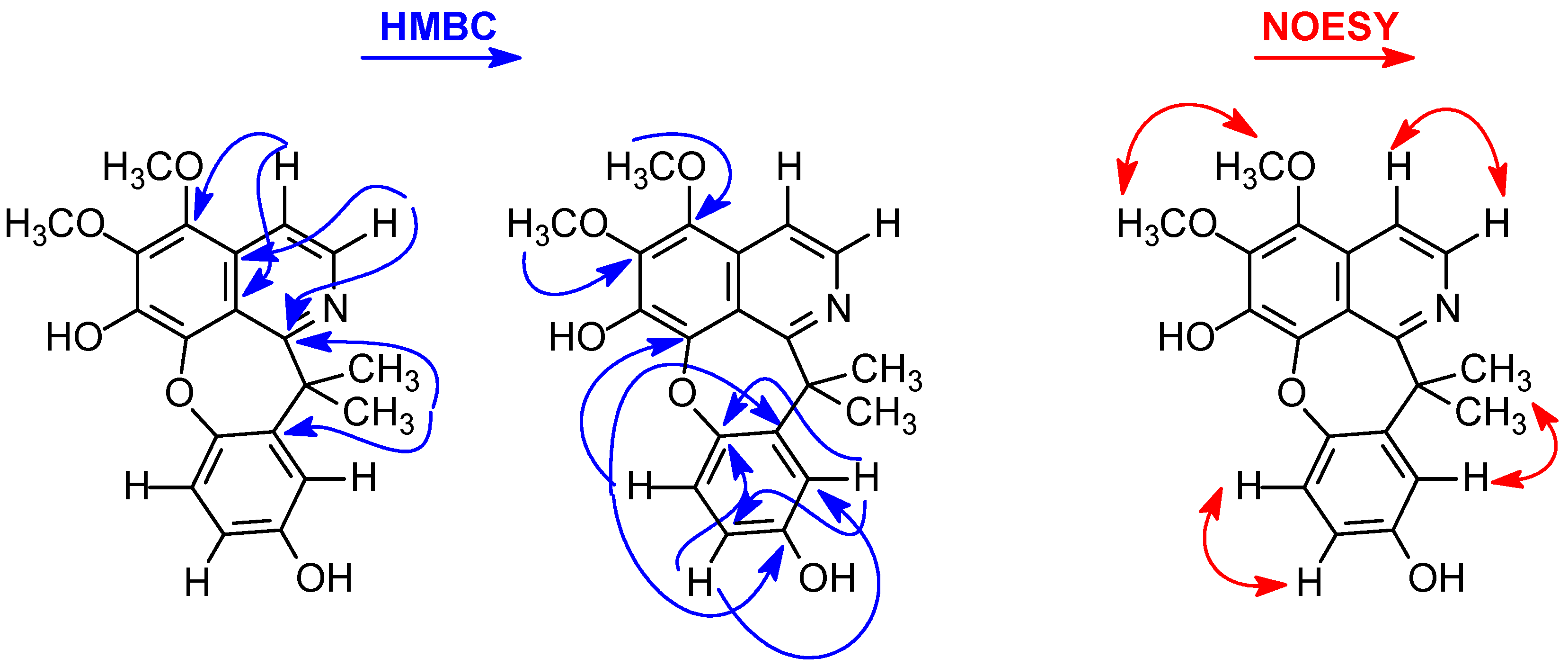
The location of the methoxyl group in the A ring was established on the basis of the long-range 1H-13C correlation map from the HMBC NMR experiment (Table 1 and Figure 2). This analysis revealed that the hydrogen at δH 7.74 (H-4) had a long-range 1H-13C correlation with 3J, with the carbon at δC 116.9 (C-8a) and δC 142.2 (C-5), and with 2J, with the carbon at δC 127.2 (C-4a) and δC 137.9 (C-3), confirming that one of the methoxy groups was substituted at C-3 of the A ring. The same analysis was performed for the presence of one of the hydroxyl groups located in the C ring. The signal of the hydrogen at δH 7.11 (H-5′) showed long-range 1H-13C correlation to 3J with the signals of the carbons at δC 140.0 (C-8), δC 140.2 (C-1′), and δC 153.9 (C-3′), and to 2J with the signals of the carbons at δC 149.9 (C-6′) and δC 114.8 (C-4′). The signal of the hydrogen at δH 6.95 (H-2′) showed a long-range 1H-13C correlation to 3J with the signals of the carbons at δC 114.8 (C-4′) and δC 149.9 (C-6′), and to 2J with the signals of the carbons at δC 140.2 (C-1′) and δC 153.9 (C-3′). The resonate carbon at δC 153.9 (C-9) did not correlate with any of the methoxy group signals, confirming the presence of one of the hydroxyl groups in the C ring (Figure 2). Thus, the locations of the other hydroxyl and methoxyl groups on the A ring were established on the basis of the NOESY data (Figure 2). In this experiment, the signal of the hydrogen at δH 7.11 (H-5′) was correlated only with the signal at δH 6.71 (H-4′), indicating the presence of a hydroxyl group at the C-7 position of ring A. Consequently, the second methoxy group was established at the C-6 (δH 4.13) position of the A ring due to the correlation in the NOESY experiment (Figure 2) of the signal at δH 7.74 (H-4), with signals at δH 3.94 (H3CO-5) and δH 8.17 (H-3). The presence of a methoxyl substituent at C-2 was also confirmed on the basis of the NOESY data by multiple correlations of the signals at δH 4.13 (H3CO-6) with those at δH 3.94 (H3CO-5). Therefore, on the basis of MS, 1D/2D NMR, and IR data and comparisons with the literature data, compound 9 was determined to be a cularine-type alkaloid known as gouregine. This is a rare alkaloid found in the literature with records to date showing it to be in the Annonaceae family, being the second reported alkaloid in the family and described in the literature for the first time in the species Guatteria ouregou (Aubl.) Dunal. [20,21].
According to Leboeuf et al. [20], the biosynthesis of gouregine apparently does not come from the classical pattern of cularins, that is, from the intramolecular oxidative coupling of an 8-hydroxybenzylisoquinoline. These authors suggested that gouregine is likely biosynthesized from its major constituent, melosmine, through a rearrangement of its skeleton with consecutive oxidation of the double bond between C-11 and C-11a, generating an arene oxide that is rearranged to gouregine. This explanation can be confirmed by treating melosmine with Fenton’s reagent (hydroxyl radicals generated by the decomposition of hydrogen peroxide with ferrous sulfate), which led to the formation of gouregine (Figure 3) in a 90% yield [20]. This oxidation between C-11 and C-11a of the aromatic D-ring of melosmine, which transforms it into an arene oxide (Figure 3), can be easily carried out by the enzyme cytochrome P450 [22].
The MS/MS spectrum of protonated molecule 9 at m/z 354 revealed a fragmentation pathway (Figure 4) consistent with that of isoquinoline-derived alkaloids, which possess methoxyl and hydroxyl groups as substituents in the A ring and iminium nitrogen in the B ring [23]. Thus, two sequential methyl losses (−15 Da) (m/z 354 → 339 and m/z 339 → 324) were observed, followed by the competitive loss of water −18 Da (m/z 324 → 306), which in this case occurred via gas-phase proton transfer [23] and carbon monoxide (−28 Da) (m/z 324 → 296). These observations agree with the structure of 9. These results corroborate the 1H and 13C 1D/2D NMR data, as well as the information from the IR spectrum, confirming the structure of the new alkaloid of the 7,7-dimethylaporphin type.
Compounds 18 were identified as isopiline (1) [19,24], O-methylisopiline (2) [24], melosmine 3 [19], 9-hydroxyiguattescine 4 [18], dihydromelosmine 5 [19], lysicamine 6 [25], acanthoic acid 7 [26], and guattouregidine 8 [19] on the basis of their spectroscopic profiles and comparison with values in the literature. Although the NMR data of isopiline, O-methylisopiline, melosmine, dihydromelosmine, and guattouregidine have already been described in the literature, they are incomplete and contain some ambiguities. Thus, the complete and unequivocal NMR data for these alkaloids were reviewed according to their 1D and 2D NMR data (Table 1 and Table 2). The 1H and 13C 1D and 2D-NMR spectra, as well as the mass spectra of all the isolated compounds, are available in the Supplementary Materials. All the isolated compounds from this species are described for the first time and contribute significantly to the chemophenetic knowledge of the species, as well as the genus and family.
From a chemophenetic (a new term for plant chemosystematics/plant chemotaxonomy) point of view, the results obtained in this study differ from the results obtained by Araújo et al. [15], who also investigated the bark of another specimen of G. olivacea collected in the same collection area as the investigated species in this study, highlighting phenanthrene alkaloids as the main constituents and chemophenetic markers. In this study, the main chemical constituents and chemophenetic markers were 7,7-dimethylaporphine alkaloids, indicating that even between the same species, there is a difference in chemical composition and it can be considered a chemotype. Notably, Maas et al. [1] recently synonymized 34 species names of G. punctata (bringing the total number of synonyms to 46), including the type of species of the genus G. glauca [1]. Several of these synonymized species have different chemical compositions and are classified as distinct species rather than synonymized species. On the basis of this study and data on the chemical composition of synonymized Guatteria species described in the literature, there is a need to include the chemical composition of Guatteria species in molecular phylogenetic studies when a given species is synonymized. In this case, G. olivacea could not be synonymized, nor could other species of Guatteria synonymized by Maas et al. [1].
According to the literature, alkaloids of the 7,7-dimethylaporphine and/or 7-hydroxy-7-methylaporphine types are clearly rare in Annonaceae, being found practically in species of the genus Guatteria, with a certain selectivity between species that make up the genus. To date, the presence of 7,7-dimethylaporphine and/or 7-hydroxy-7-methylaporphine alkaloids has been reported in eight of the 177 recognized species, such as G. ouregou [19,21], Guatteria melosma Diels [27], Guatteria scandens Diels [28], Guatteria discolor R.E. Fries [29,30], Guatteria schomburgkiana Mart. [31], Guatteria foliosa Benth. [32], Guatteria multivenia Diels [33], and Guatteria friesiana [18,34]. Among these species, the most representative in terms of the presence of 7,7-dimethylaporphine and/or 7-hydroxy-7-methylaporphine alkaloids are G. ouregou, G. melosma, and G. friesiana, the major chemical constituents of which have been isolated and identified.
Among the 7,7-dimethylaporphine and/or 7-hydroxy-7-methylaporphine alkaloids found in the Guatteria species described above, the following occurrences have been reported: melosmine (3) was described in the stem bark of G. melosma [27], G. discolor [29], and G. ouregou [21], and now also in the stem bark of G. olivacea; 9-hydroxyiguattescine (4) is the second record in the literature, being described for the first time in the stem bark of G. friesiana [18]; dihydromelosmine (5) was an original synthetic product [35] and was found as a natural product in the stem bark of G. ouregou [21], being the second report in the literature; guattouregidine (8) was also found in the stem bark of G. ouregou [21] and was the second report in the literature; and finally, the gouregine alkaloid, a rare cularine-type alkaloid that has a pattern similar to that of the 7,7-dimethylaporphine-type alkaloids, has been recorded only in the species G. ouregou and now in G. olivacea.
It is also important to highlight the similarity in terms of the isolated and identified alkaloids between the species G. ouregou and G. olivacea, which have different morphological characteristics, with emphasis on the alkaloid containing the cularine skeleton named gouregine (Figure 1), which has been found only in these two species to date. These observations further reinforce the importance of the chemical composition of species in phylogenetic studies with the aim of contributing to the correct classification of species within a given genus, as well as synonymy and even variation or chemotyping within the same species, which is observed for the studies described for G. olivacea.

2.2. Cytotoxicity Assay

The in vitro cytotoxic activities of the isolated compounds 19 were evaluated against the cancer cell lines HepG2 (human hepatocellular carcinoma), KG-1a (human myeloid leukemia), and HCT116 (human colon carcinoma), and the noncancerous cell line MRC-5 (human lung fibroblast) via the Alamar blue assay after 72 h of incubation.
Among the compounds evaluated (Table 3), the most promising result was verified for the oxoaporphine alkaloid lysicamine (6), which has strong activity against HCT116 cells, with an IC50 value of 6.64 μg/mL (22.79 µmol/L) and a selectivity index of 2.60 (Table S1). These results agree with the results of De Souza et al. [36], who reported the activity of this alkaloid against HL-60 and K562 leukemia cells, with IC50 values of 7.11 μg/mL (24.40 µmol/L) and 11.29 μg/mL (38.75 µmol/L), respectively. Similarly, Omar et al. [37] reported that lysicamine has IC50 values of 26 μg/mL (89.24 µmol/L) for MCF-7 breast cancer cells and 27 μg/mL (92.67 µmol/L) for HepG2 cells. Interestingly, it has been reported that lysicamine can inhibit Akt activation in anaplastic human thyroid cancer cells [38] and suppress human colon cancer cell lines and cancer stem cells through the inhibition of Wnt/β-catenin [39]. Similarly, liriodenine caused apoptosis in CAOV-3 ovarian cancer cells via the mitochondrial pathway via the activation of caspase-3 and caspase-9 [40]. The alkaloid melosmine (3) showed moderate activity against HCT116, with an IC50 value of 16.77 μg/mL (49.70 µmol/L). Previously, melosmine was reported to be an antimalarial agent with low cytotoxicity to human cancer cells [41]. The same moderate activity was observed for the pimaradiene diterpene acanthoic acid against HepG2 and HCT116 cells, with IC50 values of 14.63 μg/mL (48.37 µmol/L) and 21.25 μg/mL (70.25 µmol/L), respectively. On the other hand, it should be noted that acanthoic acid (7), among the active compounds, did not show cytotoxic activity against noncancerous MRC-5 cells up to the evaluated concentration of 25 μg/mL (82.65 µmol/L). Importantly, acanthoic acid has been reported to have anti-inflammatory activity with low cytotoxicity to cancer cells [42,43,44,45,46]. Furthermore, Kim et al. [47] demonstrated that acanthoic acid increased the levels of cleaved caspase 3 and cleaved PARP1 and reduced the levels of the antiapoptotic protein Bcl-xL, causing apoptotic cell death via activation of the p38 MAPK pathway in HL-60 leukemia cells. The other compounds did not show cytotoxic activity against either cancer cells or noncancerous cells, which is still a good result considering the results of other biological assays, such as those evaluating antimicrobial, antiparasitic, and antiviral activities.

3. Materials and Methods

3.1. General Experimental Procedures

Fourier transform infrared (FTIR) spectra were obtained on a Thermo Scientific Nicolet iS5 spectrometer (Thermo Fisher Scientific Inc., Waltham, MA, USA) coupled to an ATR iD3 instrument with ZnSe crystals. The 1D and 2D NMR data were acquired in CDCl3 (chloroform-d) at 298 K on an AVANCE III HD NMR spectrometer (Bruker, Billerica, MA, USA) operating at 11.75 T (1H and 13C at 500 and 125 MHz, respectively). All the 1H- and 13C-NMR chemical shifts (δ) are presented in ppm relative to the tetramethylsilane signal at 0.00 ppm as an internal reference, and the coupling constants (J) are given in Hz. The NMR spectrometer was equipped with a 5 mm multinuclear inverse detection probe (for 1D and 2D NMR experiments) with a z gradient. One-bond (HSQC) and two- and three-bond (HMBC) 1H-13C-NMR correlation experiments were optimized for average coupling constants 1J(C,H) and LRJ(C,H) of 140 and 8 Hz, respectively. For low-resolution mass spectrometry (LR-MS) analysis, the samples of the isolated compounds were resuspended in methanol (HPLC grade), creating stock solutions (1 mg/mL). Aliquots (5 µL) of the stock solutions were further diluted to 5 µg/mL and analyzed by direct infusion into a triple-quadrupole mass spectrometer, model TSQ Quantum Access (Thermo Scientific, San Jose, CA, USA), equipped with electrospray ionization (ESI) or atmospheric-pressure chemical ionization (APCI) sources in negative or positive mode. An HPLC Shimadzu (Kyoto, Japan) coupled with a MicroTOF II (Bruker Daltonics, Billerica, MA, USA) with an electrospray ionization (ESI) source was used to obtain high-resolution mass spectra (HRESIMS) in positive mode. The parameters were as follows: capillary voltage of 4.5 kV, ESI in positive mode, final plate offset of 500 V, 40 psi nebulizer, dry gas (N2) flow rate of 8 mL/min, and temperature of 200 °C. The mass spectra (m/z 50–1000) were recorded every 2 s. Silica gel 60 (Sigma-Aldrich, San Luis, MO, USA, 70–230 mesh) was used for column chromatography (CC), whereas silica gel 60 F254 (Macherey-Nagel, Düren, Germany, 0.25 mm, aluminum) was used for analysis and preparation with thin-layer chromatography (PTLC) (Macherey-Nagel, 1.00 mm, glass). The compounds were visualized by exposure to UV254/365 light, spraying with p-anisaldehyde reagent, heating on a hot plate, and spraying with Dragendorff’s reagent.

3.2. Plant Material

In the present investigation, the botanical material (bark) of G. olivacea was collected on 16 December 2021, at the Adolpho Ducke Reserve (geographic coordinates: 2°54′47″ S and 59°58′48″ W), Manaus, Amazonas State, Brazil, and identified by Prof. Dr. Antonio Carlos Webber, a plant taxonomist of the Department of Biology of the Federal University of Amazonas (DB/UFAM). The voucher specimen number 11423 was deposited at the Herbarium of DB/UFAM. The accession (specimen) was registered in the ‘Sistema Nacional de Gestão do Patrimônio Genético e do Conhecimento Tradicional Associado (SISGEN)’ with the record A70EDCD.

3.3. Extraction and Isolation

The bark of G. olivacea was dried in an air-circulating oven at approximately 45 °C for 72 h and subsequently pulverized in a four-knife mill grinder (Marconi) to obtain the powdered material (1263.37 g). Then, exhaustive maceration with hexane (5 × 4.5 L, 25 °C) followed by MeOH (5 × 4.5 L, 25 °C) was performed. The extractive solutions obtained were concentrated in a rotary evaporator at reduced pressure (40–50 °C) to obtain hexane (19.45 g) and MeOH (107.17 g) extracts.
TLC analysis with Dragendorff’s reagent revealed a high presence of alkaloids in the MeOH extract. Therefore, an aliquot of MeOH extract (105.0 g) was initially subjected to acid–base extraction [12] to obtain alkaloidal (2.36 g) and neutral (13.07 g) fractions. Subsequently, part of the alkaloidal fraction (2.20 g) was subjected to silica-gel chromatographic column (CC) treatment with a 10% NaHCO3 solution [12] and eluted with hexane (100%), hexane–CH2Cl2 (90:10, 80:20, 70:30, 60:40, 50:50, 40:60, 30:70, 20:80, and 10:90, v/v), CH2Cl2 (100%), CH2Cl2–EtOAc (90:10, 80:20, 70:30, 60:40, 50:50, 40:60, 30:70, 20:80, and 10:90, v/v), EtOAc (100%), EtOAc–MeOH (95:05, 90:10, 85:05, 80:10, 75:25, 70:30, 60:40, and 50:50), and finally, MeOH. For each eluent, 200 mL of the mobile phase was used and collected in 30 mL vials during chromatographic elution, yielding 213 fractions at the end of the chromatographic separation process. After TLC evaluation using a mixture of CH2Cl2–MeOH at proportions of 95:05, 90:10, 85:15, and 80:20 as the eluent system (v/v), similar samples were pooled to yield 17 fractions (F1 to F17).
Fraction F5 (301.6 mg) from CC eluted with hexane–CH2Cl2 (10:90, v/v), CH2Cl2 (100%), and CH2Cl2–EtOAc (90:10, v/v) was subjected to preparative TLC and eluted with CH2Cl2–MeOH (95:05, v/v, three elutions), affording 1 (5.7 mg), 2 (8.6 mg), and 3 (57.7 mg), respectively. Fraction F6 (84.3 mg) from CC eluted with CH2Cl2–EtOAc (90:10, 80:20, and 70:30 v/v) was also subjected to preparative TLC and eluted with CH2Cl2–MeOH (95:05, v/v, two elutions), yielding 3 (45.0 mg) and 4 (5.7 mg) again.
Fractions F7 (26.3 mg) and F8 (14.0 mg), both from CC eluted with CH2Cl2–EtOAc (70:30 and 60:40 v/v), were pooled and subjected to preparative TLC eluted with CH2Cl2–MeOH (95:05, v/v, three elutions), yielding 3 (11.8 mg), 4 (9.6 mg), 5 (5.1 mg), and 6 (2.8 mg), respectively.
Fraction F9 (94.7 mg) from CC eluted with CH2Cl2–EtOAc (60:40, 50:50, 40:60, 30:70, 20:80 and 10:90 v/v), EtOAc (100%), and EtOAc–CH3OH (90:10) was subjected to preparative TLC eluted with CH2Cl2–MeOH (95:05, v/v, three elutions), affording 3 (16.6 mg) and 5 (11.4 mg), respectively. Fraction 10 (64.0 mg) from CC eluted with CH2Cl2–EtOAc (90:10 v/v) was also subjected to the same methodology described above, affording 3 (5.0 mg) and 7 (3.0 mg) again.
Fraction F11 (206.1 mg) from CC eluted with CH2Cl2–EtOAc (90:10 and 80:20, v/v) was subjected to preparative TLC and eluted with CH2Cl2–MeOH (95:05, v/v, three elutions), yielding 3 (5.0 mg), 8 (2.4 mg), and 9 (3.4 mg). Both Fraction 12 (71.6 mg) from CC eluted with CH2Cl2–EtOAc (80:20 and 70:30, v/v) and Fraction 13 (86.0 mg) from CC eluted with CH2Cl2–EtOAc (70:30 and 60:34, v/v) were also subjected to the same methodology described above, yielding 9 (38.0 mg).
As observed in the isolation and purification methodology of the compounds, compound 3 was present in practically all the fractions studied, indicating that the plant biosynthesizes this compound in large quantities and is the major compound.
Isopiline (1): Brown amorphous powder; 1H-NMR and 13C-NMR data were reviewed on the basis of 1D and 2D-NMR experiments and are described in Table 2. LR-APCI(+)-MS [M + H]+ m/z 298.
O-Methylisopiline (2): Brown amorphous powder; 1H-NMR and 13C-NMR data were reviewed on the basis of 1D and 2D-NMR experiments and are described in Table 2. LR-APCI(+)-MS [M + H]+ m/z 312.
Melosmine (3): Orange amorphous powder; 1H-NMR and 13C-NMR data were reviewed on the basis of 1D and 2D-NMR experiments and are described in Table 2. LR-APCI(+)-MS [M + H]+ m/z 338.
9-Hydroxyguattescine (4): Orange–yellow amorphous powder; 1H-NMR and 13C-NMR in accordance with the literature [18]. LR-APCI(+)-MS [M + H]+ m/z 354.
Dihydromelosmine (5): Brown amorphous powder; 1H-NMR and 13C-NMR data were reviewed on the basis of 1D and 2D-NMR experiments and are described in Table 1. LR-APCI(+)-MS [M + H]+ m/z 340.
Lysicamine (6): Yellow crystals (CH2Cl2-MeOH 3:1); 1H-NMR and 13C-NMR in accordance with previously reported methods [25]. LR-APCI(+)-MS [M + H]+ m/z 292.
Acanthoic acid (7): White needles (hexane CH2Cl2 3:1); 1H-NMR and 13C-NMR in accordance with previously reported methods [26]. LR-APCI(−)-MS [M − H] m/z 301.
Guattouregidine (8): Brown amorphous powder; 1H-NMR and 13C-NMR data were reviewed on the basis of 1D and 2D-NMR experiments and are described in Table 1. LR-APCI(+)-MS [M + H]+ m/z 342.
Gouregine (9): Orange amorphous powder; 1H-NMR and 13C-NMR data are described in Table 1. LR-ESI(+)-MS [M + H]+ m/z 354. HRESIMS(+) 354.1323 [M + H]+.

3.3.1. Cells

The HepG2 (human hepatocellular carcinoma), KG-1a (human myeloid leukemia), HCT116 (human colon carcinoma), and MRC-5 (human lung fibroblast) cell lines were obtained from the American Type Culture Collection (ATCC, Manassas, VA, USA) and were cultured as recommended by the American Type Culture Collection (ATCC) animal culture guide [48]. All the cell lines were tested for mycoplasma via a mycoplasma stain kit (Sigma-Aldrich) to validate the use of cells that were free from contamination.

3.3.2. Cytotoxicity Assay

For the cytotoxicity assay, cell viability was quantified via the Alamar blue method, as previously described [49]. For all the experiments, the cells were plated in 96-well plates. The chemical constituents evaluated varied in purity between 90% and 99.8%, which was determined on the basis of the relative integrals of the signals of the constituents in relation to the signals of the impurities contained. Most of the constituents had purities greater than 95%, except for constituents 4 and 5, which had purities of approximately 90%. The chemical constituents were dissolved in dimethyl sulfoxide (DMSO, Vetec Química Fina Ltda., Duque de Caxias, RJ, Brazil), added to each well, and incubated for 72 h. Doxorubicin (doxorubicin hydrochloride, purity ≥95%, Laboratory IMA S.A.I.C., Buenos Aires, Argentina) was used as a positive control. At the end of the treatment, 20 µL of a stock solution (0.312 mg/mL) of resazurin (Sigma-Aldrich Co.) was added to each well. The absorbances at 570 nm and 600 nm were measured via a SpectraMax 190 Microplate Reader (Molecular Devices, Sunnyvale, CA, USA). The half-inhibitory concentration (IC50) was obtained via nonlinear regression with 95% confidence intervals (CIs 95%) via the software GraphPad Prism 8 (Intuitive Software for Science; San Diego, CA, USA).

4. Conclusions

The phytochemical investigation of the bark of G. olivacea led to the isolation and identification of nine compounds (19); eight isoquinoline-derived alkaloids, including a rare alkaloid with a α-gem-dimethyltetradehydrocularine structure known as gouregine (9); seven known aporphinoid alkaloids: isopiline (1), O-methylisopiline (2), melosmine (3), 9-hydroxyiguattescine (4), dihydromelosmine (5), lysicamine (6), and guattouregidine (8); and one known pimaradiene diterpene: acanthoic acid (7). These isolated compounds were described for the first time in the bark of G. olivacea. Most of the constituents identified were the alkaloids 7,7-dimethylaporphines (melosmine, 9-hydroxyiguattescine, and dihydromelosmine) and/or 7-hydroxy-7-methylaporphine (guattouregidine), with an emphasis on the alkaloid melosmine.
These results contribute significantly to the chemophenetic knowledge of the species, considering that the species G. olivacea was synonymized as G. punctata, which currently comprises approximately 46 synonyms. This result reinforces the importance of chemical composition today in the reclassification of a given species and must be taken into consideration, not only with molecular phylogenetic data.
The in vitro cytotoxic activities of the isolated compounds were evaluated against cancer (HepG2, KG-1a, and HCT116) and noncancerous (MRC-5) cell lines via the Alamar blue assay after 72 h of incubation. Among the compounds evaluated against human cancer cell lines, the most active was the oxoaporphine alkaloid lysicamine, which has strong activity against HCT116 cells, with an IC50 value of 6.64 µg/mL (22.79 µmol/L). Melosmine had a moderate effect on HCT116 cells, with an IC50 value of 16.77 µg/mL (49.70 µmol/L), whereas acanthoic acid had moderate effects on HepG2 and HCT116 cells, with IC50 values of 14.63 µg/mL (48.37 µmol/L) and 21.25 µg/mL (70.25 µmol/L), respectively.
The results obtained in this study indicate that G. olivacea is a typical Annonaceae species belonging to a genus (Guatteria) where selective species biosynthesize alkaloids of the 7,7-dimethylaporphine and 7-hydroxy-7-methylaporphine types and deserves further investigation of other compounds belonging to these classes with cytotoxic activities.

Supplementary Materials

The following supporting information can be downloaded at: https://www.mdpi.com/article/10.3390/molecules29163834/s1, Figures S1–S115: 1H and 13C 1D and 2D NMR, IR and MS spectra for compounds 19. Table S1: Selectivity indices (SI) for compounds 19.

Author Contributions

Conceptualization, E.V.C. and D.P.B.; methodology, E.V.C., J.G.C.F., S.P.M., M.d.S.A., V.R.S., L.d.S.S., M.B.P.S., and D.P.B.; formal analysis, E.V.C., M.B.P.S. and D.P.B.; investigation, E.V.C., J.G.C.F., S.P.M., M.d.S.A., V.R.S., L.d.S.S., M.B.P.S. and D.P.B.; resources, E.V.C. and D.P.B.; data curation, E.V.C., J.G.C.F., S.P.M., M.d.S.A., V.R.S., H.H.F.K., F.M.A.d.S., M.B.P.S., and D.P.B.; writing—original draft preparation, E.V.C. and D.P.B.; writing—review and editing, E.V.C., H.H.F.K., F.M.A.d.S., and D.P.B.; visualization, E.V.C., H.H.F.K., F.M.A.d.S., M.B.P.S., and D.P.B.; supervision, E.V.C. and D.P.B.; project administration, E.V.C. and D.P.B.; funding acquisition, E.V.C., M.B.P.S., and D.P.B. All authors have read and agreed to the published version of the manuscript.

Funding

This work was financially supported by the Brazilian agencies Coordenação de Aperfeiçoamento de Pessoal de Nível Superior (CAPES, code 001), Conselho Nacional de Desenvolvimento Científico e Tecnológico (CNPq), Institutos Nacionais de Ciência e Tecnologia (INCT/CNPq Grant: 465357/2014-8), Fundação de Amparo à Pesquisa do Estado do Amazonas (FAPEAM), Fundação Oswaldo Cruz (FIOCRUZ), Universidade Federal do Amazonas (UFAM), and Financiadora de Estudos e Projetos (FINEP).

Institutional Review Board Statement

Not applicable.

Informed Consent Statement

Not applicable.

Data Availability Statement

The data presented in this study are available in the Supplementary Materials.

Acknowledgments

The authors are grateful to the Central Analítica-Centro de Apoio Multidisciplinar-Universidade Federal do Amazonas (CA/CAM/UFAM) for the NMR and MS analyses, Josean Fechine Tavares of the Universidade Federal da Paraíba for the HR-MS analysis, Emilio Carlos de Lucca Junior for the IR and UV analyses, and Antonio Carlos Webber of the Department of Biology of the Universidade Federal do Amazonas (DB/UFAM) for botanical identification.

Conflicts of Interest

The authors declare that they have no conflicts of interest.

References

  1. Maas, P.J.M.; Westra, L.Y.T.; Guerrero, S.A.; Lobão, A.Q.; Scharf, U.; Zamora, N.A.; Erkens, R.H.J. Confronting a morphological nightmare: Revision of the Neotropical genus Guatteria (Annonaceae). Blumea 2015, 60, 1–219. [Google Scholar] [CrossRef]
  2. Erkens, R.H.J.; Westra, L.Y.T.; Maas, P.J.M. Increasing diversity in the species-rich genus Guatteria (Annonaceae). Blumea 2008, 53, 467–514. [Google Scholar] [CrossRef]
  3. Lobão, A.Q.; Mello-Silva, R.; Maas, P.J.M.; Forzza, R.C. Taxonomic and nomenclatural notes on Guatteria australis (Annonaceae). Phytotaxa 2011, 20, 33–46. [Google Scholar] [CrossRef]
  4. Britto, A.C.; Oliveira, A.C.; Henriques, R.M.; Cardoso, G.M.; Bomfim, D.S.; Carvalho, A.A.; Moraes, M.O.; Pessoa, C.; Pinheiro, M.L.; Costa, E.V.; et al. In vitro and in vivo antitumor effects of the essential oil from the leaves of Guatteria friesiana. Planta Med. 2012, 78, 409–414. [Google Scholar] [CrossRef] [PubMed]
  5. Fontes, J.E.N.; Ferraz, R.P.; Britto, A.C.; Carvalho, A.A.; Moraes, M.O.; Pessoa, C.; Costa, E.V.; Bezerra, D.P. Antitumor effect of the essential oil from leaves of Guatteria pogonopus (Annonaceae). Chem. Biodivers. 2013, 10, 722–729. [Google Scholar] [CrossRef] [PubMed]
  6. Siqueira, C.A.; Serain, A.F.; Pascoal, A.C.; Andreazza, N.L.; Lourenço, C.C.; Ruiz, A.L.; Carvalho, J.E.; Souza, A.C.; Mesquita, J.T.; Tempone, A.G.; et al. Bioactivity and chemical composition of the essential oil from the leaves of Guatteria australis A. St.-Hil. Nat. Prod. Res. 2015, 29, 1966–1969. [Google Scholar] [CrossRef] [PubMed]
  7. Santos, A.R.; Benghi, T.G.S.; Nepel, A.; Marques, F.A.; Lobão, A.Q.; Duarte, M.C.T.; Ruiz, A.L.T.G.; Carvalho, J.E.; Maia, B.H.L.N.S. In vitro antiproliferative and antibacterial activities of essential oils from four species of Guatteria. Chem. Biodivers. 2017, 14, e1700097. [Google Scholar] [CrossRef] [PubMed]
  8. Ribeiro, S.S.; Jesus, A.M.; Anjos, C.S.; Silva, T.B.; Santos, A.D.; Jesus, J.R.; Andrade, M.S.; Sampaio, T.S.; Gomes, W.F.; Alves, P.B.; et al. Evaluation of the cytotoxic activity of some Brazilian medicinal plants. Planta Med. 2012, 78, 1601–1606. [Google Scholar] [CrossRef] [PubMed]
  9. Ferraz, R.P.C.; Bomfim, D.S.; Carvalho, N.C.; Soares, M.B.P.; Pinheiro, M.L.B.; Costa, E.V.; Bezerra, D.P. Cytotoxic properties of the leaf essential oils of Guatteria blepharophylla and Guatteria hispida (Annonaceae). Flavour Frag. J. 2014, 29, 228–232. [Google Scholar] [CrossRef]
  10. Ferreira, A.K.R.; Lourenço, F.R.; Cláudia, M.; Young, M.; Lima, M.E.L.; Cordeiro, I.; Suffredini, I.B.; Lopes, P.S.; Moreno, P.R.H. Chemical composition and biological activities of Guatteria elliptica R. E. Fries (Annonaceae) essential oils. J. Essent. Oil Res. 2017, 30, 69–76. [Google Scholar] [CrossRef]
  11. Costa, R.G.A.; Anunciação, T.A.D.; Araujo, M.S.; Souza, C.A.; Dias, R.B.; Sales, C.B.S.; Rocha, C.A.G.; Soares, M.B.P.; Silva, F.M.A.D.; Koolen, H.H.F.; et al. In vitro and in vivo growth inhibition of human acute promyelocytic leukemia HL-60 cells by Guatteria megalophylla Diels (Annonaceae) leaf essential oil. Biomed. Pharmacother. 2020, 122, 109713. [Google Scholar] [CrossRef] [PubMed]
  12. Galvão, A.F.C.; Araújo, M.d.S.; Silva, V.R.; Santos, L.d.S.; Dias, R.B.; Rocha, C.A.G.; Soares, M.B.P.; Silva, F.M.A.d.; Koolen, H.H.F.; Zengin, G.; et al. Antitumor Effect of Guatteria olivacea R. E. Fr. (Annonaceae) Leaf essential oil in liver cancer. Molecules 2022, 27, 4407. [Google Scholar] [CrossRef]
  13. Ribeiro, J.E.L.S.; Maas, P.J.M.; Maas, H.; Miralha, J.M.; Webber, A. Annonaceae. In Flora da Reserva Ducke: Guia de Identificação das Plantas Vasculares de Uma Floresta de Terra-Firme na Amazônia Central; Ribeiro, J.E.L.S., Hopkins, M.J.G., Vicentini, A., Sothers, C.A., Costa, M.A.S., Brito, J.M., Souza, M.A.D., Martins, L.H.P., Lohmann, L.G., Assunção, P.A.C.L., et al., Eds.; DFID (Departamento for International Development), INPA: Manaus, Brazil, 1999; pp. 121–135. [Google Scholar]
  14. Maas, P.J.M.; Maas, H.; Miralha, J.M.S.; Junikka, L. Flora da Reserva Ducke, Amazonas, Brasil: Annonaceae. Rodriguesia 2007, 58, 617–662. [Google Scholar] [CrossRef]
  15. Araújo, M.S.; Silva, F.M.A.; Koolen, H.H.F.; Costa, E.V. Isoquinoline-derived alkaloids from the bark of Guatteria olivacea (Annonaceae). Biochem. Syst. Ecol. 2020, 92, 104105. [Google Scholar]
  16. Bay, M.; Oliveira, J.V.S.; Sales Junior, P.A.; Murta, S.M.F.; Santos, A.R.; Bastos, I.S.; Orlandi, P.P.; Sousa Junior, P.T. In vitro trypanocidal and antibacterial activities of essential oils from four species of the Family Annonaceae. Chem. Biodivers. 2019, 16, e1900359. [Google Scholar] [CrossRef] [PubMed]
  17. Costa, E.V.; Pinheiro, M.L.B.; Xavier, C.M.; Silva, J.R.A.; Amaral, A.C.F.; Souza, A.D.L.; Barison, A.; Campos, F.R.; Ferreira, A.G.; Machado, G.M.C.; et al. A pyrimidine-β-carboline and other alkaloids from Annona foetida with antileishmanial activity. J. Nat. Prod. 2006, 69, 292–294. [Google Scholar] [CrossRef] [PubMed]
  18. Costa, E.V.; Pinheiro, M.L.B.; Maia, B.H.L.H.S.; Marques, F.A.; Ruiz, A.L.T.G.; Marchetti, G.; De Carvalho, J.E.; Soares, M.B.P.; Costa, C.O.S.; Galvão, A.F.C.; et al. 7,7-Dimethylaporphine and other alkaloids from the bark of Guatteria friesiana. J. Nat. Prod. 2016, 79, 1524–1531. [Google Scholar] [CrossRef] [PubMed]
  19. Guinaudeau, H.; Leboeuf, M.; Cavé, A. Aporphinoid alkaloid, III. J. Nat. Prod. 1983, 46, 761–835. [Google Scholar] [CrossRef]
  20. Leboeuf, M.; Cortes, D.; Hocquemiller, R.; Cavé, A.; Chiaroni, A.; Riche, C. Structure de la gouregine, alcaloïde original apparent aux cularines. Tetrahedron 1982, 38, 2889–2896. [Google Scholar] [CrossRef]
  21. Leboeuf, M.; Hocquemiller, R.; Cavé, A. Alcaloïds des Annonacées XLVII*: Alcaloïdesde Guatteria ouregou**[Alkaloids from Annonaceae XLVII*: Alkaloids of Guatteria ouregou. Planta Med. 1983, 48, 234–245. [Google Scholar] [CrossRef]
  22. Dewick, P.M. Medicinal Natural Products: A Biosynthetic Approach, 3rd ed.; John Wiley & Sons, Ltd.: Chichester, UK, 2009; ISBN 978-0-470-74168-9. [Google Scholar]
  23. Da Silva, F.M.A.; Bataglion, G.; De Almeida, R.A.; Heerdt, G.; Souza, I.L.; Da Silva Filho, F.A.; Alencar, D.C.; Costa, E.V.; Souza, A.D.L.; Pinheiro, M.L.B.; et al. Positive electrospray ionization ion trap mass spectrometry and ab initio computational studies of the multi-pathway fragmentation of oxoaporphine alkaloids. Int. J. Mass Spectrom. 2017, 418, 30–36. [Google Scholar] [CrossRef]
  24. Navarro, V.R.; Sette, I.M.F.; Da Cunha, E.V.L.; Silva, M.S.; Barbosa-Filho, J.M.; Maia, J.G.S. Alcalóides de Duguetia flagellaris Huber (Annonaceae). Rev. Bras. Plantas Med. 2001, 3, 23–29. [Google Scholar]
  25. de Oliveira Teles, M.N.; Dutra, L.M.; Barison, A.; Costa, E.V. Alkaloids from leaves of Annona salzmannii and Annona vepretorum (Annonaceae). Biochem. Syst. Ecol. 2015, 61, 465–469. [Google Scholar] [CrossRef]
  26. Kim, Y.H.; Chung, B.S.; Sankawa, U. Pimaradiene diterpenes from Acanthopanax koreanum. J. Nat. Prod. 1988, 51, 1080–1083. [Google Scholar] [CrossRef]
  27. Zabel, V.; Watson, W.H.; Phoebe, C.H., Jr.; Knapp, J.E.; Schiff, P.L., Jr.; Slatkin, D.J. Melosmine and melosmidine 7,7-dimethyltetradehydroaporphine alkaloids from Guatteria melosma. J. Nat. Prod. 1982, 45, 94–101. [Google Scholar] [CrossRef]
  28. Hocquemiller, R.; Rasamizafy, S.; Cavé, A.; Moretti, C. Alcaloïdes des Annonacees XXXVII: Alcaloïdes du Guatteria scandens. J. Nat. Prod. 1983, 46, 335–341. [Google Scholar] [CrossRef]
  29. Hocquemiller, R.; Debitus, C.; Roblot, F.; Cavé, A.; Jacquemin, H. Alcaloïdes des Annonacées, XLVIII. Alcaloïdes des Écorces de Guatteria discolor. J. Nat. Prod. 1984, 47, 353–362. [Google Scholar] [CrossRef]
  30. Hocquemiller, R.; Debitus, C.; Roblot, F.; Cavé, A. Guadiscine et guadiscoline, alcalöides aporphiniques originaux de Guatteria discolor, Annonaceae (1). Tetrahedron Lett. 1982, 41, 4247–4250. [Google Scholar] [CrossRef]
  31. Cortes, D.; Ramahatra, A.; Cavé, A.; Bayma, J.C.; Dadoun, H. Alcaloïdes des Annonacées, LVIII. Alcaloïdes des Ecorces de Guatteria schomburgkiana. J. Nat. Prod. 1985, 48, 254–259. [Google Scholar] [CrossRef]
  32. Mahiou, V.; Roblot, F.; Hocquemiller, R.; Cavé, A.; De Arias, A.R.; Inchausti, A.; Yaluff, G.; Fournet, A.; Angelo, A. New aporphine alkaloids from Guatteria foliosa. J. Nat. Prod. 1994, 57, 890–895. [Google Scholar] [CrossRef]
  33. Zhang, Z.; ElSohly, H.N.; Jacob, M.R.; Pasco, D.S.; Walker, L.A.; Clark, A.M. New sesquiterpenoids from the root of Guatteria multivenia. J. Nat. Prod. 2002, 65, 856–859. [Google Scholar] [CrossRef] [PubMed]
  34. Costa, E.V.; Da Cruz, P.E.O.; Pinheiro, M.L.B.; Marques, F.A.; Ruiz, A.L.T.G.; Marchetti, G.M.; De Carvalho, J.E.; Barison, A.; Maia, B.H.L.N.S. Aporphine and tetrahydroprotoberberine alkaloids from the leaves of Guatteria friesiana (Annonaceae) and their cytotoxic activities. J. Braz. Chem. Soc. 2013, 24, 788–796. [Google Scholar]
  35. Leboeuf, M.; Cortes, D.; Hocquemiller, R.; Cave, A. Guattouregidine, guattouregine et dihydromelosmine, alcaloïdes aporphiniques nouveaux de Guatteria ouregou Dun, Annonacées. C. R. Acad. Sci. 1982, 295, 191–194. [Google Scholar]
  36. De Souza, C.A.S.; Nardelli, V.B.; Paz, W.H.P.; Pinheiro, M.L.B.; Rodrigues, A.C.B.C.; Bomfim, L.M.; Soares, M.B.P.; Bezerra, D.P.; Chaar, J.S.; Koolen, H.H.F.; et al. Asarone-derived phenylpropanoids and isoquinoline-derived alkaloids from the bark of Duguetia pycnastera (Annonaceae) and their cytotoxicities. Quim. Nova 2020, 43, 1397–1403. [Google Scholar] [CrossRef]
  37. Omar, H.; Hashim, N.M.; Zajmi, A.; Nordin, N.; Abdelwahab, S.I.; Azizan, A.H.; Hadi, A.H.; Ali, H.M. Aporphine alkaloids from the leaves of Phoebe grandis (Nees) Mer. (Lauraceae) and their cytotoxic and antibacterial activities. Molecules 2013, 18, 8994–9009. [Google Scholar] [CrossRef] [PubMed]
  38. Rodrigues, M.T.; Michelli, A.P.P.; Caso, G.F.; De Oliveira, P.R.; Rodrigues-Junior, D.M.; Morale, M.G.; Machado Júnior, J.; Bortoluci, K.R.; Tamura, R.E.; Da Silva, T.R.C.; et al. Lysicamine Reduces Protein Kinase B (AKT) Activation and Promotes Necrosis in Anaplastic Thyroid Cancer. Pharmaceuticals 2023, 16, 1687. [Google Scholar] [CrossRef] [PubMed]
  39. Okayama, M.; Matsumoto, T.; Kitagawa, T.; Nakamura, S.; Ohta, T.; Yoshida, T.; Watanabe, T. Cytotoxic activities of alkaloid constituents from the climbing stems and rhizomes of Sinomenium acutum against cancer stem cells. J. Nat. Med. 2024, 78, 226–235. [Google Scholar] [CrossRef] [PubMed]
  40. Nordin, N.; Majid, N.A.; Hashim, N.M.; Rahman, M.A.; Hassan, Z.; Ali, H.M. Liriodenine, an aporphine alkaloid from Enicosanthellum pulchrum, inhibits proliferation of human ovarian cancer cells through induction of apoptosis via the mitochondrial signaling pathway and blocking cell cycle progression. Drug Des. Devel. Ther. 2015, 9, 1437–1448. [Google Scholar] [CrossRef] [PubMed]
  41. Osorio, D.E.J.; Montoya, P.G.L.; Muñoz, D.K.; Arango, A.G.J. Actividad antiplasmodial de alcaloides aporfínicos de Rollinia pittieri Y Pseudomalmea boyacana (Annonaceae). Vitae 2006, 13, 49–54. [Google Scholar]
  42. Través, P.G.; Pimentel-Santillana, M.; Rico, D.; Rodriguez, N.; Miethke, T.; Castrillo, A.; Theodorakis, E.A.; Martín-Sanz, P.; Palladino, M.A.; Boscá, L. Anti-inflammatory actions of acanthoic acid-related diterpenes involve activation of the PI3K p110γ/δ subunits and inhibition of NF-κB. Chem. Biol. 2014, 21, 955–966. [Google Scholar] [CrossRef]
  43. Kasemsuk, T.; Saehlim, N.; Arsakhant, P.; Sittithumcharee, G.; Okada, S.; Saeeng, R. A novel synthetic acanthoic acid analogues and their cytotoxic activity in cholangiocarcinoma cells. Bioorg. Med. Chem. 2021, 29, 115886. [Google Scholar] [CrossRef] [PubMed]
  44. Chao, T.H.; Lam, T.; Vong, B.G.; Través, P.G.; Hortelano, S.; Chowdhury, C.; Bahjat, F.R.; Lloyd, G.K.; Moldawer, L.L.; Boscá, L.; et al. A new family of synthetic diterpenes that regulates cytokine synthesis by inhibiting IkappaBalpha phosphorylation. Chembiochem. 2005, 6, 133–144. [Google Scholar] [CrossRef] [PubMed]
  45. Lam, T.; Ling, T.; Chowdhury, C.; Chao, T.H.; Bahjat, F.R.; Lloyd, G.K.; Moldawer, L.L.; Palladino, M.A.; Theodorakis, E.A. Synthesis of a novel family of diterpenes and their evaluation as anti-inflammatory agents. Bioorg. Med. Chem. Lett. 2003, 13, 3217–3221. [Google Scholar] [CrossRef] [PubMed]
  46. Dou, J.Y.; Jiang, Y.C.; Cui, Z.Y.; Lian, L.H.; Nan, J.X.; Wu, Y.L. Acanthoic acid, unique potential pimaradiene diterpene isolated from Acanthopanax koreanum Nakai (Araliaceae): A review on its pharmacology, molecular mechanism, and structural modification. Phytochemistry 2022, 200, 113247. [Google Scholar] [CrossRef]
  47. Kim, K.N.; Ham, Y.M.; Moon, J.Y.; Kim, M.J.; Jung, Y.H.; Jeon, Y.J.; Lee, N.H.; Kang, N.; Yang, H.M.; Kim, D.; et al. Acanthoic acid induces cell apoptosis through activation of the p38 MAPK pathway in HL-60 human promyelocytic leukaemia. Food Chem. 2012, 135, 2112–2117. [Google Scholar] [CrossRef]
  48. ATCC Animal Cell Culture Guide. Get Time-Tested Tips for Culturing ATCC Animal Cells. 2024. Available online: https://www.atcc.org/resources/culture-guides/animal-cell-culture-guide (accessed on 3 June 2024).
  49. Ahmed, S.A.; Gogal, R.M., Jr.; Walsh, J.E. A new rapid and simple non-radioactive assay to monitor and determine the proliferation of lymphocytes: An alternative to [3H]-thymidine incorporation assay. J. Immunol. Methods 1994, 170, 211–224. [Google Scholar] [CrossRef]
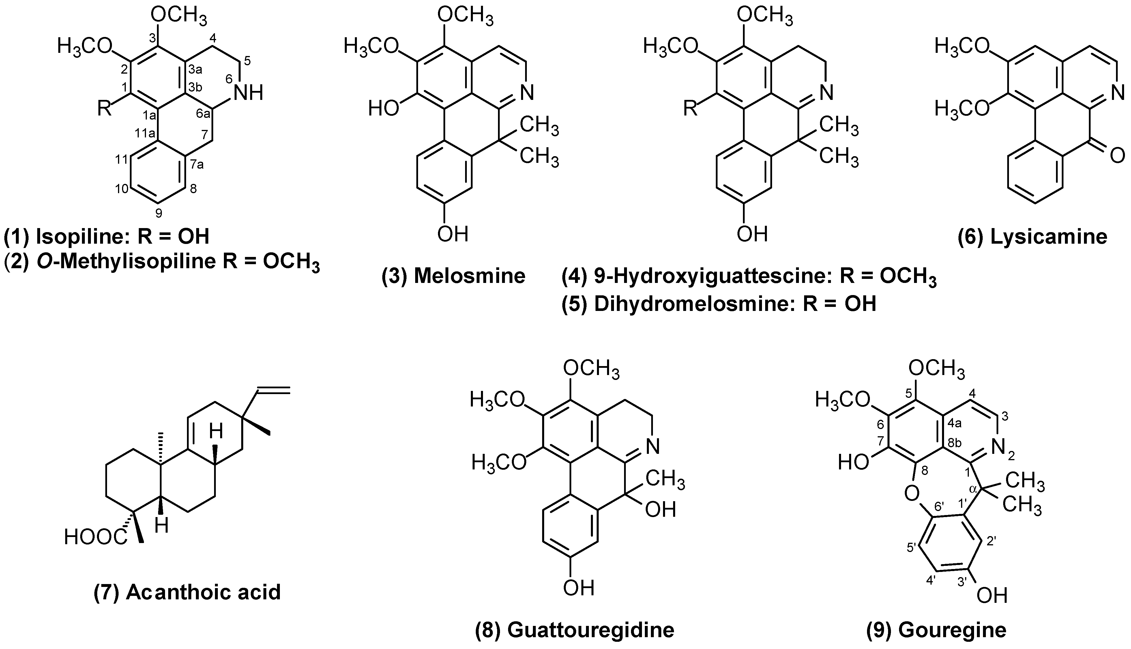
Figure 1. Chemical structures of the isolated compounds from the bark of G. olivacea.
Molecules 29 03834 g001
Figure 2. Key HMBC and NOESY correlations for alkaloid 9.
Molecules 29 03834 g002
Figure 3. Proposal for the rearrangement of melosmine to generate gouregine. Adapted from Leboeuf et al. [20].
Molecules 29 03834 g003
Figure 4. Proposed fragmentation pathway for the major product ions observed in the MS/MS spectrum of the protonated molecule at m/z 354.
Molecules 29 03834 g004
Table 1. 1H (500 MHz) and 13C NMR (125 MHz) data for alkaloids 3, 5, 8, and 9.
Position358Position9
δC mult.δH mult.
(J in Hz)
δC mult.δH mult. (J in Hz)δC mult.δH mult. (J in Hz)δC mult.δH mult. (J in Hz)
1144.4, qC 144.5, qC 144.0, qC 1160.2, qC
1a111.3, qC 116.7, qC 116.0, qC 3137.9, CH8.17 d (5.9)
2142.2, qC 142.6, qC 142.9, qC 4114.2, CH7.74 d (5.9)
3142.8, qC 146.7, qC 147.0, qC 4a127.2, qC
3a127.2, qC 122.8, qC 122.4, qC 5142.2, qC
3b119.4, qC 119.1, qC 117.4, qC 6142.7, qC
4ax
4eq
112.3, CH7.68 d (5.6)19.4, CH22.61 t (7.2)19.5, CH22.42 td (15.3; 5.5)
2.94 dddd (16.5; 15.3; 4.7; 0.8)
7135.8, qC
5ax
5eq
140.8, CH8.46 d (5.6)46.0, CH23.62 t (7.2)45.6, CH23.20 dddd (16.5; 15.3; 5.5; 0.8)
4.09 dd (15.3; 5.5)
8140.0, qC
6a163.0, qC 173.0 qC 170.7, qC 8a116.9, qC
742.1 qC 42.9, qC 72.7, qC 1′140.2, qC
7a147.0, qC 145.1, qC 144.1, qC 2′113.8, CH6.95 d (2.9)
8113.4, CH7.17 d (2.7)111.8, CH7.01 d (2.6)111.5, CH7.30 d (2.7)3′153.9, qC
9155.5, qC 156.0, qC 155.5, qC 4′114.8, CH6.71 dd (8.5; 2.9)
10113.7, CH6.86 dd (8.8; 2.7)113.4, CH6.78 dd (8.7; 2.6)127.5, CH6.80 dd (8.7; 2.7)5′122.3, CH7.11 d (8.5)
11129.0, CH8.87 d (8.8)129.8, CH8.45 d (8.7)129.6, CH8.44 d (8.7)6′149.9, qC
11a121.7, qC 121.9, qC 120.9, qC α45,6, qC
1-OH α-(CH3)227.5, CH31.87 s
2-OCH361.2, CH34.17 s61.04.04 s61.14.04 s5-OCH361.6, CH33.94 s
3-OCH361.1, CH33.99 s60.63.86 s60.73.87 s6-OCH361.2, CH34.13 s
9-OH 7-OH
7-(CH3)232.6, CH31.73 s27.31.47 s33.31.47 s3′-OH
7-OH
The data were obtained at 298 K with TMS as an internal reference (0.00 ppm) in CDCl3.
Table 2. 1H (500 MHz) and 13C NMR (125 MHz) data for alkaloids 1 and 2.
Position12
δC mult.δH mult. (J in Hz)δC mult.δH mult. (J in Hz)
1145.5, qC 150.3, qC
1a115.4, qC 122.3, qC
2138.9, qC 145.6, qC
3148.9, qC 150.6, qC
3a130.1, qC 131.0, qC
3b117.7, qC 122.4, qC
4ax
4eq
22.4, CH22.84 m23.2, CH22.84 m
5ax
5eq
42.2, CH22.99 m
3.54 m
42.5, CH22.97 m
3.48 ddd (12.2; 5.0; 1.9)
6a53.5, CH3.97 m53.7, CH3.87 d (13.8; 4.7)
7ax
7eq
36.0, CH22.88 t (13.8)
2.99 dd (13.8; 4.5)
36.5, CH22.82 t (13.8)
2.92 dd (13.8; 4.7)
7a134.3, qC 135.0, qC
8127.8, CH7.21 br d (7.2)127.83, CH7.23 dd (7.3; 1.7)
9126.8, CH7.17 td (7.2; 1.2)127.09, CH7.19 td (7.3; 1.3)
10126.9, CH7.31 ddd (7.9; 7.2; 1.1)127.06, CH7.29 ddd (7.9; 7.3; 1.7)
11127.8, CH8.31 br dd (7.9; 0.8)127.81, CH8.27 br dd (7.9; 0.8)
11a131.9, qC 131.9, qC
1-OCH3 (OH) 60.6, CH33.73 s
2-OCH360.8, CH33.95 s60.9, CH33.95 s
3-OCH359.9, CH33.87 s60.4, CH33.91 s
The data were obtained at 298 K with TMS as an internal reference (0.00 ppm) in CDCl3.
Table 3. Cytotoxic activity of the isolated compounds from the bark of G. olivacea.
CompoundsIC50, in µg mL−1 (µmol/L), and 95% CI a
HepG2KG-1aHCT116MRC-5
Isopiline (1)N.T.N.T.>25 (>84.07)>25 (>84.07)
O-Methylisopiline (2)N.T.N.T.>25 (>80.28)>25 (>80.28)
Melosmine (3)N.T.N.T.16.77 (49.70)
10.42–27.02
23.72 (70.30)
18.79–33.97
9-hydroxyiguattescine (4)>25 (>70.73)N.T.>25 (>70.73)>25 (>70.73)
Dihydromelosmine (5)>25 (>73.66)N.T.>25 (>73.66)>25 (>73.66)
Lysicamine (6)N.T.N.T.6.64 (22.79)
5.35–8.24
17.24 (59.18)
11.04–29.22
Acanthoic acid (7)14.63 (48.37)
10.67–20.07
N.T.21.25 (70.25)
13.25–34.10
>25 (>82.65)
Guattouregidine (8)>25 (>73.23)N.T.>25 (>73.23)>25 (>73.23)
Gouregine (9)>25 (>70.74)>25 (>70.74)N.T.>25 (>70.74)
Doxorubicin b0.04 (0.07)
0.03–0.05
0.22 (0.40)
0.15–0.33
0.20 (0.36)
0.11–0,35
1.42 (2.61)
0.22–2.12
a Data are presented as IC50 values in µg mL−1 (μmol L−1) and their 95% confidence intervals (CIs) obtained by nonlinear regression from three independent experiments performed in duplicate, measured via the Alamar blue assay after 72 h of incubation. The cancer cell lines used were HepG2 (human hepatocellular carcinoma), KG-1a (human myeloid leukemia), and HCT116 (human colon carcinoma) cells. Noncancerous MRC-5 cells (human lung fibroblasts) were used. b Doxorubicin was used as a positive control. N.T: Not tested.
Disclaimer/Publisher’s Note: The statements, opinions and data contained in all publications are solely those of the individual author(s) and contributor(s) and not of MDPI and/or the editor(s). MDPI and/or the editor(s) disclaim responsibility for any injury to people or property resulting from any ideas, methods, instructions or products referred to in the content.