Article

A Silver Modified Nanosheet Self-Assembled Hollow Microsphere with Enhanced Conductivity and Permeability

1
Xi’an Key Laboratory of Advanced Photo-Electronics Materials and Energy Conversion Device, Technological Institute of Materials & Energy Science (TIMES), Xijing University, Xi’an 710123, China
2
Key Laboratory of Advanced Electrode Materials for Novel Solar Cells for Petroleum and Chemical Industry of China, School of Chemistry and Life Sciences, Suzhou University of Science and Technology, Suzhou 215009, China
3
School of Material Science and Engineering, Shaanxi University of Science and Technology, Xi’an 710021, China
4
School of Artificial Intelligence, Optics and Electronics (iOPEN), Northwestern Polytechnical University, Xi’an 710072, China
*
Authors to whom correspondence should be addressed.
Molecules 2024, 29(18), 4384; https://doi.org/10.3390/molecules29184384
Submission received: 30 July 2024 / Revised: 3 September 2024 / Accepted: 11 September 2024 / Published: 15 September 2024
(This article belongs to the Section Nanochemistry)

Abstract

:
The utilization of sheet structure composites as a viable conductive filler has been implemented in polymer-based electromagnetic shielding materials. However, the development of an innovative sheet structure to enhance electromagnetic shielding performance remains a significant challenge. Herein, we propose a novel design incorporating silver-modified nanosheet self-assembled hollow spheres to optimize their performance. The unique microporous structure of the hollow composite, combined with the self-assembled surface nanosheets, facilitates multiple reflections of electromagnetic waves, thereby enhancing the dissipation of electromagnetic energy. The contribution of absorbing and reflecting electromagnetic waves in hollow nanostructures could be attributed to both the inner and outer surfaces. When multiple reflection attenuation is implemented, the self-assembled stack structure of nanosheets outside the composite material significantly enhances the occurrence of multiple reflections, thereby effectively improving its shielding performance. The structure also facilitates multiple reflections of incoming electromagnetic waves at the internal and external interfaces of the material, thereby enhancing the shielding efficiency. Simultaneously, the incorporation of silver particles can enhance conductivity and further augment the shielding properties. Finally, the optimized Ag/NiSi-Ni nanocomposites can demonstrate superior initial permeability (2.1 × 10⁻⁶ H m−1), saturation magnetization (13.2 emu g−1), and conductivity (1.2 × 10⁻³ Ω•m). This work could offer insights for structural design of conductive fillers with improved electromagnetic shielding performance.

1. Introduction

With the rapid development of fifth-generation (5G) mobile networks, various electronic instruments have become extensively utilized in diverse domains and households [1,2,3]. The electromagnetic compatibility of precision instruments, potential harm to human health caused by electromagnetic radiation, threats to national information security due to electromagnetic information leakage, and a series of associated issues have emerged as significant concerns [4,5,6]. Consequently, the problem of shielding against electromagnetic radiation has garnered considerable attention. Among all strategies aimed at reducing electromagnetic contamination, the utilization of enhanced full-band electromagnetic shielding materials in electrical and electronic instruments has proven to be highly effective [7,8,9].
The total shielding effectiveness (SE) of the material could be obtained by summing the contributions of reflection (R), absorption (A), and multiple reflection (B) in dB, as shown below:
SE dB = R dB   +   A dB + B dB
R dB = 168 10 log 10 σ μ f 2
A dB = 1 . 314   ×   t ( f σ μ ) 1 / 2
B dB = 20 log 10 1   e 2 t ( π f μ σ ) 1 / 2 e j 2 t ( π f μ σ ) 1 / 2
where μ is the relative magnetic permeability, σ is the electrical conductivity, t is the material thickness, and ƒ is the frequency [10,11]. It is obvious that high electrical and magnetic conductivity are essential properties of electromagnetic shielding materials.
As a novel research focus, polymer-based electromagnetic shielding materials typically consist of a polymer matrix and conductive or magnetic fillers, offering the advantages of cost-effectiveness, lightweightness, and ease of processing [12,13,14]. Various types of conductive fillers exist, and their structure, properties, and content directly influence the formation of a conductive network within the matrix, thereby impacting the electromagnetic shielding performance of composite materials. According to the structure of conductive packing, it can be categorized into micro-particle type packing [15,16], high-aspect-ratio structure packing [17,18], lamellar structure packing [19,20] and so on. It has been observed that the incorporation of sheet structures can enhance the performance of polymer-based electromagnetic shielding materials; however, there is still considerable room for improvement in this regard. Recent scientific reports have highlighted the significance of constructing nanosheet self-assembled hollow structures with a large specific surface area as a crucial approach for material modification [21]. Nevertheless, limited research has been conducted on this topic within this field.
Herein, a nanosheet self-assembled hollow sphere structure was constructed. The hollow NiSi spheres with nanosheet self-assembled structures on the surface were reduced at high temperature through the hydrothermal reaction between SiO2 and NiCl2. To further enhance the packing’s EMI (electromagnetic interference) shielding properties, Ag nanoparticles were used to form an enhanced coating, using electroless plating, because of their high electrical conductivity (Ag/NiSi-Ni) [22]. Importantly, it was found that the magnetic and electrical conductivity could be finely controlled by adjusting the structure of the nanocomposites. The utilization of the silver-loaded nanosheet self-assembled hollow sphere structure enables the attainment of exceptional magnetic and electrical conductivity.

2. Results and Discussion

Firstly, SiO2 nanospheres were prepared using the Stöber hydrolysis method, while the synthesis principle has been investigated by previous researchers [23,24]. The successful preparation of amorphous SiO2 with an average particle size of 375 nm by the Stöber hydrolysis method is demonstrated in Figure S1, exhibiting a narrow particle size distribution and excellent dispersion. The XRD pattern in the lower-left corner of the image indicates that the original SiO2 exhibited a broad diffraction peak centered at approximately 22°, which could be attributed to the cristobalite silica phase.
The formation mechanism of NiSi-Ni(OH)2 in the hydrothermal process is elucidated as follows: initially, the silica sphere surface undergoes attack by hydroxyl groups, leading to the generation of silicate ions during the reaction [25]. The silicate ion generated in the high-temperature alkaline solution undergoes a reaction with nickel ions, resulting in the formation of nickel silicate that selectively grows on the surface of silica spheres and forms a NiSi shell in situ. As the reaction progresses, the silica core is gradually consumed and hollow NiSiOx spheres are formed. In this process, ammonium chloride plays a crucial role by adjusting the pH value and suppressing the formation of Ni(OH)2. Upon complete reaction of SiO2 into NiSiOx, the remaining Ni2+ ions undergo subsequent reactions to form a coating of Ni(OH)2 on the surface of the NiSi shell, resulting in the formation of a composite structure known as NiSi-Ni(OH)2. According to the energy spectrum analysis conducted by EDS, Figure S2 reveals that the nanoparticles primarily consisted of elements such as Ni, Si, and O.
The X-ray Diffraction (XRD) patterns of NiSi-Ni(OH)2, NiSi-Ni, and Ag/NiSi-Ni are presented in Figure 1. For the XRD pattern of NiSi-Ni(OH)2, the peaks of 19.6°, 33.4°, 38.8°, 52.2°, 59.2°, 63.1°, 70.8°, and 72.7° were attributed to the (001), (100), (101), (102), (110), (111), (103), and (201) crystal planes of Ni(OH)2 (JCPDS No. 14-0177), respectively. The peaks of 11.9°, 19.7°, 24.3°, 34.2°, 36.7°, and 60.5° corresponded to the (002), (110), (004), (200), (202), and (208) crystal faces of Ni3Si2O5(OH)4 (JCPDS No. 22-0754), respectively. The diffraction peaks observed in the analysis indicated that they were a result of the combination of Ni(OH)2 and NiSiOx, providing evidence for the successful synthesis of the NiSi-Ni(OH)2 composite. The XRD patterns of NiSi-Ni at 44.3°, 51.9°, and 76.4° corresponded to the (111), (200), and (220) crystallographic planes of nickel (JCPDS No. 04-0850). The peaks of 34.3° and 60.7° corresponded to the (200) and (208) crystal planes of Ni3Si2O5(OH)4; however, these peaks were not prominently visible in the spectrum. It is evident that Ni(OH)2 underwent reduction to elemental nickel during the hydrogen reduction process, while retaining the existence of a NiSi shell structure. Nevertheless, due to the high intensity of the diffraction peak from pure nickel, there is limited evidence for strong diffraction phenomena associated with NiSiOx. The XRD pattern of Ag/NiSi-Ni exhibited three prominent Ni peaks, along with additional diffraction peaks at 38.0°, 44.3°, 64.5°, and 77.4°, corresponding to the (111), (200), (220), and (311) crystallographic planes of Ag (JCPDS No.89-3722), respectively. The results demonstrated the successful introduction of Ag into the system through electroless silver plating. Figure S3 presents the EDS energy spectrum analysis of the Ag/NiSi-Ni composite, revealing the presence of Ni, Si, O, and Ag elements, thereby confirming the incorporation of Ag. In order to analyze the silver load in the Ag/NiSi-Ni, X-ray fluorescence (XRF) tests were performed on the sample (Figure S6). The test results showed that the silver content of Ag/NiSi-Ni was 40.7%.
The scanning electron microscopy (SEM) images in Figure 2a reveal that the average particle size of NiSi-Ni(OH)2 spheres was 500 nm, with a visible hollow structure observed in some broken spheres. Most of the spheres had intact and unbroken shells. The diameter of the hollow region measured approximately 325 nm, which closely corresponds to the initial diameter of SiO2 involved in the reaction. This finding provides further evidence supporting the formation mechanism of the NiSi hollow shell. The high-resolution scanning electron microscopy (Figure 2b) revealed the presence of a flower-shaped Ni(OH)2 structure, formed through the self-assembly of nanosheets. This structure exhibited uniform attachment onto the surface of the hollow shell, resulting in a well-defined hierarchical architecture. The thickness of the Ni(OH)2 nanosheet was about 5 nm, and the thickness of the Ni(OH)2 layer was about 100 nm. Figure 2c,d are SEM images of NiSi-Ni, which were obtained from the reduction of NiSi-Ni(OH)2 by H2. Upon comparison, it is evident that there was no significant alteration in the microscopic morphology of the samples following H2 reduction. In other words, the flower-like structure of Ni(OH)2 remained unchanged during its conversion process. Figure 2e shows images of Ag/NiSi-Ni, which was obtained after the silver plating of NiSi-Ni. After electroless silver plating, nanosized Ag particles were coated on the surface of the particles. Figure 2f reveals the uniform embedding of Ag nanoparticles with a fine particle size into the flower-like lamellae structure.
To investigate the variations in particle size of samples at different stages during the preparation process, an analysis of particle size distribution statistics was conducted. Figure S4 shows the particle size distribution of SiO2, NiSi-Ni (OH)2, NiSi-Ni, and Ag/NiSi-Ni nanoparticles. In the overall trend, there was a gradual increase in particle size, particularly observed during the hydrothermal synthesis of NiSi-Ni(OH)2 from SiO2 and the electroless Ni plating of Ag/NiSi-Ni from NiSi-Ni, where the average particle size increased from 375 nm to 500 nm and from 540 nm to 650 nm, respectively.
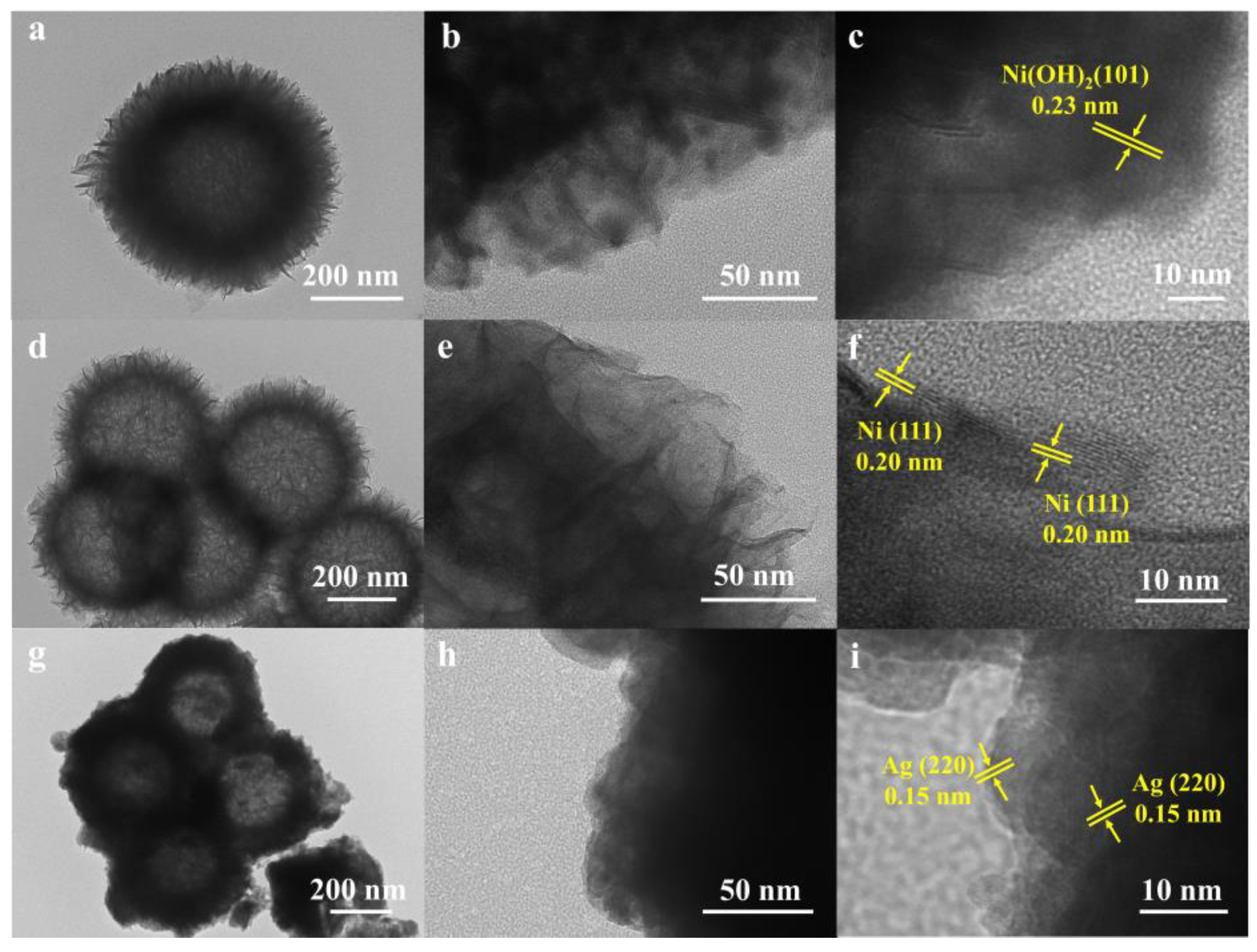
Transmission electron microscopy (TEM) testing was employed to elucidate the structural characteristics of the sample more effectively. The TEM and high resolution TEM (HRTEM) images of NiSi-Ni(OH)2 are presented in Figure 3a–c, revealing a distinct nanosheet self-assembly hollow sphere structure. Notably, the HRTEM image (Figure 3c) clearly displays lattice fringes with a spacing of 0.23 nm, corresponding to the (101) planes of Ni(OH)2. Additionally, Figure 3d–f and Figure 3g–i depict the TEM and HRTEM images of NiSi-Ni and Ag/NiSi-Ni, respectively, demonstrating that both samples maintained their well-preserved self-assembled hollow sphere structures. The flower-like spheres with laminated nanosheets possessed a significantly high specific surface area, ensuring multiple reflections for effective electromagnetic shielding. Simultaneously, the hollow architecture facilitated the occurrence of multiple reflections at material interfaces, thereby enhancing the shielding efficiency. The HRTEM images of NiSi-Ni (Figure 3f) exhibit lattice fringes measuring 0.20 nm, corresponding to the (111) plane of nickel. Figure 3g–h depict the TEM image subsequent to silver loading. Upon careful examination of the HRTEM image, distinct lattice fringes attributed to the (220) planes of silver are clearly discernible. The EDS elemental mappings (Figure S5) of the Ag/NiSi-Ni confirmed the homogeneous distribution of Ag, Ni, and Si elements.
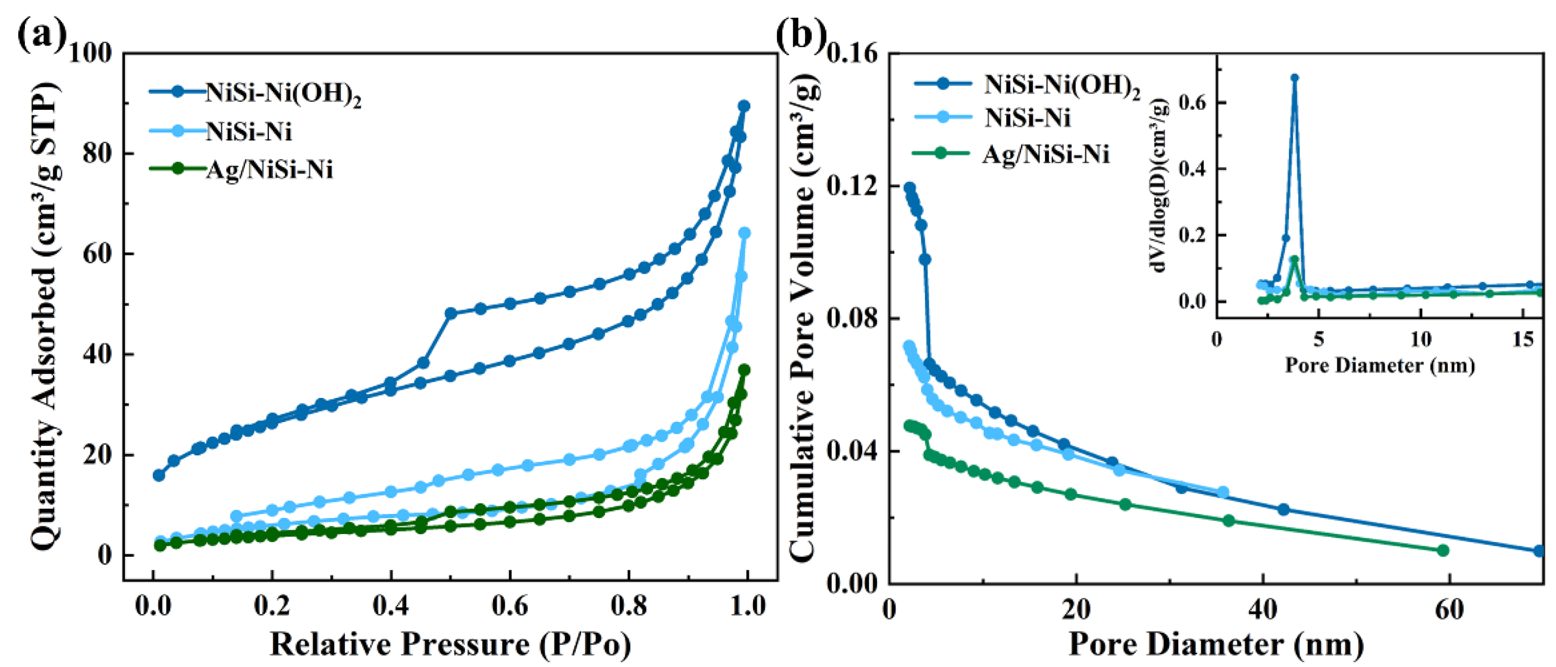
The nitrogen adsorption–desorption isotherms of the product were measured to investigate the specific surface area and pore size distribution, as depicted in Figure 4. All isotherms exhibited a characteristic IV-type classification, which is indicative of mesoporous materials. The Brunauer–Emmett–Teller (BET) specific surface areas of NiSi-Ni(OH)2, NiSi-Ni, and Ag/NiSi–Ni were 94.2, 23.5, and 14.6 m2g−1, respectively. It suggests that the self-assembly of thin nanosheets led to a significantly enhanced specific surface area. By using the Barrett–Joyner–Halenda (BJH) method, the pore size distributions were calculated via the desorption branch of the isotherms. In Figure 4b, the pore sizes of NiSi-Ni(OH)2, NiSi-Ni, and Ag/NiSi–Ni were measured to be 6.0 nm, 8.3 nm, and 10.2 nm, respectively. The BJH desorption cumulative pore volumes were determined to be 0.019, 0.076, and 0.048 cm3g−1 for the respective samples. The Ag/NiSi-Ni composite exhibits promising potential as an optimal candidate for electromagnetic shielding material, considering its specific surface area, pore size distribution, and pore volume [26,27].
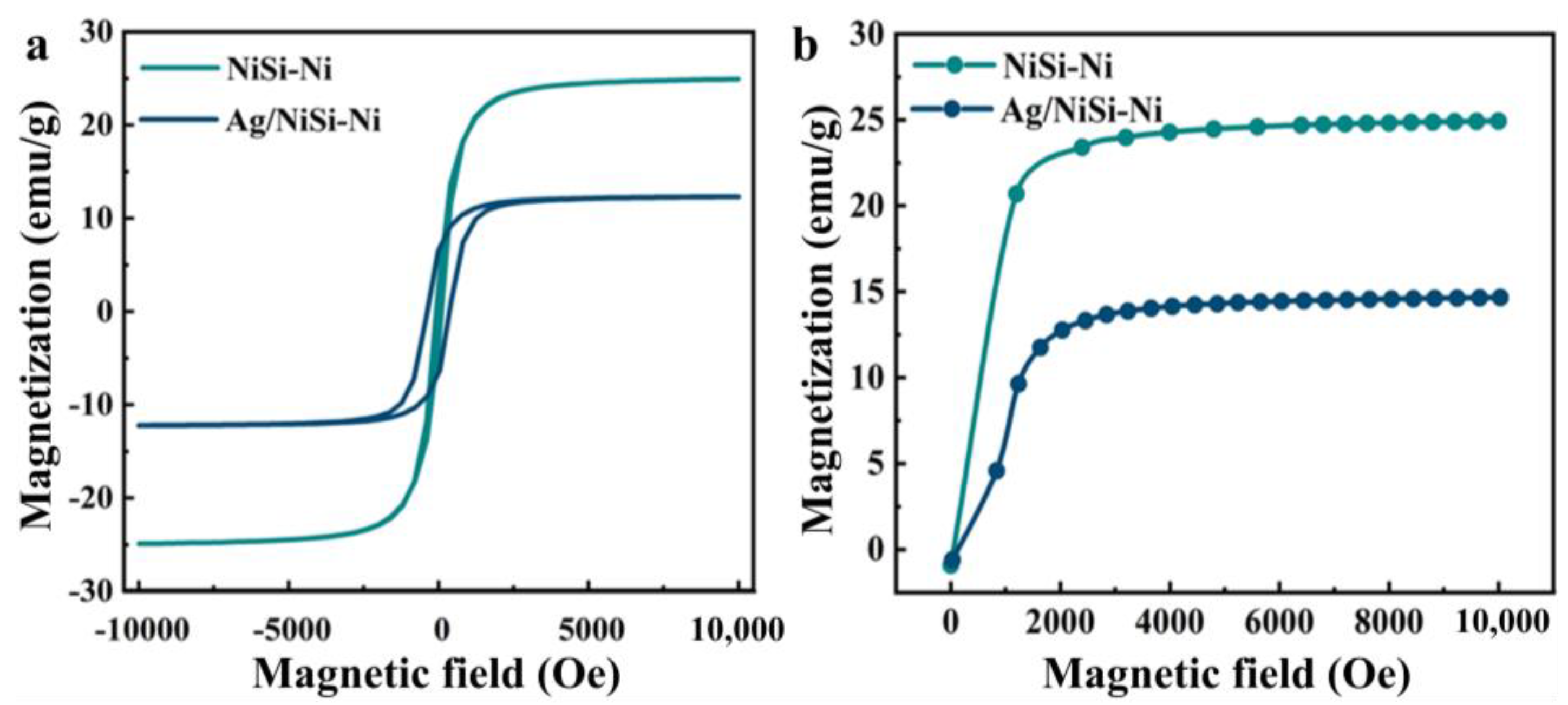
The hysteresis loops and initial magnetization curves of the prepared NiSi-Ni and Ag/NiSi-Ni composites were subsequently examined. As depicted in Figure 5a, both composites exhibited a characteristic S-shaped curve in their respective hysteresis loops. In the first quadrant, the magnetization exhibited a rapid increase, followed by a saturation effect, as both positive and external magnetic fields intensified. The hysteresis loop in the third quadrant initially experienced a steep decline, which then leveled off with an increasing strength of the reverse external magnetic field. The external magnetic field induced the formation of spin magnetic moment through electron spins located outside the nucleus of nickel atoms, while the orbital motion around the nickel nucleus generated an orbital magnetic moment. The resultant combined magnetic moment aligned with the direction of the external magnetic field. The material demonstrated ferromagnetic behavior through its magnetization in the presence of an external magnetic field, thus exhibiting the characteristic hysteresis loops observed in ferromagnetic materials [28,29]. Figure 5a shows that the saturation magnetization of NiSi-Ni and Ag/NiSi-Ni were 24.8 emu g−1 and 13.2 emu g−1, respectively. The coercivity was 3.0 Oe and 383.4 Oe, respectively. This work demonstrated clear advantages in terms of coercivity when compared to other materials (Table S1). The saturation magnetization decreased and the coercivity increased as a result of electroless silver plating. Due to the diamagnetic properties of silver, it underwent reverse magnetization when subjected to an external magnetic field, thereby diminishing the saturation magnetization of the composite material. The introduction of Ag nanoparticles simultaneously enhanced the formation of grain boundaries, thereby augmenting the blocking effect on magnetic domain movement and resulting in an increased coercivity [30]. Figure 5b shows that the initial permeability of NiSi-Ni and Ag/NiSi-Ni were 3.7 × 10⁻⁶ H m−1 and 2.1 × 10⁻⁶ H m−1, respectively.
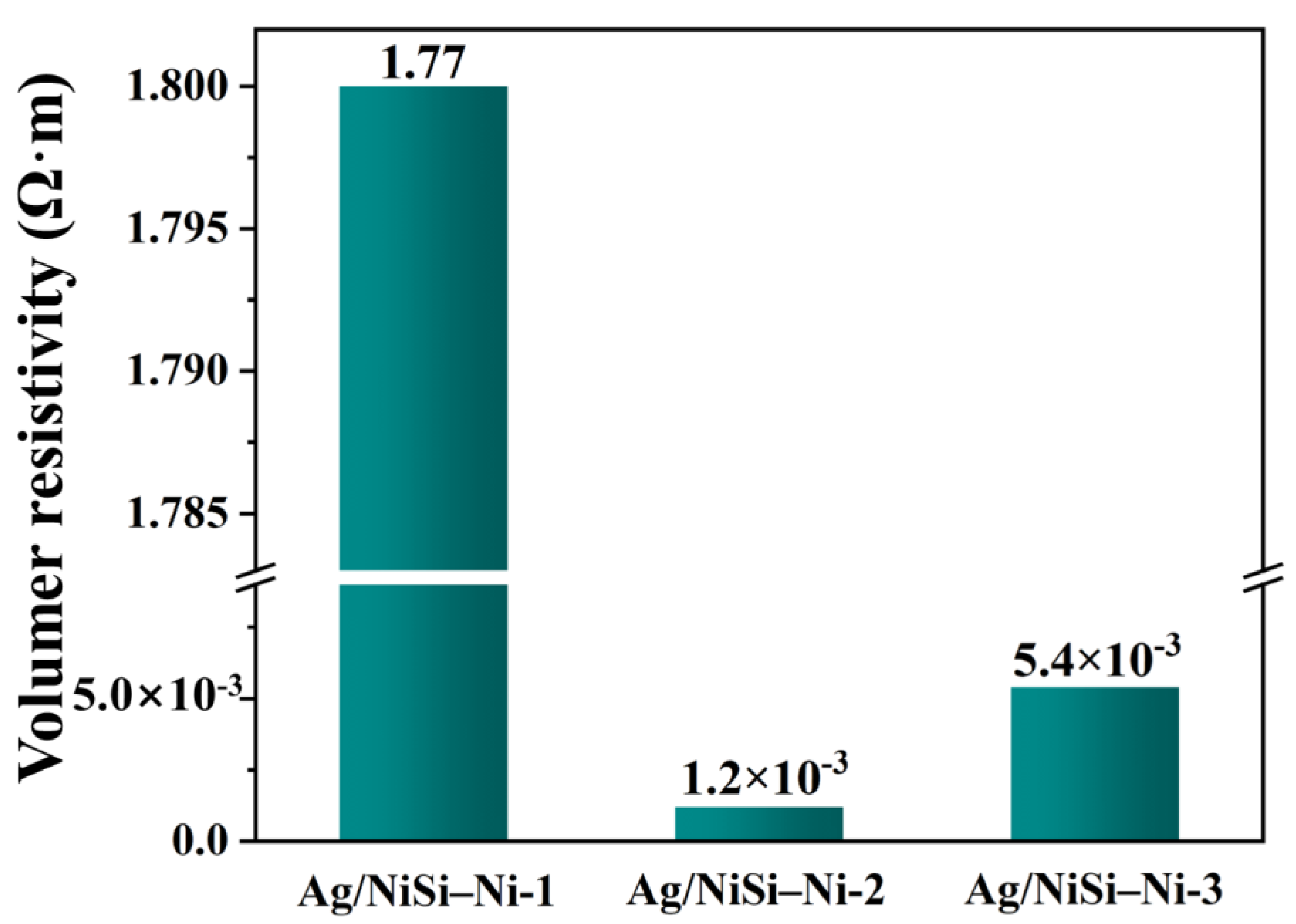
In order to characterize the effect of the amount of silver plating on the conductivity of Ag/NiSi-Ni composites, diverse samples were prepared by changing the concentration of silver ammonia solution. The composite samples of NiSi-Ni electroless silver plating were named Ag/NiSi–Ni-1, Ag/NiSi–Ni-2, and Ag/NiSi–Ni-3, corresponding to the silver ammonia solution concentrations of 0.15 g L−1, 0.35 g L−1, and 0.7 g L−1, respectively. As shown in Figure 6, there were significant differences in the silver morphology that existed in the system, due to the difference in silver content. With the increase in silver content, silver nanoparticles exhibited dispersion within cluster flower-like structures (Ag/NiSi-Ni-1), uniform coating (Ag/NiSi-Ni-2), and overgrown (Ag/NiSi-Ni-3). Considering the resistivity of the three materials depicted in Figure 7, it is evident that the Ag content in Ag/NiSi-Ni-1 was relatively low, resulting in dispersed particles and a comparatively higher resistivity. The Ag nanoparticles were uniformly coated onto the surface of the Ag/NiSi-Ni-2 material, forming a conductive pathway and resulting in a significantly reduced resistivity of 1.2 × 10⁻³ Ω•m [31]. The resistivity of this work exhibited distinct advantages when compared to other materials (Table S2). For Ag/NiSi-Ni-3, the volume resistivity increased slightly instead of decreasing when the Ag content was further increased. From Figure 6c, it was not difficult to find that the reason was that the Ag layer completely wrapped the material, and the inner hollow self-assembly of nanosheets structure was not obvious. The volume resistivity of Ag/NiSi-Ni-3 exhibited a slight increase, instead of the expected decrease, with a further increase in Ag content. The excessive presence of silver powder completely enveloped the inherent sheet self-assembly structure, leading to a reduction in the material’s specific surface area, impeding electron mobility and elevating resistivity [32,33]. An excess of silver content gave rise to an irregular blocky structure with uneven distribution (Figure 6c), resulting in localized current density surpassing acceptable limits and thereby diminishing overall conductivity. Simultaneously, the flake self-assembly structure exhibited a substantial specific surface area that could accommodate more silver particles when the silver content was appropriately balanced, consequently enhancing electrical conductivity [34,35].
Combined with the above results, the essential advantages of the Ag/NiSi-Ni structure in this study were revealed. The mechanism of this structure with higher shielding efficiency can be explained by the schematic diagram shown in Figure 8. In the case of hollow nanostructures, both absorption and reflection contributions originated from their outer and inner surfaces, as follows:
SE dB = R dB + A dB + B dB
where R = Ri + Ro and A = Ai + Ao, Ri and Ai are the contributions of the reflection and absorption of the inner surface, and Ro and Ao are the ontributions of the reflection and absorption of the outer surface. When implementing multiple reflection attenuation, the lamellar self-assembled stack structure within the composite material could significantly amplify the occurrence of multiple reflections, thereby effectively enhancing its shielding performance. The hollow structure simultaneously facilitated multiple reflections of incident electromagnetic waves at the material interface, thereby enhancing the shielding efficiency. Theoretically, this structural design exhibited distinct advantages over conventional materials for electromagnetic wave shielding [36].

3. Materials and Methods

3.1. Material Preparation

Tetraethyl orthosilicate (TEOS), nickel (II) chloride hexahydrate (NiCl2·6H2O), NH4Cl, NH3·H2O, silver nitrate, formaldehyde (HCHO), and ethanol were purchased from Aladdin Chemical Reagent Co., Ltd. (Shanghai, China) at an analytical grade and used as received.

3.1.1. Synthesis of NiSi-Ni

In a typical process, SiO2 spheres with an average diameter of approximately 300 nm were synthesized using the Stöber hydrolysis method.
The SiO2 (0.1 g) was prepared via Stöber hydrolysis, a commonly employed method for its synthesis. To obtain solution A, the dispersed SiO2 was mixed with 20 mL of deionized water and subjected to ultrasound treatment for 1 h. Solution B was formed by vigorously stirring 1.2 g NiCl2·6H2O, 10 mmol NH4Cl, and 2 mL NH3·H2O in 20 mL deionized water for a duration of 10 min. The mixture of Solution A and B was prepared by adding solution B dropwise to solution A. The resulting mixture was vigorously stirred for 30 min and subsequently transferred into a hydrothermal reactor. Following a hydrothermal reaction at 120 °C for 24 h, the resulting green precipitate was thoroughly washed with deionized water and ethanol until reaching a neutral pH. The product was subsequently subjected to vacuum drying at a temperature of 70 °C for a duration of 12 h. The resulting synthesized product was designated as NiSi-Ni(OH)2. Finally, the thermal reduction of the prepared precursors under an N2/H2 (N2:H2 = 95:5 vol %) atmosphere in a tubular oven at 500 °C for 2 h led to the formation of NiSi-Ni composites.

3.1.2. Synthesis of Ag/NiSi-Ni

The silver ammonia solution was prepared by adding 80 mL of deionized water to a beaker containing a specific quantity of silver nitrate (15 mg, 35 mg, 70 mg), followed by the gradual addition of 20 mL of 25 wt% ammonia, while adjusting the pH to 13 using sodium hydroxide. Subsequently, a sequential addition of 95 mL anhydrous ethanol, 4 mL deionized water, and 2.5 mL formaldehyde solution at a concentration of 37 wt% was performed into the beaker containing NiSi-Ni particles, followed by ultrasonic dispersion for a duration of 15 min. Subsequently, the specimen was immersed in a water bath maintained at 25 °C, followed by the addition of the prepared silver ammonia solution within a time frame of 20 min. The mixture was continuously stirred for a duration of 2 h. Upon completion of the reaction, precipitation separation was conducted through thorough cleansing with deionized water and ethanol to achieve neutralization. Finally, Ag/NiSi-Ni was obtained after drying. The schematic representation of this simplified sample preparation process is illustrated in Figure 9.

3.2. Methods

Morphological data and energy dispersive spectra (EDS) were obtained using a field emission scanning electron microscopy (FE-SEM) (GeminiSEM500, Shanghai, China) and a transmission electron microscopy (TEM) (JEOL JEM2100, Tokyo, Japan). X-ray Diffraction (XRD) measurements were carried out using a PANalytical X’pert MPDPro (Almelo, The Netherlands) diffractometer with a Cu Ka radiation source (40 kV, 40 mA). Brunauer–Emmett–Teller (BET) surface areas and pore size distributions were obtained at −196 °C (liquid nitrogen temperature) using an ASAP 2020 (Norcross, GA, USA) instrument. Electric conductivity properties were measured using a HP RLC4284A Bridge. Magnetic properties were measured using a TF-CIB static hysteresis loop measurement instrument. The X-ray fluorescence (XRF) technique employed a BRUKER (S8 TIGER) wavelength dispersive X-ray fluorescence spectrometer equipped with an RHX-ray tube, a 4 kW generator, and an 8-bit crystal converter. The Si, Ni, Ag, and Rh tubes operated at 50 kV and 50 mA.
The resistivity of the composite powder was determined using powder bulk resistivity measurement. In this test, a specific quantity of conductive powder to be measured was added to the sample reservoir, followed by the installation of the load device. Once the circuit was switched on, the resistance indicating value was recorded as soon as it stabilized, representing the stacking resistance R (Ω) of the sample. The bottom area of the sample reservoir was denoted by S (m2), and L (m) represented the height at which the powder was stacked. Equation (6) illustrates the calculation method for determining ρ, which represents the resistivity of powdered packing, as follows:
ρ = R ( S L )

4. Conclusions

In this paper, a novel electromagnetic shielding filler with a nanosheet self-assembled hollow sphere structure was controlled. It exhibited excellent electrical and magnetic conductivity. The initial permeability, saturation magnetization, and conductivity of optimized Ag/NiSi–Ni nanocomposites were obtained as 2.1 × 10⁻⁶ H m−1, 13.2 emu/g, and 1.2 × 10⁻³ Ω•m, respectively. When implementing multiple reflection attenuation, the incorporation of a hollow structure and a self-assembled stack structure could significantly amplify the occurrence of multiple reflections, thereby effectively enhancing the shielding performance. Simultaneously, the incorporation of silver particles enhanced conductivity and further augments shielding efficiency. This study provides a new idea for the preparation of electromagnetic shielding filler. Hollow spherical composite powder is employed as a conductive filler, which also possesses distinctive advantages, such as its lightweight nature within the aerospace domain.

Supplementary Materials

The following supporting information can be downloaded at: https://www.mdpi.com/article/10.3390/molecules29184384/s1. Figure S1. XRD and SEM of SiO2 were prepared by the Stöber hydrolysis method.; Figure S2. EDX spectra of NiSi-Ni(OH)2. Figure S3. EDX spectra of Ag/NiSi-Ni.; Figure S4. Particle size distribution of different nanoparticles.; Figure S5. (a) TEM image of Ag/NiSi-Ni and their EDS elemental mappings of Si(b), Ni(c) and Ag(d) elements; Figure S6. XRF test results of Ag/NiSi-Ni.; Table S1. Comparison of the performance of Coercivity (Oe); Table S2. Comparison of the performance of Resistivity (Ω•m); References [37,38,39,40,41,42,43,44] are cited in Supplementary Materials.

Author Contributions

Conceptualization, Y.Z.; Methodology, X.D.; Software, W.S.; Formal analysis, F.W. and J.L.; Investigation, Z.H.; Resources, Y.Z., W.S., C.L., J.H. and Z.M.; Writing—original draft, F.W.; Writing—review & editing, F.W. All authors have read and agreed to the published version of the manuscript.

Funding

This work was supported by the National Natural Science Foundation of China (No. 52173263, 22379103, 22308283), the Xi’an Science Technology Plan Project of Shaanxi Province (No. 2023JH-ZCGJ-0068), the Qinchuangyuan High-level Talent Project of Shaanxi (No. QCYRCXM-2022-219), the Natural Science Basic Research Plan in Shaanxi Province of China (No. 2024JC-YBMS-342), the Scientific Research Foundation of Xijing University (XJ23B09, XJ24B05), and the Youth innovation Team of Shaanxi Universities.

Institutional Review Board Statement

Not applicable.

Informed Consent Statement

Not applicable.

Data Availability Statement

Data are available upon request from the corresponding authors.

Conflicts of Interest

There are no conflicts to declare.

References

  1. Tsai, M.K.; Chiu, R.; He, E.R.; Chen, J.Y.; Chu, F.; Tsai, J.; Wang, Y.P.; Jian, S.Y.; Chen, S.M. Innovative EMI Shielding Solutions on Advanced SiP Module for 5G Application. In Proceedings of the Electronics Packaging Technology Conference, Singapore, 4–6 December 2019; pp. 601–607. [Google Scholar]
  2. Zeng, S.; Huang, Z.X.; Jiang, H.; Li, Y. From Waste to Wealth: A Lightweight and Flexible Leather Solid Waste/Polyvinyl Alcohol/Silver Paper for Highly Efficient Electromagnetic Interference Shielding. ACS Appl. Mater. Interfaces 2020, 12, 52038–52049. [Google Scholar] [CrossRef] [PubMed]
  3. Li, J.; Tsai, M.; Chiu, R.; He, E.; Hsieh, A.; Tsai, M.F.; Chu, F.; Chen, J.Y.; Jian, S.; Chen, S.; et al. EMI Shielding Technology in 5G RF System in Package Module. In Proceedings of the 2020 IEEE 70th Electronic Components and Technology Conference, Orlando, FL, USA, 3–30 June 2020; pp. 931–937. [Google Scholar]
  4. Pande, S.; Singh, B.; Mathur, R.; Dhami, T.; Saini, P.; Dhawan, S. Improved Electromagnetic Interference Shielding Properties of MWCNT-PMMA Composites Using Layered Structures. Nanoscale Res. Lett. 2009, 4, 327–334. [Google Scholar] [CrossRef] [PubMed]
  5. Huang, Y.; Li, N.; Ma, Y.; Du, F.; Li, F.; He, X.; Lin, X.; Gao, H.; Chen, Y. The influence of single-walled carbon nanotube structure on the electromagnetic interference shielding efficiency of its epoxy composites. Carbon 2007, 45, 1614–1621. [Google Scholar] [CrossRef]
  6. Wang, C.; Murugadoss, V.; Kong, J.; He, Z.; Mai, X.; Shao, Q.; Chen, Y.; Guo, L.; Liu, C.; Angaiah, S.; et al. Overview of carbon nanostructures and nanocomposites for electromagnetic wave shielding. Carbon 2018, 140, 696–733. [Google Scholar] [CrossRef]
  7. Wen, B.; Cao, M.S.; Lu, M.M.; Cao, W.Q.; Shi, H.L.; Liu, J.; Wang, X.X.; Jin, H.B.; Fang, X.Y.; Wang, W.Z.; et al. Reduced Graphene Oxides: Light-Weight and High-Efficiency Electromagnetic Interference Shielding at Elevated Temperatures. Adv. Mater. 2014, 26, 3484–3489. [Google Scholar] [CrossRef]
  8. Cao, M.S.; Wang, X.X.; Cao, W.Q.; Yuan, J. Ultrathin graphene: Electrical properties and highly efficient electromagnetic interference shielding. J. Mater. Chem. C 2015, 3, 6589–6599. [Google Scholar] [CrossRef]
  9. Xu, H.L.; Yin, X.W.; Li, X.L.; Li, M.H.; Liang, S.; Zhang, L.T.; Cheng, L.F. Lightweight Ti2CTx MXene/Poly(vinyl alcohol) Composite Foams for Electromagnetic Wave Shielding with Absorption-Dominated Feature. ACS Appl. Mater. Interfaces 2019, 11, 10198–10207. [Google Scholar] [CrossRef]
  10. Ji, K.; Zhao, H.; Zhang, J.; Chen, J.; Dai, Z. Fabrication and electromagnetic interference shielding performance of open-cell foam of a Cu–Ni alloy integrated with CNTs. Appl. Surf. Sci. 2014, 311, 351–356. [Google Scholar] [CrossRef]
  11. Li, X.F.; Chang, Z.Z.; Zhang, Y.S.; Gao, G.X. Nickel and silver coated nano-SiO2 with excellent conductivity and permeability. Surf. Eng. 2014, 31, 427–432. [Google Scholar] [CrossRef]
  12. Srivastava, S.K.; Manna, K. Recent advancements in the electromagnetic interference shielding performance of nanostructured materials and their nanocomposites: A review. J. Mater. Chem. A 2022, 10, 7431–7496. [Google Scholar] [CrossRef]
  13. Liu, S.; Qin, S.; Jiang, Y.; Song, P.A.; Wang, H. Lightweight high performance carbon-polymer nanocomposites for electromagnetic interference shielding. Compos. Part A Appl. Sci. Manuf. 2021, 145, 106376. [Google Scholar] [CrossRef]
  14. Yang, W.Q.; Yang, D.; Mei, H.; Yao, L.; Xiao, S.S.; Yao, Y.T.; Chen, C.; Cheng, L.F. 3D printing of PDC-SiOC@SiC twins with high permittivity and electromagnetic interference shielding effectiveness. J. Eur. Ceram. Soc. 2021, 41, 5437–5444. [Google Scholar] [CrossRef]
  15. Panigrahi, R.; Srivastava, S.K. Tollen’s reagent assisted synthesis of hollow polyaniline microsphere/Ag nanocomposite and its applications in sugar sensing and electromagnetic shielding. Mater. Res. Bull. 2015, 64, 33–41. [Google Scholar] [CrossRef]
  16. Goyal, R.K.; Sulakhe, R. Study on poly (vinylidene fluoride)/nickel composites with low percolation. Adv. Mater. Lett. 2015, 6, 309–317. [Google Scholar] [CrossRef]
  17. Wang, G.L.; Wang, L.; Mark, L.H.; Shaayegan, V.; Wang, G.Z.; Li, H.P.; Zhao, G.Q.; Park, C.B. Ultralow threshold and lightweight biodegradable porous PLA/MWCNT with segregated conductive networks for high performance thermal insulation and electromagnetic interference shielding applications. ACS Appl. Mater. Interfaces 2018, 10, 1195–1203. [Google Scholar] [CrossRef]
  18. Feng, D.; Wang, Q.Q.; Xu, D.W.; Liu, P.J. Microwave assisted sinter molding of polyetherimide/carbon nanotubes composites with segregated structure for high-performance EMI shielding applications. Compos. Sci. Technol. 2019, 182, 107753. [Google Scholar] [CrossRef]
  19. Shahzad, F.; Lee, S.H.; Hong, S.M.; Koo, C.M. Segregated reduced graphene oxide polymer composite as a high performance electromagnetic interference shield. Res. Chem. Intermed. 2018, 44, 4707–4719. [Google Scholar] [CrossRef]
  20. Prashantha, K.; Roger, F. Multifunctional properties of 3D printed poly (lactic acid)/graphene nanocomposites by fused deposition modeling. J. Macromol. Sci. Part A 2017, 54, 24–29. [Google Scholar] [CrossRef]
  21. Mai, T.; Chen, L.; Wang, P.L.; Liu, Q.; Ma, M.G. Hollow Metal-Organic Framework/MXene/Nanocellulose Composite Films for Giga/Terahertz Electromagnetic Shielding and Photothermal Conversion. Nano-Micro Lett. 2024, 16, 169. [Google Scholar] [CrossRef]
  22. Chen, Y.M.; Pang, L.; Li, Y.; Luo, H.; Duan, G.G.; Mei, C.T.; Xu, W.H.; Zhou, W.; Liu, K.M.; Jiang, S.H. Ultra-thin and highly flexible cellulose nanofiber/silver nanowire conductive paper for effective electromagnetic interference shielding. Compos. Part A Appl. Sci. Manuf. 2020, 135, 105960. [Google Scholar] [CrossRef]
  23. Wong, Y.J.; Zhu, L.F.; Teo, W.S.; Tan, Y.W.; Yang, Y.H.; Wang, C.; Chen, H.Y. Revisiting the Stober Method: In homogeneity in Silica Shells. J. Am. Chem. Soc. 2011, 133, 11422–11425. [Google Scholar] [CrossRef] [PubMed]
  24. Wu, S.H.; Mou, C.Y.; Lin, H.P. Synthesis of mesoporous silica nanoparticles. Chem. Soc. Rev. 2013, 42, 3862–3875. [Google Scholar] [CrossRef] [PubMed]
  25. Wang, Q.; Zhang, Y.; Xiao, J.; Jiang, H.; Li, X.; Meng, C. A novel ordered hollow spherical nickel silicate–nickel hydroxide composite with two types of morphologies for enhanced electrochemical storage performance. Mater. Chem. Front. 2019, 3, 2090–2101. [Google Scholar] [CrossRef]
  26. Kumar, P.; Shahzad, F.; Yu, S.; Hong, S.M.; Kim, Y.H.; Koo, C.M. Large-area reduced graphene oxide thin film with excellent thermal conductivity and electromagnetic interference shielding effectiveness. Carbon 2015, 94, 494–500. [Google Scholar] [CrossRef]
  27. Hu, P.Y.; Lyu, J.; Fu, C.; Gong, W.B.; Liao, J.H.; Lu, W.B.; Chen, Y.P.; Zhang, X.T. Multifunctional Aramid Nanofiber/Carbon Nanotube Hybrid Aerogel Films. ACS Nano 2020, 14, 688–697. [Google Scholar] [CrossRef]
  28. Santara, B.; Giri, P.K.; Imakita, K.; Fujii, M. Evidence of oxygen vacancy induced room temperature ferromagnetism in solvothermally synthesized undoped TiO2 nanoribbons. Nanoscale 2013, 5, 5476–5488. [Google Scholar] [CrossRef]
  29. Miao, B.F.; Huang, S.Y.; Qu, D.; Chien, C.L. Inverse Spin Hall Effect in a Ferromagnetic Metal. Phys. Rev. Lett. 2013, 111, 066602. [Google Scholar] [CrossRef] [PubMed]
  30. Srivastava, J.K.; Treutmann, W.; Untersteller, E. Anisotropic spin glass pseudobrookite: Evidence for transverse freezing and possible implications. Phys. Rev. B 2003, 68, 144404. [Google Scholar] [CrossRef]
  31. Li, J.; Lu, J.; Wang, Y.; Wang, T.; Xiao, J. Research development of conductive inks and nanoparticles applied in conductive inks. Electron. Compon. Mater. 2014, 33, 12–16. [Google Scholar]
  32. Rasouli, T.; Pourabdoli, M.; Lashgari, V.A.; Hamidi, A.G. Characterization of silver-coated copper particles synthesized by mechanical activation and electroless plating. Transit. Met. Chem. 2024, 6, 1–12. [Google Scholar] [CrossRef]
  33. Manikam, V.R.; Razak, K.A.; Cheong, K.Y. Physical and electrical attributes of sintered Ag80-Al20 high temperature die attach material with different organic additives content. J. Mater. Sci. Mater. Electron. 2013, 24, 720–733. [Google Scholar] [CrossRef]
  34. Hao, T.R.; Xu, H.L.; Sun, S.P.; Yu, H.; Qin, Q.Q.; Song, B.; Li, M.L.; Shao, G.; Fan, B.B.; Wang, H.L.; et al. Assembling flower-like MgAl-LDH nanospheres and g-C3N4 nanosheets for high efficiency removal of methyl orange. Ceram. Int. 2024, 50, 10724–10734. [Google Scholar] [CrossRef]
  35. Han, Y.; Chao, M.; Luo, C.J.; Yan, L. Self-assembled B-doped flower-like graphitic carbon nitride with high specific surface area for enhanced photocatalytic performance. J. Colloid Interface Sci. 2024, 657, 309–319. [Google Scholar] [CrossRef]
  36. Sharma, V.; Manna, K.; Srivastava, S.K.; Chandra, A. Hollow nanostructures of metal oxides as efficient absorbers for electromagnetic interference shielding. J. Phys. D Appl. Phys. 2019, 52, 015301. [Google Scholar] [CrossRef]
  37. Petryshynets, I.; Kovac, F.; Stoyka, V.; Boruta, J. Influence of microstructure evolution on the coercive forces in low silicon non-oriented steels. Acta Phys. Pol. A 2010, 118, 1013–1014. [Google Scholar] [CrossRef]
  38. Gopkalo, O.; Bezlyudko, G.; Nekhotiashchiy, V.; Gopkalo, O.; Kurash, Y. Damage Evaluation for AISI 304 Steel Under Cyclic Loading Based On Co-ercive Force Measurements. Int. J. Fatigue 2020, 139, 105752. [Google Scholar] [CrossRef]
  39. Chang, C.H.T.; Kuo, W.H.; Chang, Y.C.; Tsay, J.S.; Yau, S.L. Tuning coercive force by adjusting electric potential in solution processed Co/Pt(111) and the mechanism involved. Sci. Rep. 2017, 7, 43700. [Google Scholar] [CrossRef]
  40. Kato, T.; Limsuwan, N.; Yu, C.Y.; Akatsu, K.; Lorenz, R.D. Rare earth reduction using a novel variable magnetomotive force flux-Intensified IPM machine. IEEE Trans. Ind. Appl. 2014, 50, 1748–1756. [Google Scholar] [CrossRef]
  41. Li, Z.; Wang, W.Y.; Chen, Y.J.; Xiong, C.Y.; He, G.W.; Cao, Y.; Wu, H.; Guiver, M.D.; Jiang, Z.Y. Constructing efficient ion nanochannels in alkaline anion exchange membranes by in-situ assembly of poly (ionic liquid) in metal-organic frameworks. J. Mater. Chem. A 2016, 4, 2340–2348. [Google Scholar] [CrossRef]
  42. He, X.Y.; Gang, M.Y.; Li, Z.; He, G.W.; Yin, Y.H.; Cao, L.; Zhang, B.; Wu, H.; Jiang, Z.Y. Highly conductive and robust composite anion exchange membranes by incorporating quaternized MIL-101(Cr). Sci. Bull. 2017, 62, 266–276. [Google Scholar] [CrossRef]
  43. Yang, F.; Xu, G.; Dou, Y.B.; Wang, B.; Zhang, H.; Wu, H.; Zhou, W.; Li, J.R.; Chen, B.L. A flexible metal-organic framework with a high density of sulfonic acid sites for proton conduction. Nat. Energy 2017, 2, 877–883. [Google Scholar] [CrossRef]
  44. Guo, Y.; Jiang, Z.Q.; Ying, W.; Chen, L.P.; Liu, Y.Z.; Wang, X.B.; Jiang, Z.J.; Chen, B.L.; Peng, X.S. DNA-Threaded ZIF-8 Membrane with High Proton Conductivity and Low Methanol Permeability. Adv. Mater. 2018, 30, 1705155. [Google Scholar] [CrossRef] [PubMed]
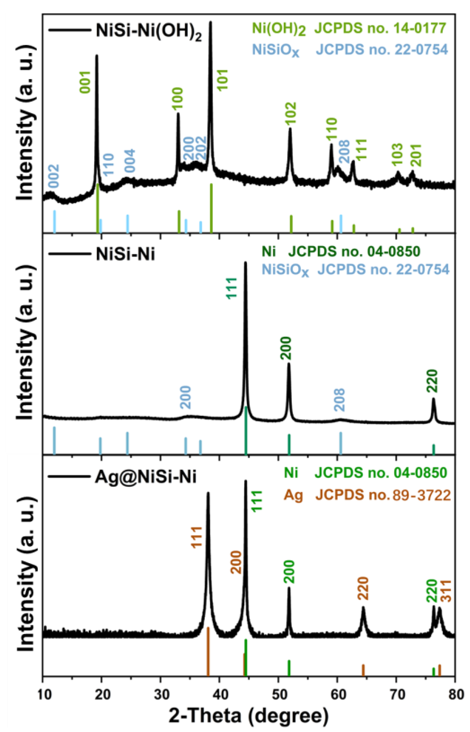
Figure 1. XRD patterns of NiSi-Ni (OH)2, NiSi-Ni, and Ag/NiSi-Ni.
Molecules 29 04384 g001
Figure 2. The SEM images of NiSi–Ni(OH)2 (a,b), NiSi–Ni (c,d), and Ag/NiSi-Ni (e,f).
Molecules 29 04384 g002
Figure 3. The TEM and HRTEM images of NiSi–Ni(OH)2 (ac); NiSi–Ni (df); and Ag/NiSi-Ni (gi).
Molecules 29 04384 g003
Figure 4. Nitrogen adsorption, desorption isotherms (a), and pore diameter distributions (b) of NiSi-Ni (OH)2, NiSi-Ni, and Ag/NiSi-Ni.
Molecules 29 04384 g004
Figure 5. Hysteresis loops (a) and initial magnetization curves (b) of NiSi-Ni and Ag/NiSi-Ni.
Molecules 29 04384 g005
Figure 6. The SEM images of Ag/NiSi–Ni-1 (a), Ag/NiSi–Ni-2 (b), and Ag/NiSi–Ni-3 (c).
Molecules 29 04384 g006
Figure 7. Volume resistivity of Ag/NiSi–Ni–1, Ag/NiSi–Ni–2, and Ag/NiSi–Ni–3.
Molecules 29 04384 g007
Figure 8. Absorption, reflection, and transmission mechanisms of high-frequency electromagnetic waves in nanosheet self-assembled hollow sphere structure particles.
Molecules 29 04384 g008
Figure 9. Schematic illustration of the preparation process of Ag/NiSi-Ni.
Molecules 29 04384 g009
Disclaimer/Publisher’s Note: The statements, opinions and data contained in all publications are solely those of the individual author(s) and contributor(s) and not of MDPI and/or the editor(s). MDPI and/or the editor(s) disclaim responsibility for any injury to people or property resulting from any ideas, methods, instructions or products referred to in the content.