
Quaternized Curcumin Derivative—Synthesis, Physicochemical Characteristics, and Photocytotoxicity, Including Antibacterial Activity after Irradiation with Blue Light

 ,
,  , , , ,
  ,
,
Abstract
1. Introduction
2. Results
2.1. Chemical Synthesis and Characterization
2.2. Physicochemical Characteristics
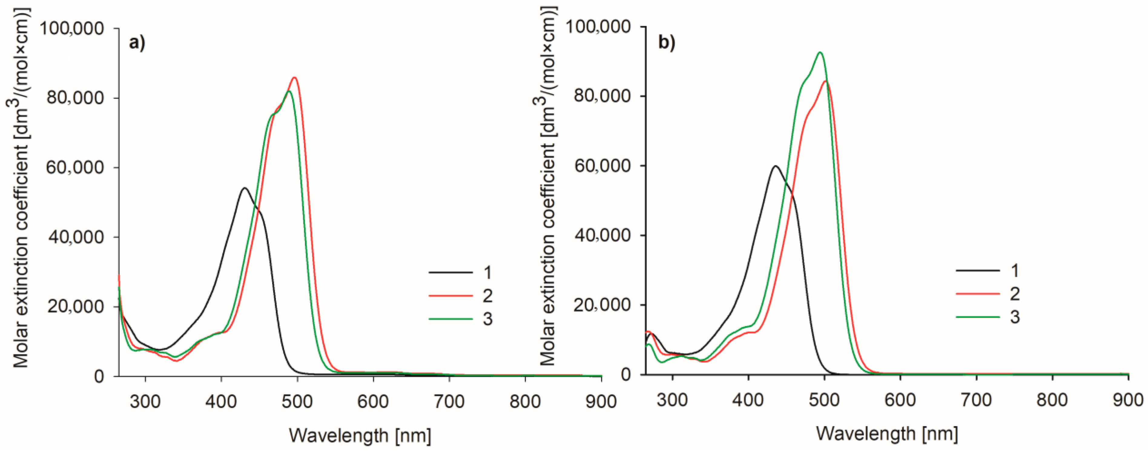
2.2.1. Spectral Properties
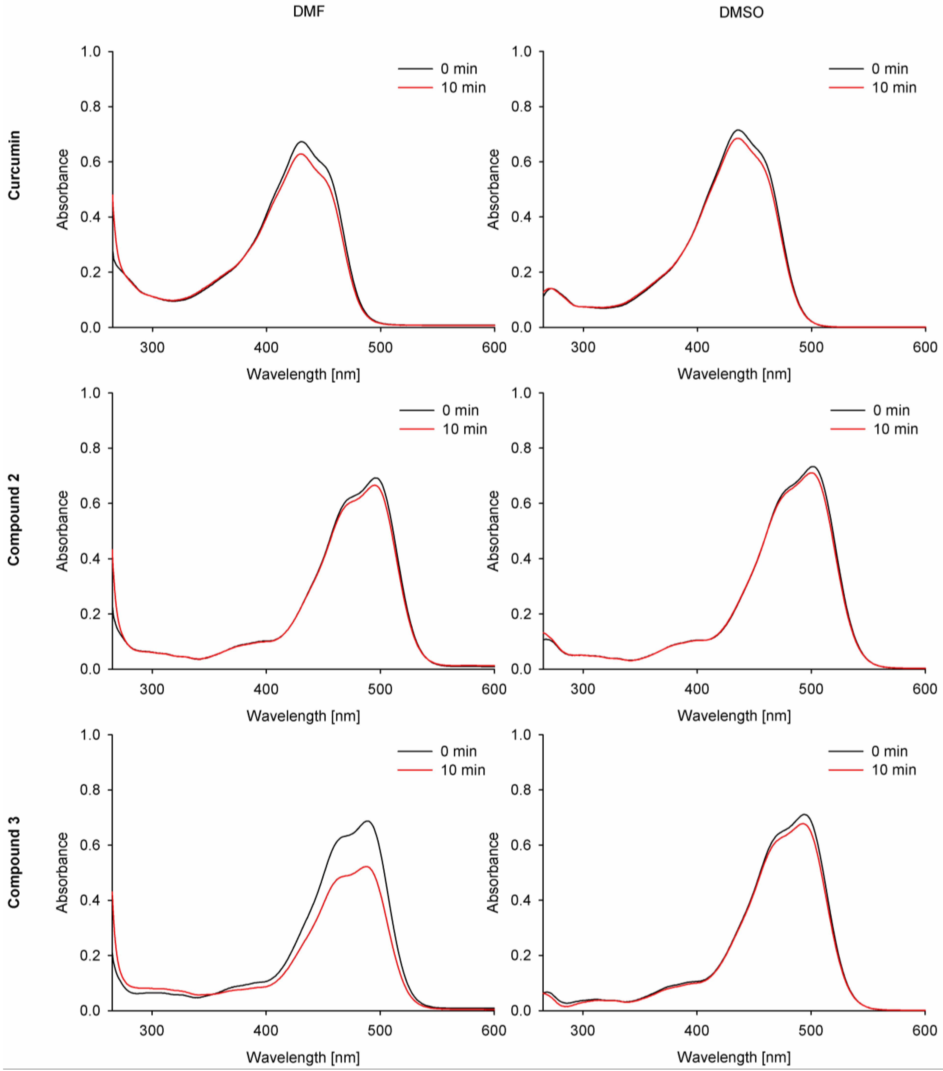
2.2.2. Photodecomposition Quantum Yields
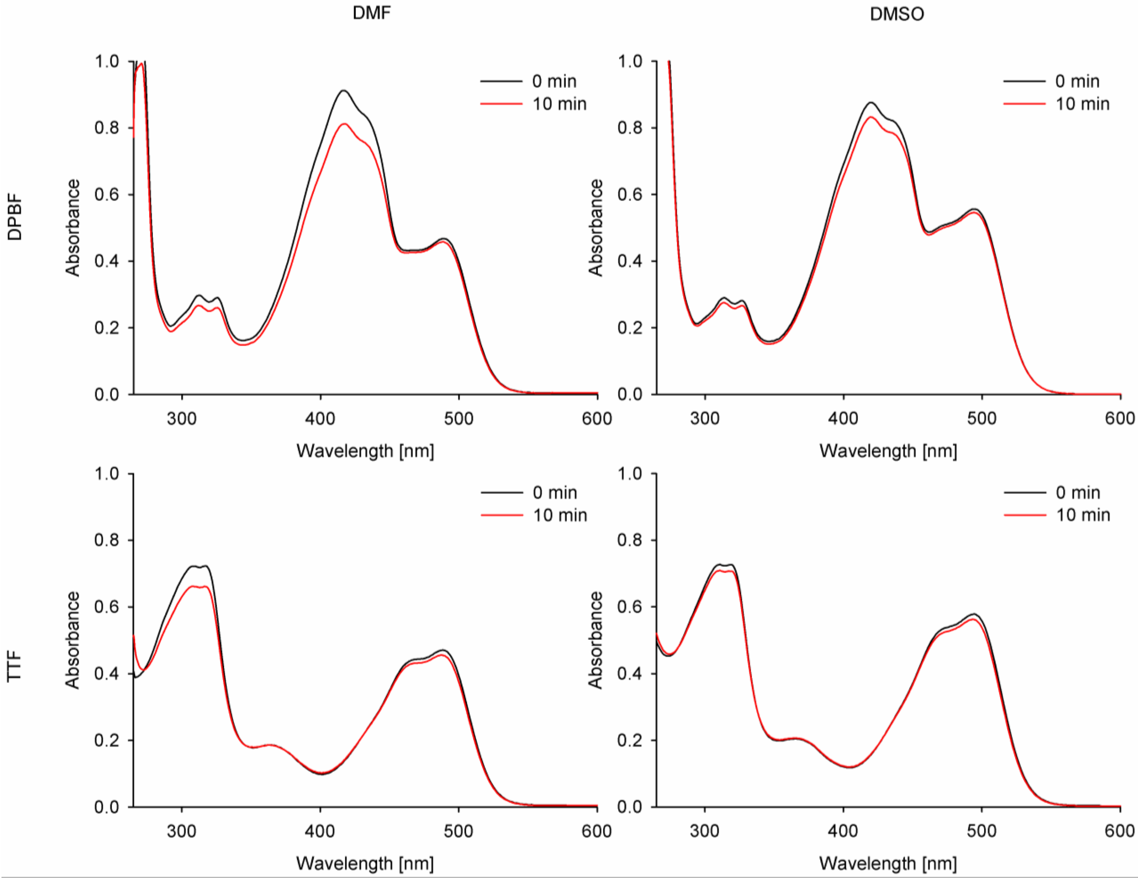
2.2.3. Singlet Oxygen Generation Quantum Yields
2.2.4. Aggregation Studies
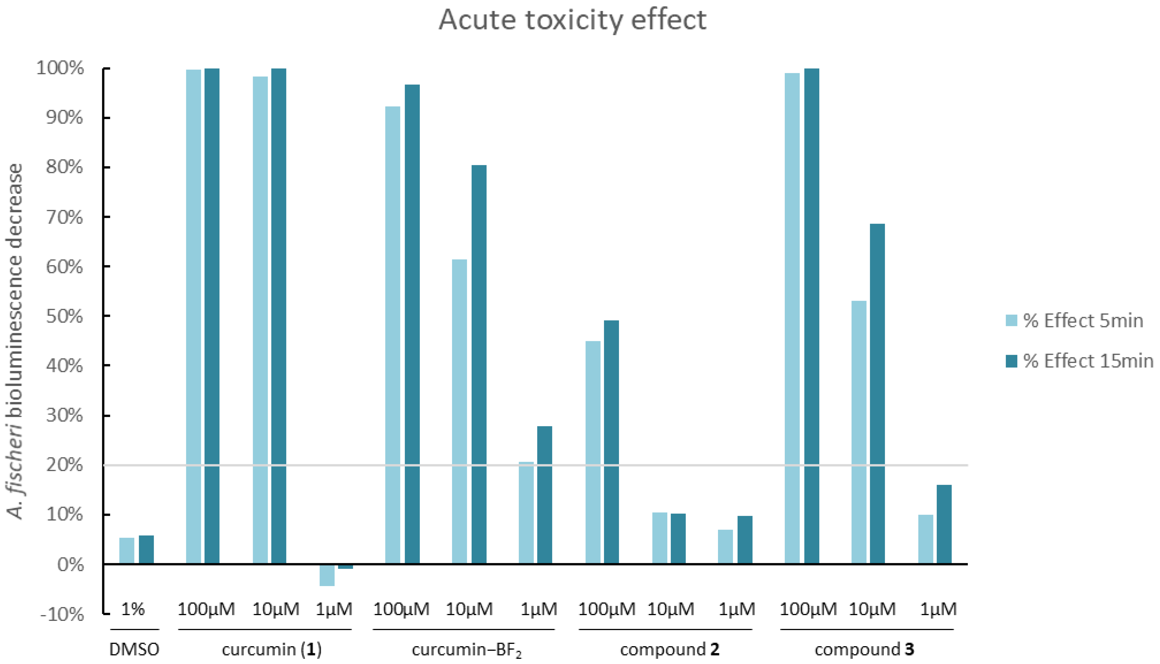
2.3. Acute Toxicity Assessment—Microtox Assay
2.4. In Vitro Photodynamic Antimicrobial Activity
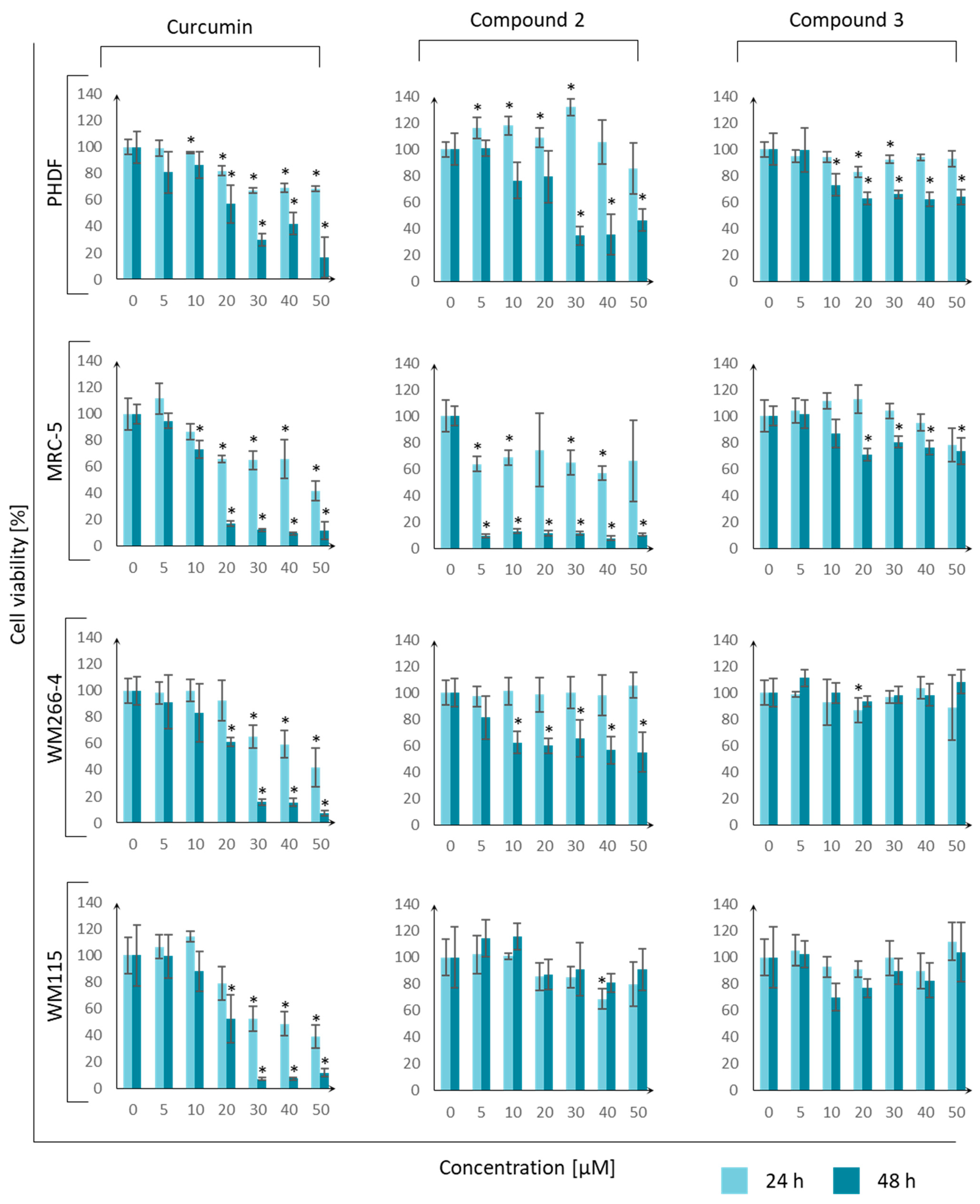
2.5. Cell Viability Assessment
3. Discussion
4. Materials and Methods
4.1. Chemistry
4.1.1. Synthesis of Modified Aldehyde—Stage I
4.1.2. Synthesis of Acetylacetone–BF2—Stage II
4.1.3. Synthesis of 2 via Aldol Condensation—Stage III
4.1.4. Synthesis of Compound 3—Stage IV
4.2. Physicochemical Studies
4.2.1. Photodecomposition Quantum Yields
4.2.2. Singlet Oxygen Generation Quantum Yields under Light Irradiation
4.2.3. Aggregation Studies
4.3. Biological Activity
4.3.1. Microtox Assay
4.3.2. In Vitro Photodynamic Antimicrobial Activity
4.3.3. MTT Assay and Cell Culture
5. Conclusions
Author Contributions
Funding
Institutional Review Board Statement
Informed Consent Statement
Data Availability Statement
Acknowledgments
Conflicts of Interest
References
- Van Duin, D.; Paterson, D.L. Multidrug-Resistant Bacteria in the Community. Infect. Dis. Clin. N. Am. 2020, 34, 709–722. [Google Scholar] [CrossRef] [PubMed]
- Devi, N.S.; Mythili, R.; Cherian, T.; Dineshkumar, R.; Sivaraman, G.K.; Jayakumar, R.; Prathaban, M.; Duraimurugan, M.; Chandrasekar, V.; Peijnenburg, W.J.G.M. Overview of Antimicrobial Resistance and Mechanisms: The Relative Status of the Past and Current. Microbe 2024, 3, 100083. [Google Scholar] [CrossRef]
- Ziental, D.; Mlynarczyk, D.T.; Czarczynska-Goslinska, B.; Lewandowski, K.; Sobotta, L. Photosensitizers Mediated Photodynamic Inactivation against Fungi. Nanomaterials 2021, 11, 2883. [Google Scholar] [CrossRef] [PubMed]
- Glowacka-Sobotta, A.A.; Ziental, B.D.; Sobotta, C.L. Porphyrinoids Used for Photodynamic Inactivation against Bacteria. In Applications of Porphyrinoids as Functional Materials; Lang, H., Rueffer, T., Eds.; The Royal Society of Chemistry: London, UK, 2021; pp. 352–404. ISBN 978-1-83916-188-9. [Google Scholar]
- Ruggieri, F.; Compagne, N.; Antraygues, K.; Eveque, M.; Flipo, M.; Willand, N. Antibiotics with Novel Mode of Action as New Weapons to Fight Antimicrobial Resistance. Eur. J. Med. Chem. 2023, 256, 115413. [Google Scholar] [CrossRef]
- Piksa, M.; Lian, C.; Samuel, I.C.; Pawlik, K.J.; Samuel, I.D.W.; Matczyszyn, K. The Role of the Light Source in Antimicrobial Photodynamic Therapy. Chem. Soc. Rev. 2023, 52, 1697–1722. [Google Scholar] [CrossRef]
- Balakrishnan, D.; Lee, C.-I. Photodynamic Impact of Curcumin Enhanced Silver Functionalized Graphene Nanocomposites on Candida Virulence. Discov. Nano 2024, 19, 71. [Google Scholar] [CrossRef]
- Anas, A.; Sobhanan, J.; Sulfiya, K.M.; Jasmin, C.; Sreelakshmi, P.K.; Biju, V. Advances in Photodynamic Antimicrobial Chemotherapy. J. Photochem. Photobiol. C Photochem. Rev. 2021, 49, 100452. [Google Scholar] [CrossRef]
- Stolarska, M.; Glowacka-Sobotta, A.; Ziental, D.; Dlugaszewska, J.; Falkowski, M.; Mielcarek, J.; Goslinski, T.; Sobotta, L. Photochemical Properties and Photocytotoxicities against Wound Bacteria of Sulfanyl Porphyrazines with Bulky Peripheral Substituents. J. Organomet. Chem. 2021, 934, 121669. [Google Scholar] [CrossRef]
- Sobotta, L.; Skupin-Mrugalska, P.; Piskorz, J.; Mielcarek, J. Non-Porphyrinoid Photosensitizers Mediated Photodynamic Inactivation against Bacteria. Dye. Pigment. 2019, 163, 337–355. [Google Scholar] [CrossRef]
- Stolarska, M.; Glowacka-Sobotta, A.; Mlynarczyk, D.T.; Dlugaszewska, J.; Goslinski, T.; Mielcarek, J.; Sobotta, L. Photodynamic Activity of Tribenzoporphyrazines with Bulky Periphery against Wound Bacteria. Int. J. Mol. Sci. 2020, 21, 6145. [Google Scholar] [CrossRef]
- Pucelik, B.; Dąbrowski, J.M. Photodynamic Inactivation (PDI) as a Promising Alternative to Current Pharmaceuticals for the Treatment of Resistant Microorganisms. In Advances in Inorganic Chemistry; Elsevier: Amsterdam, The Netherlands, 2022; Volume 79, pp. 65–108. ISBN 978-0-323-99972-4. [Google Scholar]
- Szczolko, W.; Ratajczak, M.; Koczorowski, T.; Kaminska, D.; Goslinski, T.; Dlugaszewska, J. Promising Photocytotoxicity of Water-Soluble Phtalocyanine against Planktonic and Biofilm Pseudomonas Aeruginosa Isolates from Lower Respiratory Tract and Chronic Wounds. Appl. Sci. 2022, 12, 3707. [Google Scholar] [CrossRef]
- Sobotta, L.; Ziental, D.; Sniechowska, J.; Dlugaszewska, J.; Potrzebowski, M.J. Lipid Vesicle-Loaded Meso-Substituted Chlorins of High in Vitro Antimicrobial Photodynamic Activity. Photochem. Photobiol. Sci. 2019, 18, 213–223. [Google Scholar] [CrossRef]
- Hussain, Y.; Alam, W.; Ullah, H.; Dacrema, M.; Daglia, M.; Khan, H.; Arciola, C.R. Antimicrobial Potential of Curcumin: Therapeutic Potential and Challenges to Clinical Applications. Antibiotics 2022, 11, 322. [Google Scholar] [CrossRef] [PubMed]
- Adamczak, A.; Ożarowski, M.; Karpiński, T.M. Curcumin, a Natural Antimicrobial Agent with Strain-Specific Activity. Pharmaceuticals 2020, 13, 153. [Google Scholar] [CrossRef] [PubMed]
- Zorofchian Moghadamtousi, S.; Abdul Kadir, H.; Hassandarvish, P.; Tajik, H.; Abubakar, S.; Zandi, K. A Review on Antibacterial, Antiviral, and Antifungal Activity of Curcumin. BioMed Res. Int. 2014, 2014, 186864. [Google Scholar] [CrossRef]
- Abdulrahman, H.; Misba, L.; Ahmad, S.; Khan, A.U. Curcumin Induced Photodynamic Therapy Mediated Suppression of Quorum Sensing Pathway of Pseudomonas Aeruginosa: An Approach to Inhibit Biofilm in Vitro. Photodiagnosis Photodyn. Ther. 2020, 30, 101645. [Google Scholar] [CrossRef]
- Bahari, S.; Zeighami, H.; Mirshahabi, H.; Roudashti, S.; Haghi, F. Inhibition of Pseudomonas Aeruginosa Quorum Sensing by Subinhibitory Concentrations of Curcumin with Gentamicin and Azithromycin. J. Glob. Antimicrob. Resist. 2017, 10, 21–28. [Google Scholar] [CrossRef]
- Itzia Azucena, R.-C.; José Roberto, C.-L.; Martin, Z.-R.; Rafael, C.-Z.; Leonardo, H.-H.; Gabriela, T.-P.; Araceli, C.-R. Drug Susceptibility Testing and Synergistic Antibacterial Activity of Curcumin with Antibiotics against Enterotoxigenic Escherichia Coli. Antibiotics 2019, 8, 43. [Google Scholar] [CrossRef]
- Zheng, D.; Huang, C.; Huang, H.; Zhao, Y.; Khan, M.R.U.; Zhao, H.; Huang, L. Antibacterial Mechanism of Curcumin: A Review. Chem. Biodivers. 2020, 17, e2000171. [Google Scholar] [CrossRef]
- Mohapatra, S.; Yutao, L.; Goh, S.G.; Ng, C.; Luhua, Y.; Tran, N.H.; Gin, K.Y.-H. Quaternary Ammonium Compounds of Emerging Concern: Classification, Occurrence, Fate, Toxicity and Antimicrobial Resistance. J. Hazard. Mater. 2023, 445, 130393. [Google Scholar] [CrossRef]
- Arnold, W.A.; Blum, A.; Branyan, J.; Bruton, T.A.; Carignan, C.C.; Cortopassi, G.; Datta, S.; DeWitt, J.; Doherty, A.-C.; Halden, R.U.; et al. Quaternary Ammonium Compounds: A Chemical Class of Emerging Concern. Environ. Sci. Technol. 2023, 57, 7645–7665. [Google Scholar] [CrossRef] [PubMed]
- Vereshchagin, A.N.; Frolov, N.A.; Egorova, K.S.; Seitkalieva, M.M.; Ananikov, V.P. Quaternary Ammonium Compounds (QACs) and Ionic Liquids (ILs) as Biocides: From Simple Antiseptics to Tunable Antimicrobials. Int. J. Mol. Sci. 2021, 22, 6793. [Google Scholar] [CrossRef] [PubMed]
- Osimitz, T.G.; Droege, W. Quaternary Ammonium Compounds: Perspectives on Benefits, Hazards, and Risk. Toxicol. Res. Appl. 2021, 5, 239784732110490. [Google Scholar] [CrossRef]
- Kuźmińska, J.; Kobyłka, P.; Wierzchowski, M.; Łażewski, D.; Popenda, Ł.; Szubska, P.; Jankowska, W.; Jurga, S.; Goslinski, T.; Muszalska-Kolos, I.; et al. Novel Fluorocurcuminoid-BF2 Complexes and Their Unlocked Counterparts as Potential Bladder Anticancer Agents—Synthesis, Physicochemical Characterization, and in Vitro Anticancer Activity. J. Mol. Struct. 2023, 1283, 135269. [Google Scholar] [CrossRef]
- Bakun, P.; Kucinska, M.; Kobyłka, P.; Kuźmińska, J.; Koczorowski, T.; Mlynarczyk, D.T.; Popenda, L.; Górska, K.; Kasperkowiak, M.; Murias, M.; et al. Morpholinated Curcuminoids against Urinary Bladder Cancer Cells: Synthesis and Anticancer Evaluation. Med. Chem. Res. 2024, 33, 944–963. [Google Scholar] [CrossRef]
- Kucinska, M.; Skupin-Mrugalska, P.; Szczolko, W.; Sobotta, L.; Sciepura, M.; Tykarska, E.; Wierzchowski, M.; Teubert, A.; Fedoruk-Wyszomirska, A.; Wyszko, E.; et al. Phthalocyanine Derivatives Possessing 2-(Morpholin-4-Yl)Ethoxy Groups As Potential Agents for Photodynamic Therapy. J. Med. Chem. 2015, 58, 2240–2255. [Google Scholar] [CrossRef]
- Popov, A.B.; Krstulović, L.; Koštrun, S.; Jelić, D.; Bokulić, A.; Stojković, M.R.; Zonjić, I.; Taylor, M.C.; Kelly, J.M.; Bajić, M.; et al. Design, Synthesis, Antitrypanosomal Activity, DNA/RNA Binding and in Vitro ADME Profiling of Novel Imidazoline-Substituted 2-Arylbenzimidazoles. Eur. J. Med. Chem. 2020, 207, 112802. [Google Scholar] [CrossRef]
- Liu, K.; Chen, J.; Chojnacki, J.; Zhang, S. BF3·OEt2-Promoted Concise Synthesis of Difluoroboron-Derivatized Curcumins from Aldehydes and 2,4-Pentanedione. Tetrahedron Lett. 2013, 54, 2070–2073. [Google Scholar] [CrossRef]
- Dlugaszewska, J.; Szczolko, W.; Koczorowski, T.; Skupin-Mrugalska, P.; Teubert, A.; Konopka, K.; Kucinska, M.; Murias, M.; Düzgüneş, N.; Mielcarek, J.; et al. Antimicrobial and Anticancer Photodynamic Activity of a Phthalocyanine Photosensitizer with N -Methyl Morpholiniumethoxy Substituents in Non-Peripheral Positions. J. Inorg. Biochem. 2017, 172, 67–79. [Google Scholar] [CrossRef]
- Lazewski, D.; Kucinska, M.; Potapskiy, E.; Kuzminska, J.; Popenda, L.; Tezyk, A.; Goslinski, T.; Wierzchowski, M.; Murias, M. Enhanced Cytotoxic Activity of PEGylated Curcumin Derivatives: Synthesis, Structure–Activity Evaluation, and Biological Activity. Int. J. Mol. Sci. 2023, 24, 1467. [Google Scholar] [CrossRef]
- Hung, S.-J.; Hong, Y.-A.; Lin, K.-Y.; Hua, Y.-W.; Kuo, C.-J.; Hu, A.; Shih, T.-L.; Chen, H.-P. Efficient Photodynamic Killing of Gram-Positive Bacteria by Synthetic Curcuminoids. Int. J. Mol. Sci. 2020, 21, 9024. [Google Scholar] [CrossRef] [PubMed]
- Kazantzis, K.T.; Koutsonikoli, K.; Mavroidi, B.; Zachariadis, M.; Alexiou, P.; Pelecanou, M.; Politopoulos, K.; Alexandratou, E.; Sagnou, M. Curcumin Derivatives as Photosensitizers in Photodynamic Therapy: Photophysical Properties and in Vitro Studies with Prostate Cancer Cells. Photochem. Photobiol. Sci. 2020, 19, 193–206. [Google Scholar] [CrossRef] [PubMed]
- Bai, G.; Yu, C.; Cheng, C.; Hao, E.; Wei, Y.; Mu, X.; Jiao, L. Syntheses and Photophysical Properties of BF 2 Complexes of Curcumin Analogues. Org. Biomol. Chem. 2014, 12, 1618–1626. [Google Scholar] [CrossRef]
- Priyadarsini, K.I. Photophysics, Photochemistry and Photobiology of Curcumin: Studies from Organic Solutions, Bio-Mimetics and Living Cells. J. Photochem. Photobiol. C Photochem. Rev. 2009, 10, 81–95. [Google Scholar] [CrossRef]
- Spaeth, A.; Graeler, A.; Maisch, T.; Plaetzer, K. CureCuma–Cationic Curcuminoids with Improved Properties and Enhanced Antimicrobial Photodynamic Activity. Eur. J. Med. Chem. 2018, 159, 423–440. [Google Scholar] [CrossRef] [PubMed]
- Sobotta, L.; Lijewski, S.; Dlugaszewska, J.; Nowicka, J.; Mielcarek, J.; Goslinski, T. Photodynamic Inactivation of Enterococcus Faecalis by Conjugates of Zinc(II) Phthalocyanines with Thymol and Carvacrol Loaded into Lipid Vesicles. Inorganica Chim. Acta 2019, 489, 180–190. [Google Scholar] [CrossRef]
- Ogunsipe, A.; Durmuş, M.; Atilla, D.; Gürek, A.G.; Ahsen, V.; Nyokong, T. Synthesis, Photophysical and Photochemical Studies on Long Chain Zinc Phthalocyanine Derivatives. Synth. Met. 2008, 158, 839–847. [Google Scholar] [CrossRef]
- Wolnicka-Glubisz, A.; Olchawa, M.; Duda, M.; Pabisz, P.; Wisniewska-Becker, A. The Role of Singlet Oxygen in Photoreactivity and Phototoxicity of Curcumin. Photochem. Photobiol. 2023, 99, 57–67. [Google Scholar] [CrossRef]
- Condat, M.; Mazeran, P.-E.; Malval, J.-P.; Lalevée, J.; Morlet-Savary, F.; Renard, E.; Langlois, V.; Abbad Andalloussi, S.; Versace, D.-L. Photoinduced Curcumin Derivative-Coatings with Antibacterial Properties. RSC Adv. 2015, 5, 85214–85224. [Google Scholar] [CrossRef]
- Chignell, C.F.; Bilskj, P.; Reszka, K.J.; Motten, A.G.; Sik, R.H.; Dahl, T.A. Spectral And Photochemical Properties Of Curcumin. Photochem. Photobiol. 1994, 59, 295–302. [Google Scholar] [CrossRef]
- Vesco, G.; Brambati, M.; Scapinello, L.; Penoni, A.; Mella, M.; Masson, M.; Gaware, V.; Maspero, A.; Nardo, L. Asymmetric Phenyl Substitution: An Effective Strategy to Enhance the Photosensitizing Potential of Curcuminoids. Pharmaceuticals 2022, 15, 843. [Google Scholar] [CrossRef] [PubMed]
- Patsahan, T.; Pizio, O. Aspects of the Microscopic Structure of Curcumin Solutions with Water-Dimethylsulfoxide Solvent. Molecular Dynamics Computer Simulation Study. Condens. Matter Phys. 2023, 26, 33605. [Google Scholar] [CrossRef]
- Domínguez, C.M.; Ventura, P.; Checa-Fernández, A.; Santos, A. Comprehensive Study of Acute Toxicity Using Microtox® Bioassay in Soils Contaminated by Lindane Wastes. Sci. Total Environ. 2023, 856, 159146. [Google Scholar] [CrossRef]
- Niemirycz, E.; Nichthauser, J.; Staniszewska, M.; Nałęcz-Jawecki, G.; Bolałek, J. The Microtox® Biological Test: Application in Toxicity Evaluation of Surface Waters and Sediments in Poland. Oceanol. Hydrobiol. Stud. 2007, 36, 151–163. [Google Scholar] [CrossRef]
- Biczak, R.; Turek, M.; Pawłowska, B.; Różycka-Sokołowska, E.; Marciniak, B.; Deska, M.; Krupa, P.; Jatulewicz, I.; Skalik, J.; Bałczewski, P. An Influence of Structural Changes in Ammonium Cations on Ecotoxicity of 2,2’-Thiodiacetate Mono and Bis-Salts. Ecotoxicol. Environ. Saf. 2018, 155, 37–42. [Google Scholar] [CrossRef]
- Lei, L.; Aoyama, I. Effect-Directed Investigation and Interactive Effect of Organic Toxicants in Landfill Leachates Combining Microtox Test with RP-HPLC Fractionation and GC/MS Analysis. Ecotoxicology 2010, 19, 1268–1276. [Google Scholar] [CrossRef] [PubMed]
- Siwak, D.R.; Shishodia, S.; Aggarwal, B.B.; Kurzrock, R. Curcumin-Induced Antiproliferative and Proapoptotic Effects in Melanoma Cells Are Associated with Suppression of IκB Kinase and Nuclear Factor κB Activity and Are Independent of the B-Raf/Mitogen-Activated/Extracellular Signal-Regulated Protein Kinase Pathway and the Akt Pathway. Cancer 2005, 104, 879–890. [Google Scholar] [CrossRef] [PubMed]
- Skupin-Mrugalska, P.; Szczolko, W.; Gierlich, P.; Konopka, K.; Goslinski, T.; Mielcarek, J.; Düzgüneş, N. Physicochemical Properties of Liposome-Incorporated 2-(Morpholin-4-Yl)Ethoxy Phthalocyanines and Their Photodynamic Activity against Oral Cancer Cells. J. Photochem. Photobiol. A Chem. 2018, 353, 445–457. [Google Scholar] [CrossRef]
- Sindelo, A.; Sen, P.; Nyokong, T. Photoantimicrobial Activity of Schiff-Base Morpholino Phthalocyanines against Drug Resistant Micro-Organisms in Their Planktonic and Biofilm Forms. Photodiagnosis Photodyn. Ther. 2023, 42, 103519. [Google Scholar] [CrossRef]
- Tortik, N.; Steinbacher, P.; Maisch, T.; Spaeth, A.; Plaetzer, K. A Comparative Study on the Antibacterial Photodynamic Efficiency of a Curcumin Derivative and a Formulation on a Porcine Skin Model. Photochem. Photobiol. Sci. 2016, 15, 187–195. [Google Scholar] [CrossRef]
- Glueck, M.; Schamberger, B.; Eckl, P.; Plaetzer, K. New Horizons in Microbiological Food Safety: Photodynamic Decontamination Based on a Curcumin Derivative. Photochem. Photobiol. Sci. 2017, 16, 1784–1791. [Google Scholar] [CrossRef] [PubMed]
- Mlynarczyk, D.T.; Dlugaszewska, J.; Falkowski, M.; Popenda, L.; Kryjewski, M.; Szczolko, W.; Jurga, S.; Mielcarek, J.; Goslinski, T. Tribenzoporphyrazines with Dendrimeric Peripheral Substituents and Their Promising Photocytotoxic Activity against Staphylococcus Aureus. J. Photochem. Photobiol. B Biol. 2020, 204, 111803. [Google Scholar] [CrossRef]
- Agazzi, M.L.; Ballatore, M.B.; Reynoso, E.; Quiroga, E.D.; Durantini, E.N. Synthesis, Spectroscopic Properties and Photodynamic Activity of Two Cationic BODIPY Derivatives with Application in the Photoinactivation of Microorganisms. Eur. J. Med. Chem. 2017, 126, 110–121. [Google Scholar] [CrossRef] [PubMed]
- Le Guern, F.; Ouk, T.-S.; Yerzhan, I.; Nurlykyz, Y.; Arnoux, P.; Frochot, C.; Leroy-Lhez, S.; Sol, V. Photophysical and Bactericidal Properties of Pyridinium and Imidazolium Porphyrins for Photodynamic Antimicrobial Chemotherapy. Molecules 2021, 26, 1122. [Google Scholar] [CrossRef] [PubMed]
- Pérez-Laguna, V.; García-Luque, I.; Ballesta, S.; Pérez-Artiaga, L.; Lampaya-Pérez, V.; Rezusta, A.; Gilaberte, Y. Photodynamic Therapy Using Methylene Blue, Combined or Not with Gentamicin, against Staphylococcus Aureus and Pseudomonas Aeruginosa. Photodiagnosis Photodyn. Ther. 2020, 31, 101810. [Google Scholar] [CrossRef]
- Parasuraman, P.; Antony, A.P.; Sharan, A.; Siddhardha, B.; Kasinathan, K.; Bahkali, N.A.; Dawoud, T.M.S.; Syed, A. Antimicrobial Photodynamic Activity of Toluidine Blue Encapsulated in Mesoporous Silica Nanoparticles against Pseudomonas aeruginosa and Staphylococcus aureus. Biofouling 2019, 35, 89–103. [Google Scholar] [CrossRef]
- Pérez-Laguna, V.; García-Luque, I.; Ballesta, S.; Pérez-Artiaga, L.; Lampaya-Pérez, V.; Samper, S.; Soria-Lozano, P.; Rezusta, A.; Gilaberte, Y. Antimicrobial Photodynamic Activity of Rose Bengal, Alone or in Combination with Gentamicin, against Planktonic and Biofilm Staphylococcus Aureus. Photodiagnosis Photodyn. Ther. 2018, 21, 211–216. [Google Scholar] [CrossRef] [PubMed]
- Yuan, Y.; Liu, Q.; Huang, Y.; Qi, M.; Yan, H.; Li, W.; Zhuang, H. Antibacterial Efficacy and Mechanisms of Curcumin-Based Photodynamic Treatment against Staphylococcus Aureus and Its Application in Juices. Molecules 2022, 27, 7136. [Google Scholar] [CrossRef]
- Giuliani, F.; Martinelli, M.; Cocchi, A.; Arbia, D.; Fantetti, L.; Roncucci, G. In Vitro Resistance Selection Studies of RLP068/Cl, a New Zn(II) Phthalocyanine Suitable for Antimicrobial Photodynamic Therapy. Antimicrob. Agents Chemother. 2010, 54, 637–642. [Google Scholar] [CrossRef]
- WHO Publishes List of Bacteria for Which New Antibiotics Are Urgently Needed. Available online: https://www.who.int/news/item/27-02-2017-who-publishes-list-of-bacteria-for-which-new-antibiotics-are-urgently-needed (accessed on 18 July 2024).
- Santezi, C.; Reina, B.D.; Dovigo, L.N. Curcumin-Mediated Photodynamic Therapy for the Treatment of Oral Infections—A Review. Photodiagnosis Photodyn. Ther. 2018, 21, 409–415. [Google Scholar] [CrossRef]
- Dovigo, L.N.; Pavarina, A.C.; Ribeiro, A.P.D.; Brunetti, I.L.; Costa, C.A.D.S.; Jacomassi, D.P.; Bagnato, V.S.; Kurachi, C. Investigation of the Photodynamic Effects of Curcumin against Candida albicans. Photochem. Photobiol. 2011, 87, 895–903. [Google Scholar] [CrossRef] [PubMed]
- Dovigo, L.N.; Carmello, J.C.; De Souza Costa, C.A.; Vergani, C.E.; Brunetti, I.L.; Bagnato, V.S.; Pavarina, A.C. Curcumin-Mediated Photodynamic Inactivation of Candida albicans in a Murine Model of Oral Candidiasis. Med. Mycol. 2013, 51, 243–251. [Google Scholar] [CrossRef] [PubMed]
- Andrade, M.C.; Ribeiro, A.P.D.; Dovigo, L.N.; Brunetti, I.L.; Giampaolo, E.T.; Bagnato, V.S.; Pavarina, A.C. Effect of Different Pre-Irradiation Times on Curcumin-Mediated Photodynamic Therapy against Planktonic Cultures and Biofilms of Candida spp. Arch. Oral Biol. 2013, 58, 200–210. [Google Scholar] [CrossRef]
- Breidenstein, E.B.M.; De La Fuente-Núñez, C.; Hancock, R.E.W. Pseudomonas Aeruginosa: All Roads Lead to Resistance. Trends Microbiol. 2011, 19, 419–426. [Google Scholar] [CrossRef] [PubMed]
- Morita, Y.; Tomida, J.; Kawamura, Y. Responses of Pseudomonas Aeruginosa to Antimicrobials. Front. Microbiol. 2014, 4, 422. [Google Scholar] [CrossRef]
- Liebmann, J.; Born, M.; Kolb-Bachofen, V. Blue-Light Irradiation Regulates Proliferation and Differentiation in Human Skin Cells. J. Investig. Dermatol. 2010, 130, 259–269. [Google Scholar] [CrossRef]
- Masson-Meyers, D.S.; Bumah, V.V.; Enwemeka, C.S. Blue Light Does Not Impair Wound Healing in Vitro. J. Photochem. Photobiol. B Biol. 2016, 160, 53–60. [Google Scholar] [CrossRef]
- Kim, M.M.; Darafsheh, A. Light Sources and Dosimetry Techniques for Photodynamic Therapy. Photochem. Photobiol. 2020, 96, 280–294. [Google Scholar] [CrossRef]
- Aroso, R.T.; Schaberle, F.A.; Arnaut, L.G.; Pereira, M.M. Photodynamic Disinfection and Its Role in Controlling Infectious Diseases. Photochem. Photobiol. Sci. 2021, 20, 1497–1545. [Google Scholar] [CrossRef]
- Wysocki, M.; Ziental, D.; Jozkowiak, M.; Dlugaszewska, J.; Piotrowska-Kempisty, H.; Güzel, E.; Sobotta, L. Porphyrazine/Phthalocyanine Hybrid Complexes—Antibacterial and Anticancer Photodynamic and Sonodynamic Activity. Synth. Met. 2023, 299, 117474. [Google Scholar] [CrossRef]
- Skupin-Mrugalska, P.; Koczorowski, T.; Szczolko, W.; Dlugaszewska, J.; Teubert, A.; Piotrowska-Kempisty, H.; Goslinski, T.; Sobotta, L. Cationic Porphyrazines with Morpholinoethyl Substituents—Syntheses, Optical Properties, and Photocytotoxicities. Dye. Pigment. 2022, 197, 109937. [Google Scholar] [CrossRef]
- Chauke, V.; Ogunsipe, A.; Durmuş, M.; Nyokong, T. Novel Gallium(III) Phthalocyanine Derivatives—Synthesis, Photophysics and Photochemistry. Polyhedron 2007, 26, 2663–2671. [Google Scholar] [CrossRef]
- Seotsanyana-Mokhosi, I.; Kuznetsova, N.; Nyokong, T. Photochemical Studies of Tetra-2,3-Pyridinoporphyrazines. J. Photochem. Photobiol. A Chem. 2001, 140, 215–222. [Google Scholar] [CrossRef]
- Bakun, P.; Czarczynska-Goslinska, B.; Mlynarczyk, D.T.; Musielak, M.; Mylkie, K.; Dlugaszewska, J.; Koczorowski, T.; Suchorska, W.M.; Ziegler-Borowska, M.; Goslinski, T.; et al. Gallic Acid-Functionalized, TiO2-Based Nanomaterial—Preparation, Physicochemical and Biological Properties. Materials 2022, 15, 4177. [Google Scholar] [CrossRef]
- Mazurek, S.; Oleksiewicz, U.; Czerwińska, P.; Wróblewska, J.; Klimczak, M.; Wiznerowicz, M. Disruption of RING and PHD Domains of TRIM28 Evokes Differentiation in Human iPSCs. Cells 2021, 10, 1933. [Google Scholar] [CrossRef]


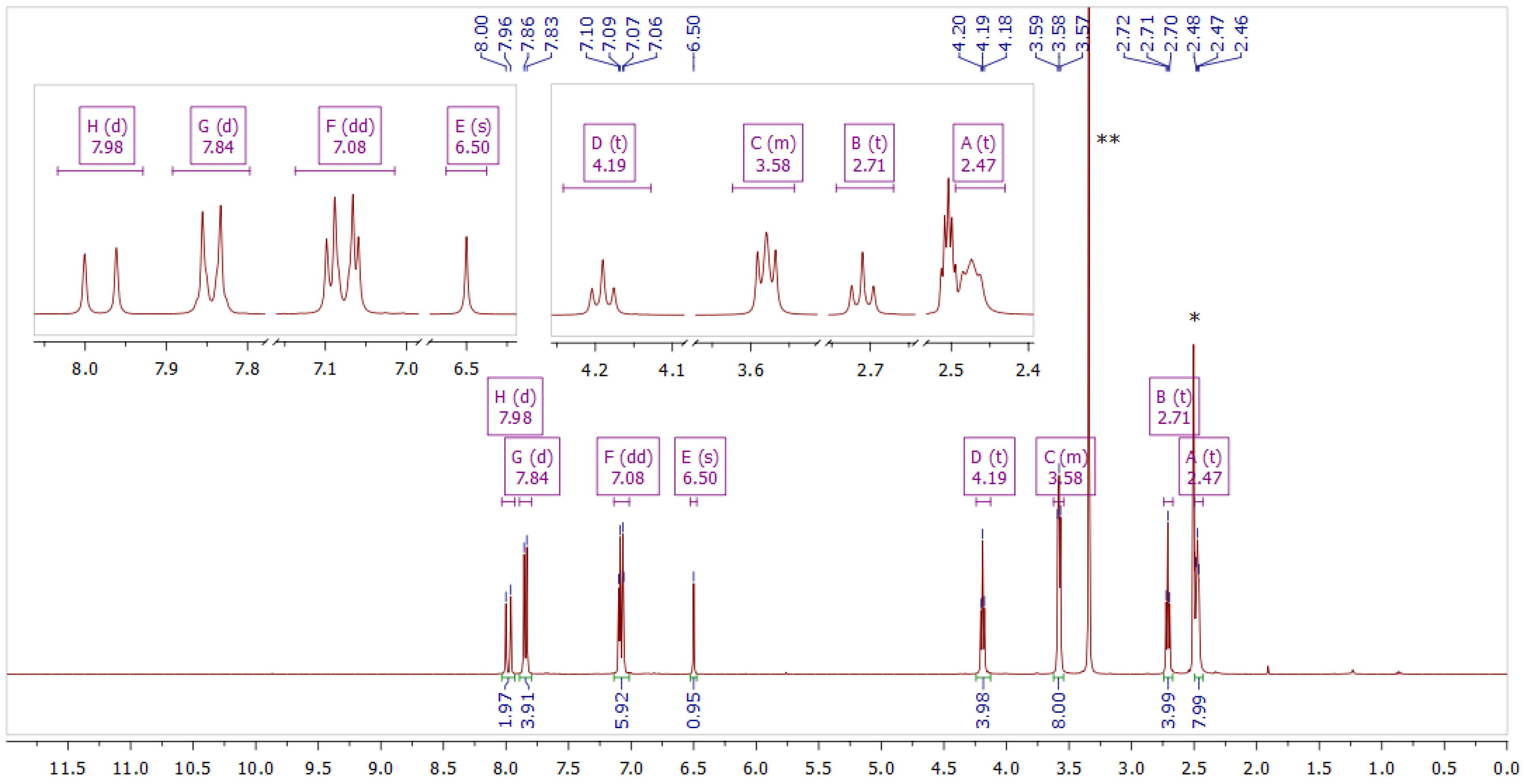
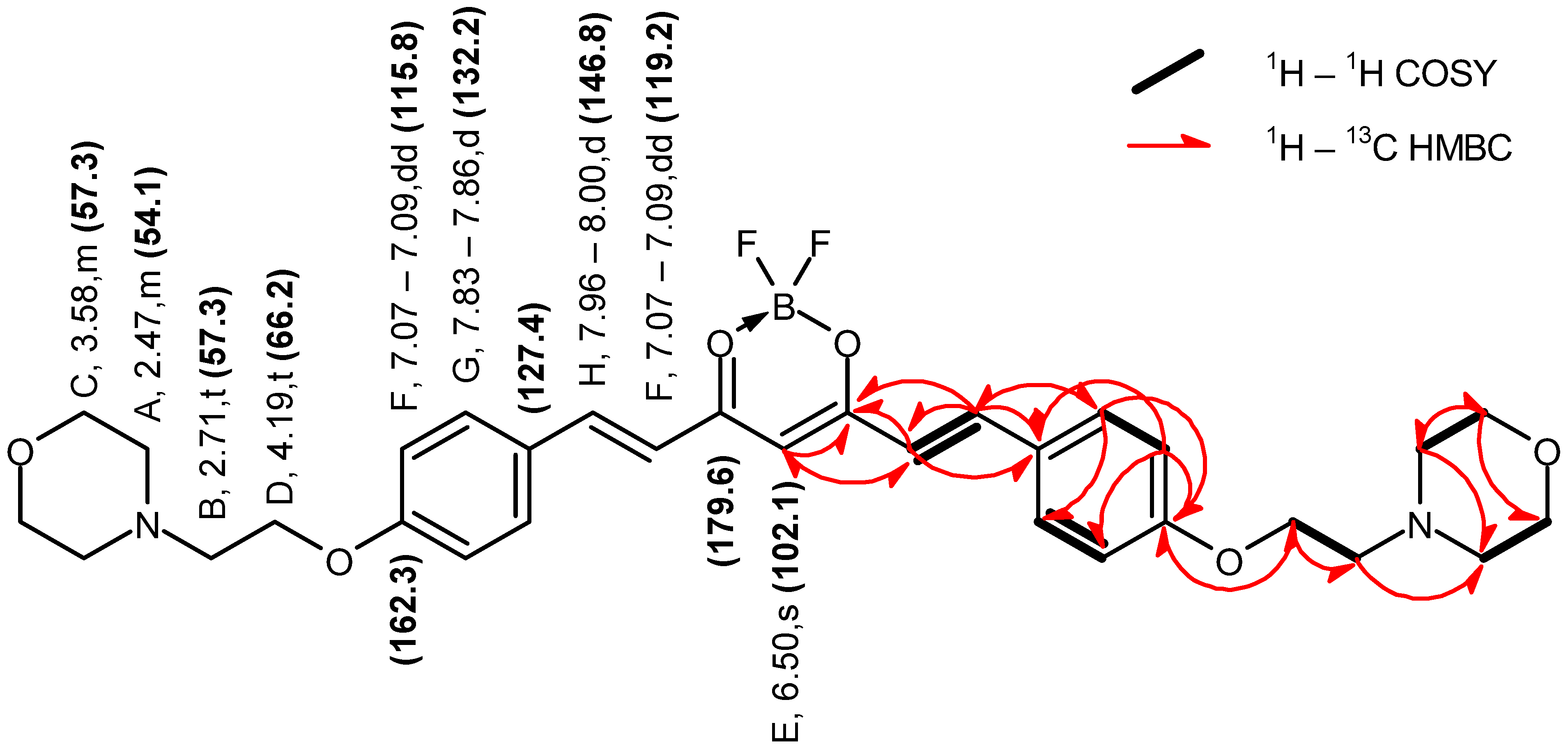
| Symbol | δH (ppm) | Multiplicity (JH-H in Hz) | 1H–13C HSQC δC (ppm) | 1H–13C HMBC δC (ppm) |
|---|---|---|---|---|
| A | 2.47 | m | 54.1 | 54.1, 66.6 |
| B | 2.71 | t (5.7) | 57.3 | 54.1, 66.2 |
| C | 3.58 | m | 66.6 | 54.1, 66.6 |
| D | 4.19 | t (5.7) | 66.2 | 162.3, 57.3 |
| E | 6.50 | s | 102.1 | 119.2, 179.6 |
| F | 7.09–7.07 | dd (12.3, 3.4) | 115.8, 119.2 | 102.1, 115.8, 127.4, 162.3, 179.6 |
| G | 7.86–7.83 | d (8.9) | 132.2 | 132.2, 146.8, 162.3 |
| H | 8.00–7.96 | d (15.6) | 146.8 | 119.2, 127.4, 132.2, 179.6 |
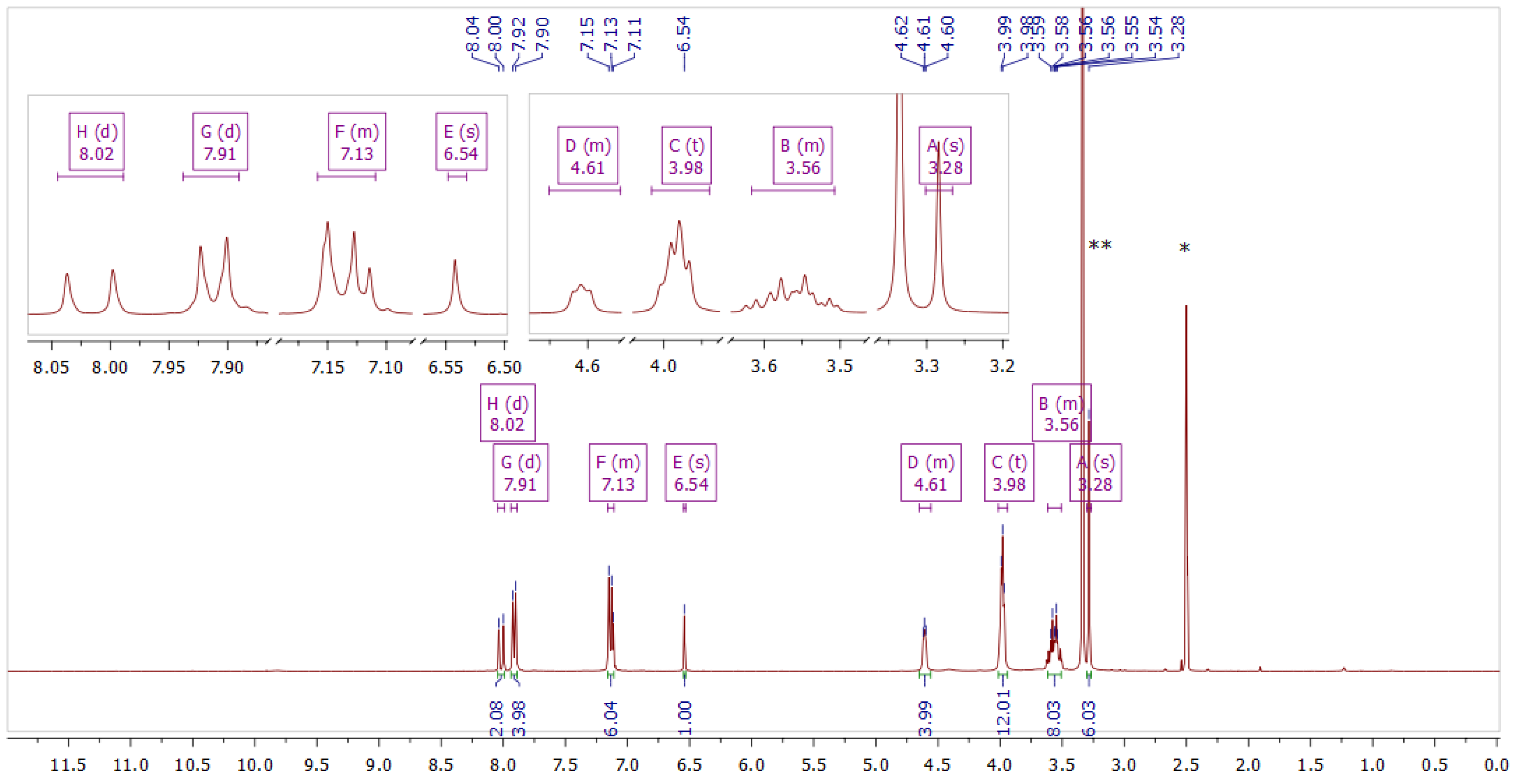
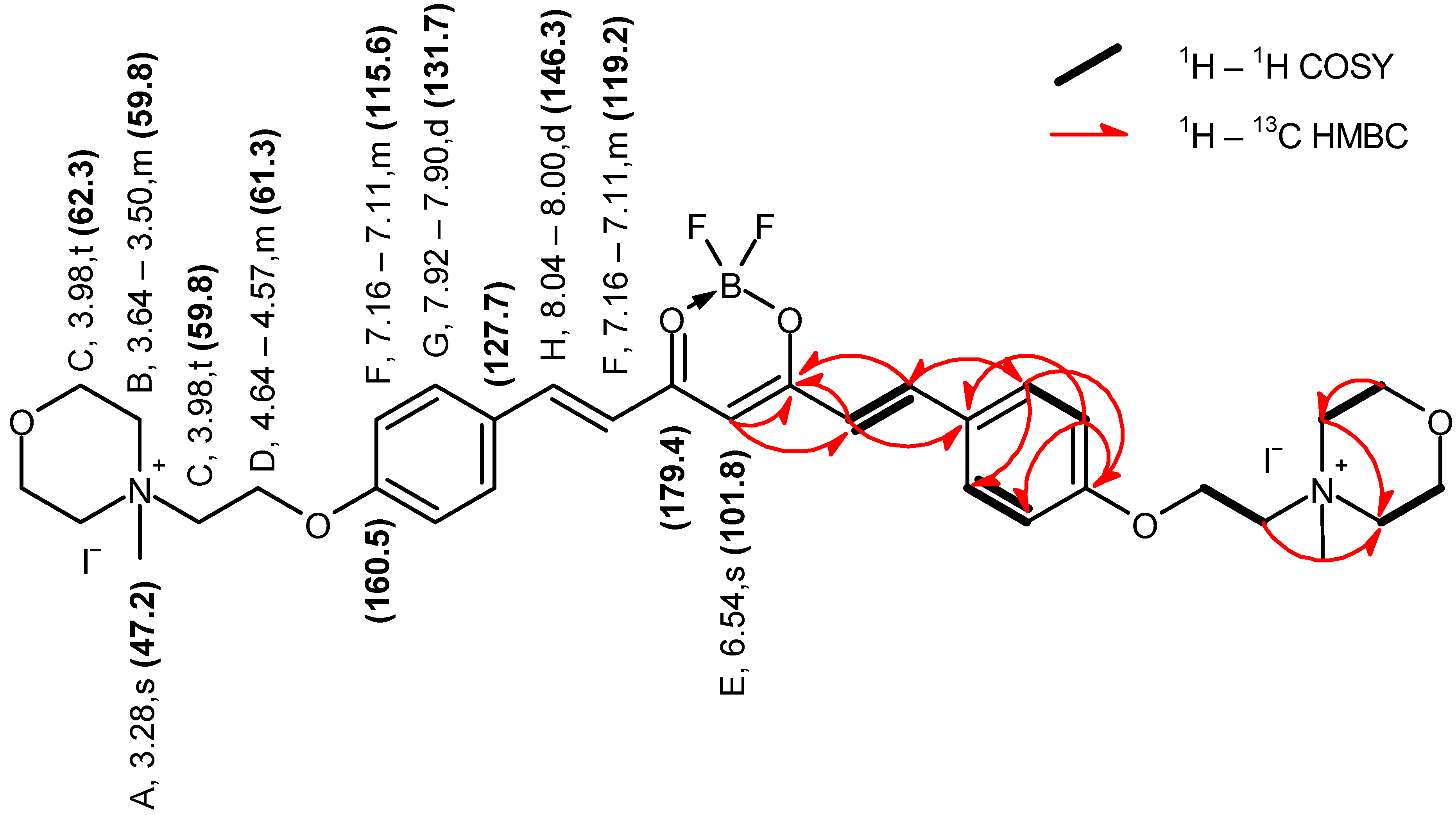
| Symbol | δH (ppm) | Multiplicity (JH-H in Hz) | 1H–13C HSQC δC (ppm) | 1H–13C HMBC δC (ppm) |
|---|---|---|---|---|
| A | 3.28 | s | 47.2 | 59.8, 62.3 |
| B | 3.64–3.50 | m | 59.8 | 59.8 |
| C | 3.98 | t (4.7) | 59.8, 62.3 | 59.8 |
| D | 4.64–4.57 | m | 61.3 | |
| E | 6.54 | s | 101.8 | 119.2, 179.6 |
| F | 7.16–7.11 | m | 115.6, 119.2 | 115.6, 127.7, 160.5, 179.4 |
| G | 7.92–7.90 | d (8.9) | 131.7 | 131.7, 146.3, 160.5 |
| H | 8.04–8.00 | d (15.6) | 146.3 | 131.7, 179.4 |
| 10−5 Φp | ||
|---|---|---|
| Compound | DMF | DMSO |
| 2 | 2.90 | 5.41 |
| 3 | 20.10 | 6.13 |
| Curcumin | 9.99 | 8.31 |
| ZnPc | 1.02 [38] | 0.35 [39] |
| Compound | Solvent | DPBF | TTF |
|---|---|---|---|
| 2 | DMF | <0.01 | <0.01 |
| DMSO | 0.024 | <0.01 | |
| 3 | DMF | 0.026 | 0.084 |
| DMSO | 0.046 | 0.057 | |
| Curcumin | DMF | - | 0.098 |
| DMSO | - | 0.116 | |
| ZnPc | DMF | 0.56 [39] | |
| DMSO | 0.67 [39] | ||
| Compound 2 | Compound 3 | |||||
|---|---|---|---|---|---|---|
| S.a. | E.c. | C.a. | S.a. | E.c. | C.a. | |
| L+P− | 0.16 | −0.03 | −0.06 | 0.16 | −0.03 | −0.06 |
| L−P+ | 0.05 | 0.52 | 0.04 | −0.01 | 0.58 | −0.09 |
| L+P+ | 2.13 | 0.19 | 0.01 | >5.54 | >4.46 | −0.04 |
| Compound 3, 10 µM—Log Red. | Compound 3, 1 µM—Log Red. | |||||||
|---|---|---|---|---|---|---|---|---|
| Irradiation time | S.a. | S.p. | E.c. | P.a. | S.a. | S.p. | E.c. | P.a. |
| 5 min | 3.37 | >4.93 | 1.15 | 4.29 | −0.01 | 3.03 | 0.83 | 0.86 |
| 10 min | 4.09 | 0.77 | 4.51 | |||||
| 15 min | >5.40 | 3.08 | 4.75 | 1.01 | >4.93 | 1.57 | 2.32 | |
| 20 min | 1.42 | |||||||
| 25 min | 4.53 | >4.85 | 1.72 | 1.82 | 2.83 | |||
| 30 min | 3.32 | |||||||
| Bacterial Strain | Time Point [min] | Log Reduction | Compound Concentration [µM] | Fluence [J/cm2] |
|---|---|---|---|---|
| S.a. G+ | 5 | 3.37 | 10 | 5 |
| S.p. G+ | 5 | 4.93 | 10 | 5 |
| 5 | 3.03 | 1 | 5 | |
| E.c. G− | 15 | 3.08 | 10 | 15 |
| P.a. G− | 5 | 4.29 | 10 | 5 |
| 30 | 3.32 | 1 | 30 |
| Log N ± SD | ||
|---|---|---|
| S. aureus | E. coli | |
| L−D+ | 6.88 ± 0.04 | 5.98 ± 0.41 |
| L−D− | 6.88 ± 0.02 | 5.69 ± 0.03 |
| Cell Line/Compound | Curcumin IC50 24 h IC50 48 h | Compound 2 IC50 24 h IC50 48 h | Compound 3 IC50 24 h IC50 48 h |
|---|---|---|---|
| PHDF—n | >50 µM 24.6 ± 11.5 µM | >50 µM 28.4 ± 11.8 µM | >50 µM >50 µM |
| MRC-5—n | 39.5 ± 8.8 µM 18.1 ± 4.4 µM | >50 µM 3.8 ± 2.3 µM | >50 µM >50 µM |
| WM266-4—c | 44.9 ± 10.7 µM 20.6 ± 9.1 µM | >50 µM >50 µM | >50 µM >50 µM |
| WM115—c | 39.5 ± 9.4 µM 18.1 ± 11.0 µM | >50 µM >50 µM | >50 µM >50 µM |
| Photosensitizer | Irradiation Parameters | Strains | Efficiency | Ref. |
|---|---|---|---|---|
| Phtalocyanine derivative 100 µM | 736 nm; 1.8 J/cm2; 10 min | P. aeruginosa | 4.6–6.4 log | [13] |
| Sulfanyl tribenzoporphyrazines | 660 nm; 3.6 J/cm2; 3 mW/cm2; 20 min | [11,54] | ||
| 1 µM | S. aureus | 4.8 log | ||
| 100 nM | S. aureus | 3.2 log | ||
| BODIPY derivatives | 350–800 nm; 70 mW/cm2 at 500 nm | [55] | ||
| 1 μM | 5 min | S. aureus | >5 log | |
| 5 μM | 15 min | E. coli | ~2.5 log up to 3.5 log with 50 mM KI | |
| 5 μM | 30 min | C. albicans | up to 5 log with 50 mM KI | |
| Pyridinium and imidazolium porphyrins | White light; 348 J/cm2; 20 h | [56] | ||
| 1.5 µM | S. aureus | 3 log (99.9%) | ||
| 2.5 µM | E. coli | 3 log (99.9%) | ||
| 20 µM | P. aeruginosa | 3 log (99.9%) | ||
| Methylene blue | 625 nm; 7 mW/cm2; 18 J/cm2 | [57] | ||
| 0.62 µg/mL | S. aureus | 6 log | ||
| 10–20 µg/mL | P. aeruginosa | 6 log | ||
| Toluidine blue | 670 nm; 97.65 J/cm2; 5 min | [58] | ||
| 50 µg/mL | S. aureus | 2.66 log | ||
| P. aeruginosa | 2.36 log | |||
| Rose bengal | 515 nm; 5.8 mW/cm2 | [59] | ||
| 0.62 µg/mL | 18 J/cm2 | S. aureus | 6 log | |
| 0.31 µg/mL | 37 J/cm3 | S. aureus | 6 log | |
| Curcumin 10 µM | 440 nm; 1.944 J/cm2; 3.6 mW/cm2; 8.8 min | S. aureus | 5.3 log | [60] |
| Zinc phthalocyanine RLP068/Cl | 600–700 nm; 30 J/cm2; 50 mW/cm2; 10 min | [61] | ||
| 64 ng/mL | S. aureus | 5–6 log | ||
| 26 µg/mL | P. aeruginosa | 5–6 log | ||
| Compound 3 | 470 nm | This study | ||
| 10 µM = 8.66 µg/mL | 15 min; 15 J/cm2 | S. aureus | >5.4 log | |
| 5 min; 5 J/cm2 | S. pyogenes | >4.93 log | ||
| 25 min; 25 J/cm2 | E. coli | 4.53 log | ||
| 25 min; 25 J/cm2 | P. aeruginosa | >4.85 log |
Disclaimer/Publisher’s Note: The statements, opinions and data contained in all publications are solely those of the individual author(s) and contributor(s) and not of MDPI and/or the editor(s). MDPI and/or the editor(s) disclaim responsibility for any injury to people or property resulting from any ideas, methods, instructions or products referred to in the content. |
© 2024 by the authors. Licensee MDPI, Basel, Switzerland. This article is an open access article distributed under the terms and conditions of the Creative Commons Attribution (CC BY) license (https://creativecommons.org/licenses/by/4.0/).
Share and Cite
Bakun, P.; Wysocki, M.; Stachowiak, M.; Musielak, M.; Dlugaszewska, J.; Mlynarczyk, D.T.; Sobotta, L.; Suchorska, W.M.; Goslinski, T. Quaternized Curcumin Derivative—Synthesis, Physicochemical Characteristics, and Photocytotoxicity, Including Antibacterial Activity after Irradiation with Blue Light. Molecules 2024, 29, 4536. https://doi.org/10.3390/molecules29194536
Bakun P, Wysocki M, Stachowiak M, Musielak M, Dlugaszewska J, Mlynarczyk DT, Sobotta L, Suchorska WM, Goslinski T. Quaternized Curcumin Derivative—Synthesis, Physicochemical Characteristics, and Photocytotoxicity, Including Antibacterial Activity after Irradiation with Blue Light. Molecules. 2024; 29(19):4536. https://doi.org/10.3390/molecules29194536
Chicago/Turabian StyleBakun, Pawel, Marcin Wysocki, Magdalena Stachowiak, Marika Musielak, Jolanta Dlugaszewska, Dariusz T. Mlynarczyk, Lukasz Sobotta, Wiktoria M. Suchorska, and Tomasz Goslinski. 2024. "Quaternized Curcumin Derivative—Synthesis, Physicochemical Characteristics, and Photocytotoxicity, Including Antibacterial Activity after Irradiation with Blue Light" Molecules 29, no. 19: 4536. https://doi.org/10.3390/molecules29194536
APA StyleBakun, P., Wysocki, M., Stachowiak, M., Musielak, M., Dlugaszewska, J., Mlynarczyk, D. T., Sobotta, L., Suchorska, W. M., & Goslinski, T. (2024). Quaternized Curcumin Derivative—Synthesis, Physicochemical Characteristics, and Photocytotoxicity, Including Antibacterial Activity after Irradiation with Blue Light. Molecules, 29(19), 4536. https://doi.org/10.3390/molecules29194536