
Overview of the Design and Application of Dual-Signal Immunoassays


Abstract
:
1. Introduction
2. Optical Dual-Signal Immunoassays
2.1. Colorimetric–Fluorescence Dual-Signal Immunoassays
2.1.1. Homogeneous Methods

| Signal Label | Target | Linear Range | Detection Limit | Ref. |
|---|---|---|---|---|
| HRP/QDs@PSS | RABV | 0.12–23.4 and 0.012–46.8 ng/mL | 91 and 8 pg/mL | [46] |
| HRP | OTA | 0.049–1.563 and 0.04–25 ng/mL | 4.316 and 1.515 pg/mL | [48] |
| Fe-MOFs | PSA | 1–20 and 0–15 ng/mL | 180 pg/mL | [49] |
| CeO2@Au | T-2 toxin | 0.1–1 and 0.005–0.7 ng/mL | 8.52 and 20.11 pg/mL | [50] |
| MPBNs | S. aureus | 0.01–100,000 CFU/μL | 0.1 CFU/μL | [51] |
| B-CDs@SiO2@MnO2 | DEP | 0.05–100 ng/mL | 3.4 and 2.7 pg/mL | [52] |
| CsPbBr3 NCs | PSA | 0.1–15 and 0.01–80 ng/mL | 290 and 81 pg/mL | [54] |
| CPNs@ZIF-8 | BPA | 0–5 ng/mL | 18 and 8 pg/mL | [55] |
| Co/NCNT | OTA | 0.001–10 ng/mL | 0.21 and 0.17 pg/mL | [56] |
| ALP | OTA | 10–45 and 2.89–34.72 ng/mL | 0.81 and 0.62 ng/mL | [57] |
| ALP | OTA | 14–300 and 4.69–37.5 ng/mL | 962 and 404 pg/mL | [58] |
| porous Pd NPs | NMP22 | 0.001–0.5 ng/mL | 0.35 and 0.31 pg/mL | [62] |
| GOx | danofloxacin | 1–10 and 1–25 ng/mL | 648 and 337 pg/mL | [63] |
| nanoceria | cTnI | 0.001–10 ng/mL | 227 and 413 fg/mL | [65] |
| HRP | AFB1 | 0.75–1.8 and 0.75–1.8 ng/mL | 62 and 13 pg/mL | [66] |
| HRP | A. acidoterrestris | 4.8 × 10²–4.8 × 10⁷ CFU/mL | 4.8 × 10² CFU/mL | [68] |
| HRP | ZEN | 0.048–3.125 and 0.012–3.125 ng/mL | 11 pg/mL and 19 pg/mL | [69] |
| ALP | ZEN | 7.5–20 and 7.5–17.5 ng/mL | 7.22 and 36 pg/mL | [73] |
| ALP | OTA | 0.39–25 and 1.56–50 ng/mL | 70 and 570 ng/mL | [74] |
| ALP | AFB1 | 0.19–0.32 and 0.09–0.41 ng/mL | 0.16 and 0.19 ng/mL | [76] |
| ALP | cTnI | 0.2–80 and 0.05–4 ng/mL | 60 and 15 ng/mL | [77] |
| ALP | PSA | 0.02–28 and 0.02–20 ng/mL | 4.1 and 9.6 pg/mL | [78] |
| ALP | amantadine | 0.03–200 and 0.03–200 ng/g | 47 and 60 ng/g | [79] |
| Au@CD | ferritin | 1–160 and 1–120 ng/mL | 20 and 64 ng/mL | [81] |
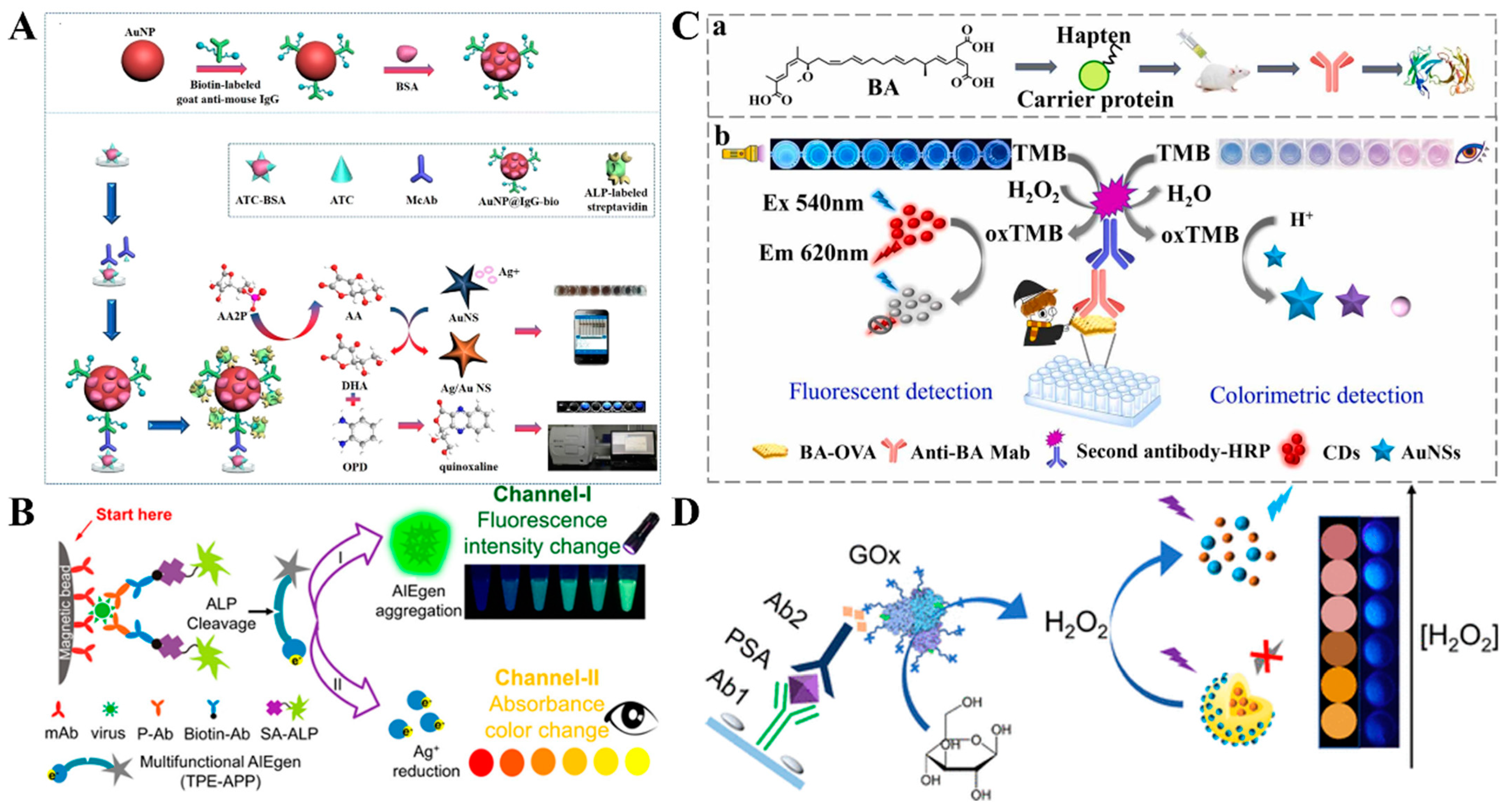
| ALP-AuNPs | ATC | 0.63–84.59 ng/mL | 1.2 and 0.44 ng/mL | [86] |
| ALP | EV71 virions | 1340–134,000 and 1.67–2505 copies/μL | 868.4 and 1.4 copies/μL | [87] |
| Cu2O@Fe(OH)3 | OTA | 0.001–10 ng/mL | 0.83 and 0.56 pg/mL | [88] |
| HRP | BA | 0–100 ng/mL | 8.4 and 5.7 ng/mL | [90] |
| PBNCs@PEI | RSG | 0.5–1000 pg/mL | 95 and 63 fg/mL | [91] |
| GOx | PSA | 0.005–20 ng/mL | 2.3 and 0.84 ng/mL | [92] |
| QDs/ZnS NSs | AFP | 0.05–12 ng/mL | 7 and 10 pg/mL | [93] |
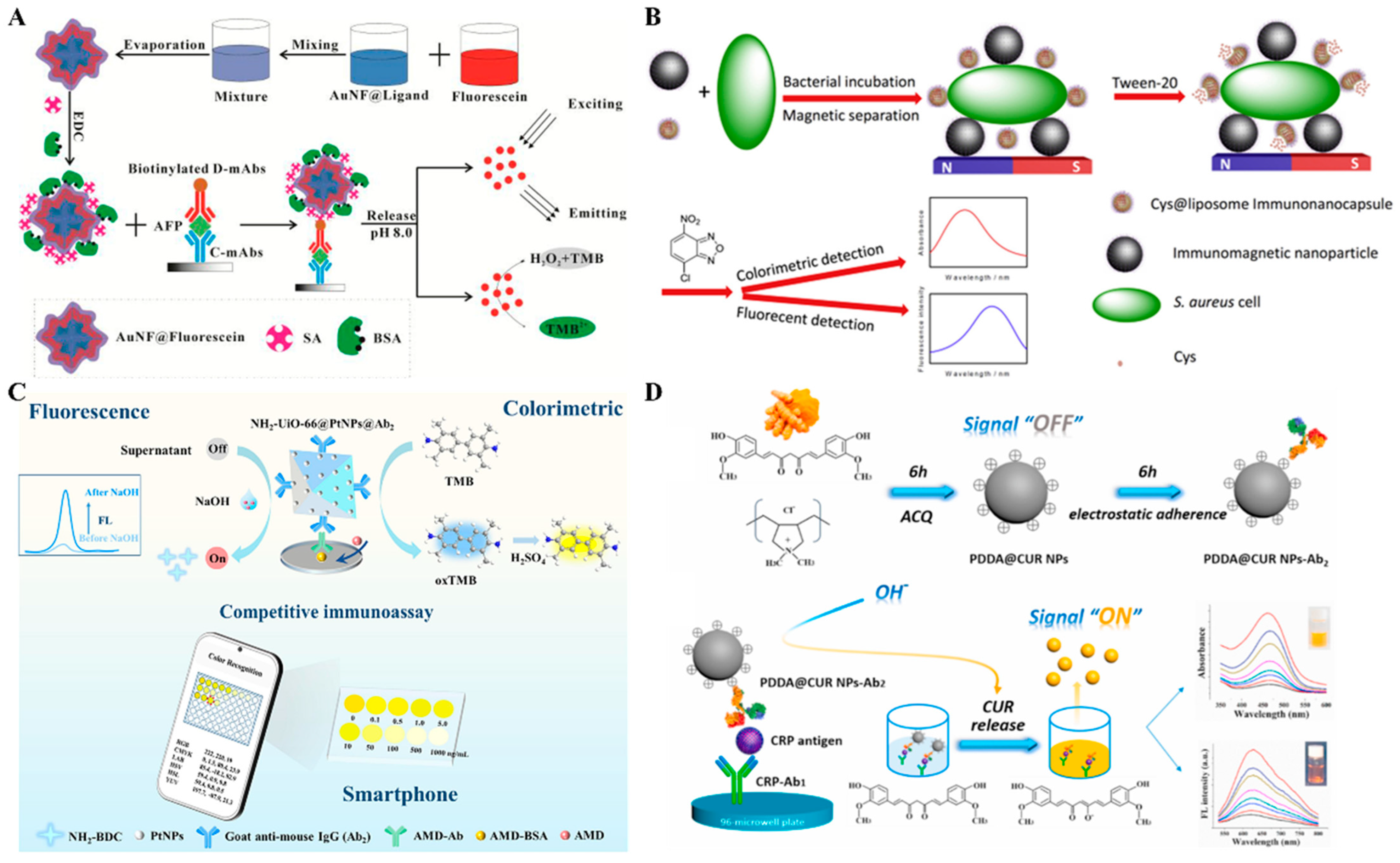
| AuNF@Fluorescein | AFP | 5–5000 and 0.01–10 pg/mL | 17.7 and 0.029 fg/mL | [94] |
| Cys@liposome | S. aureus | 40–4000 and 4–4000 CFU/mL | 10 and 1 CFU/mL | [97] |
| NH2-UiO-66@PtNPs | amantadine | 0.1–1000 ng/mL | 69 and 2.2 pg/mL | [99] |
| PDDA@CUR NPs | CUR | 0.1–2 ng/mL | 43 and 38 fg/mL | [101] |
2.1.2. LFIA Methods
| Signal Label | Target | Linear Range | Detection Limit | Ref. |
|---|---|---|---|---|
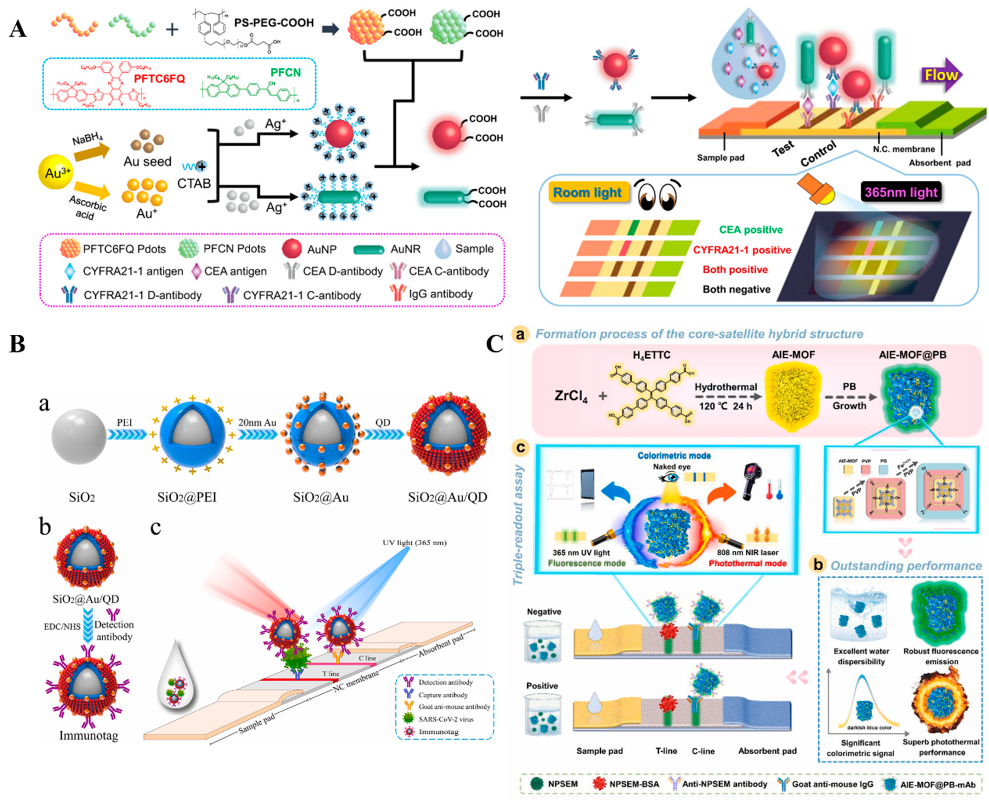
| PFTC6FQ@AuNP | CYFRA21-1 | 0–10 ng/mL | 70 pg/mL | [114] |
| Au-Ag NCs@PEI-AuNPs | dicofol | 8.45–94.79 and 1.36–19.92 ng/mL | 4.16 and 0.62 ng/mL | [115] |
| SiO2@Au/QD | S1 protein | 0.05–1000 ng/mL | 1 and 0.033 ng/mL | [119] |
| MoS2@QDs | clothianidin | Not reported | 120 and 2.58 pg/mL | [121] |
| AIENBs | CRP | 8–100 and 0.2–100 mg/L | 8 and 0.16 mg/L | [124] |
| AuNPs/C2-15-EmGFP | imidaclothiz | 3.21–35.8 and 2.62–34.6 ng/mL | 64 and 8 ng/mL | [126] |
| Au@PDA | carbendazim | 7.21–945.55 and 3.56–246.67 ng/mL | 3.21 and 1.36 ng/mL | [127] |
| APNPs | AC, CLE | 0–4.0 ng/mL (AC), 0–10 ng/mL (CLE) | 13 pg/mL (AC), 152 pg/mL (CLE) | [128] |
| ZnCdSe/ZnS@AuNPs | SBT | 3.9–62.5 ng/mL | 3.9 ng/mL | [129] |
| PD-AuNPs | E. coli O157:H7 | 97.7–12,500 CFU/mL | 330 and 90.6 CFU/ mL | [131] |
| Ti3C2@Au | DXMS | 0.05–0.8 μg/kg | 1.8 and 1.3 ng/kg | [132] |
| CsPbBr3/OPA/AuNPs | CEA | 0–1 ng/mL | 027 and 23 pg/mL | [134] |
| APDA | Gen | 0–1 ng/mL | 1 and 0.5 ng/mL | [135] |
| PDQB | SARS-CoV-2 | 0.005–1 ng/mL | 100 and 5 pg/mL | [136] |
| Si-Au/DQD | A29L | 0.005–100 ng/mL | 500 and 2.1 pg/mL | [137] |
| SAQDsRu | zearalenone | 0.01–3 ng/mL | 8 and 5.8 pg/mL | [138] |
| TRFNPs@HRP | ATC | 0.2–100 ng/mL | 80 pg/mL | [139] |
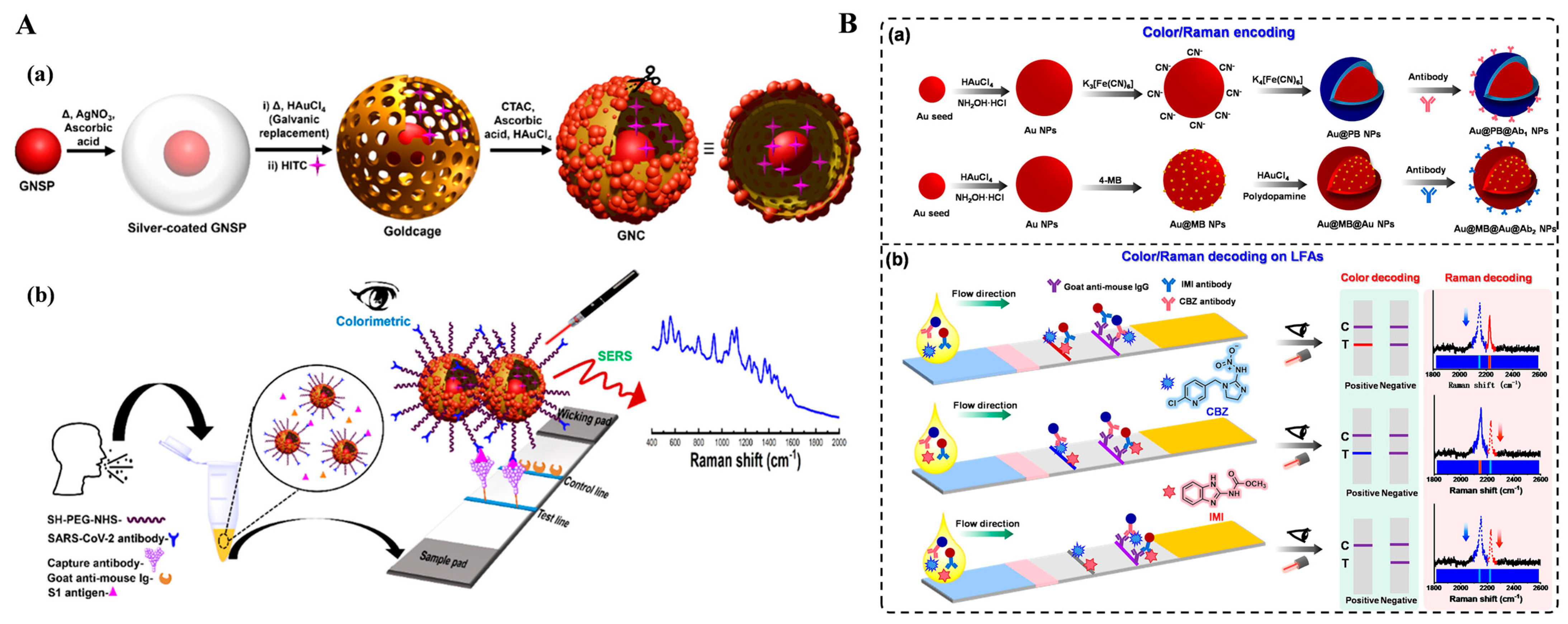
2.2. Colorimetric–SERS Dual-Signal Immunoassays
2.3. Fluorescence–SERS Dual-Signal Immunoassays
| Detection Mode | Signal Label | Target | Linear Range | Detection Limit | Ref. |
|---|---|---|---|---|---|
| Color–SERS | AuNS@Ag | S. aureus | 1–107 and 1–105 CFU/mL | 1 CFU/mL | [145] |
| HITC@GNC | S1 protein | 100–1000 and 0.0001–10 ng/mL | 91.24 and 0.05721 pg/mL | [148] | |
| Au/Au nanostar | clenbuterol | 0–1 ng/mL | 5 and 0.05 ng/mL | [149] | |
| Au@Ag NPs | cTnI | 5–50 and 0.9–50 ng/mL | 4.5 and 0.09 ng/mL | [150] | |
| Au@PB NPs | CBZ | 2–10 and 0.05–0.5 ng/mL | 1.27 and 0.04 ng/mL | [151] | |
| AuNR@Pt | C. jejuni | 102–106 and 102–5 × 10⁶ CFU/mL | 75 and 50 CFU/mL | [152] | |
| UAA@P NPs | S. aureus | 100–107 and 10–107 CFU/mL | 18 and 3 CFU/mL | [153] | |
| Au@PBNP | α-LA | 1–600 and 0.2–600 ng/mL | 283 and 11 pg/mL | [155] | |
| OMQ NPs | IgG | 1–1012 fM | 1 fM | [158] | |
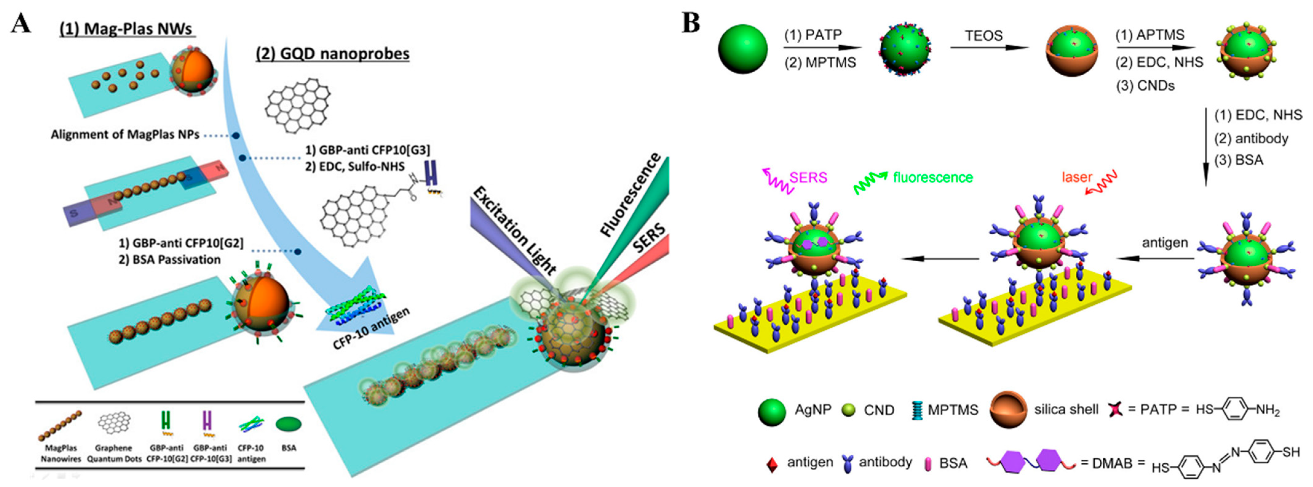
| FL–SERS | GQD | TB | 0.001–1000 ng/mL | 51.1 fg/mL | [160] |
| CNDs/Ag@SiO2 | IgG | 0.01–100 μg/mL | 100 and 2.5 ng/mL | [165] |
3. Electrochemical Dual-Signal Immunoassays
3.1. Electrochemical–PEC Dual-Signal Immunoassays
3.2. Electrochemical–ECL Dual-Signal Immunoassays
4. Optical and Electrochemical Dual-Signal Immunoassays
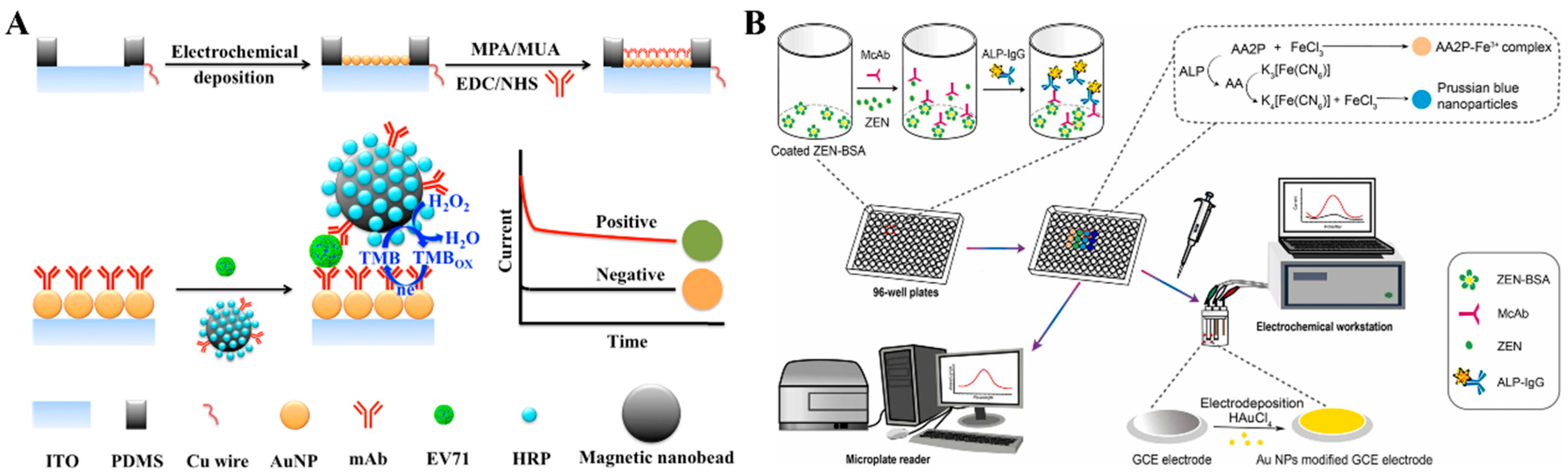
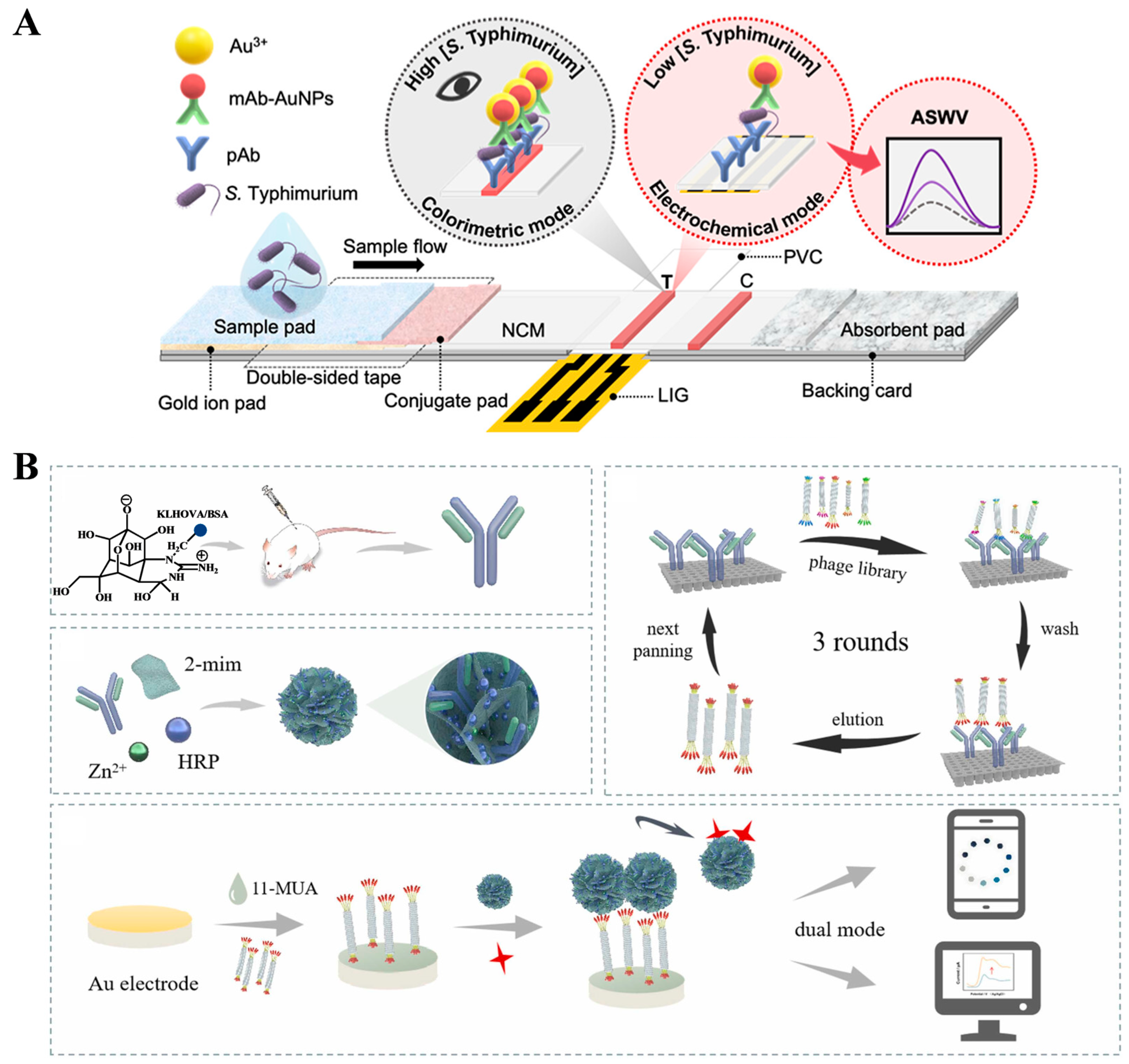
4.1. Colorimetric–Electrochemical Dual-Signal Immunoassays
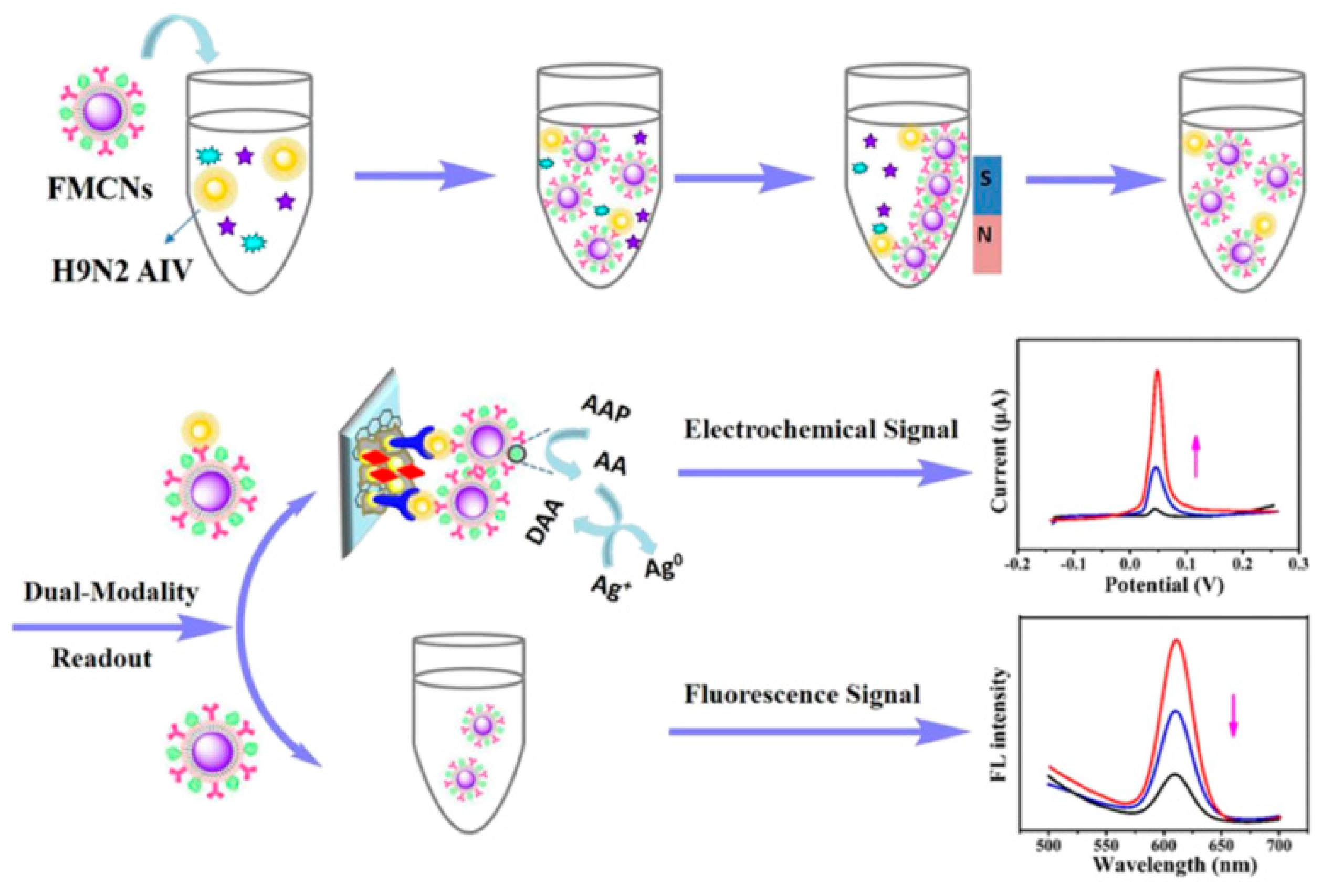
4.2. Fluorescence–Electrochemical Dual-Signal Immunoassays
4.3. Colorimetric–PEC Dual-Signal Immunoassays
4.4. Fluorescence–PEC Dual-Signal Immunoassays
| Detection Mode | Signal Label | Target | Linear Range | Detection Limit | Ref. |
|---|---|---|---|---|---|
| Color–EC | PBNPs | TRX1 | 10–50 ng/mL | 9 and 6.5 ng/mL | [179] |
| PBNPs | IgG | 0.5–10 µg/mL | 34 ng/mL | [180] | |
| ALP | CA125 | 5–1000 and 50–1000 U/mL | 1.3 and 40 U/mL | [182] | |
| HRP-Ppy NPs | PSA | 0.001–40 ng/mL | 0.8 and 0.7 pg/mL | [183] | |
| PP1-MPs | MC-LR | Not reported | 7.4 and 0.4 μg/L | [184] | |
| uPtNZs | GA | 0.01–5 and 0.005–10 mg/mL | 9.2 and 3.8 µg/mL | [185] | |
| H-AuNPs | BNP | 0.001–0.2 and 5–25 ng/mL | 0.03 and 80.3 pg/mL | [186] | |
| HRP-MPs | EV71 | 0.1–600 ng/mL | 1 and 0.01 ng/mL | [187] | |
| ALP | ZEN | 0.2–0.8 and 0.125–0.5 ng/mL | 40 and 80 pg/mL | [188] | |
| Au-AuNP | S. typhimurium | 0.1–108 CFU/mL | 1 CFU/10 mL | [189] | |
| HRP/ZIF-8 | TTX1 | 10.5–380 and 0.1–1000 ng/mL | 183 and 10 pg/mL | [190] | |
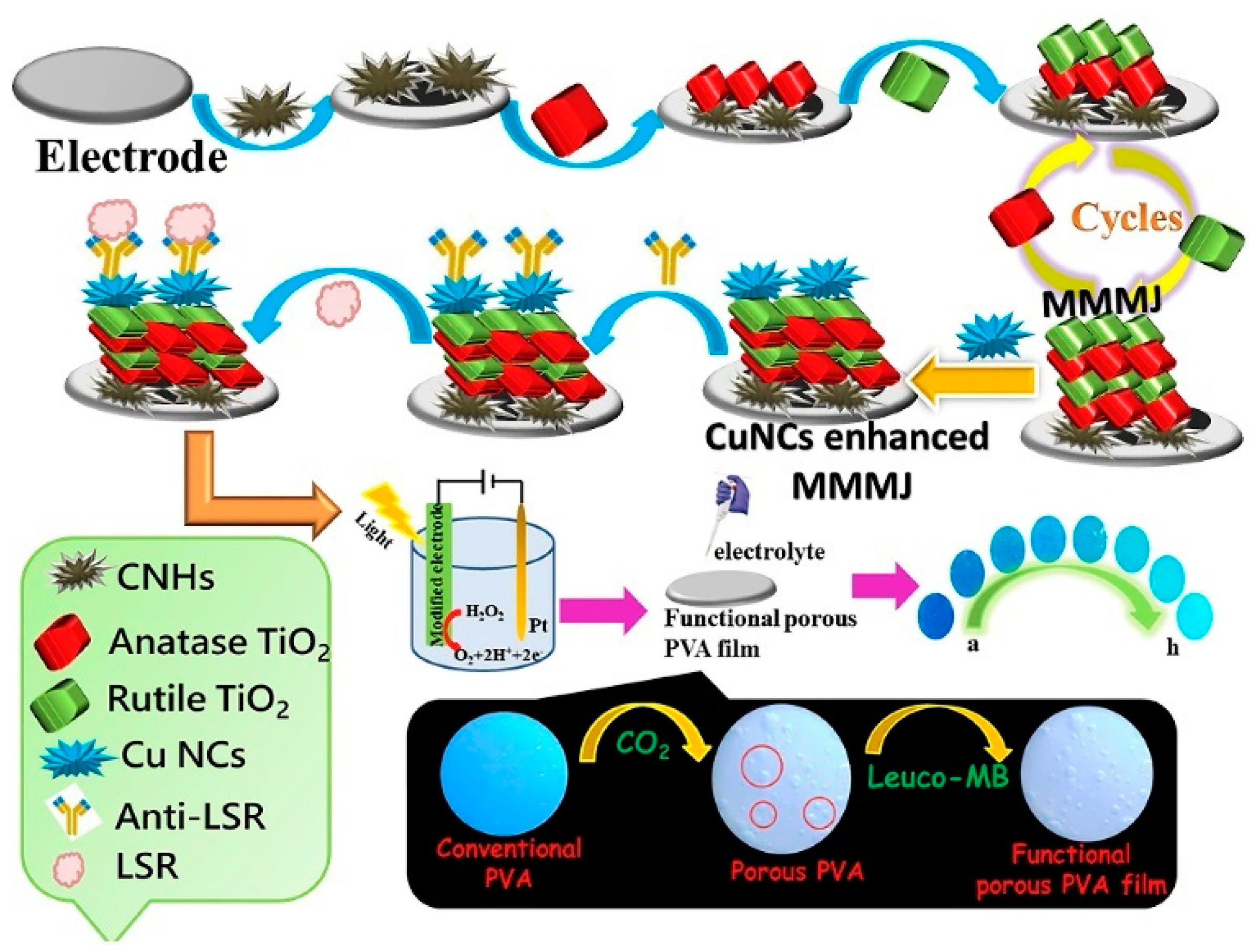
| Color–PEC | CuNCs | LSR | 0.001–10,000 pg/mL | 1 fg/mL | [197] |
| HRP@NL | OTA | 0.001–5 ng/mL | 1.7 and 0.7 pg/mL | [202] | |
| ALP-AuNP | PSA | 0.001–10 ng/mL | 0.41 and 0.16 pg/mL | [203] | |
| ALP | AFP | 0.05–100 ng/mL | 10 pg/mL | [205] |
5. Conclusions
Author Contributions
Funding
Institutional Review Board Statement
Informed Consent Statement
Data Availability Statement
Conflicts of Interest
References
- Pei, X.; Zhang, B.; Tang, J.; Liu, B.; Lai, W.; Tang, D. Sandwich-type immunosensors and immunoassays exploiting nanostructure labels: A review. Anal. Chim. Acta 2013, 758, 1–18. [Google Scholar] [CrossRef] [PubMed]
- Shao, Y.; Zhou, H.; Wu, Q.; Xiong, Y.; Wang, J.; Ding, Y. Recent advances in enzyme-enhanced immunosensors. Biotechnol. Adv. 2021, 53, 107867–107883. [Google Scholar] [CrossRef] [PubMed]
- Rizzo, F. Optical immunoassays methods in protein analysis: An overview. Chemosensors 2022, 10, 326. [Google Scholar] [CrossRef]
- Wang, Z.; Guo, Y.; Xianyu, Y. Applications of self-assembly strategies in immunoassays: A review. Coordin. Chem. Rev. 2023, 478, 214974–215000. [Google Scholar] [CrossRef]
- Gan, S.D.; Patel, K.R. Enzyme immunoassay and enzyme-linked immunosorbent assay. J. Investig. Dermatol. 2013, 133, e12–e14. [Google Scholar] [CrossRef]
- Zhao, Q.; Lu, D.; Zhang, G.; Zhang, D.; Shi, X. Recent improvements in enzyme-linked immunosorbent assays based on nanomaterials. Talanta 2021, 223, 121722–121737. [Google Scholar] [CrossRef]
- Xiong, Y.; Leng, Y.; Li, X.; Huang, X.; Xiong, Y. Emerging strategies to enhance the sensitivity of competitive ELISA for detection of chemical contaminants in food samples. TrAC-Trend. Anal. Chem. 2020, 126, 115861–115879. [Google Scholar] [CrossRef]
- Jiang, W.; Beloglazova, N.V.; Luo, P.; Guo, P.; Lin, G.; Wang, X. A dual-color quantum dots encoded frit-based immunoassay for visual detection of aflatoxin M(1) and pirlimycin residues in milk. J. Agric. Food. Chem. 2017, 65, 1822–1828. [Google Scholar] [CrossRef]
- Chen, M.; Qileng, A.; Liang, H.; Lei, H.; Liu, W.; Liu, Y. Advances in immunoassay-based strategies for mycotoxin detection in food: From single-mode immunosensors to dual-mode immunosensors. Compr. Rev. Food Sci. Food Saf. 2023, 22, 1285–1311. [Google Scholar] [CrossRef]
- Zhang, S.; Chen, Y.; Huang, Y.; Dai, H.; Lin, Y. Design and application of proximity hybridization-based multiple stimuli-responsive immunosensing platform for ovarian cancer biomarker detection. Biosens. Bioelectron. 2020, 159, 112201–112208. [Google Scholar] [CrossRef]
- Huang, Y.; Sun, T.; Li, W.; Liu, L.; Liu, G.; Yi, X.; Wang, J. Colorimetric and electrochemical detection of ligase through ligation reaction-induced streptavidin assembly. Chin. Chem. Lett. 2022, 33, 3151–3155. [Google Scholar] [CrossRef]
- Lai, K.K.; Renneberg, R.; Mak, W.C. Multifunctional protein particles with dual analytical channels for colorimetric enzymatic bioassays and fluorescent immunoassays. Biosens. Bioelectron. 2012, 32, 169–176. [Google Scholar] [CrossRef] [PubMed]
- Qiu, Y.; Jiang, K.; Wu, J.; Peng, Y.-K.; Oh, J.-W.; Lee, J.-H. Ultrasensitive plasmonic photothermal immunomagnetic bioassay using real-time and end-point dual-readout. Sens. Actuators B Chem. 2023, 377, 133110–133117. [Google Scholar] [CrossRef]
- Han, Q.; Wang, H.; Wang, J. Multi-mode/signal biosensors: Electrochemical integrated sensing techniques. Adv. Funct. Mater. 2024; early view. [Google Scholar] [CrossRef]
- Juan-Colas, J.; Johnson, S.; Krauss, T.F. Dual-mode electro-optical techniques for biosensing applications: A review. Sensors 2017, 17, 2047. [Google Scholar] [CrossRef] [PubMed]
- Lu, G.; Lin, N.; Chen, Z.; Jiang, W.; Hu, J.J.; Xia, F.; Lou, X. Nanopores/nanochannels based on electrical and optical dual signal response for application in biological detection. Chin. J. Chem. 2023, 41, 1374–1384. [Google Scholar] [CrossRef]
- Wang, J.; Zhao, K.; Ye, C.; Song, Y. Emerging interactively stretchable electronics with optical and electrical dual-signal feedbacks based on structural color materials. Nano Res. 2023, 17, 1837–1855. [Google Scholar] [CrossRef]
- Zhao, L.; Wu, D.; Xiao, S.; Yin, Y.; Li, L.; Wang, J.; Wu, Y.; Qiu, Y.; Dong, Y. Dual-mode aptasensors with cross validation capacity for reliability enhancement and analytical assurance. TrAC-Trend. Anal. Chem. 2024, 177, 117755–117768. [Google Scholar] [CrossRef]
- Xu, J.; Zhang, B.; Zhang, Y.; Mai, L.; Hu, W.; Chen, C.-J.; Liu, J.-T.; Zhu, G. Recent advances in disease diagnosis based on electrochemical-optical dual-mode detection method. Talanta 2023, 253, 124037–124054. [Google Scholar] [CrossRef]
- Zhou, Y.; Zhang, W.; Wang, X.; Li, P.; Tang, B. Recent progress in small-molecule fluorescence and photoacoustic dual-modal probes for the in-vivo detection of bioactive molecules. Chem. Asian J. 2022, 17, e202200155–e202200168. [Google Scholar] [CrossRef]
- Sun, R.; Li, Y.; Du, T.; Qi, Y. Recent advances in integrated dual-mode optical sensors for food safety detection. Trends Food Sci. Technol. 2023, 135, 14–31. [Google Scholar] [CrossRef]
- Saleh, R.O.; Almajidi, Y.Q.; Mansouri, S.; Hammoud, A.; Rodrigues, P.; Mezan, S.O.; Maabreh, H.G.; Deorari, M.; Shakir, M.N.; Alasheqi, M.Q. Dual-mode colorimetric and fluorescence biosensors for the detection of foodborne bacteria. Clin. Chim. Acta 2024, 553, 117741–117753. [Google Scholar] [CrossRef] [PubMed]
- Hsu, C.Y.; Saleh, R.O.; Pallathadka, H.; Kumar, A.; Mansouri, S.; Bhupathi, P.; Jasim Ali, S.H.; Al-Mashhadani, Z.I.; Alzubaidi, L.H.; Hizam, M.M. Advances in electrochemical-optical dual-mode biosensors for detection of environmental pathogens. Anal. Methods 2024, 16, 1306–1322. [Google Scholar] [CrossRef] [PubMed]
- Zheng, W.; Jiang, X. Integration of nanomaterials for colorimetric immunoassays with improved performance: A functional perspective. Analyst 2016, 141, 1196–1208. [Google Scholar] [CrossRef]
- Yin, Y.; Cao, Y.; Xu, Y.; Li, G. Colorimetric immunoassay for detection of tumor markers. Int. J. Mol. Sci. 2010, 11, 5077–5094. [Google Scholar] [CrossRef]
- Shan, Z.; Lu, M.; Wang, L.; MacDonald, B.; MacInnis, J.; Mkandawire, M.; Zhang, X.; Oakes, K.D. Chloride accelerated Fenton chemistry for the ultrasensitive and selective colorimetric detection of copper. Chem. Commun. 2016, 52, 2087–2090. [Google Scholar] [CrossRef]
- Sun, L.; Chen, L.G.; Wang, H.B. Fenton-like reaction triggered chemical redox-cycling signal amplification for ultrasensitive fluorometric detection of H2O2 and glucose. Analyst 2024, 149, 546–552. [Google Scholar] [CrossRef]
- Fan, K.; Wang, H.; Xi, J.; Liu, Q.; Meng, X.; Duan, D.; Gao, L.; Yan, X. Optimization of Fe3O4 nanozyme activity via single amino acid modification mimicking an enzyme active site. Chem. Commun. 2016, 53, 424–427. [Google Scholar] [CrossRef]
- Asati, A.; Santra, S.; Kaittanis, C.; Nath, S.; Perez, J.M. Oxidase-like activity of polymer-coated cerium oxide nanoparticles. Angew. Chem. Int. Ed. 2009, 48, 2308–2312. [Google Scholar] [CrossRef]
- Yang, S.; Chen, R.; Jia, L. Cupric oxide nanosheet as an oxidase mimic for fluorescent detection of acetone by a 3D-printed portable device. Microchim. Acta 2024, 191, 122–131. [Google Scholar] [CrossRef]
- Wang, D.; Jana, D.; Zhao, Y. Metal-organic framework derived nanozymes in biomedicine. Acc. Chem. Res. 2020, 53, 1389–1400. [Google Scholar] [CrossRef] [PubMed]
- Kong, Y.; Li, Z.; Liu, Q.; Song, J.; Zhu, Y.; Lin, J.; Song, L.; Li, X. Artificial neural network-facilitated V2C MNs-based colorimetric/fluorescence dual-channel biosensor for highly sensitive detection of AFB1 in peanut. Talanta 2024, 266, 125056–125064. [Google Scholar] [CrossRef] [PubMed]
- Xue, T.; Jiang, S.; Qu, Y.; Su, Q.; Cheng, R.; Dubin, S.; Chiu, C.Y.; Kaner, R.; Huang, Y.; Duan, X. Graphene-supported hemin as a highly active biomimetic oxidation catalyst. Angew. Chem. Int. Ed. 2012, 51, 3822–3825. [Google Scholar] [CrossRef] [PubMed]
- Ma, W.; Xue, Y.; Guo, S.; Jiang, Y.; Wu, F.; Yu, P.; Mao, L. Graphdiyne oxide: A new carbon nanozyme. Chem. Commun. 2020, 56, 5115–5118. [Google Scholar] [CrossRef] [PubMed]
- Li, Z.; Zhang, W.; Zhang, Q.; Li, P.; Tang, X. Self-assembly multivalent fluorescence-nanobody coupled multifunctional nanomaterial with colorimetric fluorescence and photothermal to enhance immunochromatographic assay. ACS Nano 2023, 17, 19359–19371. [Google Scholar] [CrossRef]
- Xiang, X.; Chen, L.; Zhang, C.; Luo, M.; Ji, X.; He, Z. A fluorescence-based colorimetric droplet platform for biosensor application to the detection of alpha-fetoprotein. Analyst 2012, 137, 5586–5591. [Google Scholar] [CrossRef]
- He, M.; Shang, N.; Zheng, B.; Yue, G. An ultrasensitive colorimetric and fluorescence dual-readout assay for glutathione with a carbon dot-MnO2 nanosheet platform based on the inner filter effect. RSC Adv. 2021, 11, 21137–21144. [Google Scholar] [CrossRef]
- Shan, Y.; Wang, B.; Huang, H.; Yan, K.; Li, W.; Wang, S.; Liu, F. Portable high-throughput multimodal immunoassay platform for rapid on-site COVID-19 diagnostics. Anal. Chim. Acta 2023, 1238, 340634–340643. [Google Scholar] [CrossRef]
- Lu, D.; Jiang, H.; Zhang, T.; Pan, J.; Zhao, L.; Shi, X.; Zhao, Q. Dual modal improved enzyme-linked immunosorbent assay for aflatoxin B1 detection inspired by the interaction of amines with Prussian blue nanoparticles. Int. J. Biol. Macromol. 2024, 264, 130479–130487. [Google Scholar] [CrossRef]
- Gao, F.; Ye, S.; Huang, L.; Gu, Z. A nanoparticle-assisted signal-enhancement technique for lateral flow immunoassays. J. Mater. Chem. B 2024, 12, 6735–6756. [Google Scholar] [CrossRef]
- Zhang, J.; Zhou, R.; Tang, D.; Hou, X.; Wu, P. Optically-active nanocrystals for inner filter effect-based fluorescence sensing: Achieving better spectral overlap. TrAC-Trend. Anal. Chem. 2019, 110, 183–190. [Google Scholar] [CrossRef]
- Qiu, H.; Yang, H.; Gao, X.; Nie, C.; Gu, Y.; Shen, Y. Inner filter effect-based fluorescence assays toward environmental pesticides and antibiotics. Coordin. Chem. Rev. 2023, 493, 215305–215320. [Google Scholar] [CrossRef]
- Chen, S.; Yu, Y.L.; Wang, J.H. Inner filter effect-based fluorescent sensing systems: A review. Anal. Chim. Acta. 2018, 999, 13–26. [Google Scholar] [CrossRef] [PubMed]
- Zhou, Y.; Huang, X.; Hu, X.; Tong, W.; Leng, Y.; Xiong, Y. Recent advances in colorimetry/fluorimetry-based dual-modal sensing technologies. Biosens. Bioelectron. 2021, 190, 113386. [Google Scholar] [CrossRef] [PubMed]
- Xu, H.; Li, C.; Mao, R.; Wang, X.; Fan, Y.; Lu, H.; Liu, J.; Zhou, H. Colorimetric and ECL dual-mode aptasensor for smartphone-based onsite sensitive detection of aflatoxin B1 in combination with ZnO@MWCNTs/g-C3N4 nanosheets and CuO@CuPt nanocomposites. Biosens. Bioelectron. 2024, 262, 116569–116576. [Google Scholar] [CrossRef]
- Zhou, J.; Ren, M.; Wang, W.; Huang, L.; Lu, Z.; Song, Z.; Foda, M.F.; Zhao, L.; Han, H. Pomegranate-inspired silica nanotags enable sensitive dual-modal detection of rabies virus nucleoprotein. Anal. Chem. 2020, 92, 8802–8809. [Google Scholar] [CrossRef]
- Pang, S.; Wang, M.; Yuan, J.; Yang, Z.; Yu, H.; Zhang, H.; Dong, T.; Liu, A. Sensitive dual-signal ELISA based on specific phage-displayed double peptide probes with internal filtering effect to assay monkeypox virus antigen. Anal. Chem. 2024, 96, 10064–10073. [Google Scholar] [CrossRef]
- Xiao, Y.; Zhang, X.; Ma, L.; Fang, H.; Yang, H.; Zhou, Y. Fluorescence and absorbance dual-mode immunoassay for detecting Ochratoxin A. Spectrochim. Acta A Mol. Biomol. Spectrosc. 2022, 279, 121440–121445. [Google Scholar] [CrossRef]
- Xie, W.; Tian, M.; Luo, X.; Jiang, Y.; He, N.; Liao, X.; Liu, Y. A dual-mode fluorescent and colorimetric immunoassay based on in situ ascorbic acid-induced signal generation from metal-organic frameworks. Sens. Actuators B Chem. 2020, 302, 127180–127186. [Google Scholar] [CrossRef]
- Shu, R.; Liu, S.; Zhao, C.; Lan, X.; Li, Y.; Wang, J.; Qiu, N.; Zhang, D. Two birds with one stone: Gold “bridging” both nanozyme and immunosensor for sensitive double-response detection of T-2 toxin. Chem. Eng. J. 2024, 481, 148473–148482. [Google Scholar] [CrossRef]
- Huang, Q.; Yang, Y.; Abbas, M.S.; Pei, S.; Ro, C.U.; Dong, C.; Geng, H. Multifunctional magnetic tags with photocatalytic and enzyme-mimicking properties for constructing a sensitive dual-readout ELISA. Food Chem. 2024, 457, 140085–140094. [Google Scholar] [CrossRef] [PubMed]
- Chen, B.; Li, L.; Yang, Q.; Zhang, M. Self-corrected dual-optical immunosensors using carbon dots@SiO2@MnO2 improving diethyl phthalate detection accuracy. Talanta 2023, 261, 124652–124660. [Google Scholar] [CrossRef] [PubMed]
- Cao, A.; Sun, Y.; Pei, F.; Mu, X.; Du, B.; Tong, Z.; Hao, Q.; Xia, M.; Lei, W.; Liu, B. A nanozyme-linked immunosorbent assay based on Au@CeO2@Pt nanozymes for colorimetric and fluorescent detection of SARS-CoV-2 nucleocapsid protein. Microchem. J. 2023, 194, 109263–109271. [Google Scholar] [CrossRef]
- Li, M.; Wang, Y.; Hu, H.; Feng, Y.; Zhu, S.; Li, C.; Feng, N. A dual-readout sandwich immunoassay based on biocatalytic perovskite nanocrystals for detection of prostate specific antigen. Biosens. Bioelectron. 2022, 203, 113979. [Google Scholar] [CrossRef] [PubMed]
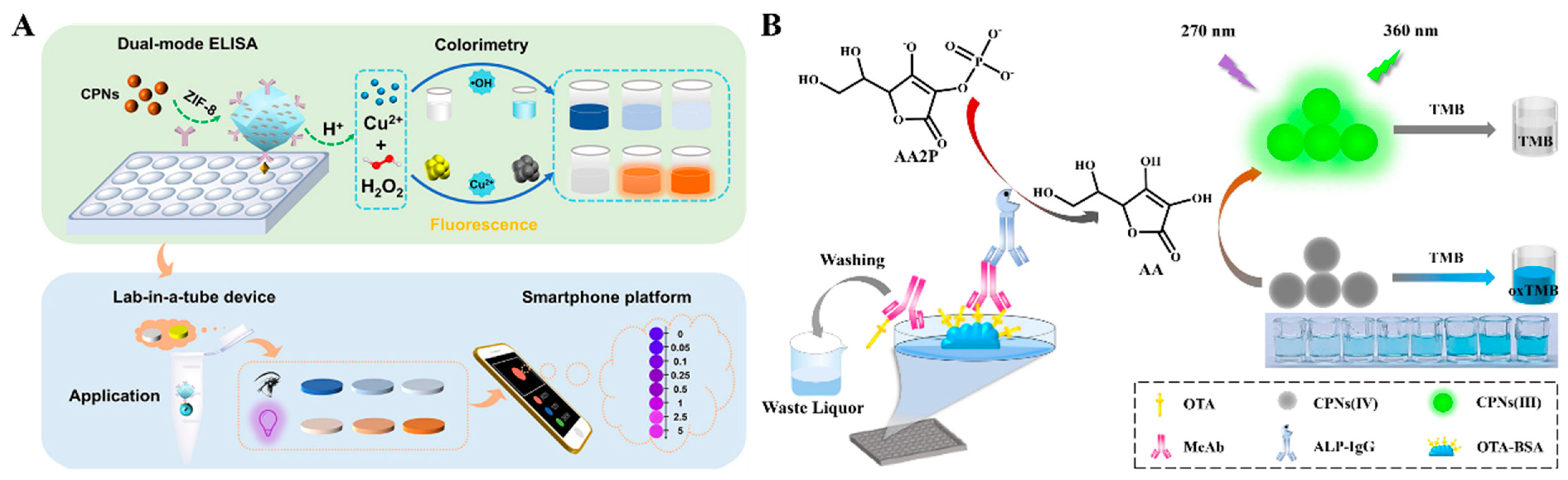
- Wei, D.; Xiong, D.; Zhu, N.; Wang, Y.; Hu, X.; Zhao, B.; Zhou, J.; Yin, D.; Zhang, Z. Copper peroxide nanodots encapsulated in a metal-organic framework for self-supplying hydrogen peroxide and signal amplification of the dual-mode immunoassay. Anal. Chem. 2022, 94, 12981–12989. [Google Scholar] [CrossRef]
- Chen, M.; Liu, Z.; Guan, Y.; Chen, Y.; Liu, W.; Liu, Y. Zeolitic imidazolate frameworks-derived hollow Co/N-doped CNTs as oxidase-mimic for colorimetric-fluorescence immunoassay of ochratoxin A. Sens. Actuators B Chem. 2022, 359, 131609–131619. [Google Scholar] [CrossRef]
- Chen, W.; Zhang, X.; Zhang, Q.; Zhang, G.; Wu, S.; Yang, H.; Zhou, Y. Cerium ions triggered dual-readout immunoassay based on aggregation induced emission effect and 3,3’,5,5’-tetramethylbenzidine for fluorescent and colorimetric detection of ochratoxin A. Anal. Chim. Acta 2022, 1231, 340445–340453. [Google Scholar] [CrossRef]
- Zheng, X.; Sun, L.; Zhao, Y.; Yang, H.; Zhu, Y.; Zhang, J.; Xu, D.; Zhang, X.; Zhou, Y. A fluorescence and colorimetric dual-mode immunoassay for detection of ochratoxin A based on cerium nanoparticles. Microchem. J. 2024, 201, 110419–110426. [Google Scholar] [CrossRef]
- Yang, X.; Wang, E. A nanoparticle autocatalytic sensor for Ag+ and Cu2+ ions in aqueous solution with high sensitivity and selectivity and its application in test paper. Anal. Chem. 2011, 83, 5005–5011. [Google Scholar] [CrossRef]
- Ye, Q.; Ren, S.; Huang, H.; Duan, G.; Liu, K.; Liu, J.B. Fluorescent and colorimetric sensors based on the oxidation of o-phenylenediamine. ACS Omega 2020, 5, 20698–20706. [Google Scholar] [CrossRef]
- Yang, H.; He, Q.; Pan, J.; Lin, M.; Lao, Z.; Li, Q.; Cui, X.; Zhao, S. PtCu nanocages with superior tetra-enzyme mimics for colorimetric sensing and fluorescent sensing dehydroepiandrosterone. Sens. Actuators B Chem. 2022, 351, 130905–130915. [Google Scholar] [CrossRef]
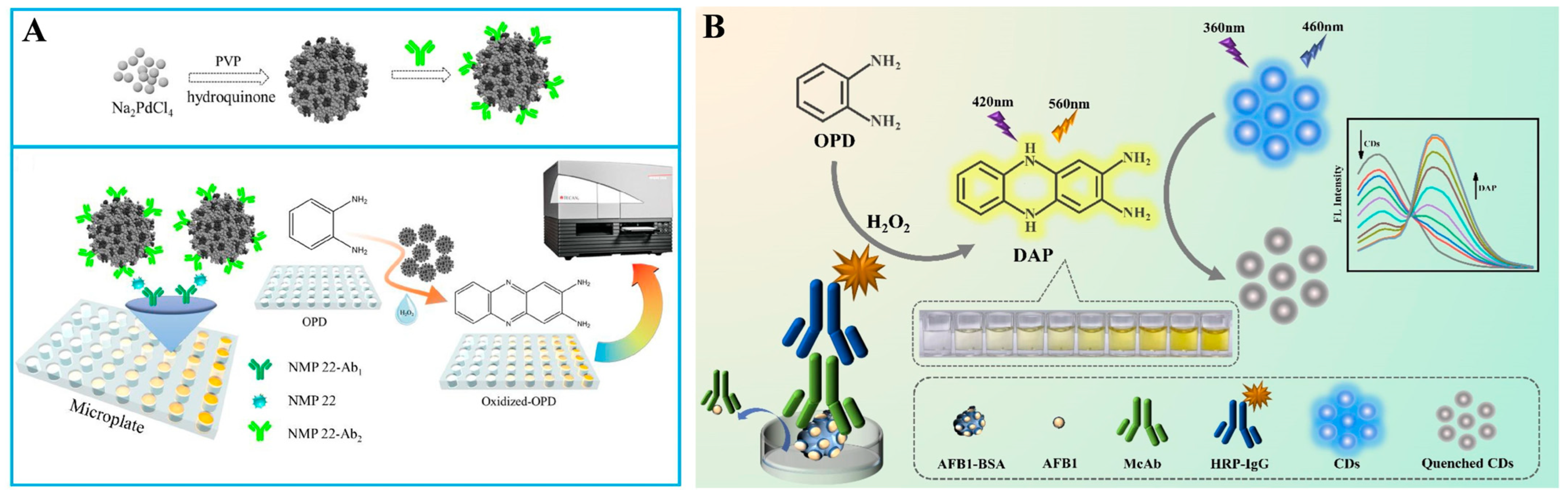
- Zhuge, W.; Tan, X.; Zhang, R.; Li, H.; Zheng, G. Fluorescent and colorimetric immunoassay of nuclear matrix protein 22 enhanced by porous Pd nanoparticles. Chin. Chem. Lett. 2019, 30, 1307–1309. [Google Scholar] [CrossRef]
- Wang, S.; Fang, B.; Yuan, M.; Wang, Z.; Peng, J.; Lai, W. Dual-mode immunoassay system based on glucose oxidase-triggered Fenton reaction for qualitative and quantitative detection of danofloxacin in milk. J. Dairy Sci. 2020, 103, 7826–7833. [Google Scholar] [CrossRef] [PubMed]
- Liu, W.; Kang, Q.; Wang, P.; Zhou, F. Ratiometric fluorescence immunoassay based on MnO2–o-phenylenediamine–fluorescent carbon nanodots for the detection of α-fetoprotein via fluorescence resonance energy transfer. New J. Chem. 2022, 46, 1120–1126. [Google Scholar] [CrossRef]
- Miao, L.; Jiao, L.; Tang, Q.; Li, H.; Zhang, L.; Wei, Q. A nanozyme-linked immunosorbent assay for dual-modal colorimetric and ratiometric fluorescent detection of cardiac troponin I. Sens. Actuators B Chem. 2019, 288, 60–64. [Google Scholar] [CrossRef]
- Xu, D.; Zhang, J.; Luo, Z.; Zhao, Y.; Zhu, Y.; Yang, H.; Zhou, Y. Ratiometric fluorescence and absorbance dual-model immunoassay based on 2,3-diaminophenazine and carbon dots for detecting Aflatoxin B1. Food Chem. 2024, 439, 138125–138131. [Google Scholar] [CrossRef]
- Xu, X.; Zhang, P.; Ruan, F.; Chang, G.; Zhou, T.; Chen, D.; Li, L.; Wang, X. Construction of a Colorimetric and Fluorescence Dual-Mode Immunoassay Detection of Alpha-Hemolysin in Milk. Foodborne Pathog. Dis. 2024, 12, 1535–1544. [Google Scholar] [CrossRef]
- Shi, Y.; Sun, Y.; Qu, X.; Zhou, L.; Wang, Z.; Gao, Z.; Hu, Z.; Yue, T.; Yuan, Y. Development of a colorimetric and fluorescence dual-mode immunoassay for the precise identification of Alicyclobacillus acidoterrestris in apple juice. Food Control 2021, 124, 107898–107906. [Google Scholar] [CrossRef]
- Wu, S.; Zhao, Y.; Xiao, Y.; Ma, L.; Zhang, Q.; Li, P.; Yang, H.; Zhou, Y. Dual-readout immunoassay based on IFE and p-phenylenediamine for detection of zearalenone. J. Food Compos. Anal. 2024, 132, 106311–106316. [Google Scholar] [CrossRef]
- Liu, L.; Chang, Y.; Lou, J.; Zhang, S.; Yi, X. Overview on the development of alkaline-phosphatase-linked optical immunoassays. Molecules 2023, 28, 6565. [Google Scholar] [CrossRef]
- Shaban, S.M.; Byeok Jo, S.; Hafez, E.; Ho Cho, J.; Kim, D.-H. A comprehensive overview on alkaline phosphatase targeting and reporting assays. Coordin. Chem. Rev. 2022, 465, 214567–214604. [Google Scholar] [CrossRef]
- Wu, Y.; Chen, W.; Wang, C.; Xing, D. Assays for alkaline phosphatase that use L-ascorbic acid 2-phosphate as a substrate. Coordin. Chem. Rev. 2023, 495, 215370–215423. [Google Scholar] [CrossRef]
- Ma, L.; Zhang, X.; Xiao, Y.; Fang, H.; Zhang, G.; Yang, H.; Zhou, Y. Fluorescence and colorimetric dual-mode immunoassay based on G-quadruplex/N-methylmesoporphyrin IX and p-nitrophenol for detection of zearalenone. Food Chem. 2023, 401, 134190–134195. [Google Scholar] [CrossRef] [PubMed]
- Luo, Z.; Zhang, J.; Ma, J.; Xu, D.; Zhao, Y.; Zhu, Y.; Yang, H.; Zhou, Y. Fluorescence and absorbance dual-mode immunoassay for detection of Ochratoxin A based on 2-aminoterephthalic acid and p-nitrophenol. J. Food Compos. Anal. 2024, 125, 105784–105790. [Google Scholar] [CrossRef]
- Zhang, J.; Lu, X.; Lei, Y.; Hou, X.; Wu, P. Exploring the tunable excitation of QDs to maximize the overlap with the absorber for inner filter effect-based phosphorescence sensing of alkaline phosphatase. Nanoscale 2017, 9, 15606–15611. [Google Scholar] [CrossRef]
- Xiong, J.; Sun, B.; Zhang, S.; Wang, S.; Qin, L.; Jiang, H. Highly efficient dual-mode detection of AFB1 based on the inner filter effect: Donor-acceptor selection and application. Anal. Chim. Acta 2024, 1298, 342384–342394. [Google Scholar] [CrossRef]
- Zhao, J.; Wang, S.; Lu, S.; Bao, X.; Sun, J.; Yang, X. An enzyme cascade-triggered fluorogenic and chromogenic reaction applied in enzyme activity assay and immunoassay. Anal. Chem. 2018, 90, 7754–7760. [Google Scholar] [CrossRef]
- Chen, C.; Zhao, D.; Wang, B.; Ni, P.; Jiang, Y.; Zhang, C.; Yang, F.; Lu, Y.; Sun, J. Alkaline phosphatase-triggered in situ formation of silicon-containing nanoparticles for a fluorometric and colorimetric dual-channel immunoassay. Anal. Chem. 2020, 92, 4639–4646. [Google Scholar] [CrossRef]
- Xie, S.; Yue, Y.; Xu, G. In situ formed carbon nanoparticles enables colorimetric and fluorometric dual-mode immunoassay detection of amantadine in poultry foodstuffs. Microchem. J. 2023, 192, 108895–108903. [Google Scholar] [CrossRef]
- Liu, L.; Hao, Y.; Deng, D.; Xia, N. Nanomaterials-based colorimetric immunoassays. Nanomaterials 2019, 9, 316. [Google Scholar] [CrossRef]
- Priyadarshini, E.; Rawat, K.; Bohidar, H.B.; Rajamani, P. Dual-probe (colorimetric and fluorometric) detection of ferritin using antibody-modified gold@carbon dot nanoconjugates. Microchim. Acta 2019, 186, 687–695. [Google Scholar] [CrossRef] [PubMed]
- Han, Z.; Xia, C.; Xu, Z.; Liu, X.; Zuo, H.; Cai, L.; Sun, T.; Liu, Y. Fluorescent and colorimetric detection of Norfloxacin with a bifunctional ligand and enzymatic signal amplification system. Microchem. J. 2022, 179, 107660–107666. [Google Scholar] [CrossRef]
- Ma, F.; He, L.; Lindner, E.; Wu, D.-Y. Highly porous poly(L-lactic) acid nanofibers as a dual-signal paper-based bioassay platform for in vitro diagnostics. Appl. Surf. Sci. 2021, 542, 148732–148740. [Google Scholar] [CrossRef]
- Lee, J.H.; Cho, H.Y.; Choi, H.K.; Lee, J.Y.; Choi, J.W. Application of gold nanoparticle to plasmonic biosensors. Int. J. Mol. Sci. 2018, 19, 2021. [Google Scholar] [CrossRef]
- Qi, L.; Liao, A.; Huang, X.; Li, X.; Jiang, X.; Yuan, X.; Huang, K. Enzymatic reaction-modulated in-situ formation of nanomaterials and their applications in colorimetric and fluorescent sensing. Coordin. Chem. Rev. 2024, 508, 215787–215813. [Google Scholar] [CrossRef]
- Zha, Y.; Lu, S.; Hu, P.; Ren, H.; Liu, Z.; Gao, W.; Zhao, C.; Li, Y.; Zhou, Y. Dual-modal immunosensor with functionalized gold nanoparticles for ultrasensitive detection of chloroacetamide herbicides. ACS Appl. Mater. Interfaces 2021, 13, 6091–6098. [Google Scholar] [CrossRef]
- Xiong, L.H.; He, X.; Zhao, Z.; Kwok, R.T.K.; Xiong, Y.; Gao, P.F.; Yang, F.; Huang, Y.; Sung, H.H.; Williams, I.D.; et al. Ultrasensitive virion immunoassay platform with dual-modality based on a multifunctional aggregation-induced emission luminogen. ACS Nano 2018, 12, 9549–9557. [Google Scholar] [CrossRef]
- Zhu, H.; Cai, Y.; Qileng, A.; Quan, Z.; Zeng, W.; He, K.; Liu, Y. Template-assisted Cu2O@Fe(OH)3 yolk-shell nanocages as biomimetic peroxidase: A multi-colorimetry and ratiometric fluorescence separated-type immunosensor for the detection of ochratoxin A. J. Hazard. Mater. 2021, 411, 125090–125099. [Google Scholar] [CrossRef]
- Chen, M.; Huang, X.; Chen, Y.; Cao, Y.; Zhang, S.; Lei, H.; Liu, W.; Liu, Y. Shape-specific MOF-derived Cu@Fe-NC with morphology-driven catalytic activity: Mimicking peroxidase for the fluorescent-colorimetric immunosignage of ochratoxin. J. Hazard. Mater. 2023, 443, 130233–130244. [Google Scholar] [CrossRef]
- Cao, X.M.; Li, L.H.; Liang, H.Z.; Li, J.D.; Chen, Z.J.; Luo, L.; Lu, Y.N.; Zhong, Y.X.; Shen, Y.D.; Lei, H.T.; et al. Dual-modular immunosensor for bongkrekic acid detection using specific monoclonal antibody. J. Hazard. Mater. 2023, 455, 131634–131642. [Google Scholar] [CrossRef]
- Liang, H.; Liu, Y.; Qileng, A.; Shen, H.; Liu, W.; Xu, Z.; Liu, Y. PEI-coated Prussian blue nanocubes as pH-Switchable nanozyme: Broad-pH-responsive immunoassay for illegal additive. Biosens. Bioelectron. 2023, 219, 114797–114804. [Google Scholar] [CrossRef] [PubMed]
- Gao, Y.; Wu, Y.; Huang, P.; Wu, F.-Y. Carbon dot-encapsulated plasmonic core-satellite nanoprobes for sensitive detection of cancer biomarkers via dual-mode colorimetric and fluorometric immunoassay. ACS Appl. Nano Mater. 2022, 5, 11539–11548. [Google Scholar] [CrossRef]
- Zhu, D.; Hu, Y.; Zhang, X.J.; Yang, X.T.; Tang, Y.Y. Colorimetric and fluorometric dual-channel detection of α-fetoprotein based on the use of ZnS-CdTe hierarchical porous nanospheres. Microchim. Acta 2019, 186, 124–132. [Google Scholar] [CrossRef] [PubMed]
- Zhou, Y.; Huang, X.; Xiong, S.; Li, X.; Zhan, S.; Zeng, L.; Xiong, Y. Dual-mode fluorescent and colorimetric immunoassay for the ultrasensitive detection of alpha-fetoprotein in serum samples. Anal. Chim. Acta 2018, 1038, 112–119. [Google Scholar] [CrossRef] [PubMed]
- Liu, Q.; Boyd, B.J. Liposomes in biosensors. Analyst 2013, 138, 391–409. [Google Scholar] [CrossRef]
- Radha, R.; Al-Sayah, M.H. Development of liposome-based immunoassay for the detection of cardiac troponin I. Molecules 2021, 26, 6988. [Google Scholar] [CrossRef]
- Deng, W.; Cheng, C.; Yang, H.; Wang, H.; Tan, Y.; Xie, Q.; Ma, M.; Yao, S. Ultrasensitive immunoassay of Staphylococcus aureus based on colorimetric and fluorescent responses of 4-chloro-7-nitrobenzo-2-oxa-1,3-diazole to l-cysteine. Talanta 2019, 202, 244–250. [Google Scholar] [CrossRef]
- Deng, D.; Chang, Y.; Liu, W.; Ren, M.; Xia, N.; Hao, Y. Advancements in biosensors based on the assembles of small organic molecules and peptides. Biosensors 2023, 13, 773. [Google Scholar] [CrossRef]
- Li, H.; Yang, J.; Hu, X.; Han, R.; Wang, S.; Pan, M. Portable smartphone-assisted fluorescence-colorimetric multidimensional immunosensing microarray based on NH2-UiO-66@PtNPs multifunctional composite for efficient and visual detection of amantadine. Chem. Eng. J. 2023, 473, 145401–145410. [Google Scholar] [CrossRef]
- Miao, L.; Zhu, C.; Jiao, L.; Li, H.; Du, D.; Lin, Y.; Wei, Q. Smart drug delivery system-inspired enzyme-linked immunosorbent assay based on fluorescence resonance energy transfer and allochroic effect induced dual-modal colorimetric and fluorescent detection. Anal. Chem. 2018, 90, 1976–1982. [Google Scholar] [CrossRef]
- Chen, L.; Li, Y.; Miao, L.; Pang, X.; Li, T.; Qian, Y.; Li, H. “Lighting-up” curcumin nanoparticles triggered by pH for developing improved enzyme-linked immunosorbent assay. Biosens. Bioelectron. 2021, 188, 113308–113314. [Google Scholar] [CrossRef] [PubMed]
- Cheng, J.; Yang, G.; Guo, J.; Liu, S.; Guo, J. Integrated electrochemical lateral flow immunoassays (eLFIAs): Recent advances. Analyst 2022, 147, 554–570. [Google Scholar] [CrossRef] [PubMed]
- Omidfar, K.; Riahi, F.; Kashanian, S. Lateral flow assay: A summary of recent progress for improving assay performance. Biosensors 2023, 13, 837. [Google Scholar] [CrossRef] [PubMed]
- Wen, C.; Dou, Y.; Liu, Y.; Jiang, X.; Tu, X.; Zhang, R. Au nanoshell-based lateral flow immunoassay for colorimetric and photothermal dual-mode detection of interleukin-6. Molecules 2024, 29, 3683. [Google Scholar] [CrossRef]
- Kim, M.J.; Haizan, I.; Ahn, M.J.; Park, D.H.; Choi, J.H. Recent advances in lateral flow assays for viral protein detection with nanomaterial-based optical sensors. Biosensors 2024, 14, 197. [Google Scholar] [CrossRef]
- Parolo, C.; Sena-Torralba, A.; Bergua, J.F.; Calucho, E.; Fuentes-Chust, C.; Hu, L.; Rivas, L.; Alvarez-Diduk, R.; Nguyen, E.P.; Cinti, S.; et al. Tutorial: Design and fabrication of nanoparticle-based lateral-flow immunoassays. Nat. Protoc. 2020, 15, 3788–3816. [Google Scholar] [CrossRef]
- Wang, P.; Li, J.; Guo, L.; Li, J.; He, F.; Zhang, H.; Chi, H. The developments on lateral flow immunochromatographic assay for food safety in recent 10 years: A review. Chemosensors 2024, 12, 88. [Google Scholar] [CrossRef]
- Yin, X.; Liu, S.; Kukkar, D.; Wang, J.; Zhang, D.; Kim, K.-H. Performance enhancement of the lateral flow immunoassay by use of composite nanoparticles as signal labels. TrAC-Trend. Anal. Chem. 2024, 170, 117441. [Google Scholar] [CrossRef]
- Hu, S.W.; Qiao, S.; Xu, B.Y.; Peng, X.; Xu, J.J.; Chen, H.Y. Dual-functional carbon dots pattern on paper chips for Fe3+ and ferritin analysis in whole blood. Anal. Chem. 2017, 89, 2131–2137. [Google Scholar] [CrossRef]
- Gong, X.; Zhang, B.; Piao, J.; Zhao, Q.; Gao, W.; Peng, W.; Kang, Q.; Zhou, D.; Shu, G.; Chang, J. High sensitive and multiple detection of acute myocardial infarction biomarkers based on a dual-readout immunochromatography test strip. Nanomedicine 2018, 14, 1257–1266. [Google Scholar] [CrossRef]
- Hang, Y.; Boryczka, J.; Wu, N. Visible-light and near-infrared fluorescence and surface-enhanced Raman scattering point-of-care sensing and bio-imaging: A review. Chem. Soc. Rev. 2022, 51, 329–375. [Google Scholar] [CrossRef] [PubMed]
- Preechakasedkit, P.; Osada, K.; Katayama, Y.; Ruecha, N.; Suzuki, K.; Chailapakul, O.; Citterio, D. Gold nanoparticle core-europium(III) chelate fluorophore-doped silica shell hybrid nanocomposites for the lateral flow immunoassay of human thyroid stimulating hormone with a dual signal readout. Analyst 2018, 143, 564–570. [Google Scholar] [CrossRef] [PubMed]
- You, P.Y.; Li, F.C.; Liu, M.H.; Chan, Y.H. Colorimetric and fluorescent dual-mode immunoassay based on plasmon-enhanced fluorescence of polymer dots for detection of PSA in whole blood. ACS Appl. Mater. Interfaces 2019, 11, 9841–9849. [Google Scholar] [CrossRef] [PubMed]
- Yang, Y.C.; Liu, M.H.; Yang, S.M.; Chan, Y.H. Bimodal multiplexed detection of tumor markers in non-small cell lung cancer with polymer dot-based immunoassay. ACS Sens. 2021, 6, 4255–4264. [Google Scholar] [CrossRef]
- Pan, Y.; Wei, X.; Guo, X.; Wang, H.; Song, H.; Pan, C.; Xu, N. Immunoassay based on Au-Ag bimetallic nanoclusters for colorimetric/fluorescent double biosensing of dicofol. Biosens. Bioelectron. 2021, 194, 113611–113618. [Google Scholar] [CrossRef]
- Wang, S.; Shen, W.; Zheng, S.; Li, Z.; Wang, C.; Zhang, L.; Liu, Y. Dual-signal lateral flow assay using vancomycin-modified nanotags for rapid and sensitive detection of Staphylococcus aureus. RSC Adv. 2021, 11, 13297–13303. [Google Scholar] [CrossRef]
- Wang, J.; Jiang, C.; Yuan, J.; Tong, L.; Wang, Y.; Zhuo, D.; Huang, L.; Ni, W.; Zhang, J.; Huang, M.; et al. Hue recognition competitive fluorescent lateral flow immunoassay for aflatoxin M1 detection with improved visual and quantitative performance. Anal. Chem. 2022, 94, 10865–10873. [Google Scholar] [CrossRef]
- Wang, C.; Yang, X.; Gu, B.; Liu, H.; Zhou, Z.; Shi, L.; Cheng, X.; Wang, S. Sensitive and simultaneous detection of SARS-CoV-2-specific IgM/IgG using lateral flow immunoassay based on dual-mode quantum dot nanobeads. Anal. Chem. 2020, 92, 15542–15549. [Google Scholar] [CrossRef]
- Han, H.; Wang, C.; Yang, X.; Zheng, S.; Cheng, X.; Liu, Z.; Zhao, B.; Xiao, R. Rapid field determination of SARS-CoV-2 by a colorimetric and fluorescent dual-functional lateral flow immunoassay biosensor. Sens. Actuators B Chem. 2022, 351, 130897–130905. [Google Scholar] [CrossRef]
- Cheng, X.; Yang, X.; Tu, Z.; Rong, Z.; Wang, C.; Wang, S. Graphene oxide-based colorimetric/fluorescence dual-mode immunochromatography assay for simultaneous ultrasensitive detection of respiratory virus and bacteria in complex samples. J. Hazard. Mater. 2023, 459, 132192–132202. [Google Scholar] [CrossRef]
- Zheng, S.; Xia, X.; Tian, B.; Xu, C.; Zhang, T.; Wang, S.; Wang, C.; Gu, B. Dual-color MoS2@QD nanosheets mediated dual-mode lateral flow immunoassay for flexible and ultrasensitive detection of multiple drug residues. Sens. Actuators B Chem. 2024, 403, 135142–135152. [Google Scholar] [CrossRef]
- Fang, B.; Hu, S.; Wang, C.; Yuan, M.; Huang, Z.; Xing, K.; Liu, D.; Peng, J.; Lai, W. Lateral flow immunoassays combining enrichment and colorimetry-fluorescence quantitative detection of sulfamethazine in milk based on trifunctional magnetic nanobeads. Food Control 2019, 98, 268–273. [Google Scholar] [CrossRef]
- Cheng, Y.; Yin, X.; Li, Y.; Wang, S.; Xue, S.; Wu, Q.; Wang, J.; Zhang, D. Multiple-readout lateral flow immunoassay for the sensitive detection of nitrofurazone metabolites through ultrabright AIE-MOF coupled in-situ growth strategy. Biosens. Bioelectron. 2024, 262, 116556–116564. [Google Scholar] [CrossRef] [PubMed]
- Fan, L.; Yan, W.; Chen, Q.; Tan, F.; Tang, Y.; Han, H.; Yu, R.; Xie, N.; Gao, S.; Chen, W.; et al. One-component dual-readout aggregation-induced emission nanobeads for qualitative and quantitative detection of C-reactive protein at the point of care. Anal. Chem. 2024, 96, 401–408. [Google Scholar] [CrossRef] [PubMed]
- Sheng, W.; Chang, Q.; Shi, Y.; Duan, W.; Zhang, Y.; Wang, S. Visual and fluorometric lateral flow immunoassay combined with a dual-functional test mode for rapid determination of tetracycline antibiotics. Microchim. Acta 2018, 185, 404–413. [Google Scholar] [CrossRef]
- Ding, Y.; Hua, X.; Chen, H.; Gonzalez-Sapienza, G.; Barnych, B.; Liu, F.; Wang, M.; Hammock, B.D. A dual signal immunochromatographic strip for the detection of imidaclothiz using a recombinant fluorescent-peptide tracer and gold nanoparticles. Sens. Actuators B Chem. 2019, 297, 126714–126720. [Google Scholar] [CrossRef]
- Mao, X.; Wang, Y.; Jiang, L.; Zhang, H.; Zhao, Y.; Liu, P.; Liu, J.; Hammock, B.D.; Zhang, C. A polydopamine-coated gold nanoparticles quenching quantum dots-based dual-readout lateral flow immunoassay for sensitive detection of carbendazim in agriproducts. Biosensors 2022, 12, 83. [Google Scholar] [CrossRef]
- Liu, S.; Shu, R.; Zhao, C.; Sun, C.; Zhang, M.; Wang, S.; Li, B.; Dou, L.; Ji, Y.; Wang, Y.; et al. Precise spectral overlap-based donor-acceptor pair for a sensitive traffic light-typed bimodal multiplexed lateral flow immunoassay. Anal. Chem. 2024, 96, 5046–5055. [Google Scholar] [CrossRef]
- Gui, Y.; Zhao, Y.; Liu, P.; Wang, Y.; Mao, X.; Peng, C.; Hammock, B.D.; Zhang, C. Colorimetric and reverse fluorescence dual-signal readout immunochromatographic assay for the sensitive determination of sibutramine. ACS Omega 2024, 9, 7075–7084. [Google Scholar] [CrossRef]
- Shu, X.; Guo, P.; Zhang, G.; Zhang, W.; Hu, H.; Peng, J.; Xiong, Y.; Ma, B.; Lai, W. Novel litchi-like Au-Ag nanospheres driven dual-readout lateral flow immunoassay for sensitive detection of pyrimethanil. Food Chem. 2024, 450, 139380–139388. [Google Scholar] [CrossRef]
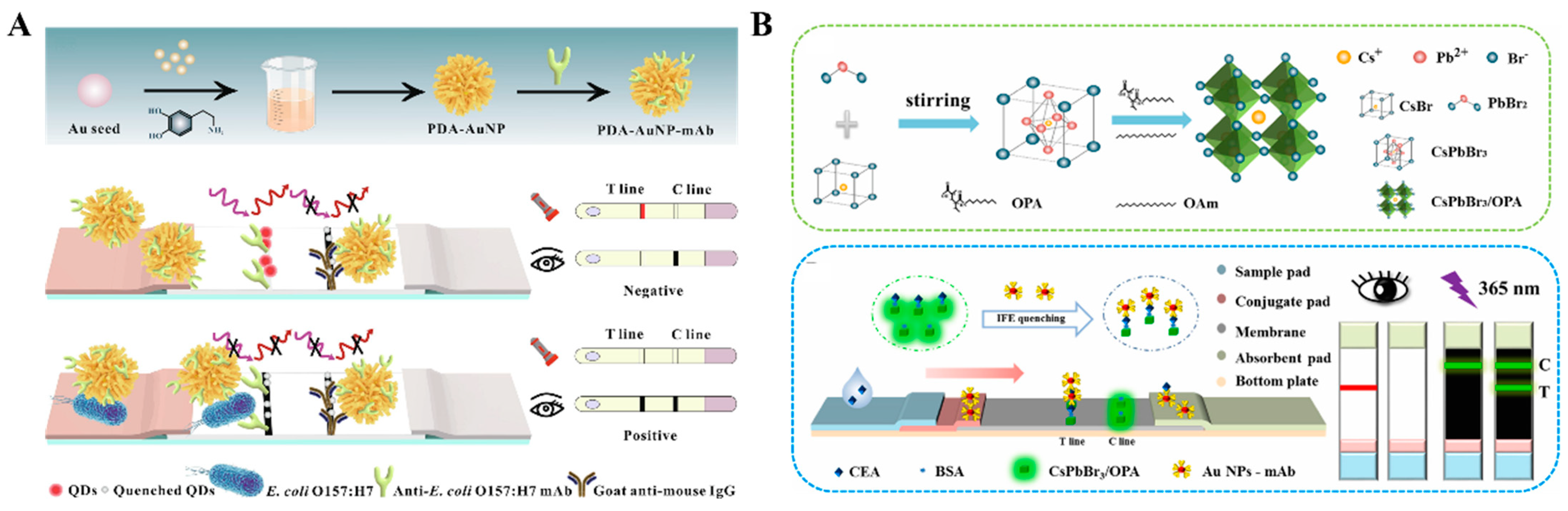
- Shao, Y.; Wang, Z.; Xie, J.; Zhu, Z.; Feng, Y.; Yu, S.; Xue, L.; Wu, S.; Gu, Q.; Zhang, J.; et al. Dual-mode immunochromatographic assay based on dendritic gold nanoparticles with superior fluorescence quenching for ultrasensitive detection of E. coli O157:H7. Food Chem. 2023, 424, 136366–136375. [Google Scholar] [CrossRef] [PubMed]
- Deng, Y.; Wang, Y.; Lin, M.; Chen, Y.; Qian, Z.J.; Liu, J.; Li, X. High-density Au anchored to Ti3C2-based colorimetric-fluorescence dual-mode lateral flow immunoassay for all-domain-enhanced performance and signal intercalibration. Anal. Chem. 2024, 96, 5106–5114. [Google Scholar] [CrossRef]
- Zhang, B.; Ma, W.; Li, F.; Gao, W.; Zhao, Q.; Peng, W.; Piao, J.; Wu, X.; Wang, H.; Gong, X.; et al. Fluorescence quenching-based signal amplification on immunochromatography test strips for dual-mode sensing of two biomarkers of breast cancer. Nanoscale 2017, 9, 18711–18722. [Google Scholar] [CrossRef] [PubMed]
- Wang, Y.; Shen, J.; Song, R.; Xu, Q.; Hu, X.; Shu, Y. Highly bright and stable CsPbBr3 perovskite nanocrystals with amphiphilic polymer binding based dual-readout lateral flow immunoassay for detection of carcinoembryonic antigen. Talanta 2024, 266, 125017–125024. [Google Scholar] [CrossRef] [PubMed]
- Liu, S.; Zhao, C.; Shu, R.; Dou, L.; Luo, X.; Luo, L.; Sun, J.; Wang, Y.; Ji, Y.; Wang, J. Fortified dual-spectral overlap with enhanced colorimetric/fluorescence dual-response immunochromatography for on-site bimodal-type gentamicin monitoring. J. Agric. Food. Chem. 2024, 72, 9400–9410. [Google Scholar] [CrossRef]
- Yang, X.; Cheng, X.; Tu, Z.; Wei, H.; Rong, Z. PDA-mediated colorimetric-fluorescence co-enhanced immunochromatography assay for the simultaneous and rapid detection of SARS-CoV-2 and HAdV. Chem. Eng. J. 2024, 481, 148756. [Google Scholar] [CrossRef]
- Yang, X.; Cheng, X.; Wei, H.; Tu, Z.; Rong, Z.; Wang, C.; Wang, S. Fluorescence-enhanced dual signal lateral flow immunoassay for flexible and ultrasensitive detection of monkeypox virus. J. Nanobiotechnol. 2023, 21, 450. [Google Scholar] [CrossRef]
- Bu, T.; Bai, F.; Zhao, S.; Cao, Y.; He, K.; Sun, X.; Wang, Q.; Jia, P.; Li, M.; Wang, X.; et al. Multifunctional bacteria-derived tags for advancing immunoassay analytical performance with dual-channel switching and antibodies bioactivity sustaining. Biosens. Bioelectron. 2021, 192, 113538. [Google Scholar] [CrossRef]
- Zha, Y.; Li, Y.; Zhou, J.; Liu, X.; Park, K.S.; Zhou, Y. Dual-mode fluorescent/intelligent lateral flow immunoassay based on machine learning algorithm for ultrasensitive analysis of chloroacetamide herbicides. Anal. Chem. 2024, 96, 12197–12204. [Google Scholar] [CrossRef]
- Yu, Q.; Wu, T.; Tian, B.; Li, J.; Liu, Y.; Wu, Z.; Jin, X.; Wang, C.; Wang, C.; Gu, B. Recent advances in SERS-based immunochromatographic assay for pathogenic microorganism diagnosis: A review. Anal. Chim. Acta 2024, 1286, 341931–341949. [Google Scholar] [CrossRef]
- Tian, Y.; Yin, X.; Li, J.; Dou, L.; Wang, S.; Jia, C.; Li, Y.; Chen, Y.; Yan, S.; Wang, J.; et al. A dual-mode lateral flow immunoassay by ultrahigh signal-to background ratio SERS probes for nitrofurazone metabolites ultrasensitive detection. Food Chem. 2024, 441, 138374–138383. [Google Scholar] [CrossRef] [PubMed]
- Liu, L.; Wang, Y.; Xue, Z.; Peng, B.; Kou, X.; Gao, Z. Research progress of dual-mode sensing technology strategy based on SERS and its application in the detection of harmful substances in foods. Trends Food Sci. Technol. 2024, 148, 104487–104500. [Google Scholar] [CrossRef]
- Zhuang, X.; Hu, Y.; Wang, J.; Hu, J.; Wang, Q.; Yu, X. A colorimetric and SERS dual-readout sensor for sensitive detection of tyrosinase activity based on 4-mercaptophenyl boronic acid modified AuNPs. Anal. Chim. Acta 2021, 1188, 339172. [Google Scholar] [CrossRef] [PubMed]
- Xu, X.; Yue, S.; Tu, K.; Yuan, B.; Bi, S.; Yu, J.; Qiu, H.; Zhang, H.; Zhang, L.; Wu, H.F.; et al. Multi-shell nanourchin-integrated dual mode lateral flow immunoassay for sensitive and rapid detection of clinical cardiac myosin-binding protein C. Anal. Chem. 2024, 96, 11853–11861. [Google Scholar] [CrossRef] [PubMed]
- Wang, J.; Jiang, H.; Chen, Y.; Zhu, X.; Wu, Q.; Chen, W.; Zhao, Q.; Wang, J.; Qin, P. CRISPR/Cas9-mediated SERS/colorimetric dual-mode lateral flow platform combined with smartphone for rapid and sensitive detection of Staphylococcus aureus. Biosens. Bioelectron. 2024, 249, 116046. [Google Scholar] [CrossRef]
- Zhao, T.; Liang, P.; Ren, J.; Zhu, J.; Yang, X.; Bian, H.; Li, J.; Cui, X.; Fu, C.; Xing, J.; et al. Gold-silver alloy hollow nanoshells-based lateral flow immunoassay for colorimetric, photothermal, and SERS tri-mode detection of SARS-CoV-2 neutralizing antibody. Anal. Chim. Acta 2023, 1255, 341102–341108. [Google Scholar] [CrossRef]
- Fernandez-Lodeiro, C.; Gonzalez-Cabaleiro, L.; Vazquez-Iglesias, L.; Serrano-Pertierra, E.; Bodelon, G.; Carrera, M.; Blanco-Lopez, M.C.; Perez-Juste, J.; Pastoriza-Santos, I. Au@Ag core-shell nanoparticles for colorimetric and surface-enhanced Raman-scattering-based multiplex competitive lateral flow immunoassay for the simultaneous detection of histamine and parvalbumin in fish. ACS Appl. Nano Mater. 2024, 7, 498–508. [Google Scholar] [CrossRef]
- Atta, S.; Zhao, Y.; Li, J.Q.; Vo-Dinh, T. Dual-modal colorimetric and surface-enhanced Raman scattering (SERS)-based lateral flow immunoassay for ultrasensitive detection of SARS-CoV-2 using a plasmonic gold nanocrown. Anal. Chem. 2024, 96, 4783–4790. [Google Scholar] [CrossRef]
- Su, L.; Hu, H.; Tian, Y.; Jia, C.; Wang, L.; Zhang, H.; Wang, J.; Zhang, D. Highly sensitive colorimetric/surface-enhanced Raman spectroscopy immunoassay relying on a metallic core-shell Au/Au nanostar with clenbuterol as a target analyte. Anal. Chem. 2021, 93, 8362–8369. [Google Scholar] [CrossRef]
- Bai, T.; Wang, M.; Cao, M.; Zhang, J.; Zhang, K.; Zhou, P.; Liu, Z.; Liu, Y.; Guo, Z.; Lu, X. Functionalized Au@Ag-Au nanoparticles as an optical and SERS dual probe for lateral flow sensing. Anal. Bioanal. Chem. 2018, 410, 2291–2303. [Google Scholar] [CrossRef]
- Wang, M.; Feng, J.; Ding, J.; Xiao, J.; Liu, D.; Lu, Y.; Liu, Y.; Gao, X. Color- and background-free Raman-encoded lateral flow immunoassay for simultaneous detection of carbendazim and imidacloprid in a single test line. Chem. Eng. J. 2024, 487, 150666–150675. [Google Scholar] [CrossRef]
- He, D.; Wu, Z.; Cui, B.; Xu, E.; Jin, Z. Establishment of a dual mode immunochromatographic assay for Campylobacter jejuni detection. Food Chem. 2019, 289, 708–713. [Google Scholar] [CrossRef] [PubMed]
- Huang, X.; Chen, L.; Zhi, W.; Zeng, R.; Ji, G.; Cai, H.; Xu, J.; Wang, J.; Chen, S.; Tang, Y.; et al. Urchin-shaped Au-Ag@Pt sensor integrated lateral flow immunoassay for multimodal detection and specific discrimination of clinical multiple bacterial infections. Anal. Chem. 2023, 95, 13101–13112. [Google Scholar] [CrossRef] [PubMed]
- Yang, Y.; Li, G.; Wang, P.; Fan, L.; Shi, Y. Highly sensitive multiplex detection of foodborne pathogens using a SERS immunosensor combined with novel covalent organic frameworks based biologic interference-free Raman tags. Talanta 2022, 243, 123369–123378. [Google Scholar] [CrossRef] [PubMed]
- Zhang, X.; Shi, Y.; Wu, D.; Fan, L.; Liu, J.; Wu, Y.; Li, G. A bifunctional core-shell gold@Prussian blue nanozyme enabling dual-readout microfluidic immunoassay of food allergic protein. Food Chem. 2024, 434, 137455–137466. [Google Scholar] [CrossRef] [PubMed]
- Israelsen, N.D.; Wooley, D.; Hanson, C.; Vargis, E. Rational design of Raman-labeled nanoparticles for a dual-modality, light scattering immunoassay on a polystyrene substrate. J. Biol. Eng. 2016, 10, 2–13. [Google Scholar] [CrossRef]
- Alvarez-Puebla, R.A.; Pazos-Perez, N.; Guerrini, L. SERS-fluorescent encoded particles as dual-mode optical probes. Appl. Mater. Today 2018, 13, 1–14. [Google Scholar] [CrossRef]
- Wang, Z.; Zong, S.; Li, W.; Wang, C.; Xu, S.; Chen, H.; Cui, Y. SERS-fluorescence joint spectral encoding using organic-metal-QD hybrid nanoparticles with a huge encoding capacity for high-throughput biodetection: Putting theory into practice. J. Am. Chem. Soc. 2012, 134, 2993–3000. [Google Scholar] [CrossRef]
- Kim, K.; Lee, Y.M.; Lee, H.B.; Shin, K.S. Silver-coated silica beads applicable as core materials of dual-tagging sensors operating via SERS and MEF. ACS Appl. Mater. Interfaces 2009, 1, 2174–2180. [Google Scholar] [CrossRef]
- Zou, F.; Zhou, H.; Tan, T.V.; Kim, J.; Koh, K.; Lee, J. Dual-mode SERS-fluorescence immunoassay using graphene quantum dot labeling on one-dimensional aligned magnetoplasmonic nanoparticles. ACS Appl. Mater. Interfaces 2015, 7, 12168–12175. [Google Scholar] [CrossRef]
- Yang, Y.; Zhu, J.; Weng, G.-J.; Li, J.-J.; Zhao, J.-W. Gold nanoring core-shell satellites with abundant built-in hotspots and great analyte penetration: An immunoassay platform for the SERS/fluorescence-based detection of carcinoembryonic antigen. Chem. Eng. J. 2021, 409, 128173–128183. [Google Scholar] [CrossRef]
- Kim, K.; Lee, H.B.; Choi, J.Y.; Shin, K.S. Silver-coated dye-embedded silica beads: A core material of dual tagging sensors based on fluorescence and Raman scattering. ACS Appl. Mater. Interfaces 2011, 3, 324–330. [Google Scholar] [CrossRef] [PubMed]
- Zong, S.; Wang, Z.; Zhang, R.; Wang, C.; Xu, S.; Cui, Y. A multiplex and straightforward aqueous phase immunoassay protocol through the combination of SERS-fluorescence dual mode nanoprobes and magnetic nanobeads. Biosens. Bioelectron. 2013, 41, 745–751. [Google Scholar] [CrossRef] [PubMed]
- Wang, Z.; Zong, S.; Chen, H.; Wu, H.; Cui, Y. Silica coated gold nanoaggregates prepared by reverse microemulsion method: Dual mode probes for multiplex immunoassay using SERS and fluorescence. Talanta 2011, 86, 170–177. [Google Scholar] [CrossRef]
- Zhang, X.; Du, X. Carbon nanodot-decorated Ag@SiO2 nanoparticles for fluorescence and surface-enhanced Raman scattering immunoassays. ACS Appl. Mater. Interfaces 2016, 8, 1033–1040. [Google Scholar] [CrossRef]
- Arshad, F.; Nurul Azian Zakaria, S.; Ahmed, M.U. Gold-silver-platinum trimetallic nanozyme-based dual-mode immunosensor for ultrasensitive osteoprotegerin detection. Microchem. J. 2024, 201, 110523–110531. [Google Scholar] [CrossRef]
- Dong, Y.; Wang, W.; Ye, C.; Song, Y. Recent advances in photoelectrochemistry-coupled dual-modal biosensors: From constructions to biosensing applications. Nano Res. 2024, 17, 5512–5528. [Google Scholar] [CrossRef]
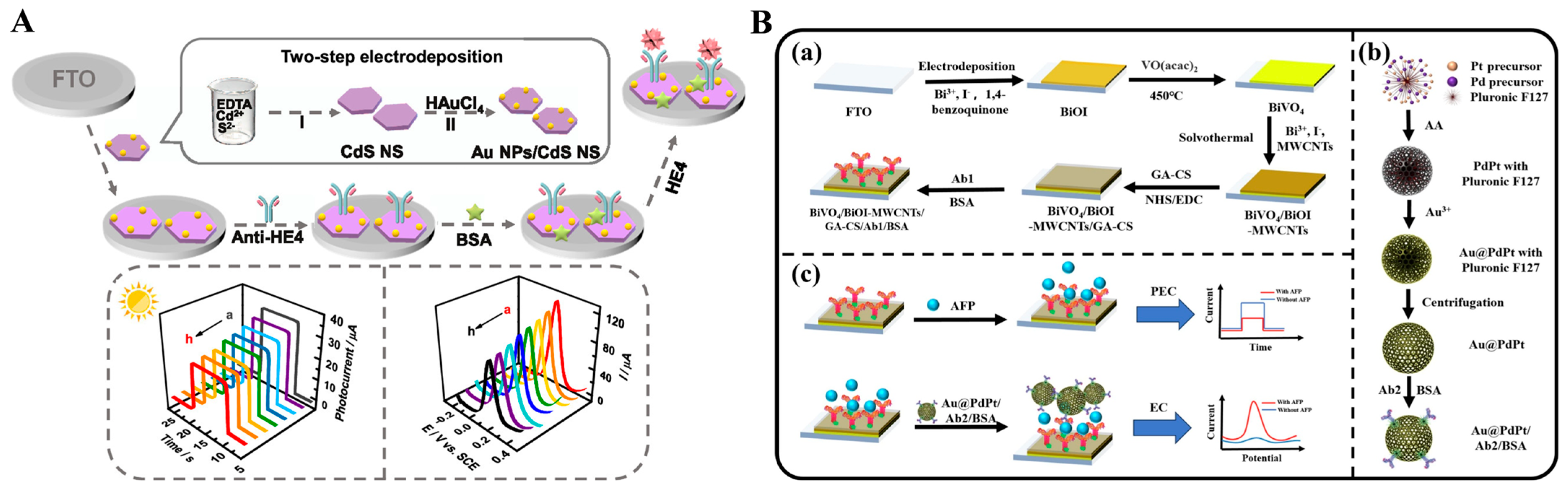
- Wei, J.-J.; Wang, G.-Q.; Zheng, J.-Y.; Wang, A.-J.; Mei, L.-P.; Feng, J.-J. Dual-mode photoelectrochemical and electrochemical immunosensors for human epididymis protein 4 based on SPR-promoted Au nanoparticle/CdS nanosheet heterostructures. ACS Appl. Nano Mater. 2022, 5, 14934–14941. [Google Scholar] [CrossRef]
- Qileng, A.; Liang, H.; Huang, S.; Liu, W.; Xu, Z.; Liu, Y. Dual-function of ZnS/Ag2S nanocages in ratiometric immunosensors for the discriminant analysis of ochratoxins: Photoelectrochemistry and electrochemistry. Sens. Actuators B Chem. 2020, 314, 128066–128073. [Google Scholar] [CrossRef]
- Wu, H.; Yang, X. Biofunctional photoelectrochemical/electrochemical immunosensor based on BiVO4/BiOI-MWCNTs and Au@PdPt for alpha-fetoprotein detection. Bioelectrochemistry 2024, 160, 108773–108781. [Google Scholar] [CrossRef]
- Firoozbakhtian, A.; Hosseini, M.; Guan, Y.; Xu, G. Boosting electrochemiluminescence immunoassay sensitivity via Co-Pt nanoparticles within a Ti3C2 MXene-modified single electrode electrochemical system on raspberry Pi. Anal. Chem. 2023, 95, 15110–15117. [Google Scholar] [CrossRef] [PubMed]
- Gong, J.; Zhang, T.; Chen, P.; Yan, F.; Liu, J. Bipolar silica nanochannel array for dual-mode electrochemiluminescence and electrochemical immunosensing platform. Sens. Actuators B Chem. 2022, 368, 132086–132093. [Google Scholar] [CrossRef]
- Huang, J.; Zhang, T.; Zheng, Y.; Liu, J. Dual-mode sensing platform for cancer antigen 15-3 determination based on a silica nanochannel array using electrochemiluminescence and electrochemistry. Biosensors 2023, 13, 317. [Google Scholar] [CrossRef] [PubMed]
- Hao, X.; Liu, Z.; Li, Y.; Hou, W.; Zheng, T.; Hu, L. A dual mode immunosensing platform constructed on CdIn2S4 near-infrared electrochemiluminescence emitter and AuNPs@g-C3N4 for zearalenone sensing. Microchem. J. 2024, 204, 111166–111176. [Google Scholar] [CrossRef]
- Wang, W.; Liu, Y.; Shi, T.; Sun, J.; Mo, F.; Liu, X. Biosynthesized quantum dot for facile and ultrasensitive electrochemical and electrochemiluminescence immunoassay. Anal. Chem. 2020, 92, 1598–1604. [Google Scholar] [CrossRef] [PubMed]
- Pang, S.; Yu, H.; Zhang, Y.; Jiao, Y.; Zheng, Z.; Wang, M.; Zhang, H.; Liu, A. Bioscreening specific peptide-expressing phage and its application in sensitive dual-mode immunoassay of SARS-CoV-2 spike antigen. Talanta 2024, 266, 125093–125099. [Google Scholar] [CrossRef]
- Hu, Y.; Zhu, L.; Mei, X.; Liu, J.; Yao, Z.; Li, Y. Dual-mode sensing platform for electrochemiluminescence and colorimetry detection based on a closed bipolar electrode. Anal. Chem. 2021, 93, 12367–12373. [Google Scholar] [CrossRef]
- Romanelli, S.; Bettazzi, F.; Martellini, T.; Shelver, W.L.; Cincinelli, A.; Galarini, R.; Palchetti, I. Evaluation of a QuEChERS-like extraction approach for the determination of PBDEs in mussels by immuno-assay-based screening methods. Talanta 2017, 170, 540–545. [Google Scholar] [CrossRef]
- Kim, J.U.; Kim, J.M.; Thamilselvan, A.; Nam, K.H.; Kim, M.I. Colorimetric and electrochemical dual-mode detection of thioredoxin 1 based on the efficient peroxidase-mimicking and electrocatalytic property of Prussian Blue nanoparticles. Biosensors 2024, 14, 185. [Google Scholar] [CrossRef]
- Seddaoui, N.; Attaallah, R.; Amine, A. Development of an optical immunoassay based on peroxidase-mimicking Prussian blue nanoparticles and a label-free electrochemical immunosensor for accurate and sensitive quantification of milk species adulteration. Microchim. Acta 2022, 189, 209. [Google Scholar] [CrossRef]
- Campas, M.; de la Iglesia, P.; Le Berre, M.; Kane, M.; Diogene, J.; Marty, J.L. Enzymatic recycling-based amperometric immunosensor for the ultrasensitive detection of okadaic acid in shellfish. Biosens. Bioelectron. 2008, 24, 716–722. [Google Scholar] [CrossRef] [PubMed]
- Al-Ogaidi, I.; Aguilar, Z.P.; Suri, S.; Gou, H.; Wu, N. Dual detection of cancer biomarker CA125 using absorbance and electrochemical methods. Analyst 2013, 138, 5647–5653. [Google Scholar] [CrossRef] [PubMed]
- Hong, W.; Lee, S.; Cho, Y. Dual-responsive immunosensor that combines colorimetric recognition and electrochemical response for ultrasensitive detection of cancer biomarkers. Biosens. Bioelectron. 2016, 86, 920–926. [Google Scholar] [CrossRef] [PubMed]
- Reverte, L.; Garibo, D.; Flores, C.; Diogene, J.; Caixach, J.; Campas, M. Magnetic particle-based enzyme assays and immunoassays for microcystins: From colorimetric to electrochemical detection. Environ. Sci. Technol. 2013, 47, 471–478. [Google Scholar] [CrossRef]
- Choi, H.; Son, S.E.; Hur, W.; Tran, V.K.; Lee, H.B.; Park, Y.; Han, D.K.; Seong, G.H. Electrochemical immunoassay for determination of glycated albumin using nanozymes. Sci. Rep. 2020, 10, 9513–9524. [Google Scholar] [CrossRef]
- Xu, X.H.; Xia, J.; Song, S.; Liu, Y.; Chen, J.; Zhong, S.; Chen, H.; Zhang, Z.H. Dual-signal immuno-competitive determination of brain natriuretic peptide based on magnetic nanozyme. Electroanalysis 2023, 35, e202200500–e202200508. [Google Scholar] [CrossRef]
- Hou, Y.H.; Wang, J.J.; Jiang, Y.Z.; Lv, C.; Xia, L.; Hong, S.L.; Lin, M.; Lin, Y.; Zhang, Z.L.; Pang, D.W. A colorimetric and electrochemical immunosensor for point-of-care detection of enterovirus 71. Biosens. Bioelectron. 2018, 99, 186–192. [Google Scholar] [CrossRef]
- Shang, C.; Li, Y.; Zhang, Q.; Tang, S.; Tang, X.; Ren, H.; Hu, P.; Lu, S.; Li, P.; Zhou, Y. Alkaline phosphatase-triggered dual-signal immunoassay for colorimetric and electrochemical detection of zearalenone in cornmeal. Sens. Actuators B Chem. 2022, 358, 131525–131532. [Google Scholar] [CrossRef]
- Preechakasedkit, P.; Panphut, W.; Lomae, A.; Wonsawat, W.; Citterio, D.; Ruecha, N. Dual colorimetric/electrochemical detection of Salmonella typhimurium using a laser-induced graphene integrated lateral flow immunoassay strip. Anal. Chem. 2023, 95, 13904–13912. [Google Scholar] [CrossRef]
- Li, Y.-R.; Dong, X.-X.; Cai, W.-Y.; Liang, Y.-F.; Chen, X.-H.; Li, X.-M.; Wang, Y.; Liu, Y.-J.; Lei, H.-T.; Xu, Z.-L. Non-toxic immunosensor for highly efficient detection of tetrodotoxin by electrochemical and colorimetric dual-mode sensing platform. Sens. Actuators B Chem. 2023, 389, 133849–133857. [Google Scholar] [CrossRef]
- Li, N.; Brahmendra, A.; Veloso, A.J.; Prashar, A.; Cheng, X.R.; Hung, V.W.; Guyard, C.; Terebiznik, M.; Kerman, K. Disposable immunochips for the detection of Legionella pneumophila using electrochemical impedance spectroscopy. Anal. Chem. 2012, 84, 3485–3488. [Google Scholar] [CrossRef] [PubMed]
- Korram, J.; Anbalagan, A.C.; Banerjee, A.; Sawant, S.N. Bio-conjugated carbon dots for the bimodal detection of prostate cancer biomarkers via sandwich fluorescence and electrochemical immunoassays. J. Mater. Chem. B 2024, 12, 742–751. [Google Scholar] [CrossRef] [PubMed]
- Cui, R.; Pan, H.C.; Zhu, J.J.; Chen, H.Y. Versatile immunosensor using CdTe quantum dots as electrochemical and fluorescent labels. Anal. Chem. 2007, 79, 8494–8501. [Google Scholar] [CrossRef] [PubMed]
- Chopra, A.; Tuteja, S.; Sachdeva, N.; Bhasin, K.K.; Bhalla, V.; Suri, C.R. CdTe nanobioprobe based optoelectrochemical immunodetection of diabetic marker HbA1c. Biosens. Bioelectron. 2013, 44, 132–135. [Google Scholar] [CrossRef]
- Peng, X.; Luo, G.; Wu, Z.; Wen, W.; Zhang, X.; Wang, S. Fluorescent-magnetic-catalytic nanospheres for dual-modality detection of H9N2 avian influenza virus. ACS Appl. Mater. Interfaces 2019, 11, 41148–41156. [Google Scholar] [CrossRef]
- Kong, Q.; Cui, K.; Zhang, L.; Wang, Y.; Sun, J.; Ge, S.; Zhang, Y.; Yu, J. “On-Off-On” photoelectrochemical/visual lab-on-paper sensing via signal amplification of CdS quantum dots@leaf-shape ZnO and quenching of Au-modified prism-anchored octahedral CeO2 nanoparticles. Anal. Chem. 2018, 90, 11297–11304. [Google Scholar] [CrossRef]
- Chen, Y.; Zhang, S.; Dai, H.; Hong, Z.; Lin, Y. A multiple mixed TiO2 mesocrystal junction based PEC-colorimetric immunoassay for specific recognition of lipolysis stimulated lipoprotein receptor. Biosens. Bioelectron. 2020, 148, 111809–111815. [Google Scholar] [CrossRef]
- Li, J.; Wang, H.; Liu, M.; Qin, Y.; Tan, R.; Hu, L.; Gu, W.; Zhu, C. Galvanic replacement reaction-regulated photoelectric response and enzyme-mimicking property of ionic liquid functionalized Cu@Cu2O aerogels for dual-mode immunoassay. Chem. Eng. J. 2023, 455, 140743–140749. [Google Scholar] [CrossRef]
- Leng, D.; Xu, R.; Ren, X.; Ma, H.; Liu, L.; Wei, Q.; Ju, H. Self-shedding of multifunctional biomolecular nanocarriers for H-FABP monitoring in magnetic separation self-powered photoelectrochemical and colorimetric immunoassay platform. Chem. Eng. J. 2023, 469, 143876–143883. [Google Scholar] [CrossRef]
- Jia, L.; Wang, Y.; Jiang, M.; Yuan, W.; Jin, Y.; Yan, W.; Ze, X.; Chen, Y.; Niu, L. An ultrasensitive dual-mode stagey for 17β-estradiol assay: Photoelectrochemical and colorimetric biosensor based on a WSe2/TiO2-modified electrode coupled with nucleic acid amplification. Anal. Chim. Acta 2024, 1319, 342966–342975. [Google Scholar] [CrossRef]
- Zhang, N.; Ruan, Y.F.; Ma, Z.Y.; Zhao, W.W.; Xu, J.J.; Chen, H.Y. Simultaneous photoelectrochemical and visualized immunoassay of β-human chorionic gonadotrophin. Biosens. Bioelectron. 2016, 85, 294–299. [Google Scholar] [CrossRef] [PubMed]
- Wei, J.; Chen, H.; Chen, H.; Cui, Y.; Qileng, A.; Qin, W.; Liu, W.; Liu, Y. Multifunctional peroxidase-encapsulated nanoliposomes: Bioetching-induced photoelectrometric and colorimetric immunoassay for broad-spectrum detection of ochratoxins. ACS Appl. Mater. Interfaces 2019, 11, 23832–23839. [Google Scholar] [CrossRef] [PubMed]
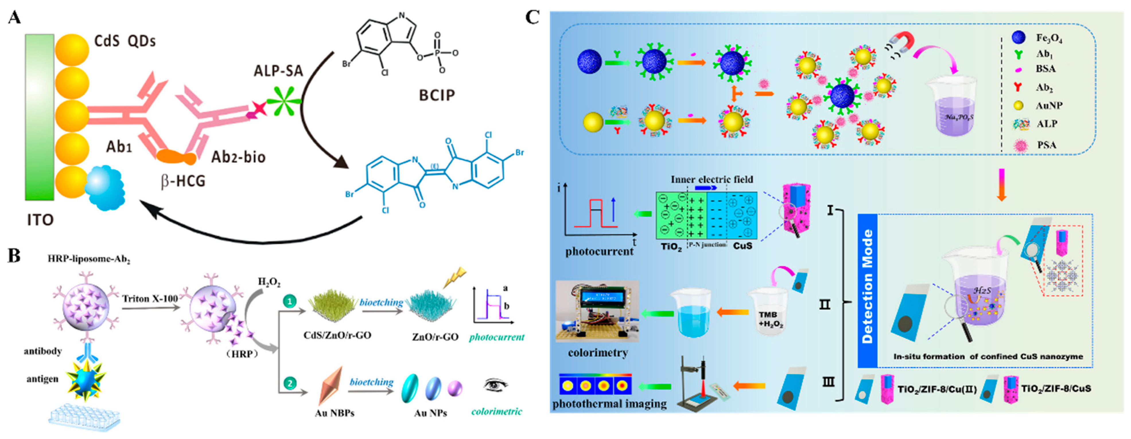
- Meng, X.; Wang, J.; Diao, L.; Li, C. Construction of multi-mode photoelectrochemical immunoassays for accurate detection of cancer markers: Assisted with MOF-confined plasmonic nanozyme. Anal. Chem. 2024, 96, 1336–1344. [Google Scholar] [CrossRef]
- Shen, Y.Z.; Xie, W.Z.; Wang, Z.; Ning, K.P.; Ji, Z.P.; Li, H.B.; Hu, X.Y.; Ma, C.; Qin, X. A generalizable sensing platform based on molecularly imprinted polymer-aptamer double recognition and nanoenzyme assisted photoelectrochemical-colorimetric dual-mode detection. Biosens. Bioelectron. 2024, 254, 116201–116217. [Google Scholar] [CrossRef]
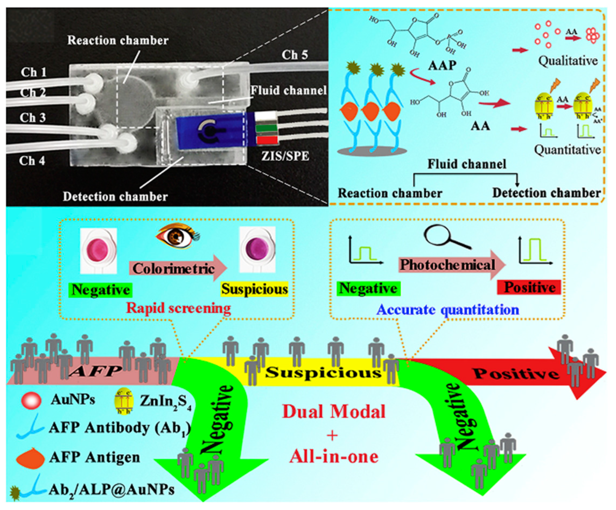
- Li, X.; Pan, X.; Lu, J.; Zhou, Y.; Gong, J. Dual-modal visual/photoelectrochemical all-in-one bioassay for rapid detection of AFP using 3D printed microreactor device. Biosens. Bioelectron. 2020, 158, 112158–112165. [Google Scholar] [CrossRef]
- Qileng, A.; Liu, W.; Sun, Z.; Zhou, W.; Fang, Y.; Lei, H.; Liu, Y.; Zhang, S. Portable dual-modular immunosensor constructed from bimetallic metal-organic framework heterostructure grafted with enzyme-mimicking label for rosiglitazone detection. Adv. Funct. Mater. 2022, 32, 2203244–2203253. [Google Scholar] [CrossRef]
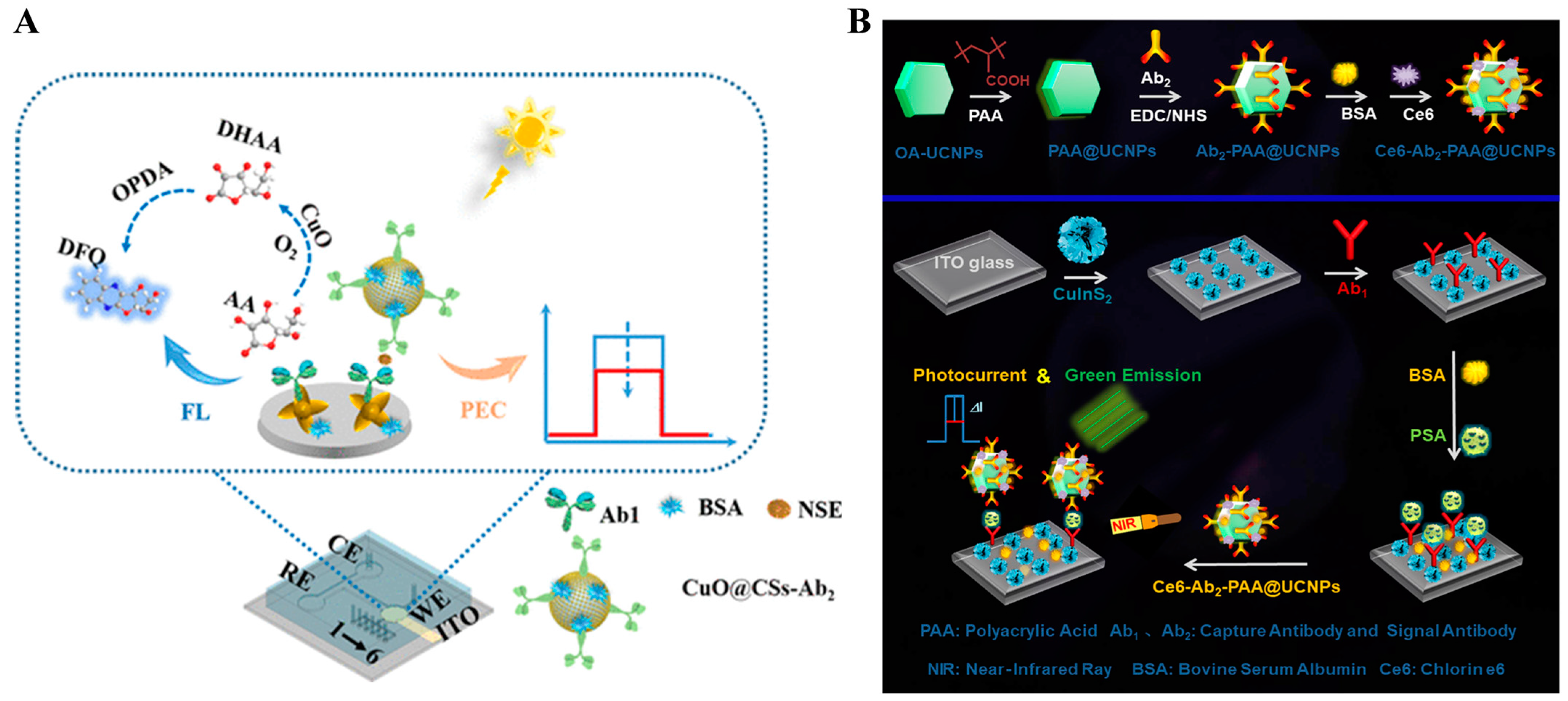
- Wu, T.; Yu, S.; Dai, L.; Feng, J.; Ren, X.; Ma, H.; Wang, X.; Wei, Q.; Ju, H. CuO nanozymes as multifunctional signal labels for efficiently quenching the photocurrent of ZnO/Au/AgSbS2 hybrids and initiating a strong fluorescent signal in a dual-mode microfluidic sensing platform. ACS Sens. 2022, 7, 1732–1739. [Google Scholar] [CrossRef] [PubMed]
- Han, Q.; Zhao, X.; Na, N.; Ouyang, J. Integrating near-infrared visual fluorescence with a photoelectrochemical sensing system for dual readout detection of biomolecules. Anal. Chem. 2021, 93, 3486–3492. [Google Scholar] [CrossRef]
- Han, Q.; Zhao, X.; Zhang, X.; Na, N.; Ouyang, J. A rationally designed triple-qualitative and double-quantitative high precision multi-signal readout sensing platform. Sens. Actuators B Chem. 2022, 360, 131663–131672. [Google Scholar] [CrossRef]


Disclaimer/Publisher’s Note: The statements, opinions and data contained in all publications are solely those of the individual author(s) and contributor(s) and not of MDPI and/or the editor(s). MDPI and/or the editor(s) disclaim responsibility for any injury to people or property resulting from any ideas, methods, instructions or products referred to in the content. |
© 2024 by the authors. Licensee MDPI, Basel, Switzerland. This article is an open access article distributed under the terms and conditions of the Creative Commons Attribution (CC BY) license (https://creativecommons.org/licenses/by/4.0/).
Share and Cite
Ma, X.; Ge, Y.; Xia, N. Overview of the Design and Application of Dual-Signal Immunoassays. Molecules 2024, 29, 4551. https://doi.org/10.3390/molecules29194551
Ma X, Ge Y, Xia N. Overview of the Design and Application of Dual-Signal Immunoassays. Molecules. 2024; 29(19):4551. https://doi.org/10.3390/molecules29194551
Chicago/Turabian StyleMa, Xiaohua, Yijing Ge, and Ning Xia. 2024. "Overview of the Design and Application of Dual-Signal Immunoassays" Molecules 29, no. 19: 4551. https://doi.org/10.3390/molecules29194551