Article

Experimental and DFT Study of Monensinate and Salinomycinate Complexes Containing {Fe33–O)}7+ Core

1
Faculty of Chemistry and Pharmacy, Sofia University St. Kliment Ohridski, 1164 Sofia, Bulgaria
2
Institute of Physical Chemistry, Bulgarian Academy of Sciences, 1113 Sofia, Bulgaria
3
Institute of Catalysis, Bulgarian Academy of Sciences, 1113 Sofia, Bulgaria
4
Institute of General and Inorganic Chemistry, Bulgarian Academy of Sciences, 1113 Sofia, Bulgaria
5
Research and Development Department, Biovet Ltd., 4550 Peshtera, Bulgaria
*
Authors to whom correspondence should be addressed.
Molecules 2024, 29(2), 364; https://doi.org/10.3390/molecules29020364
Submission received: 20 December 2023 / Revised: 4 January 2024 / Accepted: 9 January 2024 / Published: 11 January 2024
(This article belongs to the Section Computational and Theoretical Chemistry)

Abstract

:
Two trinuclear oxo-centred iron(III) coordination compounds of monensic and salinomycinic acids (HL) were synthesized and their spectral properties were studied using physicochemical/thermal methods (FT–IR, TG–DTA, TG–MS, EPR, Mössbauer spectroscopy, powder XRD) and elemental analysis. The data suggested the formation of [Fe33–O)L3(OH)4] and the probable complex structures were modelled using the DFT method. The computed spectral parameters of the optimized constructs were compared to the experimentally measured ones. In each complex, three metal centres were joined together at the axial position by a μ3–O unit to form a {Fe3O}7+ core. The antibiotics monoanions served as bidentate ligands through the carboxylate and hydroxyl groups located at the termini. The carboxylate moieties played a dual role bridging each two metal centres. Hydroxide anions secured the overall neutral character of the coordination species. Mössbauer spectra displayed asymmetric quadrupole doublets that were consistent with the existence of two types of high-spin iron(III) sites with different environments—two Fe[O5] and one Fe[O6] centres. The solid-state EPR studies confirmed the +3 oxidation state of iron with a total spin St = 5/2 per trinuclear cluster. The studied complexes are the first iron(III) coordination compounds of monensin and salinomycin reported so far.

1. Introduction

Metal ions are essential for maintaining homeostasis in organisms ranging from the microscopic world of bacteria to the macroscopic scale of plants, animals, and, of course, human beings [1,2,3,4]. The importance of Mn+ is rooted not in their varying concentration in each organism, but in the pathologies arising from an abnormal amount of certain metal ions in a given life form. Special attention must be paid to microelements, as variations in their concentration can cause severe health issues.
Iron ions play a vital role in almost all living organisms [5] (except Lactobacillus and some strains of Bacillus [6]). Among the special functions of iron are the transport, storage and activation of molecular oxygen, reduction in ribonucleotides and dinitrogen, activation and decomposition of peroxides, and electron transport using various electron carriers [7]. On the other hand, iron biochemistry can be crucial for cancer occurrence and development since an abnormal regulation in cancer cells has been observed—downregulated efflux of iron ions from the cells, generation of reactive oxygen species (ROS), ferroptosis induction, etc. [8,9,10,11,12,13,14,15,16,17,18,19].
If we look at some of the simplest living organisms, for example, representatives of microbiota, we witness an interesting natural way of scavenging insoluble iron(III) from the environment—these organisms synthesize specific compounds called siderophores [1,20,21,22,23,24,25,26,27], which effectively bind iron(III) and transfer it to the intracellular compartment. Based on their structures, siderophores have been divided into five groups—catecholates, phenolates, hydroxamates, carboxylates, and a mixed type [27]. Their primary role is to ensure iron(III) uptake even at low concentration levels. This process also occurs in infected humans—the so-called “nutritional immunity” triggers and the host organism itself limits the amount of trace elements accessible to the pathogen [9]. The result is a dramatic change in the metabolism of the invader and its subsequent cell death.
Compounds that bind iron and transport it through the cells, without ion release or changing its reduction potential, can function as chemotherapeutics. Deferoxamine and deferasirox are two such compounds that are known to exclusively bind iron ions [11,16,28]. These drugs have already been included in therapeutic protocols and are used in the case of iron overload in some haematological diseases, metal intoxication, and in the treatment of aceruloplasminemia patients. The availability of such approved compounds in human medicine raises the question of whether a synergistic effect could be achieved by using a metal chelator as a ligand which can bind iron ions, but also can specifically induce apoptosis in cancer cells or/and cancer stem cells as well [10,11,13,14,15,16,19,29,30].
Such biologically active compounds can be found among the ionophore antibiotics, which have been in the spotlight for the last decade due to their anticancer properties. A member of this group—salinomycin, shows a substantial effect on cancer cells and cancer stem cells [18,31,32]. Another representative is monensin, which possesses less pronounced anticancer activity compared to salinomycin but has potential to be included in cancer therapy, due to the high selectivity index of some of its recently studied metal complexes [33]. Both antibiotics are polyether derivatives of monocarboxylic acids and, similarly to carboxylate siderophores, they may serve as potential ligands for iron(III) binding.
In this study, the ability of the natural antibiotics monensin (MonH) and salinomycin (SalH) (Figure 1) to interact with iron(III) was studied via a plethora of physicochemical methods. Two isostructural coordination compounds bearing the corresponding ionophore were synthesized. The resulting complexes consisted of two types of ligands directly bound to metal ions situated in a trinuclear configuration typical for iron(III) monocarboxylates. The composition and the binding mode of the ligands were proposed based on experimental data. Essential information for the primary coordination shell and the electronic configuration of the newly synthesized complexes was derived from EPR and Mössbauer spectroscopy. The mutual position of the atoms in the cores of the complexes was evaluated by appropriate theoretical models. Due to the complicated structure of polyether ionophores, solely the closest environment around the metal centres was modelled. The computed data obtained for the simplified system revealed the spatial placement of the metal cations and the donor atoms but also disclosed the magnetic properties of the complexes, which largely impact their spin state population at room temperature. The theoretically predicted electronic structure and properties of iron(III) monensinate and salinomycinate were used to explain the experimentally observed results and some peculiarities in the spectral behaviour of the coordination compounds.

2. Results and Discussion

2.1. General

Iron(III) monensinate (1) and salinomycinate (2) were obtained from methanolic solutions containing MonH×H2O or SalH, respectively, Et3N and FeCl3×6H2O. Precipitation from water produces yellow coordination species in satisfactory yields. The good agreement between the experimental and theoretical data allowed us to deduce plausible structures of novel iron(III) complexes of monensin (1) and salinomycin (2) as trinuclear oxo-clusters of composition [Fe33–O)L3(OH)4].

2.2. Elemental and Thermal Analysis

The elemental analysis data of the isolated complexes 12 can be found in Table 1. The thermal studies (TG–DTA and TG–MS) exclude the presence of coordinated water molecules in the composition of the solid samples (Figure S1). The powder XRD analysis (Figure S2) reveals the lack of crystallinity in both complexes evident from their amorphous halo located at small angles. The experimental data indicate the formation of isostructural equimolar coordination species of both antibiotics (Fe3+:ligand ratio 1:1), the absence of water, and the direct involvement of hydroxide anions in the close environment of the metal ions.

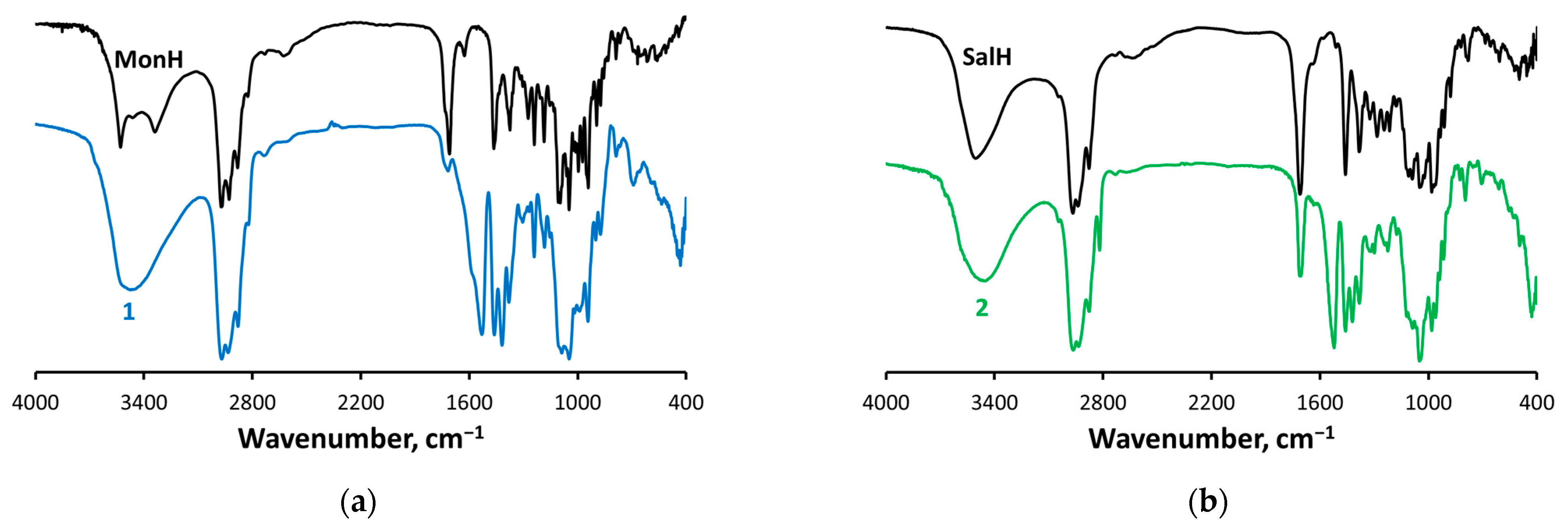
2.3. IR Spectroscopy

The free non-coordinated acidic forms of the polyether ionophores possess characteristic bands in the IR region, assigned to stretching vibrations of the hydroxyl (4000–3000 cm−1), carboxyl (1710 cm−1), and carbonyl (1710 cm−1, SalH) groups.
The band at 1710 cm−1 disappears (monensinate, Mon) or significantly diminishes in intensity (salinomycinate, Sal) when switching to the spectra of 12. Instead, two new bands in the range of 1530–1520 cm−1 and 1420 cm−1 are observed attributed to νas(−COO) and νs(−COO), respectively (Figure 2). The difference between the two vibrations is 112 (1) and 103 (2) cm−1 and is consistent with the presence of a bridging deprotonated carboxyl function in the ionophores structure [34,35,36]. The strong absorption peaks at ca. 1050 cm−1 are characteristic for δ(FeOH), suggesting the inclusion of hydroxide anions as additional ligands in the primary coordination shell of the metal centres. The peaks at ca. 435 cm−1 in the spectra of 12 are specific for ν(FeO), pointing to the formation of Fe–O bonds [37]. The broad intense bands in the IR spectra of 12 centred at 3470–3465 cm−1 are assigned to various ν(OH) vibrations.

2.4. EPR Spectroscopy

The EPR spectra of 12 presented in Figure 3a,b were recorded within the temperature range 77–295 K. Both spectra consist of one relatively broad symmetric signal with Lorentz shape. Its peak-to-peak linewidths at room temperature are 99 mT (1) and 122 mT (2) and undergo identical broadening as the temperature decreases (Figure 3c,d). The signal position is temperature independent, and the g-factor values remain in a very narrow interval in the whole temperature range (2.020–2.005 for 1 and 2.030–2.010 for 2). Further study of the temperature dependence of the signal intensity reveals that the Currie–Weiss law is obeyed in the studied samples and that the experimentally estimated values of the Currie–Weiss constant are as follows: −378 ± 12 K (1); −380 ± 9 K (2). The negative Currie–Weiss constants and the signal broadening at low temperatures indicate an antiferromagnetic interaction between the magnetically coupled Fe3+ ions. The linewidth temperature dependence could be explained by increased magnetic interactions at low temperatures. The detection of a broad EPR signal for complexes 1 and 2, with a g-factor around 2.0, is due to exchange-coupled individual Fe3+ ions (6S5/2) [38]. In most ferric monocarboxylate complexes these interactions occur through an oxo-anion centred in the core of the coordination compounds. Since the EPR characteristics of our complexes are consistent with the concept of a trinuclear antiferromagnetically coupled cluster of Fe3+ ions and based on the IR and elemental analysis data, the species 12 can be described as oxo-complexes of the antibiotics, namely [Fe33-O)L3(OH)4].

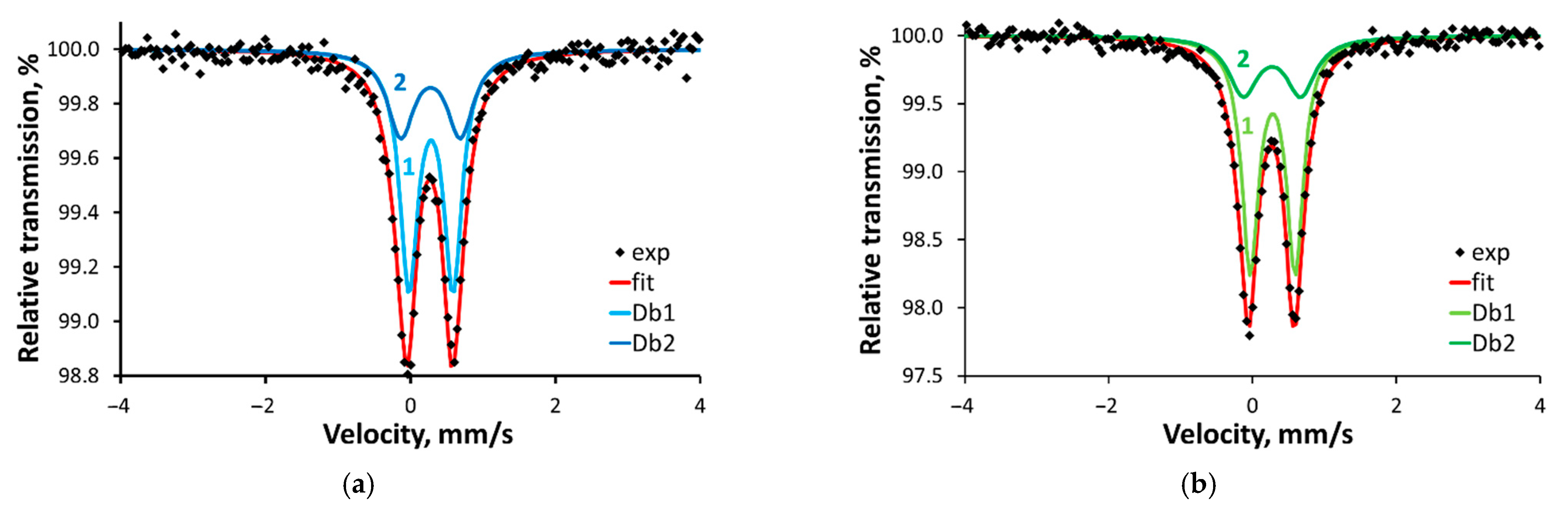
2.5. Mössbauer Spectroscopy

The zero-field Mössbauer spectra of both samples at 293 K consist of asymmetric doublets that can be decomposed into two Lorentzian-shaped quadrupole doublets with an area ratio of 2:1 (Table 2, Figure 4). A least-square fit of the spectra yields isomer shifts (IS, δ) within the range of 0.3 to 0.6 mm/s typically observed for high-spin iron(III) complexes [39,40]. The half-filled 3d electron level implies that the charge distribution asymmetry around the metal centres dominantly contributes to the observed quadrupole splitting (QS, ∆). The QS of the two doublets, which describes the experimental signals of 12, leads to the conclusion that two of the ions possess more symmetric charge distribution (ca. 0.60) than the third one (ca. 0.80). The temperature decrease does not change significantly the QS values (Table 2, Figure S3), but leads to an increase in the IS values, attributable to a second-order Doppler effect. The noticeably more prominent Mössbauer effect can be explained in terms of decreased vibrational motions of the molecules, thus increasing the probability of recoilless absorption of the gamma quant.
The subspectra with an intensity ratio of 2:1 (Figure 4) can be assigned to two penta-coordinated ions Fe(1) and Fe(2) (subspectrum (1)) and one six-coordinated ion Fe(3) (subspectrum (2)), respectively, as it is shown in the sketch, presented in Figure 5. The isomer shifts, together with the quadrupole splittings, support the high-spin state for the three iron sites. The similarity in the isomer shifts of Fe(l)/Fe(2) and Fe(3) might be assigned to close bond lengths at all irons. The penta-coordinated ferric ions Fe(1) and Fe(2) are indistinguishable in the Mössbauer spectra while the quadrupole splitting of the six-coordinated Fe(3) exceeds the value of Fe(1) and Fe(2) [41].
In summary, we were able to isolate two new Fe(III) complexes of monensin (1) and salinomycin (2). The spectral data reveal the following:
(i)
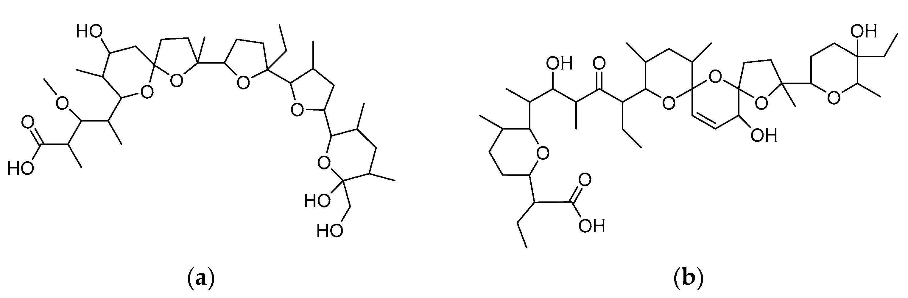
The antibiotics undergo deprotonation and act as bridging monoanions (IR);
(ii)
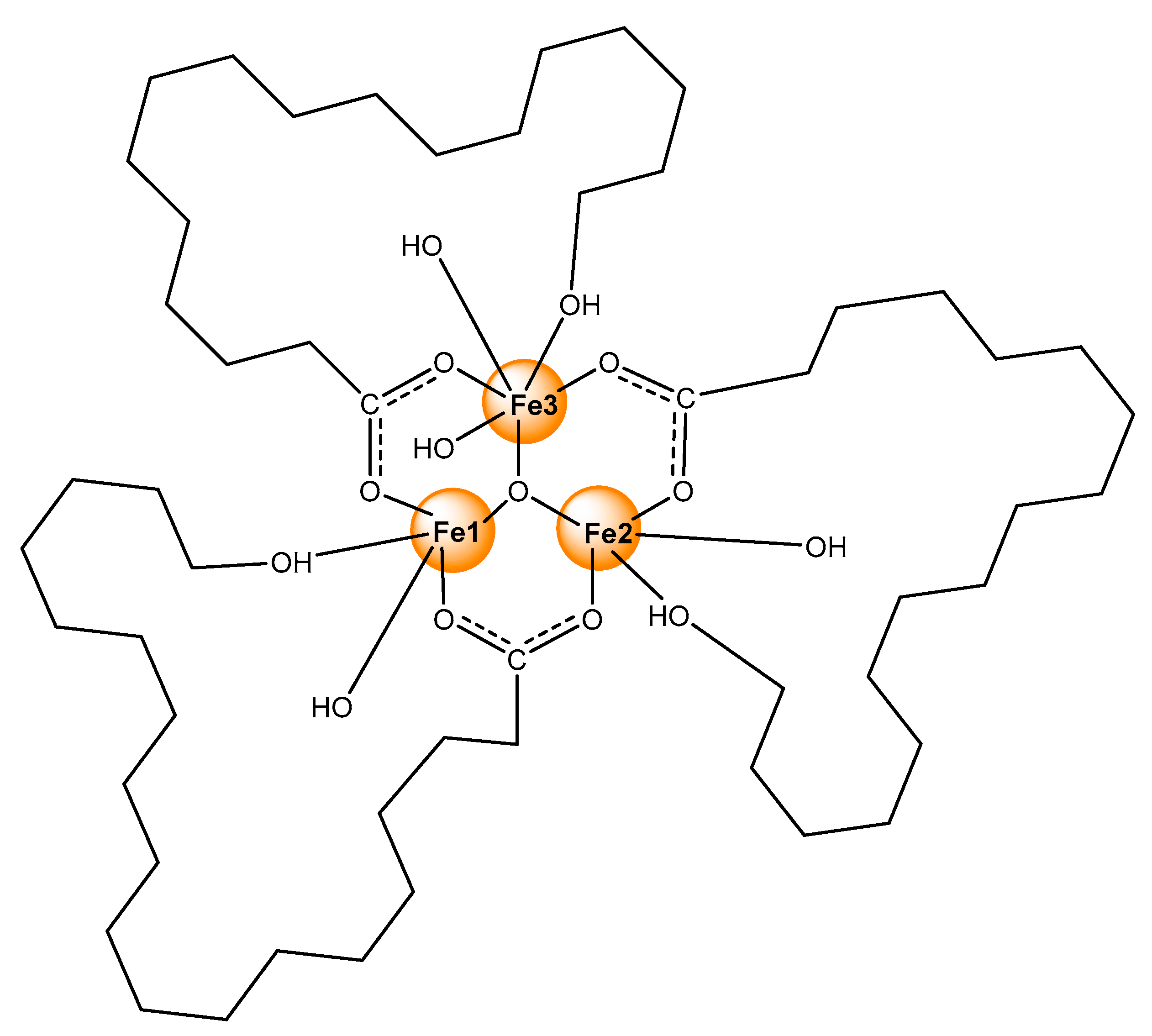
No water ligands are included in the primary coordination shell of the metal ions (TGV);
(iii)
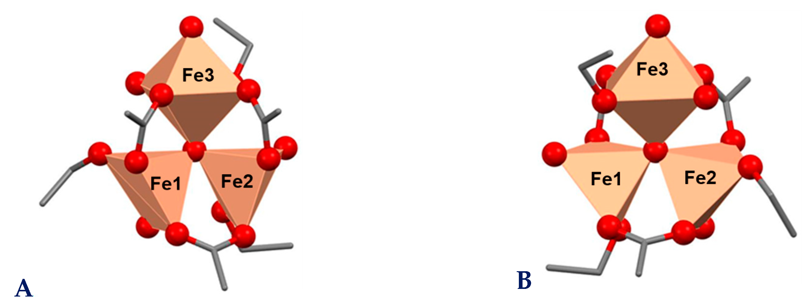
An antiferromagnetic interaction between the metal centres occurs that can be realized within a triangular oxo-iron cluster (EPR);
(iv)
The three high-spin iron ions are inequivalent and possess non-symmetric charge distribution at 2:1 ratio (Mössbauer spectroscopy).
Based on the experimental evidence, we assume that the new coordination species belong to the known class of trinuclear oxo-ferri-carboxylates with [Fe3(O)L3]4+ composition. Most likely, each ionophore serves as a bridge between every two metal centres through the carboxylate function, but also acts as a bidentate ligand via the hydroxyl group placed at the opposite end of the molecule. The overall neutral character of the coordination species formed is ensured by hydroxide anions originating from the basic medium used for the complexes preparation. Three of these anions occupy the hydrophilic cavity of antibiotics and bind to irons completing their penta-coordinated shell. In addition, an extra hydroxide is bound to one of the metal centres to fulfil its sixth coordination position (Fe(3) in Figure 5).
Such “head-to-tail” cyclization accompanied by a cave-hosted water molecule or hydroxide ion is a common feature of polyether ionophores [33,42,43], but the unambiguous coordination mode and the folding of the antibiotics can only be proved by X-ray diffraction on monocrystals. Unfortunately, our attempts to grow suitable crystals were unsuccessful; that is why we used several spectral techniques to explore the structure of the target complexes. The magnetic data disclose the antiferromagnetic properties of 12, which can arise only through an indirect coupling, namely via an oxo-anion centred into the core of a trinuclear iron cluster. For deeper insight into the structure of the new iron(III) monensinate and salinomycinate, we carried out theoretical studies to rationalize some of the experimentally observed peculiarities.

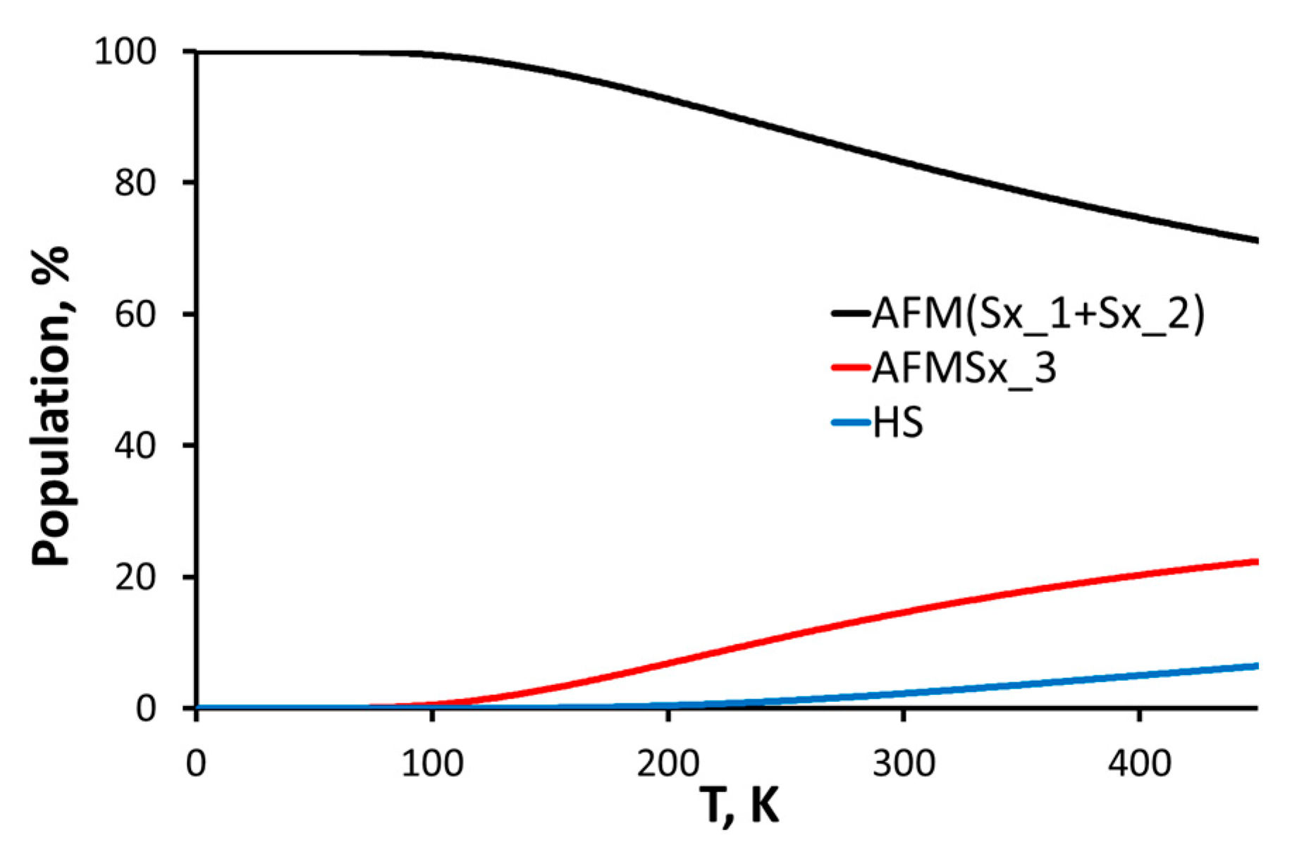
2.6. Theoretical Studies

The entire structures of complexes 12 are oversized for reliable first principles quantum chemical calculations. Therefore, we limited ourselves to modelling the primary coordination shell of the iron ions. The crystal structure of ferric acetate [44] was used as a template and three of the acetate ligands were replaced by ethanol molecules to reproduce the antibiotics “tail” hydroxyl groups. The experimental evidence that our complexes contain no coordinated water and that the three irons are placed in 2:1 different environments prompted us to remove two water ligands in the initial structure and to change the last one to a hydroxide anion. In addition, we built two possible architectures that differ in the orientation of the carboxylate anion: in the first one the COO-groups are parallel to the plane of the {Fe33–O)} core (Figure 6A), while in the second one the COO-function bridging Fe(1) and Fe(2) lies outside this plane (Figure 6B). The stability of the two structures was assessed by the full geometry optimization of the corresponding high spin states (HS, St = 15/2). The free energy difference between A and B is ca. 6 kcal/mol which indicates that construct B is the preferred configuration. Moreover, B is the more reasonable one, if we consider the antibiotics total skeletons and the geometry of their known compounds [43]. Structural characteristics of B are shown in Table 3. The iron ions Fe(1) and Fe(2) are situated in triangular bipyramidal environments with Fe−O bond lengths varying within the range of 1.9–2.1 Å, typical for iron oxygen-containing compounds [44,45,46,47,48]. The third metal centre Fe(3) is enveloped by six oxygens which form a slightly distorted octahedral structure, where the equatorial plane is occupied by equidistant carboxylate, alcohol, and hydroxide oxygens, while the second hydroxide and the oxo-oxygens are placed at axial positions. The inequivalent surrounding of the individual metal centres corroborates well the experimentally observed Mössbauer asymmetric doublets for 12.
Fe(III) features five unpaired electrons; this allows different multiplicities and magnetic behaviour of the trinuclear construct—ferromagnetic, with total spin St = 15/2 (labelled HS) and antiferromagnetic with total spin St = 5/2 (labelled AFMSx). To model the possible antiferromagnetic sextets, we applied a spin flip on each metal centre (the corresponding AFM sextets labelled according to the Fe numbering in Figure 5 and Figure 6). This procedure does not change the geometry but changes the energetics and the electron and spin distribution. The Gibbs free energy at 293 K was calculated and each state population in the range 0−450 K (Figure 7) was determined (450 K being approximately the melting temperature of the complexes). AFMSx_1 and AFMSx_2 exhibit ignorable energy difference of less than 0.1 kcal/mol and are essentially degenerate and structurally identical; so only structural results for AFMSx_1 are shown further on. Thus, the most unfavourable state is the high-spin one (St = 15/2) followed by AFMSx_3. The spin population is temperature dependent: at room temperature all species are present while at 77 K and 0 K only the penta-coordinated irons define the magnetic conduct. Yet, the calculated g-factors are very close (Table 4). These findings are in agreement with the EPR data which imply an enhanced antiferromagnetic interaction between magnetically coupled iron(III) ions upon temperature decrease. In addition, the credibility of the presence of construct B as antiferromagnetic sextet is supported by the calculated g-factor of 2.005, which matches the experimentally observed ones g = 2.005 (1) and g = 2.010 (2), respectively.
The NBO spin population of the [Fe33–O)]7+ core is presented in Table 5. It is easily seen that the spin distribution of the iron ions is analogous in all spin states—slightly lower at the penta-coordinated state and always somewhat higher at the octahedral species. The missing portion of the total spin is distributed predominantly on the oxygens with highest values (0.1−0.2) on the equatorial hydroxide one. The spin density on the (μ3–O)-centre correlates with the relative energy of the spin states (Table 4) indicating that the complex stability depends critically on the spin localization at this centre.
Similar to the spin distribution, the charge distribution in the first coordination shell (Table 6) is fairly uniform: all iron ions bear charges of ca. 2.2 (minor excess on the six-coordinated one), all carboxylate oxygen charges are ca. −0.8, the alcohol groups are with summed charges −0.3, and the hydroxide ions are with total charge −0.7. This confirms the overall identical environment of Fe(1) and Fe(2). The charge envelope of Fe(3) differs only in the additional hydroxide ligand. Unlike the spin density distribution, the charge on the μ3–O fragment is constant in all spin states.
The s-electron density on the iron ions, calculated from the NAO occupancy, is 6.246, irrespective of the coordination pattern, and corroborates the identical isomer shift established from the Mössbauer spectra analysis (Table 2, Figure 4).
Valuable information regarding the solidity of the bonds in the first coordination sphere of the complexes can be obtained from the vibrational analysis of the most stable structure. To be able to compare the computed to the experimentally measured frequencies for 12, we need to make models that differ in mass, as do the real ligands, while conserving the simplicity of the constructs. For this purpose, isotopic substitution of some carbons and hydrogens in structure B was performed. When choosing the isotopes and their arrangement, a maximally symmetric distribution was maintained with respect to the main chain of the model ligand, as well as the mass number of the isotope used. 2H, 13C, and 14C were chosen (Figure S4), thereby minimizing direct impact on the atoms directly involved in the metal–oxygen bonds.
The most important vibrational frequencies are presented unscaled in Table 7 and compared to the recorded IR spectra of 12. The deviations are within an acceptable range, and the trends witnessed in the experimental data (Figure 2) are preserved. Lower values for the vibrational frequencies in salinomycin directly correlate with the larger effective mass of the ligand. This allows us to see a correlation between the Mössbauer effect observed and the bond strength on the other hand. The vibrational frequencies of the carboxylate groups are in excellent agreement with the experimentally recorded ones and with the differences in the wave numbers for asymmetric and symmetric stretching, which confirms the validity of the model and the assumption about the structure of the first coordination sphere. Moreover, this correspondence authenticates the isotopic substitution as an approach for distinctive treatment, with respect to predicting the vibrational behaviour of large systems, having an identical core but different mass, which, to our knowledge, has not been reported so far.

3. Materials and Methods

3.1. Reagents

Sodium monensinate (MonNa) and salinomycinate (SalNa) were complimentary supplied by Huvefarma Ltd. (Peshtera, Bulgaria). Monensic acid (MonH×H2O) and salinomycinic acid (SalH) were prepared according to Gertenbah and Popov [49]. The rest of the reagents of p.a. grade (FeCl3×6H2O, Et3N, acetonitrile, methanol, and ether) were purchased from local suppliers. In all experiments deionized water was used.

3.2. Synthesis of Complexes 12

MonH×H2O (0.5 mmol, 344 mg in 15 mL MeOH) or SalH (0.5 mmol, 375.5 mg), respectively, was mixed with Et3N (1 mmol, 139.5 µL) and stirred at r. t. for 15 min. After that FeCl3×6H2O (0.25 mmol, 70.1 mg in 15 mL MeOH) was slowly added and the resulting orange solution was precipitated from water to produce a yellow solid phase, which was filtered off, washed subsequently with water and ether, and dried in a desiccator. The composition and elemental analysis data for coordination compounds 1 (MonH) and 2 (SalH) are presented in Table 1. Yield: 1—281 mg, 75%; 2—292 mg, 70%. The isolated complexes are insoluble in water and are sparingly soluble in most of the organic solvents.

3.3. Spectroscopic Studies

Infrared (IR) spectra were obtained in KBr pellets (4000–400 cm−1) using a Nicolet 6700 FT-IT spectrometer (Thermo Scientific, Madison, WI, USA).
Thermogravimetric experiments (TG−DTA/MS) were performed on Setaram Labsys Evo 1600 (25–600 °C) with a heating rate of 10 K/min in argon flow (Caluire-et-Cuire, France). The apparatus is equipped with an Omnistar GSD 301 O2 mass spectrometer, Pfeiffer Vacuum.
57Fe Mössbauer measurements were performed using a Wissel spectrometer (Wissenschaftliche Elektronik GmbH, Starnberg, Germany), operating in a constant acceleration mode. The transmission Mössbauer spectra were measured at room temperature (293 K) and 77 K with an analyser with 1024 channels, source 57Co/Rh (15 mCi). The samples were adjusted to an optimum thickness for a “thin” absorber density of about 10 mg/cm2 of the total iron content (60 mg of solid samples studied). The hyperfine interaction parameters: isomer shift (δ) relative to α-Fe and quadrupole splitting (∆) were determined via computer fitting with Lorentzian lineshapes using the program NORMOS (based on the least squares method). The isomer shift (δ), quadrupole splitting (∆), and line width (Γ) are calculated with ±0.01 mm/s error.
The electron paramagnetic resonance (EPR) studies were conducted on Bruker BioSpin EMXplus10/12 EPR spectrometer (Karlsruhe, Germany) working at 9.4 GHz.
The sample crystallinity degree was evaluated by powder X-ray diffraction (XRD) analysis using PANalytical Empyrean X-ray diffractometer (Malvern Panalytical, Malvern, UK) with CuKα radiation (λ = 0.15418 nm) operating at 40 kV, 30 mA.
Elemental analysis (C, H) was performed on an organic elemental analyser Vario MACRO cube (Elementar analysensysteme GmbH, Stuttgart, Germany). The metal content was determined on Perkin-Elmer SCIEX-ELAN DRC-e ICP-MS (Waltham, MA, USA) after wet digestion of samples with conc. HNO3 using appropriate standards.

3.4. Quantum Chemical Calculations

The overall structure of the complexes 12 requires a large number of basis functions for its description, which exceeds the potential of the computational software and hardware available. For this reason, simplifications were considered and only the first coordination sphere was modelled as detailed in Section 2.6. The geometry of all structures in states with multiplicities 6 (AFMSx) and 16 (HS) was optimized with the hybrid functional B3LYP [50], employing the 6-31G(d) basis set, in vacuo, including the Grimme D3 correction for dispersion interactions within the Gaussian 16 software. The energy minimum of each structure was verified with a vibrational frequency analysis. These structures were a starting point for the calculation of all energies and EPR properties using the ORCA 5.0.3 software package. Based on the geometries obtained and their symmetry, a further spin-flip on each iron centre was also performed. The calculations were meant to provide reliable results for high-spin systems in which the exchange interactions are of paramount importance, which is why we switched to a hybrid functional with 50% Hartree–Fock exchange such as BHandHLYP [51,52,53] with a triple-zeta basis set for the ligands and CP(PPP) basis set for Fe [54]. All calculations were performed in vacuo. For each structure and multiplicity, the thermochemical properties and EPR parameters were evaluated. Particularly, the free energy comprises contributions of the total energy, the zero-point vibrational energy, the translational, rotational, and vibrational energy to the entropy assessment at the respective temperature, the latter energy quantified in the harmonic approximation.

4. Conclusions

Aiming to achieve the combined effect of iron binding agents and cancer therapeutics, the first complexes of Fe(III) with two antibiotics with promising anticancer activity were synthesized and characterized structurally utilizing an assortment of spectroscopies and first principles molecular modelling. The antibiotics monensin and salinomycin have kindred molecular skeletons and documented “head-to-tail” patterns of coordination with d-elements. The complexes obtained in alkaline medium do not form crystals for X-ray description; therefore, the vibrational, Mössbauer, and EPR spectroscopies were employed for characterization. The elemental analysis revealed 1:1 ratio of Fe ions and ionophorous antibiotics. The hypothesis that the structure of the complexes contains a trinuclear {Fe33–O)}7+ core allowed their simulating with a simple molecular model, for which quantum chemical methods of trustworthy quality could be applied. The agreement between the theoretically assessed and the experimentally measured characteristics is very good, validating the coordination pattern and the antiferromagnetic spin state. In addition to the structural features, molecular modelling shed light on the charge and spin distribution and on the temperature dependence of the spin states population, reflected in the physicochemical characteristics variation upon temperature decrease. Furthermore, it was shown that isotopic substitution is an efficient strategy for predicting the vibrational behaviour of compounds with the same core but with ligands of different mass; this can be deemed a methodological merit of the modelling approach in the study.

Supplementary Materials

The following supporting information can be downloaded at: https://www.mdpi.com/article/10.3390/molecules29020364/s1, Figure S1: TGV−DTA (top) and TGV−MS (bottom) curves of MonH/complex 1 (left); SalH/complex 2 (right). Colour code: weight decrease (black); endo- and exothermic effects (grey); H2O loss (blue); CO2 loss (red).; Figure S2: Powder XRD spectra of (a) complex 1 and (b) complex 2; Figure S3: 57Fe Mössbauer spectra of (a) 1 and (b) 2 at 77 K; Figure S4: Isotopic substitution of selected carbons and hydrogens in structure B.

Author Contributions

Conceptualization, N.P. and I.P.; methodology and analysis, N.P., A.T., E.E., Z.C.-Z., D.P., R.S. and R.K.; resources, P.D.; writing—original draft preparation, N.P. and I.P.; and writing—review and editing, I.P. and A.T. All authors have read and agreed to the published version of the manuscript.

Funding

This research received no external funding.

Institutional Review Board Statement

Not applicable.

Informed Consent Statement

Not applicable.

Data Availability Statement

Data are available from the authors upon request.

Acknowledgments

The molecular modelling was performed on facilities provided within project KP-06-COST/5/23.05.2023 in support of COST action COSY-CA21101.

Conflicts of Interest

The authors declare that the research was conducted in the absence of any commercial or financial relationships that could be construed as a potential conflict of interest. Author Petar Dorkov comes from the Research and Development Department of Biovet Ltd. company; however, there are no conflicts of interest related to the research in this study.

References

  1. Chaaban, T.; Mohsen, Y.; Ezzeddine, Z.; Ghssein, G. Overview of Yersinia pestis metallophores: Yersiniabactin and Yersinopine. Biology 2023, 12, 598. [Google Scholar] [CrossRef]
  2. Chifman, J.; Laubenbacher, R.; Torti, S.V. A systems biology approach to iron metabolism. Adv. Exp. Med. Biol. 2014, 844, 201–225. [Google Scholar] [CrossRef] [PubMed]
  3. Maret, W. Metalloproteomics, metalloproteomes, and the annotation of metalloproteins. Metallomics 2010, 2, 117–125. [Google Scholar] [CrossRef] [PubMed]
  4. Zoroddu, M.A.; Aaseth, J.; Crisponi, G.; Medici, S.; Peana, M.; Nurchi, V.M. The essential metals for humans: A brief overview. J. Inorg. Biochem. 2019, 195, 120–129. [Google Scholar] [CrossRef] [PubMed]
  5. Crichton, R.R.; Pierre, J.L. Old iron, young copper: From Mars to Venus. BioMetals 2001, 14, 99–112. [Google Scholar] [CrossRef] [PubMed]
  6. Archibald, F. Lactobacillus plantarum, an organism not requiring iron. FEMS Microbio. Lett. 1983, 19, 29–32. [Google Scholar] [CrossRef]
  7. Pierre, J.L.; Fontecave, M.; Crichton, R.R. Chemistry for an essential biological process: The reduction of ferric iron. BioMetals 2002, 15, 341–346. [Google Scholar] [CrossRef]
  8. Paterson, S.; Armstrong, N.J.; Iacopetta, B.J.; McArdle, H.J.; Morgan, E.H. lntravesicular pH and iron uptake by immature erythroid cells. J. Cell. Physiol. 1984, 120, 225–232. [Google Scholar] [CrossRef]
  9. Page, M.G.P. The role of iron and siderophores in infection, and the development of siderophore antibiotics. Clin. Infect. Dis. 2019, 69, S529–S537. [Google Scholar] [CrossRef]
  10. Liang, C.; Zhang, X.; Yang, M.; Dong, X. Recent progress in ferroptosis inducers for cancer therapy. Adv. Mater. 2019, 31, e1904197. [Google Scholar] [CrossRef]
  11. Qiu, Y.; Cao, Y.; Cao, W.; Jia, Y.; Lu, N. The application of ferroptosis in diseases. Pharmacol. Res. 2020, 159, 104919. [Google Scholar] [CrossRef] [PubMed]
  12. Huang, G.; Pan, S.T. ROS-mediated therapeutic strategy in chemo-/radiotherapy of head and neck cancer. Oxid. Med. Cell. Longev. 2020, 2020, 5047987. [Google Scholar] [CrossRef]
  13. Guo, Q.; Li, L.; Hou, S.; Yuan, Z.; Li, C.; Zhang, W.; Zheng, L.; Li, X. The role of iron in cancer progression. Front. Oncol. 2021, 11, 778492. [Google Scholar] [CrossRef]
  14. Sacco, A.; Battaglia, A.M.; Botta, C.; Aversa, I.; Mancuso, S.; Costanzo, F.; Biamonte, F. Iron metabolism in the tumor microenvironment—Implications for anti-cancer immune response. Cells 2021, 10, 303. [Google Scholar] [CrossRef] [PubMed]
  15. Cosialls, E.; El Hage, R.; Dos Santos, L.; Gong, C.; Mehrpour, M.; Hamai, A. Ferroptosis: Cancer stem cells rely on iron until “to die for” it. Cells 2021, 10, 2981. [Google Scholar] [CrossRef]
  16. Yan, H.F.; Zou, T.; Tuo, Q.Z.; Xu, S.; Li, H.; Belaidi, A.A.; Lei, P. Ferroptosis: Mechanisms and links with diseases. Signal Transduct. Target Ther. 2021, 6, 49. [Google Scholar] [CrossRef] [PubMed]
  17. Ma, T.L.; Chen, J.X.; Zhu, P.; Zhang, C.B.; Zhou, Y.; Duan, J.X. Focus on ferroptosis regulation: Exploring novel mechanisms and applications of ferroptosis regulator. Life Sci. 2022, 307, 120868. [Google Scholar] [CrossRef] [PubMed]
  18. Antoszczak, M.; Muller, S.; Caneque, T.; Colombeau, L.; Dusetti, N.; Santofimia-Castano, P.; Gaillet, C.; Puisieux, A.; Iovanna, J.L.; Rodriguez, R. Iron-sensitive prodrugs that trigger active ferroptosis in drug-tolerant pancreatic cancer cells. J. Am. Chem. Soc. 2022, 144, 11536–11545. [Google Scholar] [CrossRef]
  19. Wu, Z.; Zhong, M.; Liu, Y.; Xiong, Y.; Gao, Z.; Ma, J.; Zhuang, G.; Hong, X. Application of natural products for inducing ferroptosis in tumor cells. Biotechnol. Appl. Biochem. 2022, 69, 190–197. [Google Scholar] [CrossRef]
  20. Renshaw, J.C.; Robson, G.D.; Trinci, A.P.J.; Wiebe, M.G.; Livens, F.R.; Collison, D.; Taylor, R.J. Fungal siderophores: Structures, functions and applications. Mycol. Res. 2002, 106, 1123–1142. [Google Scholar] [CrossRef]
  21. De Lorenzo, V.; Martinez, J.L. Aerobactin production as a virulence factor: A reevaluation. Eur. J. Clin. Microbiol. Infect. Dis. 1988, 7, 621–629. [Google Scholar] [CrossRef] [PubMed]
  22. Nikaido, H. Uptake of iron-siderophore complexes across the bacterial outer membrane. Trends Microbiol. 1993, 1, 5–7. [Google Scholar] [CrossRef] [PubMed]
  23. Dreechsel, H.; Jung, G. Peptide siderophores. J. Peptide Sci. 1998, 4, 147–181. [Google Scholar] [CrossRef]
  24. De Luca, N.G.; Wood, P.M. Iron uptake by fungi: Contrasted mechanisms with internal or external reduction. Adv. Microb. Physiol. 2000, 43, 39–74. [Google Scholar] [CrossRef]
  25. Haas, H.; Eisendle, M.; Turgeon, B.G. Siderophores in fungal physiology and virulence. Annu. Rev. Phytopathol. 2008, 46, 149–187. [Google Scholar] [CrossRef] [PubMed]
  26. Holinsworth, B.; Martin, J.D. Siderophore production by marine-derived fungi. Biometals 2009, 22, 625–632. [Google Scholar] [CrossRef]
  27. Hider, R.C.; Kong, X. Chemistry and biology of siderophores. Nat. Prod. Rep. 2010, 27, 637–657. [Google Scholar] [CrossRef]
  28. Codd, R.; Richardson-Sanchez, T.; Telfer, T.J.; Gotsbacher, M.P. Advances in the chemical biology of desferrioxamine B. ACS Chem. Biol. 2018, 13, 11–25. [Google Scholar] [CrossRef]
  29. Li, Z.; Chen, L.; Chen, C.; Zhou, Y.; Hu, D.; Yang, J.; Chen, Y.; Zhuo, W.; Mao, M.; Zhang, X.; et al. Targeting ferroptosis in breast cancer. Biomark. Res. 2020, 8, 58. [Google Scholar] [CrossRef]
  30. Hu, H.; Xu, Q.; Mo, Z.; Hu, X.; He, Q.; Zhang, Z.; Xu, Z. New anti-cancer explorations based on metal ions. J. Nanobiotechn. 2022, 20, 457. [Google Scholar] [CrossRef]
  31. Antoszczak, M.; Huczynski, A. Salinomycin and its derivatives—A new class of multiple-targeted “magic bullets”. Eur. J. Med. Chem. 2019, 176, 208–227. [Google Scholar] [CrossRef] [PubMed]
  32. Tefas, L.R.; Barbalata, C.; Tefas, C.; Tomuta, I. Salinomycin-based drug delivery systems: Overcoming the hurdles in cancer therapy. Pharmaceutics 2021, 13, 1120. [Google Scholar] [CrossRef] [PubMed]
  33. Petkov, N.; Pantcheva, I.; Ivanova, A.; Stoyanova, R.; Kukeva, R.; Alexandrova, R.; Abudalleh, A.; Dorkov, P. Novel cerium(IV) coordination compounds of monensin and salinomycin. Molecules 2023, 28, 4676. [Google Scholar] [CrossRef]
  34. Amani, V.; Safari, N.; Khavasi, H.R. Solution and solid state characterization of oxo-centered trinuclear iron(III) acetate complexes [Fe33-O)(µ-OAc)6(L)3]+. Spectrochim. Acta A Mol. Biomol. Spectrosc. 2012, 85, 17–24. [Google Scholar] [CrossRef] [PubMed]
  35. Kiana, S.; Yazdanbakhsh, M.; Jamialahmadi, M.; Tayyari, S.F. Vibrational assignment and structure of trinuclear oxo-centered of basic formate iron(III) and chromium(III) complexes: A density functional theory study. Spectrochim. Acta A Mol. Biomol. Spectrosc. 2014, 130, 287–294. [Google Scholar] [CrossRef]
  36. Dimiza, F.; Hatzidimitriou, A.G.; Sanakis, Y.; Papadopoulos, A.N.; Psomas, G. Trinuclear and tetranuclear iron(III) complexes with fenamates: Structure and biological profile. J. Inorg. Biochem. 2021, 218, 111410. [Google Scholar] [CrossRef]
  37. Feng, Q.; Li, B.; Du, R.; Jiang, F.; Liu, T. Syntheses, crystal structures, spectroscopic and magnetic properties of two trinuclear iron complexes with carboxylate and oximate mixed-bridge ligands. Trans. Met. Chem. 2019, 44, 49–55. [Google Scholar] [CrossRef]
  38. Chakradhar, R.P.; Sivaramaiah, G.; Rao, J.L.; Gopal, N.O. Fe3+ ions in alkali lead tetraborate glasses—An electron paramagnetic resonance and optical study. Spectrochim. Acta A Mol. Biomol. Spectrosc. 2005, 62, 51–57. [Google Scholar] [CrossRef]
  39. Greenwood, N.N.; Gibb, T.C. Mossbauer Spectroscopy; Springer: Dordrecht, The Netherlands, 1971. [Google Scholar] [CrossRef]
  40. Murray, K.S. Binuclear oxo-bridged iron(III) complexes. Coord. Chem. Rev. 1974, 12, 1–35. [Google Scholar] [CrossRef]
  41. Bill, E.; Krebs, C.; Winter, M.; Gerdan, M.; Trautwein, A.X.; Florke, U.; Haupt, H.-J.; Chaudhuri, P. A triangular iron(III) complex potentially relevant to iron(III)-binding sites in ferreascidin. Chem. Eur. J. 1997, 3, 193–201. [Google Scholar] [CrossRef]
  42. Dorkov, P.; Pantcheva, I.N.; Sheldrick, W.S.; Mayer-Figge, H.; Petrova, R.; Mitewa, M. Synthesis, structure and antimicrobial activity of manganese(II) and cobalt(II) complexes of the polyether ionophore antibiotic sodium monensin A. J. Inorg. Biochem. 2008, 102, 26–32. [Google Scholar] [CrossRef]
  43. Pantcheva, I.N.; Zhorova, R.; Mitewa, M.; Simova, S.; Mayer-Figge, H.; Sheldrick, W.S. First solid state alkaline-earth complexes of monensic acid A (MonH): Crystal structure of [M(Mon)2(H2O)2] (M = Mg, Ca), spectral properties and cytotoxicity against aerobic Gram-positive bacteria. Biometals 2010, 23, 59–70. [Google Scholar] [CrossRef] [PubMed]
  44. Bijelić, J.; Gašo-Sokač, D.; Djerdj, I. Single crystal growth, structural characterization and magnetic properties study of an antiferromagnetic trinuclear iron(III) acetate complex with uncoordinated hexamine. Inorg. Chim. Acta 2021, 520, 120292. [Google Scholar] [CrossRef]
  45. Raptopoulou, C.P.; Sanakis, Y.; Boudalis, A.K.; Psycharis, V. Salicylaldoxime (H2salox) in iron(III) carboxylate chemistry: Synthesis, X-ray crystal structure, spectroscopic characterization and magnetic behavior of trinuclear oxo-centered complexes. Polyhedron 2005, 24, 711–721. [Google Scholar] [CrossRef]
  46. Boudalis, A.K.; Sanakis, Y.; Raptopoulou, C.P.; Terzis, A.; Tuchagues, J.-P.; Perlepes, S.P. A trinuclear cluster containing the {Fe33-O)}7+ core: Structural, magnetic and spectroscopic (IR, Mössbauer, EPR) studies. Polyhedron 2005, 24, 1540–1548. [Google Scholar] [CrossRef]
  47. Lalia-Kantouri, M.; Papadopoulos, C.; Hatzidimitriou, A.; Bakas, T.; Pachin, S. A trinuclear iron(III) complex containing the semi-cubane [Fe33-O)]7+ core: Structural, spectroscopic, magnetic and electrochemical study. Z. Anorg. Allg. Chem. 2010, 636, 531–538. [Google Scholar] [CrossRef]
  48. Pap, J.S.; Kaizer, J.; Giorgi, M.; Speier, G. Molecular and electronic structure of a trinuclear oxo-centred iron(III) complex with trigonal bipyramidal metal sites. Inorg. Chem. Comm. 2010, 13, 1069–1073. [Google Scholar] [CrossRef]
  49. Gertenbach, P.G.; Popov, A.I. Solution chemistry of monensin and its alkali metal ion complexes. Potentiometric and spectroscopic studies. J. Am. Chem. Soc. 1975, 97, 4738–4744. [Google Scholar] [CrossRef]
  50. Stephens, P.J.; Devlin, F.J.; Chabalowski, C.F.; Frisch, M.J. Ab initio calculation of vibrational absorption and circular dichroism spectra using density functional force fields. J. Phys. Chem. 1994, 98, 11623–11627. [Google Scholar] [CrossRef]
  51. Becke, A.D. A new mixing of Hartree-Fock and local density-functional theories. J. Chem. Phys. 1993, 98, 1372–1377. [Google Scholar] [CrossRef]
  52. Zhao, H.; Fang, C.; Gao, J.; Liu, C. Spin-state energies of heme-related models from spin-flip TDDFT calculations. Phys. Chem. Chem. Phys. 2016, 18, 29486–29494. [Google Scholar] [CrossRef] [PubMed]
  53. Yang, Y.; Ratner, M.A.; Schatz, G.C. Computational modeling of octahedral iron oxide clusters: Hexaaquairon(III) and its dimers. J. Phys. Chem. C 2013, 117, 21706–21717. [Google Scholar] [CrossRef]
  54. Neese, F. Prediction and interpretation of the 57Fe isomer shift in Mössbauer spectra by density functional theory. Inorg. Chim. Acta 2002, 337, 181–192. [Google Scholar] [CrossRef]
Figure 1. Chemical structures of (a) MonH and (b) SalH.
Molecules 29 00364 g001
Figure 2. IR spectra of (a) MonH/complex 1 and (b) SalH/complex 2.
Molecules 29 00364 g002
Figure 3. X-band temperature-dependent EPR spectra of (a) 1 and (b) 2 and the temperature dependence of the linewidth: (c) for 1—150 mT (77 K), 99 mT (295 K); (d) for 2—175 mT (77 K), 122 mT (295 K).
Molecules 29 00364 g003aMolecules 29 00364 g003b
Figure 4. 57Fe Mössbauer spectra of (a) 1 and (b) 2 at 293 K.
Molecules 29 00364 g004
Figure 5. Sketch of the proposed structure of complexes 12 (note that the antibiotic backbones lie outside the plane of the Fe3O-unit and significant geometry distortions at each iron centre might be expected).
Molecules 29 00364 g005
Figure 6. Modelled constructs of 12 with parallel (A) and non-parallel (B) orientation of the carboxylate donor atoms relative to the triangular μ3–O-iron plane. Hydrogen is omitted for clarity. Colour code: C—grey, O—red, Fe—orange.
Molecules 29 00364 g006
Figure 7. Boltzmann distribution (0−450 K) of the spin states of the construct B.
Molecules 29 00364 g007
Table 1. Elemental analysis of 12.
ComplexCompositionMW, g/molC, %H, %Fe, %
Calc. FoundCalc. FoundCalc. Found
[Fe33-O)Mon3(OH)4], 1C108H187Fe3O382261.1857.3756.338.347.357.417.39
[Fe33-O)Sal3(OH)4], 2C126H211Fe3O382501.5760.5060.128.508.446.706.69
Table 2. Mössbauer spectra parameters of 12 at r. t. (the data at 77 K are given in parenthesis).
CompoundComponentδ, mm/s∆, mm/sΓ, mm/sA, %M, %
1Db10.39 (0.59)0.61 (0.59)0.30 (0.28)65 (66)0.86 (5.21)
Db20.39 (0.58)0.82 (0.84)0.45 (0.33)35 (34)0.31 (2.25)
2Db10.39 (0.58)0.63 (0.61)0.28 (0.29)70 (65)1.69 (4.70)
Db20.38 (0.57)0.80 (0.93)0.49 (0.38)30 (35)0.42 (1.95)
Table 3. Bond lengths [Å] for structure B (optimized HS state).
BondFe(1)Fe(2)Fe(3)
μ3-O−Fe1.91.92.0
COO−Fe2.02.02.1
2.12.12.1
ROH−Fe2.11.92.1
OH−Fe1.91.91.9
2.1
Table 4. Relative energy, population, and g-factors for all spin states of construct B.
MultiplicityΔG, kcal/mol
293 K
Population, % 293 KPopulation, %
77 K
g-Factor
0 K
HS2.152.100.002.005
AFM(Sx_1 + Sx_2)0.0083.899.92.005
AFMSx_31.0414.10.102.006
Table 5. NBO spin population of the [Fe33–O)]7+ core.
Centre/Spin StateHSAFMSx_1AFMSx_2AFMSx_3
Fe(1)4.43–4.424.424.42
Fe(2)4.43 4.43 –4.434.43
Fe(3)4.49 4.484.49 –4.48
μ3–O0.40 0.020.110.27
Table 6. NBO charges of the first coordination shell and metal ions.
Fe(1)Fe(2)Fe(3)
Fe 2.18 2.19 2.24
μ3–O–1.46–1.46–1.46
COO–0.82–0.83–0.82
COO–0.84–0.84–0.83
ROH–0.85/–0.31 *–0.83/–0.30 *–0.97/–0.29 *
OH–1.17/–0.70 *–1.18/–0.71 *–1.19/–0.71 *
OH–1.02/–0.65 *
* The values are for the NBO summed OH-charges.
Table 7. Frequencies of some important vibrations (cm−1) and similarity factors (SF, the ratio of the experimental to the calculated value for each pair of results).
VibrationMonensinate ComplexSalinomycinate Complex
Exp.Calc.SFExp.Calc.SF
ν C = O a s y m , COO¯153016020.96152215330.99
ν C = O s y m , COO¯141815010.94141914360.99
Δ   ( ν C = O a s y m ν C = O s y m ) 11210110397
Disclaimer/Publisher’s Note: The statements, opinions and data contained in all publications are solely those of the individual author(s) and contributor(s) and not of MDPI and/or the editor(s). MDPI and/or the editor(s) disclaim responsibility for any injury to people or property resulting from any ideas, methods, instructions or products referred to in the content.

