Article

Ceratocarpus arenarius: Botanical Characteristics, Proximate, Mineral Composition, and Cytotoxic Activity

by
Aigerim Kantureyeva
1,*,
Gulbaram Ustenova
1,
Alenka Zvonar Pobirk
2,
Serzhan Mombekov
1,
Moldir Koilybayeva
1,
Akerke Amirkhanova
1,
Nadezhda Gemejiyeva
3,
Assem Mamurova
4 and
Nina Kočevar Glavač
5,*
1
School of Pharmacy, S.D. Asfendiyarov Kazakh National Medical University, Tole-bi 94, Almaty 050012, Kazakhstan
2
Department of Pharmaceutical Technology, Faculty of Pharmacy, University of Ljuljana, Aškerčeva cesta 7, 1000 Ljubljana, Slovenia
3
Laboratory of Plant Resources, Institute of Botany and Phyto-Introductions, Almaty 050000, Kazakhstan
4
Department of Biodiversity of Bioresources, al-Farabi Kazakh National University, Almaty 050040, Kazakhstan
5
Department of Pharmaceutical Biology, Faculty of Pharmacy, University of Ljubljana, Aškerčeva cesta 7, 1000 Ljubljana, Slovenia
*
Authors to whom correspondence should be addressed.
Molecules 2024, 29(2), 384; https://doi.org/10.3390/molecules29020384
Submission received: 3 November 2023 / Revised: 9 January 2024 / Accepted: 10 January 2024 / Published: 12 January 2024
(This article belongs to the Section Natural Products Chemistry)

Abstract

:
Ceratocarpus arenarius (Chenopodiaceae) is an under-investigated annual plant that occurs in dry areas stretching from eastern and south-eastern Europe to East Asia. This article presents the botanical characterization and examination of proximate parameters, minerals and cytotoxic activity of C. arenarius that grows wild in Kazakhstan. The results of morphological analysis using a light microscope, based on cross-sections of stems, roots and leaves, provide the necessary data to develop a regulatory document for this herbal substance as a raw material for use in the pharmaceutical, cosmetic and food industries. The investigated proximate characteristics included moisture content (6.8 ± 0.28%), ash (5.9 ± 0.40%), fat (12.5 ± 21.28%) and protein (392.85 ± 25.50). The plant is also rich in minerals (mg/100 g dry weight); Na (20.48 ± 0.29), K (302.73 ± 1.15), Zn (4.45 ± 0.35), Fe (1.18 ± 0.03), Cu (0.11 ± 0.02), Mn (0.76 ± 0.01), Ca (131.23 ± 0.09) and Mg (60.69 ± 0.72). The ethanolic extract of C. arenarius showed no acute toxicity against the brine shrimp nauplii.

1. Introduction

Chenopodiaceae, in the order Caryophyllales, is one of the most interesting families with respect to having species with a large diversity in the structure of carbon-absorbing organs responsible for different types of photosynthesis [1]. The Chenopodiaceae family includes annual or perennial herbs, subshrubs, shrubs, small trees and climbers. They are found in deserts, semi-deserts, salt-marshes, and coastal or inland saline and ruderal sites [2]. The family includes about 98 genera and 1400 species distributed mainly in the extra tropical regions of the Northern Hemisphere [3].
Plant species belonging to the Chenopodiaceae family show high drought and salinity resistance and tolerance to nutrient deficiency and often grow in psammophytic or halophytic plant communities. They are considered pioneer plants in the colonization and settlement of harsh edaphic environments affected by salt or drought, and therefore, they play crucial roles in erosion control and rehabilitation of desert ecosystems [4].
The large territory of arid and semi-arid zones and diverse soil and climatic conditions, including extreme environmental conditions, are the reason for the rich genetic resources of Kazakhstan, Central Asia. About 6000 species of vascular plants from 160 families are distributed in the country [5,6].
Ceratocarpus arenarius L. (Figure 1) is an annual herbaceous plant belonging to the family of Chenopodiaceae. It is native to Kazakhstan (Figure 2), as well as other areas stretching from eastern and south-eastern Europe to East Asia, and is locally known as tumbleweed. It is usually found in dry climates with precipitation of 100–400 mm, in deserts, arid slopes, sands, wastelands and along roadsides. Plants produce fruits (utricles) near the soil surface (basicarps) and a continuous series of morphologically distinct fruits from the lower to upper parts of the canopy [7]. The seeds are numerous, approx. 4000 per plant, with a thick pericarp that fits tightly to the seed coat. In autumn, which is the end of the growing season, the above-ground parts of C. arenarius are easily separated from the soil surface and dispersed long distances by the wind [8].
C. arenarius is widely used in folk medicine and agriculture. It is a potential source of biologically active compounds such as polyphenolic compounds, e.g., flavonoids and phenolic acids, organic acids, saponins, steroids, and vitamins C and B2 [9,10]. To support the possibility of using this plant in medicine and the pharmaceutical, cosmetic and food industries, it is necessary to develop a regulatory document or a monograph for the herbal substance as a raw material. Important sections of such monographs, which determine the authenticity of plant raw materials, and ensure reliable quality control thereof, are morphological characteristics, both at a macroscopic and microscopic level.
Such characterization has not yet been available for C. arenarius. Therefore, the purpose of this work was to conduct a detailed morphological and anatomical study of C. arenarius that grows wild in Kazakhstan, as well as the plant’s nutrient and mineral contents and cytotoxic activity.

2. Results and Discussion

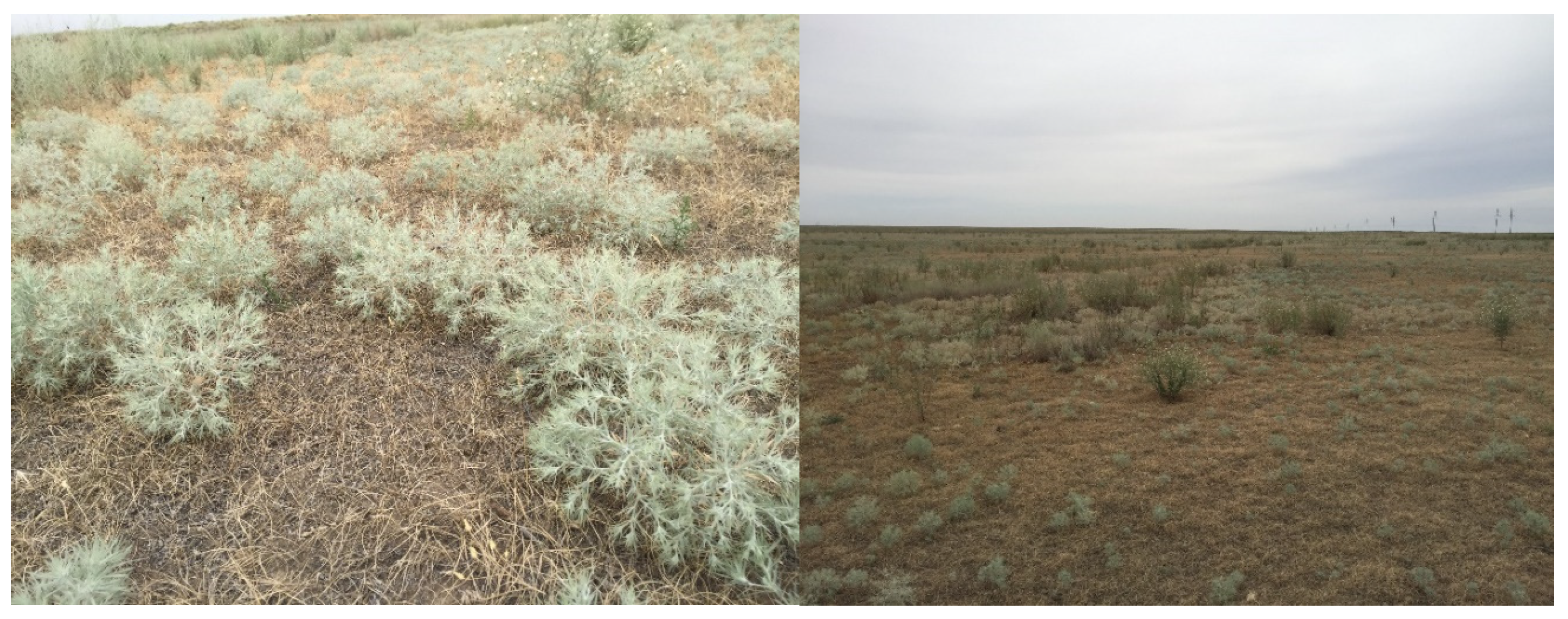
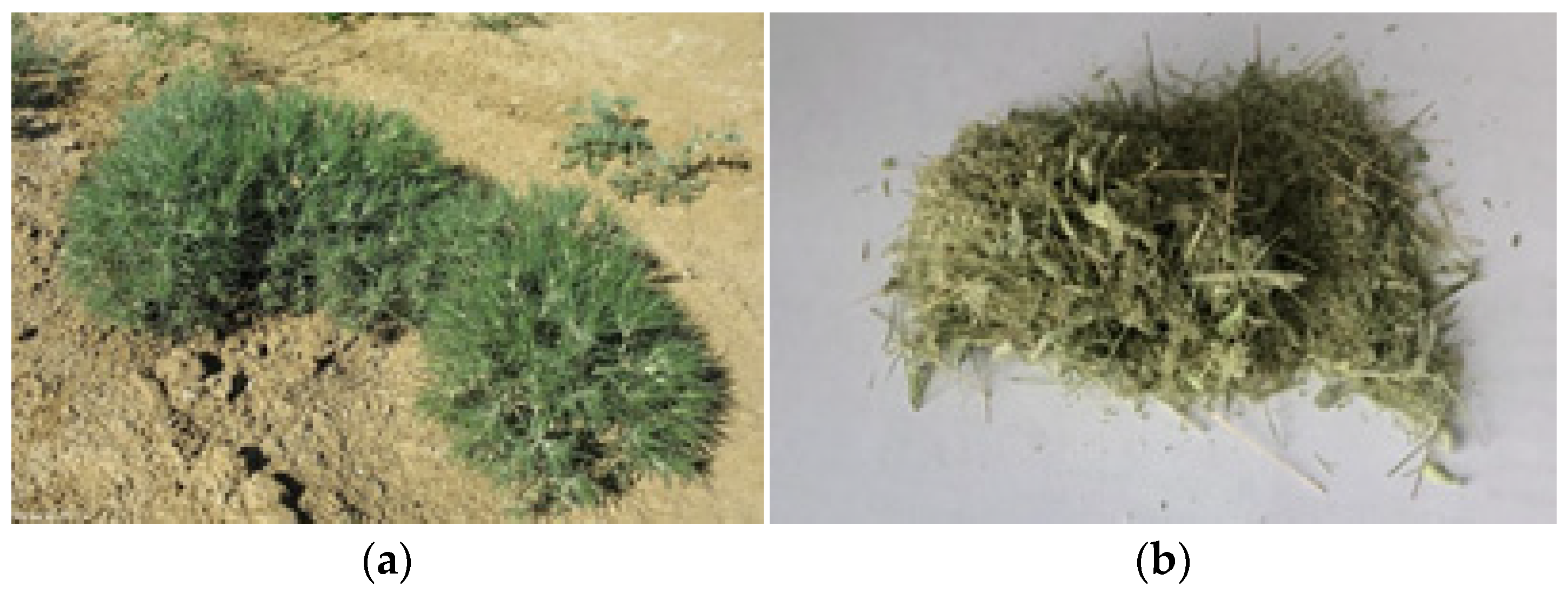
2.1. Morphological Characteristics

The C. arenarius plant (Figure 1) is grayish-green in color, and its plant height reaches 9.5 ± 1.16 cm. The plant is strongly and repeatedly fork-branched from the base, and the leaves often form a spherical shape. The leaves are alternate, narrowed to the base, single-nerved, with stellate hairs. They are aromatic in nature upon bruising/crushing in hands. The average leaf length is 3.5 ± 0.07 cm, and the number of leaves per plant is 37.2 ± 1.1. The stem is angular in shape and dark green in color. The average stem length is 11.4 ± 0.76 cm. The average seed length is 6.27 ± 0.12 mm. Morphological features of C. arenarius stems, leaves and seeds are presented in Table 1 and Figure 3.

2.2. Anatomical Characteristics

The anatomical characteristics of C. arenarius were assessed for the leaf, stem and root. The recorded observations are described below.

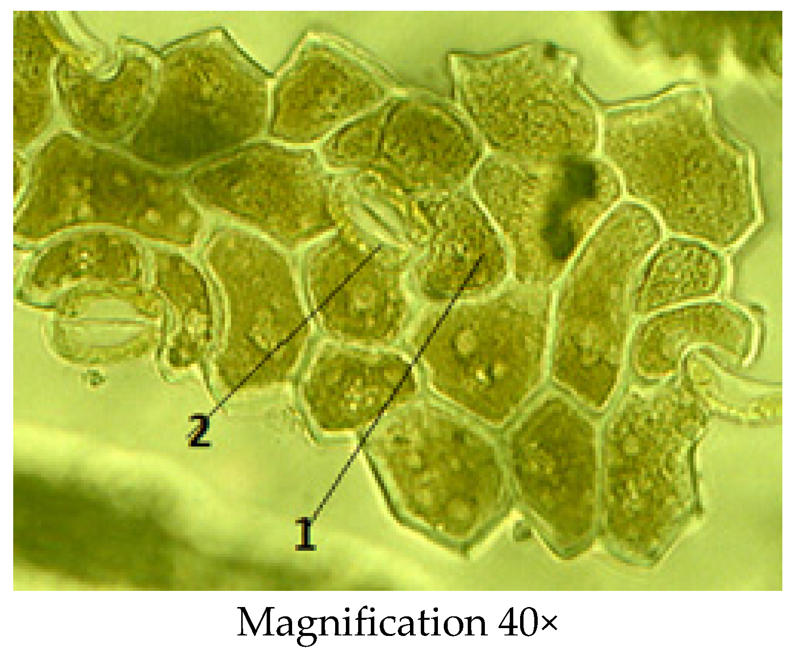
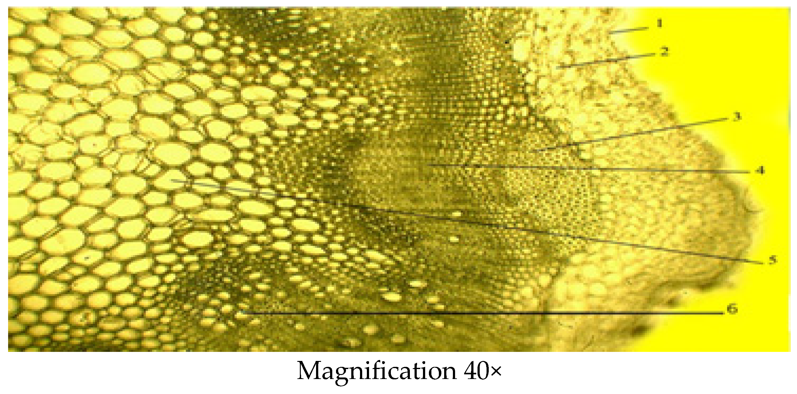
2.2.1. Microscopic Structure of the Leaf

The leaf blade is of dorsiventral type, and upper and lower epidermises were observed in detail, which are in the leaf structure (Figure 4). The cells of the epidermis of the upper and lower sides of the leaf have irregularly thickened walls and become slightly elongated along the length of the leaf lobe. The mesophyll has the palisade and the spongy parenchyma composed of more compact and smaller vascular bundles, as compared to other leaf tissues. Under the upper epidermis, the palisade mesophyll is organized into two rows. The spongy mesophyll is barely noticeable. Well-developed trichomes are numerous, 6 to 7.
The leaf trichomes of the stellate hairs have a one- or two-cell stalk followed by more densely packed cells, each of which is elongated on one or both sides, forming together a star-shaped cap. In the leaves stained with safranin and astra-blue, the walls of these trichomes were colored red to varying degrees and stood out on the blue-colored cells of the epidermis. After treatment with the phloroglucin/HCl reagent, the trichomes were not stained, which demonstrates the absence of lignification of cell walls. Stellate hairs were insensitive to 10% potassium hydroxide and retained their structure.
Biometric indicators of the leaf structure of C. arenarius are presented in Table 2. The thickness of the leaf is 1.99 ± 0.18 µm. The cells of the lower epidermis (0.25 ± 0.05 µm) are larger and arranged in 1–2 rows, and the thickness of the palisade mesophyll (1.98 ± 0.02 µm) is expressed better than the thickness of the spongy mesophyll (0.49 ± 0.13 µm). The vascular bundle (0.148 ± 0.13 µm) is collateral, either single or in a group of two, arranged at the ridges.
There are numerous stomata on the lower surface of the leaf. The neighboring epidermal cells of the stoma show a rectangular shape and a chain-like appearance. The stoma consists of a pair of specialized cells called guard cells, which regulate the degree of openness of the stoma, and between them is the stomata gap. The walls of the guard cells are thickened unevenly: those directed to the gap are thicker than the walls directed from the gap. The gap widens and narrows, regulating transpiration and gas exchange. Companion cells are radially arranged around the stomatal cells, forming an actinocytic stomata type (Figure 5).

2.2.2. Microscopic Structure of the Stem

The stem is covered with an epidermis followed by the primary cortex, the central cylinder and the core (Figure 6). The main tissues of the primary cortex are represented by the lamellar collenchyma, which is located either in a solid ring or in separate sections, and vascular tissue, i.e., phloem and xylem. The stem is also characterized by the presence of lignified sclerenchyma cells. The central cylinder is formed from the sclerenchyma and medullary parenchyma of pericyclic sagging adjacent to the starch-bearing vagina. The other part of it is filled with parenchyma in which conducting bundles are arranged in one circle. The main biometric indicators of the stem include the thickness of the epidermis, thickness of the primary parenchyma, thickness of the collenchyma, the diameter of xylem and phloem, thickness of conducting beam and the diameter of core parenchyma zone (Table 3).

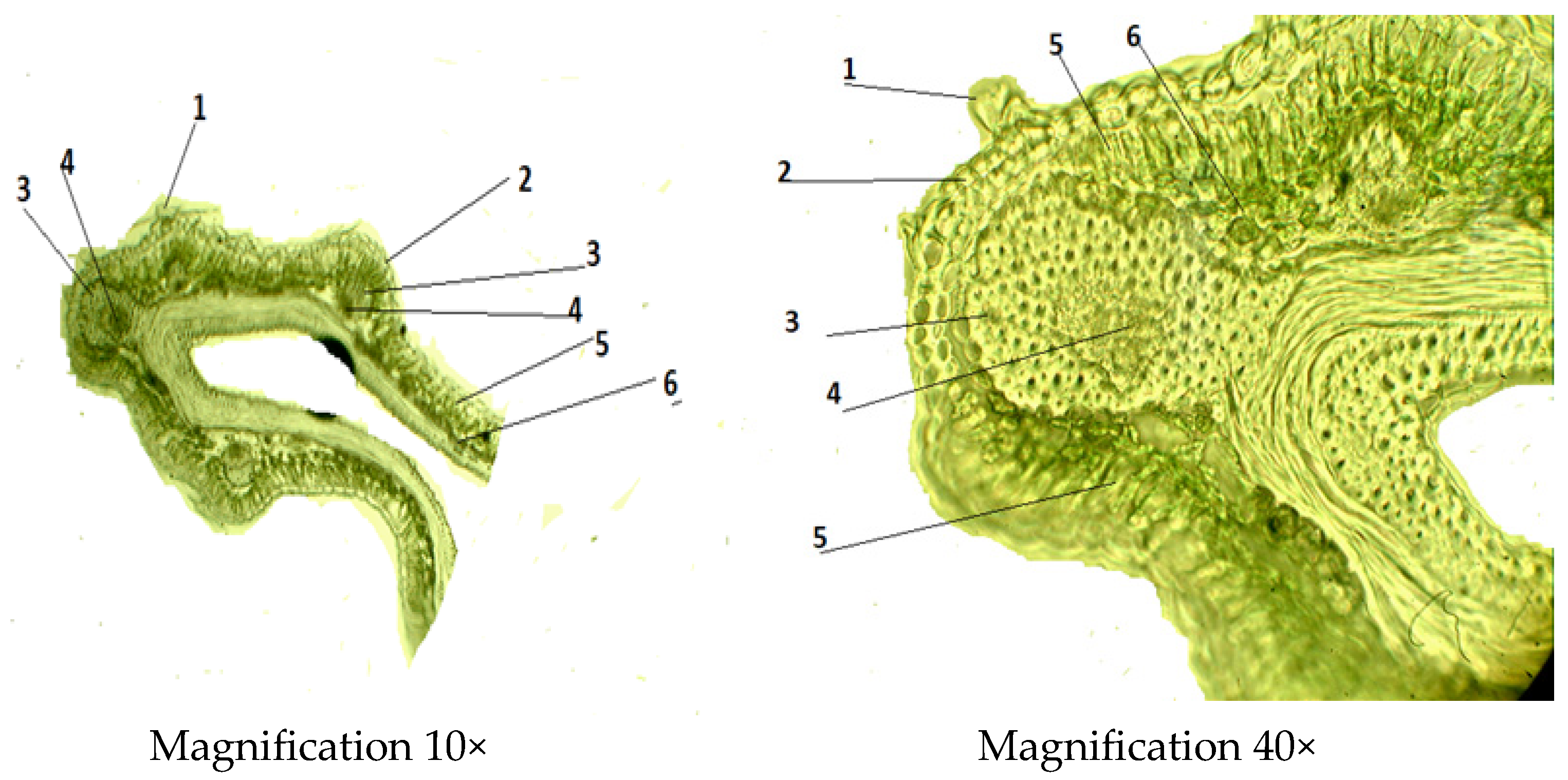
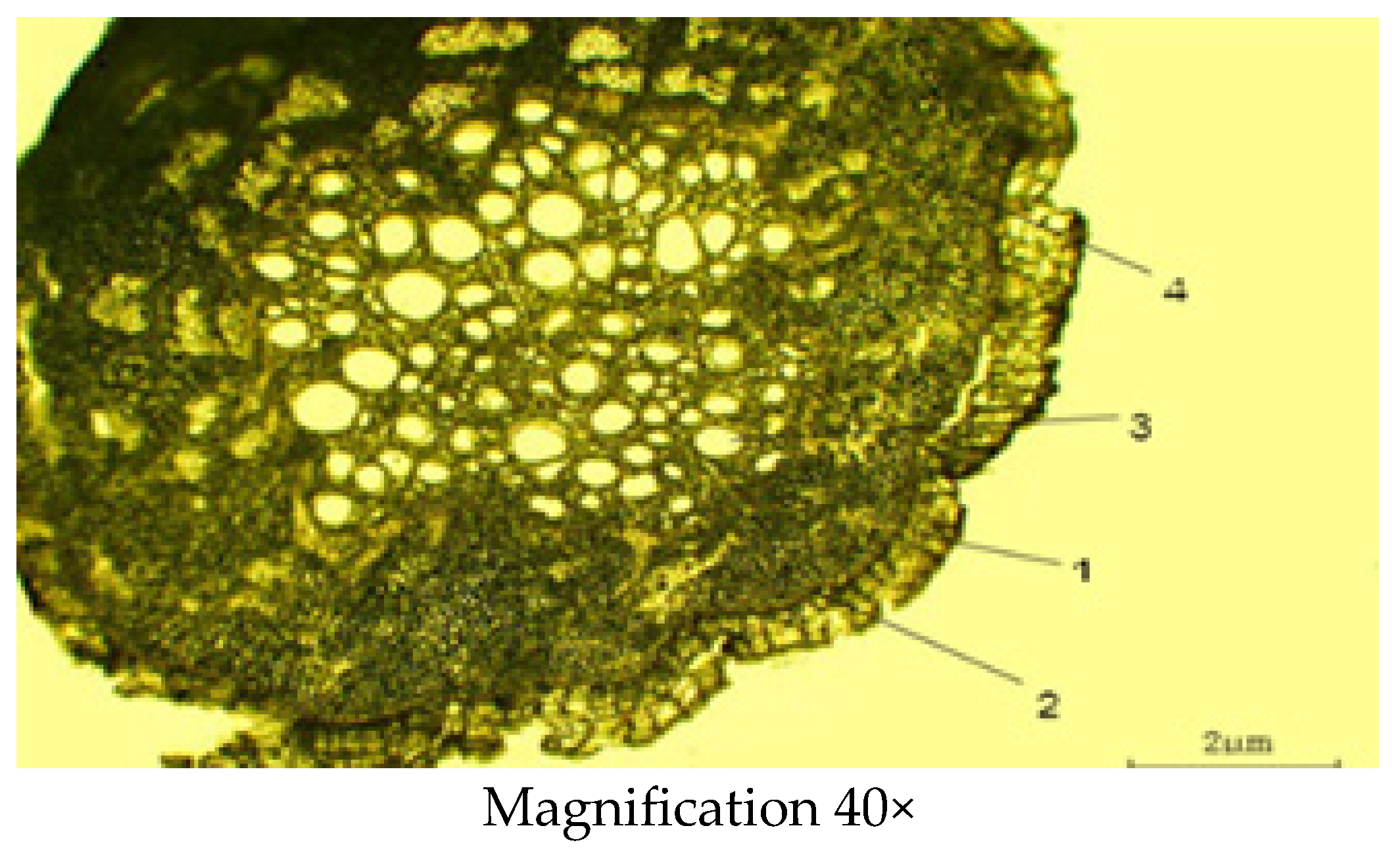
2.2.3. Microscopic Structure of the Root

Sections were made from fresh samples using a freezing microtome; no dyes were used. In the cross-section (Figure 7), the roots are thin, cylindrical, light brown and have a well-developed plug, cortex, phloem and a central core of primary and secondary xylem. In the root cylindrical structure, there is the outermost exoderm layer. The periderm layer (0.16 ± 0.02 µm) is located under the exoderm layer. The epidermis is narrow. The conductive bundles form small areas separated by broad medullary rays consisting of tangentially elongated cells. The xylem is developed and well-visible at 0.24 ± 0.01 µm in diameter. Secondary phloem (0.32 ± 0.01 µm in diameter) is clearly differentiable in loose sieve elements and elongated horizontally and arranged in phloem rays. The location of the phloem and xylem is radial and scattered. The conducting bundles are arranged in an order close to the central circle. The cell thickness of the primary cortex (0.48 ± 0.03 µm) is densely well developed. Biometric measurements of the root are shown in Table 4 below.

2.3. Determination of Proximate Parameters

The proximate composition of C. arenarius is presented in Table 5.
Ash content and moisture content were 6.8% and 5.9%, respectively. High ash content indicates significant concentrations of several minerals. Ashing eliminates all of the sample’s organic constituents; ash also contains inorganic plant parts. Ash indicates that a plant is highly digestible [11]. Depending on the environment and the physiology of the plant, different plant species have different moisture contents. Moisture content is influenced by environmental factors such as temperature, humidity, weather, harvest time and storage conditions [12], and a moisture content of more than 15% is not desirable because of the risk of microbiological contamination. The moisture content of 6.8% found in the tested plant material corresponds to a typical moisture content of 6–8% [13].
The fatty-acid profile of the C. arenarius plant is presented in Table 6 and is expressed as a percentage (%) of total fatty acids (TFA). The profiling of fatty acids showed the presence of eight compounds; of these, four were saturated fatty acids (SFAs), two were monounsaturated fatty acids (MUFAs), and two were polyunsaturated fatty acids (PUFA). Fatty acids containing one or more covalent double bonds between carbon–carbon at various positions on the carbon chain are called unsaturated fatty acids. In general, a high percentage of unsaturated fatty acids (83.8%) was found. In addition, percentages of MUFAs (62.5%) were higher than those of PUFAs (21.3%). The major unsaturated fatty acids were oleic (62.2%) followed by linoleic (20.8%), linolenic (0.5%) and palmitoleic (2.29%) acids. Oleic acid is considered beneficial, as it has been shown to lower cholesterol levels in low-density lipoproteins [14]. Polyunsaturated fatty acids (PUFAs) are basic components involved in the architecture and function of cellular membranes and play key roles in several biological processes. Linoleic acid is a major constituent of human tissues, and it is considered to be an essential fatty acid [15]. Fatty acids that consist of a single covalent bond between carbon–carbon atoms and are generally solid at room temperature are called saturated fatty acids. Palmitic acid (9.3%), stearic acid (5.6%), myristic acid (0.8%) and pentadecanoic acid (0.5%) found in vegetable oils are the most important saturated fatty acids. Saturated fatty acids can be synthesized in the human body, and even if no fat is consumed, these types of fatty acids can be synthesized from molecules formed by carbohydrate metabolism. In response to this, unsaturated fats are essential fatty acids that the body needs. They are liquid at room temperature and most of them are of vegetable origin [16,17].
The fatty acid composition is a good indicator of the quality and stability of the oil. Consequently, it is vital to determine the type and content of fatty acids [18]. The season of collection and other abiotic factors including soil, salinity, light and temperature may also contribute to variations in fatty acid content [19,20]. The current investigation represents the first study to assess the fatty-acid composition in C. arenarius.
Amino acids play a number of roles in the body and are required for the synthesis of proteins. It is necessary to have a balanced diet that contains all of the amino acids—the non-essential ones and those that are not synthesized in the body (essential amino acids). C. arenarius contains essential and non-essential amino acids. Table 7 shows that amino acids with the highest amounts detected include glutamic acid (2298 mg/100 g dry weight), aspartic acid (1204 mg/100 g weight) and alanine (680 mg/ 100 g dry weight). This is the first study that reports the amino acid composition of C. arenarius.
The analysis of the mineral composition is shown in Table 8. The findings revealed that C. arenarius contained eight important minerals. The macro minerals analyzed included sodium (Na), potassium (K) and calcium (Ca). The micro minerals included magnesium (Mg), copper (Cu), zinc (Zn), iron (Fe) and manganese (Mn). K and Ca had the highest mineral concentrations of 302.725 and 131.230 mg/100 g of dry weight, respectively. The lowest concentration minerals were Cu, Mn and Fe, with respective concentrations of 0.11, 0.76 and 1.18 mg/100 g of dry weight.
For the optimal functioning of human systems as well as for healthy development and growth, mineral elements are needed in trace amounts [21]. The most prevalent mineral in bones is Ca, which is necessary for many cellular processes such as neuron and muscle function, hormone responses and, blood clotting [22]. The ionic equilibrium of the human body and tissue excitability are maintained by Na and K. Na is crucial for the transfer of metabolites, and K is crucial due to its diuretic properties. Any food’s K/Na ratio is a crucial element linked to hypertension and atherosclerosis. Na raises blood pressure, while K lowers it [23]. Magnesium helps to avoid immune system dysfunction, impaired spermatogenesis, impaired muscle development, growth retardation, cardiomyopathy and bleeding disorders [24]. The stabilization of macromolecular production and structure is facilitated by Zn. Both DNA and RNA polymerases are Zn-dependent enzymes, and the role of the metal ion in DNA and RNA synthesis is well known [25]. The cofactor for enzymes such as arginase and glycosyl transferase is manganese (Mn). Other enzymes, such as glutamine synthetase and phosphoenol pyruvate carboxy kinase, are also triggered by Mn ions. Mn is necessary for the synthesis of hemoglobin as well [26]. Fe performs a wide range of biological functions, including the role in hemoglobin and the transfer of oxygen from the lungs to tissue cells [27]. Fe deficiency affects humans more frequently than any other nutritional deficiency [28]. The Cu protein is made up in a large part by Cu. The three main Cu-containing metalloenzymes are tyrosine oxidase, lysyl oxidase and cytochrome C oxidase.
As part of enzymatic defense, microelements such as Zn, Mn, Cu and Fe are crucial in the fight against oxidative stress. Some plants contain significant amounts of minerals; their presence and abundance are influenced by the plant’s ancestry, history and phytochemical traits [29].

2.4. Cytotoxic Activity

Artemia salina is a simple biological organism (marine invertebrate) about 1 mm in size. Their freeze-dried cysts (A. salina eggs) can last for several years and can be hatched into larvae without special equipment. The brine shrimp lethality test (BSLT) or brine shrimp survival method is a general bioassay that has been used successfully for preliminary assessment of cytotoxicity [30,31]. This bioassay can be employed to determine the cytotoxic activity of plant extracts, and it is a very useful tool for screening a wide range of chemical compounds [32].
The results of cytotoxic activity are shown in Table 9. The comparison drug, actinomycin D, showed cytotoxicity at all concentrations—the mortality of nauplii was 63–96%. In contrast, the ethanolic extract of C. arenarius showed no toxicity against A. salina. Even with the increase in the extract concentration, no mortality was observed.

3. Materials and Methods
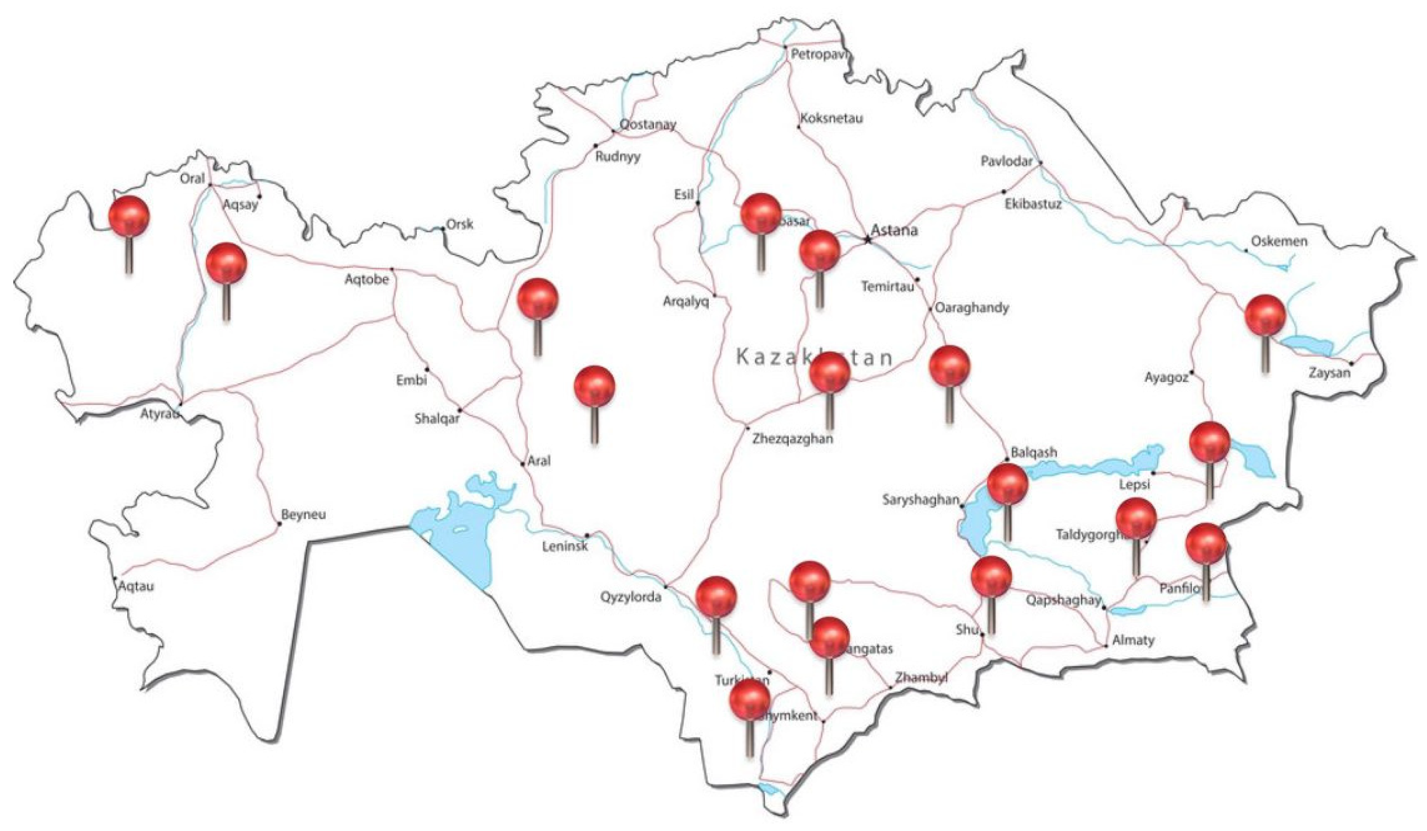
3.1. Plant Material

The plant of C. arenarius (Figure 8) was collected in the Almaty region (Kazakhstan) in June 2021. The material was identified at the Institute of Botany and Phyto introduction of the Republic of Kazakhstan, and a voucher specimen was registered and stored under No. 01-09/305. The raw material was crushed in an electric mill (Figure 8) and then preserved in clean boxes.

3.2. Morphological Studies

The macroscopic study of vegetative organs was carried out in accordance with the requirements of the State Pharmacopoeia of the Republic of Kazakhstan [33]. Morphological measurements were taken on roots, stems, leaves and seeds, using 10 individual plants. Values were statistically processed using Microsoft Excel 2016, and final results were expressed as a mean value ± SD.

Anatomical Studies

Anatomical studies of vegetative organs were conducted in accordance with the State Pharmacopoeia of the Republic of Kazakhstan and methods described by Vekhov et al. [34]. Dry raw material (whole plants) was soaked in a mixture of glycerin:water:ethanol 96% (1:1:1). Anatomical preparations were prepared using a freezing microtome (Minux S700, Shenzhen, China) [35]. The thickness of anatomical sections varied between 10 and 15 μm. Microphotographs of anatomical sections were taken using an MC-300 microscope (Micros Company, Vienna, Austria) magnification 10× for ocular and 40× for objective) with a CAMV400/1.3 m video camera (Micros Company, Vienna, Austria). Mathematical processing of the obtained images was carried out according to the methodology described in [36]. Cross sections of the leaf, stem and root were prepared. Clarification of the preparations was carried out with glycerin. More than 100 temporary preparations were prepared. Each parameter for an individual preparation was measured tenfold.

3.3. Determination of Proximate Parameters

3.3.1. Determination of Moisture

The method outlined by AOAC (2016) [37] was used to determine moisture content. A desiccator was used to allow an empty crucible to cool and dry to a consistent weight before being weighed (W1). Dry plant sample (2.0 g) was dried at 105 °C until it reached a constant weight, then it was weighed (W2) in the crucible. The plant sample-containing crucible was allowed to cool in a desiccator before the weight (W3) was calculated. The percentage used to calculate moisture content was as follows:
m o i s t u r e % = W 1 W 2 × 100 W 1
where
  • W1 = weight (g) of sample before drying;
  • W2 = weight (g) of sample after drying.

3.3.2. Determination of Ash

The ash content assay was conducted using the AOAC (2016) [37] method. In order to determine the dry weight (W1), a heat-resistant porcelain crucible was dried for 10 min at 105 °C in an oven before cooling in a desiccator. Then, 2.0 g of the ground-up plant sample was weighed again (W2) after being measured in the porcelain crucible. To guarantee appropriate ashing, the crucible containing the sample was burned in a furnace at 250 °C for 1 h and then at 550 °C for 7 h. After being taken out, the crucible was weighed (W3) after cooling in a desiccator. Ash content percentage was calculated as follows:
a s h c o n t e n t % = W 3 W 1 × 100 W 2 W 1
where
  • W1 = weight of empty crucible;
  • W2 = weight of crucible + sample;
  • W3 = weight of crucible + ash.

3.3.3. Determination of Fatty Acid Composition

One volume of sample was extracted by 20 times the volume of chloroform:methanol (2:1) for 5 min. Then, the mixture was filtered through a paper filter to obtain a clear extract that was evaporated in a round bottom flask on a rotary evaporator at a bath temperature of 30–40 °C until dried. Then, 10 mL of methanol and 2–3 drops of acetyl chloride were added, and methylation reaction was performed at 60–70 °C for 30 min. Then, methanol was evaporated on a rotary evaporator, and the dry residue was dissolved in 5 mL of hexane. An aliquot of the upper hexane layer was directly taken and analyzed using GC-MS system (Carlo Erba 4200, Cornaredo, Italy) with capillary column (30 m × 0.25 mm, 0.25 µm). Helium is used as a carrier gas. It was operated under the following conditions: oven temperature of 188 °C for 1 h. The injector temperature was set at 188 °C, and detector temperature was set at 230 °C [38].
The identification of the compounds was based on a comparison of their mass spectra and retention indices with those of the synthetic compounds spectral library of the National Institute of Standards and Technology (NIST11).

3.3.4. Determination of Amino Acid Composition

In total, 1 g of the sample was hydrolyzed in 5 mL of 6 N hydrochloric acid at 105 °C for 24 h, in ampoules sealed under a jet of argon. The resulting hydrolysate was dried on a rotary evaporator three times at a temperature of 40–50 °C. The dried mass was dissolved in 5 mL of sulfosalicylic acid. The supernatant was passed at a rate of 1 drop/second through a column of Daux 50 ion-exchange resin after being centrifuged for 5 min. The resin was then rinsed until the pH was neutral. To elute amino acids from the column, 3 mL of 6 N NH4OH solution was passed through the column at a rate of 2 drops/second. The eluate was collected in a round bottom flask together with demineralized water, which was used to wash the column to neutral pH. Then, the contents of the flask were evaporated to dryness on a rotary evaporator at a pressure of 1 atm and temperature of 40–50 °C. In total, 1 drop of freshly prepared 1.5% SnCl2 solution, 1 drop of 2,2-dimethoxypropane and 1–2 mL of propanol saturated with hydrochloric acid were added, and the mixture was heated to 110 °C, kept at this temperature for 20 min, and then, the contents were evaporated from the flask again on a rotary evaporator. In the next step, 1 mL of freshly prepared acetylating reagent (acetic anhydride:triethylamine:acetone = 1:2:5, v/v) was introduced into the flask and heated at 60 °C for 1.5–2 min. Then, the sample was evaporated again on the rotary evaporator to dryness, and 2 mL of ethyl acetate and 1 mL of saturated NaCl solution were added to the flask. The contents were thoroughly mixed, and as two layers of liquids were clearly formed, the upper one (ethyl acetate) was taken for gas chromatographic analysis, which was carried out on a gas-liquid chromatograph (Carlo Erba Reagents, Cornaredo, Italy) [38].

3.3.5. Determination of Mineral Content

The procedure was modified from that given by Idris et al. [39]. The plant samples that had been ground up (3 g) were weighed and ashed at 550 °C. The resultant residue, known as white ash, was dissolved in 4 mL of concentrated HCl, filtered, and the filtrate was then diluted with distilled water in a volumetric flask. The examination of minerals was then performed on the extract’s final solution. Nutrient analysis of K, Ca, Mg, Fe, Mn, Zn, Na and Cu contents were determined using Atomic Absorption Spectrophotometer (Analytik Jena nova 350, Jena, Germany). The analyses were performed in triplicate.

3.4. Extraction

The freshly cleansed plant was air-dried for 72 h at room temperature before being ground in an electric blender. The powder (100 g) was extracted for 48 h with 1 L of 70% ethanol in a glass conical flask using a shaker at 25 °C and filtered. Using a rotary evaporator under low pressure, the crude extract was concentrated at 40 °C. The extract was preserved after drying at 4 °C until further analysis.

3.5. Cytotoxic Activity

A. salina was taken to determine cytotoxic activity. This technique is based on determining the difference between the numbers of dead A. salina larvae in the sample analyzed (experiment) and water that does not contain toxic substances (control). The criterion for acute lethal toxicity of a substance solution is the death of 50% or more larvae in the experiment compared to the control. The flask was filled with artificial seawater, and A. salina eggs were added. They were kept for 72 h with a soft air supply until the shrimp hatched from the eggs. Actinomycin D was used as a comparison drug. Samples (70% ethanol extracts of C. arenarius) were tested at concentrations of 1, 5 and 10 mg/mL. Each sample was tested in three parallel experiments conducted at a temperature of 20 °C, in natural light period. The salinity of the control artificial water was 8.0–8.5 (pH). At the time of the biotest, the A. salina larvae were up to 1 day old. The planting density of larvae was 20–40 specimens per test tube.

4. Conclusions

This study provided analysis of morpho-anatomical characteristics, nutritional contents and cytotoxic activity of wild-growing C. arenarius. The results of botanical characterization have an important diagnostic significance and will allow the evaluation of authenticity of C. arenarius as a plant raw material in the development of regulatory documentation. This will expand the raw material base for obtaining new phytopreparations for therapeutic, cosmetic or nutritional use. In addition, our studies of the plant’s nutrient and mineral contents and cytotoxic activity confirm its promising potential. However, as a largely unexplored plant, C. arenarius should first be subjected to detailed phytochemical characterization in further studies.

Supplementary Materials

The following supporting information can be downloaded at: https://www.mdpi.com/article/10.3390/molecules29020384/s1, Figure S1: GC/MS Chromatogram of fatty acid Ceratocarpus arenarius L.; Figure S2: GC/MS Chromatogram of amino acid Ceratocarpus arenarius L.

Author Contributions

Conceptualization: A.K., N.K.G., A.Z.P. and G.U.; methodology, A.K., A.M. and N.G.; software, A.K., N.K.G. and G.U; validation, S.M., A.A. and A.M.; formal analysis, S.M. and M.K.; investigation, A.K., N.K.G., A.Z.P. and G.U.; resources, N.G. and M.K.; data curation, A.K., M.K. and A.A.; writing—original draft preparation, A.K. and N.K.G.; writing—review and editing, N.K.G.; visualization, N.G., A.M. and A.A.; supervision, A.Z.P. and G.U.; project administration, A.K., N.K.G. and G.U.; funding acquisition, G.U. All authors have read and agreed to the published version of the manuscript.

Funding

This research received no external funding.

Institutional Review Board Statement

Not applicable.

Informed Consent Statement

Not applicable.

Data Availability Statement

The authors confirm that the data supporting the findings of this study are available within the article and supplementary materials.

Conflicts of Interest

The authors declare no conflicts of interest.

References

  1. Grigore, M.; Toma, C. Histo-anatomical strategies of Chenopodiaceae halophytes: Adaptive, ecological and evolutionary implications. WSEAS Trans. Biol. Biomed. 2007, 12, 4. [Google Scholar] [CrossRef]
  2. Heklau, H.; Gasson, P.; Schweingruber, F.; Baas, P. Wood anatomy of Chenopodiaceae (Amaranthaceae s.l.). IAWA J. 2012, 33, 205–232. [Google Scholar] [CrossRef]
  3. Lomonosova, M.N.; An’kova, T.V.; Voronkova, M.S.; Korolyuk, E.A.; Banaev, E.V.; Skaptsov, M.V. Ploidy level of the representatives of Chenopodiaceae based on genome size and chromosome numbers. Turczaninowia 2020, 23, 24–31. Available online: http://turczaninowia.asu.ru (accessed on 15 February 2023).
  4. Zhang, T.; Shi, N.; Bai, D.; Chen, Y.; Feng, G. Arbuscular Mycorrhizal Fungi Promote the Growth of Ceratocarpus arenarius (Chenopodiaceae) with No Enhancement of Phosphorus Nutrition. PLoS ONE 2012, 7, e41151. [Google Scholar] [CrossRef]
  5. Zhaglovskaya, A.; Aidosova, S.; Akhtayeva, N.; Mamurova, A.; Yesimova, D. Anatomical and morphological stem features of two Haloxylon Species (Chenopodiaceae Vent.) of Drought Stress, Kazakhstan. Biosci. Biotechnol. Res. Asia 2015, 12, 1965–1974. [Google Scholar] [CrossRef]
  6. Gemejiyeva, N.G.; Grudzinskaya, L.M. Current State and Prospects for Studies on the Diversity of Medicinal Flora in Kazakhstan. In Book Vegetation of Central Asia and Environs; Egamberdieva, D., Öztürk, M., Eds.; Springer: Almaty, Kazakhstan, 2018; pp. 239–262. [Google Scholar]
  7. Gan, L.; Lu, J.; Jerry, M.B.; Baskin, C.C.; Tan, D. Phenotypic plasticity in diaspore production of a amphi basicarpic cold desert annual that produces polymorphic diaspores. Sci. Rep. 2020, 10, 11142. [Google Scholar] [CrossRef]
  8. Ebrahimi, E.; Eslami, S.V. Effect of environmental factors on seed germination and seedling emergence of invasive Ceratocarpus arenarius. Eur. Weed Res. Soc. Weed Res. 2011, 52, 50–59. [Google Scholar] [CrossRef]
  9. Grudzinskaya, L.M.; Gemedzhieva, N.G.; Nelina, N.V.; Karzhaubekov, Z.Z. (Eds.) Annotated List of Medicinal Plants of Kazakhstan; KazNMU: Almaty, Kazakhstan, 2014; Volume 20, p. 200. [Google Scholar]
  10. Pan, L.; Li, L.; Xu, L.; Zhang, J.; Li, J.; Gao, M.; Yu, J.; Jin, J.; Lei, D. UHPLC-QTOF-MS/MS based characterization of anti-tumor constituents in Ceratocarpus arenarius L. and identification of EGFR-TK inhibitors by virtual screening. Nat. Prod. Prod. 2022, 36, 6111–6115. [Google Scholar] [CrossRef]
  11. Ullah, Z.; Baloch, M.K.; Khader, J.A.; AbdEIslam, N.M.; Noor, S. Proximate and nutrient analysis of selected medicinal plants of Tank and South Waziristan area of Pakistan. Afr. J. Pharm. Pharmacol. 2013, 7, 179–184. [Google Scholar] [CrossRef]
  12. Radha, M.K.; Sunil, P.; Ashok, P.; Sneh, P.B.; Sushil, C.; Poonam, C.; Parameswari, E.; Ahmad, A.; Mahesh, K.S.; Rahul, D.D.; et al. Evaluation of Nutritional, Phytochemical, and Mineral Composition of Selected Medicinal Plants for Therapeutic Uses from Cold Desert of Western Himalaya. Plants 2021, 10, 1429. [Google Scholar] [CrossRef]
  13. Princewill-Ogbonna, I.L.; Ogbonna, P.C.; Ogujiofor, I.B. Proximate Composition, Vitamin, Mineral and biologically Active Compounds Levels in Leaves of Mangifera indica (Mango), Persea americana (Avocado pea), and Annona muricata (Sour sop). J. Appl. Sci. Environ. Manag. 2019, 23, 65–74. [Google Scholar]
  14. Grundy, S.M. Monounsaturated fatty acids and cholesterol metabolism: Implication for dietary recommendations. J. Nutr. 1989, 4, 529–533. [Google Scholar] [CrossRef]
  15. Ramsden, C.E.; Ringel, A.; Feldstein, A.E.; Taha, A.Y.; MacIntosh, B.A.; Hibbeln, J.R.; Majchrzak-Hong, S.F.; Faurot, K.R.; Rapoport, S.I.; Cheon, Y.; et al. Lowering dietary linoleic acid reduces bioactive oxidized linoleic acid metabolites in humans. Prostagland. Leukot. Essent. Fat. Acids 2012, 87, 135–141. [Google Scholar] [CrossRef]
  16. Karaca, E.; Aytac, S. The factors affecting on fatty acid composition of oil crops. J. Agric. Fac. Ondokuz Mayıs Univ. 2007, 22, 123–131. [Google Scholar]
  17. Nas, S.; Gokalp, Y.H.; Unsal, M. Vegetable Oil Technology; Pamukkale University Faculty of Architecture Printing House: Denizli, Turkey, 2001; p. 322. [Google Scholar]
  18. Hunter, J.E. Studies on effects of dietary fatty acids as related to their position on triglycerides. Lipids 2001, 36, 655–668. [Google Scholar] [CrossRef]
  19. Sakthivel, R.; Devi, K.P. Evaluation of physicochemical properties, proximate and nutritional composition of Gracilaria edulis collected from Palk Bay. Food Chem. 2015, 174, 68–74. [Google Scholar] [CrossRef]
  20. Francavilla, M.; Franchi, M.; Monteone, M.; Caroppo, C. The red seaweed Gracilaria gracilis as a multi products source. Mar. Drugs 2013, 11, 3754–3776. [Google Scholar] [CrossRef]
  21. Woods, P.W. Herbal healing. Essence 1999, 30, 42–46. [Google Scholar]
  22. Hussain, J.; Rehman, N.; Al-Harrasi, A.; Ali, L.; Ullah, R.; Mabood, F.; Hussain, H.; Ismail, M. Nutritional prospects and mineral compositions of selected vegetables from Dhoda sharif Kohat. J. Med. Plants Res. 2011, 5, 6509–6514. [Google Scholar]
  23. Saupi, N.; Zakaria, M.H.; Bujang, J.S. Analytic chemical composition and mineral content of yellow velvet leaf (Limnocharis flava L. Buchenau)’s edible parts. J. Appl. Sci. 2009, 9, 2969–2974. [Google Scholar] [CrossRef]
  24. Chaturvedi, V.C.; Shrivastava, R.; Upreti, R.K. Viral infections and trace elements: A complex trace element. Curr. Sci. 2004, 87, 1536–1554. Available online: http://www.jstor.org/stable/24109032 (accessed on 20 May 2023).
  25. Saikia, P.; Deka, D.C. Mineral content of some wild green leafy vegetables of North-East India. J. Chem. Pharm. Res. 2013, 5, 117–121. [Google Scholar] [CrossRef]
  26. Indrayan, A.K.; Sharma, S.; Durgapal, D.; Kumar, N.; Kumar, M. Determination of nutritive value and analysis of mineral elements for some medicinally valued plants from Uttaranchal. Curr. Sci. 2005, 89, 1252–1255. [Google Scholar]
  27. Kruczek, A. Effect of row fertilization with different kinds of fertilizers on the maize yield. Acta Sci. Pol. Agric. 2005, 4, 37–46. [Google Scholar]
  28. Léchaudel, M.; Joas, J.; Caro, Y.; Génard, M.; Jannoyer, M. Leaf: Fruit ratio and irrigation supply affect seasonal changes in minerals, organic acids and sugars of mango fruit. J. Sci. Food Agric. 2004, 85, 251–260. [Google Scholar] [CrossRef]
  29. Lokhande, R.; Singare, P.; Andhale, M. Study on Mineral content of some ayurvedic indian medicinal plants by instrumental Neutron Activation analysis and AAS techniques. Health Sci. J. 2010, 4, 157–168. [Google Scholar]
  30. Wanyoike, G.N.; Chhabra, S.C.; Lang’at-Thoruwa, C.C.; Omar, S.A. Brine shrimp toxicity and antiplasmodial activity of five Kenyan medicinal plants. J. Ethnopharmacol. 2004, 90, 129–133. [Google Scholar] [CrossRef]
  31. Anderson, J.E.; Goetz, C.M.; McLaughlin, J.L.; Suffness, M.A. Blind Comparison of Simple Bench-top Bioassays and Human Tumour Cell Cytotoxicities as Antitumor Prescreens. Phytochem. Anal. 1991, 2, 107–111. [Google Scholar] [CrossRef]
  32. Arzu, K. Cytotoxic Activities on Selected Lamiaceae Species from Turkey by Brine Shrimp Lethality Bioassay. Ordu Univ. J. Sci. Tech. 2019, 9, 105–111. Available online: https://dergipark.org.tr/en/pub/ordubtd/issue/51531/579909 (accessed on 12 June 2023).
  33. State Pharmacopoeia of the Republic of Kazakhstan; Zhibek-Zholy: Almaty, Kazakhstan, 2008; Volume 1, 592p.
  34. Vekhov, V.N.; Lotova, L.I.; Filin, V.R. Practicum on Anatomy and Morphology of Higher Plants; MSU: Moscow, Russia, 1980; 560p. [Google Scholar]
  35. Barykina, R.P.; Veselova, T.D.; Devyatov, A.G.; Jalilova, H.H.; Ilyina, G.M.; Chubatova, N.V. Handbook of Botanical Microtechnology (Fundamentals and Methods); Publishing House, Moscow State University: Moscow, Russia, 2004; p. 312. [Google Scholar]
  36. Dospekhov, B.A. Book Methodology of Field Experience (with the Basics of Statistical Processing of Research Results); Agropromizdat: Moscow, Russia, 1985; p. 157. Available online: https://www.scirp.org/(S(i43dyn45teexjx455qlt3d2q))/reference/ReferencesPapers.aspx?ReferenceID=1877236 (accessed on 2 March 2023).
  37. Association of Official Analytical Chemist (AOAC). Official Methods of Analysis of AOAC International; AOAC: Washington, DC, USA, 2016; ISBN 0935584870. Available online: https://www.techstreet.com/standards/official-methods-of-analysis-of-aoac-international-20th-edition-2016?product_id=1937367 (accessed on 25 June 2023).
  38. Moldabergenova, A.K.; Litvinenko, Y.A.; Akhtayeva, N.Z.; Kiekbayeva, I.N.; Ross, S.A. Amino and fatty acid composition of the aerial parts of Echinops albicaulis, growing in Kazakhstan. Int. J. Biol. Chem. 2016, 9, 1–4. Available online: https://elibrary.kaznu.kz/wp-content/uploads/2021/06/international-journal-of-biology-and-chemistry_2016-2.pdf (accessed on 30 June 2023). [CrossRef]
  39. Idris, O.A.; Wintola, O.A.; Afolayan, A.J. Comparison of the Proximate Composition, Vitamins (Ascorbic Acid, α-Tocopherol and Retinol), Anti-Nutrients (Phytate and Oxalate) and the GC-MS Analysis of the Essential Oil of the Root and Leaf of Rumex crispus L. Plants 2019, 8, 51. [Google Scholar] [CrossRef]
Figure 1. C. arenarius, Almaty region, Kazakhstan.
Molecules 29 00384 g001
Figure 2. Distribution of C. arenarius over Kazakhstan. The location where the plant is widespread is indicated in red.
Molecules 29 00384 g002
Figure 3. Raw material sample of C. arenarius. (a) Whole plant. (b) Leaf. (c) Stem.
Molecules 29 00384 g003
Figure 4. Cross section of the C. arenarius leaf; 1—trichome; 2—epidermis; 3—xylem; 4—phloem; 5—palisade mesophyll; 6—spongy mesophyll.
Molecules 29 00384 g004
Figure 5. The lower epidermis of the C. arenarius leaf; 1—stomatal gap; 2—guard cells (actinocytic).
Molecules 29 00384 g005
Figure 6. Cross section of the C. arenarius stem; 1—epidermis; 2—lamellar collenchyma; 3—pericyclic sclerenchyma; 4—phloem; 5—medullary parenchyma; 6—xylem.
Molecules 29 00384 g006
Figure 7. Cross section of the C. arenarius root; 1—periderm; 2—periderm; 3—xylem; 4—phloem.
Molecules 29 00384 g007
Figure 8. The plant of C. arenarius (a); powdered plant (b).
Molecules 29 00384 g008
Table 1. Macroscopic characterization of C. arenarius. Values are represented as a mean value ± standard deviation.
SampleIndicator/Mean ± SD
Herb colorgrayish-green
OdorAromatic
Plant height (cm)9.5 ± 1.16
PhyllotaxisAlternate
Flowerssingle, pale yellow
Leaf length (cm)3.5 ± 0.07
Number of leaves per plant37.2 ± 1.1
Stem length (cm)11.4 ± 0.76
Seed length (mm)6.27 ± 0.12
Table 2. Biometric measurements of the anatomical structures of C. arenarius leaf. Values are represented as a mean value ± standard deviation.
IndicatorMean ± SD (µm)
Thickness of the leaf1.99 ± 0.18
Thickness of the upper epidermis0.03 ± 0.02
Thickness of the lower epidermis0.25 ± 0.05
Thickness of the spongy mesophyll0.49 ± 0.13
Thickness of the palisade mesophyll1.98 ± 0.02
Diameter of the vascular bundle0.148 ± 0.13
Table 3. Biometric measurements of the anatomical structures of C. arenarius stem. Values are represented as a mean value ± standard deviation (n = 10).
IndicatorMean ± SD (µm)
Thickness of the epidermis0.02 ± 0.003
Thickness of the primary parenchyma0.27 ± 0.02
Thickness of collenchyma0.38 ± 0.14
Diameter of xylem0.22 ± 0.04
Diameter of phloem0.38 ± 0.09
Thickness of conducting beam1.06 ± 0.01
Diameter of core parenchyma zone3.27 ± 0.003
Table 4. Biometric measurements of the anatomical structures of C. arenarius root. Values are represented as a mean value ± standard deviation.
IndicatorMean ± SD (µm)
Thickness of the periderm0.16 ± 0.02
Thickness of the primary cortex or cortex parenchyma0.48 ± 0.03
Diameter of the central cylinder2.89 ± 0.25
Diameter of xylem0.24 ± 0.01
Diameter of phloem0.32 ± 0.01
Table 5. Proximate composition of C. arenarius.
ParameterValue
Moisture (%)6.8 ± 0.28
Protein (mg/100 g)392.85 ± 25.50
Ash (%)5.9 ± 0.40
Fat (%)12.5 ± 21.28
Table 6. Fatty acid profile of C. arenarius.
№.ParameterC Number: Number of Double BondsClass of CompoundContent, %
1Myristic acid14:0Saturated0.8
2Pentadecanoic acid15:0Saturated0.5
3Palmitic acid16:0Saturated9.3
4Palmitoleic acid16:1Monounsaturated0.3
5Stearic acid18:0Saturated5.6
6Oleic acid18:1Monounsaturated62.2
7Linoleic acid18:2Polyunsaturated20.8
8Linolenic acid18:3Polyunsaturated0.5
Table 7. Amino acid composition of C. arenarius.
Amino AcidContent
mg/100 g
Alanine680
Glycine222
Leucine374
Isoleucine345
Valine236
Glutamic acid2298
Threonine218
Proline435
Methionine56
Serine370
Aspartic acid1204
Cysteine28
Oxyproline1
Phenylalanine352
Tyrosine384
Histidine183
Ornithine1
Arginine256
Lysine16 2
Tryptophan52
Table 8. Mineral content of C. arenarius. Results are represented as a mean value± standard deviation.
Mineral (mg/100 g Dry Weight)
KCaMgFeNaMnZnCu
302.73 ± 1.15131.23 ± 0.0960.69 ± 0.721.18 ± 0.0320.48 ± 0.290.76 ± 0.014.45 ± 0.350.11 ± 0.02
Table 9. Cytotoxic activity of the 70% ethanolic extract of C. arenarius.
SampleConcentration mg/mL% of Surviving Nauplii in the Control% of Surviving Larvae in the Sample% Mortality
Actinomycin D1096096
596492
1963363
Extract1096960
596960
196960
Disclaimer/Publisher’s Note: The statements, opinions and data contained in all publications are solely those of the individual author(s) and contributor(s) and not of MDPI and/or the editor(s). MDPI and/or the editor(s) disclaim responsibility for any injury to people or property resulting from any ideas, methods, instructions or products referred to in the content.

