Article

Synthesis of 2-Amino-N′-aroyl(het)arylhydrazides, DNA Photocleavage, Molecular Docking and Cytotoxicity Studies against Melanoma CarB Cell Lines

by
Achilleas Mitrakas
1,
Maria-Eleni K. Stathopoulou
2,
Chrysoula Mikra
3,
Chrystalla Konstantinou
2,
Stergios Rizos
4,
Stella Malichetoudi
1,
Alexandros E. Koumbis
3,
Maria Koffa
1 and
Konstantina C. Fylaktakidou
2,3,*
1
Laboratory of Cellular Biology, Molecular Biology and Genetics Department, Democritus University of Thrace, University Campus, 68100 Alexandroupolis, Greece
2
Laboratory of Organic, Bioorganic and Natural Product Chemistry, Molecular Biology and Genetics Department, Democritus University of Thrace, 68100 Alexandroupolis, Greece
3
Laboratory of Organic Chemistry, Faculty of Chemistry, Aristotle University of Thessaloniki, 54124 Thessaloniki, Greece
4
Department of Chemistry and Chemical Biology, Harvard University, 12 Oxford St., Cambridge, MA 02138, USA
*
Author to whom correspondence should be addressed.
Molecules 2024, 29(3), 647; https://doi.org/10.3390/molecules29030647
Submission received: 30 December 2023 / Revised: 21 January 2024 / Accepted: 22 January 2024 / Published: 30 January 2024

Abstract

:
Diacylhydrazine bridged anthranilic acids with aryl and heteroaryl domains have been synthesized as the open flexible scaffold of arylamide quinazolinones in order to investigate flexibility versus rigidity towards DNA photocleavage and sensitivity. Most of the compounds have been synthesized via the in situ formation of their anthraniloyl chloride and subsequent reaction with the desired hydrazide and were obtained as precipitates, in moderate yields. All compounds showed high UV-A light absorption and are eligible for DNA photocleavage studies under this “harmless” irradiation. Despite their reduced UV-B light absorption, a first screening indicated the necessity of a halogen at the p-position in relation to the amine group and the lack of an electron-withdrawing group on the aryl group. These characteristics, in general, remained under UV-A light, rendering these compounds as a novel class of UV-A-triggered DNA photocleavers. The best photocleaver, the compound 9, was active at concentrations as low as 2 μΜ. The 5-Nitro-anthranilic derivatives were inactive, giving the opposite results to their related rigid quinazolinones. Molecular docking studies with DNA showed possible interaction sites, whereas cytotoxicity experiments indicated the iodo derivative 17 as a potent cytotoxic agent and the compound 9 as a slight phototoxic compound.

1. Introduction

N,N′-Diacylhydrazines (DACHZs, general structure A, Figure 1) represent an interesting class of compounds with potential applications in chemistry and biology. Several N-tert-butyl-substituted derivatives, such as methoxyfenozide and tebufenozide (B and C, respectively, Figure 1), are well known for their insecticidal activity. Due to their ability to mimic the action of two principal hormones in insects while having almost no impact on most non-target and beneficiary organisms, those derivatives have been subjected to numerous structural modifications [1,2,3,4]. Moreover, an N,N′-bis substituted DACHZ moiety is found in the naturally occurring alkaloids montamine (D) that displays anti-oxidative activity and cytotoxicity against CaCo-2 colon cancer (IC50 = 43.9 μΜ) and elaiοmycin (E) that displays strong in vitro inhibition against bovine and human strains of Mycobacterium tuberculosis [3,5]. A similar urethan-like pattern (general structure F) is considered as a useful synthon in Organic Chemistry [6].
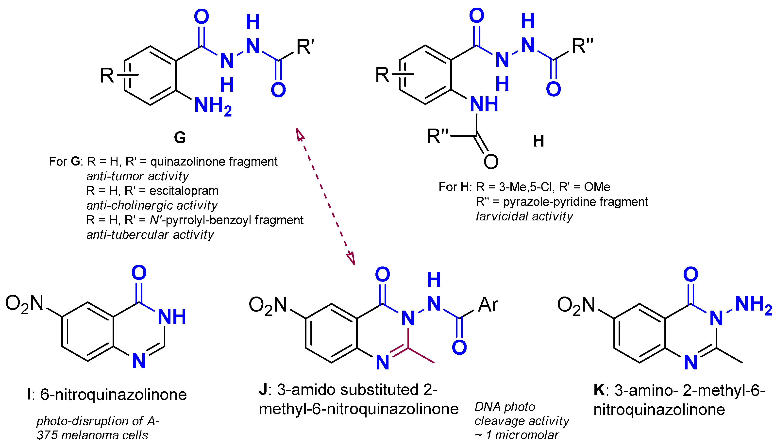
Anthranilic acid DACHZs (AA DACHZs, Figure 2, G, H) bear an additional functional amine group which contributes to their chemistry, biology and technology. Derivatives such as G may give rise to 1,3,4-oxadiazoles [7,8,9,10], 1,2,3-benzotriazine-4-ones [11], 3-amido-1,2-dihydro substituted quinazolinones [12,13,14,15,16,17], 3-amido substituted quinazolinones [10,18], quinazolinone containing fused polycyclic compounds [19,20] or N-N axially chiral 3,3′-bisquinazolinones [21]. Furthermore, their well-positioned electron-donating atoms allow metal complexation which, depending on the metal and the conditions applied, range from the formation of simple metal complexes [22,23,24] to the important metal–organic macrocycles (MOMs) [25,26] with Mn, Ga and In [27,28,29]. Compounds of the general structures G and H have also been used as conjugates of the chemotherapeutic drug daunorubicin [30], whereas other AA DACHZs have been tested or were found to exhibit biological activity as inhibitors of EGFR [31], of HIV-1 Integrase [32], of cholinesterase [33], of enoyl ACP reductase [34] or as potent insecticidal agents that target ryanodine receptors [35,36] (Figure 2: for some structures, the R and R′ or/and R″ are defined, along with the biological activities of the compounds, below the general structure).
Organic DNA photocleavers that can be photosensitized under UV-A irradiation are less studied compared to organometallic compounds, because the majority of the former compounds lack strong absorptions above the UV-B light wavelength. However, the interest for small organic molecules that can photocleave DNA [37] is showing a come-back due to the need for alternative anticancer and antimicrobial therapies able to overcome drug resistances [38,39,40,41]. Thus, pyrazoles (UV-A), trifluoromethyl pyrazolines and pyrazoles (UV-B), as well as bis-pyranopyrazoles (UV-A), were found to photocleave DNA exhibiting additional antibacterial, cytotoxic and antimicrobial activities, respectively [42,43,44]. Interestingly, halogenated derivatives were the best photocleavers among pyrazoles [42], as well as nitro-substituted trifluoromethyl pyrazoles [43]. Deazaflavin analogs linked to the naphthalene core (UV-A) [45] were also found to photocleave DNA, and quinolinium dicarbocyanine dyes (near IR) bearing a pentamethine bridge that was meso-substituted with halogen caused photodynamic cell damage [46,47]. The attachment of a halogen (Cl or Br) at the polymethine meso-carbon was anticipated to introduce a “heavy atom effect” in which ROS production and DNA photocleavage was enhanced by increasing the rate of intersystem crossing between the photosensitizer’s singlet and triplet excited states [46]. A methylene violet-conjugated perylene diimide (near IR) was found to be a promising antitumor nanoagent through a photothermal/photodynamic combination mechanism [48]. Finally, β-carbolinebisindole compounds [49], chlorinated hexahydroquinolines [50], triazolylnucleosides [51] and bis-pyrimidine derivatives [52] when photosensitized under UV-A irradiation promote the photocleavage of DNA, with nucleosides and pyrimidines exhibiting antimicrobial activity as well.
Our team has recently shown a strong interest in the synthesis and biological evaluation of quinazoline and sulfonyloxy carbamidoxime organic ligands and their metal complexes [53,54,55], as well as in the DNA photocleavage of organic photosensitizers. Some of the derivatives were found to exhibit insecticidal activity on a major crop pest, the Whitefly Bemisia tabaci, under UV-A irradiation [56]. The so-called “privileged structures” that correspond to the quinazolinone ring system were part of our target scaffolds for the investigation of photoreactivity. Among the quinazolinones tested, the simple 6-nitro derivative (Figure 2, I) was found to photodegrade human melanoma cell lines under UV-A irradiation at a concentration 50 μM, whereas a 6-bromo analogue was found to photocleave DNA under UV-B irradiation [57]. In addition, several 3-amido-2-methyl-6-nitro substituted quinazolinones exhibited excellent DNA photocleavage activity at concentrations 1 μM, which was better compared to their parent 3-amino-2-methyl-6-nitro-quinazolinone (Figure 2, J, K, respectively) [58]. Molecular docking studies on the 6-nitro derivative J were indicative of satisfactory binding to DNA with participation of the nitro group and this was correlated with the observed photoactivity.
Figure 2. G, H: Structures of non-substituted (G) [31,33,34] or arylamine substituted (H) AA DACHZs [36]; I, J and K: Structures of known DNA photocleaving nitro-quinazolinone derivatives [57,58]; G and J are the open flexible and closed rigid structures corresponding to AA DACHZ and quinazolinone (red arrow between G and J as well as red color on J indicate similarities and differences between the two scaffolds).
Molecules 29 00647 g002
Comparing structures G and J, one may suggest that the scaffold of G, lacking the carbon joint between the two nitrogen atoms, literally represents the open and flexible form of J. We have therefore decided to synthesize a number of AA DACHZ bridged derivatives of G that bear electron-donating and electron-withdrawing groups (R) at a p- and m- position to the amine group and a variety of aroyl and heteroaroyl rings (R′) attached at the AA CO-NH-NH- bridge and test their DNA photocleavage activity under UV-A and UV-B irradiation. Furthermore, molecular docking studies were planned in order to observe similarities and differences between the open AA derivative (G) and the closed locked quinazolinone ring system (J) [58]. Finally, to assess the potential increase in cytotoxic effects, a series of toxicity and phototoxicity studies were conducted on melanoma cells with the derivatives that exhibited the best DNA photocleavage activity.

2. Results and Discussion

2.1. Chemistry

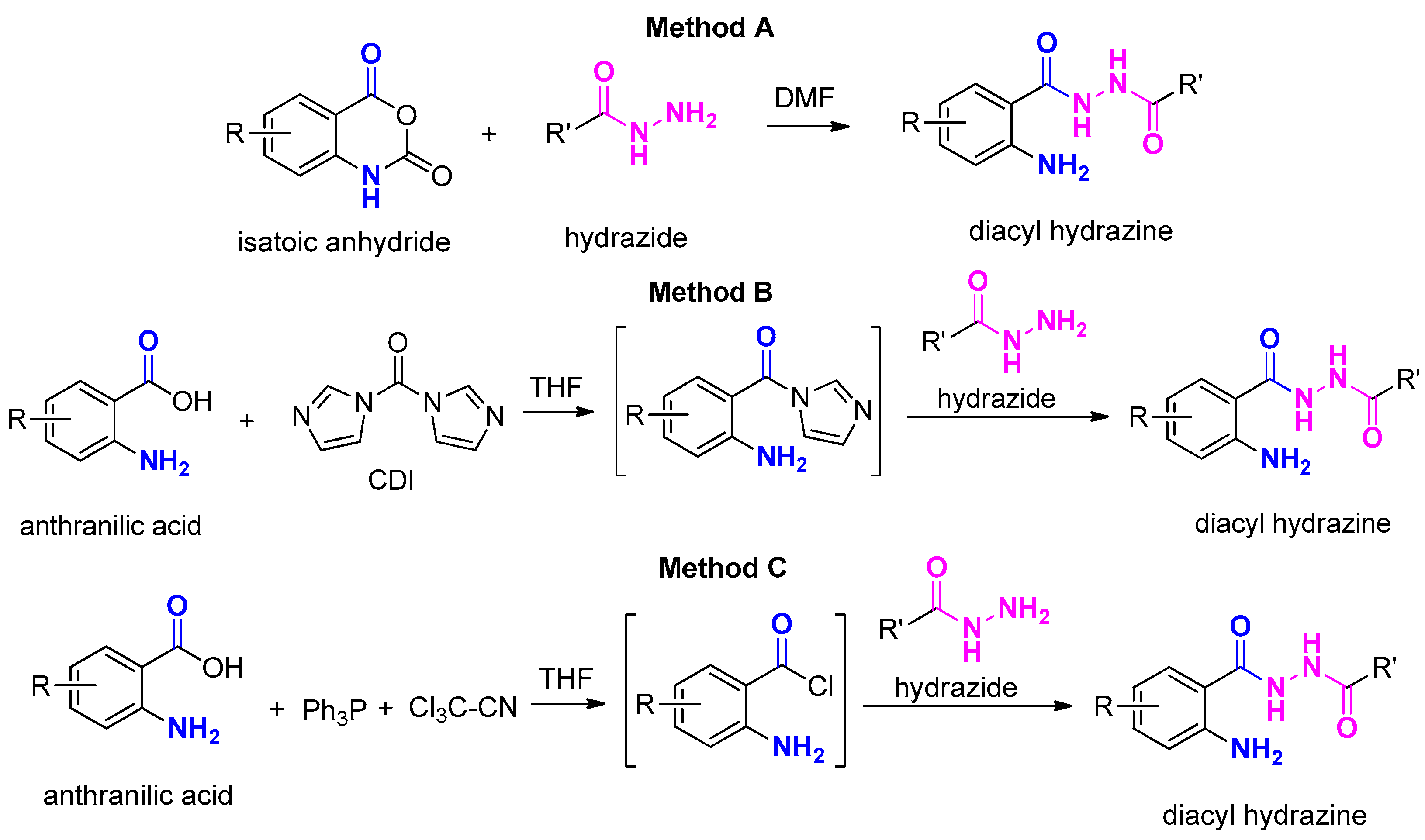
There are several methods in the literature to form amide bonds using a carboxylic acid and an amine [59,60,61] which apply to the synthesis of hydrazides as well [33,62,63,64]. More specifically, the synthesis of anthranilamides is performed upon activation of the carboxylic acid with known classic reagents or with the use of isatoic anhydrides. In our case, depending of the availability of the reagents, we have used the nucleophilic attack of the proper hydrazide to (a) isatoic anhydride [20] (Figure 3, Method A), (b) the intermediate imidazole amides derived from AAs using Ν,Ν’-carbonyldiimidazole (CDI) as the coupling agent [65] and adopting a modified methodology for AAs [66] (Figure 3, Method B) and finally (c) the in situ formation of anthraniloyl chlorides upon treatment with Ph3P and CCl3CN anthraniloyl chlorides (Figure 3, Method C). The latter has been established for the synthesis of carboxylic acid amides, where triaryl phosphonium chloride is believed to be generated upon the reaction of Ph3P and CCl3CN. This reacts further with carboxylic acid to produce the corresponding acid chloride, triphenylphosphine oxide and dichloroacetonitrile [67].
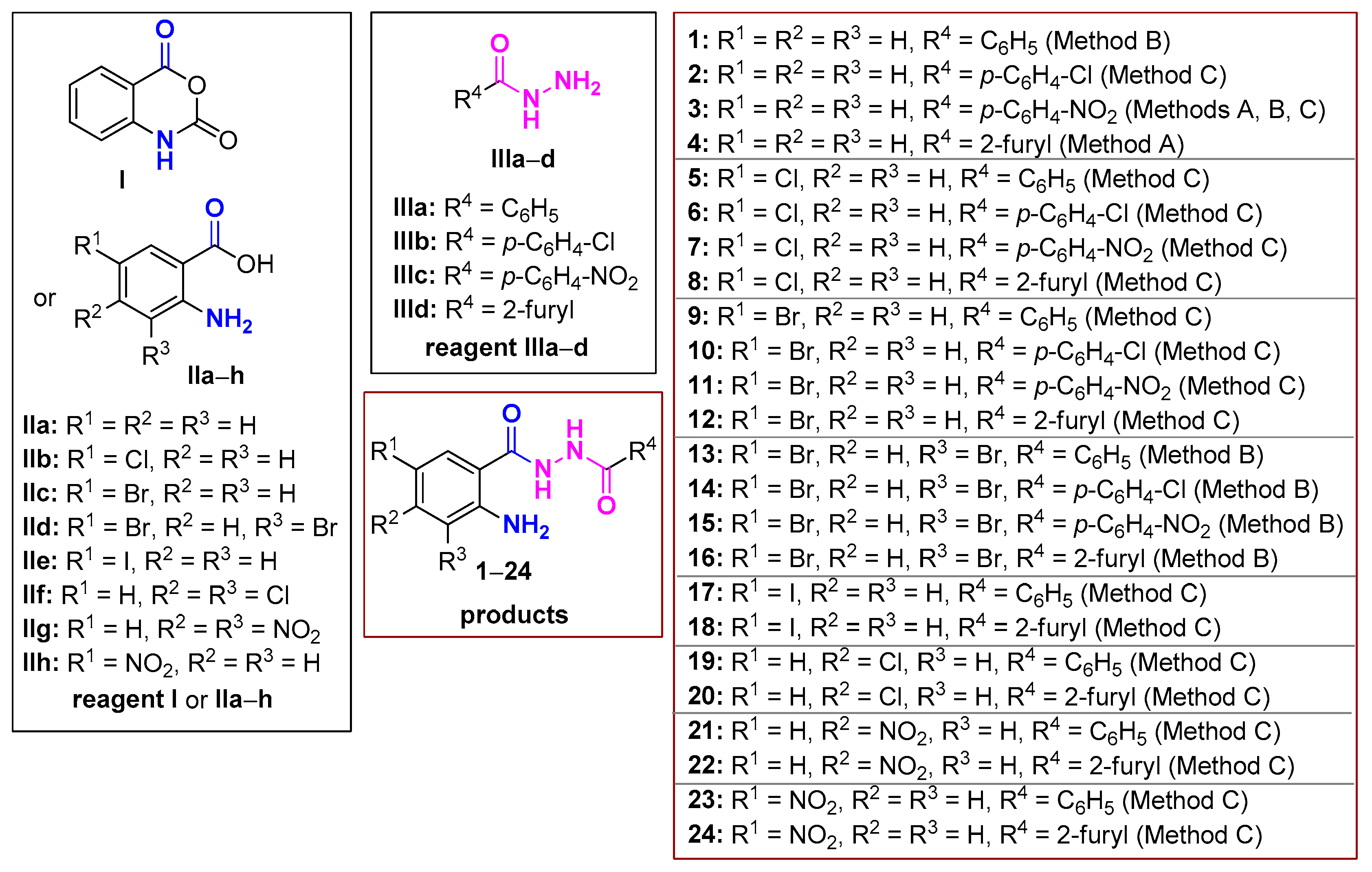
Our synthetic work is summarized in Figure 4. Τhe availability of isatoic anhydride has initially driven our synthetic attempts (Method A). According to this protocol, the combination of I and IIIc,d in DMF smoothly gave the derivatives 3 and 4. However, the yields were generally moderate and, furthermore, in order to apply the same protocol for all derivatives we needed to synthesize the corresponding anhydrides. Thus, we turned our efforts towards the CDI-assisted coupling reaction (Method B). This procedure was applied for the reactions between the AAs IIa,d and IIIad in THF and the compounds 1, 3 and 1316, where obtained. Products derived from the simple AA ΙIa needed column chromatography purification, but the 3,5-dibromo analogue IId derived products were insoluble in the reaction mixture and obtained in good yields upon simple filtration. Nevertheless, the reaction required an overnight stirring at room temperature and then heating for 1 h at 55 °C, then the addition of water and heating for another 1 h at 55 °C.
An alternative approach (Method C), involving the in situ generation of acid chlorides from the corresponding carboxylic acid followed by hydrazide attack furnished in several cases the desired products. This approach was adopted and adjusted to AAs, predicting the highest reactivity of hydrazide NH2 in comparison to aniline NH2 group present in the AAs. In general, the formation of the anthraniloyl chlorides in THF was fast (~1.5 h), whereas the rate of the second step depended on the substituents of starting materials II (1 to 12 h). Upon completion of the reaction, the mixture was extracted with water and ethyl acetate. The residue obtained after removal of the organic solvent was simply triturated with CH2Cl2 to furnish a precipitate which corresponded to the pure product. A small quantity of product remained in the aliquot, which was not further purified. The easiness of this synthetic protocol, the availability of the inexpensive starting materials and the facile work up to obtain pure products rendered this method as the best green choice to prepare most of the targeted derivatives, avoiding column chromatography and use of organic solvents.
Apart from the compounds 14, the rest are novel. All compounds were fully characterized with 1H-NMR, 13C-NMR, IR and HRMS, and all data are in accordance with the proposed structure (Section 3 and Supplementary Materials S.1). The two NH moieties appear as two broad singlet peaks between 10.11 and 10.88 ppm except for the derivatives 13 and 21 where both hydrogens give a broad singlet integrated for two protons. The NH2 group appears as a broad singlet from 6.42 to 6.87 ppm for all derivatives 122. The two 5-nitro-substituted compounds 23, 24 having this electron-withdrawing group in the p-position relative to the amine group appear downfield at ~7.70 ppm. In the IR spectra, the absorptions at the area above 3200 cm−1 are also characteristic for the NH and NH2 groups, and amide carbonyl groups are found to absorb between 1630 and 1690 cm−1.

2.2. DNA Photointeractions of AA DACHZs with Plasmid DNA

2.2.1. DNA Photo-Cleavage Experiments at 312 nm (UV-B Irradiation)

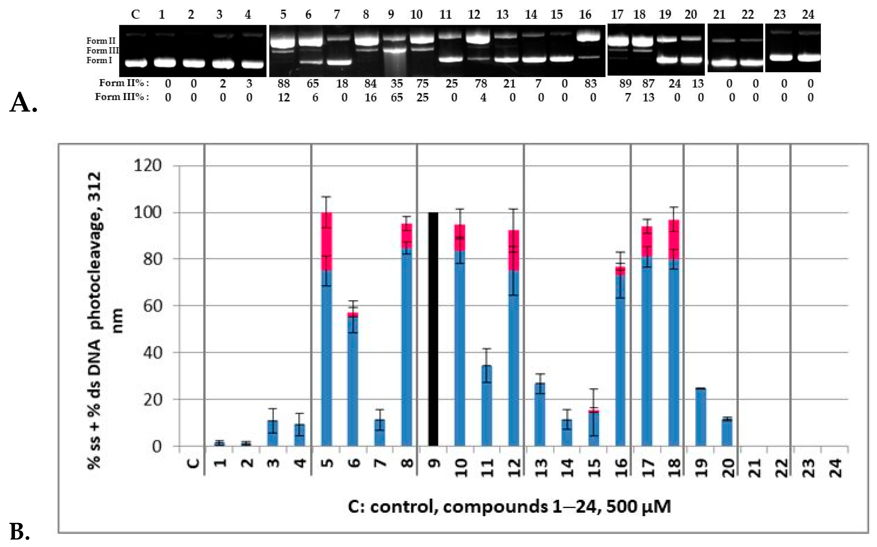
All compounds at a concentration of 500 μΜ were mixed with pBR322, incubated for 30 min and then irradiated for 30 min under UV-B irradiation at 312 nm. In Figure 5A, a compilation of representative agarose gel electrophoresis pictures for each compound (124) under UV-B irradiation is depicted. All experiments have been contacted at least twice. All runs are given in Supplementary Materials S.2: S.2.1–S.2.8 with calculations of Form II (nicked plasmid, % ss—single strand—damage) and Form III (linear plasmid, % ds—double strand—damage) photocleavage to be performed comparing to controls of irradiated plasmid DNA under the same conditions. On the top of the agarose gel, the numbers 1–24 one-to-one correspond to the compounds 124, and C to the control irradiated plasmid DNA. In Figure 5B, plots of DNA photocleavage of all experiments is depicted. The % ss (Form II) is shown in blue and the % ds (Form III) is shown in red. Vertical gray lines divide the graph into groups of derivatives for easier reading. Thus, the compounds 14 resulted from the reactions of the AA IIa; 58 from 5-Cl-AA IIb; 912 from the 5-Br-AA IIc; and 1316 from the 3,5-diBr-AA IId. Furthermore, each of those quartets have similar hydrazide residues, meaning Ph (from IIIa) for the first compound of the group; p-Cl-Ph (from IIIb) for the second; p-NO2-Ph (from IIIc) for the third; and finally the heteroaryl 2-furyl group (from IIId).
The compounds 14 were found to be almost inactive, regardless of the hydrazide residue (Supplementary Materials S.2.3 and S.2.7). In addition, the starting material IIa was also photochemically inactive (Supplementary Materials S.2.4). However, when we moved to the substituted AA IIb derivative (compounds 58), we observed a significant DNA photocleavage that seems to be specifically higher for the Ph and furyl derivatives (Figure 5A,B, Lanes 5 and 8, respectively, Supplementary Materials S.2.2, S.2.3 and S.2.5) giving at this concentration both the nicked plasmid (Form II) and linear form (Form III). The substitution on the aryl groups seems to lower the activity. Interestingly, in the case of the nitroaryl group (Lane 7) this inactivity towards irradiation was particularly notable. Compared to the starting material IIb that exhibited a strong DNA photocleavage at 500 μM under UV-B irradiation, only the derivative 5 retained this activity, whereas 68 showed lower photoaction towards DNA (Supplementary Materials S.2.2–S.2.5).
Checking 5-Br-AA (IIc) and the related products 912, we found that IIc results in high DNA photocleavage, comparable to its derivatives 10 and 12; however, an extreme reactivity was observed for compound 9 (Figure 5A, Supplementary Materials S.2.2–S.2.4), where plasmid DNA nicely reacted photochemically with the photosensitizer to give both nicked and linear fragments at 35% and 65%, respectively (Supplementary Materials S.2.3). In other runs with the compound 9 (Supplementary Materials S.2.2 and S.2.4), plasmid DNA has totally vanished, indicating that a lower concentration is needed for this compound (vide intra). For this reason, in Figure 5B, where the average of all calculations of all experiments with their standard deviations is depicted, this plot is represented with a black color. The compound 10 showed a lower activity than 9, and the nitro derivative 11 dramatically lost activity, whereas furyl heterocycle reestablished photointeraction. The introduction of a second bromine atom that inactivates the aromatic ring of the AA (IId) diminished the activity for all compounds of the group but the furyl one 16 that showed the same activity with the corresponding AA (Supplementary Materials S.2.1 and S.2.3).
The above-described set of experiments indicated that the phenyl and furyl hydrazide residues promote photosensitization. For this reason, further experiments that resulted from the combination of IIIa,d with the 5-I-AA (IIe), 4-Cl-AA (IIf), 4-NO2-AA (IIg) and 5-NO2-AA (IIh) were set up in order to examine the influence of a heavier halogen (I) or the position of the chlorine atom (5- or 4-). In addition, the 4-NO2 and 5-NO2 substituted AA could complete the study showing the effect of a highly electron-withdrawing group. Under UV-B irradiation, the I-derivatives 17 and 18 (Supplementary Materials S.2.4–S.2.6) had comparable activity with the 5-Cl analogues 5 and 8 and better than 5-I AA (IIe). Interestingly, derivatives bearing the Cl atom at the meta position relatively to NH2 (19 and 20) dropped all activity, as was also true for 4-Cl AA (IIf) (Supplementary Materials S.2.4–S.2.6). And even more surprising was the fact that all four nitro substituted compounds (2124), as well as IIg,h regardless of the position of the nitro group, were found to exhibit zero activity (Supplementary Materials S.2.4, S.2.6 and S.2.8). Therefore, it seems that rigidity is the key for the high activity of nitro-quinazolinones [57] and their 3-amide substituted derivatives [58] that allows the nitro group to express its photochemistry in biological systems as it does in organic synthesis [68].
The compound 9 that exhibited the higher reactivity among all checked derivatives (124) has been subjected to concentration/reactivity relationship experiments in order to find an optimized concentration to perform pH influence experiments and mechanistic studies. One can observe in Figure 6 that the compound 9 exhibited 50% damage of the plasmid DNA at concentration as low as 1 μM (Supplementary Materials S.3.1 and S.3.2).
As far as the pH influence on the DNA photocleavage experiment concerns, it is obvious that for the compound 9 (5 μΜ) a pH above 8 had little effect on the photocleavage (Figure 7A, top picture, Lanes 2–7 for pH 5–10, respectively, and bottom diagram, Supplementary Materials S.3.3), whereas below 8 and up to pH 5 no change was observed. Moreover, mechanistic studies show that a lack of molecular oxygen does not affect the reactivity (Figure 7B, top picture, Lane 3 and bottom diagram, Supplementary Materials S.3.4), which probably means that the homolysis of the C-Br bond may occur [57]. In the presence of oxygen, a singlet oxygen scavenger like NaN3 indicated the formation of this species, whereas a hydroxyl radical scavenger like DMSO verified their presence (Figure 7B, top picture, Lanes 6 and 8, respectively, Supplementary Materials S.3.4). Therefore, the compound 9 seems to react under Type I and Type II photosensitized oxidation reactions [69].

2.2.2. DNA Photo-Cleavage Experiments at 365 nm (UV-A Irradiation)

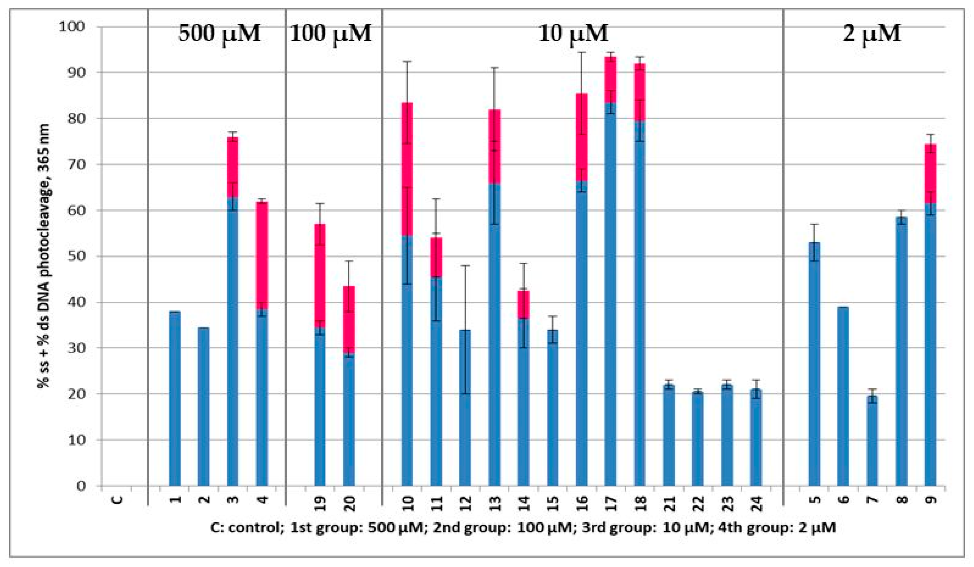
All compounds at a concentration of 500, 100, 10 and 2 μΜ were mixed with pBluescript SK II DNA, incubated for 30 min and then irradiated for 120 min under UV-A irradiation at 365 nm (broad band lamb). Concentrations below 500 μΜ were chosen after being apparent that the compounds were very active. The same applies for concentrations below 100 and 10 μΜ. All AA DACHZs absorb light in this area. The compounds 3-Arylamide-6-Br-2-Me-quinazolinones [58] that closely resemble open forms of 911 in the UV–Vis spectra did not absorb with a high ε, with values very far from 315 nm. All sets of derivatives gave a broad shoulder with a high ε from 300 to 400 nm in UV–Vis spectra (Supplementary Materials S.4), except 23 and 24 which showed a right shift and absorption from 350 to 500 nm. The compound J (Figure 2, Ar = Ph) which is the rigid analogue of 23 had a peak at 325 nm [58].
In Figure 8 (Supplementary Materials S.5 for the agarose gel pictures), it is shown that the compounds 14 that were inactive under UV-B irradiation at 500 μΜ exhibited some DNA photocleavage under UV-A light. This may be attributed to the higher UV–Vis absorption in this area. However, compared to the rest of the compounds, they still seemed reluctant to be excited. In addition, derivatives bearing the Cl atom in the meta position relative to NH2 (19 and 20) showed a lower activity (moderate action at concentration 100 μΜ) compared to their corresponding p-analogues 5 and 8, which were active at concentrations as low as 2 μΜ. The activity among the AA DACHZs of this group (58) indicated again that the nitroaryl moiety lowered DNA photocleavage (derivative 7).
What was left to be analyzed was the group of the 5-Br-AA DACHZs (912), of the 3,5-diBr-AA DACHZs (1316) and of the 5-I-AA DACHZs (17, 18). In addition to the compound 9 (2 μΜ), the rest of the derivatives 1018 were active at a concentration of 10 μΜ. Both nitrophenyl hydrazides’ (IIIc) products 11 and 15 had reduced activity in relation to other members of their group. The rest of the compounds were sufficiently active at a 10 μM concentration with the 5-I-AA DACHZs (17, 18) being the most potent ones. The next group with low activity (10 μM) was the nitro-AA derivatives 2124. Again, the nitro derivatives seemed to be very inactive.
The much higher activity of the compounds 58 and 1020 that photocleaved DNA at much lower concentrations under UV-A irradiation compared to UV-B might be attributed to the very high absorption between 300 and 400 nm. However, this does not seem to explain the reactivity of the derivative 9 that keeps the higher activity in both UV-B and UV-A irradiation at similar concentrations. Certainly, since none of the compounds was tested for photosensitivity by itself but as a DNA complex, the phenomenon is much more complex. Nonetheless, the bromine atom in this position may contribute to a high atom effect in the photochemistry of the compound [42,46,47].

2.3. Molecular Docking “In Silico” Calculations of DNA/AA DACHZs

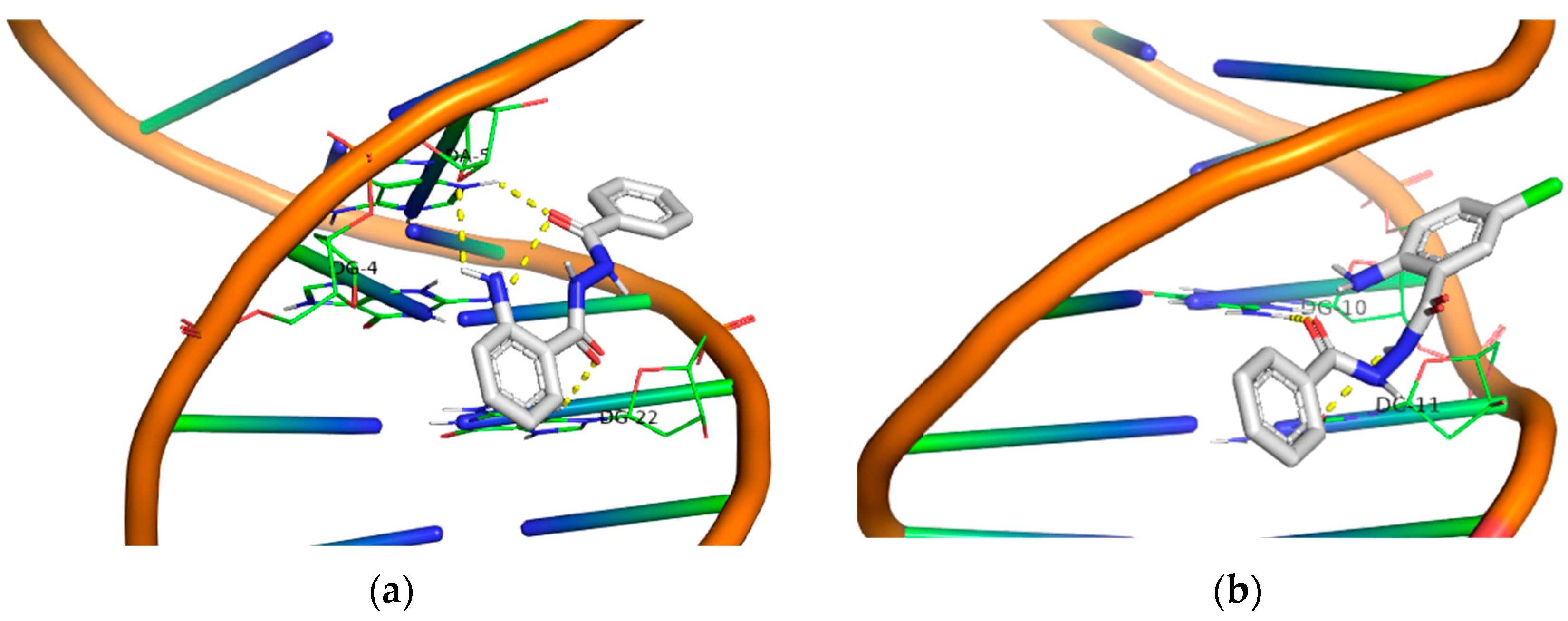
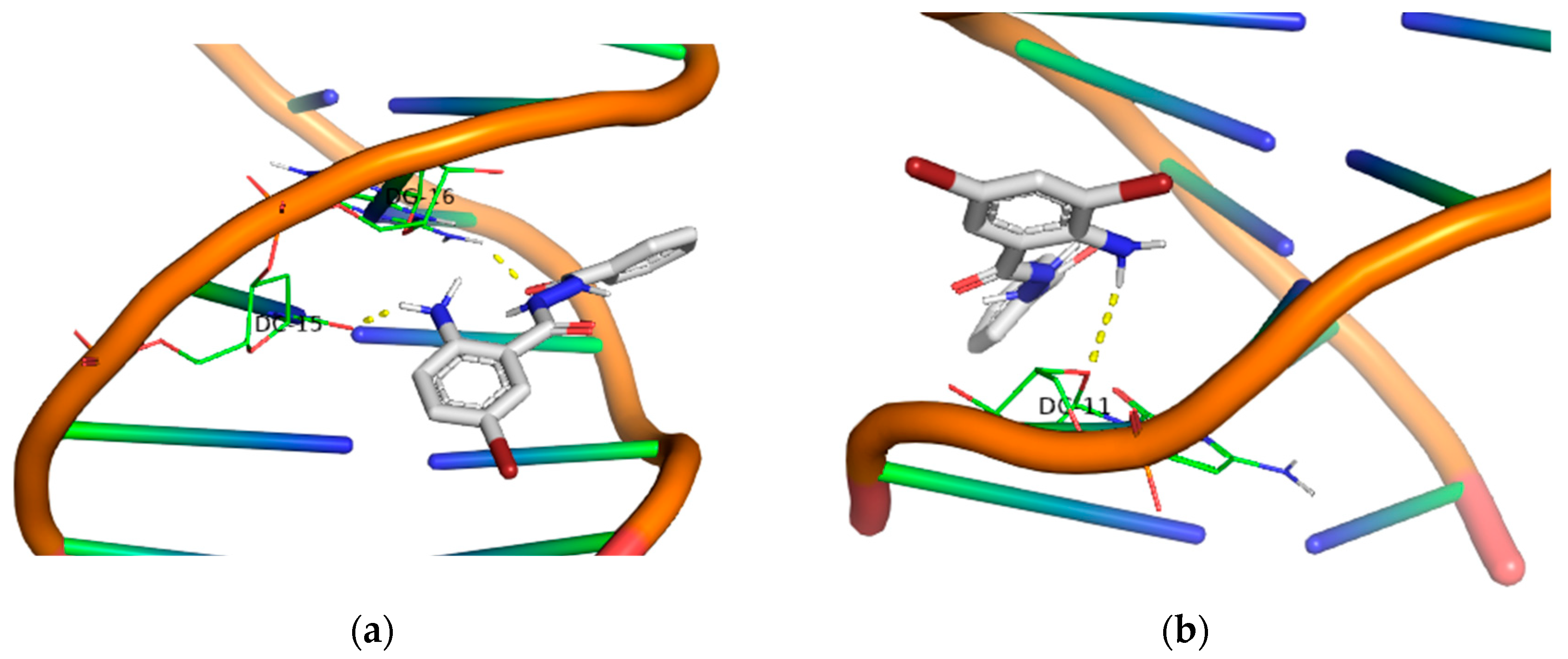
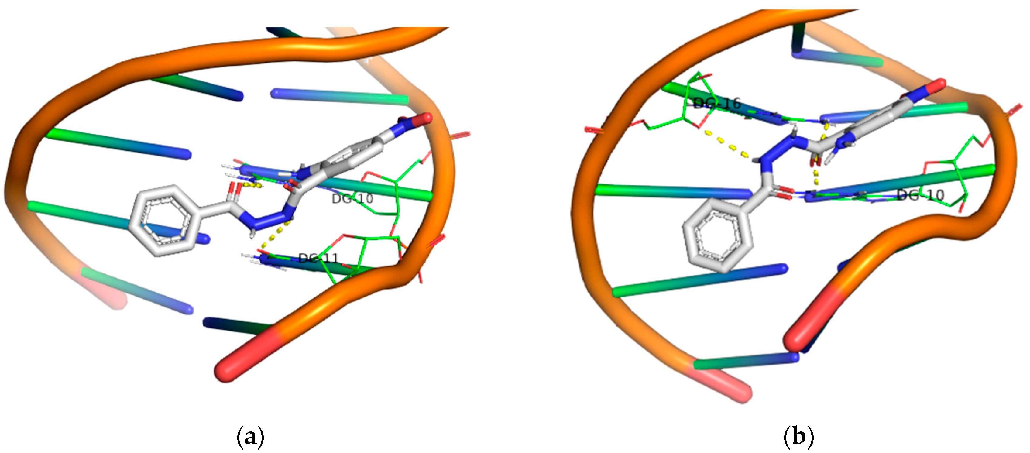
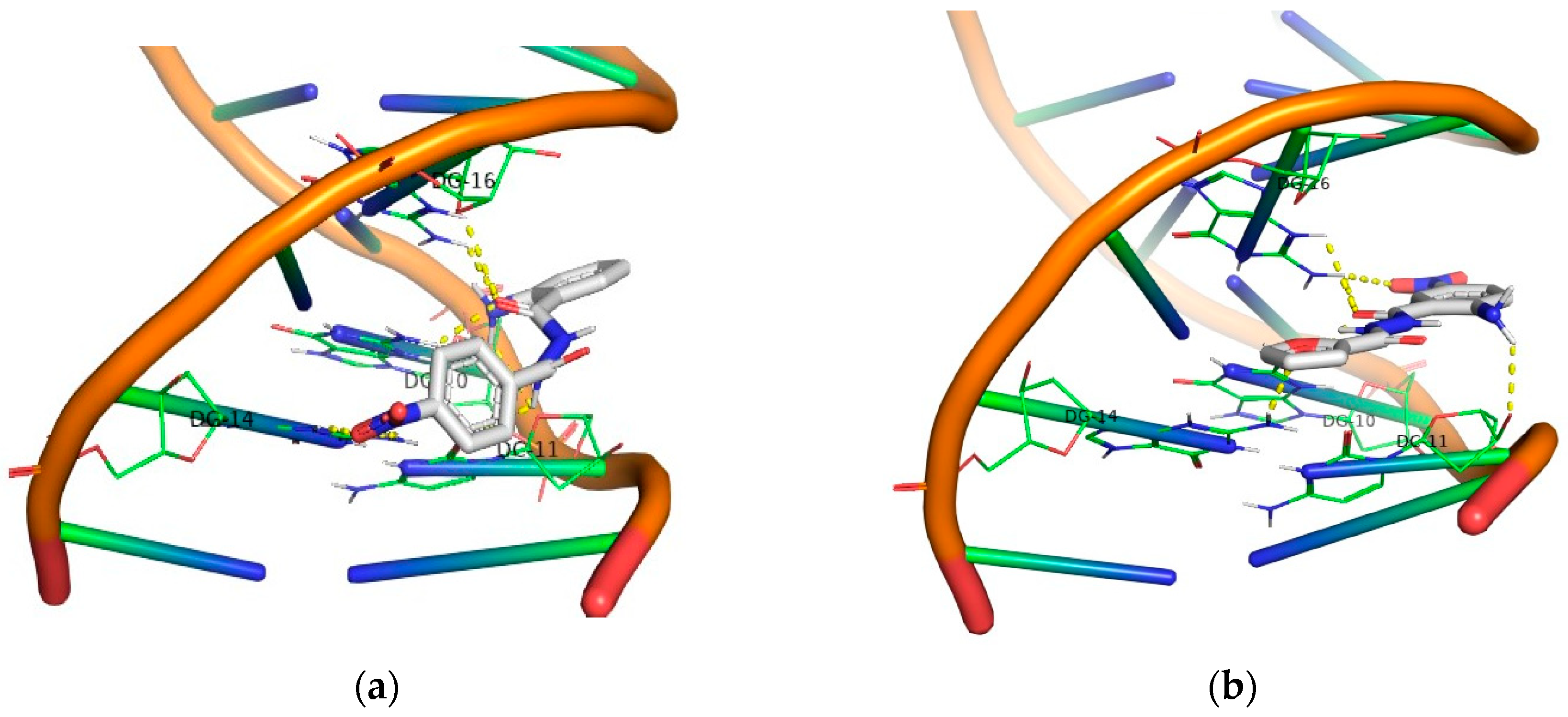
Molecular docking studies for the derivatives 124 were conducted using the AutoDock Vina program. The objective was to identify polar contacts and calculate the energy of their DNA binding. All calculated energy binding values, along with DNA base interactions, are given in Table 1. To gain a deeper understanding of the conformational preferences of the compounds within DNA, a 3D calculation program (PyMOL) was employed to identify all polar contacts with both DNA strands.
Generally, one may observe a network of polar contacts and hydrogen bonds arising from the free -NH2 group and the carbonyl groups in all derivatives. Upon closer examination of the docking results for each group, the first group (compounds 1, 2, 3 and 4) exhibits good binding with DNA. However, when irradiated in the UV-B area, all compounds were inactive. In the UV-A area, only the compounds 3 and 4 demonstrated significant DNA photocleavage, resulting in both the nicked plasmid (Form II) and linear form (Form III), although this was at the highest concentration (500 μM). In the second group (compounds 5, 6, 7 and 8), a stronger binding to DNA was observed in all 5-Cl derivatives, as they exhibited a higher binding energy compared to the compounds in the previous group. The compounds 5 and 8 showed a higher photocleavage effect after irradiation in the UV-B area, and these same compounds proved to be active even at small concentrations (2 μM) when irradiation occurred in the UV-A area.
Moving on to the third group of compounds (9, 10, 11 and 12), it is noteworthy that, excluding the compound 12, the 5-Br derivatives 10 and 11 exhibited a UV-A photocleavage effect (10 μM) at much lower concentrations than UV-B. the compound 9 demonstrated exceptional results under UV-B irradiation, despite having the lowest binding affinity with DNA among all the other compounds in this group. Under UV-A light, the compound 9 remained active even at a concentration of 2 μM.
In the fourth group, all the 4,5-bis-bromo derivatives demonstrated a good binding affinity, with the compound 15 having a binding energy value of −10.1 kcal/mol. However, despite exhibiting the highest binding energy among all the compounds, this one does not appear to be significantly active in either the UV-A or UV-B area. The other three compounds exhibited remarkable photocleavage results under UV-A irradiation at a concentration of 10 μM. Another characteristic of these compounds is the formation of polar contacts solely with the –NH2 group. The only exception to this is the compound 16, which forms hydrogen bonds with both the carbonyl group and the oxygen from the furyl group.
Regarding the 5-I derivatives (19, 20), one may observe the formation of polar contacts through both the –NH2 group and the –C=O group in both cases. Moreover, these compounds are particularly active in the UV-A area of irradiation. Plasmid DNA reacted photochemically with the photosensitizers at a concentration of 10 μM, yielding both nicked and linear fragments.
In the case of 4-Cl derivatives (21, 22), although the binding energy remained at a good level, their activity decreased significantly under UVB irradiation, while in the UVA area, their activity increased. This can be partially attributed to the significant absorption of the compounds in the UV-A region.
Finally, all the –NO2 derivatives (2124) utilized the -NH2 and –C=O groups to form hydrogen bonds with the DNA bases. Even though the -NO2 group does not participate in this network of polar contacts (with the compound 24 being the only exception), the binding of these compounds to DNA is significantly strong. Although all nitro derivatives proved to be ineffective under UV-B light, in the UV-A area they demonstrate a moderate photocleavage ability, fragmenting DNA at a concentration of 100 μM. In Figure 9, Figure 10, Figure 11 and Figure 12 images of all phenyl substituted AA DACHZ compounds 1, 5, 9, 13, 17, 19, 21 and 23 are shown (Supplementary Materials S.7).
The comparison of open and closed structures is also considered useful as it can lead to conclusions. In the closed structures [58], we observed the crucial role of the –NO2 group in strengthening the binding energy, as this group formed polar contacts with the DNA bases. On the contrary, in open structures, the –NO2 group does not appear to play a significant role. Specifically, among the four –NO2 derivatives (2124), only the compound 24 showed the participation of the –NO2 group in interaction with DNA. Furthermore, among the four compounds bearing the –NO2 group in the ring of hydrazide (3, 7, 11, 15), again only one (3) out of four compounds exhibited participation of the -NO2 group in polar contacts (Figure 13, Supplementary Materials S.6).
When considering binding energy, it is noteworthy that, despite open structures having more available groups for hydrogen bond formation, the binding in closed structures is slightly stronger than in the open ones, respectively.

2.4. Cell Culture Experiments of Selective AA DACHZs with Melanoma Cell Lines

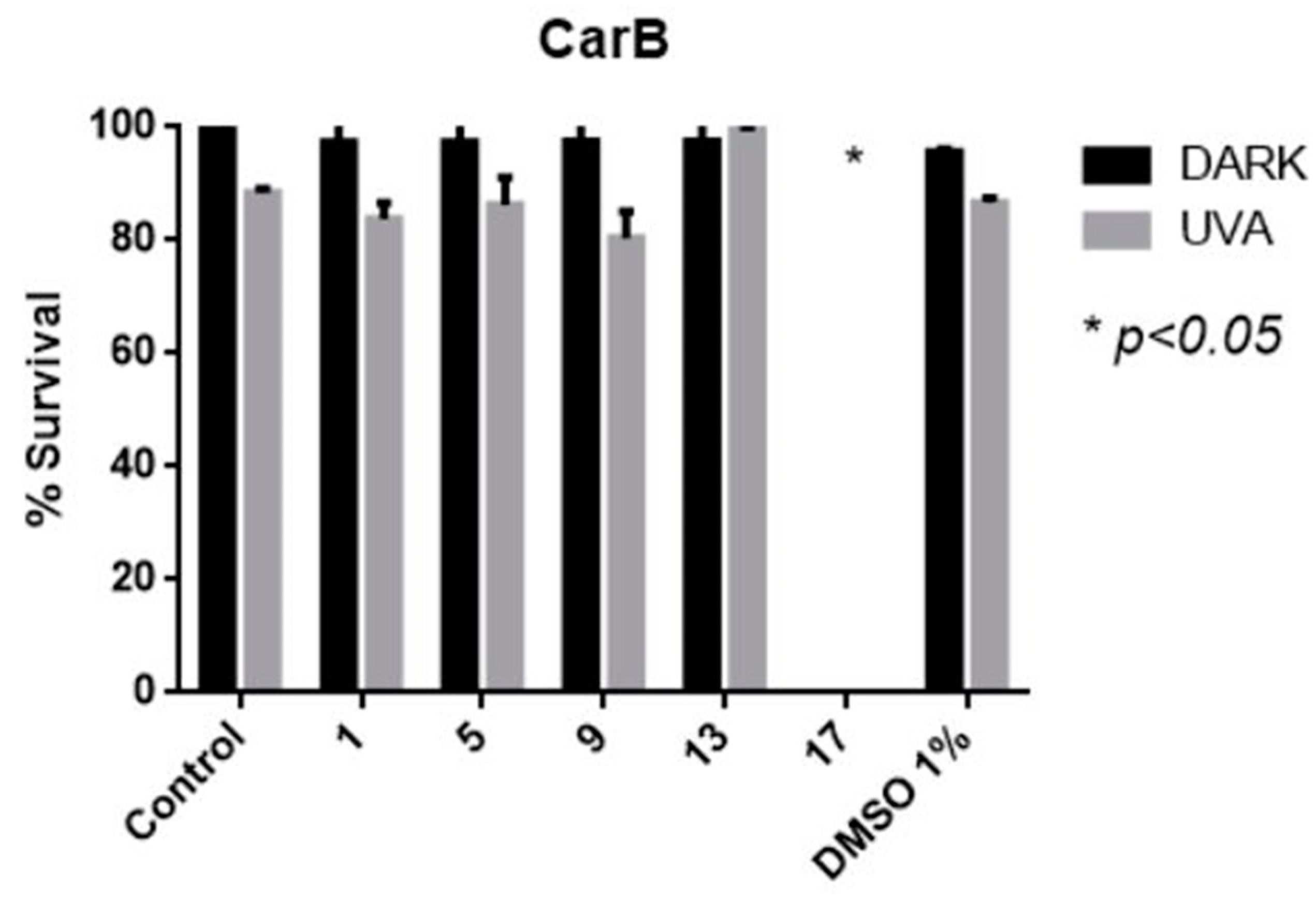
The highly malignant melanoma cell line CarB were used for cell culture experiments. These cells derive from squamous cell carcinoma of the mouse skin. Generally, skin cancer is preferred in drug photoactivation studies due to the penetration of UV radiation [70]. Moreover, our aim was to test the compounds activity on highly aggressive and metastatic cells. The cells were incubated with 100 μM of each selected compound 1, 5, 9, 13 and 17 and were irradiated at 365 nm (broad band) for 1 h. The selection was based on DNA photocleavage experiments where all compounds 5, 9 and 13, 17 were found active at 2 and 10 μM, respectively (Figure 8, Supplementary Materials S.5). Derivative 1 was selected for comparison, since it bears the same (phenyl) group attached on the diacylhydrazine moiety. It was observed that 17 possesses a high cytotoxic role; however, this role is not specific after UV irradiation. Among the compounds 1, 5, 9 and 13, it seems that 9 has a slight phototoxicity; nevertheless, this toxicity is not that impressive at 100 μM (Figure 14).

3. Materials and Methods

All commercially available reagent-grade chemicals and solvents were used without further purification. pB322 supercoiled plasmid was purchased from New England Biolabs (Ipswich, MA, USA). pBluescript SK II was laboratory produced. UV–Visible (UV–Vis) spectra were recorded on a Hitachi U–2001 dual beam UV–Vis spectrophotometer (Hitachi, Tokyo, Japan). NMR spectra were recorded on an Agilent 500/54 (Agilent Technologies, Santa Clara, CA, USA) (500 MHz and 125 MHz for 1H and 13C, respectively) or on a Bruker 300 AM (Bruker, Billerica, MA, USA) (300 MHz and 75 MHz for 1H and 13C, respectively) spectrometer using DMSO-d6 as a solvent. J values are reported in Hz. High-resolution mass spectra measured with an LTQ ORBITRAP XL with an ETD-Thermo Fisher Scientific Ion Source (Thermo Scientific, Waltham, MA, USA): Electrospray Ionization (ESI) positive mode Mass Analyser: Orbitrap. All samples containing pBR322 or pBluescript SK II plasmid were irradiated at pH 6.8 with Philips 2 × 9 W/01/2P UV−B narrowband lamps (Amsterdam, The Netherlands) at 312 nm and Philips 2 × 9 W/10/2P UV-A broad band lamps at 365 nm. All reactions were monitored on commercially available pre-coated TLC plates (layer thickness 0.25 mm) of Kieselgel 60 F254 (Merck, Darmstadt, Germany). Melting points were measured on GallenKamp MFB-595 melting point apparatuses (GallenKamp, Cambridge, UK) and are uncorrected. The calculation of yields was based on the amount of the crystallized product collected.

3.1. Synthesis of AA DACHZs 124

Method A: Isatoic anhydride (I) (2 mmol), the corresponding hydrazide (IIIc or IIId) (2 mmol) and Et3N (2 mmol) were added in DMF (1.2 mL) and the mixture was stirred at r.t. for 18 h. Iced water was added; the precipitate was filtrated and recrystallized from the proper solvent.
Method B: Modified from ref [66]. A mixture of the corresponding anthranilic acid IIa or IId (2 mmol) and CDI (2 mmol) in 5 mL THF (dry, commercially available) was stirred at 0 °C for 1 h and then for 2 h at r.t. Hydrazide (IIIad) was added diluted in 5 mL THF and the mixture was stirred overnight at r.t. The temperature was raised to 55 °C and the mixture was heated for 1 h. After this period, water was added (2 mL) and the mixture was heated for 1 h more, at the same temperature. After concentration of the solvents under reduced pressure, 0.1 M NaOH (30 mL) was added, and the residue was extracted with EA (3 × 30 mL). The organic layers were separated, dried with Na2SO4, and removed under reduced pressure. In the case of the compounds 1 and 3, trituration with CH2Cl2 gave a precipitate which was filtrated, purified by column chromatography and recrystallized from the proper solvent to give the pure compound. For IId and all derivatives 1316, no extraction was necessary. Directly after completion of the reaction, filtration of the precipitate gave the crude product which was purified by recrystallization.
Method C: Modified from ref [67]. A total of 4 mmol of Ph3P and 2 mmol of the corresponding anthranilic acid IIac or IIeh were individually mixed, added in toluene and dried with the azeotropic removal of any moisture with the solvent under reduced pressure. The solid powder was then added in 20 mL dry THF (commercially available) under argon. Cl3C–CN (5 mmol) was poured in the flask and the mixture was stirred at r.t. for 1.5 h. After being dried with azeotropic removal of any moisture with toluene under reduced pressure, the corresponding hydrazide IIIad (2 mmol) was added into the mixture with a subsequent addition of dry Et3N (6 mmol). The mixture was stirred from 1 to 12 h and then H2O 50 mL was added, and the mixture was extracted with EA (3 × 50 mL). The organic layers were dried with Na2SO4 and were removed under reduced pressure. The addition of CH2Cl2 gave a precipitate which was filtered and the obtained solid was recrystallized from the proper solvent.
  • 2-Amino-N-benzoylbenzohydrazide (1): Method B; white amorphous solid; mp: 180–182 °C (EA/hex), lit: 210–212 °C [20]; yield: 47%; IR (KBr) cm−1: 3408, 3272, 1674, 1645, 1614; 1H-NMR (DMSO-d6, 500 MHz) δ 10.38 (bs, 1H, NH), 10.17 (bs, 1H, NH), 7.93 (brs, 2H), 7.61 (bs, 2H), 7.52 (s, 2H), 7.20 (s, 1H), 6.75 (s, 1H), 6.55 (s, 1H), 6.43 (bs, 2H, NH2) ppm; 13C-NMR (DMSO-d6, 125 MHz) δ 168.3, 166.0, 149.9, 132.7, 132.3, 131.8, 128.5, 128.2, 127.5, 116.5, 114.7, 112.56 ppm; HRMS(ESI) m/z [M+H]+: C14H14N3O2+, calc: 256.1081; found: 256.1080.
  • 2-Amino-N-(4-chlorobenzoyl)benzohydrazide (2): Method C (1.5 h + 1 h); off-white amorphous solid; mp: 226.4 °C (EA/hex), lit: 238–240 °C [14]; yield: 52%; IR (KBr) cm−1: 3414, 3312, 3255, 1678, 1647, 1612; 1H-NMR (DMSO-d6, 500 MHz) δ 10.47 (s, 1H, NH), 10.19 (brs, 1H, NH), 7.94 (d, J = 8.3 Hz, 2H), 7.60 (d, J = 8.3 Hz, 2H), 7.20 (t, J = 7.5 Hz, 1H), 6.74 (d, J = 8.1 Hz, 1H), 6.55 (t, J = 7.4 Hz, 1H), 6.43 (brs, 2H, NH2) ppm; 13C-NMR (DMSO-d6, 125 MHz) δ 168.3, 165.0, 150.0, 136.7, 132.4, 131.4, 129.4, 128.6, 128.2, 116.4, 114.6, 112.4 ppm; HRMS(ESI) m/z [M+H]+: C14H13ClN3O2+, calc: 290.0691; found: 290.0689, 290.0660 (3/1).
  • 2-Amino-N-(4-nitrobenzoyl)benzohydrazide (3): Method A, B, C; yellow amorphous solid; mp: 239.0 °C (EA/EtOH), lit: 238–240 °C [12], 238 °C [71]; yield: 42%, 52%, 47%, respectively for each method used; IR (KBr) cm−1: 3407, 3310, 3301, 3244, 1682, 1642, 1613; 1H-NMR (DMSO-d6, 500 MHz) δ 10.73 (s, 1H, NH), 10.29 (brs, 1H, NH), 8.37 (d, J = 8.8 Hz, 2H), 8.15 (d, J = 8.7 Hz, 2H), 7.62 (d, J = 7.1 Hz, 1H), 7.20 (dt, J = 8.3, 1.3 Hz, 1H), 6.75 (d, J = 7.7 Hz, 1H), 6.56 (t, J = 7.2 Hz, 1H), 6.45 (brs, 2H, NH2) ppm; 13C-NMR (DMSO-d6, 125 MHz) δ 168.2, 164.5, 150.0, 149.4, 138.3, 132.5, 129.0, 128.2, 123.8, 116.5, 114.6, 112.1 ppm; HRMS(ESI) m/z [M+H]+: C14H13N4O4+, calc: 301.0931; found: 301.0933.
  • N-(2-Aminobenzoyl)furan-2-carbohydrazide (4): Method A; beige amorphous solid; mp: 194.2–195 °C (EA/EtOH); lit: 285–287 °C [20]; yield: 44%; IR (KBr) cm−1: 3414, 3317, 3280, 1680, 1645, 1614; 1H-NMR (DMSO-d6, 500 MHz) δ 10.25 (s, 1H, NH), 10.11 (brs, 1H, NH), 7.91 (d, J = 0.9 Hz, 1H), 7.58 (dd, J = 7.9, 0.9 Hz, 1H), 7.25 (d, J = 3.4 Hz, 1H), 7.19 (dt, J = 7.0, 1.3 Hz, 1H), 6.74 (dd, J = 8.1, 0.7 Hz, 1H), 6.67 (dd, J = 3.4, 1.7 Hz, 1H), 6.54 (dt, J = 7.8, 0.9 Hz, 1H), 6.42 (brs, 2H, NH2) ppm; 13C-NMR (DMSO-d6, 125 MHz) δ 168.3, 157.5, 150.0, 146.4, 145.7, 132.4, 128.2, 116.4, 114.6, 114.5, 112.3, 111.9 ppm; HRMS(ESI) m/z [M+H]+: C12H12N3O3+, calc: 246.0873; found: 246.0874.
  • 2-Amino-N-benzoyl-5-chlorobenzohydrazide (5): Method C (1.5 h + 2 h); off-white amorphous solid; mp: 203.1 °C (EA/EtOH); yield: 52%; IR (KBr) cm−1: 3486, 3372, 3310, 3280, 1684, 1642, 1612; 1H-NMR (DMSO-d6, 500 MHz) δ 10.47 (s, 1H, NH), 10.34 (brs, 1H, NH), 7.92 (d, J = 7.5 Hz, 2H), 7.67 (brs, 1H), 7.60 (t, J = 7.3 Hz, 1H), 7.52 (t, J = 7.6 Hz, 2H), 7.24 (dd, J = 8.8, 1.8 Hz, 1H), 6.78 (d, J = 8.8 Hz, 1H), 6.60 (brs, 2H, NH2) ppm; 13C-NMR (DMSO-d6, 125 MHz) δ 167.3, 166.0, 148.9, 132.5, 132.2, 132.0, 128.6, 127.5, 127.5, 118.2, 117.8, 113.2 ppm; HRMS(ESI) m/z [M+H]+: C14H13ClN3O2+, calc: 290.0691; found: 290.0692, 292.0661 (3/1).
  • 2-Amino-5-chloro-N-(4-chlorobenzoyl)benzohydrazide (6): Method C (1.5 h + 2.5 h); off white amorphous solid; mp: 233.2 °C (EA/EtOH); yield: 41%; IR (KBr) cm−1: 3482, 3347, 3222, 3164, 1663, 1636, 1595; 1H-NMR (DMSO-d6, 500 MHz) δ 10.55 (s, 1H, NH), 10.36 (brs, 1H, NH), 7.93 (d, J = 8.2 Hz, 2H), 7.66 (s, 1H), 7.61 (d, J = 8.1 Hz, 2H), 7.24 (d, J = 8.0 Hz, 1H), 6.77 (d, J = 8.8 Hz, 1H), 6.60 (brs, 2H, NH2) ppm; 13C-NMR (DMSO-d6, 125 MHz) δ 167.2, 165.0, 149.0, 136.8, 132.3, 131.3, 129.5, 128.8, 127.5, 118.3, 117.8, 113.0 ppm; HRMS(ESI) m/z [M+H]+: C14H12Cl2N3O2+, calc: 324.0301; found: 324.0302, 326.0273, 328.0244, M, M+2, M+4 (9/6/1).
  • 2-Amino-5-chloro-N-(4-nitrobenzoyl)benzohydrazide (7): Method C (1.5 h + 1 h); light yellow amorphous solid; mp: 235.4 °C (EA/hex); yield: 48%; IR (KBr) cm−1: 3503, 3370, 3219, 3047, 1666, 1637, 1600; 1H-NMR (DMSO-d6, 500 MHz) δ 10.77 (brs, 1H, NH), 10.44 (brs, 1H, NH), 8.37 (d, J = 8.1 Hz, 2H), 8.14 (d, J = 8.1 Hz, 2H), 7.66 (s, 1H), 7.24 (d, J = 7.3 Hz, 1H), 6.78 (d, J = 8.6 Hz, 1H), 6.58 (brs, 2H, NH2) ppm; 13C-NMR (DMSO-d6, 125 MHz) δ 167.1, 164.5, 149.5, 148.9, 138.2, 132.3, 129.0, 127.4, 123.8, 118.3, 117.8, 112.9 ppm; HRMS(ESI) m/z [M+H]+: C14H12ClN4O4+, calc: 335.0542; found: 335.0541, 337.0511 (3/1).
  • N-(2-Amino-5-chlorobenzoyl)furan-2-carbohydrazide (8): method C (1.5 h + 1 h); beige amorphous solid; mp: 232.3 °C (EA/hex); yield: 49%; IR (KBr) cm−1: 3465, 3435, 3206, 3017, 1618, 1584; 1H-NMR (DMSO−d6, 500 MHz) δ 10.34 (s, 1H, NH), 10.28 (brs, 1H, NH), 7.92 (s, 1H), 7.64 (s, 1H), 7.26 (d, J = 2.8 Hz, 1H), 7.24 (d, J = 9.0 Hz, 1H), 6.77 (d, J = 8.9 Hz, 1H), 6.68 (s, 1H), 6.59 (brs, 2H, NH2) ppm; 13C-NMR (DMSO-d6, 125 MHz) δ 167.2, 157.5, 149.0, 146.3, 145.9, 132.3, 127.4, 118.3, 117.8, 114.7, 113.0, 112.0 ppm; HRMS(ESI) m/z [M+H]+: C12H11ClN3O3+, calc: 280.0483; found: 280.0481, 282.0451 (3/1).
  • 2-Amino-N-benzoyl-5-bromobenzohydrazide (9): Method C (1.5 h + 2 h); off-white amorphous solid; mp: 207.0 °C (1,4-dioxane/EtOH); yield: 45%; IR (KBr) cm−1: 3411, 3282, 1673, 1644, 1604; 1H-NMR (DMSO-d6, 500 MHz) δ 10.43 (s, 1H, NH), 10.31 (brs, 1H, NH), 7.91 (d, J = 7.5Hz, 2H), 7.78 (s, 1H), 7.60 (t, J = 7.6 Hz, 1H), 7.52 (t, J = 7.6 Hz, 2H), 7.33 (d, J = 8.9 Hz, 1H), 6.73 (d, J = 8.9 Hz, 1H), 6.59 (brs, 2H, NH2) ppm; 13C-NMR (DMSO-d6, 125 MHz) δ 167.1, 165.9, 149.1, 134.8, 132.5, 131.9, 130.3, 128.5, 127.5, 118.6, 113.9, 104.9 ppm; HRMS(ESI) m/z [M+H]+: C14H13BrN3O2+, calc: 334.0186; found: 334.0183, 336.0163 (1/1).
  • 2-Amino-5-bromo-N-(4-chlorobenzoyl)benzohydrazide (10): Method C (1.5 h + 2 h); off-white amorphous solid; mp: 205–207 °C (EA/EtOH); yield: 56%; IR (KBr) cm−1: 3376, 3276, 1679, 1645, 1593; 1H-NMR (DMSO-d6, 500 MHz) δ 10.54 (brs, 1H, NH), 10.35 (brs, 1H, NH), 7.93 (d, J = 8.5 Hz, 2H), 7.77 (d, J = 1.9 Hz, 1H), 7.60 (d, J = 8.5 Hz, 2H), 7.34 (dd, J = 8.8, 2.0 Hz, 1H), 6.73 (d, J = 9 Hz, 1H), 6.60 (brs, 2H, NH2) ppm; 13C-NMR (DMSO-d6, 125 MHz) δ 167.1, 164.9, 149.2, 136.8, 134.9, 131.2, 130.3, 129.4, 128.7, 118.6, 113.8, 104.9 ppm; HRMS(ESI) m/z [M+H]+: C14H12BrClN3O2+, calc: 367.9796; found: 367.9793, 369.9770, 371.9838 M, M+2, M+4 (3/4/2).
  • 2-Amino-5-bromo-N-(4-nitrobenzoyl)benzohydrazide (11): Method C (1.5 h + 2 h); beige amorphous solid; mp: 221.5 °C (EA/hex); yield: 79%; IR (KBr) cm−1: 3387, 3290, 1682, 1646, 1605; 1H-NMR (DMSO-d6, 500 MHz) δ 10.78 (s, 1H, NH), 10.44 (brs, 1H, NH), 8.37 (d, J = 8.5 Hz, 2H), 8.14 (d, J = 8.5 Hz, 2H), 7.79 (s, 1H), 7.35 (d, J = 8.9 Hz, 1H), 6.74 (d, J = 8.9 Hz, 1H), 6.61 (brs, 2H, NH2) ppm; 13C-NMR (DMSO-d6, 125 MHz) δ 167.0, 164.4, 149.4, 149.2, 138.1, 134.9, 130.3, 129.0, 123.8, 118.6, 113.5, 104.9 ppm; HRMS(ESI) m/z [M+H]+: C14H12BrN4O4+, calc: 379,0036; found: 379.0037, 381.0017 (1/1).
  • N-(2-Amino-5-bromobenzoyl)furan-2-carbohydrazide (12): Method C (1.5 h + 2 h); beige amorphous solid; mp: 212.4–217.7 °C (EA/EtOH); yield: 50%; IR (KBr) cm−1: 3466, 3360, 3213, 1664, 1614, 1581; 1H-NMR (DMSO-d6, 500 MHz) δ 10.32 (s, 1H, NH), 10.27 (brs, 1H, NH), 7.92 (d, J = 0.8 Hz, 1H), 7.75 (d, J = 2.1 Hz, 1H), 7.33 (dd, J = 8.9, 2.1 Hz, 1H), 7.26 (d, J = 3.3 Hz, 1H), 6.72 (d, J = 8.9 Hz, 1H), 6.68 (dd, J = 3.3, 1.6 Hz, 1H), 6.59 (brs, 2H, NH2) ppm; 13C-NMR (DMSO-d6, 125 MHz) δ 167.1, 157.5, 149.2, 146.3, 145.8, 134.9, 130.3, 118.6, 114.6, 113.7, 111.9, 104. ppm; HRMS(ESI) m/z [M+H]+: C12H11BrN3O3+, calc: 323.9978; found: 323.9978, 325.9957 (1/1).
  • 2-Amino-N-benzoyl-3,5-dibromobenzohydrazide (13): Method B; white amorphous solid; mp: 275.9 °C (EtOH); yield: 46%; IR (KBr) cm−1: 3464, 3340, 3219, 1668, 1632, 1602; 1H-NMR (DMSO-d6, 500 MHz) δ 10.54 (brs, 2H, NH, NH), 7.91 (d, J = 7.3 Hz, 2H), 7.81 (s, 2H), 7.59 (t, J = 7.1 Hz, 1H), 7.52 (t, J = 7.4 Hz, 2H), 6.58 (brs, 2H, NH2) ppm; 13C-NMR (DMSO-d6, 125 MHz) δ 166.5, 165.9, 145.5, 137.0, 132.3, 132.0, 130.2, 128.6, 127.5, 116.0, 110.3, 105.2 ppm; HRMS(ESI) m/z [M+H]+: C14H12Br2N3O2+, calc: 411,9291; found: 411.9290, 413.9269, 415.9250 (1/2/1).
  • 2-Amino-3,5-dibromo-N-(4-chlorobenzoyl)benzohydrazide (14): Method B; light yellow amorphous solid; mp: 282.9 °C (1,4-dioxane); yield: 73%; IR (KBr) cm−1: 3483, 3342, 3031, 1658, 1630, 1601; 1H-NMR (DMSO-d6, 500 MHz) δ 10.64 (brs, 1H, NH), 10.57 (brs, 1H, NH), 7.81 και 7.94 (two doublets overlapped, 4H), 7.62 (s, 1H), 6.58 (brs, 2H, NH2) ppm; 13C-NMR (DMSO-d6, 125 MHz) δ 166.5, 164.9, 145.5, 137.1, 136.9, 131.0, 130.1, 129.4, 128.7, 115.9, 110.3, 105.2 ppm; HRMS(ESI) m/z [M+H]+: C14H11Br2ClN3O2+, calc: 445.8901; found: 445.8901, 447.8880, 449.8857, 451.8831 (3/7/5/1).
  • 2-Amino-3,5-dibromo-N-(4-nitrobenzoyl)benzohydrazide (15): Method B; yellow amorphous solid; mp: 283.5 °C (1,4-dioxane); yield: 69%; IR (KBr) cm−1: 3467, 3351, 3227, 3024, 1673, 1636, 1604; 1H-NMR (DMSO-d6, 500 MHz) δ 10.88 (brs, 1H, NH), 10.68 (brs, 1H, NH), 8.37 (d, J = 8.5 Hz, 2H), 8.14 (d, J = 8.5 Hz, 2H), 7.82 (s, 2H), 6.59 (brs, 2H, NH2) ppm; 13C-NMR (DMSO-d6, 125 MHz) δ 166.4, 164.4, 149.5, 145.6, 137.9, 137.2, 130.2, 129.1, 123.8, 115.6, 110.4, 105.2 ppm; HRMS(ESI) m/z [M+H]+: C14H11Br2N4O4+, calc: 456,9142; found: 456.9141, 458.9120, 460.9100 (1/2/1). Despite all our efforts, a small amount of 1,4-dioxane remained after recrystallization.
  • N-(2-Amino-3,5-dibromobenzoyl)furan-2-carbohydrazide (16): Method B; beige amorphous solid; mp: 223.3 °C (EA/EtOH); yield: 54%; IR (KBr) cm−1: 3469, 3410, 3339, 3198, 1676, 1640, 1600; 1H-NMR (DMSO-d6, 500 MHz) δ 10.48 (bs, 1H, NH), 10.41 (s, 1H, NH), 7.92 (s, 1H), 7.80 (s, 1H), 7.78 (s, 1H), 7.27 (d, J = 3.1 Hz, 1H), 6.68 (s, 1H), 6.57 (brs, 2H, NH2) ppm; 13C-NMR (DMSO-d6, 125 MHz) δ 166.4, 157.3, 146.0, 145.9, 145.5, 137.0, 130.1, 115.7, 114.8, 111.9, 110.3, 105.2 ppm; HRMS(ESI) m/z [M+H]+: C12H10Br2N3O3+, calc: 401,9083; found: 401.9083, 403.9062, 405.9042 (1/2/1).
  • 2-Amino-N-benzoyl-5-iodobenzohydrazide (17): Method C (1.5 h + 2.5 h); off-white amorphous solid; mp: 208.0 °C (EA/EtOH); yield: 46%; IR (KBr) cm−1: 3417, 3297, 3274, 1673, 1641, 1603; 1H-NMR (DMSO-d6, 400 MHz) δ 10.42 (s, 1H, NH), 10.31 (brs, 1H, NH), 7.92 (d, J = 7.5 Hz, 2H), 7.91 (s, 1H), 7.60 (t, J = 7.3 Hz, 1H), 7.52 (t, J = 7.4 Hz, 2H), 7.44 (d, J = 8.6 Hz, 1H), 6.61 (d, J = 8.7 Hz, 1H), 6.58 (brs, 2H, NH2) ppm; 13C-NMR (DMSO-d6, 100 MHz) δ 167.0, 165.9, 149.4, 140.2, 136.0, 132.5, 131.9, 128.5, 127.5, 119.0, 114.9, 74.6 ppm; HRMS(ESI) m/z [M+H]+: C14H13IN3O2+, calc: 382.0047; found: 382.0046.
  • N-(2-Amino-5-iodobenzoyl)furan-2-carbohydrazide (18): Method C (1.5 h + 1.5 h); beige amorphous solid; mp: 198.8 °C (EA/hex); yield: 48%; IR (KBr) cm−1: 3435, 3330, 3181, 1686, 1641, 1601; 1H-NMR (DMSO-d6, 500 MHz) δ 10.30 (s, 1H, NH), 10.24 (brs, 1H, NH), 7.91 (s, 1H), 7.87 (s, 1H), 7.45 (d, J = 8.6 Hz, 1H), 7.25 (s, 1H), 6.67 (s, 1H), 6.61 (d, J = 8.7 Hz, 1H), 6.57 (brs, 2H, NH2) ppm; 13C-NMR (DMSO-d6, 125 MHz) δ 167.0, 157.5, 149.5, 146.3, 145.8, 140.3, 136.0, 119.0, 114.7, 114.6, 111.9, 74.6 ppm; HRMS(ESI) m/z [M+H]+: C12H11IN3O3+, calc: 371.9840; found: 371.9836.
  • 2-Amino-N-benzoyl-4-chlorobenzohydrazide (19): Method C (1.5 h + 1.5 h); white amorphous solid; mp: 230.1 °C (EA/EtOH); yield: 58%; IR (KBr) cm−1: 3458, 3353, 3205, 1607, 1566; 1H-NMR (DMSO-d6, 500 MHz) δ 10.40 (s, 1H, NH), 10.26 (s, 1H, NH), 7.91 (d, J = 7.5 Hz, 2H), 7.62 (d, J = 8.6 Hz, 1H), 7.59 (t, J = 7.2 Hz, 1H), 7.51 (t, J = 7.5 Hz, 2H), 6.82 (s, 1H), 6.69 (brs, 2H, NH2), 6.58 (d, J = 8.4 Hz, 1H) ppm; 13C-NMR (DMSO-d6, 125 MHz) δ 167.5, 166.0, 151.2, 136.9, 132.6, 131.9, 130.0, 128.5, 127.5, 115.2, 114.4, 111.3 ppm; HRMS(ESI) m/z [M+H]+: C14H13ClN3O2+, calc: 290.0691; found: 290.0690, 292.0661 (3/1).
  • N-(2-Amino-4-chlorobenzoyl)furan-2-carbohydrazide (20): Method C (1.5 h + 1.5 h); white amorphous solid; mp: 207.8 °C (EA/EtOH); yield: 57%; IR (KBr) cm−1: 3397, 3303, 1681, 1648, 1613; 1H-NMR (DMSO-d6, 500 MHz) δ 10.28 (s, 1H, NH), 10.21 (s, 1H, NH), 7.91 (s, 1H), 7.59 (d, J = 8.4 Hz, 1H), 7.25 (s, 1H), 6.81 (s, 1H), 6.68 (brs, 3H, 1H + NH2), 6.57 (d, J = 8.3 Hz, 1H) ppm; 13C-NMR (DMSO-d6, 100 MHz) δ 167.6, 157.6, 151.3, 146.3, 145.8, 136.9, 130.0, 115.2, 114.6, 114.4, 111.9, 111.0 ppm; HRMS(ESI) m/z [M+H]+: C12H11ClN3O3+, calc: 280.0483; found: 280.0483, 280.0452 (3/1).
  • 2-Amino-N-benzoyl-4-nitrobenzohydrazide (21): Method C (1.5 h + 3 h); yellow amorphous solid; mp: 244.4 °C (EA/EtOH); yield: 45%; IR (KBr) cm−1: 3406, 3275, 1671, 1646, 1622; 1H-NMR (DMSO-d6, 500 MHz) δ 10.53 (s, 2H, NH, NH), 7.92 (d, J = 7.5 Hz, 2H), 7.90 (s, 1H), 7.78 (d, J = 8.5 Hz, 1H), 7.63 (s, 1H), 7.60 (t, J = 7.8 Hz, 1H), 7.53 (t, J = 7.4 Hz, 2H), 7.34 (d, J = 8.5 Hz, 1H), 6.87 (brs, 2H, NH2) ppm; 13C-NMR (DMSO-d6, 125 MHz) δ 166.9, 166.0, 150.3, 149.8, 132.4, 132.0, 129.8, 128.6, 127.5, 118.1, 110.2, 108.4 ppm; HRMS(ESI) m/z [M+H]+: C14H13N4O4+, calc: 301.0931; found: 301.0932.
  • N-(2-Amino-4-nitrobenzoyl)furan-2-carbohydrazide (22): Method C (1.5 h + 6 h); yellow amorphous solid; mp: 242.0 °C (EA/EtOH); yield: 47%; IR (KBr) cm−1: 3446, 3366, 3212, 3129, 1682, 1627, 1564; 1H-NMR (DMSO-d6, 500 MHz) δ 10.47 (s, 1H, NH), 10.42 (s, 1H, NH), 7.92 (s, 1H), 7.75 (d, J = 8.6 Hz, 1H), 7.62 (s, 1H), 7.32 (d, J = 8.6 Hz, 1H), 7.26 (d, J = 2.1 Hz, 1H), 6.85 (brs, 2H, NH2), 6.68 (dd, J = 3.0, 1.5 Hz, 1H) ppm; 13C-NMR (DMSO-d6, 125 MHz) δ 166.9, 157.5, 150.3, 149.9, 146.2, 145.9, 129.8, 117.8, 114.8, 112.0, 110.2, 108.4 ppm; HRMS(ESI) m/z [M+H]+: C12H11N4O5+, calc: 291.0724; found: 291.0725.
  • 2-Amino-N-benzoyl-5-nitrobenzohydrazide (23): Method C (1.5 h + 12 h); yellow amorphous solid; mp: 311.1 °C (1,4-dioxane/EtOH); yield: 20%; IR (KBr) cm−1: 3398, 3362, 3297, 3263, 3177, 1689, 1645, 1619; 1H-NMR (DMSO-d6, 500 MHz) δ 10.68 (s, 1H, NH), 10.51 (s, 1H, NH), 8.64 (s, 1H), 8.08 (d, J = 9.0 Hz, 1H), 7.92 (d, J = 7.5 Hz, 2H), 7.72 (brs, 2H, NH2), 7.61 (t, J = 7.3 Hz, 1H), 7.53 (t, J = 7.4 Hz, 2H), 6.86 (d, J = 9.2 Hz, 1H), ppm; 13C-NMR (DMSO-d6, 75 MHz) δ 166.8, 166.0, 155.3, 135.0, 132.4, 132.0, 128.6, 127.9, 127.5, 126.0, 116.1, 111.0 ppm; HRMS(ESI) m/z [M+H]+: C14H13N4O4+, calc: 301.0931; found: 301.0932.
  • N-(2-Amino-5-nitrobenzoyl)furan-2-carbohydrazide (24): Method C (1.5 h + 12 h); yellow amorphous solid; mp: >350 °C (1,4-dioxane/EtOH); yield: 26%; IR (KBr) cm−1: 3401, 3294, 3253, 1688, 1643, 1618; 1H-NMR (DMSO-d6, 300 MHz) δ 10.64 (s, 1H, NH), 10.41 (s, 1H, NH), 8.61 (d, J = 2.6 Hz, 1H), 8.08 (dd, J = 9.3, 2.6 Hz, 1H), 7.94 (s, 1H), 7.73 (brs, 2H, NH2), 7.27 (d, J = 3.5 Hz, 1H), 6.86 (d, J = 9.3 Hz, 1H), 6.69 (s, 1H) ppm; 13C-NMR (DMSO-d6, 75 MHz) δ 166.8, 157.4, 155.3, 146.2, 145.9, 135.0, 128.0, 126.0, 116.1, 114.8, 112.0, 110.7 ppm; HRMS(ESI) m/z [M+H]+: C12H11N4O5+, calc: 291.0724; Found: 291.0729.

3.2. DNA Photo-Cleavage Experiments

The compounds 124 were individually incubated with plasmid DNA at the desired concentration, in Eppendorf vials and/or were irradiated with UV-B (312 nm, 2 × 9 W) or UV-A (365 nm, 2 × 9 W), and in 10 cm distance under aerobic conditions at room temperature for 30 min and 2 h, respectively. The conditions of the photobiological reaction and gel electrophoresis, quantification of DNA-cleaving activity and calculation of ss % and ds % damage protocols have been described previously [72]. All experiments were performed at least twice.

3.3. Molecular Docking Studies

Organic compounds were fully optimized for their minimized energy at the B3LYP/6-31g* level of theory with the LanL2DZ basis set for iodine in the case of the compounds 13 and 18 as implemented in the Gaussian 09 [73] suite of programs (Revision B.01). The crystal data of the B-DNA dodecamer d(CGCGAATTCGCG)2 (PDB 1D:1BNA) were downloaded from the Protein Data Bank [74]. The docking analysis was performed using the AutoDock Vina program [75] (https:vina.scripps.edu, accessed on 21 January 2024). The DNA was adapted for docking by removing water molecules and polar hydrogens, and Gasteiger charges were added by AutoDock 4.2 Tools (ADT) before performing docking calculations. A grid box with a size of 60 × 80 × 114 with 0.375 Å spacing was used to encompass the whole DNA. The rigid docking protocol and 100 runs of the Lamarckian genetic algorithm for searching ligand conformations were performed. PyMOL [76] was used for the representation of the docking results and interactions between DNA and compounds.

3.4. Cell Culture Experiments

The CarB cell line, from mouse skin squamous cell carcinoma was a kind gift from V. Zoumpourlis, from National Hellenic Research Foundation, and was used to test the cytotoxic effect of the compounds. Cells were cultured under aseptic conditions using DMEM basal medium (31885-023; Gibco, Grand Island, NY, USA) supplemented with 10% fetal bovine serum (FB1000/500, Biosera, East Sussex, UK), 100 units/mL penicillin and 100 μg/mL streptomycin (15140-122, Gibco) and 2 mM L-Glutamine (25030; Gibco). The cell line was maintained at standard conditions (37 °C, 5% CO2) in a humidified atmosphere and cells were used at 70–90% confluency. A total of 10,000 cells were seeded per well. A UV-A lamp was placed 10 cm over the 96-well plate. A 1 h incubation with 100 μM and 200 μM of each compound was followed by 1 h irradiation with UV-A (365 nm). Then, compounds were removed, the medium was replaced and a cytotoxicity assay was performed 24 h later. The Resazurin Cell Viability Assay (CA035, Canvax, Boecillo, Spain) was used for fluorescence measurements according to the manufacturer’s guidelines. Essentially, a non-irradiated 96-well plate was used as a control, under the same conditions. Incubation with 10% resazurin (7 h) was followed by fluorescence measurement at λem = 590 nm and λex = 530/560 nm in a VarioSkan lux reader (ThermoFisher Scientific, Waltham, MA, USA).

4. Conclusions

Due to the importance of diacylhydrazine bridged anthranilic acids, a series of such compounds have been synthesized from the conjugation of commercially available anthranilic acids and hydrazides. The counterparts were carefully chosen to possess substituents with all possible electronic effects. The electron-withdrawing effect of the NO2 group in the p-position in relation to the amine group of the AA negatively affected the yield of the products, in a method driven by the in situ formation of the anthraniloyl chloride. For all new AA DACHZ derivatives, the yields were calculated based on the amount of product precipitated upon treatment with CH2Cl2 and were moderate; however, the synthesis was completed in short times compared to other methods tried. All derivatives exhibited a high UV–Vis absorption in the UV-A area of the spectrum and DNA photocleavage was performed under both UV-B and UV-A irradiation. All screenings highlighted the importance of a halogen in the p-position in relation to the amine group and the absence of an electron-withdrawing group on the aryl group. Differences were observed in DNA photocleavage under UV-B and UV-A irradiation. The derivative 9 maintained activity under both types of irradiation at very low concentrations (1 and 2 μΜ, for UV-B and UV-A, respectively); however under UV-A irradiation, all halogenated compounds were active at concentrations as low as 2 and 10 μΜ. Molecular docking studies with DNA showed potential interaction sites, although the reactivity was not correlated for all derivatives with their photoactivity towards DNA. Cytotoxicity experiments indicated the iodo derivative 17 as a potent cytotoxic agent and the bromo compound 9 as a slight phototoxic agent. In general, based on the studies described herein, one may keep in mind the high UV-A light absorption of AA DACHZs that allows DNA photocleavage. Since no DNA photocleavage may occur without the compound showing binding to DNA, this new class of DNA photocleavers may hold promise for the development of novel anticancer, antimicrobial and probably insecticidal agents. Finally, open-form anthranilic acid derivatives and their rigid form quinazolinones exhibited very different photoreactivities particularly regarding derivatives containing the nitro group, with the former being inactive and the latter being highly potent DNA photocleavers.

Supplementary Materials

The following supporting information can be downloaded at: https://www.mdpi.com/article/10.3390/molecules29030647/s1, S.1: Copies of NMR spectra of the compounds 124 and of HRMS measurements; S.2: Gel electrophoresis pictures of the compounds 124 and AA IIah (UV-B); S.3: Gel electrophoresis pictures of the compound 9. Concentration, pH and mechanistic studies (UV-B); S.4: UV–Vis spectra of the compounds 124; S.5: Gel electrophoresis pictures of the compounds 124 (UV-A); S.6: Optimized molecular geometries of the compounds 124 at the B3LYP/6–31G (d) level; S.7: Molecular docking studies for the compounds 124.

Author Contributions

Conceptualization, K.C.F.; methodology, M.K. and K.C.F.; software, C.M.; validation, A.M., S.R., A.E.K. and K.C.F.; formal analysis, A.M., C.M. and K.C.F.; investigation, A.M., M.-E.K.S., C.M., C.K. and S.M.; resources, A.E.K., M.K. and K.C.F.; data curation, C.M., S.R., A.E.K. and K.C.F.; writing—original draft preparation, K.C.F.; writing—review and editing, A.E.K. and K.C.F.; visualization, K.C.F.; supervision, K.C.F.; project administration, M.K. and K.C.F.; funding acquisition, M.K. and K.C.F. All authors have read and agreed to the published version of the manuscript.

Funding

This research was funded by “Competitiveness, Entrepreneurship & Innovation” (EPAnEK), MIS Code 5047285 (InTechThrace).

Institutional Review Board Statement

Not applicable.

Informed Consent Statement

Not applicable.

Data Availability Statement

Data are contained within the article or Supplementary Material.

Acknowledgments

A.M., A.E.K., M.K. and K.C.F are thankful to InTechThrace: Integrated Technologies in biomedical research: multilevel biomarker analysis in Thrace. We thank Assist. C. Kokotos for assisting obtaining two 13C spectra and V. Zoumpourlis for the kind gift of CarB cells. We are also thankful to the PROFI (Proteomics Facility at IMBB-FORTH) for performing all of the HRMS analyses. The authors would like to thank the editorial board for a free waiver for the manuscript.

Conflicts of Interest

The authors declare no conflicts of interest.

References

  1. Pandey, A.; Srivastava, S.; Aggarwal, N.; Srivastava, C.; Adholeya, A.; Kochar, M. Assessment of the Pesticidal Behaviour of Diacyl Hydrazine-Based Ready-to-Use Nanoformulations. Chem. Biol. Technol. Agric. 2020, 7, 10. [Google Scholar] [CrossRef]
  2. Huang, Z.; Liu, Y.; Li, Y.; Xiong, L.; Cui, Z.; Song, H.; Liu, H.; Zhao, Q.; Wang, Q. Synthesis, Crystal Structures, Insecticidal Activities, and Structure-Activity Relationships of Novel N′-Tert-Butyl-N′-Substituted-Benzoyl-N-[Di(Octa)Hydro]Benzofuran{(2,3-Dihydro)Benzo[1,3]([1,4])Dioxine}carbohydrazide Derivatives. J. Agric. Food Chem. 2011, 59, 635–644. [Google Scholar] [CrossRef] [PubMed]
  3. Sun, G.X.; Sun, Z.H.; Yang, M.Y.; Liu, X.H.; Ma, Y.; Wei, Y.Y. Design, Synthesis, Biological Activities and 3D-QSAR of New N,N′-Diacylhydrazines Containing 2,4-Dichlorophenoxy Moieties. Molecules 2013, 18, 14876–14891. [Google Scholar] [CrossRef]
  4. Clements, J.S.; Islam, R.; Sun, B.; Tong, F.; Gross, A.D.; Bloomquist, J.R.; Carlier, P.R. N′-Mono- and N, N′-Diacyl Derivatives of Benzyl and Arylhydrazines as Contact Insecticides against Adult Anopheles Gambiae. Pestic. Biochem. Physiol. 2017, 143, 33–38. [Google Scholar] [CrossRef] [PubMed]
  5. Freitas, M.B.; Simollardes, K.A.; Rufo, C.M.; McLellan, C.N.; Dugas, G.J.; Lupien, L.E.; Davie, E.A.C. Bidirectional Synthesis of Montamine Analogs. Tetrahedron Lett. 2013, 54, 5489–5491. [Google Scholar] [CrossRef]
  6. Spiliopoulou, N.; Constantinou, C.T.; Triandafillidi, I.; Kokotos, C.G. Synthetic Approaches to Acyl Hydrazides and Their Use as Synthons in Organic Synthesis. Synthesis 2020, 52, 3219–3230. [Google Scholar] [CrossRef]
  7. Venkatagiri, N.; Krishna, T.; Thirupathi, P.; Bhavani, K.; Reddy, C.K. Synthesis, Characterization, and Antimicrobial Activity of a Series of 2-(5-Phenyl-1,3,4-Oxadiazol-2-Yl)-N-[(1-Aryl-1H-1,2,3-Triazol-4-Yl)Methyl]Anilines Using Click Chemistry. Russ. J. Gen. Chem. 2018, 88, 1488–1494. [Google Scholar] [CrossRef]
  8. Perković, I.; Poljak, T.; Savijoki, K.; Varmanen, P.; Maravić-Vlahoviček, G.; Beus, M.; Kučević, A.; Džajić, I.; Rajić, Z. Synthesis and Biological Evaluation of New Quinoline and Anthranilic Acid Derivatives as Potential Quorum Sensing Inhibitors. Molecules 2023, 28, 5866. [Google Scholar] [CrossRef]
  9. Zheng, C.; Yuan, A.; Zhang, Z.; Shen, H.; Bai, S.; Wang, H. Synthesis of Pyridine-Based 1,3,4-Oxadiazole Derivative as Fluorescence Turn-on Sensor for High Selectivity of Ag+. J. Fluoresc. 2013, 23, 785–791. [Google Scholar] [CrossRef]
  10. Nagahara, K.; Takada, A. Synthesis of 3,3′-Biquinazoline-4,4′-Diones and 1,3,4-Oxadiazoles from Isatoic Anhydride. Chem. Pharm. Bull. 1977, 25, 2713–2717. [Google Scholar] [CrossRef]
  11. Fadda, A.A.; Abdel-Latif, E.; Fekri, A.; Mostafa, A.R. Synthesis and Docking Studies of Some 1,2,3-Benzotriazine-4-One Derivatives as Potential Anticancer Agents. J. Heterocycl. Chem. 2019, 56, 804–814. [Google Scholar] [CrossRef]
  12. Smirnov, G.A.; Sizova, E.P.; Luk’yanov, O.A.; Fedyanin, I.V.; Antipin, M.Y. Reactions of N′-Acyl and N′-Tosyl Substituted Hydrazides of 2 Aminobenzoic Acid with Carbonyl Compounds. Russ. Chem. Bull. Int. Ed. 2003, 52, 2444–2445. [Google Scholar] [CrossRef]
  13. Mieriņa, I.; Tetere, Z.; Zicāne, D.; Raviņa, I.; Turks, M.; Jure, M. Synthesis and Antioxidant Activity of New Analogs of Quin-C1. Chem. Heterocycl. Compd. 2013, 48, 1824–1831. [Google Scholar] [CrossRef]
  14. Beam, C.F.; Kadhodayan, B.; Taylor, R.A.; Heindel, N.D. Preparation of Esters of Certain Substituted 1,2,3,4-Tetrahydro-4-Oxo-2-Quin Azoline Acetic Acids from Isatoic Anhydrides, Substituted Hydrazines, and Acetylene Diesters. Synth. Commun. 1993, 23, 237–244. [Google Scholar] [CrossRef]
  15. Zicāne, D.; Raviņa, I.; Tetere, Z.; Petrova, M. Synthesis of N′-Cyclohexenecarbonyl-Substituted Hydrazides of 2-Aminobenzoic Acids and Preparation of 3-Cyclohexenyl-Amido-1,2-Dihydroquinazolin-4-Ones Based on Them. Chem. Heterocycl. Compd. 2007, 43, 755–758. [Google Scholar] [CrossRef]
  16. Zicāne, D.; Tetere, Z.; Raviņa, I.; Turks, M. Synthesis of novel 4-aminotetrahydro-pyrrolo[1,2-α]quinazolinone derivatives. Chem. Heterocycl. Compd. 2013, 49, 310–316. [Google Scholar] [CrossRef]
  17. Boltersdorf, T.; Ansari, J.; Senchenkova, E.Y.; Jiang, L.; White, A.J.P.; Coogan, M.; Gavins, F.N.E.; Long, N.J. Development, Characterisation and: In Vitro Evaluation of Lanthanide-Based FPR2/ALX-Targeted Imaging Probes. Dalt. Trans. 2019, 48, 16764–16775. [Google Scholar] [CrossRef]
  18. Zicane, D.; Ravina, I.; Tetere, Z.; Rijkure, I. Synthesis of 3-{3-[(4-methylcyclohex-3-enyl-carbonyl)amino]-4-oxo-3,4-dihydroquinazolin-2-yl} Propanoic Acid Anilides. Chem. Heterocycl. Compd. 2012, 48, 380–383. [Google Scholar] [CrossRef]
  19. Shakhidoyatov, K.M.; Urakov, B.A.; Mukarramov, N.I.; Ashirmatov, M.A.; Bruskov, V.P. Oxidative Cyclocondensation of Thio(Seleno)Amides and Ureas 1. 2-Thioxo-4-Quinazolone. Chem. Heterocycl. Compd. 1996, 32, 728–731. [Google Scholar] [CrossRef]
  20. Shemchuk, L.A.; Chernykh, V.P.; Krys’kiv, O.S. Synthesis of 2-R-3-Hydroxy[1,2,4]Triazino[6,1-b]-Quinazoline-4,10-Diones. Russ. J. Org. Chem. 2006, 42, 752–756. [Google Scholar] [CrossRef]
  21. Pu, L.Y.; Zhang, Y.J.; Liu, W.; Teng, F. Chiral Phosphoric Acid-Catalyzed Dual-Ring Formation for Enantioselective Construction of N-N Axially Chiral 3,3′-Bisquinazolinones. Chem. Commun. 2022, 58, 13131–13134. [Google Scholar] [CrossRef]
  22. Althagafi, I.; Morad, M.; Al-dawood, A.Y.; Yarkandy, N.; Katouah, H.A.; Hossan, A.S.; Khedr, A.M.; El-Metwaly, N.M.; Ibraheem, F. Synthesis and Characterization for New Zn(II) Complexes and Their Optimizing Fertilization Performance in Planting Corn Hybrid. Chem. Pap. 2021, 75, 2121–2133. [Google Scholar] [CrossRef]
  23. Rehman, S.U.; Ikram, M.; Rehman, S. Synthesis and Biological Studies of Complexes of 2-Amino-N(2-Aminobenzoyl) Benzohydrazide with Co(II), Ni(II), and Cu(II). Front. Chem. China 2010, 5, 348–356. [Google Scholar] [CrossRef]
  24. Saeed-Ur-Rehman; Mazhar-Ul-Islam; Ikram, M.; Rehman, S.; Shah, S.M.; Mahdi, K.; Ullah, F. Effect on the Inhibitory Activity of Potential Microbes on the Complexation of Methyl Anthranilate Derived Hydrazide with Cu, Ni and Zn(II) Metal Ions. J. Chem. Soc. Pak. 2013, 35, 420–425. [Google Scholar] [CrossRef]
  25. Zasada, L.B.; Guio, L.; Kamin, A.A.; Dhakal, D.; Monahan, M.; Seidler, G.T.; Luscombe, C.K.; Xiao, D.J. Conjugated Metal-Organic Macrocycles: Synthesis, Characterization, and Electrical Conductivity. J. Am. Chem. Soc. 2022, 144, 4515–4521. [Google Scholar] [CrossRef] [PubMed]
  26. Li, Y.; Jia, L.; Tang, X.; Dong, J.; Cui, Y.; Liu, Y. Metal-Organic Macrocycles with Tunable Pore Microenvironments for Selective Anion Transmembrane Transport. Mater. Chem. Front. 2022, 6, 1010–1020. [Google Scholar] [CrossRef]
  27. Oh, M.; Liu, X.; Park, M.; Kim, D.; Moon, D.; Lah, M.S. Entropically Driven Self-Assembly of a Strained Hexanuclear Indium Metal-Organic Macrocycle and Its Behavior in Solution. Dalt. Trans. 2011, 40, 5720–5727. [Google Scholar] [CrossRef] [PubMed]
  28. Park, M.; John, R.P.; Moon, D.; Lee, K.; Kim, G.H.; Lah, M.S. Two Octanuclear Gallium Metallamacrocycles of Topologically Different Connectivities. J. Chem. Soc. Dalt. Trans. 2007, 5412–5418. [Google Scholar] [CrossRef] [PubMed]
  29. Choi, J.; Park, J.; Park, M.; Moon, D.; Myoung, S.L. A 2D Layered Metal-Organic Framework Constructed by Using a Hexanuclear Manganese Metallamacrocycle as a Supramolecular Building Block. Eur. J. Inorg. Chem. 2008, 2008, 5465–5470. [Google Scholar] [CrossRef]
  30. Rodrigues, P.C.A.; Roth, T.; Fiebig, H.H.; Unger, C.; Mülhaupt, R.; Kratz, F. Correlation of the Acid-Sensitivity of Polyethylene Glycol Daunorubicin Conjugates with Their in Vitro Antiproliferative Activity. Bioorg. Med. Chem. 2006, 14, 4110–4117. [Google Scholar] [CrossRef]
  31. Fang, Z.Y.; Zhang, Y.H.; Chen, C.H.; Zheng, Q.; Lv, P.C.; Ni, L.Q.; Sun, J.; Wu, Y.F. Design, Synthesis and Molecular Docking of Novel Quinazolinone Hydrazide Derivatives as EGFR Inhibitors. Chem. Biodivers. 2022, 19, e202200189. [Google Scholar] [CrossRef]
  32. Zhao, H.; Neamati, N.; Sunder, S.; Hong, H.; Wang, S.; Milne, G.W.A.; Pommier, Y.; Burke, T.R. Hydrazide-Containing Inhibitors of HIV-1 Integrase. J. Med. Chem. 1997, 40, 937–941. [Google Scholar] [CrossRef]
  33. Nisa, M.-U.; Munawar, M.A.; Iqbal, A.; Ahmed, A.; Ashraf, M.; Gardener, Q.-T.A.A.; Khan, M.A. Synthesis of Novel 5-(Aroylhydrazinocarbonyl)Escitalopram as Cholinesterase Inhibitors. Eur. J. Med. Chem. 2017, 138, 396–406. [Google Scholar] [CrossRef]
  34. Joshi, S.D.; Dixit, S.R.; Kulkarni, V.H.; Lherbet, C.; Nadagouda, M.N.; Aminabhavi, T.M. Synthesis, Biological Evaluation and in Silico Molecular Modeling of Pyrrolyl Benzohydrazide Derivatives as Enoyl ACP Reductase Inhibitors. Eur. J. Med. Chem. 2017, 126, 286–297. [Google Scholar] [CrossRef]
  35. Zhou, Y.; Wei, W.; Zhu, L.; Li, Y. Synthesis and Bioactivities Evaluation of Novel Anthranilic Diamides Containing N-(Tert-Butyl)Benzohydrazide Moiety as Potent Ryanodine Receptor Activator. Chin. J. Chem. 2019, 37, 605–610. [Google Scholar] [CrossRef]
  36. Zhou, Y.; Wei, W.; Zhu, L.; Li, Y.; Li, Z. Synthesis and Insecticidal Activity Study of Novel Anthranilic Diamides Analogs Containing a Diacylhydrazine Bridge as Effective Ca2+ Modulators. Chem. Biol. Drug Des. 2018, 92, 1914–1919. [Google Scholar] [CrossRef]
  37. Armitage, B. Photocleavage of Nucleic Acids. Chem. Rev. 1998, 98, 1171–1200. [Google Scholar] [CrossRef]
  38. Zhang, J.; Jiang, C.; Figueiró Longo, J.P.; Azevedo, R.B.; Zhang, H.; Muehlmann, L.A. An Updated Overview on the Development of New Photosensitizers for Anticancer Photodynamic Therapy. Acta Pharm. Sin. B 2018, 8, 137–146. [Google Scholar] [CrossRef] [PubMed]
  39. Bhardwaj, S.K.; Singh, H.; Deep, A.; Khatri, M.; Bhaumik, J.; Kim, K.-H.; Bhardwaj, N. UVC-Based Photoinactivation as an Efficient Tool to Control the Transmission of Coronaviruses. Sci. Total Environ. 2021, 792, 148548. [Google Scholar] [CrossRef] [PubMed]
  40. Shleeva, M.; Savitsky, A.; Kaprelyants, A. Photoinactivation of Mycobacteria to Combat Infection Diseases: Current State and Perspectives. Appl. Microbiol. Biotechnol. 2021, 105, 4099–4109. [Google Scholar] [CrossRef] [PubMed]
  41. Hamblin, M.R.; Abrahamse, H. Oxygen-Independent Antimicrobial Photoinactivation: Type III Photochemical Mechanism? Antibiotics 2020, 9, 53. [Google Scholar] [CrossRef]
  42. Sharma, T.; Vinit; Sakshi; Bawa, S.; Kumar, V.; Singh, J.; Kataria, R.; Singh, B.; Kumar, V. Synthesis, Characterization, Antibacterial and DNA Photocleavage Study of 1-(2-Arenethyl)-3, 5-Dimethyl-1H-Pyrazoles. Chem. Data Collect. 2020, 28, 100408. [Google Scholar] [CrossRef]
  43. Aggarwal, R.; Kumar, S.; Mittal, A.; Sadana, R.; Dutt, V. Synthesis, Characterization, in Vitro DNA Photocleavage and Cytotoxicity Studies of 4-Arylazo-1-Phenyl-3-(2-Thienyl)-5-Hydroxy-5-Trifluoromethylpyrazolines and Regioisomeric 4-Arylazo-1-Phenyl-5(3)-(2-Thienyl)-3(5)-Trifluoromethylpyrazoles. J. Fluor. Chem. 2020, 236, 109573. [Google Scholar] [CrossRef]
  44. Yusuf, M.; Kaur, M.; Sohal, H.S. Synthesis, Antimicrobial Evaluations, and DNA Photo Cleavage Studies of New Bispyranopyrazoles. J. Heterocycl. Chem. 2017, 54, 706–713. [Google Scholar] [CrossRef]
  45. Ragheb, M.A.; Abdelwahab, R.E.; Darweesh, A.F.; Soliman, M.H.; Elwahy, A.H.M.; Abdelhamid, I.A. Hantzsch-Like Synthesis, DNA Photocleavage, DNA/BSA Binding, and Molecular Docking Studies of Bis(Sulfanediyl)Bis(Tetrahydro-5-Deazaflavin) Analogs Linked to Naphthalene Core. Chem. Biodivers. 2022, 19, 202100958. [Google Scholar] [CrossRef] [PubMed]
  46. Ahoulou, E.O.; Drinkard, K.K.; Basnet, K.; Lorenz, A.S.; Taratula, O.; Henary, M.; Grant, K.B. DNA Photocleavage in the Near-Infrared Wavelength Range by 2-Quinolinium Dicarbocyanine Dyes. Molecules 2020, 25, 2926. [Google Scholar] [CrossRef] [PubMed]
  47. Basnet, K.; Fatemipouya, T.; St. Lorenz, A.; Nguyen, M.; Taratula, O.; Henary, M.; Grant, K.B. Single Photon DNA Photocleavage at 830 Nm by Quinoline Dicarbocyanine Dyes. Chem. Commun. 2019, 55, 12667–12670. [Google Scholar] [CrossRef] [PubMed]
  48. Li, H.; Yue, L.; Wu, M.; Wu, F. Self-Assembly of Methylene Violet-Conjugated Perylene Diimide with Photodynamic/Photothermal Properties for DNA Photocleavage and Cancer Treatment. Colloids Surf. B Biointerfaces 2020, 196, 111351. [Google Scholar] [CrossRef] [PubMed]
  49. Kovvuri, J.; Nagaraju, B.; Nayak, V.L.; Akunuri, R.; Rao, M.P.N.; Ajitha, A.; Nagesh, N.; Kamal, A. Design, Synthesis and Biological Evaluation of New β-Carboline-Bisindole Compounds as DNA Binding, Photocleavage Agents and Topoisomerase I Inhibitors. Eur. J. Med. Chem. 2018, 143, 1563–1577. [Google Scholar] [CrossRef]
  50. Ragheb, M.A.; Omar, R.S.; Soliman, M.H.; Elwahy, A.H.M.; Abdelhamid, I.A. Synthesis, Characterization, DNA Photocleavage, in Silico and in Vitro DNA/BSA Binding Properties of Novel Hexahydroquinolines. J. Mol. Struct. 2022, 1267, 133628. [Google Scholar] [CrossRef]
  51. Kumar, S.; Sukhvinder; Kumar, V.; Gupta, G.K.; Beniwal, V.; Abdmouleh, F.; Ketata, E.; El Arbi, M. Antibacterial, Tyrosinase, and DNA Photocleavage Studies of Some Triazolylnucleosides. Nucleosides Nucleotides Nucleic Acids 2017, 36, 543–551. [Google Scholar] [CrossRef]
  52. Kaur, M.; Yusuf, M.; Malhi, D.S.; Sohal, H.S. Bis-Pyrimidine Derivatives: Synthesis and Impact of Olefinic/Aromatic Linkers on Antimicrobial and DNA Photocleavage Activity. Russ. J. Org. Chem. 2022, 58, 1831–1838. [Google Scholar] [CrossRef]
  53. Kakoulidou, C.; Gritzapis, P.S.; Hatzidimitriou, A.G.; Fylaktakidou, K.C.; Psomas, G. Zn(II) Complexes of (E)-4-(2-(Pyridin-2-Ylmethylene)Hydrazinyl)Quinazoline in Combination with Non-Steroidal Anti-Inflammatory Drug Sodium Diclofenac: Structure, DNA Binding and Photo-Cleavage Studies, Antioxidant Activity and Interaction with Albumin. J. Inorg. Biochem. 2020, 211, 111194. [Google Scholar] [CrossRef] [PubMed]
  54. Kakoulidou, C.; Chasapis, C.T.; Hatzidimitriou, A.G.; Fylaktakidou, K.C.; Psomas, G. Transition Metal(Ii) Complexes of Halogenated Derivatives of (E)-4-(2-(Pyridin-2-Ylmethylene)Hydrazinyl)Quinazoline: Structure, Antioxidant Activity, DNA-Binding DNA Photocleavage, Interaction with Albumin and in Silico Studies. Dalt. Trans. 2022, 27, 16688–16705. [Google Scholar] [CrossRef] [PubMed]
  55. Perontsis, S.; Geromichalos, G.D.; Pekou, A.; Hatzidimitriou, A.G.; Pantazaki, A.; Fylaktakidou, K.C.; Psomas, G. Structure and Biological Evaluation of Pyridine-2-Carboxamidine Copper(II) Complex Resulting from N′-(4-Nitrophenylsulfonyloxy)2-Pyridine-Carboxamidoxime. J. Inorg. Biochem. 2020, 208, 111085. [Google Scholar] [CrossRef] [PubMed]
  56. Panagopoulos, A.; Alipranti, K.; Mylona, K.; Paisidis, P.; Rizos, S.; Koumbis, A.E.; Roditakis, E.; Fylaktakidou, K.C. Exploration of the DNA Photocleavage Activity of O-Halo-Phenyl Carbamoyl Amidoximes: Studies of the UVA-Induced Effects on a Major Crop Pest, the Whitefly Bemisia Tabaci. DNA 2023, 3, 85–100. [Google Scholar] [CrossRef]
  57. Panagopoulos, A.; Balalas, T.; Mitrakas, A.; Vrazas, V.; Katsani, K.R.; Koumbis, A.E.; Koukourakis, M.I.; Litinas, K.E.; Fylaktakidou, K.C. 6-Nitro-Quinazolin−4(3H)−one Exhibits Photodynamic Effects and Photodegrades Human Melanoma Cell Lines. A Study on the Photoreactivity of Simple Quinazolin−4(3H)−ones. Photochem. Photobiol. 2021, 97, 826–836. [Google Scholar] [CrossRef] [PubMed]
  58. Mikra, C.; Bairaktari, M.; Petridi, M.-T.; Detsi, A.; Fylaktakidou, K.C. Green Process for the Synthesis of 3-Amino-2-Methyl -Quinazolin-4(3H)-One Synthones and Amides Thereof:DNA Photo-Disruptive and Molecular Docking Studies. Processes 2022, 10, 384. [Google Scholar] [CrossRef]
  59. Acosta-Guzmán, P.; Ojeda-Porras, A.; Gamba-Sánchez, D. Contemporary Approaches for Amide Bond Formation. Adv. Synth. Catal. 2023, 365, 4359–4391. [Google Scholar] [CrossRef]
  60. Valeur, E.; Bradley, M. Amide Bond Formation: Beyond the Myth of Coupling Reagents. Chem. Soc. Rev. 2009, 38, 606–631. [Google Scholar] [CrossRef]
  61. Massolo, E.; Pirola, M.; Benaglia, M. Amide Bond Formation Strategies: Latest Advances on a Dateless Transformation. Eur. J. Org. Chem. 2020, 2020, 4641–4651. [Google Scholar] [CrossRef]
  62. Tereshchenko, A.D.; Myronchuk, J.S.; Leitchenko, L.D.; Knysh, I.V.; Tokmakova, G.O.; Litsis, O.O.; Tolmachev, A.; Liubchak, K.; Mykhailiuk, P. Synthesis of 3-Oxadiazolyl/Triazolyl Morpholines: Novel Scaffolds for Drug Discovery. Tetrahedron 2017, 73, 750–757. [Google Scholar] [CrossRef]
  63. Samala, G.; Devi, P.B.; Saxena, S.; Meda, N.; Yogeeswari, P.; Sriram, D. Design, Synthesis and Biological Evaluation of Imidazo[2,1-b]Thiazole and Benzo[d]Imidazo[2,1-b]Thiazole Derivatives as Mycobacterium Tuberculosis Pantothenate Synthetase Inhibitors. Bioorg. Med. Chem. 2016, 24, 1298–1307. [Google Scholar] [CrossRef]
  64. Liang, J.w.; Li, W.q.; Nian, Q.y.; Xie, S.h.; Yang, L.; Meng, F.h. Synthesis and Identification of a Novel Skeleton of N-(Pyridin-3-Yl) Proline as a Selective CDK4/6 Inhibitor with Anti-Breast Cancer Activities. Bioorg. Chem. 2022, 119, 105547. [Google Scholar] [CrossRef]
  65. Montalbetti, C.A.G.N.; Falque, V. Amide Bond Formation and Peptide Coupling. Tetrahedron 2005, 61, 10827–10852. [Google Scholar] [CrossRef]
  66. Wright, W.B.; Tomcufcik, A.S.; Chan, P.S.; Marsico, J.W.; Press, J.B. Thromboxane Synthetase Inhibitors and Antihypertensive Agents. 4. N-[(1H-imidazol-1-yl)alkyl] derivatives of quinazoline-2,4(1H,3H)-diones, quinazolin-4(3H)-ones, and 1,2,3-benzotriazin-4(3H)-ones. J. Med. Chem. 1987, 30, 2277–2283. [Google Scholar] [CrossRef] [PubMed]
  67. Jang, D.O.; Park, D.J.; Kim, J. Mild and Efficient Procedure for the Preparation of Acid Chlorides from Carboxylic Acids. Tetrahedron Lett. 1999, 40, 5323–5326. [Google Scholar] [CrossRef]
  68. Gkizis, P.L.; Triandafillidi, I.; Kokotos, C.G. Nitroarenes: The Rediscovery of Their Photochemistry Opens New Avenues in Organic Synthesis. Chem 2023, 9, P3401–P3414. [Google Scholar] [CrossRef]
  69. Foot, C.S. Definition of Type I and Type II photosensitized oxidation. Photochem. Photobiol. 1991, 54, 659. [Google Scholar] [CrossRef] [PubMed]
  70. Logotheti, S.; Papaevangeliou, D.; Michalopoulos, I.; Sideridou, M.; Tsimaratou, K.; Christodoulou, I.; Pyrillou, K.; Gorgoulis, V.; Vlahopoulos, S.; Zoumpourlis, V. Progression of Mouse Skin Carcinogenesis Is Associated with Increased Erα Levels and Is Repressed by a Dominant Negative Form of Erα. PLoS ONE 2012, 7, 41957. [Google Scholar] [CrossRef]
  71. Reddy, C.K.; Reddy, P.S.N.; Ratnam, C.V. A Facile Synthesis of 2-Aryl-3,4-Dihydro-5H-1,3,4-Benzotriazepin-5-Ones. Synthesis 1983, 1983, 842–844. [Google Scholar] [CrossRef]
  72. Pasolli, M.; Dafnopoulos, K.; Andreou, N.P.; Gritzapis, P.S.; Koffa, M.; Koumbis, A.E.; Psomas, G.; Fylaktakidou, K.C. Pyridine and p-Nitrophenyl Oxime Esters with Possible Photochemotherapeutic Activity: Synthesis, DNA Photocleavage and DNA Binding Studies. Molecules 2016, 21, 864. [Google Scholar] [CrossRef]
  73. Frisch, M.J.; Trucks, G.W.; Schlegel, H.B.; Scuseria, G.E.; Robb, M.A.; Cheeseman, J.R.; Scalmani, G.; Barone, V.; Mennucci, B.; Petersson, G.A.; et al. Gaussian 09, Revision B.01; Gaussian, Inc.: Wallingford, CT, USA, 2009. [Google Scholar]
  74. Drew, H.R.; Dickerson, R.E. Structure of a B-DNA Dodecamer. III. Geometry of Hydration. J. Mol. Biol. 1981, 151, 535–556. [Google Scholar] [CrossRef]
  75. Trott, O.; Olson, A.J. Software News and Update AutoDock Vina: Improving the Speed and Accuracy of Docking with a New Scoring Function, Efficient Optimization, and Multithreading. J. Comput. Chem. 2010, 31, 455–461. [Google Scholar] [CrossRef]
  76. Schrodinger, L. The PyMOL Molecular Graphics System; Version 1.2r3pre. Available online: https://pymol.sourceforge.net/overview/index.htm (accessed on 21 January 2024).
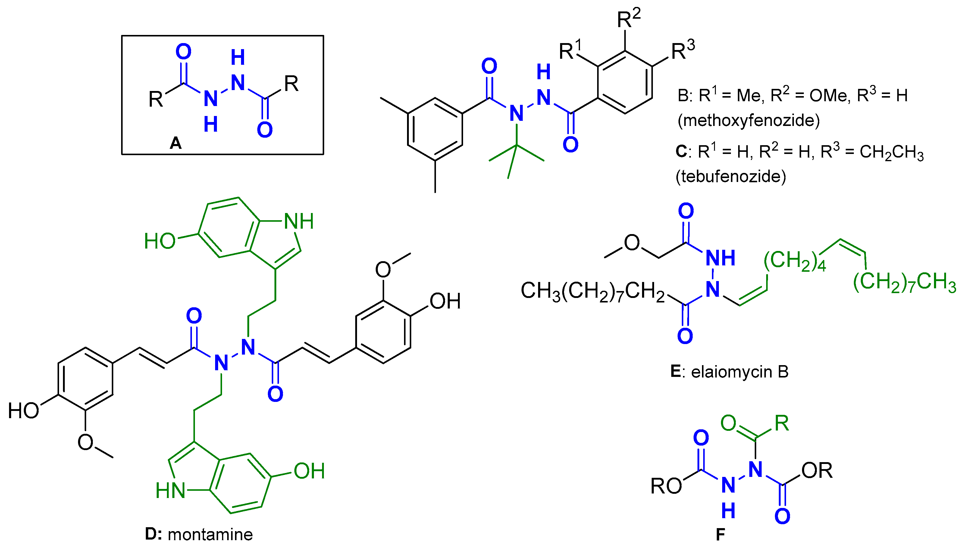
Figure 1. A: General structure of DACHZs; B, C: commercially available insecticides bearing a DACHZ moiety; D, E: natural products bearing a DACHZ group; F: a DACHZ-related motif with synthon characteristics. Blue color: the DACHZ skeleton; Green color: Substituents other than H on the nitrogen atom.
Molecules 29 00647 g001
Figure 3. Methods for the synthesis of AA DACHz. Method A: Synthesis via isatoic anhydride; Method B: Synthesis via the in situ formation of anthraniloyl imidazole; Method C: Synthesis via the in situ formation of the anthraniloyl chloride.
Molecules 29 00647 g003
Figure 4. All synthesized AA DACHZs 124. 14: from the combination of I or IIa with all hydrazides IIIad; 58: from the combination of IIb with all hydrazides IIIad; 912: from the combination of IIc with all hydrazides IIIad; 1316: from the combination of IId with all hydrazides IIIad; 1718: from the combination of IIe with the hydrazides IIIa,d; 1920: from the combination of IIf with the hydrazides IIIa,d; 2122: from the combination of IIg with the hydrazides IIIa,d; 2324: from the combination of IIh with the hydrazides IIIa,d.
Molecules 29 00647 g004
Figure 5. (A): A compilation of representative agarose gel pictures for the compounds 124 under 312 nm irradiation for 30 min, at a concentration of 500 μM. Calculations of Form II and Form III % are shown below the picture, compared to the control. Numbers 1–24 on the top correspond to the compounds 124 (all gel pictures are shown at Supplementary Materials S.2); (B): Plots of DNA photocleavage of the compounds 124 of all experiments: Error bars represent the standard deviation from at least two experiments; blue column: % ss photocleavage; red column: % ds photocleavage. The black column indicates that the concentration was too high to give measurable strands.
Molecules 29 00647 g005
Figure 6. Diagram on the (left): Plots of DNA photocleavage of the compound 9 under various concentrations (indicated at horizontal axis): Error bars represent the standard deviation from at least two experiments; blue column: % ss cleavage; red column: % ds cleavage (for all gel pictures see Supplementary Materials S.3). Picture on the (right): DNA agarose gel picture (one experiment) for the compound 9 under 312 nm irradiation for 30 min, at concentrations of 100, 50, 10, 1, 0.5 μM, Lanes 2–6, respectively (Supplementary Materials S.3.2). Lane 1: control (DNA + UV). Calculations of Form II and Form III % are shown below the picture, compared to the control.
Molecules 29 00647 g006
Figure 7. (A): (Top) picture: DNA agarose gel picture (one experiment) for the compound 9, 312 nm, 30 min, at a concentration of 5 μM and pH 5–10. Lane 1: Control DNA at pH 7; Lanes 2–7: pH 5–10, respectively. Calculations of Form II and Form III % are shown below the picture, compared to the control. (Bottom) diagram: Plots of DNA photocleavage of the compound 9 of all experiments at pHs shown on the horizontal axis; Error bars represent the standard deviation from at least two experiments; blue column: % ss cleavage; red column: % ds cleavage, (Supplementary Materials S.3.3); (B): (Top) picture: DNA agarose gel picture (one experiment) for the compound 9, 312 nm, 30 min, at concentration of 5 μM. Mechanistic studies: Lane 1: control, Lanes 2 and 3: under air and under argon; Lanes 4–8: under air and various scavengers like: L-cyst, KI, NaN3, D2O and DMSO, respectively. (Bottom) diagram: Plots of DNA photocleavage of the compound 9 of all mechanistic experiments shown on the horizontal axis. Blue and red columns as in (A) (Supplementary Materials S.3.4).
Molecules 29 00647 g007
Figure 8. Plots of DNA photocleavage of the compounds 124 under various concentrations, at 365 nm. C: control; 1st group of four compounds separated with gray lines are compounds that gave DNA photocleavage at a 500 μΜ concentration; 2nd group—two compounds: 100 μΜ concentration; 3rd group—thirteen compounds: 10 μΜ concentration; 4th group—five compounds: 2 μΜ concentration. Error bars represent the standard deviation from at least two experiments; blue column: % ss cleavage; red column: % ds cleavage (all gel agarose pictures at Supplementary Materials S.5).
Molecules 29 00647 g008
Figure 9. 3D structures of the polar contacts of selected compounds of each group. (a) Compound 1; (b) Compound 5.
Molecules 29 00647 g009
Figure 10. 3D structures of the polar contacts of selected compounds of each group. (a) Compound 9; (b) Compound 13.
Molecules 29 00647 g010
Figure 11. 3D structures of the polar contacts of selected compounds of each group. (a) Compound 17; (b) Compound 19.
Molecules 29 00647 g011
Figure 12. 3D structures of the polar contacts of selected compounds of each group. (a) Compound 21; (b) Compound 23.
Molecules 29 00647 g012
Figure 13. 3D structures of the polar contacts of selected compounds of each group. (a) Compound 3; (b) Compound 24.
Molecules 29 00647 g013
Figure 14. The cytotoxic effect of the compounds 1, 5, 9, 13 and 17 on the CarB cell line. The compound 17 exhibits a high cytotoxic effect.
Molecules 29 00647 g014
Table 1. Calculated energies and interactions of the compounds 124 with DNA.
CompoundEnergy (Kcal/mol)Interactions (PyMol)
Polar Contacts
1−8.9 DG4, DA5, DG22
2−9.0 DG4, DA5, DG22
3−8.1DG10, DC11, DG14, DG16
4−8.9DG10, DC11, DC15, DG16
5−9.5DG10, DC11
6−9.0DC15, DG16
7−9.8DG16
8−9.2DG10, DC15, DG16
9−8.8DC15, DG16
10−9.0DC15, DG16
11−9.4DG10, DC11, DC15, DG16
12−9.0DG10, DC11, DC15, DG16
13−9.2DC11
14−9.3DG10, DC11
15−10.1DG10, DC11
16−8.9DG10, DG16
17−9.4DG10, DC15, DG16
18−9.2DG10, DC15, DG16
19−9.6DG10, DC11
20−9.5DG10, DG16
21−9.9DG10, DG16
22−9.5DG10, DC11, DC15, DG16
23−9.8DG10, DG16
24−9.4DG10, DC11, DG14, DG16
Disclaimer/Publisher’s Note: The statements, opinions and data contained in all publications are solely those of the individual author(s) and contributor(s) and not of MDPI and/or the editor(s). MDPI and/or the editor(s) disclaim responsibility for any injury to people or property resulting from any ideas, methods, instructions or products referred to in the content.