Article

Antibacterial Activity of Plant Polyphenols Belonging to the Tannins against Streptococcus mutans—Potential against Dental Caries

by
Krzysztof Czerkas
1,*,
Ewa Olchowik-Grabarek
2,*,
Magdalena Łomanowska
2,
Nodira Abdulladjanova
3 and
Szymon Sękowski
2
1
Doctoral School of Exact and Natural Sciences, University of Bialystok, 15-254 Bialystok, Poland
2
Laboratory of Molecular Biophysics, Department of Microbiology and Biotechnology, Faculty of Biology, University of Bialystok, 15-254 Bialystok, Poland
3
Institute of Bioorganic Chemistry, Academy of Sciences of the Republic of Uzbekistan, Tashkent 100143, Uzbekistan
*
Authors to whom correspondence should be addressed.
Molecules 2024, 29(4), 879; https://doi.org/10.3390/molecules29040879
Submission received: 11 January 2024 / Revised: 1 February 2024 / Accepted: 9 February 2024 / Published: 16 February 2024

Abstract

:
Dental caries (DC) is the most common oral pathology. The main bacteria responsible for DC is Streptococcus mutans. One of the strategies that can decrease or eliminate the risk of DC development is using compounds that will inhibit both the growth and virulence factors of S. mutans. Tannins are plant polyphenols that have strong antibacterial activity. The purpose of this study was to assess the antibacterial activity of three tannins against S. mutans. In this investigation, microbiological tests (MIC and MBC) and physicochemical techniques like the fluorescence measurements of tannins’ interaction with S. mutans cell membrane and membrane proteins, zeta potential, and thermodynamic analyses were used to obtain knowledge about the antibacterial potential of the investigated compounds against S. mutans as well as about the mechanisms associated with antibacterial activity. The obtained results demonstrate that the used compounds exhibit high antibacterial activity against S. mutans. The mechanisms of their antibacterial activity are linked to the strong change in the S. mutans membrane fluidity and potential, and to their interaction with membrane proteins that can result in great disturbance of bacterial physiology and ultimately the inhibition of bacterial growth, triggering their death. Therefore, it can be concluded that the investigated compounds can be potentially used as natural factors in the prevention of dental caries.

Graphical Abstract

1. Introduction

Oral diseases develop mainly due to the activity of different bacterial strains, for example Lactobacillus fermentum, Streptococcus sobrinus, Bifidobacterium dentium, Treponema denticola, Porphyromonas gingivalis, or Streptococcus mutans [1].
Among the many oral diseases, one of the most common oral infectious diseases globally is dental caries (DC) [1,2]. DC is a multifactorial oral illness, but the three most crucial factors associated with it are as follows: (i) hygienic, connected with the self-care of teeth and gums, (ii) dietary, and (iii) microbiological, connected with previous bacterial infections [3]. The main pathogen linked with DC is Streptococcus mutans. This aerotolerant anaerobes bacterium is characterized by multistep activity, finally resulting in DC development. Its main action is associated with a decrease in pH in the dental oral cavity zone as a result of the production of acid metabolites, leading to demineralization [4], and the synthesis of glucan (by glucosyltransferases enzymes), which enables S. mutans adhesion, glucan-binding proteins (GBPs), surface protein antigen c (SPAc), and mutacins (bacteriocins), which are S. mutans virulence factors and collagen-binding proteins [5]. Additionally, an important virulence molecule biosynthesized by S. mutans is sortase A, a surface protein mainly connected with biofilm formation [6]. It must be emphasized that dental caries and periodontal diseases play a crucial role in oral biofilm formation, leading to demineralization [7] and causing a significant decrease in antibiotic bioavailability to bacteria cells as well as increasing bacteria survival and proliferation [8].
Many currently used compounds (e.g., chlorhexidine-CHX) and various study factors (for example, nanoparticles or bacteriophages) against DC act through the inhibition of S. mutans biofilm formation [9] and may have documented (like for CHX) or potentially different side effects. According to the well-known adage “prevention better than cure”, the other way to minimize or completely eliminate the risk of DC development is using compounds that will inhibit the growth of S. mutans and its virulence factors. This will allow the teeth to be protected against the colonization and biofilm formation of the S. mutans bacteria.
Plant polyphenols are very promising compounds that are intensively investigated for their therapeutic potential against different bacteria. These compounds possess antibacterial activity against bacterial strains like S. aureus, P. aeruginosa, B. subtilis, E. coli, K. pneumoniae, E. faecalis, S. epidermitis, and others [10]. The wide antimicrobial potential of polyphenols makes them good candidates for therapeutic use against bacteria responsible for oral infectious diseases. It was shown that different polyphenols belonging to flavonoid and tannin groups act against S. mutans, P. gingivalis, S. sobrinus, S. sanguinus, and S. salivarius [1]. Zayed demonstrated that green tea extracts possess the ability to inhibit S. mutans biofilm formation [4]. Another study presented that flavonoids and proanthocyanidins, isolated from cranberry (Vaccinium macrocarpon), possess the ability to inhibit the cariogenic virulence factors of pathogens responsible for DC [11]. One polyphenol group of compounds is tannins, i.e., secondary plant metabolites, which despite its high biological activity is less investigated in comparison to other polyphenolic compounds like flavonoids. Tannins possess high antibacterial activity. In our studies, we have demonstrated that PGG (1,2,3,4,6-penta-O-galloyl-β-d-glucose) as well as 1,2-di-O-galloyl-4,6-valoneoyl-β-d-glucose inhibited S.aureus growth and can act together with different antibiotics in a synergistic or an additive way [12]. PGG had also the ability to inhibit staphylococcal α-hemolysin release by influencing the bacteria cell and/or hla gene expression [13]. The antibacterial activity of tannins is the result of their ability to strongly interact with lipid membranes, leading to changes in their physicochemical properties like fluidity and permeability [12,14,15], and with different proteins like serum albumins [16,17], alpha-synuclein [18], salivary α-amylase [19], and including bacterial ones, e.g., streptococcus glucosyltransferase, protease, or neuraminidase, resulting in their inhibition [20]. Therefore, the aim of these studies was to investigate the antibacterial potential of 1,2,3,4,6-penta-O-galloyl-β-d-glucose (PGG), 1,2-di-O-galloyl-4,6-valoneoyl-β-d-glucose (dGVG), and 2-O-bis-digalloyl-4,6-valoneoyl-β-d-glucose (b-dGVG) against S. mutans. Except the standard MIC and MBC values, the antibacterial activity investigations were extended to the measurement of membrane fluidity and integrity, interaction studies with S. mutans membrane proteins, and the assessment of the thermodynamic nature of these interactions as well as the analysis of zeta potential at the bacteria surface. Therefore, our work contains the complete study of PGG, dGVG, and b-dGVG against S. mutans bacteria.
Our studies possess high novelty. For the first time, we have demonstrated that 1,2,3,4,6-penta-O-galloyl-β-d-glucose, 1,2-di-O-galloyl-4,6-valoneoyl-β-d-glucose, and 2-O-bis-digalloyl-4,6-valoneoyl-β-d-glucose can be utilized as anti-S. mutans compounds and potentially used as anticaries agents.

2. Results and Discussion

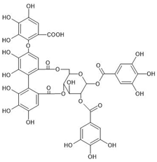
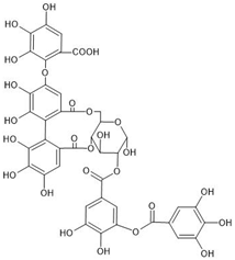
The investigated compounds, i.e., 1,2,3,4,6-penta-O-galloyl-β-d-glucose (PGG), 1,2-di-O-galloyl-4,6-valoneoyl-β-d-glucose (dGVG), and 2-O-bis-digalloyl-4,6-valoneoyl-β-d-glucose (b-dGVG), possess a similar molecular mass and number of gallic acid residues (5) and glucose moieties (1), as well as hydroxyl groups (15) (Table 1). However, they differ in their molecule’s flexibility due to the absence of a valoneoyl group in the case of PGG and its presence in the case of dGVG and b-dGVG. The valoneoyl group decreases the rotation abilities of gallic acid residues, leading to an increase in the stiffness of molecules. Therefore, the investigations will not only elucidate if the studied compounds possess antibacterial activity against S. mutans and connect with it the anticaries potential, but they will also give information about the influence of the valoneoyl group and the molecules’ flexibility on this activity.

2.1. Antibacterial Activity against S. mutans—MIC and MBC Studies

S. mutans is a bacterium found in the oral cavity that is strongly linked to the development of dental caries. The bacterium can adhere to and form a biofilm on the surfaces of teeth, acidifying the environment and leading to enamel demineralization [21]. In order to check if the studied tannins—PGG, dGVG, and b-dGVG—have antibacterial potential against S. mutans, two parameters were investigated: MIC (minimum inhibitory concentration) and MBC (minimum bactericidal concentration). These two microbiological values are the basic and key microbiological parameters that are most often designated to verify the tested compounds’ antibacterial potential. The obtained results are demonstrated in Table 2.
As observed, the MIC value for PGG was four times lower than for b-dGVG and eight times lower than for dGVG. Therefore, it can be concluded that PGG has the strongest S. mutans growth inhibition activity among all tested compounds. When comparing the MBC values, PGG also demonstrated the highest bactericidal activity, which was two times and four times larger than b-dGVG and dGVG, respectively. Also, in our earlier study, PGG showed higher antimicrobial activity against S. aureus strain 8325-4 than dGVG [12]. Our results are consistent with data reported in the literature, where isolated polyphenols, including gallic acid, salicylic acid, quercetin, or tannic acid [22] and extracts of polyphenolic compounds [23], showed antibacterial activity against different strains of S. mutans. Additionally, the MIC values for the tested tannins against studied S. mutans were lower compared to the activity of the B. crassifolia extract [24].
The significantly stronger antibacterial activity of PGG compared to dGVG and b-dGVG may result from the compounds having structural differences, leading to varied physicochemical parameters. The same influence of molecular structure on the activity of PGG and dGVG was observed for DMPC liposomes and described in detail in our previous work [25]. Generally, PGG exhibits greater flexibility, has a smaller surface area, higher dipole moment, and greater hydrophobicity than dGVG and, most likely, b-dGVG. Therefore, its enhanced efficacy results from its ability to interact more effectively with the cell membranes of S. mutans. Consequently, we continued our investigation into the interactions of tannins with bacterial membranes.

2.2. Evaluation of Tannins Interaction with S. mutans Membranes

One of the potential mechanisms of tannins’ antibacterial activity could be linked to the damage or modification of the cell membrane of S. mutans. Bacterial membranes perform critical roles in diverse physiological processes, such as osmoregulation, biosynthesis, transport, and respiration. Consequently, membrane alterations may induce metabolic stress, ultimately resulting in cell death [26]. Many polyphenolic compounds, e.g., kaempferol, quercetin, and apigenin, demonstrate activity against the cell membranes of S. mutans, resulting in bacterial death [1]. In the initial phase of evaluating the influence of tannins on the integrity of the S. mutans membrane, we used Sytox Green, a marker that binds to DNA when cell membrane damage occurs. The results presented in Figure 1 suggest that bacteria stained with Sytox Green in the presence of PGG, dGVG, and b-dGVG do not exhibit statistically significant changes in membrane integrity compared to the control. In previous studies, S. mutans solution without any compounds was used as negative control, whereas S. mutans with added Triton X-100, which acts as bacteria membrane-damaging factor [27], was taken as positive control.
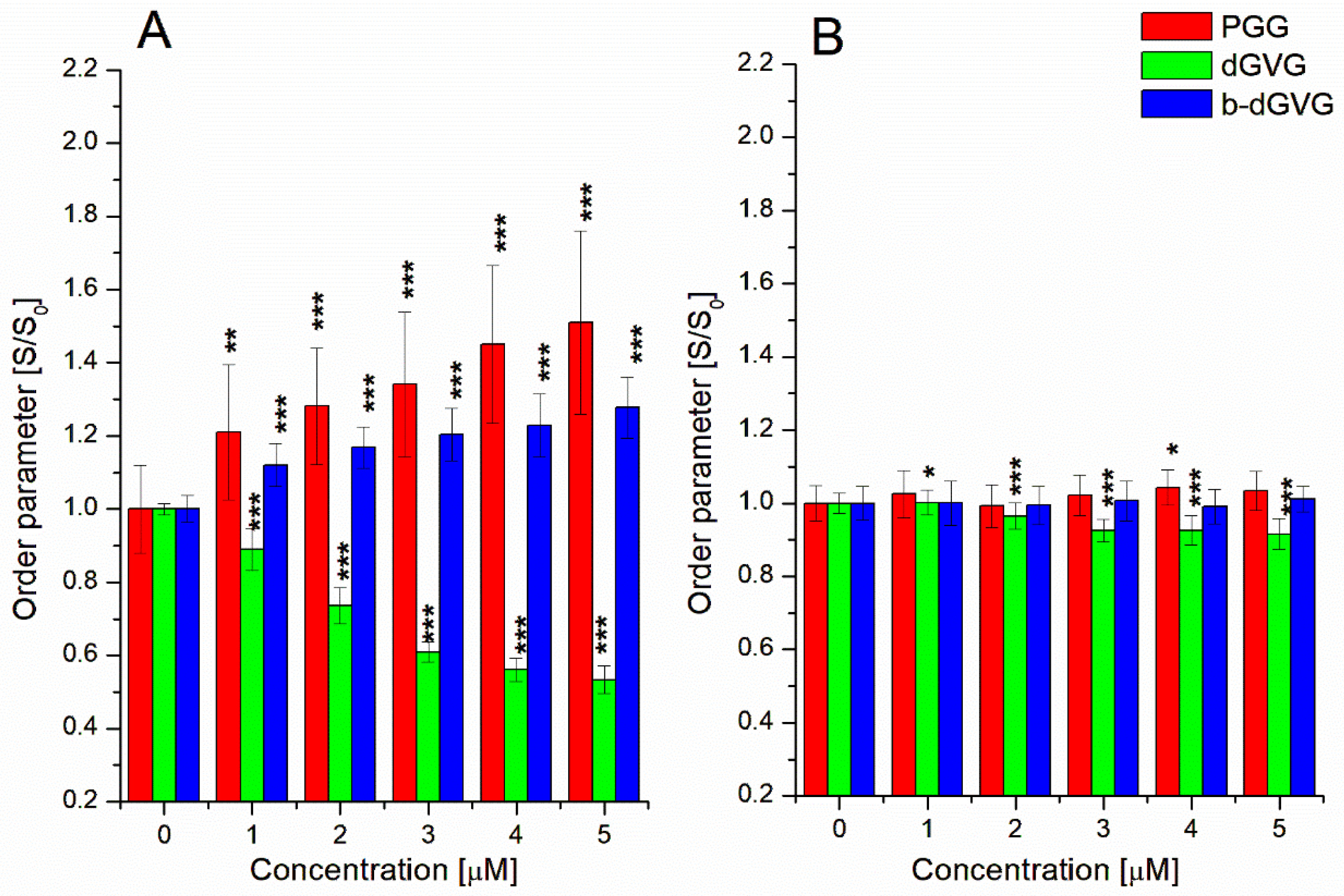
To further explore the antibacterial mechanisms of the utilized tannins, we broadened our studies by investigating the compounds’ influence on membrane fluidity. In order to check if analyzed compounds change the membrane rigidity, fluorescence staining was used, and the two fluorescent labels, i.e., TMA-DPH and DPH, which incorporate and anchor to different phospholipid parts of lipid membranes, were applied [25]. TMA-DPH binds to the outer polar layer of the membrane, whereas DPH enters deeper into the hydrophobic region of the phospholipids layer of the membrane. Changes in fluorescence anisotropy were monitored and used to calculate the lipid ordering parameter (S) using Equation (2).
Figure 2A presents changes in the ordering parameter at the polar parts of the S. mutans membrane in the presence of PGG, dGVG, and b-dGVG. It can be concluded that at concentrations of 1–5 µM, PGG induced significant changes in the ordering parameter for the outer hydrophilic zone of the membrane compared to dGVG and b-dGVG. Interestingly, dGVG resulted in a decrease in the lipid ordering parameter. It was clearly demonstrated that PGG exhibits the strongest interaction with S. mutans membranes. Furthermore, the data indicate the stiffening of the polar membrane parts by PGG and b-dGVG, and the increase in fluidity by dGVG.
Subsequently, using Equation (2), the data obtained for DPH-labeled samples were analyzed, and the results are demonstrated in Figure 2B. Changes obtained for PGG were statistically significant for a concentration of 4 µM. In the presence of b-dGVG, very small changes in the S. mutans membrane order parameter were statistically insignificant. The opposite effect in comparison with b-dGVG was detected for dGVG, where the 2–5 µM concentration range indicated statistical significance for the increase in fluidity. According to the above results, it can be concluded that dGVG demonstrated the strongest impact on the inner part of the S. mutans cell membrane. This suggests that dGVG can penetrate deeper into the lipid bilayer and interact with the hydrophobic part of the S. mutans membrane. The strong interaction of tannin molecules with the bacteria cell membrane is attributed to their antibacterial activity. It must be emphasized that this influence manifests in the changes in membrane potential and the modification of membrane fluidity as well as cell membrane disruption [20]. For example, punicalagin modified membrane permeability, whereas proanthocyanidins increased the growth inhibition of bacteria by inactivating ATPase enzymes in the membrane [20]. Also, Delehanty described that proanthocyanidins interacted with the outer bacteria membrane, resulting in the destabilization of their integrity [28].

2.3. Evaluation of Tannin Interaction with S. mutans Membrane Proteins through Tryptophan Fluorescence

As we demonstrated above, PGG, dGVG, and b-dGVG can interact with the S. mutans cell membrane, modify its fluidity, and influence the membrane order parameter. It is commonly known that the cell membrane has many different proteins that play a crucial role in regulating bacteria’s physiological processes. Tannins have a strong affinity to proteins, forming complexes with membrane proteins and resulting in antibacterial activity [29]. The method enabling the analysis of interactions between different molecules and proteins involves analyzing fluorescence changes descended from aromatic amino acids, including tryptophan. Additionally, tryptophan fluorescence is particularly sensitive to changes in its microenvironment [30]. In order to verify if our analyzed compounds can also interact with S. mutans cell membrane proteins, fluorescence investigations were conducted, and the fluorescence signal from the protein tryptophan residues was monitored. Based on these measurements, some physicochemical parameters that characterize these interactions were calculated. The obtained results are presented in Figure 3 and Table 3.
The results allowed us to conclude that tannins decreased the tryptophan fluorescence of S. mutans membrane proteins. The effect was concentration- and temperature- dependent (see Figure 3). For the highest concentration at 310 K, a reduction in fluorescence was observed at the levels of 0.309 ± 0.007 for PGG, 0.380 ± 0.005 for dGVG, and 0.303 ± 0.024 for b-dGVG. The results were statistically significance for all tannin concentrations.
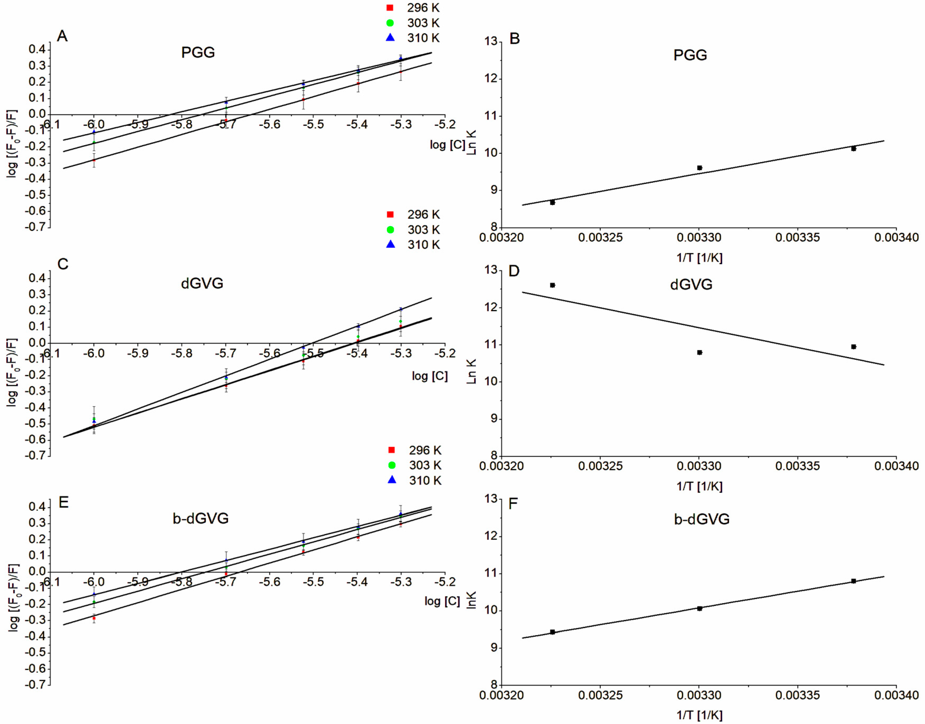
Based on the fluorescence results, the Stern–Volmer plots were drawn (Figure 3B,D,F) and the Stern–Volmer constants (KSV) describing the accessibility of the quencher to the quenched molecules (fluorophores) were calculated (see Table 3). As observed, the KSV is larger at higher temperatures, but it should be emphasized that the KSV order of magnitude was not temperature-dependent. As can be observed, PGG and b-dGVG exhibited similar KSV values, while for dGVG, they were approximately 1.5 times lower at each temperature compared to PGG and b-dGVG. This result demonstrates that dGVG had lower accessibility to the quencher (S. mutans membrane protein tryptophans). The obtained results correspond very well with the MIC and MBC values and the changes in the physicochemical nature of the S. mutans cell membrane. The strongest antibacterial activity (see Table 2) was demonstrated by PGG. This compound’s strongest influence on the S. mutans membrane increased the order parameter. The largest PGG activity is the result of the five free galloyl groups. It is well known that the presence of free galloyl moieties is a crucial factor for the antibacterial activity of tannins [20]. Additionally, PGG is highly hydrophobic and thus can penetrate deeper into the membrane in comparison, e.g., with dGVG, as we described earlier [12]. The other compound (b-dGVG), despite being similar to PGG in terms of being accessible to S. mutans membrane proteins (b-dGVG KSV values are close to those for PGG), has lower antibacterial activity. This is probably due to the lower number of free galloyl moieties compared to PGG and a lower influence on the membrane order parameter. It should be noted that b-dGVG, in opposition to PGG, possesses a valoneoyl group. This group also determines the antibacterial activity of tannins [20], and as described by Shimozu for two compounds with a valoneoyl group, i.e., isorugosin A and isorugosin B, they showed antibacterial activity against methicillin-resistant Staphylococcus aureus (MRSA), and the substitution of two or three galloyl moieties by the valoneoyl group was the factor that induced activity against MRSA [31]. How, then, can we explain that the b-dGVG possesses a more significant activity than dGVG, which has a very similar molecular structure. If it is assumed that the valoneoyl group, which is at the same position attached to glucose of both molecules (i.e., 4,6-valoneoyl-β-d-glucose), determines, in the same way, the antibacterial activity, then the explanation should be found in the position of the two remaining galloyl groups. In dGVG, two galloyl moieties are separate and free (1,2-di-O-galloyl), whereas in b-dGVG, they are joined together (2-O-bis-digalloyl). The configuration of the digalloyl group/groups is also the factor that influences the tannins’ antibacterial activity [20]. Our studies demonstrate that b-dGVG with digalloyl groups has greater anti-S. mutans activity in comparison with dGVG, probably resulting from the presence of the digalloyl structure which probably increased antibacterial activity of b-dGVG. It must also be emphasized that this activity is also the result of a stronger interaction with the S. mutans membrane and larger modification of the order parameter, as well as the rigidifying of the membrane, contrary to dGVG, which had the weakest interaction with the membrane and evoked an increase in S. mutans membrane fluidity. As described above, PGG, dGVG, and b-dGVG possess the ability to interact with S. mutans membrane proteins, measured by the tryptophan fluorescence decrease. In order to qualify if tannins decrease Trp fluorescence through complex formation with S. mutans proteins (static quenching) or only by collisional encounters (dynamic quenching), the quenching constants (kq) were calculated. The obtained kq values for all studied compounds were significantly higher than the maximum scatter collision constant (2 × 10¹⁰ M−1s−1), indicating a static quenching mechanism through the formation of tannin–S. mutans complexes. Similar results were obtained in our previous studies on PGG and dGVG interaction with membrane proteins of S. aureus [12] as well as for staphylococcal alpha-hemolysin [13]. Such strong affinity to S. mutans membrane proteins allows us to assume that PGG, dGVG, and b-dGVG inhibited such S. mutans proteins, like glucosyltransfrease, surface protein antigen c (PAc) [5], or sortase A (associated with S. mutans adhesion, biofilm formation, and evasion of host defense) [6].
As we demonstrated above, the studied tannins formed complexes with S. mutans membrane proteins. To determine whether the formed complexes are reversible or irreversible, the binding constants (logKb) were calculated using a double logarithmic plot Equation (5) and drawing double logarithmic plots (Figure 4A,C,E). This enables the estimation of the relative binding strengths between S. mutans membrane proteins and tannins at different temperatures. The binding constants for PGG and b-dGVG decreased with temperature, reaching values of 3.769 ± 0.491 for PGG and 4.095 ± 0.116 for b-dGVG at 310 K. In contrast, dGVG exhibited higher values than the PGG and b-dGVG at all used temperatures. Furthermore, an increase in temperature to 310 K resulted in an elevation in logKb to the level of 5.474 ± 0.317. Non-logarithmic values of Kb are in the range from 104 to 106 and mostly are in the range 1–15 × 104 M−1, indicating the reversibility of such interactions [32]. The results were similar to those obtained for the interaction of four flavonoids with human serum albumin [33].
The interaction between molecules is mainly responsible for hydrogen bonds, van der Waals forces, electrostatic interactions, and hydrophobic forces [34]. Calculating thermodynamic parameters like enthalpy (ΔH) and entropy (ΔS) allows us to qualify which type of bonds (forces) are responsible for molecule–molecule interactions. Hydrophobic bonds predominantly manifest with positive values of ΔH and ΔS, whereas van der Waals forces and hydrogen bonds are associated with negative values of ΔH and ΔS. Electrostatic forces arise when ΔH is negative while ΔS is positive. Additionally, it is possible to assess whether the reaction occurs spontaneously, which is indicated by the negative value of free energy ΔG (Gibb’s potential) [35]. In our studies, the thermodynamic parameters (ΔH and ΔS) were calculated based on fluorescence data and the Van’t Hoff equation (Equation (6)) used to draw Van’t Hoff plots presented in Figure 4B,D,F while the free energy (ΔG) was calculated using Equation (7). Table 4 below demonstrates the values of enthalpy, entropy, and Gibb’s potential.
According to the obtained results, it can be concluded that the forces responsible for bond formation with bacterial proteins were not the same for all tannins. PGG and b-dGVG interacted with S. mutans membrane proteins through van der Waals forces and hydrogen bonds (ΔH and ΔS values were negative). In contrast, for dGVG, both ΔH and ΔS were positive, which clearly suggests that the binding process occurs primarily through hydrophobic interactions. These differences probably result from their different interactions with S. mutans membranes. As we observed for PGG and b-dGVG, these two tannins greatly increased the order parameter of membrane polar parts (see Figure 2A) but practically did not influence the order parameter of hydrophobic parts of S. mutans membrane (Figure 2B). The opposite effect was detected for dGVG, which decreased the order parameter in both polar and hydrophobic parts of the S. mutans membrane (Figure 2), clearly indicating that hydrophobic forces are engaged in such interaction, confirming thermodynamic analyses.
The calculated free energy ΔG was negative for all studied compounds, indicating that PGG, dGVG, and b-dGVG interact with S. mutans spontaneously. However, notably, for PGG and b-dGVG, ΔG values decreased with temperature, suggesting a decrease in complex stability. Meanwhile, ΔG values with dGVG increased, indicating an increase in complex stability, which is a known fact as hydrophobic interactions increase protein stability with temperature [36].

2.4. Analyses of S. mutans’ Zeta Potential Changes in the Presence of PGG, d-GVG, and b-dGVG

One important parameter that is engaged in the interaction between molecules and between molecules and cells is zeta potential (ϛ-potential). Zeta potential is a physicochemical property connected with the cell composition and is one of the crucial factors in the interaction of bacteria with different ions and molecules [37]. Additionally, zeta potential changes in bacterial cells can indicate, among others things, alterations in membrane permeability, biofilm formation, adhesion degree, viability, and the bacteria’s tendency to aggregate under the influence of compounds [38]. Therefore, in our studies, the changes in ϛ-potential have been analyzed as the parameter demonstrating changes at the S. mutans charge during interaction with PGG, dGVG, and b-dGVG. The obtained results are presented below (Figure 5).
As it can be observed, the average zeta potential for the control samples of S. mutans, i.e., without the addition of tannins, was −17.72 ± 0.454 mV, which, in comparison with the other Gram-positive bacteria such as S. aureus, L. monocytogenes, and B. cereus with zeta potential ranging from −23 to −53 mV, indicates a significantly lower value (if we compare voltage values in mV) [39].
The results obtained after the addition of tannins varied slightly. However, they were statistically significant for PGG and b-dGVG for all used concentrations, and the zeta potential negative values gently increased with increasing tannin concentration. Notably, b-dGVG achieved a slightly higher value at concentrations of 4–5 µM. Changes in zeta potential values may be associated with alterations in the order parameter and tryptophan fluorescence quenching of S. mutans membrane proteins due to the investigated tannins. The interaction of tannins with proteins and bacterial membranes can lead to changes in zeta potential values, resulting in changes in such parameters as the metabolic and physiological state of bacteria and their capacity for adhesion [38]. For example, compounds like ferulic acid, rosmarinic acid, and epigallocatechin gallate influenced the zeta potential reduction, limiting bacterial adhesion [39].
The above results from our studies demonstrate that PGG, dGVG, and b-dGVG possess antibacterial activity against S. mutans. Despite PGG acting the most strongly, dGVG and b-dGVG also showed anti-S. mutans potential. Physicochemical analyses allowed us to detect that the studied tannins change the fluidity of the S. mutans membrane and interact spontaneously with bacteria membrane proteins, forming protein–tannin complexes. This observation can have a strong implication for using tannins as natural factors against the development of dental caries. The changes in S. mutans membrane fluidity can disturb membrane integrity, and ATPase enzymes can be inhibited as described for the proanthocyanidins [20,25]. Additionally, the complex formation between S. mutans membrane proteins and the studied tannins can inhibit essential bacteria proteins like glucosyltransferase, surface protein antigen c, or sortase A, resulting in a decrease in the pathogenicity of S. mutans, which will reduce the risk of dental caries.

3. Materials and Methods

3.1. Chemicals

Studied tannins were isolated from Rhus typhina L.—1,2,3,4,6-penta-O-galloyl-β-d-glucose (PGG), Euphorbia jaxartica—1,2-di-O-galloyl-4,6-valoneoyl-β-d-glucose (dGVG), and P. lanceolata L.—2-O-bis-digalloyl-4,6-valoneoyl-β-d-glucose (b-dGVG). All of the tannins were obtained and isolated by the previously described method [15]. N,N,N-trimethyl-4-(6-phenyl-1,3,5-hexatrien-1-yl) phenylammonium p-toluenesulfonate (TMA-DPH), 1,6-diphenyl-1,3,5-hexatriene (DPH), DMSO, and phosphate buffer saline (PBS) were from SIGMA (Merck); MH broth and MH agar were from Oxoid (Basingstoke, England); Sytox Green was obtained from Thermofisher (Walthman, MA, USA).

3.2. Bacterial Strain and Growth Conditions

Studies were conducted using Streptococcus mutans ATCC 25175 strain. For investigations, the suspension of bacteria was incubated overnight (t = 37 °C, Mueller–Hinton (MH) broth, 200 rpm continuous shaking).

3.3. Antimicrobial Activity—Determination of Minimum Inhibitory Concentration (MIC) and Minimum Bactericidal Concentration (MBC)

In order to investigate the antibacterial activity of studied tannins against S. mutans, two parameters, MIC (minimum inhibitory concentration) and MBC (minimum bactericidal concentration), were analyzed using the broth microdilution method, following guidelines established by the National Committee for Clinical Laboratory Standards. A stock solution of compounds was added to Mueller–Hinton broth (MHB) to achieve a final concentration of 1600 µM. In the next step, the samples were serial two-fold diluted in the MH broth to obtain a concentration range from 800 µM up to 3.25 µM with a final volume of 100 µL per 96-well microtiter plate. Then, 100 µL of S. mutans solution with a concentration of 1 × 10⁶ colony-forming units per mL (CFU/mL) was added to each well. The plates were incubated at 37 °C for 24 h to facilitate bacterial growth. The MIC values were identified as the lowest concentration of studied tannins that prevented bacterial growth, indicated by the absence of turbidity. In contrast, MBC was determined as the lowest concentration of used compounds where bacterial growth on the plates had not been observed.

3.4. Studies of S. mutans Membrane Permeability—Sytox Staining

Investigations were conducted according to Olchowik-Grabarek et al. [12]. S. mutans grew overnight (at 37 °C in Mueller–Hinton broth (MHB) with shaking at 200 rpm). The bacteria solution was centrifuged, and the cell pellet was resuspended in PBS with 5% MH broth, with the final OD600 = 0.01. Next, S. mutans was pipetted into 96-well plates, and the studied tannins were added to gain the final concentration range of 1/4–3 MIC and incubated for 1 h. The probes with 1% Triton X-100 were prepared as the positive control. Next, the Sytox Green label was added to each well at the final concentration 5 µM. After 2 h of incubation (t = 37 °C), the fluorescence intensity was registered using SpectraMax M2 microplate reader (Molecular Device, San Jose, CA, USA) at the wavelengths λexc. = 504 nm and λem. = 523 nm.

3.5. Measurements of S. mutans Membrane Fluidity

Membrane fluidity was measured according to our previous study [12], with some modifications. Briefly, S. mutans ATCC 25175 was cultured overnight at 37 °C in Mueller–Hinton broth (MHB) with shaking at 200 rpm. Next, the bacteria suspension was centrifugated (2300× g for 10 min), the supernatant was removed, and the bacterial cell pellet was resuspended in PBS buffer (C = 10 mM, pH = 7.4) to achieve S. mutans suspension with the optical density OD600 = 0.01. The suspension was subsequently labeled (for 15 min, 37 °C) using TMA-DPH or DPH probe at the final concentration of 1 μM, and the fluorescence anisotropy signal was measured using the excitation and emission wavelengths λexc = 340 nm, λem= 430 nm and λexc = 348 nm, λem= 426 nm for TMA-DPH and DPH, respectively. Bacteria without studied compounds were taken as control. For analysis, to investigate the tannins’ influence on membrane fluidity, the compounds were added to the labeled S. mutans at concentrations of 1–5 μM and incubated for 10 min (t = 37 °C), and the fluorescence anisotropy was read for control. The investigations were conducted using a PerkinElmer LS-55 spectrofluorometer (PerkinElmer, Buckinghamshire, UK). Changes in the fluidity of S. mutans membranes induced by tannins were assessed based on the fluorescence anisotropy values of the samples (r).
The anisotropy values were calculated using the Jablonski equation [15]:
r = I V V G I V H I V V + 2 G I V H
where IVV and IVH are the vertical and horizontal fluorescence intensities, respectively, for the vertical polarization of the excitation light beam. Before analysis, the G factor (grating correction factor) that corrects the polarizing effects of the monochromator was registered.
The obtained values were used to calculate the order parameter using the equation below [15]:
S = 1 2 r r 0 + 5 r r 0 2 1 + r r 0 2 r r 0
where r0 is the fluorescence anisotropy of probes in the absence of any rotational motion of probes.

3.6. Fluorescence Analysis of Tannins’ Interactions with S. mutans Membrane Proteins

The S. mutans cultures were incubated overnight at 37 °C in Mueller–Hinton (MH) broth, with shaking at 200 rpm. For the experiment, bacteria suspension was centrifuged (2300× g, 10 min), the supernatant was removed, and bacterial cells were resuspended in PBS (pH = 7.4) to obtain the solution with OD600 = 0.1. Next, tannins were added to the bacteria samples at concentrations of 1–5 μM, and the probes were incubated for 10 min at 37 °C. Bacteria without tannins were taken as control. The fluorescence intensities of S. mutans membrane proteins were adjusted for appropriate baselines and determined using a PerkinElmer LS-55B spectrofluorometer (PerkinElmer, Waltham, MA, USA). Since the fluorescence descended from the membrane protein tryptophan’s residues, the readings were carried out using the excitation and emission wavelengths λexc = 295 nm and λem = 350 nm, respectively. The experiments were conducted at three temperatures (23 °C, 30 °C, and 37 °C).
Fluorescence quenching was characterized using the Stern–Volmer equation [40], and the Stern-Volmer plots were employed for its graphical representation.
F 0 F = K s v Q + 1
where: F0 and F—fluorescence without and with presence of quencher; Ksv—Stern–Volmer constant; [Q]—quencher concentration
In order to check which mechanism (static or dynamic) is responsible for both the fluorescence quenching and tannin–protein molecule interactions, the quenching constant (kq) [40] was calculated using the following equation:
k q = K S V τ 0
where: kq—quenching constant; KSV—Stern–Volmer constant; τ0—fluorescence lifetime of fluorophore molecules (5 × 10⁻⁹ s).
To determine if the tannin–S. mutans interaction is reversible, the binding constant (log Kb) was calculated based on the double logarithmic plots and double logarithmic equation [41].
log [ ( F 0 F ) F ] = log K b + n l o g [ Q ]
where: F0 and F—fluorescence without and with presence of quencher; Kb—binding constant; n—number of binding sites; [Q]—quencher concentration
It is commonly known that all chemical reactions are connected with thermal effects. Analysis of thermodynamic nature of such reaction enables us to determine whether a reaction occurs spontaneously or not [35]. Therefore, to thoroughly explain the nature of PGG, dGVG, and b-dGVG interactions with S. mutans membrane proteins, such thermodynamic parameters like enthalpy (ΔH), entropy (ΔS), and free energy change (ΔG) have been calculated using the following equations [35]:
l n K b = Δ H R T + Δ S R
Δ G = Δ H T Δ S
where: ΔH—enthalpy changes, ΔS—entropy changes, ΔG—free energy changes, T—temperature at Kelvin scale, R—gas constant, and lnKb—natural logarithm of binding constant.

3.7. Analysis of ϛ-Potential

Changes in the zeta potential (ϛ-potential) of S. mutans in the presence of the studied compounds were measured using electrophoretic light scattering with a zetasizer ULTRA (Malvern, Worcestershire, UK). Bacteria cell suspension was prepared in the same way as for fluorescence analyses. Next, 3 mL of the S. mutans was incubated with tannins in the concentration range of 1–5 μM for 15 min at 25 °C. The analyses were performed at a temperature of 25 °C in disposable folded capillary cells (DTS1070 Malvern Panalytical Ltd., Malvern, UK).

3.8. Statistical Analysis

A minimum of three independent trials were conducted for each of the experiments. The data are presented as the mean ± standard deviation. Grubb’s test was applied to ensure that the raw data values did not deviate significantly from the overall dataset. Statistical analysis was conducted using the GraphPad software QuickCalc. Graphs and figures were created using Origin 8.5.1 software (Northampton, MA, USA).

4. Conclusions

One of the most serious oral pathologies is dental caries (DC). Among many reasons responsible for DC development, the main one is S. mutans infection. This bacterium is responsible for demineralization, glucan synthesis, pH change, and biofilm formation, which results in caries development at the final stage. Tannins, natural plant compounds, have well-documented antibacterial activity. Therefore, our studies investigated the antibacterial potential of three tannins against S. mutans. Using microbiological and physicochemical analyses, we demonstrated that studied compounds possess a strong affinity to S. mutans bacteria cells and demonstrate quite high antibacterial activity. Among the tested compounds, the highest antistreptococcal activity was observed for PGG. The observed antibacterial potential of PGG, dGVG, and b-dGVG was connected with modifications of S. mutans membrane fluidity and membrane surface charge, as well as with strong interactions with bacteria surface proteins and the protein–tannin complex formation. Thermodynamic analysis shows that all tannins bound to bacteria membrane proteins spontaneously. According to the obtained results, it can be concluded that tannins are efficient anti-S. mutans compounds and can be potentially used as natural protection against caries, but further research is needed to develop the most efficient systems to apply tannins in the oral cavity space.

Author Contributions

Conceptualization, K.C., E.O.-G. and S.S.; methodology, K.C. and E.O.-G.; formal analysis, K.C. and E.O.-G.; investigation, K.C., M.Ł. and E.O.-G.; chemical isolation of used tannins, N.A.; writing—original draft preparation, K.C., E.O.-G. and S.S.; writing—review and editing, E.O.-G. and S.S.; supervision, E.O.-G. and S.S. All authors have read and agreed to the published version of the manuscript.

Funding

This research received no external funding.

Institutional Review Board Statement

Not applicable.

Informed Consent Statement

Not applicable.

Data Availability Statement

Data is contained within the article.

Conflicts of Interest

The authors declare no conflicts of interest.

References

  1. Kováč, J.; Slobodníková, L.; Trajčíková, E.; Rendeková, K.; Mučaji, P.; Sychrová, A.; Bittner Fialová, S. Therapeutic Potential of Flavonoids and Tannins in Manaent of Oral Infectious Diseases. Molecules 2023, 28, 158. [Google Scholar] [CrossRef]
  2. Huang, L.; Yu, F.; Sun, X.; Dong, Y.; Lin, P.T.; Yu, H.H. Antibacterial activity of a modified unfilled resin containing a novel polymerizable quaternary ammonium salt MgemAE-HB. Sci. Rep. 2016, 6, 33858. [Google Scholar] [CrossRef]
  3. Wu, C.D. Grape Products and Oral Health. J. Nutr. 2009, 139, 1818S–1823S. [Google Scholar]
  4. Zayed, S.M.; Aboulwafa, M.M.; Hashem, A.M.; Saleh, S.E. Biofilm Formation by Streptococcus mutans and Its Inhibition by Green Tea Extracts. AMB Expr. 2021, 11, 73. [Google Scholar] [CrossRef]
  5. Matsumoto-Nakano, M. Role of Streptococcus mutans Surface Proteins for Biofilm Formation. Jpn. Dent. Sci. Rev. 2018, 54, 22–29. [Google Scholar] [CrossRef]
  6. Das, S.; Srivastava, V.K.; Parray, Z.A.; Jyoti, A.; Islam, A.; Kaushik, S. Identification of potential inhibitors of sortase A: Binding studies, in-silico docking and protein-protein interaction studies of sortase A from Enterococcus faecalis. Int. J. Biol. Macromol. 2018, 120, 1906–1916. [Google Scholar] [CrossRef]
  7. Larsen, T.; Fiehn, N.E. Dental biofilm infections—An update. APMIS 2017, 125, 376–384. [Google Scholar] [CrossRef]
  8. Lahiri, D.; Nag, M.; Banerjee, R.; Mukherjee, D.; Garai, S.; Sarkar, T.; Ankita, D.; Hassan, L.S.; Sushil, K.P.; Hisham, A.E.; et al. Amylases: Biofilm inducer or biofilm inhibitor? Front. Cell. Infect. 2021, 11, 660048. [Google Scholar] [CrossRef]
  9. Gao, Z.; Chen, X.; Wang, C.; Song, J.; Xu, J.; Liu, X.; Yu, Q.; Suo, H. New Strategies and Mechanisms for Targeting Streptococcus mutans Biofilm Formation to Prevent Dental Caries. Microbiol. Res. 2023, 278, 127526. [Google Scholar] [CrossRef]
  10. Manso, T.; Lores, M.; de Miguel, T. Antimicrobial Activity of Polyphenols and Natural Polyphenolic Extracts on Clinical Isolates. Antibiotics 2022, 11, 46. [Google Scholar] [CrossRef]
  11. Philip, N.; Walsh, L.J. Cranberry Polyphenols: Natural Weapons against Dental Caries. Dent. J. 2019, 7, 20. [Google Scholar] [CrossRef]
  12. Olchowik-Grabarek, E.; Sękowski, S.; Kwiatek, A.; Płaczkiewicz, J.; Abdulladjanova, N.; Shlyonsky, V.; Swiecicka, I.; Zamaraeva, M. The Structural Changes in the Membranes of Staphylococcus aureus Caused by Hydrolysable Tannins Witness Their Antibacterial Activity. Membranes 2022, 12, 1124. [Google Scholar] [CrossRef]
  13. Olchowik-Grabarek, E.; Sekowski, S.; Mies, F.; Bitiucki, M.; Swiecicka, I.; Abdulladjanova, N.; Shlyonsky, V.; Zamaraeva, M. Electrophysiological and Spectroscopic Investigation of Hydrolysable Tannins Interaction with α-Hemolysin of S. aureus. Bioelectrochemistry 2023, 150, 108318. [Google Scholar] [CrossRef]
  14. Hendrich, A.B. Flavonoid-membrane interactions: Possible consequences for biological effects of some polyphenolic compounds. Acta Pharmacol. Sin. 2006, 27, 27–40. [Google Scholar] [CrossRef]
  15. Olchowik-Grabarek, E.; Sekowski, S.; Bitiucki, M.; Dobrzynska, I.; Shlyonsky, V.; Ionov, M.; Burzynski, P.; Roszkowska, A.; Swiecicka, I.; Abdulladjanova, N.; et al. Inhibition of interaction between Staphylococcus aureus α-hemolysin and erythrocytes membrane by hydrolysable tannins: Structure-related activity study. Sci. Rep. 2020, 10, 11168. [Google Scholar] [CrossRef]
  16. Sekowski, S.; Ionov, M.; Kaszuba, M.; Mavlyanov, S.; Bryszewska, M.; Zamaraeva, M. Biophysical studies of interaction between hydrolysable tannins isolated from Oenothera gigas and Geranium sanguineum with human serum albumin. Colloids Surf. B Biointerfaces 2014, 123, 623–628. [Google Scholar] [CrossRef]
  17. Sekowski, S.; Olchowik-Grabarek, E.; Wieckowska, W.; Veiko, A.; Oldak, L.; Gorodkiewicz, E.; Karamov, E.; Abdulladjanova, N.; Mavlyanov, S.; Lapshina, E.; et al. Spectroscopic, Zeta-potential and Surface Plasmon Resonance analysis of interaction between potential anti-HIV tannins with different flexibility and human serum albumin. Colloids Surf. B Biointerfaces 2020, 194, 111175. [Google Scholar] [CrossRef]
  18. Sekowski, S.; Buczkowski, A.; Palecz, B.; Abdulladjanova, N. Inhibitory effect of Euphorbia tannins on α-synuclein aggregation in aqueous solutions. J. Mol. Liq. 2020, 299, 112112. [Google Scholar] [CrossRef]
  19. Soares, S.; Mateus, N.; De Freitas, V. Interaction of different polyphenols with bovine serum albumin (BSA) and human salivary α-amylase (HSA) by fluorescence quenching. J. Agric. Food Chem. 2007, 55, 6726–6735. [Google Scholar] [CrossRef]
  20. Farha, A.K.; Yang, Q.Q.; Kim, G.; Li, H.B.; Zhu, F.; Liu, H.Y.; Gan, R.Y.; Corke, H. Tannins as an alternative to antibiotics. Food Biosci. 2020, 38, 100751. [Google Scholar] [CrossRef]
  21. Iacopetta, D.; Ceramella, J.; Catalano, A.; D’Amato, A.; Lauria, G.; Saturnino, C.; Andreu, I.; Sinicropi, M.S. Diarylureas: New Promising Small Molecules against Streptococcus mutans for the Treatment of Dental Caries. Antibiotics 2023, 12, 112. [Google Scholar] [CrossRef]
  22. Sendamangalam, V.; Choi, O.K.; Seo, Y.; Kim, D.-S. Antimicrobial and Antioxidant Activities of Polyphenols against Streptococcus mutans. Free Radic. Antioxid. 2011, 1, 48–55. [Google Scholar] [CrossRef]
  23. Veloz, J.J.; Alvear, M.; Salazar, L.A. Antimicrobial and Antibiofilm Activity against Streptococcus mutans of Individual and Mixtures of the Main Polyphenolic Compounds Found in Chilean Propolis. BioMed Res. Int. 2019, 2019, 7602343. [Google Scholar] [CrossRef]
  24. Liu, Y.; Xu, Y.; Song, Q.; Wang, F.; Sun, L.; Liu, L.; Yang, X.; Yi, J.; Bao, Y.; Ma, H.; et al. Anti-biofilm activities from Bergenia crassifolia leaves against Streptococcus mutans. Front. Microbiol. 2017, 8, 1738. [Google Scholar] [CrossRef]
  25. Sekowski, S.; Veiko, A.; Olchowik-Grabarek, E.; Dubis, A.; Wilczewska, A.Z.; Markiewicz, K.H.; Zamaraeva, M. Hydrolysable tannins change physicochemical parameters of lipid nano-vesicles and reduce DPPH radical-Experimental studies and quantum chemical analysis. Biochim. Biophys. Acta Biomembr. 2022, 1864, 183778. [Google Scholar] [CrossRef]
  26. Strahl, H.; Errington, J. Bacterial membranes: Structure, domains, and function. Annu. Rev. Microbiol. 2017, 71, 519–538. [Google Scholar] [CrossRef]
  27. Zhou, Y.; Zhang, B.; Wang, Y.; Hu, R. Effects of Sulforaphene on the Cariogenic Properties of Streptococcus Mutans In Vitro and Dental Caries Development In Vivo. Antibiotics 2023, 12, 1359. [Google Scholar] [CrossRef]
  28. Delehanty, J.B.; Johnson, B.J.; Hickey, T.E.; Pons, T.; Ligler, F.S. Binding and neutralization of lipopolysaccharides by plant proanthocyanidins. J. Nat. Prod. 2007, 70, 1718–1724. [Google Scholar] [CrossRef]
  29. Feng, Y.; Jin, C.; Lv, S.; Zhang, H.; Ren, F.; Wang, J. Molecular Mechanisms and Applications of Polyphenol-Protein Complexes with Antioxidant Properties: A Review. Antioxidants 2023, 12, 1577. [Google Scholar] [CrossRef]
  30. Albani, J.R. Principles and Applications of Fluorescence Spectroscopy; John Wiley & Sons: Hoboken, NJ, USA, 2008. [Google Scholar]
  31. Shimozu, Y.; Kuroda, T.; Tsuchiya, T.; Hatano, T. Structures and antibacterial properties of isorugosins H-J, oligomeric ellagitannins from Liquidambar formosana with characteristics bridging groups between sugar moieties. J. Nat. Prod. 2017, 80, 2723–2733. [Google Scholar] [CrossRef]
  32. Dufour, C.; Dangles, O. Flavonoid–serum albumin complexation: Determination of binding constants and binding sites by fluorescence spectroscopy. Biochim. Biophys. Acta Gen. Subj. 2005, 1721, 164–173. [Google Scholar] [CrossRef] [PubMed]
  33. Lin, C.Z.; Hu, M.; Wu, A.Z.; Zhu, C.C. Investigation on the differences of four flavonoids with similar structure binding to human serum albumin. J. Pharm. Anal. 2014, 4, 392–398. [Google Scholar] [CrossRef] [PubMed]
  34. Aguirre-Ramírez, M.; Silva-Jiménez, H.; Banat, I.M.; Díaz De Rienzo, M.A. Surfactants: Physicochemical interactions with biological macromolecules. Biotechnol. Lett. 2021, 43, 523–535. [Google Scholar] [CrossRef]
  35. Suryawanshi, V.D.; Walekar, L.S.; Gore, A.H.; Anbhule, P.V.; Kolekar, G.B. Spectroscopic analysis on the binding interaction of biologically active pyrimidine derivative with bovine serum albumin. J. Pharm. Anal. 2016, 6, 56–63. [Google Scholar] [CrossRef]
  36. Chen, W.Y.; Huang, H.M.; Lin, C.C.; Lin, F.Y.; Chan, Y.C. Effect of temperature on hydrophobic interaction between proteins and hydrophobic adsorbents: Studies by isothermal titration calorimetry and the van’t Hoff equation. Langmuir 2003, 19, 9395–9403. [Google Scholar] [CrossRef]
  37. Ayala-Torres, C.; Hernández, N.; Galeano, A.; Novoa-Aponte, L.; Soto, C.Y. Zeta potential as a measure of the surface charge of mycobacterial cells. Ann. Microbiol. 2014, 64, 1189–1195. [Google Scholar] [CrossRef]
  38. Maillard, A.P.F.; Espeche, J.C.; Maturana, P.; Cutro, A.C.; Hollmann, A. Zeta potential beyond materials science: Applications to bacterial systems and to the development of novel antimicrobials. Biochim. Biophys. Acta Biomembr. 2021, 1863, 183597. [Google Scholar]
  39. Kurinčič, M.; Jeršek, B.; Klančnik, A.; Možina, S.S.; Fink, R.; Dražić, G.; Raspor, P.; Bohinc, K. Effects of natural antimicrobials on bacterial cell hydrophobicity, adhesion, and zeta potential. Arh. Hig. Rada. Toksikol. 2016, 67, 39–45. [Google Scholar] [CrossRef]
  40. Lakowicz, J.R. Principles of Fluorescence Spectroscopy, 2nd ed.; Kluwer Academic/Planum Publishers: New York, NY, USA, 1999. [Google Scholar]
  41. Naveenraj, S.; Anandan, S.; Kathiravan, A.; Renganathan, R.; Ashokkumar, M. The interaction of sonochemically synthesized gold nanoparticles with serum albumins. J. Pharm. Biomed. Anal. 2010, 53, 804–810. [Google Scholar] [CrossRef]
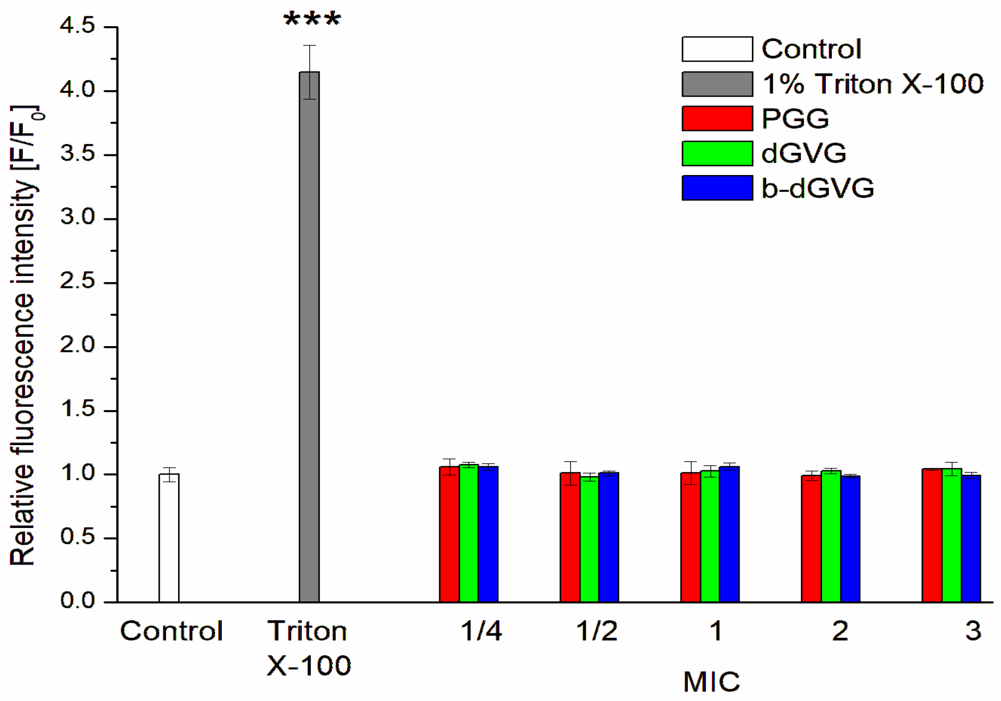
Figure 1. Impact of PGG, dGVG, and b-dGVG on S. mutans cell membrane integrity, using 1% Triton X-100 as positive control. The data presented are the means ± SD, n = 6. Statistical significance was estimated using a one-way ANOVA test (results compared to control, *** p < 0.001).
Molecules 29 00879 g001
Figure 2. Dependence of the order parameter of S. mutans membranes in the presence of tannins measured using TMA-DPH (A) and DPH (B) probes. The data presented are the means ± SD, n = 6. Statistical significance was estimated using a one-way ANOVA test (results compared to control, * p < 0.05; ** p < 0.01; *** p < 0.001).
Molecules 29 00879 g002
Figure 3. Relative quenching of Trp fluorescence of S. mutans membrane proteins in the presence of tannins (A,C,E) and Stern–Volmer plots of Trp fluorescence quenching of S. mutans membrane proteins in the presence of tannins (B,D,F). The data presented are the means ± SD, n = 6. Statistical significance was estimated using a one-way ANOVA test (results compared to control, *** p < 0.001); K in legend means Kelvin degree.
Molecules 29 00879 g003
Figure 4. Double logarithmic plots (A,C,E) and Van’t Hoff plots (B,D,F) for tannins–protein interactions of S. mutans membranes.
Molecules 29 00879 g004
Figure 5. Zeta potential changes in S. mutans in the presence of tannins. The data presented are the means ± SD, n = 6. Statistical significance was estimated using a one-way ANOVA test (results compared to control, * p < 0.05; ** p < 0.01; *** p < 0.001). (A)—PGG; (B)—dGVG; (C)—b-dGVG.
Molecules 29 00879 g005
Table 1. Chemical structures and basic properties of PGG, dGVG, and b-dGVG.
PGGdGVGb-dGVG
Chemical structureMolecules 29 00879 i001Molecules 29 00879 i002Molecules 29 00879 i003
Chemical formulaC41H32O26C41H30O27C41H30O27
Molecular weight940.681 g/mol954.664 g/mol954.664 g/mol
Number of hydroxyl groups (-OH)151515
Number of gallic acid residues555
Number of glucose moieties111
OriginRhus typhina L.Euphorbia E. turkestanicaPlantago lanceolata L.
Table 2. Antibacterial activity (MIC and MBC) of PGG, dGVG, and b-dGVG against S. mutans.
TanninMIC [µM]MBC [µM]
PGG50200
dGVG400800
b-dGVG200400
Table 3. Binding parameters of tannin—S. mutans membrane protein interactions (KSV—Stern–Volmer constant, kq—quenching constant, and logKb—logarithm of binding constant).
Binding Parameters
TanninTemperature [K]Ksv [×106] [M−1]kq [×1014] [M−1·s−1]logKb
PGG2960.393 ± 0.0450.786 ± 0.0904.398 ± 0.404
3030.464 ± 0.0420.927 ± 0.0854.175 ± 0.230
3100.484 ± 0.0200.967 ± 0.0403.769 ± 0.491
dGVG2960.261 ± 0.0300.522 ± 0.0684.758 ± 0.216
3030.281 ± 0.0390.562 ± 0.0794.689 ± 0.186
3100.321 ± 0.0100.642 ± 0.0205.474 ± 0.317
b-dGVG2960.417 ± 0.0160.834 ± 0.0324.692 ± 0.240
3030.468 ± 0.0320.937 ± 0.0634.368 ± 0.416
3100.4914 ± 0.0590.983 ± 0.1174.095 ± 0.116
Table 4. Thermodynamic parameters of the binding of tannins with S. mutans membranes.
TanninT(K)ΔH (kJ·mol−1)ΔS (kJ·mol−1K−1)ΔG (kJ·mol−1)
PGG296−78.7697−0.18131−25.1026
303−23.8335
310−22.5643
dGVG29689.111740.389428−26.1589
303−28.8840
310−31.6109
b-dGVG296−74.8819−0.16328−26.5519
303−25.4089
310−24.2660
Disclaimer/Publisher’s Note: The statements, opinions and data contained in all publications are solely those of the individual author(s) and contributor(s) and not of MDPI and/or the editor(s). MDPI and/or the editor(s) disclaim responsibility for any injury to people or property resulting from any ideas, methods, instructions or products referred to in the content.