Article

Four New Polyprenylated Acylphloroglucinols from Hypericum perforatum L.

1
Shaanxi Key Laboratory of Natural Products & Chemical Biology, College of Chemistry & Pharmacy, Northwest A&F University, Yangling 712100, China
2
Shaanxi Jiahe Phytochem Co., Ltd., Xi’an 710077, China
*
Authors to whom correspondence should be addressed.
These authors contributed equally to this work.
Molecules 2024, 29(8), 1756; https://doi.org/10.3390/molecules29081756
Submission received: 9 March 2024 / Revised: 28 March 2024 / Accepted: 4 April 2024 / Published: 12 April 2024
(This article belongs to the Section Natural Products Chemistry)

Abstract

:
Hyperforatums A–D (14), four new polyprenylated acylphloroglucinols, together with 13 known compounds were isolated and identified from the aerial parts of Hypericum perforatum L. (St. John’s wort). Their structures were confirmed with a comprehensive analysis comprising spectroscopic methods, including 1D and 2D NMR, HRESIMS, and electronic circular dichroism (ECD) calculations. Hyperforatum A featured an unusual chromene-1,4-dione bicyclic system, and hyperforatums B and C were two rare monocyclic PPAPs with five-membered furanone cores. Compound 1 exhibited a moderate inhibition effect on NO production in BV-2 microglial cells stimulated by LPS.

1. Introduction

The genus Hypericum is a large family, consisting of approximately 500 species [1]. Plants of this genus are widely distributed throughout the world and some of them are used as folk medicinal herbs [2]. H. perforatum (St. John’s wort) is extensively used to treat mild to moderate mental depression in many countries [3]. In addition, the extracts of H. perforatum showed anti-neurodegenerative disease, antitumor, and antimicrobial activities [4,5,6]. Chemical researches studying this plant revealed the presence of diverse PPAPs [7,8,9], flavonoids [10], phenolic acids, and so on [11]. Now, more than 1100 polycyclic polyprenylated acylphloroglucinols (PPAPs) have been isolated and identified from the genus Hypericum [12], but complex and novel carbon skeletons of PPAPs are consistently found from this plant, for example, hyperfols A and B [9], hyperforen A [13], hyperforones A–J [14]. Moreover, these PPAPs demonstrated significant neuroprotective effects, especially against Alzheimer’s disease. Thus, the discovery of intricate PPAPs is essential as they are the leading compounds for the treatment of Alzheimer’s disease.
As a part of our systematic investigation for bioactive PPAPs and terpenoids from genus Hypericum plants [15,16], compounds 117 were obtained and characterized from this plant, including 4 previously undescribed PPAPs, namely hyperforatums A–D (14) (Figure 1), as well as 13 known compounds, a PPAP (5), 4 triterpenoids (69), a flavonoid (10), a vitamin E derivative (11), a diterpenoid (12), 3 sesquiterpenoids (13, 15, 16), a coumarin (14), and a dihydroactinidiolide (17).

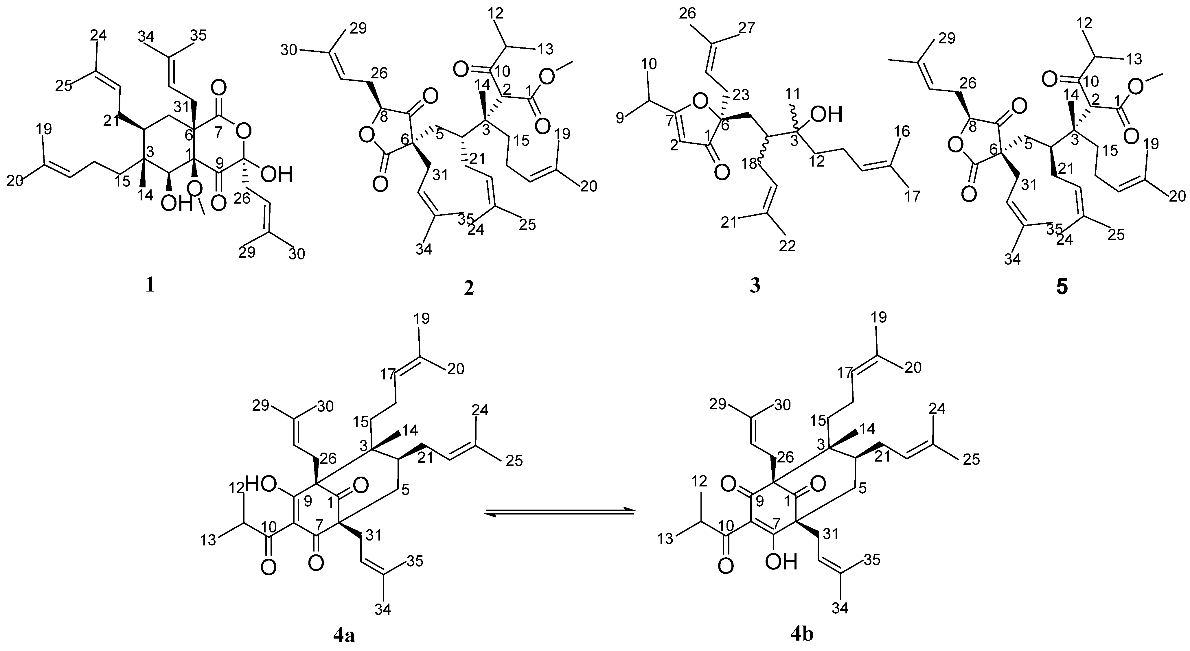
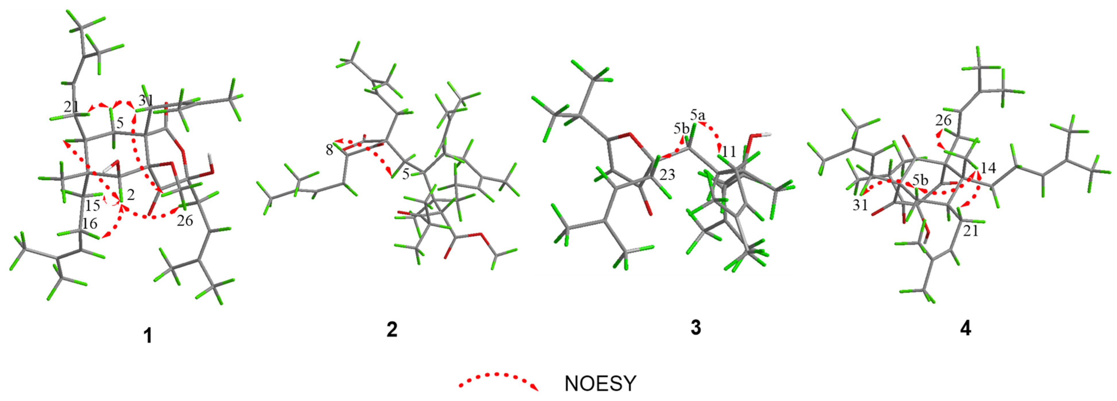
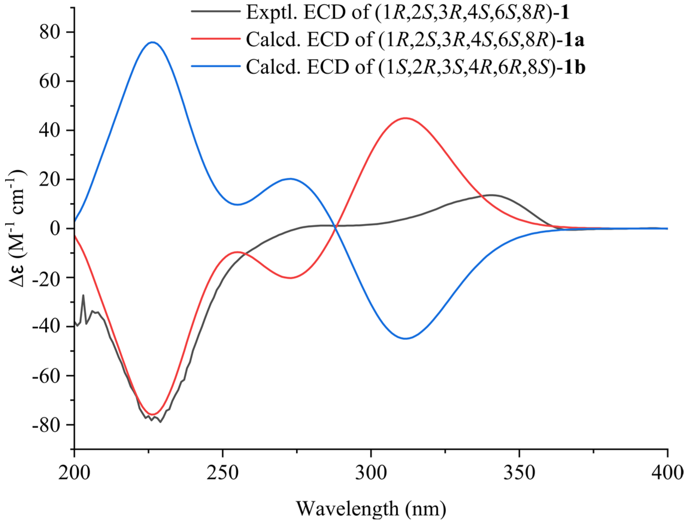
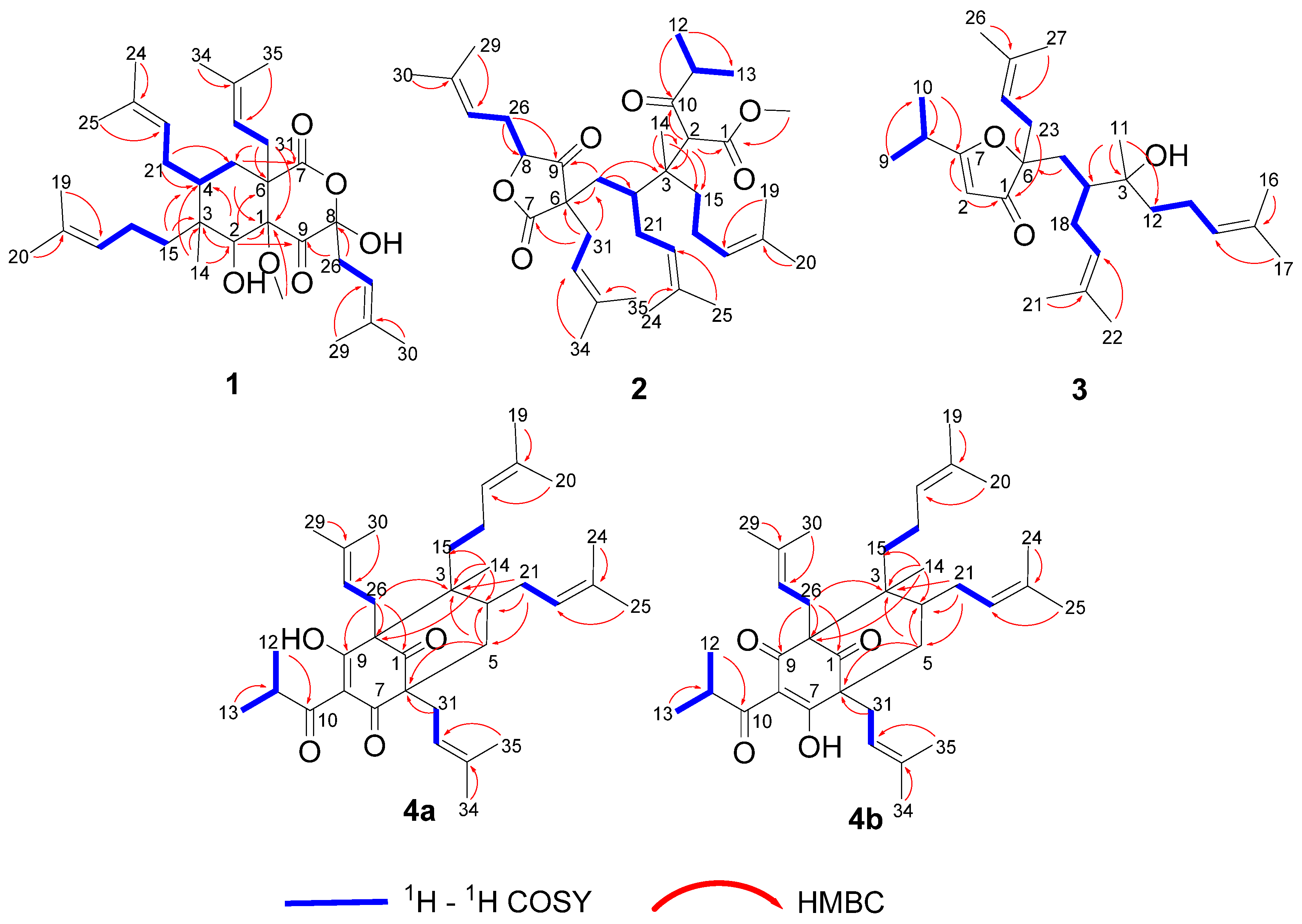
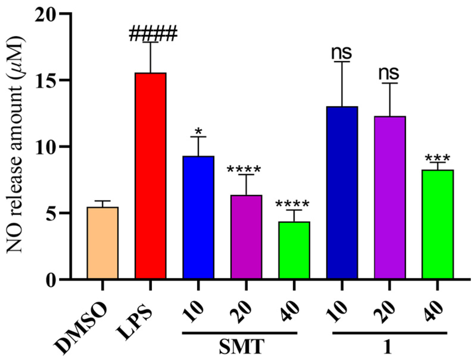
2. Results and Discussion

Hyperforatum A (1) was obtained as a colorless oil. Its molecular formula was deduced as C32H50O6 based on the 13C NMR spectrum and HRESIMS data (m/z: [M – H2O + H]+ calcd. 513.3574; found 513.3573), corresponding to 8 degrees of unsaturation (Figure S8). The 1H NMR spectrum of compound 1 displayed characteristic signals for four olefinic protons (δH 5.23, 2H, overlap; 5.05, 2H, overlap) and nine methyls (δH 0.91–1.71, s). Further analysis of its 13C NMR and DEPT spectra indicated 32 carbons attributable to 9 methyls, 6 methylenes, 6 methines, 1 methoxyl, and 10 quaternary carbons. The HRESIMS and NMR data revealed that compound 1 should be a bicyclic-type PPAP (Figures S1–S3).
The planar structure of compound 1 was established by interpreting its 2D NMR data (Figure 2). The HMBC cross peaks from H-2 to C-1/C-3/C-4/C-6/C-9, from H-14 to C-2/C-3/C-4, from H2-31 to C-1/C-5/C-6/C-7, as well as the characteristic quaternary carbons (δc 205.1, 180.3, 97.7) confirmed the bicyclic core with a methyl (C-14) and an isoprenyl fragment attached at C-3 and C-6, respectively. Two isoprenyl groups were connected to C-3 and C-4, which were established by the HMBC cross peaks from H3-20 to C-17/C-18/C-19, from H2-15 to C-3/C-4, from H3-25 to C-22/C-23/C-24, and from H2-21 to C-4/C-5, as well as the 1H-1H COSY cross peaks of H2-15/H2-16/H-17 and H2-5/H-4/H2-21/H-22. The fragment (CH2-26-CH-27-C-28-CH3-29-CH3-30) was positioned at C-8, which was deduced from the HMBC cross peaks from H3-30 to C-27/C-28/C-29, and from H2-26 to C-8/C-9, as well as the 1H-1H COSY interactions of H2-26/H-27 (Figures S4–S6). Furthermore, a methoxy group was located at C-1 due to the HMBC correlation. Thus, the planar construction of compound 1 was finally built (Figure 2).
The relative configuration of compound 1 was elucidated by the NOESY data (Figure 3). The NOESY correlations of H-2/H-15b, H-2/H-4, H-2/H2-26b, H-5a/H-21a, H-5a/H-31b, and H-31b/H3-OCH3 indicated that H-2 and the isoprenyl group at C-3 and C-8 were α-oriented; moreover, the isoprenyl group at C-4 and C-6 and the methoxy group were in the same β-orientation (Figure S7). From the above analysis, the relative configuration of compound 1 was determined to be 1R*, 2S*, 3R*, 4S*, 6S*, 8R*. Moreover, the absolute configuration of (1R, 2S, 3R, 4S, 6S, 8R)-1a was determined using the calculated ECD data, showing good agreement with the experimental data (Figure 4).
Hyperforatum B (2) was purified as a colorless oil. The molecular formula of C36H56O6 was confirmed by its HRESIMS data (m/z: [M + Na]+ calcd. 607.3969; found 607.3958), indicating 9 degrees of unsaturation (Figure S18). The 1H NMR data of compound 2 displayed signals for four olefinic protons (δH 5.16, 1H, t, J = 7.0 Hz; 5.01, 1H, t, J = 7.0 Hz; 4.95, 2H, overlap), nine methyls (δH 1.08–1.72, s), and an isopropyl group (δH 2.66, 1H, m; 1.07, 3H, d, J = 6.7 Hz; 1.04, 3H, d, J = 6.7 Hz). The 13C NMR and DEPT spectra indicated 36 carbons, including 11 methyls, 6 methylenes, 8 methines, 1 methoxyl, and 10 quaternary carbons. The comprehensive analysis of 2D NMR revealed that compound 2 shared the same planar structure as compound 5 [17] (Figure 2 and Figures S11–S16). However, minor deviations were observed: C-4 (δC 41.6), C-5 (δC 37.5), C-21 (δC 30.2) in compound 2 were replaced by C-4 (δC 40.7), C-5 (δC 37.2), C-21 (δC 35.5) in compound 5, respectively. Moreover, the chemical shifts of H2-21 and H-22 also changed significantly (Table 1). These deviations might have resulted from the differences in the configurations of the isoprenyl group at C-4. When the orientation of the isoprenyl group was changed at the C-4 position, the two large groups attached at the ends of the C-4 position had steric hindrance effects, which might have led to a significant difference of chemical shifts around the C-4 position. The deduction cannot be confirmed by the key NOESY correlations because the isoprenyl group was located in a flexible side chain. The NOESY correlations of H-8/H-5a indicated that the stereochemistry of C-8 was α-oriented (Figure 3 and Figure S17). The absolute configuration of compound 2 was unable to be calculated due to the chiral center being on a flexible chain.
The molecular formula of hyperforatum C (3) was deduced as C27H44O3 based on its HRESIMS data (m/z: [M + Na]+ calcd 439.3183, found 439.3177), suggesting 6 unsaturation sites (Figure S28). The 1H NMR spectrum showed the presence of 3 olefinic protons (δH 5.11, 2H, overlap; 4.93, 1H, t, J = 7.2 Hz), 7 singlet methyls (δH 1.03–1.69, s), and an isopropyl (δH 2.69, 1H, m; 1.22, d, 3H, J = 6.8 Hz; 1.20, d, 3H, J = 6.8 Hz). The 13C NMR data of compound 3 displayed 27 carbons assigned to 9 methyls, 5 methylenes, 6 methines (4 olefinic), and 7 nonprotonated carbons (1 carbonyl, 4 olefinic, 2 oxygenated). The above analysis suggested that compound 3 should be a monocyclic PPAP (Figures S21–S23). The HMBC interactions from H3-22 to C-19/C-20/C-21 along with the 1H-1H COSY cross peaks of H2-4/H-5/H2-18/H-19 constructed the fragment A (C-4-C-5-C-18-C-19-C-20-C-21-C-22). The HMBC interactions from H3-17 to C-14/C-15/C-16 along with the 1H-1H COSY cross peaks of H2-12/H2-13/H-14 constructed the fragment B (C12-C-13-C-14-C-15-C-16-C-17). The HMBC interactions from H3-27 to C-24/C-25/C-26 accompanied by the 1H-1H COSY cross peaks of H2-23/H-24 constructed the fragment C (C-23-C-24-C-25-C-26-C-27). The fragment C was located at C-6 due to the HMBC correlations from H2-23 to C-6. The fragments A and B were connected through the carbon C-3, of which the result was supported by the HMBC interactions from H3-11 to C-3/C-4/C-12. In addition, the HMBC correlations from H3-9 to C-7/C-10/C-11, from H-2 to C-1/C-7, from H2-23 to C-1/C-6, and from H2-5 to C-6, as well as the 1H-1H COSY cross peaks of H3-9/H-8/H3-10, formed the architecture of furanone with a isopropyl at carbon C-7 (Figure 2 and Figures S24–S26). Compound 3 might be obtained with a rearrangement of monocyclic polyprenylated acylphloroglucinols (MPAPs). The relative configuration of compound 3 could not be determined by the NOESY correlations because its chiral center was located in the flexible chain. Meantime, ECD calculations were quite challenging in the determination of the absolute configuration of compound 3. Unfortunately, its crystal failed to be obtained after the solvent conditions were changed multiple times.
Hyperforatum D (4) was isolated as a colorless oil. The molecular formula of C35H52O4 was supported by its HRESIMS data (m/z [M + H]+ calcd 537.3938, found 537.3928), with 10 degrees of unsaturation (Figure S38). The 1H NMR and 13C spectra of compound 4 in CDCl3 showed a paired mixture of two keto-enol tautomers (4a and 4b) in an approximate 1:1 ratio. The keto–enol tautomerism of a β,β′-triketo moiety was easily converted for the PPAPs seen in hypascyrins A–E [18]. The 1H NMR spectrum of compound 4a revealed the presence of 4 olefinic protons (δH 5.20, 1H, J = 7.0 Hz; 5.02, 1H, overlap, 4.84, 1H, t, J = 7.2 Hz; 4.76, 1H, J = 6.2 Hz), 9 singlet methyls (δH 1.22–1.70, s), an isopropyl group (δH 3.82, 1H, m; 1.20, 3H, d, J = 6.8 Hz; 1.06, 3H, d, J = 6.8 Hz) (Table 2). The 13C NMR spectrum combined with HSQC and HMBC revealed that tautomer 4a was a type B PPAP derivative with a bicyclo [3.3.1]nonane-2,4,9-trione system, whose structure was similar to spiranthenone B [19]. The main differences were the presence of an isopropyl group at C-8 and a prenyl group at C-3 in tautomer 4a. The deduction was verified by the HMBC correlations from H3-20 to C-17/C-18/C-19, from H2-15 to C-2/C-3/C-4/C-17, and from H3-12 to C-10/C-11/C-13, along with 1H-1H COSY correlations of H2-15/H2-16 and H3-12/H-11/H3-13 (Figure 2 and Figures S31–S36). For tautomer 4b, the hydroxyl group was located at the C-7 position, while the carbonyl was located at the C-9 position; these conclusions were confirmed by the HMBC correlations from H2-26 (δH 2.65, 2.62) to C-1 (δC 207.8)/C-2 (δC 67.1)/C-3 (δC 50.6)/C-9 (δC 200.7), and from H2-31 (δH 2.51, 2.48) to C-1 (δC 207.8)/C-6 (δC 64.2)/C-7 (δC 194.2)/C-5 (δC 37.4). Comprehensive analysis of the 2D NMR data also revealed the 2D structure of the tautomer (4b) (Figure 2 and Figures S31–S36). The relative configurations of tautomers 4a and 4b were determined using the NOESY data (Figure 3). The NOESY correlations of H3-14/H2-21, H3-14/H-5b, and H-5b/H2-30 indicated that these protons were in the same β orientation (Figure S38). Its relative configuration was equal to that of spiranthenone B based on the NOESY cross peaks. Finally, the relative configuration of compound 4 was confirmed, as shown in Figure 3.
When comparing the spectroscopic data to those reported in the literature, thirteen known compounds were identified to be methyl (αSRS,3S)-tetrahydro-β-methyl-γ,3,5-tris(3-methyl-2-buten-1-yl)-α-(2-methyl-1-oxopropyl)-β-(4-methyl-3-penten-1-yl)-2,4-dioxo-3-furanpentanoate (5) [17], lupeol acetate (6) [20], lup -20(29)-en-3-one (7) [21], α–amyrin acetate (8) [22], methyl oleanolate (9) [23], (–)-(6aR,11aR)-homopterocarpin (10) [24], 5-formyl-7,8-dimethyltocol (11) [25], cassipourol (12) [26], ent-α-cyperone (13) [27], mullein (14) [28], ledol (15) [29], kobusone (16) [30], and dihydroactinidiolide (17) [31].
There are many reports on the anti-Alzheimer’s effects of H. perforatum [14,32]. Chronic inflammation is an important cause of the development of Alzheimer’s disease’s pathogenesis [33]. The production of nitric oxide (NO) in LPS-stimulated microglial cells is used as a cellular model to evaluate the effects of anti-neuroinflammation. Since we ended up with insufficient quantities of compounds 2, 3, and 4 to complete activity evaluation, we assessed the biological activity of compound 1 only. Consequently, compound 1 significantly inhibited NO production at 40 μM (Figure 5). However, its anti-inflammatory mechanisms need to be further explored.

3. Materials and Methods

3.1. General

NMR spectra were carried out on a Bruker Avance Neo at 400 MHz and 800 MHz (Bruker BioSpin, Fällanden, Switzerland) using tetramethylsilane (TMS) as internal standard. Optical rotations (ORs) were recorded on an Autopol III automatic polarimeter (Rudolph Research Analytical). A Chirascan spectrometer was used to obtain the UV and experimental CD spectra. HRESIMS data were obtained on a LC-30A + TripleTOF5600+ (AB Sciex Pte. Ltd., Framingham, MA, USA). Separations and purifications of the samples were conducted on silica gel (200–300 and 300–400 mesh, Qingdao Marine Chemical Ltd., Qingdao, China), ODS RP-C18 (50 μm, YMC Co., Ltd., Kyoto, Japan), and Sephadex LH-20 (40–70 μm, Amersham Pharmacia Biotech AB, Stockholm, Sweden). A shimadzu LC-20AP liquid chromatography system equipped with a reversed-phase (RP) C-18 column (10 mm × 250 mm, 5 μm) was applied to complete sample purification.

3.2. Plant Materials

Air-dried aerial portions of H. perforatum were collected in August 2018 from Shangluo City, China. The plant was identified by Pro. Zhen-hai Wu. The sample (no. 20180805HPL) was preserved at Shaanxi Key Laboratory of Natural Products and Chemical Biology, Northwest A&F University.

3.3. Extraction and Isolation

Air-dried aerial portions of H. perforatum (100 kg) were powdered and extracted using 95% EtOH (300 L × 3, each of 2 h) via three cycles of refluxing. The filtered solution was then concentrated under reduced pressure to obtain the crude extract, which was suspended in water and then partitioned with n-hexane and EtOAc. The n-hexane fraction (1.03 kg) was subjected to silica gel column elution with petroleum ether/EtOAc (100:0 to 1:1, v/v) to obtain six fractions (Fr. 1–6). Fr.3 (291.0 g) was applied to a silica gel column and eluted with petroleum ether/EtOAc (100:1 to 20:1, v/v) to yield six subfractions (Fr. 3A–3F). Fr.3B (27.2 g) was further fractionated using a RP-C18 CC (MeOH/H2O, 90:10 to 100:0, v/v) and a silica gel CC (PE/EtOAc, 100:0 to 30:1) to obtain four subfractions (Fr.3Ba–3Bd). Fr.3Bb (93.3 mg) was purified via preparative HPLC using MeOH/H2O (91:9, v/v, 2 mL/min) isocratic elution to yield compounds 6 (6.0 mg, tR = 50 min), 3 (2.2 mg, 55 min) and 2 (6 mg, tR = 60 min). Fr.3Bd (85.0 mg) was subjected to preparative HPLC using CH3CN:H2O (93:7, v/v, 2 mL/min) isocratic elution to yield compounds 1 (4.9 mg, tR = 46 min) and 4 (12.7 mg, tR = 75 min); the column temperature for HPLC was 37 °C. Fr.3E (131.7 g) was subjected to a RP-C18 CC eluted with MeOH:H2O (60:40 to 100:0, v/v) to obtain four subfractions (Fr.3Ea–3Ed). Fr.3Ea (187.0 mg) was loaded onto a Sephadex LH-20 column using CH2Cl2:MeOH (1:1, v/v) and then purified via semipreparative HPLC (CH3CN:H2O, 46:54, v/v, 2 mL/min) to afford compounds 16 (15.0 mg, tR = 27 min) and 17 (6.3 mg, tR = 36 min). Fr.3Ec (3.96 g) was fractionated using a silica gel column and eluted with n-hexane:EtOAc (50:1 to 1:1, v/v) to obtain five subfractions (Fr.3Ec1–3Ec5). Fr.3Ec2 (446.0 mg) was repurified by semi-preparative HPLC (MeOH:H2O, 46:54, v/v, 2 mL/min) to acquire compounds 13 (2.7 mg, tR = 22 min), 14 (2.8 mg, tR = 26 min), and 15 (11.0 mg, tR = 38 min). Fr.3Ec4 (2.11 g) was fractionated by a Sephadex LH-20 column using CH2Cl2:MeOH (1:1, v/v) and further separated by a silica gel column and eluted with petroleum ether/EtOAc (50:1 to 1:1, v/v) to obtain compounds 6 (187.6 mg), 7 (79.1 mg), 8 (11.0 mg), 9 (47.6 mg), 10 (7.0 mg), 11 (8.5 mg), and 12 (7.5 mg).

3.4. Structural Elucidation

Hyperforatum A (1), colorless oil; [α]20D +2 (c 0.1 MeOH); UV (MeOH) λmax (log ε) 200 (4.25), nm; ECD (MeOH) λmax (∆ε) 226 (−25.16), 340 (4.36) nm; 1H and 13C NMR data (see Table 1); HRESIMS m/z 513.3573 [M – H2O + H]+ (calcd. for C32H49O5, 513.3574).
Hyperforatum B (2), colorless oil; [α]20D +7.5 (c 1.00 MeOH); UV (MeOH) λmax (log ε) 200 (3.71) nm; ECD (MeOH) λmax (∆ε) 237 (−3.36) nm; 1H and 13C NMR data (see Table 1); HRESIMS m/z 607.3958 [M + Na]+ (calcd. for C36H56O6Na, 607.3969).
Hyperforatum C (3), colorless oil; [α]20D +5.9 (c 0.35 MeOH); UV (MeOH) λmax (log ε) 200 (4.20), 262 (3.34) nm; ECD (MeOH) λmax (∆ε) 231 (−0.97), 302 (0.82) nm; 1H and 13C NMR data (see Table 2); HRESIMS m/z 439.3177 [M + Na]+ (calcd. for C27H44O3Na, 439.3183).
Hyperforatum D (4), colorless oil; [α]20D +4.0 (c 1.00 MeOH); UV (MeOH) λmax (log ε) 200 (2.85), 244 (2.14), 289 (2.19) nm; ECD (MeOH) λmax (∆ε) 210 (−1.03), 234 (+0.17), 252 (−0.36), 303 (+0.22) nm; 1H and 13C NMR data (see Table 2); HRESIMS m/z 537.3928 [M + H]+ (calcd. for C35H53O4, 537.3938).

3.5. Cell Culture

Microglial BV-2 cells from China Center for Type Culture Collection (Wuhan, China) were cultivated in DMEM (Gibco, New York, NY, USA) containing 10% FBS (Gibco) and antibiotics (100 U/mL streptomyces and penicillin) in humidified incubators under 5% CO2 at 37 °C.

3.6. Measurement of Nitric Oxide (NO) Production

BV-2 cells were seeded in 96-well plates (2 × 10⁵ cells/mL) overnight. The cells were treated with LPS (2 μg/mL) and various concentrations of hyperforatum A (1) (10, 20, 40 μM) for 24 h, with S-Methylisothiourea (SMT) as the positive control. The production of NO was measured in cell supernatants with a Griess reagent. The absorbance was recorded at 540 nm using a microplate reader. The MTT method was applied to determine the cell viability after incubation using the test compound.

3.7. Statistical Analysis

All data were presented as mean ± SD and analyzed with GraphPad Prism 9.0 software. The significant differences between different groups were performed using one-way ANOVA multiple comparisons.

4. Conclusions

The phytochemical components of the PPAPs and terpenoids were investigated from the aerial parts of H. perforatum. Seventeen secondary metabolites, including five PPAPs and nine terpenoids, were isolated and identified from the title plants. This study reported two unusual carbon cores of PPAPs, of which hyperforatum A was a chromene-1,4-dione bicyclic system, and hyperforatum B and C possessed rare monocyclic features. The new compound, hyperforatum A (1), displayed a moderate inhibitory capacity on LPS-induced NO production.

Supplementary Materials

The following supporting information can be downloaded at: https://www.mdpi.com/article/10.3390/molecules29081756/s1, Figures S1–S7: The 1D and 2D NMR spectra of compound 1 in CDCl3, Figure S8: The HRESIMS spectrum of compound 1, Figures S9 and S10: The UV and ECD spectra of compound 1, Figures S11–S17: The 1D and 2D NMR spectra of compound 2 in CDCl3, Figure S18: The HRESIMS spectrum of compound 2, Figures S19 and S20: The UV and ECD spectra of compound 2, Figure S21–S27: The 1D and 2D NMR spectra of compound 3 in CDCl3, Figure S28: The HRESIMS spectrum of compound 3, Figures S29 and S30: The UV and ECD spectra of compound 3, Figures S31–S37: The 1D and 2D NMR spectra of compound 4 in CDCl3, Figure S38: The HRESIMS spectrum of compound 4, Figures S39 and S40: The UV and ECD spectra of compound 4.

Author Contributions

Conception, formal analysis, and writing—original draft preparation, X.W.; methodology, formal analysis, and investigation, W.L.; software and investigation, S.C.; methodology and investigation, Y.G.; writing—review and editing, supervision, and project administration, J.T.; conception, writing—review and editing, and funding acquisition, J.G. All authors have read and agreed to the published version of the manuscript.

Funding

This research was funded by the National Natural Science Foundation of the People’s Republic of China (No. 22077102).

Institutional Review Board Statement

Not applicable.

Informed Consent Statement

Not applicable.

Data Availability Statement

Supporting data include HRESIMS, UV, CD, and 1D and 2D NMR spectra.

Acknowledgments

The authors would like to thank Luqi Li from Life Science Research Core Services, Northwest A&F University, for the analysis of HPLC-MS.

Conflicts of Interest

Author Xiaoying Wang was employed by the company, Shaanxi Jiahe Phytochem Co., Ltd. The funder was not involved in the study design, collection, analysis, interpretation of data, the writing of this article or the decision to submit it for publication.

References

  1. Bridi, H.; Meirelles, G.d.C.; von Poser, G.L. Structural Diversity and Biological Activities of Phloroglucinol Derivatives from Hypericum Species. Phytochemistry 2018, 155, 203–232. [Google Scholar] [CrossRef]
  2. Zhang, R.; Ji, Y.; Zhang, X.; Kennelly, E.J.; Long, C. Ethnopharmacology of Hypericum Species in China: A Comprehensive Review on Ethnobotany, Phytochemistry and Pharmacology. J. Ethnopharmacol. 2020, 254, 112686. [Google Scholar] [CrossRef] [PubMed]
  3. Korczak, M.; Pilecki, M.; Granica, S.; Gorczynska, A.; Pawłowska, K.A.; Piwowarski, J.P. Phytotherapy of mood disorders in the light of microbiota-gut-brain axis. Phytomedicine 2023, 111, 154642. [Google Scholar] [CrossRef] [PubMed]
  4. Suryawanshi, M.V.; Gujarathi, P.P.; Mulla, T.; Bagban, I. Hypericum perforatum: A comprehensive review on pharmacognosy, preclinical studies, putative molecular mechanism, and clinical studies in neurodegenerative diseases. Naunyn-Schmiedeberg’s Arch. Pharmacol. 2024. [Google Scholar] [CrossRef]
  5. Knauthe, A.; Mittag, S.; Bloch, L.; Albring, K.F.; Schmidt, M.; Werz, O.; Huber, O. Hyperforin and Myrtucommulone Derivatives Act as Natural Modulators of Wnt/β-Catenin Signaling in HCT116 Colon Cancer Cells. Int. J. Mol. Sci. 2022, 23, 2984. [Google Scholar] [CrossRef] [PubMed]
  6. Tsioutsiou, E.E.; Amountzias, V.; Vontzalidou, A.; Dina, E.; Stevanović, Z.D.; Cheilari, A.; Aligiannis, N. Medicinal Plants Used Traditionally for Skin Related Problems in the South Balkan and East Mediterranean Region-A Review. Front. Pharmacol. 2022, 13, 936047. [Google Scholar] [CrossRef] [PubMed]
  7. Duan, Y.; Deng, Y.; Bu, P.; Guo, Y.; Shi, Z.; Cao, Y.; Zhang, Y.; Hu, H.; Hu, Z.; Qi, C.; et al. Discovery of Bioactive Polycyclic Polyprenylated Acylphloroglucinols from Hypericum wilsonii. Bioorg. Chem. 2021, 115, 105246. [Google Scholar] [CrossRef] [PubMed]
  8. Guo, Y.; Zhang, N.; Chen, C.; Huang, J.; Li, X.N.; Liu, J.; Zhu, H.; Tong, Q.; Zhang, J.; Luo, Z.; et al. Tricyclic Polyprenylated Acylphloroglucinols from St John’s Wort, Hypericum perforatum. J. Nat. Prod. 2017, 80, 1493–1504. [Google Scholar] [CrossRef] [PubMed]
  9. Lou, H.Y.; Li, Y.N.; Yi, P.; Jian, J.Y.; Hu, Z.X.; Gu, W.; Huang, L.J.; Li, Y.M.; Yuan, C.M.; Hao, X.J. Hyperfols A and B: Two Highly Modified Polycyclic Polyprenylated Acylphloroglucinols from Hypericum perforatum. Org. Lett. 2020, 22, 6903–6906. [Google Scholar] [CrossRef]
  10. Smelcerovic, A.; Spiteller, M.; Zuehlke, S. Comparison of Methods for the Exhaustive Extraction of Hypericins, Flavonoids, and Hyperforin from Hypericum perforatum L. J. Agric. Food Chem. 2006, 54, 2750–2753. [Google Scholar] [CrossRef]
  11. Orčić, D.Z.; Mimica-Dukić, N.M.; Francišković, M.M.; Petrović, S.S.; Jovin, E.Đ. Antioxidant Activity Relationship of Phenolic Compounds in Hypericum perforatum L. Chem. Cent. J. 2011, 5, 34. [Google Scholar] [CrossRef] [PubMed]
  12. Xu, Z.H.; Grossman, R.B.; Qiu, Y.F.; Luo, Y.; Lan, T.; Yang, X.W. Polycyclic Polyprenylated Acylphloroglucinols Bearing a Lavandulyl-Derived Substituent from Garcinia Xanthochymus Fruits. J. Nat. Prod. 2022, 85, 2845–2855. [Google Scholar] [CrossRef] [PubMed]
  13. Lou, H.; Ma, F.; Yi, P.; Hu, Z.; Gu, W.; Huang, L.; He, W.; Yuan, C.; Hao, X. Bioassay and UPLC-Q-Orbitrap-MS/MS Guided Isolation of Polycyclic Polyprenylated Acylphloroglucinols from St. John’s Wort and Their Neuroprotective Activity. Arab. J. Chem. 2022, 15, 104057. [Google Scholar] [CrossRef]
  14. Guo, Y.; Huang, F.; Sun, W.; Zhou, Y.; Chen, C.; Qi, C.; Yang, J.; Li, X.N.; Luo, Z.; Zhu, H.; et al. Unprecedented Polycyclic Polyprenylated Acylphloroglucinols with Anti-Alzheimer’s Activity from St. John’s Wort. Chem. Sci. 2021, 12, 11438–11446. [Google Scholar] [CrossRef] [PubMed]
  15. Xie, J.Y.; Li, P.F.; Yan, X.T.; Gao, J.M. Discovery from Hypericum elatoides and synthesis of hyperelanitriles as α-aminopropionitrile-containing polycyclic polyprenylated acylphloroglucinols. Commun. Chem. 2024, 7, 1. [Google Scholar] [CrossRef]
  16. Yan, X.T.; Chen, J.X.; Wang, Z.X.; Zhang, R.Q.; Xie, J.Y.; Kou, R.W.; Zhou, H.F.; Zhang, A.L.; Wang, M.C.; Ding, Y.X.; et al. Hyperhubeins A–I, Bioactive Sesquiterpenes with Diverse Skeletons from Hypericum hubeiense. J. Nat. Prod. 2023, 86, 119–130. [Google Scholar] [CrossRef] [PubMed]
  17. Verotta, L.; Lovaglio, E.; Sterner, O.; Appendino, G.; Bombardelli, E. Modulation of Chemoselectivity by Protein Additives. Remarkable Effects in the Oxidation of Hyperforin. J. Org. Chem. 2004, 69, 7869–7874. [Google Scholar] [CrossRef] [PubMed]
  18. Niwa, K.; Tanaka, N.; Tatano, Y.; Yagi, H.; Kashiwada, Y. Hypascyrins A-E, Prenylated Acylphloroglucinols from Hypericum ascyron. J. Nat. Prod. 2019, 82, 2754–2760. [Google Scholar] [CrossRef] [PubMed]
  19. Albernaz, L.C.; Deville, A.; Dubost, L.; de Paula, J.E.; Bodo, B.; Grellier, P.; Espindola, L.S.; Mambu, L. Spiranthenones A and B, Tetraprenylated Phloroglucinol Derivatives from the Leaves of Spiranthera Odoratissima. Planta. Med. 2012, 78, 459–464. [Google Scholar] [CrossRef]
  20. Sultana, N.; Saify, Z.S.; Saleem, M.; Kamal, M. Two new triterpenes from Alstonia scholaris flowers. Nat. Prod. Res. 2013, 27, 1277–1286. [Google Scholar] [CrossRef]
  21. Yamashita, H.; Matsuzaki, M.; Kurokawa, Y.; Nakane, T.; Goto, M.; Lee, K.H.; Shibata, T.; Bando, H.; Wada, K. Four New Triterpenoids from the Bark of Euonymus Alatus Forma Ciliato-Dentatus. Phytochem. Lett. 2019, 31, 140–146. [Google Scholar] [CrossRef] [PubMed]
  22. Seo, D.G.; Kim, S.; Lee, D.K.; Kim, N.Y.; Lee, J.S.; Hwang, K.W.; Park, S.Y. Inhibitory Effect of α-Amyrin Acetate Isolated from Fraxinus Rhynchophylla on Th17 Polarization. Phytomedicine 2019, 63, 153056. [Google Scholar] [CrossRef] [PubMed]
  23. Takeoka, G.; Dao, L.; Teranishi, R.; Wong, R.; Flessa, S.; Harden, L.; Edwards, R. Identification of Three Triterpenoids in Almond Hulls. J. Agric. Food Chem. 2000, 48, 3437–3439. [Google Scholar] [CrossRef]
  24. Aung, H.T.; Aye, M.M.; Thu, Z.M.; Komori, Y.; Sein, M.M.; Vidari, G.; Takaya, Y. Bioactive Constituents from the Rhizomes of Sansevieria Cylindrica. Rec. Nat. Prod. 2020, 14, 269–275. [Google Scholar] [CrossRef]
  25. Panyasawat, P.; Wisetsai, A.; Lekphrom, R.; Senawong, T.; Schevenels, F.T. Acroquinolones A and B, two polyphenolic isoprenylated acetophenone-quinolone hybrids with anti-proliferative activities from Acronychia pedunculata (L.) Miq. Nat. Prod. Res. 2022, 36, 2743–2752. [Google Scholar]
  26. Chaturvedula, V.S.P.; Norris, A.; Miller, J.S.; Ratovoson, F.; Andriantsiferana, R.; Rasamison, V.E.; Kingston, D.G.I. Cytotoxic Diterpenes from Cassipourea Madagascariensis from the Madagascar Rainforest. J. Nat. Prod. 2006, 69, 287–289. [Google Scholar] [CrossRef] [PubMed]
  27. Krohn, K.; Kock, I.; Elsässer, B.; Flörke, U.; Schulz, B.; Draeger, S.; Pescitelli, G.; Antus, S.; Kurtán, T. Bioactive Natural Products from the Endophytic Fungus Ascochyta sp. from Meliotus dentatus—Configurational Assignment by Solid-State CD and TDDFT Calculations. Eur. J. Org. Chem. 2007, 2007, 1123–1129. [Google Scholar] [CrossRef]
  28. Kalinová, B.; Kindl, J.; Jiroš, P.; Žáček, P.; Vašíčková, S.; Buděšínský, M.; Valterová, I. Composition and Electrophysiological Activity of Constituents Identified in Male Wing Gland Secretion of the Bumblebee Parasite Aphomia Sociella. J. Nat. Prod. 2009, 72, 8–13. [Google Scholar] [CrossRef] [PubMed]
  29. Kaplan, M.A.C.; Pugialli, H.R.L.; Lopes, D.; Gottlieb, H.E. The Stereochemistry of Ledol from Renealmia chrysotrycha: An NMR Study. Phytochemistry 2000, 55, 749–753. [Google Scholar] [CrossRef] [PubMed]
  30. Chuang, L.F.; Fan, T.Y.; Li, J.J.; Sung, P.J. Kobusone: Occurrence of a Norsesquiterpenoid in the Gorgonian Coral Rumphella antipathies (Gorgoniidae). Biochem. Syst. Ecol. 2007, 35, 470–471. [Google Scholar] [CrossRef]
  31. Eidman, K.F.; MacDougall, B.S. Synthesis of Loliolide, Actinidiolide, Dihydroactinidiolide, and Aeginetolide via Cerium Enolate Chemistry. J. Org. Chem. 2006, 71, 9513–9516. [Google Scholar] [CrossRef]
  32. Cao, Z.; Wang, F.; Xiu, C.; Zhang, J.; Li, Y. Hypericum perforatum Extract Attenuates Behavioral, Biochemical, and Neurochemical Abnormalities in Aluminum Chloride-Induced Alzheimer’s Disease Rats. Biomed. Pharmacother. 2017, 91, 931–937. [Google Scholar] [CrossRef] [PubMed]
  33. Subhramanyam, C.S.; Wang, C.; Hu, Q.; Dheen, S.T. Microglia-Mediated Neuroinflammation in Neurodegenerative Diseases. Semin. Cell Dev. Biol. 2019, 94, 112–120. [Google Scholar] [CrossRef] [PubMed]
Figure 1. Chemical structures of compounds 14.
Molecules 29 01756 g001
Figure 2. 1H-1H COSY and key HMBC correlations of compounds 14.
Molecules 29 01756 g002
Figure 3. Key NOESY correlations of compounds 14.
Molecules 29 01756 g003
Figure 4. Calculated and experimental ECD spectra of compound 1.
Molecules 29 01756 g004
Figure 5. Inhibition effect of compound 1 on the production of NO in BV-2 cells treated with LPS. DMSO (blank control); LPS (model group); SMT (positive control); 1 (compound 1). Results are expressed as mean ± SEM, ns = no significant difference, * p < 0.05, *** p < 0.001, and **** p < 0.0001, #### p < 0.0001, compared to the LPS (one-way ANOVA).
Molecules 29 01756 g005
Table 1. 13C NMR and 1H NMR data of compounds 1, 2, and 5.
No1 a2 a5 a
δCδH (J in Hz)δCδH (J in Hz)δCδH (J in Hz)
180.9 169.8 169.8
266.63.39 s61.43.91 s61.54.02 s
337.3 44.2 44.1
441.7 41.61.82 m40.71.88 m
533.91.84 d (7.5)37.51.98 m37.22.00 m
1.69 m 1.68 m
647.7 54.0 53.9
7180.3 176.2 175.9
897.7 84.64.53 dd (8.7, 4.6)84.74.35 dd (8.5, 4.4)
9205.1 212.6 212.6
10 208.6 208.6
11 42.82.66 m42.92.66 m
12 18.31.04 d (6.7)18.31.04 d (6.8)
13 18.61.07 d (6.7)18.71.07 d (6.8)
1418.30.91 s21.21.05 s21.31.08 s
1542.11.55 m35.62.39 m35.21.56 m
1.26 m 1.53 m
1622.62.01 m22.91.93 m23.01.96 m
1.82 m 1.84 m
17123.95.05 m124.75.02 t (7.0)124.85.01 t (7.0)
18132.3 131.5 131.4
1918.11.61 s18.01.61 s17.91.61 s
2026.01.68 s25.91.65 s25.91.66 s
2128.02.14 m30.22.03 m35.62.48 m
1.59 s 1.82 m 2.35 m
22122.85.05 m124.44.95 t (7.0)124.45.08 t (7.1)
23133.2 132.7 131.6
2418.11.57 s18.21.55 s18.21.61 s
2525.81.69 s25.91.65 s26.01.72 s
2626.12.58 dd (15.5, 7.0)29.62.46 m30.32.58 m
2.50 dd (15.5, 7.6) 2.34 m 2.42 m
27116.15.24 m117.55.17 t (7.0)117.45.18 t (7.4)
28136.2 136.4 136.6
2918.01.62 s18.11.59 s18.11.63 s
3026.11.71 s25.91.72 s26.01.67 s
3135.52.42 m35.92.46 m35.22.48 m
2.37 m 2.39 m 2.39 m
32119.15.24 m117.04.97 t (7.0)116.84.96 t (8.0)
33135.8 137.8 137.4
3417.91.62 s17.91.61 s18.01.58 s
3526.31.71 s26.11.67 s26.11.67 s
OCH354.53.43 s52.23.69 s52.23.69 s
a NMR data were recorded in CDCl3 (1H NMR 400 MHz, 13C NMR 100 MHz).
Table 2. 13C NMR and 1H NMR data of compound 3 and tautomers 4a and 4b.
No3 a4a b4b b
δCδH (J in Hz)δCδH (J in Hz)δCδH (J in Hz)
1207.6 208.1 207.8
2102.45.42 s69.9 67.1
374.8 51.2 50.6
443.21.49 m40.31.76 m39.51.76, m
535.22.08 m 39.22.11 dd (14.5, 7.3)37.42.02 m
1.66 m 2.04 m 2.00 m
694.3 59.7 64.2
7197.9 200.1 194.2
830.62.69 m115.2 114.6
919.61.22 d (6.8)195.4 200.7
1020.01.20 d (6.8)207.2 208.2
1123.51.03 s35.03.82 m35.63.95 m
1240.41.44 m18.91.06 d (6.8)18.41.15 d (6.8)
1322.12.03 m19.01.20 d (6.8)19.51.10 d (6.8)
14124.95.10 t (7.5)18.81.21 s18.91.21 s
15131.6 36.51.38 m36.11.36 m
1.35 m 1.12 m
1618.11.60 s29.41.84 m29.12.04 m
1.75 m 1.98 m
1726.01.67 s124.44.84 t (7.2)124.14.87 t (6.2)
1831.52.12 m 133.0 132.9
1.95 m
19123.95.13 t (7.5)17.81.42 s18.01.46 s
20132.3 26.01.66 s26.11.66 s
2118.11.70 s22.91.88 m 22.61.80 m
1.86 m
2226.11.61 s123.95.02 overlap123.85.02 overlap
2335.72.52 dd (14.6, 8.4) 132.2 132.1
2.34 dd (14.6, 8.4)
24116.74.93 t (7.5)17.71.57 s17.71.56 s
25136.0 25.81.66 s25.81.66 s
2617.81.64 s26.22.64 m 26.12.65 m
2.52 m 2.62 m
2725.91.59 s119.84.76 t (6.2)119.14.66 t (6.2)
28 134.6 134.4
29 18.31.67 s18.21.65 s
30 26.11.67 s26.01.66 s
31 29.92.58 m30.02.51 m
2.51 m 2.48 m
32 119.65.20 t (7.0)120.15.15 t (7.2)
33 134.8 134.6
34 18.21.65 s18.31.66 s
35 26.01.70 s26.11.67 s
a NMR data were recorded in CDCl3 (1H NMR 400 MHz, 13C NMR 100 MHz). b NMR data were recorded in CDCl3 (1H NMR 800 MHz, 13C NMR 200 MHz).
Disclaimer/Publisher’s Note: The statements, opinions and data contained in all publications are solely those of the individual author(s) and contributor(s) and not of MDPI and/or the editor(s). MDPI and/or the editor(s) disclaim responsibility for any injury to people or property resulting from any ideas, methods, instructions or products referred to in the content.

