Article

Gas Chromatography–Mass Spectrometry Chemical Profiling of Commiphora myrrha Resin Extracts and Evaluation of Larvicidal, Antioxidant, and Cytotoxic Activities

by
Naimah Asid H. Alanazi
1,
Abdullah A. Alamri
2,3,
Abadi M. Mashlawi
4,
Nujud Almuzaini
1,
Gamal Mohamed
5 and
Salama A. Salama
4,*
1
Department of Biology, College of Science, University of Hail, Hail 2240, Saudi Arabia
2
Physical Sciences Department, College of Science, Jazan University, Jazan 45142, Saudi Arabia
3
Nanotechnology Research Unit, College of Science, Jazan University, Jazan 45142, Saudi Arabia
4
Biology Department, College of Science, Jazan University, Jazan 45142, Saudi Arabia
5
Human Anatomy Department, Faculty of Medicine, Jazan University, Jazan 82817, Saudi Arabia
*
Author to whom correspondence should be addressed.
Molecules 2024, 29(8), 1778; https://doi.org/10.3390/molecules29081778
Submission received: 21 February 2024 / Revised: 1 April 2024 / Accepted: 9 April 2024 / Published: 13 April 2024
(This article belongs to the Section Analytical Chemistry)

Abstract

:
Plant extracts and essential oils can be alternative environmentally friendly agents to combat pathogenic microbes and malaria vectors. Myrrh is an aromatic oligum resin that is extracted from the stem of Commiphora spp. It is used in medicine as an insecticide, cytotoxic, and aromatic. The current study assessed the effect of Commiphora myrrha resin extracts on the biological potency of the third larval stage of Aedes aegypti, as well as its antioxidant and cytotoxic properties against two types of tumor cells (HepG-2 and Hela cell lines). It also used GC–MS to determine the chemical composition of the C. myrrha resin extracts. Fifty components from the extracted plant were tentatively identified using the GC–MS method, with curzerene (33.57%) typically listed as the primary ingredient, but other compounds also make up a significant portion of the mixture, including 1-Methoxy-3,4,5,7-tetramethylnaphthalene (15.50%), β-Elemene (5.80%), 2-Methoxyfuranodiene (5.42%), 2-Isopropyl-4,7-Dimethyl-1-Naphthol (4.71%), and germacrene B (4.35%). The resin extracts obtained from C. myrrha exhibited significant efficacy in DPPH antioxidant activity, as evidenced by an IC50 value of 26.86 mg/L and a radical scavenging activity percentage of 75.06%. The 50% methanol extract derived from C. myrrha resins exhibited heightened potential for anticancer activity. It demonstrated substantial cytotoxicity against HepG-2 and Hela cells, with IC50 values of 39.73 and 29.41 µg mL−1, respectively. Notably, the extract showed non-cytotoxic activity against WI-38 normal cells, with an IC50 value exceeding 100 µg mL−1. Moreover, the selectivity index for HepG-2 cancer cells (2.52) was lower compared to Hela cancer cells (3.40). Additionally, MeOH resin extracts were more efficient against the different growth stages of the mosquito A. aegypti, with lower LC50, LC90, and LC95 values of 251.83, 923.76, and 1293.35 mg/L, respectively. In comparison to untreated groups (1454 eggs/10 females), the average daily number of eggs deposited (424 eggs/L) decreases at higher doses (1000 mg/L). Finally, we advise continued study into the possible use of C. myrrha resins against additional pests that have medical and veterinary value, and novel chemicals from this extract should be isolated and purified for use in medicines.

1. Introduction

Many tropical and subtropical plants that attract the attention of scientists and researchers worldwide have been investigated for their potential as eco-friendly natural resources [1,2,3]. Recent studies have concentrated on identifying the biological uses of extracted plants as well as their active chemical components [4,5]. In biological systems, free radicals are continually produced, and when they are in excess, they may severely harm tissues and biomolecules, resulting in a variety of pathological conditions such as aging, cancer, inflammation, Alzheimer’s disease, and cardiovascular illnesses [6,7]. Moreover, interest in antioxidants has grown due to their ability to scavenge free radicals and prevent oxidative damage [8,9].
Mosquito-borne diseases present a significant threat to human health, affecting both people and domesticated animals. Their widespread distribution, increasing vector resistance, and limited vaccine availability highlight the urgent need for new control strategies [10,11,12]. Historically, the high infection rates of dengue fever have had a significant impact on economic growth, leading to lost production and increased costs associated with mosquito control [13]. Chemical pesticides like pyrethroids and organophosphates have been widely used for mosquito control, but their effectiveness is declining due to growing resistance [14,15]. Additionally, these chemicals can harm non-target organisms and the environment [16,17].
Many plant-derived chemicals have therapeutic characteristics and have been utilized in traditional medicine for a long time. One of the most important plant families, the Burseraceae, or frankincense, family, is renowned as the primary source of two valuable resins: frankincense (derived from Boswellia spp., especially B. sacra) and myrrh (derived from Commiphora spp., particularly C. myrrha and C. erythraea). Plants within this family are characterized by their ability to produce aromatic and resinous exudates [18]. Traditional medicinal practices utilize these resins for a diverse array of purposes, including the treatment of bacterial infections, wounds, mouth ulcers, and inflammation. Additionally, these resins are employed in the production of incense [19,20]. The genus Commiphora, a member of the Burseraceae family, encompasses approximately 200 species. This genus is distributed from northern Africa to central Asia, with the highest abundance found in northern Saudi Arabia and India [21].
One of the most widely recognized and extensively used herbal products in Saudi Arabia is Commiphora myrrha (Botanical name: C. myrrha (Nees) Engl./C. molmol (Engl.) Engl.; English name: gum myrrh; Arabic name: myrrh or mur Hijaz). It was widely employed in old traditional medicine as a disinfectant and antibacterial agent, as well as to relieve pain [21,22]. The plant may commonly be seen in Jizan, which is a city in the Kingdom of Saudi Arabia on the Red Sea coast. C. myrrha is an aromatic oleogum resin that is exuded from the stem of C. myrrha [23]. It is a potent antibacterial medication used to treat brucellosis, glandular fever, sinusitis, gingivitis, mouth ulcers, and parasite infections [24,25]. Furthermore, the volatile oils from myrrh and their crude extracts showed a variety of biological actions, including cytotoxic, anesthetic, anti-inflammatory, antiviral, and antibacterial properties [26,27,28,29]. Previous phytochemical investigations have indicated that flavonoids, steroids, and lignans are frequently found in plant stems. Triterpenoids are the main component isolated from resins of the Commiphora species [19,30]. Numerous Commiphora species’ essential oils have been documented to include monoterpenes, oxygenated sesquiterpenes, and sesquiterpene hydrocarbons [31,32,33]. The development of safe, eco-friendly pesticides and microbial control are crucial for public health. In order to detect bioactive ingredients and allelochemicals from medicinal plant resins, crude extracts from plants like Commiphora myrrha are being studied. In the current study, the focus was on evaluating the chemical composition of the resin methanol extract from the Arabian Desert eco-species of C. myrrha. This assessment was conducted using gas chromatography–mass-spectrometry, aiming to identify the biochemical components responsible for its biological activity. Through in vitro tests, the study assessed the larvicidal effectiveness of the plant resin against Aedes aegypti larvae, along with investigating its antioxidant and cytotoxic properties.

2. Results and Discussion

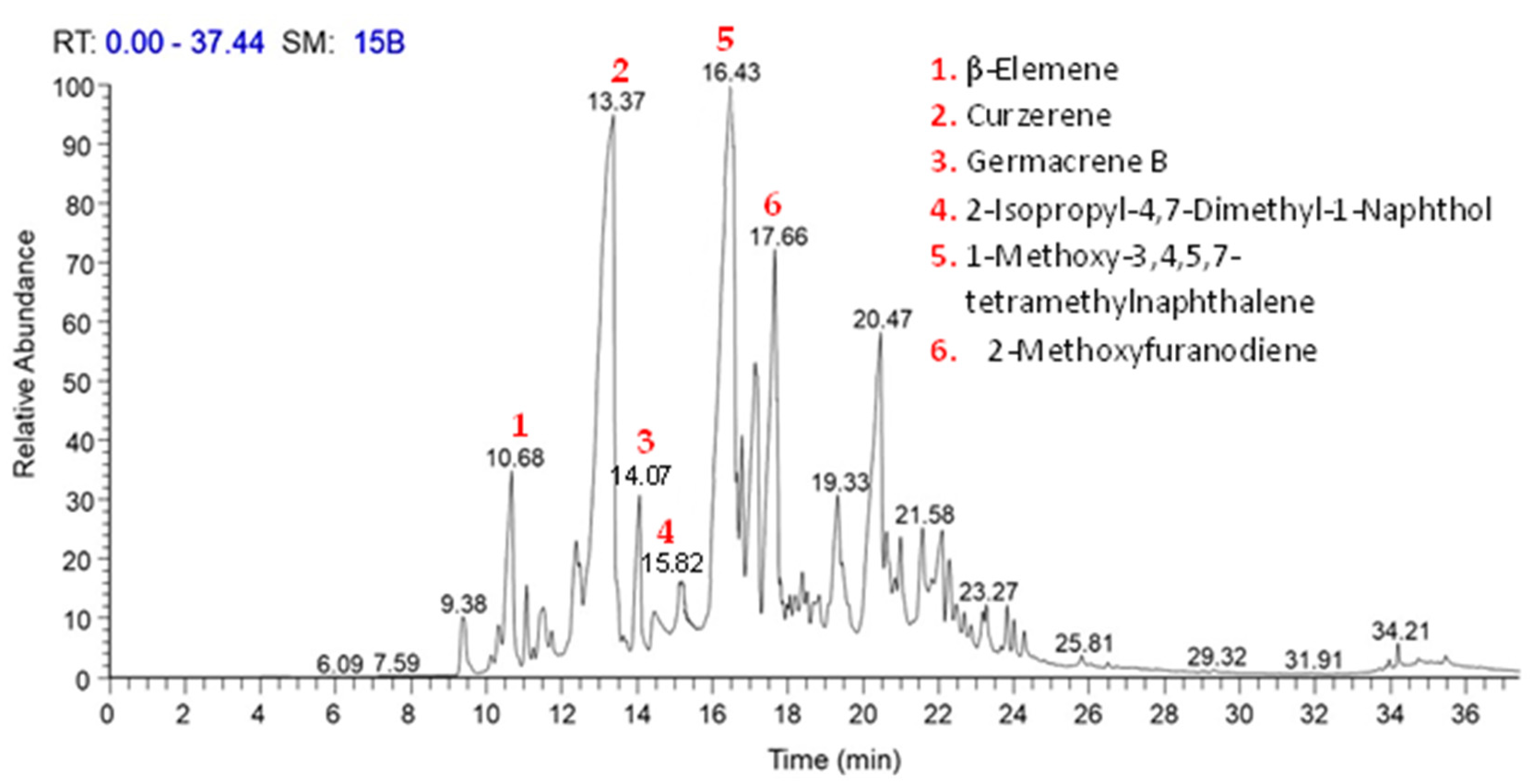
2.1. Gas Chromatography–Mass Spectroscopy “GC–MS”

The chemical constituents and structural details of the C. myrrha resin extracts were determined using gas chromatography–mass-spectrometry. Figure 1 depicts the link between the retention period at which a certain component was detected and the relative quantity of the numerous components identified by the extracted plant. The GC–MS investigation successfully identified 50 constituents, accounting for 100% of the total MeOH extract of C. myrrha resin (Table 1). Furthermore, the chemicals that were discovered underwent Electron Ionization (EI) mass spectrometry, and the results may be found in Table S1. Curzerene, identified as the main ingredient, was found to constitute 33.57% of the extract and was eluted at 13.37 min. Other substances were subsequently categorized with higher compositional percentages, including 1-Methoxy-3,4,5,7-tetramethylnaphthalene (15.50%), β-Elemene (5.80%), 2-Methoxyfuranodiene (5.42%), 2-Isopropyl-4,7-Dimethyl-1-Naphthol (4.71%), and germacrene B (4.35%). These compounds represented 69.35% of the total identified compounds (Table 1). The fatty acid derivatives’ components were observed at relatively low abundances, falling between 23.18 and 23.66 min. Meanwhile, the components of diterpenes and triterpenes had retention times ranging from 23.83 to 35.46 min. The most prevalent components of the sesquiterpene were identified within the retention time range of 9.36 to 22.69 min.
It is acknowledged that the diversity or quantity of components differs between the current investigation’s findings and the chemical profile of the previously examined myrrha extract. Nevertheless, some literature reports, such as those by Morteza-Semnani and Saeedi [34], indicate that in the Iranian C. myrrha essential oil, curzerene (40.1%), furanoeudesma-1,3-diene (15.0%), and α-elemene (8.4%) are the primary constituents. Similarly, the essential oil of Ethiopian C. myrrha, as reported by Marongiu et al. [32], is characterized by furanoeudesma-1,3-diene (38.6%), curzerene (17.5%), lindestrene (14.4%), and α-elemene (4.3%) being the primary constituents.
According to Baser et al. (2003), the primary components of Ethiopian myrrh oil are furanoeudesma-1,3-diene (34.0%), furanodiene (19.7%), and lindestrene (12.0%). Additionally, Mohamed et al. [35] identified major constituents in myrrh oil, including α-elemene (12.86%), 7-isopropyl-1,4-dimethyl-2-azulenol (12.22%), curzerene (11.64%), germacra-1(10)7,11-trien-15-oic acid, 8,12-epoxy-6-hydroxy-ç-lactone (6.20%), δ-elemene (5.57%), δ-neoclovene (5.57%), germacrene B (3.97%), and eremophilene (3.35%). Significantly, the present investigation did not identify the presence of furanoeudesma-1,3-diene, furanoeudesma-1,4-diene, and lindestrene constituents. It is important to consider genetic variables and other environmental impacts during the developmental and growth phases of the wild plant, as these factors may influence the accumulation of active secondary metabolites. Certain chemicals may be created, and their concentrations significantly altered under specific circumstances [36,37].
A significant percentage (93.22%) of the MeOH extract of the C. myrrha resin employed in this study was made up of sesquiterpene hydrocarbons. We found there to be 40 separate sesquiterpene components, with curzerene being the bulk (33.57%) of the combined makeup of the isolated components. The current findings validate and expand upon earlier studies, supporting the notion that sesquiterpenoids from the Commiphora genus predominantly fall into distinct structural groups. These groups include curzerene, lindestrene, germacrane, eudesmane, guaiane, cadinane, elemane, bisabolane, and oplopane, as previously classified in studies [19,35,38]. The other classes are oxygenated hydrocarbons (3.22%), fatty acid derivatives (1.87%), diterpenes (1.48%), and triterpenes (0.61%) (Table 1). The different climatic conditions in the area, the stage of plant development, and the plant’s adaptive metabolism may be to blame for the variances in the classes of resin extracts [39]. The well-known medicinal benefits and fascinating biological activity of the Commiphora plant may be due to the presence of numerous families of sesquiterpenoids in the crude extract and oil of the plant.

2.2. Biological Characteristics of the Plant Extracts

2.2.1. Effect of the Plant-Resin Extracts on Larvae

Plant extracts, particularly those rich in phytochemicals, offer a valuable and underutilized resource for mosquito control. Their potential as natural alternatives to synthetic insecticides has been highlighted by numerous studies demonstrating efficacy against mosquito larvae [40,41]. This larvicidal activity is likely linked to the diverse chemical composition of secondary metabolites found in plants. In the present study, we investigated the larvicidal effects of the methanol (MeOH) extract of C. myrrha resin against third-instar Aedes aegypti larvae. Our results demonstrated a concentration-dependent increase in larval and larval-pupal mortality with increasing exposure time. The highest observed mortality rates were 95% and 98.33% for larvae and larval pupae, respectively (Table 2). However, pupal mortality remained moderate, reaching only 5.0% at 1000 ppm after 24 h (Table 2).
Interestingly, adult emergence was either normal or faster in the control group (untreated; 96.67%) compared to the plant-resin extract treatments, where adult emergence was reduced to 5% and 15% at 1000 ppm and 500 ppm, respectively (Table 2). These findings suggest the potential for the MeOH extract to disrupt mosquito development beyond just larvicidal activity. Our observations align with reports by Baranitharan and Dhanasekaran [42], who found ethyl acetate and chloroform extracts of Commiphora caudata resin to be highly effective against larvae of A. aegypti, Anopheles stephensi, and Culex quinquefasciatus. Additionally, the larvicidal activity of oil resins derived from Commiphora species has been documented against Culex pipiens [43,44] and other mosquito species [45]. Similarly, Mkangara et al. [46] reported the larvicidal activity of petroleum ether, ethyl acetate, and methanol extracts of C. swynnertonii against laboratory-reared larvae of Anopheles gambiae, Culex quinquefasciatus, and Aedes aegypti.
The environmental safety of a pesticide is crucial when used against pests and vectors. A pesticide does not have to kill a lot of the target species to be effective [47]. The data given summarize the susceptibility of the third-instar larvae of A. aegypti to the tested plant-resin extracts (Table 3). For the 24 h periods in this study, the resin extracts have an LC50 of 251.83 mg/L (the median concentration of extract that results in 50% mortality), LC90 of 923.76 mg/L (the concentration of extract that results in 90% mortality), and LC95 of 1293.35 mg/L (the concentration of extract that results in 95% mortality) (Table 3). The regression line’s slope for the A. aegypti population’s in vitro strain of the third larval stage was 2.485 ± 0.212 when resin extracts were employed. Also, the results indicated that the calculated X2 (Chi)2 was 1.109 in the treatment with the extract. These results are consistent with those obtained by Samwel et al. [45], who used Commiphora merkeri oil-resin extracts against A. aegypti, A. gambiae, and C. quinquefasciatus larvae and discovered that the LC50 values were 33.79, 31.99, and 17.70 mg/mL, respectively, after 24 h. According to Muturi et al. [44], the LC50 values for Commiphora erythraea essential oils were 19.05, 22.61, and 29.83 mg/L, whereas the LC90 values for Culex restuans, C. pipiens, and A. aegypti were 31.58, 45.04, and 51.58 mg/L, respectively. Furthermore, according to Mkangara et al. [46], the LC50 of MeOH extracts from the stem bark of Commiphora swynnertonii against A. aegypti and C. quinquefasciatus demonstrated weak activity after 24 h of exposure, with LC50 values of 1235.68 mg/L and 828.13 mg/L, respectively. Similar studies have shown that the resin extracts of C. molmol possess pesticide activity against the blowfly Lucilia sericata [48], the fowl tick Argas persicus [49], and Culex pipiens [50]. Therefore, based on the present study, it can be inferred that these previous resin extracts may serve as effective larvicidal agents for controlling populations of Aedes aegypti.
Commiphora species are widely found across sub-Saharan Africa’s tropical and subtropical regions, including Madagascar, Arabia, Iran, Pakistan, and India. By sealing wounds and assisting in defense against invading insects, illness, herbivory, and infection, the resin serves as the trees’ primary form of protection [51]. According to the current study, the average number of eggs laid is 424 eggs/10 females at 1000 mg/L MeOH resin extract as opposed to untreated groups (1454 eggs/10 females). The maximum hatching (97.00%) was recorded with a 100 mg/L concentration of resin extract, whereas a 1000 mg/L concentration of resin extract resulted in the lowest egg hatching (69.58%), and the fecundity was impacted (29.16%) (Table 4). It was also found that the methanol extract of C. myrrh resin influenced the fatality of the egg embryo, reaching 28.54% compared to the untreated groups (1.10%). Sesquiterpenes, polyphenols, aromatic terpenoids, and fatty acids found in the bark extracts of C. myrrha resin may have interfered with protein synthesis or the cross-linking of proteins, which might explain the decreased egg hatching seen in the current study. This finding is corroborated by Siddiqui and Alam [52], who reported that fresh extracts from the fruit, leaf, bark, root, and gum inhibited hatching and fecundity in Meloidogyne incognita. Similar effects were observed in the cases of the western cherry fruit fly and the peach fruit fly (Bactrocera zonata) [53,54].
Finally, our data and those of others support the hypothesis that the effectiveness of C. molmol resin extracts against insect pests may be explained by the presence of numerous secondary metabolites, including sesquiterpenes, phenols, aromatic terpenoids, fatty alcohol, curzerene, germacrene, and elemene [19,50,55]. Curzerene is a volatile, aromatic terpenoid found in many herbs and spices, such as Rhododendron species, which has been shown to possess anti-inflammatory, antibacterial, acaricidal, and cytotoxicity properties in vitro [56]. As per Muturi et al. [40], both oleo-resin extract and essential oil derived from the genus Commiphora demonstrated toxicity against larvae of A. caspius and C. pipiens. This toxicity was attributed to the presence of bioactive chemical constituents such as curzerene, germacrene, eremophilene, and elemene.

2.2.2. Antioxidant Activity—DPPH Assay

The antioxidant activity of C. myrrha resin methanol (MeOH) extract at different concentrations (5–50 mg/L) was assessed by comparing it to ascorbic acid in terms of its ability to scavenge DPPH free radicals. Concentration-dependent scavenging activity was clearly demonstrated. At 50 mg/L, the inhibition percent of MeOH extracts was determined as 75.06% (Table 5). In the findings, the DPPH radical scavenging effects of the standard and plant extracts are shown using half-maximum inhibitory concentration (IC50) values. More effective DPPH radical scavenging capability is indicated by a lower IC50 value. With an IC50 of 26.86 mg/L, the findings, as shown in Table 5, supported the assumption that the resin extract exhibited the strongest antioxidant scavenging activity.
The MeOH extract of C. myrrha is rich in sesquiterpenoids, diterpenes, triterpenes, and sterols, which are known for their potential antioxidant properties. These classes of compounds can act as electron donors, potentially contributing to the antioxidant activity by neutralizing free radicals. These compounds serve as electron donors, effectively neutralizing free radicals by converting them into more stable substances and preventing further radical chain reactions. Earlier investigations conducted by Fraternale et al. [57] and Mohamed et al. [35] support this claim, demonstrating that the MeOH and hexane extracts of myrrha resin had the most significant DPPH radical scavenging activity in comparison to its oils. The authors attributed this finding to three specific furano-sesquiterpenoids (myrhone, 3-methoxy-furanogermacradien-6-one, and 2-methoxy-furanogermacren-6-one) found in the MeOH and hexane extracts of C. myrrha. These extracts exhibited the most-potent DPPH radical scavenging action. Furthermore, the ability of bioactive compounds, such as phenolics, fatty acids, terpenes, oxygenated hydrocarbons, or carbohydrates, to eliminate or stabilize free radicals is often used to assess their antioxidant activity [3,58]. A further investigation found that the methanol extract of Boswellia sacra resin had a reduced ability to neutralize DPPH radicals compared to the antioxidant activity of the resin essential oil at the same dose [59]. Curcuma and Eugenia uniflora oils have shown significant curzerene antioxidant action against the DPPH radical [60,61].

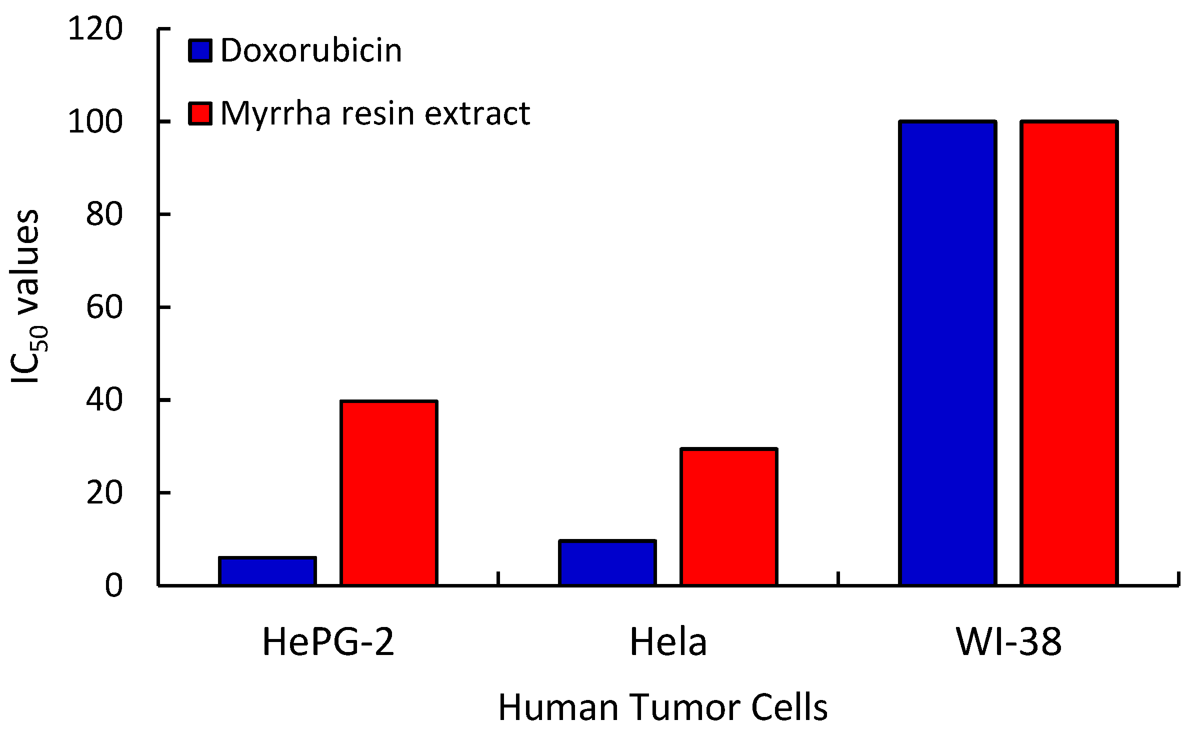
2.2.3. Cytotoxic Activity

Currently, cancer is a prominent public health issue with a substantial worldwide influence on both developed and developing nations, and the prevalence of cancer-related factors is increasing [62,63]. An MTT test was used in this study to assess the cytotoxic properties of the produced plant-resin extracts. The materials were examined in vitro against the HepG-2 and Hela cell lines, two types of tumor cells (Table 6). To assess the results of the tested samples in relation to different cancer cells, doxorubicin was used as a guide medicine. The metabolic activity of the cells was evaluated using the strategy of using cellular oxidoreductase enzymes to convert the MTT tetrazolium dye into insoluble formazan, which exhibits a purple hue. The quantity of viable cells is expected to augment during growth, decrease during cytotoxic therapies, and stay constant (or reach a plateau) during cytostatic treatments. In control trials, samples containing only the appropriate amounts of blank solutions were employed, and they had no effect on cell growth. The control sample is advantageous for determining the percentage of cell viability since it yields 100% vitality of healthy cells. Scientific data suggest that phytochemicals possess substantial anticancer capabilities. Around half of the anticancer medications that were authorized between 1940 and 2014 were derived from natural sources [64,65]. The anticancer activities of resin extracts naturally occurring in wild plants against HGC-27, KMBC, PANC-1, CRL-1739, and COLO-205 cell lines have been recently reported [33,66,67].
Herein, five concentrations of the crude methanolic extracts of plant resin (1.56, 3.125, 6.25, 12.50, 25, 50, and 100 µg/mL) generated by a series of serial dilution were used in the experiments to determine the IC50 value (concentration (μg/mL) that causes 50% cell death). On the HepG-2 and Hela cell lines, the % growth inhibition was shown to be steadily increasing with an increasing concentration up to 100 µg mL−1. For HepG-2 and Hela human tumor cells and normal cells (WI-38), the extract of C. myrrha resin showed inhibitory activities of 94.21%, 92.44%, and 10.11%, respectively. However, among all samples, the lowest concentration (1.56 µg/mL) exhibits the least amount of cytotoxic action (Table 6). The IC50 value of this assay for HepG-2 cells pretreated with MeOH extracts of plant resin was 39.73 μg/mL and the R2 value was 0.984. In the case of Hela cells, the IC50 value was 29.41 μg/mL and the R2 value was 0.996. On the other hand, doxorubicin was used as a standard drug to evaluate the effects of the compounds under investigation on the various cancer cells, which produced IC50 values for HepG-2, Hela, and WI-38 of 6.03, 9.60, and >100 µg/mL, respectively (Table 6 and Figure 2). These findings are consistent with those presented by Wang et al. [68], who suppressed HGC-27 (human gastric carcinoma cells) cells with an IC50 of 27.51 μg/mL to investigate the cytotoxic impact of a sesquiterpenoid from the Kenya ecospecies of C. myrrha resin extract. In the current investigation, a significant fraction (93.22%) of the C. myrrha resin MeOH extract was made up of sesquiterpene hydrocarbons. In similar work on resin extracts from other plant species, Alipanah and Zareian [69] and Wang et al. [70] discovered that boswellic acid derived from the gum resin of Boswellia serrata may effectively suppress the growth of the 4T1 cell line (breast cancer cells) and HCT-116 (colon cancer cells). The IC50 values, determined using the MTT test, were found to be 92.3 μg/mL and 15 μg/mL, respectively.
In order to evaluate the toxicity of the plant extract on normal cells and determine its potential for therapeutic use, the selectivity index (SI) was computed. Elevated SI values arise from substantial disparities in the cytotoxicity between cancerous and normal cells, indicating that cancerous cells will experience a more rapid demise compared to normal cells. In this study, the selectivity index values for HepG-2 and Hela cancer cells were 2.52 and 3.40, respectively (Table 6). These results showed that C. myrrha resin extract has high cytotoxic selectivity (SI > 1) for human cancer cells, indicating the need for further, in-depth study of the resin extract. For Hela cells, especially high SI values were attained.
The resin obtained from this plant has non-cytotoxic effects on normal cells (WI-38) and shows moderate cytotoxicity against two types of human tumor cells (HepG-2 and Hela), as shown by the IC50 values. It is important to note that the toxicity of extracted samples as agents that kill HepG2 and Hela tumor cells is often linked to the structural characteristics (such as size, aggregation, and surface shape) of the components in each extract, as well as the concentration and type of cancer cell line [71]. This result is consistent with the findings of [3] and Salama et al. [72], who investigated the anticancer effects of MeOH extracts derived from Rumex vesicarius and Reichardia tingitana. The extracts were evaluated in vitro against HepG-2 and PC3 cells using the MTT assay.

3. Materials and Methods

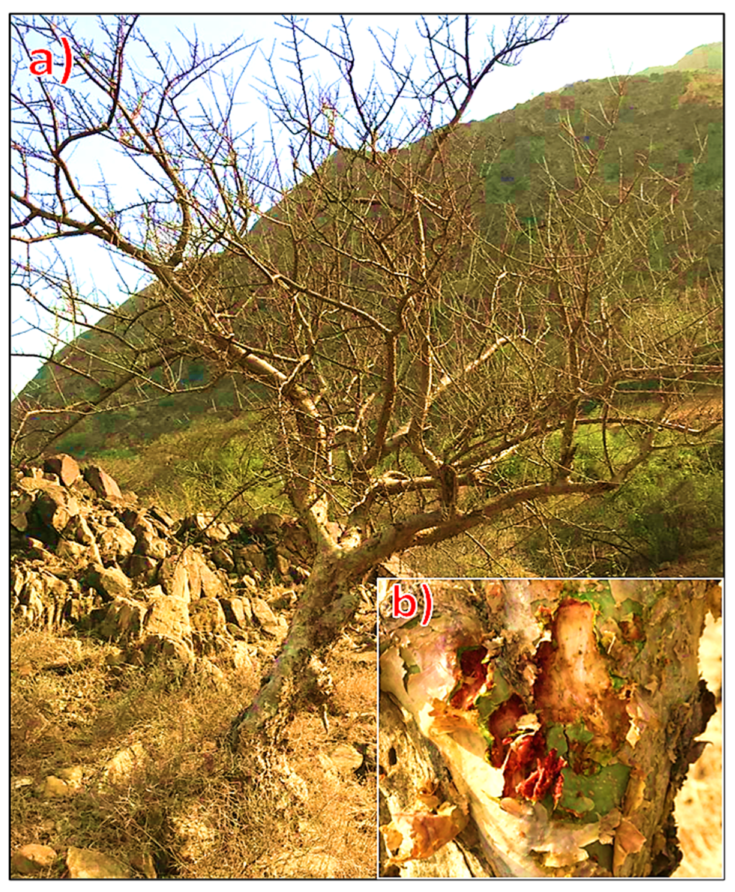
3.1. Plants Materials

The gum resin of the Commiphora myrrha (Nees) (Engl. (C. myrrha)) tree was gathered in May 2022 from several locations in Wadi Lajab (GPS: 17°35′ N, 42°55′ E), southwest of the Jazan region, Saudi Arabia, by scratching the tree bark (Figure 3). The plant was identified according to Collenette [73]. At the Biology Department, Faculty of Science, Flora and Phytotaxonomic Section, Jazan University, plant resins were identified.

3.2. Preparation of Crude Extracts

Commiphora myrrha resin (25 g) was ground into a fine powder and macerated separately in glass bottles with 150 mL of methanol. The bottles were shaken for 24 h at room temperature in an orbital shaker (Heidolph® Unimax Orbital Shakers 1010, Schwabach, Germany). Whatman No. 4 filter paper was used to filter the extracts. With new aliquots of the same solvent, residues were extracted twice. To obtain methanolic extract (3.86 g, 15.44%, w/w), supernatants from each solvent were combined and evaporated under vacuum (HeidolphTM Instruments Hei-VAP) at 40 °C. The obtained crude extracts were utilized for further analysis after being re-dissolved in methanol at a concentration of 1 μg/μL [74].

3.3. Gas Chromatography–Mass Spectrometry (GC–MS) Analysis

The analysis of the chemical composition of the extracted resin from C. myrrha was conducted using Thermo Scientific capillary gas chromatography (Thermo Scientific, Austin, TX, USA) equipped with a direct capillary column TG-5MS (30 m × 0.25 mm × 0.25 m film thickness) [75]. The temperature of the column oven was initially held constant at 50 °C, then increased at a rate of 5 °C per minute until it reached 250 °C, where it was held for 2 min. Subsequently, it was further increased at a rate of 30 °C per minute to achieve the final temperature of 300 °C, and maintained for 2 min. Temperatures of 260 and 270 °C were maintained for the MS injector and transfer line, respectively. Helium (He) served as the carrier inert gas at a constant flow rate of 1 mL/min.
The remaining sample after solvent evaporation (the concentrated extract) is then injected into the GC using the autosampler AS1300. The EI mass spectrometry data were collected in packed scan mode over the m/z range of 50–500, with an ionization energy of 70 eV. The ion source temperature was set at 200 °C. The chemical composition of each specific plant material was determined by comparing the mass spectrum data of the various extracted plant components with those in the mass spectrometry databases WILEY 09 and NIST 14. The GC–MS analysis identified five potential components for each observed peak, and the selected structure for the recommended components was determined based on likelihood factors and the fragmentation patterns of the primary structure.

3.4. Mosquitocidal Assay

3.4.1. Aedes aegypti Colony

Aedes aegypti mosquito larvae were obtained from the Medical and Molecular Entomology Section, Biology Department, Faculty of Science, Jazan University, Saudi Arabia, and were utilized in all experiments. The mosquito larvae were cultivated in round enamel plates (24 × 19 × 10 cm) containing 1.5 L of de-chlorinated water. They were fed daily with Tetramin® fish food and powder biscuit. The colony was maintained at 27 ± 2 °C, 75 ± 5% relative humidity, and a 13:11 h (L/D) photoperiod. Adult mosquitoes were provided with a 10% sugar solution. The mosquito larvae were consistently available for testing and were kept under the same laboratory conditions.

3.4.2. Larvicidal Bioassay

The resin extracted from C. myrrha for larvicidal activity was dissolved in 0.2 mL of methanol. Various concentration ranges of each extract (100, 200, 300, 400, 500, and 1000 ppm) were prepared to assess its impact on third-instar A. aegypti larvae. Plastic cups of 200 mL capacity, containing 100 mL of dechlorinated tap water, were used for the experiment. Twenty mosquito larvae were introduced into plastic containers with different extract concentrations. Typically, three replicates were conducted for each concentration. All plastic cups were then placed in a mosquito colony under controlled conditions, and subsequent mortality was recorded. Control larvae in 100 mL of water were given 0.1 mL of methanol or two drops of Tween 80. Mortality was observed daily until adult emergence, and deceased larvae and pupae were removed from the containers [76]. The percentage of larval death at 24 h post-treatment (PT) was determined using the following equation [77]:
Larval   mortality   ( % ) = Number   of   dead   larvae Number   of   treated   larvae × 100
The count of eggs laid in both treated and untreated cups was conducted using a stereomicroscope [78]. The total number of eggs and the number laid by a single female (fecundity) were recorded. The percentage of egg hatching was calculated using the following formula:
Egg   hatching   ( % ) = Number   of   Egg   hatching Number   of   Egg   laid × 100
Similarly, the following formula was used to get the fecundity percentage:
Fecundity   ( % ) = Number   of   dead   larvae Number   of   treated   larvae × 100

3.5. Antioxidant DPPH Assay

Commiphora myrrha resin was diluted from a concentrated solution of the extracted plant in methanol to the desired amounts (5, 10, 20, 30, 40, and 50 mg L−1). The DPPH solution (1 mL, 0.135 millimolar) was introduced into each concentration of the prepared sample solution. The measured concentrations in the tested samples were the standard. The specimens were stored under ambient conditions in the absence of light for a duration of 30 min. Subsequently, the optical density at a specific wavelength of 517 nm was determined using a UV/Vis spectrophotometer (Spekol 11 spectrophotometer, analytic Jena AG, Jena, Germany). The antioxidant scavenging activities were quantified by calculating the percentages using the given equation, using a DPPH solution in methanol as the reference.
%   Inhibition = A   control     A   sample A   control × 100
The approach was implemented with little modifications compared to prior experiments [39,79]. An exponential curve [80] was used to ascertain the inhibitory concentrations (IC50, mg L−1) by examining the relationship between the concentration of the sample and the quantity of remaining DPPH radicals.

3.6. Cytotoxic Activity Procedure

Hepatocellular carcinoma (HePG-2) and epithelioid cervix carcinoma (Hela) are specific types of human tumor cell lines, and they were acquired from the ATCC, a company specializing in the production of biological goods and vaccines, located in Cairo, Egypt. Doxorubicin is a frequently used chemotherapeutic agent for treating cancer. The chemical reagents used were RPMI-1640 medium, MTT, DMSO (Sigma Co., St. Louis, MO, USA), and fetal bovine serum (FBS; Gibco Life Technologies, Paisley, UK).
The cell growth and cytotoxicity of the extracted C. myrrha were evaluated using a standard colorimetric MTT test, following the protocols outlined by Bondock et al. [81]. The conversion of MTT (2-(4,5-dimethylthiazol2-yl)-3,5-diphenyl-2H-tetrazolium bromide) from a yellow color to a purple color was accomplished by the mitochondrial succinate dehydrogenases present in live cells. The cell strains were generated by adding 10% fetal bovine serum to RPMI-1640 medium. Penicillin (100 units/mL) and streptomycin (100 g/mL) antibiotics were given to an incubator containing 5% CO2. The cell lines were placed in a 96-well plate with a density of 1.0 × 10⁴ cells per well and kept at a temperature of 37 °C for 48 h in the presence of 5% CO2. The cells were first cultivated for 24 h, after which they were subjected to different dosages of the test substances. After a 24 h drug treatment period, a 20 L solution of MTT (5 mg/mL) was added, and the mixture was incubated for an additional 4 h. In order to disperse the formed violet formazan, 100 L of DMSO was then added to each well. A colorimetric study was conducted using a plate reader (EXL 800, New York, NY, USA) to measure the absorbance values at 570 nm. The IC50 values were generated using nonlinear regression (sigmoid type) using the Origin 8.0® software developed by OriginLab Corporation (https://www.originlab.com/; Accessed 27 March 2021). The equation used to calculate the percentage of inhibition in cell growth involves the optical density (OD) values obtained from the control and testing samples, where the OD represents the absorbance readings.
%   Inhibition = OD   control OD   sample OD   control × 100
In the current study, the selectivity index (SI) was calculated using the following formula:
Selectivity   index   ( SI ) = IC 50   for   normal   cell   line   WI 38 IC 50   for   respective   cancerous   cell   line
where IC50 is the concentration (μg/mL) that causes 50% cell death.

3.7. Data Analysis

A one-way analysis of variance (ANOVA) was performed on the biological data, Duncan’s multiple range tests were performed with the help of the computer program Costat var. 6.311 (CoHort Software, Monterey, CA, USA), and Probit analysis was used to determine the potentially fatal values. Each of the studies was repeated three times, and each time there were three replications.

4. Conclusions

The global fight against mosquito-borne diseases necessitates the exploration of novel, eco-friendly pest control methods. Natural products, with their vast potential and efficacy, are attracting increasing research interest. This study contributes significantly to this field by investigating the larvicidal activity of a methanol extract from C. myrrha resin against Aedes aegypti larvae—a previously unreported application. Our investigation employed GC–MS analysis to tentatively identify 50 components within the extract, with sesquiterpene hydrocarbons being the most abundant class (93.22%). Notably, curzerene was the major constituent at 33.57%. While previous studies have documented the cytotoxic, antioxidant, and anti-dengue virus vector properties of C. myrrha resins, our focus on the specific larvicidal effect against A. aegypti larvae adds a new dimension to the understanding of the resin’s potential applications. This study highlights the novel potential of the MeOH extract of C. myrrha resin as a natural mosquito larvicide. Further research is warranted to explore the extract’s efficacy against other mosquito species and potential synergistic interactions with other plant extracts. Additionally, investigating the underlying mechanisms of larvicidal activity and exploring the extract’s safety profile are crucial steps for future development. Overall, this study contributes to the ongoing search for environmentally friendly mosquito-control strategies by pioneering the exploration of C. myrrha resin as a natural alternative.

Supplementary Materials

The following supporting information can be downloaded at: https://www.mdpi.com/article/10.3390/molecules29081778/s1, Table S1: Electron Ionization (EI) mass spectra of the detected compounds.

Author Contributions

Conceptualization, N.A.H.A., A.A.A., A.M.M., N.A., G.M. and S.A.S.; validation, N.A.H.A., A.A.A., A.M.M., N.A., G.M. and S.A.S.; formal analysis, N.A.H.A., A.A.A., A.M.M., N.A., G.M. and S.A.S.; investigation, N.A.H.A., A.A.A., A.M.M., N.A., G.M. and S.A.S. data curation, N.A.H.A., A.A.A., A.M.M., N.A., G.M. and S.A.S.; writing—original draft preparation, N.A.H.A., A.A.A., A.M.M., N.A., G.M. and S.A.S.; writing—review and editing, N.A.H.A., A.A.A., A.M.M., N.A., G.M. and S.A.S. All authors have read and agreed to the published version of the manuscript.

Funding

This work was supported by the Scientific Research Deanship at the University of Hail in Saudi Arabia (RG-23 217), which provided funding.

Data Availability Statement

The data presented in this study are available upon request from the corresponding author.

Acknowledgments

The authors extend their appreciation to the Scientific Research Deanship at the University of Hail in Saudi Arabia for funding this research work through the Project with number: RG-23 217.

Conflicts of Interest

The authors declare that there are no conflicts of interest.

References

  1. Ojewumi, M.; Banjo, M.; Oresegun, M.; Ogunbiyi, T.; Ayoola, A.; Awolu, O.; Ojewumi, E. Analytical investigation of the extract of lemon grass leaves in repelling mosquito. Int. J. Pharm. Sci. Res. 2017, 8, 2048–2055. [Google Scholar]
  2. Groucutt, H.S.; Petraglia, M.D. The prehistory of the Arabian peninsula: Deserts, dispersals, and demography. Evol. Anthropol. Issues News Rev. 2012, 21, 113–125. [Google Scholar] [CrossRef]
  3. Ghazanfar, S.A.; Böer, B.; Khulaidi, A.W.A.; El-Keblawy, A.; Alateeqi, S. Plants of Sabkha ecosystems of the Arabian Peninsula. In Sabkha Ecosystems; Springer: Berlin/Heidelberg, Germany, 2019; pp. 55–80. [Google Scholar]
  4. Das, K.; Tiwari, R.; Shrivastava, D. Techniques for evaluation of medicinal plant products as antimicrobial agent: Current methods and future trends. J. Med. Plants Res. 2010, 4, 104–111. [Google Scholar]
  5. Salama, S.A.; Al-Faifi, Z.E.; El-Amier, Y.A. Chemical composition of Reichardia tingitana methanolic extract and its potential antioxidant, antimicrobial, cytotoxic and larvicidal activity. Plants 2022, 11, 2028. [Google Scholar] [CrossRef] [PubMed]
  6. Iyiola, A.O.; Babafemi, O.P.; Ogundahunsi, O.E.; Ojeleye, A.E. Food Security: A Pathway Towards Improved Nutrition and Biodiversity Conservation. In Biodiversity in Africa: Potentials, Threats and Conservation; Springer: Berlin/Heidelberg, Germany, 2022; pp. 79–107. [Google Scholar]
  7. Chen, Z.; Tian, R.; She, Z.; Cai, J.; Li, H. Role of oxidative stress in the pathogenesis of nonalcoholic fatty liver disease. Free Radic. Biol. Med. 2020, 152, 116–141. [Google Scholar] [CrossRef] [PubMed]
  8. Bagchi, D.; Swaroop, A.; Preuss, H.G.; Bagchi, M. Free radical scavenging, antioxidant and cancer chemoprevention by grape seed proanthocyanidin: An overview. Mutat. Res./Fundam. Mol. Mech. Mutagen. 2014, 768, 69–73. [Google Scholar] [CrossRef]
  9. Abd-ElGawad, A.M.; Elshamy, A.I.; Elgorban, A.M.; Hassan, E.M.; Zaghloul, N.S.; Alamery, S.F.; El Gendy, A.E.-N.G.; Elhindi, K.M.; EI-Amier, Y.A. Essential oil of Ipomoea carnea: Chemical profile, chemometric analysis, free radical scavenging, and antibacterial activities. Sustainability 2022, 14, 9504. [Google Scholar] [CrossRef]
  10. Tandina, F.; Doumbo, O.; Traoré, S.F.; Parola, P.; Robert, V. Mosquitoes (Diptera: Culicidae) and mosquito-borne diseases in Mali, West Africa. Parasites Vectors 2018, 11, 467. [Google Scholar] [CrossRef] [PubMed]
  11. Wilson, A.L.; Courtenay, O.; Kelly-Hope, L.A.; Scott, T.W.; Takken, W.; Torr, S.J.; Lindsay, S.W. The importance of vector control for the control and elimination of vector-borne diseases. PLoS Neglected Trop. Dis. 2020, 14, e0007831. [Google Scholar] [CrossRef]
  12. WHO. Global Strategy for Dengue Prevention and Control 2012–2020; World Health Organisation: Geneva, Switzerland, 2012; p. 35. Available online: http://apps.who.int/iris/bitstream/10665/75303/1/9789241504034_eng.pdf (accessed on 30 August 2020).
  13. Bhatt, S.; Gething, P.; Brady, O.; Messina, J.; Farlow, A.; Moyes, C.; Drake, J.; Brownstein, J.; Hoen, A.; Sankoh, O. The global distribution and burden of dengue. Nature 2013, 496, 504–507. [Google Scholar] [CrossRef]
  14. Benelli, G.; Murugan, K.; Panneerselvam, C.; Madhiyazhagan, P.; Conti, B.; Nicoletti, M. Old ingredients for a new recipe? Neem cake, a low-cost botanical by-product in the fight against mosquito-borne diseases. Parasitol. Res. 2015, 114, 391–397. [Google Scholar] [CrossRef]
  15. van den Berg, H.; da Silva Bezerra, H.S.; Al-Eryani, S.; Chanda, E.; Nagpal, B.N.; Knox, T.B.; Velayudhan, R.; Yadav, R.S. Recent trends in global insecticide use for disease vector control and potential implications for resistance management. Sci. Rep. 2021, 11, 23867. [Google Scholar] [CrossRef] [PubMed]
  16. Pavela, R.; Žabka, M.; Bednář, J.; Tříska, J.; Vrchotová, N. New knowledge for yield, composition and insecticidal activity of essential oils obtained from the aerial parts or seeds of fennel (Foeniculum vulgare Mill.). Ind. Crops Prod. 2016, 83, 275–282. [Google Scholar] [CrossRef]
  17. Koureas, M.; Amoutzias, G.D.; Vontas, A.; Kyritsi, M.; Pinaka, O.; Papakonstantinou, A.; Dadouli, K.; Hatzinikou, M.; Koutsolioutsou, A.; Mouchtouri, V.A. Wastewater monitoring as a supplementary surveillance tool for capturing SARS-CoV-2 community spread. A case study in two Greek municipalities. Environ. Res. 2021, 200, 111749. [Google Scholar] [CrossRef]
  18. Daly, D.C.d.B.; Fine, P.V.A.; Martínez-Habibe, M.C. Burseraceae: A model for studying the Amazon flora. Rodriguésia 2012, 63, 21–30. [Google Scholar] [CrossRef]
  19. Shen, T.; Li, G.-H.; Wang, X.-N.; Lou, H.-X. The genus Commiphora: A review of its traditional uses, phytochemistry and pharmacology. J. Ethnopharmacol. 2012, 142, 319–330. [Google Scholar] [CrossRef] [PubMed]
  20. DeCarlo, A.; Dosoky, N.S.; Satyal, P.; Sorensen, A.; Setzer, W.N. The essential oils of the Burseraceae. In Essential Oil Research; Springer: Berlin/Heidelberg, Germany, 2019; pp. 61–145. [Google Scholar]
  21. Su, S.; Hua, Y.; Wang, Y.; Gu, W.; Zhou, W.; Duan, J.-A.; Jiang, H.; Chen, T.; Tang, Y. Evaluation of the anti-inflammatory and analgesic properties of individual and combined extracts from Commiphora myrrha, and Boswellia carterii. J. Ethnopharmacol. 2012, 139, 649–656. [Google Scholar] [CrossRef] [PubMed]
  22. Steyn, M. Southern Africa Commiphora; United Litho: Arcadia, South Africa, 2003. [Google Scholar]
  23. Alsharif, K. Potential anti-inflammatory properties effect of myrrh. Lett. Appl. NanoBioSci. 2020, 9, 1687–1694. [Google Scholar]
  24. Abdel-Hay, M.H.; Saleh, A.; El Ashry, E.; Rashed, N.; Salama, O. Colorimetric determination of crude powdered myrrh, purified myrrh extract, oily fraction, and its different pharmaceutical dosage forms. Spectrosc. Lett. 2002, 35, 183–197. [Google Scholar] [CrossRef]
  25. Abdul-Ghani, R.A.; Loutfy, N.; Hassan, A. Myrrh and trematodoses in Egypt: An overview of safety, efficacy and effectiveness profiles. Parasitol. Int. 2009, 58, 210–214. [Google Scholar] [CrossRef]
  26. Tipton, D.; Lyle, B.; Babich, H.; Dabbous, M.K. In vitro cytotoxic and anti-inflammatory effects of myrrh oil on human gingival fibroblasts and epithelial cells. Toxicol. Vitr. 2003, 17, 301–310. [Google Scholar] [CrossRef] [PubMed]
  27. Massoud, A.; El Ebiary, F.H.; Abou-Gamra, M.; Mohamed, G.F.; Shaker, S.M. Evaluation of schistosomicidal activity of myrrh extract: Parasitological and histological study. J. Egypt. Soc. Parasitol. 2004, 34, 1051–1076. [Google Scholar] [PubMed]
  28. Hassanzadeh-Taheri, M.; Salimi, M.; Vazifeshenas-Darmiyan, K.; Mohammadifard, M.; Hosseini, M. Investigating the effect of ethanolic extract of Commiphora myrrha (Nees) Engl. gum-resin against hepatorenal injury in diabetic rats. J. Diabetes Metab. Disord. 2021, 20, 1573–1581. [Google Scholar] [CrossRef]
  29. Madia, V.N.; De Angelis, M.; De Vita, D.; Messore, A.; De Leo, A.; Ialongo, D.; Tudino, V.; Saccoliti, F.; De Chiara, G.; Garzoli, S. Investigation of Commiphora myrrha (Nees) Engl. oil and its main components for antiviral activity. Pharmaceuticals 2021, 14, 243. [Google Scholar] [CrossRef]
  30. Dekebo, A.; Lang, M.; Polborn, K.; Dagne, E.; Steglich, W. Four lignans from Commiphora erlangeriana. J. Nat. Prod. 2002, 65, 1252–1257. [Google Scholar] [CrossRef] [PubMed]
  31. Başer, K.; Demirci, B.; Dekebo, A.; Dagne, E. Essential oils of some Boswellia spp., myrrh and opopanax. Flavour Fragr. J. 2003, 18, 153–156. [Google Scholar] [CrossRef]
  32. Marongiu, B.; Piras, A.; Porcedda, S.; Scorciapino, A. Chemical composition of the essential oil and supercritical CO2 extract of Commiphora myrrha (Nees) Engl. and of Acorus calamus L. J. Agric. Food Chem. 2005, 53, 7939–7943. [Google Scholar] [CrossRef]
  33. Rehman, N.U.; Halim, S.A.; Khan, A.; Khan, M.; Al-Hatmi, S.; Al-Harrasi, A. Commikuanoids AC: New cycloartane triterpenoids with exploration of carbonic anhydrase-II inhibition from the resins of Commiphora kua by in vitro and in silico molecular docking. Fitoterapia 2022, 157, 105125. [Google Scholar] [CrossRef]
  34. Morteza-Semnani, K.; Saeedi, M. Constituents of the essential oil of Commiphora myrrha (Nees) Engl. var. molmol. J. Essent. Oil Res. 2003, 15, 50–51. [Google Scholar] [CrossRef]
  35. Mohamed, A.A.; Ali, S.I.; EL-Baz, F.K.; Hegazy, A.K.; Kord, M.A. Chemical composition of essential oil and in vitro antioxidant and antimicrobial activities of crude extracts of Commiphora myrrha resin. Ind. Crops Prod. 2014, 57, 10–16. [Google Scholar] [CrossRef]
  36. Liu, W.; Liu, J.; Yin, D.; Zhao, X. Influence of ecological factors on the production of active substances in the anti-cancer plant Sinopodophyllum hexandrum (Royle) TS Ying. PLoS ONE 2015, 10, e0122981. [Google Scholar]
  37. Abd-ElGawad, A.M.; El-Amier, Y.A.; Assaeed, A.M.; Al-Rowaily, S.L. Interspecific variations in the habitats of Reichardia tingitana (L.) Roth leading to changes in its bioactive constituents and allelopathic activity. Saudi J. Biol. Sci. 2020, 27, 489–499. [Google Scholar] [CrossRef] [PubMed]
  38. Racine, P.; Auffray, B. Quenching of singlet molecular oxygen by Commiphora myrrha extracts and menthofuran. Fitoterapia 2005, 76, 316–323. [Google Scholar] [CrossRef] [PubMed]
  39. Abd-ElGawad, A.M.; Elshamy, A.I.; El-Amier, Y.A.; El Gendy, A.E.-N.G.; Al-Barati, S.A.; Dar, B.A.; Al-Rowaily, S.L.; Assaeed, A.M. Chemical composition variations, allelopathic, and antioxidant activities of Symphyotrichum squamatum (Spreng.) Nesom essential oils growing in heterogeneous habitats. Arab. J. Chem. 2020, 13, 4237–4245. [Google Scholar] [CrossRef]
  40. Ghosh, A.; Chowdhury, N.; Chandra, G. Plant extracts as potential mosquito larvicides. Indian J. Med. Res. 2012, 135, 581. [Google Scholar] [PubMed]
  41. Chan, C.A.; Ho, L.Y.; Sit, N.W. Larvicidal activity and phytochemical profiling of sweet basil (Ocimum basilicum L.) leaf extract against asian tiger mosquito (Aedes albopictus). Horticulturae 2022, 8, 443. [Google Scholar] [CrossRef]
  42. Baranitharan, M.; Dhanasekaran, S. Mosquito larvicidal properties of Commiphora caudata (Wight &Arn.) (Bursaceae) against Aedes aegypti (Linn.), Anopheles stephensi (Liston), Culex quinquefasciatus (Say). Int. J. Curr. Microbiol. App. Sci. 2014, 3, 262–268. [Google Scholar]
  43. Baranitharan, M.; Dhanasekaran, S.; Gokulakrishnan, J.; Mahesh Babu, S.; Thushimenan, S. Nagapattinam medicinal plants against the dengue fever mosquito, Aedes aegypti. Int. J. Mosq. Res. 2016, 3, 29–34. [Google Scholar]
  44. Muturi, E.J.; Hay, W.T.; Doll, K.M.; Ramirez, J.L.; Selling, G. Insecticidal activity of Commiphora erythraea essential oil and its emulsions against larvae of three mosquito species. J. Med. Entomol. 2020, 57, 1835–1842. [Google Scholar] [CrossRef]
  45. Samwel, B.; Innocent, E.; Machumi, F.; Kisinza, W.N.; Heydenreich, M. Two mosquito larvicidal arabinofuranosidetridecanol from Commiphora merkeri exudate. Nat. Prod. Res. 2022, 36, 2821–2829. [Google Scholar] [CrossRef]
  46. Mkangara, M.; Chacha, M.; Kazyoba, P.E. Larvicidal potential of Commiphora swynnertonii (Burtt) stem bark extracts against Anopheles gambiae ss, Culex quinquefasciatus Say and Aedes aegypti L. Int. J. Sci. Res. 2015, 4, 356–361. [Google Scholar]
  47. Kabaru, J.; Gichia, L. Insecticidal activity of extracts derived from different parts of the mangrove tree Rhizophora mucronata (Rhizophoraceae) Lam. against three arthropods. Afr. J. Sci. Technol. 2001, 2, 44–49. [Google Scholar] [CrossRef]
  48. Hoda, S.M.; Fahmy, M.; Attia, M.; Rabab, M.; Shalaby, H.; Massoud, A. The insecticidal activity of two medicinal plants (Commiphora molmol) and (Balanites aegyptiaca) against the blowfly Lucilia sericata (Diptera: Calliphoridae). Int. J. Adv. Res. Biol. Sci. 2016, 3, 144–158. [Google Scholar]
  49. Massoud, A.; Kutkat, M.; El-Khateeb, R.; Labib, I. Acaricidal efficacy of myrrh (Commiphora molmol) on the fowl tick Argas persicus (Acari: Argasidae). J. Egypt. Soc. Parasitol. 2005, 35, 667–686. [Google Scholar] [PubMed]
  50. Baz, M.M.; Hegazy, M.M.; Khater, H.F.; El-Sayed, Y.A. Comparative Evaluation of five oil-resin plant extracts against the mosquito larvae, Culex pipiens Say (Diptera: Culicidae). Pak. Vet. J. 2021, 41, 191–196. [Google Scholar]
  51. Pichersky, E.; Raguso, R.A. Why do plants produce so many terpenoid compounds? New Phytol. 2018, 220, 692–702. [Google Scholar] [CrossRef] [PubMed]
  52. Siddiqui, M.; Alam, M. Further studies on the nematode toxicity of different parts of Margosa and Persian lilac. Neem Newsl. 1985, 2, 43–47. [Google Scholar]
  53. Renden, E.; Roither, B.; Randan, E. Effect of a neem Azadirachta indica based insecticides on oviposition deterrence, survival, behavior and reproduction of adult western cherry fruit fly (Diptera: Tephritidae). J. Econ. Entomol. 1998, 91, 123–130. [Google Scholar]
  54. Mahmoud, M.F.; Shoeib, M.A. Sterilant and oviposition deterrent activity of neem formulation on Peach fruit fly Bactrocera zonata (Saunders) (Diptera: Tephritidae). J. Biopestic. 2008, 1, 177–181. [Google Scholar]
  55. Hanuš, L.O.; Řezanka, T.; Dembitsky, V.M.; Moussaieff, A. Myrrh-Commiphora chemistry. Biomed. Pap. 2005, 149, 3–28. [Google Scholar] [CrossRef]
  56. He, J.; Shang, X.; Dai, L.; Yang, X.; Li, B.; Wei, Y.; Zhang, J.; Pan, H. Chemical constituents, antibacterial, acaricidal and anti-inflammatory activities of the essential oils from four Rhododendron species. Front. Vet. Sci. 2022, 9, 882060. [Google Scholar] [CrossRef]
  57. Fraternale, D.; Sosa, S.; Ricci, D.; Genovese, S.; Messina, F.; Tomasini, S.; Montanari, F.; Marcotullio, M.C. Anti-inflammatory, antioxidant and antifungal furanosesquiterpenoids isolated from Commiphora erythraea (Ehrenb.) Engl. resin. Fitoterapia 2011, 82, 654–661. [Google Scholar] [CrossRef] [PubMed]
  58. Okwu, D.E. Citrus fruits: A rich source of phytochemicals and their roles in human health. Int. J. Chem. Sci. 2008, 6, 451–471. [Google Scholar]
  59. Al-Harrasi, A.; Ali, L.; Ceniviva, E.; Al-Rawahi, A.; Hussain, J.; Hussain, H.; Rehman, N.U.; Abbas, G.; Al-Harrasi, R. Antiglycation and antioxidant activities and HPTLC analysis of Boswellia sacra oleogum resin: The sacred frankincense. Trop. J. Pharm. Res. 2013, 12, 597–602. [Google Scholar] [CrossRef]
  60. Li, Z.-H.; Wang, Q.; Ruan, X.; Pan, C.-D.; Jiang, D.-A. Phenolics and plant allelopathy. Molecules 2010, 15, 8933–8952. [Google Scholar] [CrossRef] [PubMed]
  61. da Costa, J.S.; Barroso, A.S.; Mourão, R.H.V.; da Silva, J.K.R.; Maia, J.G.S.; Figueiredo, P.L.B. Seasonal and antioxidant evaluation of essential oil from Eugenia uniflora L. curzerene-rich, thermally produced in situ. Biomolecules 2020, 10, 328. [Google Scholar] [CrossRef] [PubMed]
  62. Tavakoli, J.; Miar, S.; Zadehzare, M.M.; Akbari, H. Evaluation of effectiveness of herbal medication in cancer care: A review study. Iran. J. Cancer Prev. 2012, 5, 144. [Google Scholar] [PubMed]
  63. Burgio, E.; Piscitelli, P.; Migliore, L. Ionizing radiation and human health: Reviewing models of exposure and mechanisms of cellular damage. An epigenetic perspective. Int. J. Environ. Res. Public Health 2018, 15, 1971. [Google Scholar] [CrossRef]
  64. Newman, D.J.; Cragg, G.M. Natural products as sources of new drugs from 1981 to 2014. J. Nat. Prod. 2016, 79, 629–661. [Google Scholar] [CrossRef]
  65. Choudhari, A.S.; Mandave, P.C.; Deshpande, M.; Ranjekar, P.; Prakash, O. Phytochemicals in cancer treatment: From preclinical studies to clinical practice. Front. Pharmacol. 2020, 10, 1614. [Google Scholar] [CrossRef]
  66. Lin, H.; Suhail, M.; Fung, K.; Woolley, C.; Young, D. Extraction of biologically active compounds by hydrodistillation of Boswellia species gum resins for anti-cancer therapy. OA Altern. Med. 2013, 1, 4. [Google Scholar] [CrossRef]
  67. Mohammed, Y. In-vitro anti-cancer activity of extracts Dracaen cinnabari Balf. F resin from Socotra Island in Yemen Republic. Biochem. Anal. Biochem. 2016, 5, 3. [Google Scholar]
  68. Wang, C.-C.; Liang, N.-Y.; Xia, H.; Wang, R.-Y.; Zhang, Y.-F.; Huo, H.-X.; Zhao, Y.-F.; Song, Y.-L.; Zheng, J.; Tu, P.-F. Cytotoxic sesquiterpenoid dimers from the resin of Commiphora myrrha Engl. Phytochemistry 2022, 204, 113443. [Google Scholar] [CrossRef] [PubMed]
  69. Alipanah, H.; Zareian, P. Anti-cancer properties of the methanol extract of Boswellia serrata gum resin: Cell proliferation arrest and inhibition of angiogenesis and metastasis in BALB/c mice breast cancer model. Physiol. Pharmacol. 2018, 22, 183–194. [Google Scholar]
  70. Wang, D.; Ge, S.; Bai, J.; Song, Y. Boswellic acid exerts potent anticancer effects in HCT-116 human colon cancer cells mediated via induction of apoptosis, cell cycle arrest, cell migration inhibition and inhibition of PI3K/AKT signalling pathway. J. BUON 2018, 23, 340–345. [Google Scholar] [PubMed]
  71. Khacha-Ananda, S.; Tragoolpua, K.; Chantawannakul, P.; Tragoolpua, Y. Antioxidant and anti-cancer cell proliferation activity of propolis extracts from two extraction methods. Asian Pac. J. Cancer Prev. 2013, 14, 6991–6995. [Google Scholar] [CrossRef] [PubMed]
  72. Salama, S.A.; Al-Faifi, Z.E.; Masood, M.F.; El-Amier, Y.A. Investigation and biological assessment of Rumex vesicarius L. extract: Characterization of the chemical components and antioxidant, antimicrobial, cytotoxic, and anti-dengue vector activity. Molecules 2022, 27, 3177. [Google Scholar] [CrossRef]
  73. Collenette, S. Wild Flowers of Saudi Arabia; National Commission for Wildlife Conservation and Development (NCWCD): Riyadh, Saudi Arabia, 1999; p. 779. [Google Scholar]
  74. Sultana, B.; Anwar, F.; Ashraf, M. Effect of extraction solvent/technique on the antioxidant activity of selected medicinal plant extracts. Molecules 2009, 14, 2167–2180. [Google Scholar] [CrossRef]
  75. de Dobbeleer, I.; Gummersbach, J.; Huebschmann, H.-J.; Mayer, A.; Silcock, P. Analyzing PBDEs in House Dust Samples with the Thermo Scientific TSQ Quantum XLS Ultra GC-MS/MS in EI-SRM Mode; Thermo Fisher Scientific: Dreieich, Germany, 2012; pp. 1–6. [Google Scholar]
  76. El-Sheikh, T.M.; Hassan, M.I.; Moselhy, W.A.; Amer, M.S.; Shehata, A.Z. Evaluation of the biological activity of some Cupressus semprevirens (Cupressaceae) extracts against the mosquito vector Culex pipiens L. (Diptera: Culicidae). Egypt. Acad. J. Biol. Sci. A Entomol. 2011, 4, 33–48. [Google Scholar] [CrossRef]
  77. Briggs, J.D. Reduction of adult house-fly emergence by the effects of Bacillus spp. on the development of immature forms. J. Insect Pathol. 1960, 2, 418–432. [Google Scholar]
  78. Cheah, S.-X.; Tay, J.-W.; Chan, L.-K.; Jaal, Z. Larvicidal, oviposition, and ovicidal effects of Artemisia annua (Asterales: Asteraceae) against Aedes aegypti, Anopheles sinensis, and Culex quinquefasciatus (Diptera: Culicidae). Parasitol. Res. 2013, 112, 3275–3282. [Google Scholar] [CrossRef] [PubMed]
  79. Miguel, M.G. Antioxidant activity of medicinal and aromatic plants. A review. Flavour Fragr. J. 2010, 25, 291–312. [Google Scholar] [CrossRef]
  80. Parejo, I.; Codina, C.; Petrakis, C.; Kefalas, P. Evaluation of scavenging activity assessed by Co(II)/EDTA-induced luminol chemiluminescence and DPPH·(2,2-diphenyl-1-picrylhydrazyl) free radical assay. J. Pharmacol. Toxicol. Methods 2000, 44, 507–512. [Google Scholar] [CrossRef] [PubMed]
  81. Bondock, S.; Adel, S.; Etman, H.A.; Badria, F.A. Synthesis and antitumor evaluation of some new 1,3,4-oxadiazole-based heterocycles. Eur. J. Med. Chem. 2012, 48, 192–199. [Google Scholar] [CrossRef] [PubMed]
Figure 1. Chromatogram of the methanol extract of C. myrrha resins by GC–MS.
Molecules 29 01778 g001
Figure 2. IC50 values of the tested plant-resin extracts and doxorubicin as standard against human cancer and normal cells.
Molecules 29 01778 g002
Figure 3. Commiphora myrrha (Nees) Engl. (a) overview of the growing tree, (b) a close-up of tree bark demonstrating resin accumulation from scraping.
Molecules 29 01778 g003
Table 1. The characterized chemical components identified from C. myrrha resins.
No.CompoundRT a (min.)SI bConc.% cMolecular WeightMolecular Formula
 Sesquiterpenes hydrocarbons
1δ-Elemene9.365481.47 ± 0.03204C15H24
2α-Copaene 10.128190.24 ± 0.01204C15H24
3β-Bourbonene10.326740.71 ± 0.01204C15H24
4β-Elemene10.687495.80 ± 0.08204C15H24
5Caryophyllene11.075651.52 ± 0.03204C15H24
6β-Copaene11.247400.18 ± 0.01204C15H24
7γ-Elemene11.517921.68 ± 0.02204C15H24
8α-Humulene11.739170.35 ± 0.01204C15H24
9Germacrene D12.397042.35 ± 0.02204C15H24
10Aromandendrene12.476200.74 ± 0.01204C15H24
11γ-Gurjunene12.647310.11 ± 0.01204C15H24
12Curzerene13.3796133.57 ± 0.32216C15H20O
13Cubebol13.455290.13 ± 0.00222C15H26O
14α-Elemene13.505790.22 ± 0.01204C15H24
15(+)-Ledene13.646140.14 ± 0.01204C15H24
16Selina-3,7(11)-diene13.738570.13 ± 0.01204C15H24
17Germacrene B14.076364.35 ± 0.06204C15H24
18Furanoeudesma-1,4-diene14.445811.01 ± 0.02214C15H18O
19Furanoeudesma-1,3-diene14.986030.08 ± 0.01214C15H18O
20Lindestrene15.305800.45 ± 0.01214C15H18O
212-Isopropyl-4,7-Dimethyl-1-Naphthol 15.825354.71 ± 0.07214C15H18O
221-Methoxy-3,4,5,7-tetramethylnaphthalene16.4384715.50 ± 0.25214C15H18O
23Furanoelemene16.655930.49 ± 0.02216C15H20O
24Eremophilene16.806372.61 ± 0.12216C15H24
25Lindera-lactone17.145630.35 ± 0.01246C16H18O3
262-Methoxyfuranodiene17.665625.42 ± 0.08246C16H22O2
27β-Guaiene17.996220.21 ± 0.01204C15H24
28Cycloisolongifol-5-ol18.076380.32 ± 0.02220C15H24O
29γ-Eudesmol acetate 18.395360.99 ± 0.03264C17H28O2
30Isovalencenol18.516840.58 ± 0.02220C15H24O
3112-Methoxy-19-norpodocarpa-8,11,13-triene18.676550.50 ± 0.02244C17H24O
32Gazaniolide19.455500.24 ± 0.01230C15H18O2
33Furosardonin A20.095940.29 ± 0.01232C15H20O2
34Furosardonin B20.639241.21 ± 0.03232C15H20O2
35Bohlmann k2631 20.867130.28 ± 0.03232C15H20O2
366-(3-hydroxyprop-1-en-2-yl)-4,8a-dimethyl-3-oxo-1,2,3,5,6,7,8,8a-octahydronaphthalen-2-yl acetate21.016421.46 ± 0.02292C17H24O4
374-a-Methyl-1-methylene-1,2,3,4,4a,9,10,10a-octahydrophenanthrene21.585901.76 ± 0.03212C16H20
388,9-Dehydro-9-vinyl- cycloisolongifolene 21.825720.17 ± 0.01228C17H24
39Reynosin 22.486880.50 ± 0.02248C15H20O3
40Beta-Doradecin 22.695560.40 ± 0.01320C18H24O5
 Oxygenated hydrocarbons
413-Ethyl-2,6-naphthlenediol17.738391.58 ± 0.02188C12H12O2
423-Ethyl-6-(Methoxycarbonyl)-2-Naphthol 22.307031.64 ± 0.03230C14H14O3
 Fatty acid derivatives
439,12(Z,Z)-Octadecadienoic Acid, methyl ester 23.186360.80 ± 0.02294C19H34O2
449-Octadecenoic acid, methyl ester23.287930.99 ± 0.01296C19H36O2
45Octadecenoic acid, methyl ester23.666680.08 ± 0.01298C19H38O2
 Diterpenes
46(3E,7E,11E)-1-Isopropyl-4,8,12-trimethylcyclo- tetradeca-3,7,11-trienol 23.836410.89 ± 0.03290C20H34O
47Isopropyl-1,5,9-trimethyl-15-oxabicyclo [10.2.1]pentadeca-5,9-dien-2-ol 24.015780.59 ± 0.02306C20H34O2
 Triterpenes
4824-Noroleana-3,12-diene33.975970.13 ± 0.01394C29H46
4924-Norursa-3,12-diene34.216070.34 ± 0.01394C29H46
5024-Norursa-3,12-dien-11-one35.465570.14 ± 0.01408C29H44O
Total identified 100%
Sesquiterpenes hydrocarbons 93.22%
Oxygenated hydrocarbons 3.22%
Fatty acid derivatives 1.87%
Diterpenes 1.48%
Triterpenes 0.61%
a retention time, b similarity index, c average concentration of three replications ± standard deviation.
Table 2. Effects of C. myrrha resin MeOH extracts on different growth stages of the mosquito A. aegypti.
Conc. (ppm)Mortality % (Mean ± SE)Adult Emergence%
LarvalPupalLarval-Pupal
Control0.00 ± 0.00 F3.33 ± 1.67 C3.33 ± 1.67 F96.67 ± 1.67 A
10011.67 ± 1.67 E3.33 ± 1.91 C15.00 ± 2.89 E85.00 ± 5.77 B
20035.00 ± 5.00 D8.33 ± 1.47 AB43.33 ± 4.41 D56.67 ± 3.33 C
30056.67 ± 1.67 C11.66 ± 1.34 A68.33 ± 1.67 C31.67 ± 4.41 D
50073.33 ± 1.67 B11.67 ± 1.01 A85.00 ± 2.89 B15.00 ± 2.89 E
100090.00 ± 2.89 A5.00 ± 0.40 BC95.00 ± 2.89 A5.00 ± 2.89 F
Different superscript letters within each treatment (column) express significant variation at a probability level of 0.05 (Duncan’s test).
Table 3. Efficacy of methanolic extracts of C. myrrha resin on third larval instar of Aedes aegypti.
Conc. (mg/L)Mortality (%)LC50
(Low.–Up.)
LC90
(Low.–Up.)
LC95
(Low.–Up.)
Slope ± SEX2 (Chi)2
Control0.00 ± 00 F281.83
(250.40–316.23)
923.76
(760.43–1197.88)
1293.35
(1021.25–1782.63)
2.485 ± 0.2121.109
10011.67 ± 1.67 E
20035.00 ± 5.00 D
30056.67 ± 1.67 C
50073.33 ± 1.67 B
100090.00 ± 2.89 A
Different superscript letters express significant variation at a probability level of 0.05 (Duncan’s test).
Table 4. The impact of MeOH extract of C. myrrha resin on oviposition, egg hatching (%) and non-hatching eggs and fecundity of A. Aegypti.
Conc. (mg/L)No. of Eggs LaidHatching (%)Fecundity (%)Non-Hatching (%)No. of Non-Hatched Eggs
Embryo (%)Non-Embryo (%)
Control1454.00 A97.94 A100.00 A2.06 B0.96 D1.10 B
1001433.00 A97.00 A98.56 A3.00 B1.47 BC1.54 B
2001386.00 AB93.22 A95.32 AB6.78 B1.37 BC5.34 AB
3001240.00 B91.69 A85.28 B8.31 B1.29 CD7.02 AB
500811.00 C86.19 AB55.78 C13.81 AB1.73 AB12.08 AB
1000424.00 D69.58 B29.16 D30.42 A1.89 A28.54 A
Different superscript letters within each treatment (column) express significant variation at a probability level of 0.05 (Duncan’s test).
Table 5. DPPH test results showing the percentage of radical scavenging activity and IC50 values at different concentrations of methanol extract of C. myrrha resin and the standard ascorbic acid.
TreatmentConc. (mg/L)Radical Scavenging Activity (%)IC50 (mg/L)
C. myrrha resin510.42 ± 0.42 F26.86
1025.79 ± 1.64 E
2044.31 ± 2.37 D
3053.57 ± 2.88 C
4064.34 ± 3.20 B
5075.06 ± 3.72 A
LSD0.051.82 ***
Ascorbic acid13.11 ± 0.01 F11.94
2.511.68 ± 0.03 E
538.87 ± 0.19 D
1048.22 ± 0.51 C
1561.64 ± 1.42 B
2072.91 ± 1.55 A
LSD0.051.36 ***
Values are average (n = 3) ± standard deviation. Different superscript letters within each treatment (column) express significant variation at a probability level of 0.05 (Duncan’s test). LSD: least significant difference. *** p < 0.001.
Table 6. The cytotoxic activity and IC50 values of the methanol extract of C. myrrha resin were evaluated against tumor and normal cells at various doses, with doxorubicin used as the reference. Hepatocellular carcinoma (HePG-2), Epithelioid cervix carcinoma (Hela) and normal cell (WI-38).
SamplesConc. (µg/mL)In Vitro Cytotoxicity
HePG-2HelaWI-38
Doxorubicin10094.21 ± 3.27 A92.44 ± 3.30 A10.11 ± 0.41 A
5089.52 ± 3.04 B82.63 ± 2.95 B8.75 ± 0.31 B
2586.36 ± 1.98 B76.3 ± 2.64 C6.55 ± 0.23 C
12.569.51 ± 1.72 C59.21 ± 2.11 D3.78 ± 0.07 D
6.2552.30 ± 1.26 D42.70 ± 1.53 E1.87 ± 0.03 E
3.12540.13 ± 0.98 E26.40 ± 0.94 F0.67 ± 0.02 F
1.5626.62 ± 0.37 F23.91 ± 0.85 F0.02 ± 0.0 F
IC506.039.60>100
C. myrrha resin10062.83 ± 2.24 A70.92 ± 2.53 A1.09 ± 0.04 A
5057.91 ± 2.17 B60.43 ± 2.11 B0.95 ± 0.03 A
2549.18 ± 1.66 C53.51 ± 1.84 C0.77 ± 0.03 A
12.539.66 ± 1.52 D42.64 ± 1.52 D0.12 ± 0.01 B
6.2532.85 ± 1.09 E32.45 ± 1.03 E0.00 B
3.12525.13 ± 0.87 F25.70 ± 0.77 F0.00 B
1.5619.81 ± 0.54 G12.89 ± 0.38 G0.00 B
IC5039.7329.41>100
SI2.523.40-
IC50: inhibitory concentration (µg): 1–10 (very strong), 11–20 (strong), 21–50 (moderate), 51–100 (weak), and above 100 (non-cytotoxic). A favorable SI > 1.0 indicates a drug with efficacy against tumor cells greater than the toxicity against normal cells. Different superscript letters within each treatment (column) express significant variation at a probability level of 0.05 (Duncan’s test).
Disclaimer/Publisher’s Note: The statements, opinions and data contained in all publications are solely those of the individual author(s) and contributor(s) and not of MDPI and/or the editor(s). MDPI and/or the editor(s) disclaim responsibility for any injury to people or property resulting from any ideas, methods, instructions or products referred to in the content.

