Article

Synthesis, Physicochemical Properties, and Ion Recognition Ability of Azulene-Based Bis-(Thio)Semicarbazone

1
“C. D. Nenitzescu” Institute of Organic and Supramolecular Chemistry, Splaiul Independentei 202B, 060023 Bucharest, Romania
2
Faculty of Chemistry, University of Bucharest, 4-12 Bvd. Regina Elisabeta, 030018 Bucharest, Romania
3
Stefan S. Nicolau Institute of Virology, 285 Mihai Bravu Avenue, 030317 Bucharest, Romania
4
Department of Botany and Microbiology, Faculty of Biology, University of Bucharest, 1-3 Aleea Portocalelor, 060101 Bucharest, Romania
*
Author to whom correspondence should be addressed.
Molecules 2025, 30(1), 83; https://doi.org/10.3390/molecules30010083
Submission received: 5 November 2024 / Revised: 24 December 2024 / Accepted: 26 December 2024 / Published: 29 December 2024

Abstract

:
Azulene-1,3-bis(semicarbazone), 1, and azulene-1,3-bis(thiosemicarbazone), 2, were synthesized by the acid-catalyzed condensation reactions of semicarbazide and thiosemicarbazide, respectively, with azulene-1,3-dicarboxaldehyde in stoichiometric amounts. Compounds 1 and 2 were identified by high-resolution mass spectrometry and characterized by IR, 1H-NMR, 13C-NMR, and UV-vis spectroscopic techniques. Crystal structure determination of azulene-1,3-bis(thiosemicarbazone) shows that the thiosemicarbazone units exhibit a syn-closed conformation, with both arms oriented in the same direction and adopting an E configuration with respect to the imine linkages. Both hydrazones are redox active and showed fluorescence emission at 450 nm upon excitation at 350 nm. The bis-semicarbazone showed no affinity for anions nor for mercury(II) metal cation. Instead, the bis-thiosemicarbazone showed a lower affinity for chloride anions, but enhanced affinity for binding/poisoning Hg2+ ions. Both compounds were tested against osteosarcoma MG63 cell lines, exhibiting low antiproliferative activity with comparable IC50 values of 473.08 μM and 472.40 μM for compounds 1 and 2, respectively. Despite this limited antiproliferative effect, further analysis using propidium iodide staining revealed a concentration-dependent decrease in cell viability, with high concentrations inducing a marked reduction in cell number, accompanied by morphological changes characteristic of apoptosis and necrosis.

1. Introduction

Azulene derivatives have garnered significant interest due to their applications in various fields, including medicinal chemistry [1], materials science [2,3], photoswitches [4,5], and organic electronics [6,7], where their distinct structural and electronic features enable novel functionalities and reactivity patterns [8]. Azulene is a blue-colored hydrocarbon with unusual electronic behavior such as a dipole moment [9] and visible light absorption owing to its structural fused five- and seven-membered ring system [10]. Azulene exhibits interesting redox properties, being able to undergo electrochemical oxidation and reduction [11,12], in most cases as reversible processes with promising potential in electrochemical sensing and cytotoxicity for cancer treatment [13]. Distinct from many other aromatic hydrocarbons, azulene exhibits fascinating fluorescence observed from the second excited singlet state (S2), which is an exception to Kasha’s rule [14]. The fluorescence properties can be significantly altered by substituting different functional groups onto the azulene core, azulene derivatives being promising for developing fluorescence-based sensors [15]. Furthermore, azulene derivatives have shown a range of biological activities ranging from anti-inflammatory to anti-cancer properties which are explored in drug development and medical treatments [16,17,18,19,20].
The aromatic core of azulene allows for strong π–π interactions, while the functional groups attached to it, such as amino, carboxyl, or hydroxyl groups, can be tailored to interact with specific anions or metal ions [21,22]. This ability to finely tune the recognition sites makes azulene derivatives ideal candidates for the development of chemosensors for environmental monitoring, especially in detecting harmful substances like heavy metal ions [23]. Azulene derivatives have been successfully employed as chemosensors for detecting toxic anions such as fluoride (F), cyanide (CN), and other hazardous species [24,25]. The conjugated azulene system, combined with electron-donating or electron-withdrawing substituents, allows for significant changes in their optical properties upon interaction with anions. These changes are often easily detectable via UV-Vis or fluorescence spectroscopy, making azulene-based sensors practical tools for real-time environmental monitoring [15].
We thought it worth designing novel azulene-based semicarbazone and thiosemicarbazone with the aim of interplay between the azulene scaffold and the chelating ability of the semicarbazone or thiosemicarbazone groups. Hydrazone has emerged as a significant building block in coordination chemistry [26,27] or found applications in catalysis [28,29] and medicinal chemistry [30,31,32]. Particularly, thiosemicarbazones have been widely studied for their potential biological activities, including antibacterial [33,34], antifungal [35], and anticancer properties [36]. Thiosemicarbazones have also been used as chelating agents [37,38], catalysts [39], and sensors [40]. However, while significant progress has been made in the study of mono-(thio)semicarbazone-based compounds, the exploration of bis-(thio)semicarbazones remains a relatively underdeveloped area of research. These types of compounds are versatile building blocks for construction of metal complexes with a large variety of properties ranging from molecular magnets [41] to catalysts [42] or biological active compounds [43]. Bis-thiosemicarbazones have been investigated as anion receptors, with some exhibiting the ability to detect specific anions with the naked eye [44]. The anion binding supposes hydrogen bonding interactions between the thiourea moiety and the guest anions. In addition, the presence of donor atoms such as nitrogen and sulfur (in thiosemicarbazones) or nitrogen and oxygen (in semicarbazones) enables these compounds to form stable coordination complexes with toxic heavy metals, including mercury (Hg2+), cadmium (Cd2+), and lead (Pb2+) [44].
Inspired by the distinctive visible absorption band of azulene derivatives, we designed and synthesized azulene-bis(semicarbazone), 1, and thiosemicarbazone, 2, compounds, and evaluated their potential as receptors for anions and toxic mercury(II) cation, using UV-Vis and 1H-NMR spectroscopy. Additionally, we took advantage of the unique structural features of azulene to explore the fluorescent and electrochemical properties of these compounds, and also assessed their potential anticancer activities on osteosarcoma cell lines.

2. Results and Discussion

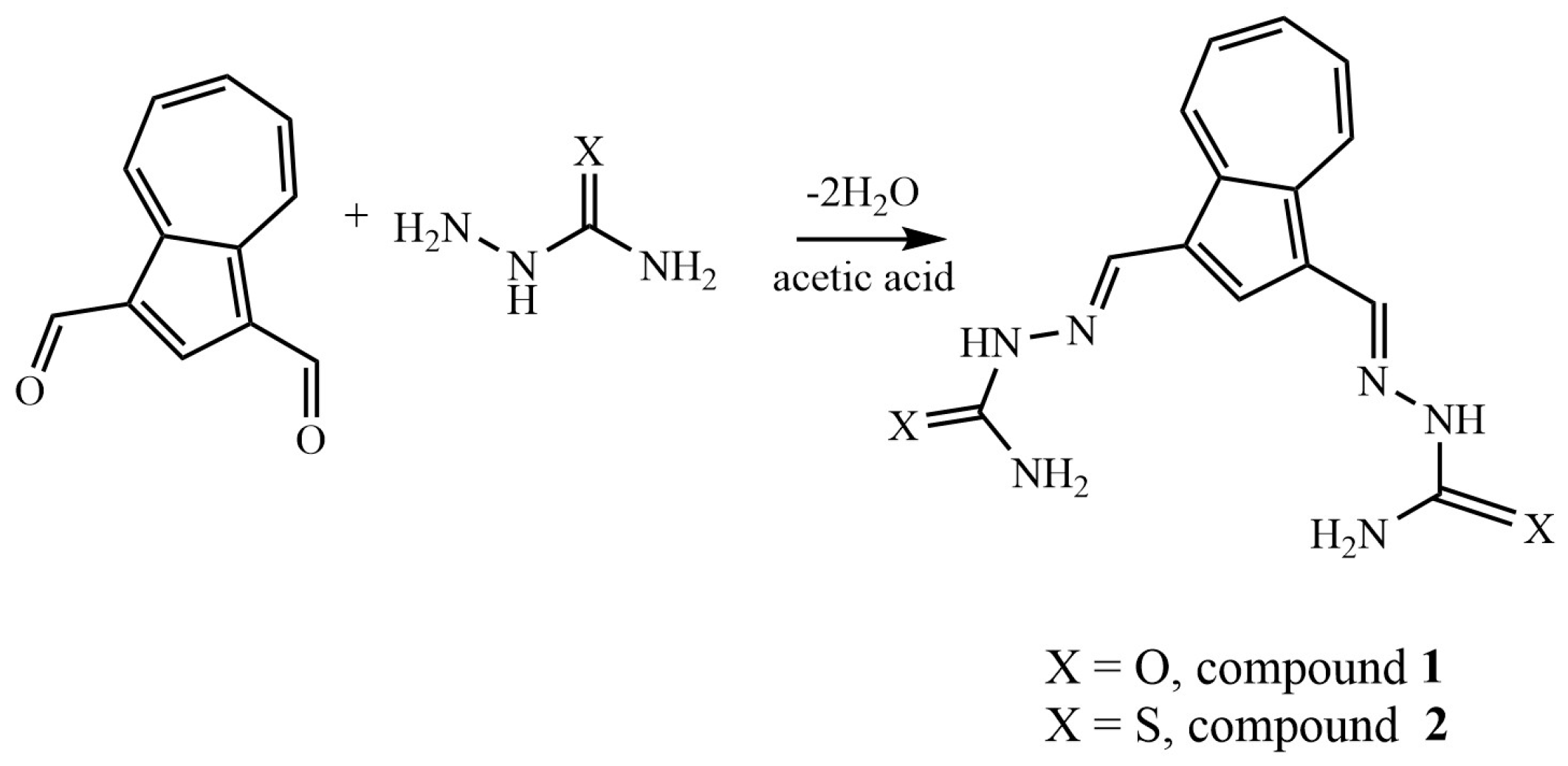
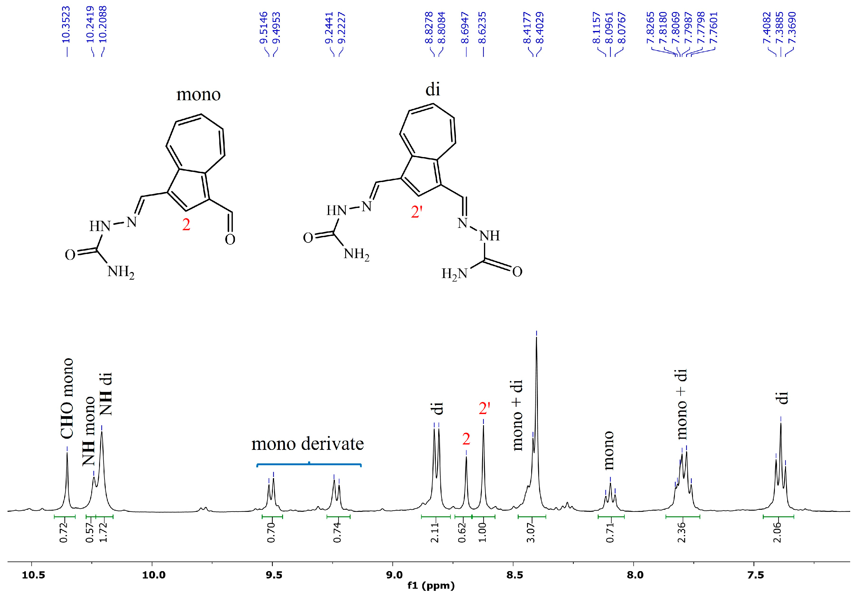
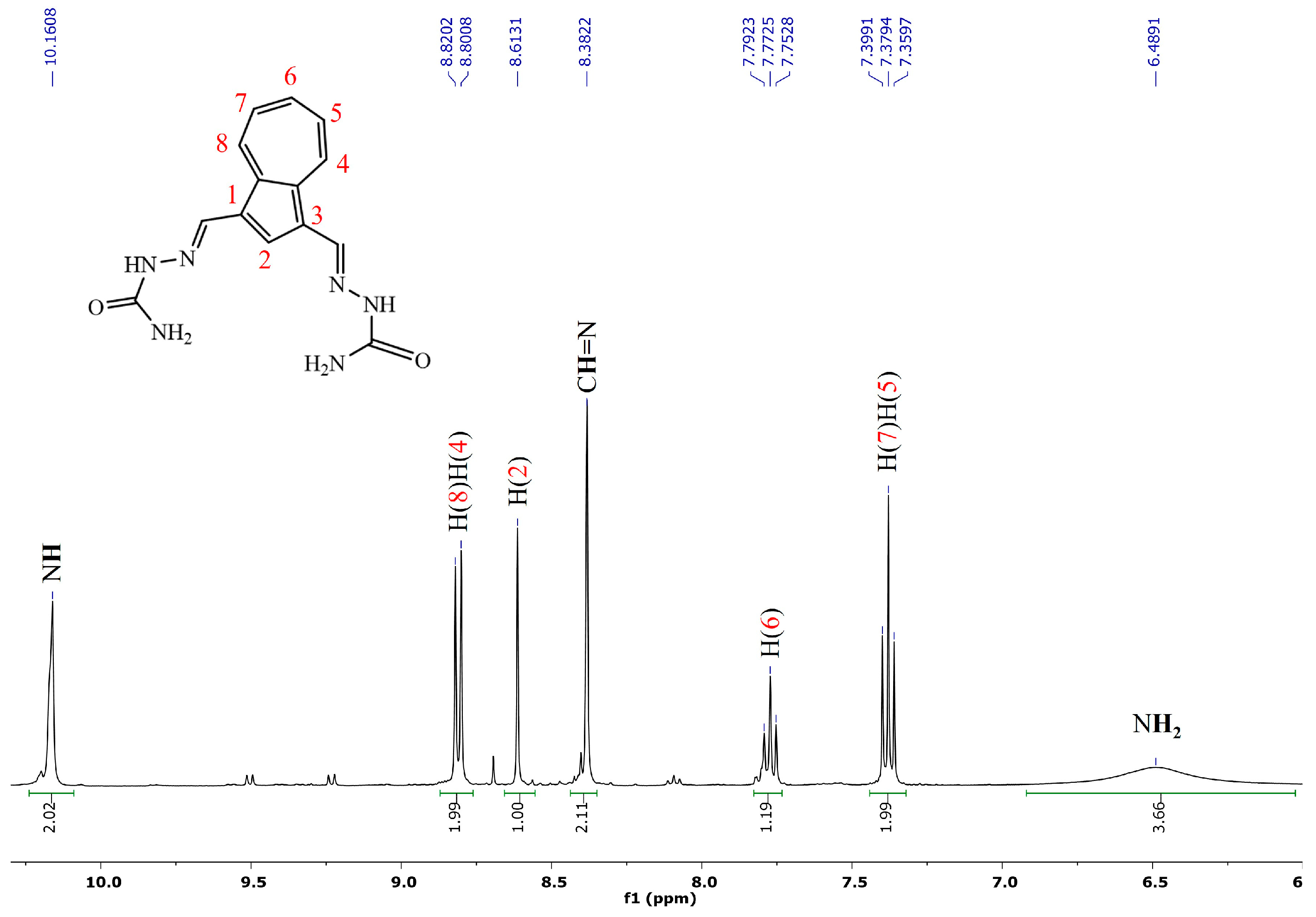
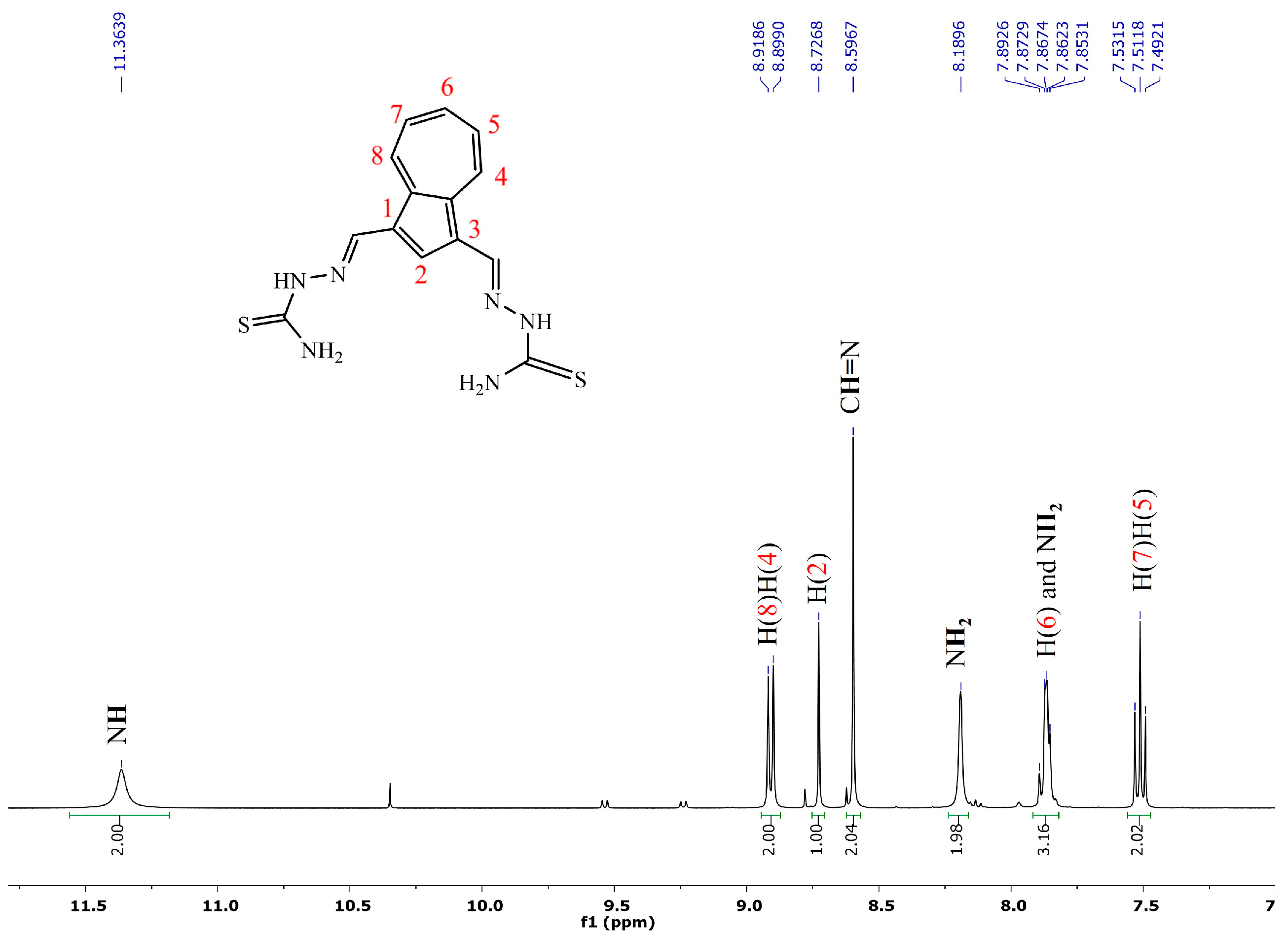
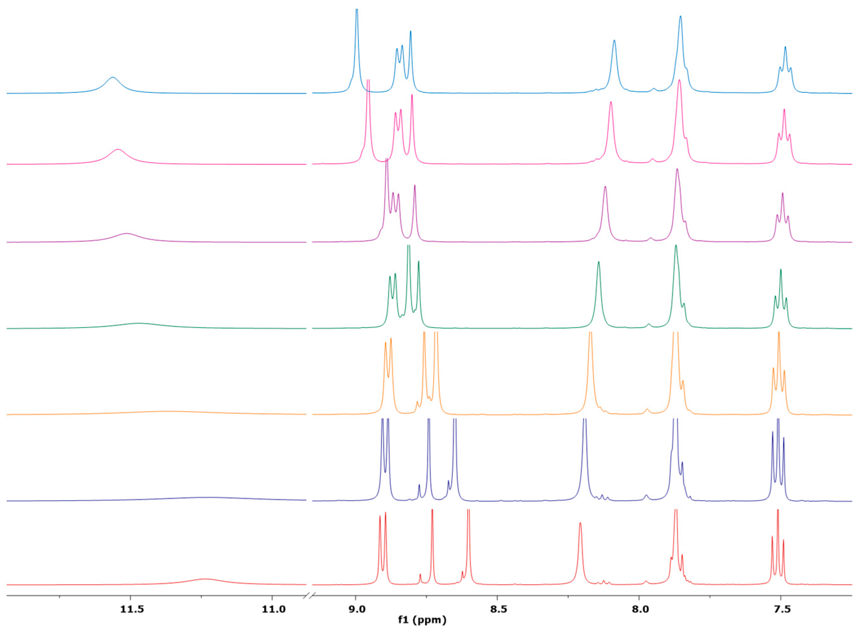
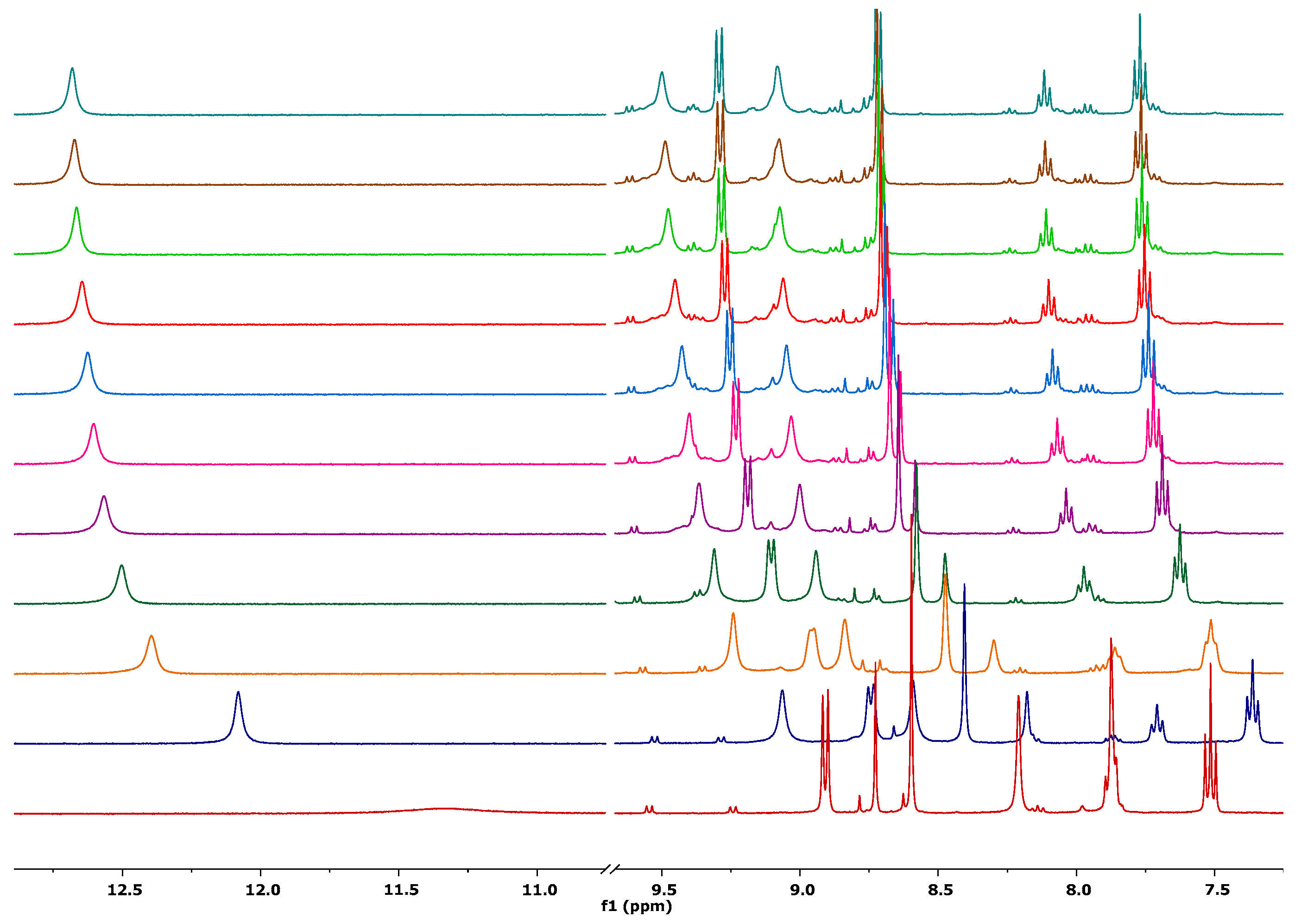
2.1. Synthesis and NMR Characterization

The azulene-bis(hydrazone) 1 and 2 were synthesized via straightforward Schiff base condensation reaction between azulen-1,3-dicarboxaldehyde and commercially available semicarbazide and thiosemicarbazide, respectively (Scheme 1). The reaction proceeds in the presence of glacial acetic acid as a catalyst, utilizing an excess of semi- and thiosemicarbazide (2.5 equivalents), in refluxing ethanol. The identification of the products was carried out by high-resolution mass spectrometry.
The reaction protocol was established for the Schiff base condensation of azulene-1,3-dicarboxaldehyde and semicarbazide. In the absence of acetic acid, a mixture of mono- and di-substituted semicarbazone was obtained in a ratio of 0.5:1, as confirmed by NMR spectroscopy. The 1H NMR spectroscopy showed the aldehyde proton (CH=O) at 10.35 ppm and two distinct signals corresponding to the imine proton (CH=N) at 8.40 and 8.38 ppm (Figure 1). Integration of the proton signals enabled determination of the molar ratio of the two reaction products present in the mixture.
In the presence of a few drops (five) of acetic acid, the reaction preferentially yields the desired symmetric compound. The reaction time was determined based on the disappearance of azulene-1,3-dicarboxaldehyde from the reaction medium via thin-layer chromatography. The 1H NMR spectrum of compound 1 shows the chemical shifts characteristic of the azulene moiety within the range of 7.36–8.82 ppm area. The NH protons are observed at 10.17 ppm, as a well-defined signal, while the two NH2 protons resonated at 6.49 ppm as a broad singlet. The characteristic CH=N proton resonance appears at 8.38 ppm in the shape of a well-defined singlet (Figure 2). The 13C NMR spectrum shows the characteristic azulene carbon resonance signals, the amide carbonyl peak at 156.9 ppm, and imine carbon resonance at 135.6 ppm.
For the thiosemicarbazone compound 2, the imine proton exhibits a downfield shift to 8.60 ppm, whereas the NH2 protons appear as two distinct, broad singlets at 8.20 and 7.86 ppm, respectively (Figure 3), due to electron delocalization and the possible thione–thiol equilibrium in solution (Scheme 2). The reduced electronegativity of the sulfur likely contributes to this phenomenon. The NH protons also exhibit a significant shift, with its resonance occurring at 11.23 ppm, which represents a downfield shift of over 1 ppm from its position in compound 1. However, in the 13C-NMR spectrum of the compound, the corresponding signal appears at 177.13 ppm, corresponding to the thioamide carbon (C=S) (Figure S2, Supplementary Materials). Thus, the 13C-NMR spectrum and the presence of the NH resonance collectively indicate that the thioenol form is not present. The two distinct singlets corresponding to C(=S)–NH2 protons suggest that the protons are magnetically nonequivalent, likely due to hindered rotation around the C-N bond.

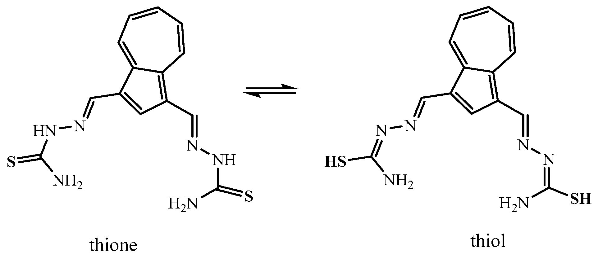
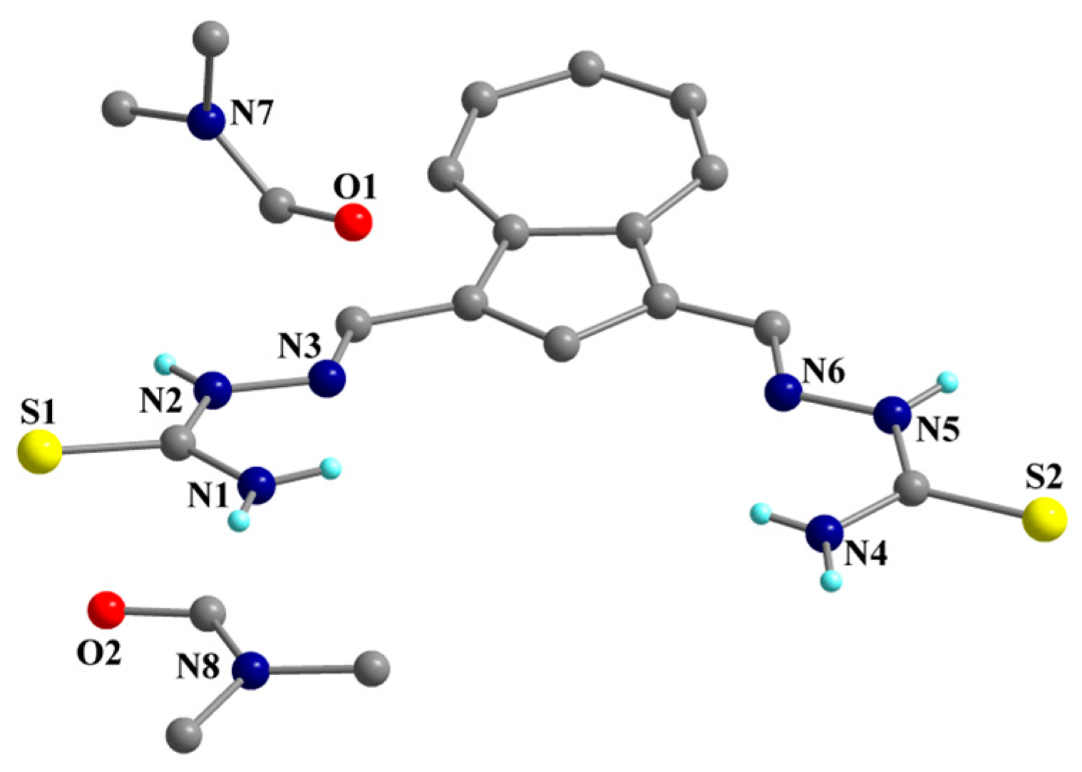
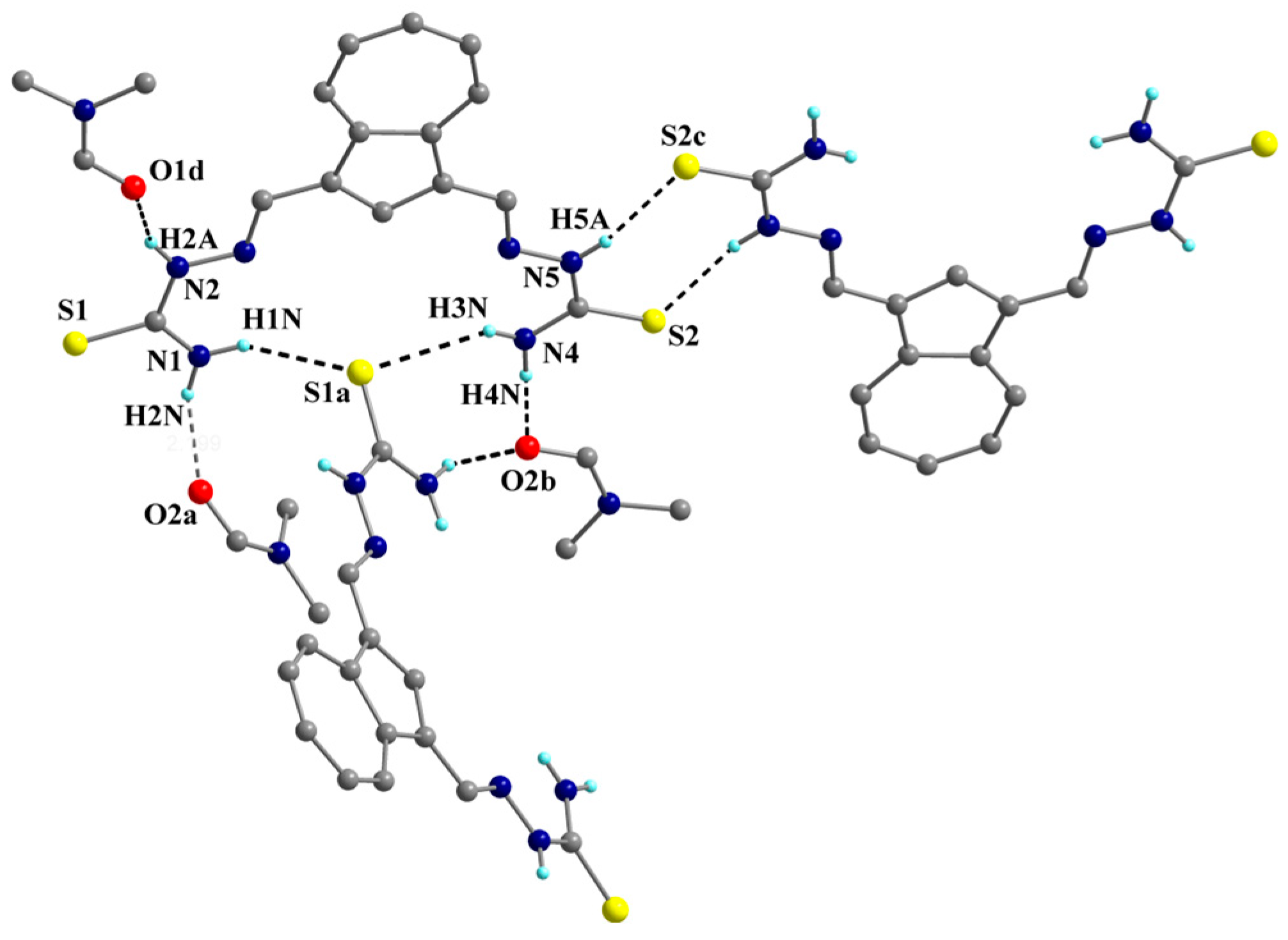
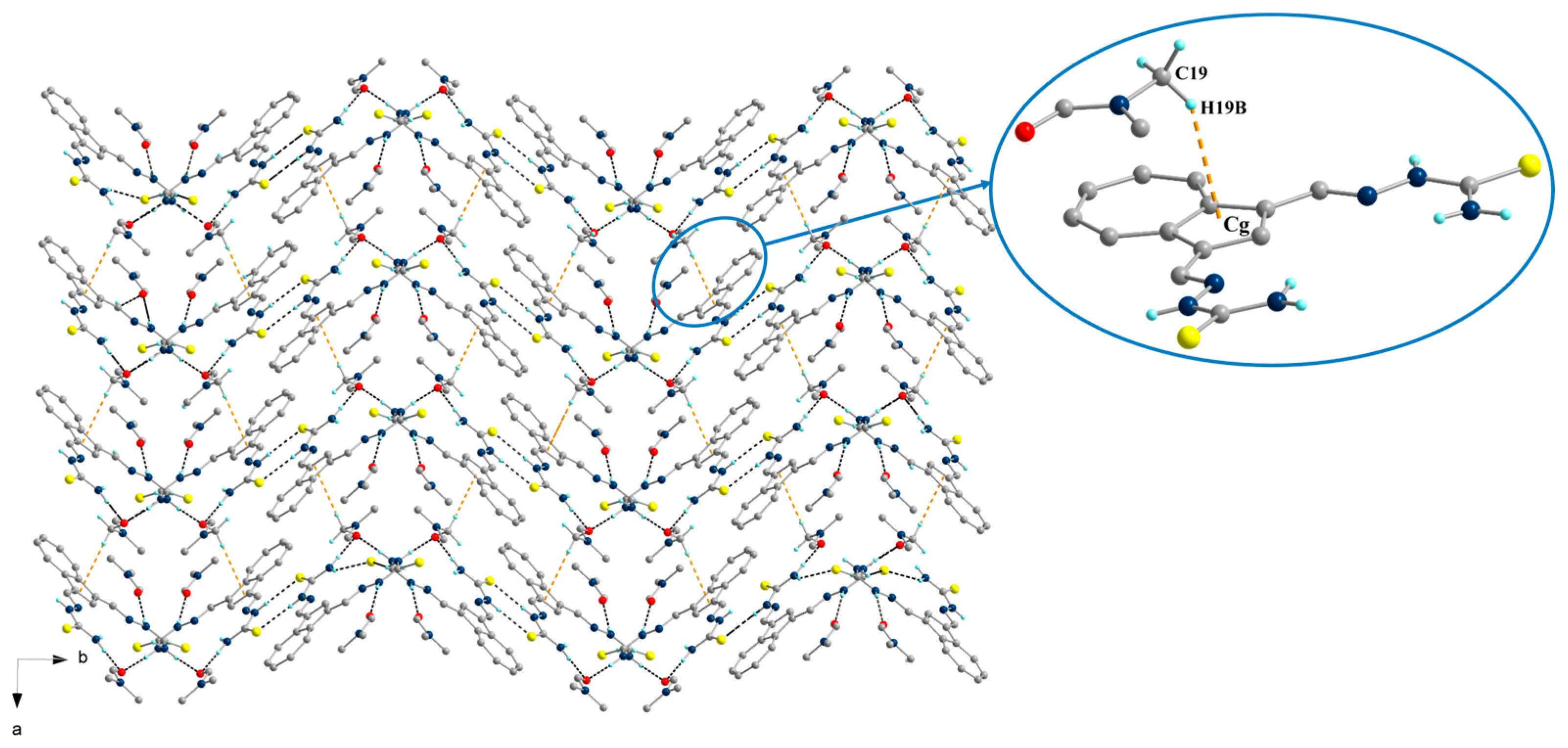
2.2. Single-Crystal X-Ray Structure Determination and IR Spectroscopy

The structure of compound 2 was confirmed definitively by single-crystal X-ray analysis. X-ray diffraction was performed at room temperature on a single crystal grown from a solution of this compound in a solvent mixture of methanol/dimethylformamide (MeOH/DMF) by slow solvent evaporation. Crystal data, details for data collection, and parameters for structure solution and refinement are summarized in Table 1. This compound crystallized in the monoclinic P21/c space group with Z = 4. The crystallographic asymmetric unit comprises one thiosemicarbazone molecule together with two DMF molecules of crystallization as depicted in Figure 4. Selected bond distances are presented in Table 2. The isolation of thiosemicarbazone as the thione tautomer is demonstrated by the thiocarbonyl (C=S) bond distances [C1–S1 = 1.691(3) Å and C14–S2 = 1.688(3) Å], which are characteristic of thioamide double bonds [45]. Interestingly, the Schiff-base imine bonds C2–N3 and C13–N6 are identical [1.284(4) Å].
The N-N bonds are 1.368 and 1.3916 A, similar to other thiosemicarbazones and longer than the N-N bonds in hydrazide (1.325 Å). The thiosemicarbazone units exhibit a syn-closed conformation, with both arms oriented in the same direction and adopting an E configuration with respect to the imine linkages. The conformation of the arms is in contrast to the bis-thiosemicarbazone derived from 2-hydroxyisophthalaldehyde, where intramolecular hydrogen bonding between the imine nitrogen and phenolic oxygen leads to an anti arrangement of the arms [46].
Intermolecular N-H···O and NH···S hydrogen bonds were observed in the crystal packing (Figure 5). Additionally, the intermolecular NH···S contacts form the R22(8) closed ring according to the symmetry center, {···H–N–C=S}2 synthons. The molecules are bonded to each other with these interactions into 2D sheets (Figure 6), which are further connected via C-H···π interactions (Figure 7). The data related to the hydrogen bonds are given in Table 2.
The IR spectroscopy of both compounds revealed characteristic bands corresponding to v(N–H), ν(C=N), ν(N–N), ν(C=O), and ν(C=S) stretching vibrations (see Section 3 and Figures S3 and S4 in the Supplementary Materials). Notably, the imine stretching vibration was observed at 1577 cm−1 for compound 1 and 1591 cm−1 for compound 2. The C=O stretching vibration appeared as a sharp, intense band at 1676 cm−1, while the C=S stretching vibration was observed at 831 cm−1. Furthermore, the absence of the 2500 cm−1 band, characteristic of v(S-H), in the IR spectrum of compound 2 confirmed the thione form (HN–C=S) in the solid state, as supported by X-ray crystallography. On the other hand, the presence of a ν(N–H) stretching vibration at 3121 cm−1 provides evidence for the azo–hydrazone tautomer of the hydrazone group.

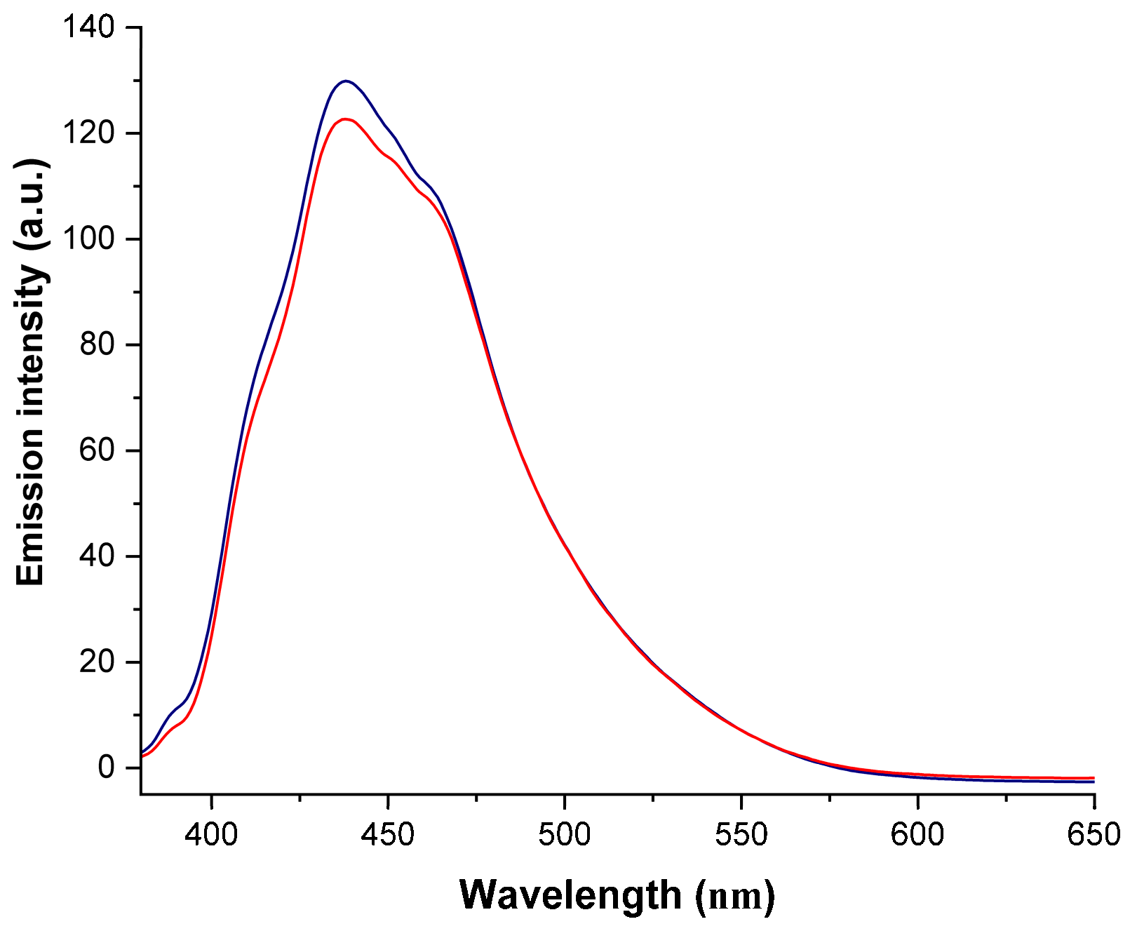
2.3. Electronic Absorption Spectroscopy and Fluorescence

UV-Vis spectra for both compounds display absorption maxima at 331 nm (lgɛ = 4.79) for compound 1 and 360 nm (lgɛ = 4.90) for compound 2. A less intense absorption peak is present in the UV region for compound 1, at 296 nm. Both compounds showed a broad absorption peak at 410 nm (Figure S5, Supplementary Materials). In a solid state, broad peaks are observed at 450 and 600 nm, characteristic of the azulene-visible absorption peak near 600 nm, which is responsible for its blue color [15] (Figure S6, Supplementary Materials).
Both compounds showed fluorescent emission upon excitation at a 350 nm wavelength (Figure 8). The fluorescence emission occurs around 450 nm for both compounds, similar to the emission spectra of the parent azulene when it relaxes from the S2 state to the ground state [14]. It is well known that the S2→S0 fluorescence of azulenes is much stronger compared to the weak S1→S0 transition and it is detectable in the visible region (blue fluorescence) [14]. The relative fluorescence quantum yield is 0.11 for 1 and 0.07 for 2, referred to as standard quinine sulphate fluorescence in acidic medium.

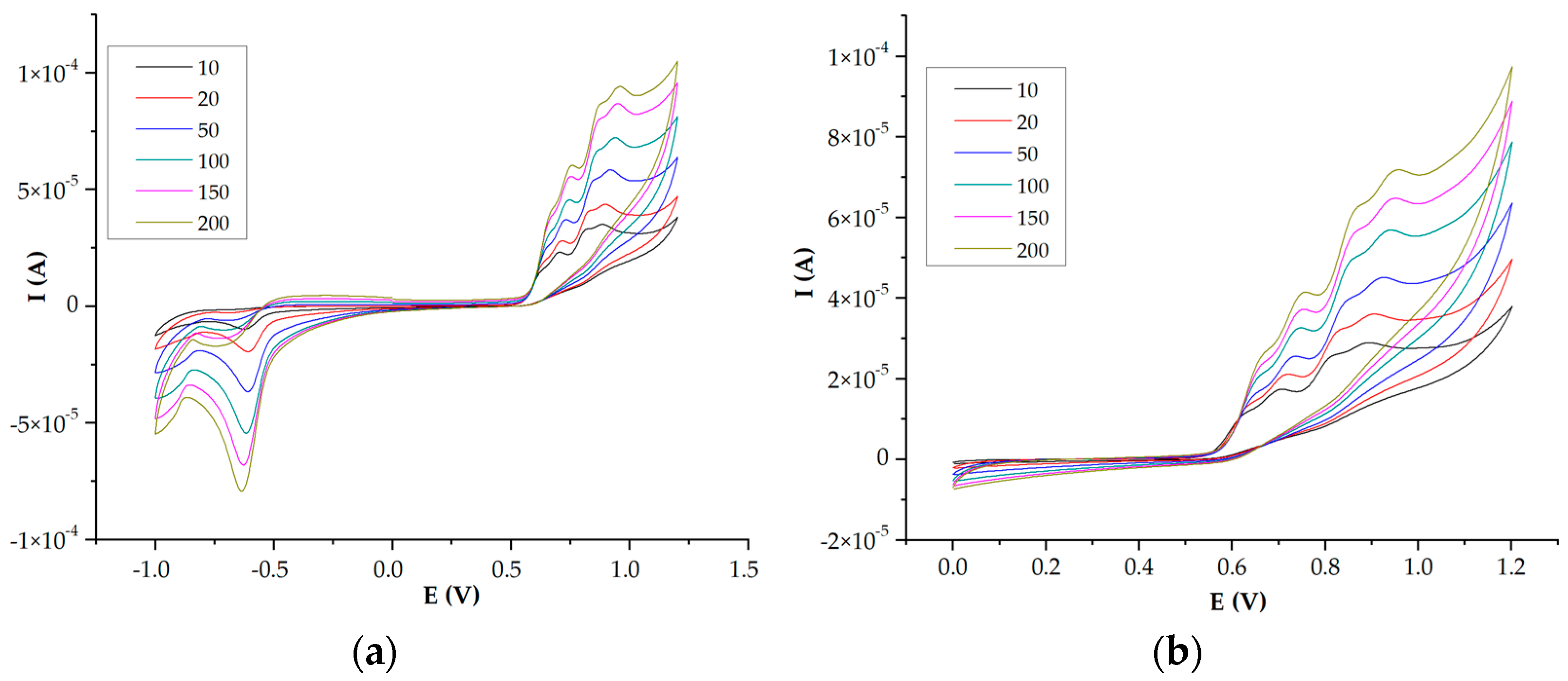
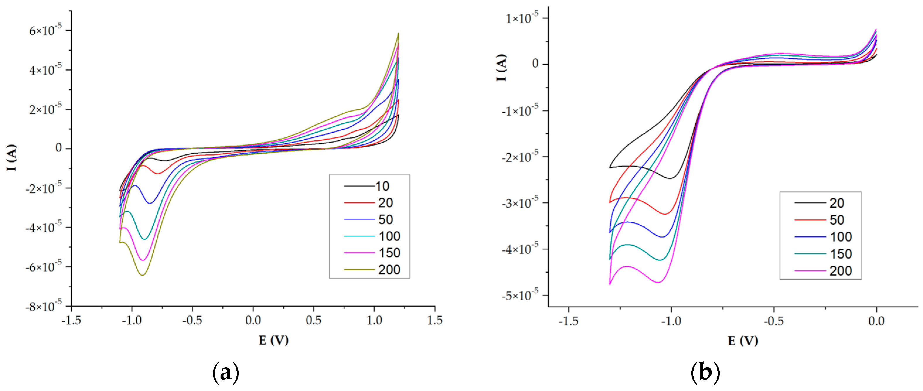
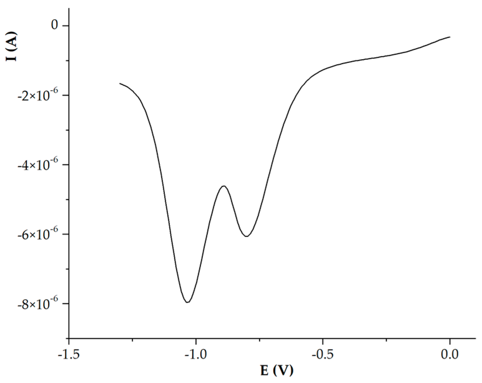
2.4. Electrochemistry

Azulene derivatives often exhibit interesting redox properties, making them potential candidates for applications in electrochemistry and materials science [47].
The redox properties of the azulene-derived semicarbazone, 1, were investigated by cyclic voltammetry (CV) over a broad potential range of [0–1.2–(−1)] V (Figure 9a) and over a narrower potential range, [0–1.2–0] V, in order to facilitate the correlation of anodic and cathodic peaks (Figure 9b).
Within the potential range from 0 to −1 V and back to 0, no oxidation peaks were observed (see Supporting Information Figure S7). In contrast, oxidation peaks are detected in the other two studied domains at 0.71 and 0.82 V. Two reduction peaks are observed at approximately −0.8 V (visible in DPV measurements), which are attributed to the reduction of the imine double bond, and at −0.60 V, ascribed to the reduction of the azulene moiety. The cyclic voltammetry data are well correlated with the DPV measurements (Figure 10).
The assignment of the oxidation and reduction peaks, both the cyclic voltammetry and DPV experiments, were performed for related phenylene-derived hydrazones, measured in the [0–1–(−0.85)] range (see Supporting Information Figure S8). The benzene ring is significantly more stable and therefore the redox behavior is as well due to the semicarbazone moiety. The anodic peak is very poorly defined (not visible on CV measurements), while the cathodic peaks in the potential range of −0.3 to −0.85 V are not present, indicating that reduction occurs with greater difficulty. Nevertheless, a comparison of the redox potentials of the two compounds enables the assignment of the oxidation and reduction potentials of azulene, as described above.
The substitution of oxygen with a sulfur atom, as in thiosemicarbazone 2, yields a more stable compound. No oxidation process was observed, and the reduction occurs at around −0.8 V, which is slightly more difficult to achieve, likely due to the increased electronegativity of sulfur (Figure 11a,b). The reduction peak potential is shifted to a more negative value with increasing scan rate. From DPV measurements, two cathodic peaks are observed at −0.75 V and −1 V (Figure 12).
The reduction peak potentials for compound 2 are shifted to more negative values compared to those of compound 1, indicating higher stability of thiosemicarbazone. Similar measurements were conducted for phenylene–thiosemicarbazone in the potential range of [0–1–(−1)] (see Supporting Information, Figure S9). When azulene was substituted with benzene, no oxidation process was observed, and a small reduction peak appeared at 0.9 V.

2.5. Receptor Ability
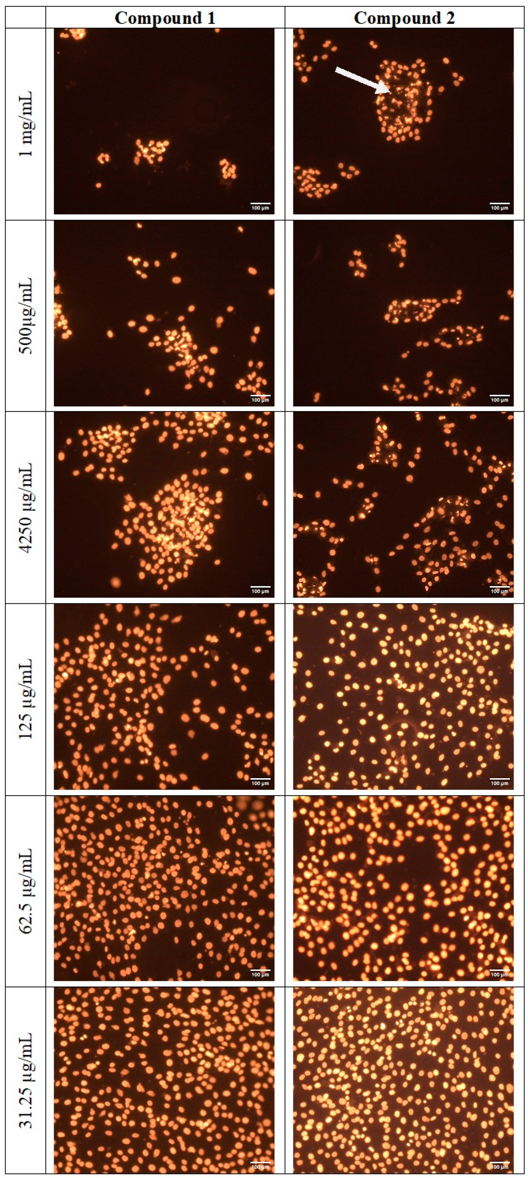
Next, we tested the ability of these compounds to function as a receptor for anions, owing to the good hydrogen bond donor ability of the N-NH-C=O(S)hydrazone and the iminothiourea moiety. Due to the optical properties of the azulene chromophore, we hoped that our systems could signal the presence of specific anion guests by changes in color. It turned out that the interactions could not be detected by simple naked-eye observation of the solutions of these receptors with anions. Next, we carried out the UV-Vis titration experiments to quantitatively determine the receptors’ affinities toward anions. The UV-Vis spectra of the azulene-containing semicarbazone and thiosemicarbazone receptors 1 and 2 displayed prominent absorption bands at 330 nm and 360 nm, respectively, in DMSO as a solvent, characteristic of the π-π* transition of the azulene rings. Solution binding studies were performed by UV-Vis titrations using [n-Bu4N]+Cl (Figure S10, Supplementary Materials). For both hydrazones, no relevant spectral changes were observed in the presence of the chloride anion, only a slight decrease in the absorption intensity as a result of dilution upon the addition of the anion solution.
Given the low concentrations employed in the UV-Vis titrations, where hydrogen bonding interactions are significantly weaker, we complemented our studies with NMR spectroscopy, which operates at much higher concentrations, thereby enhancing our ability to detect subtle interactions. The anion binding ability of 1 was very poor and tested only for Cl and Br (Figures S11 and S12 in the Supplementary Materials). While the addition of a bromide anion caused no significant changes in the position of the proton resonances, in the case of the chloride anion, a small downfield shift of the NH peaks was observed (ΔδNH = 0.23 ppm), exhibiting a modest recognition effect. Thus, detailed studies were taken using 1H-NMR spectroscopy, adding solid [n-Bu4N]+X (X = Cl, Br, ClO4, HSO4, BF4, and PF6) to DMSO-d6 solution of azulene-based thiosemicarbazone, 2. For this compound, the NH resonance shifts after the addition of a chloride anion from 11.23 in free thiosemicarbazone to 11.56 ppm after the addition of 6 equivalents of solid tetra-butylammonium chloride, with no further significant changes upon increase of the anion concentration to 10 equivalents (Figure 13). The two resonances of the NH2 groups (NH21 = 8.20 ppm and NH22 = 7.86 ppm) shift upfield (ΔδNH21 = 0.12 ppm) and the NH22 proton shifts upfield (ΔδNH22 = 0.02 ppm) upon anion coordination. The calculated association constant using the http://app.supramolecular.org/bindfit/ (accessed in October 2024) software [48] was found to be 32.13 M−1, indicative of a weak recognition effect for chloride anions.
In the case of the bromide anion, the shifts of the NH and NH2 protons are less significant, thus having a lower affinity (Figure S13, Supplementary Materials). Analysis of the crystal structure shows the NH groups outside of the binding cleft, being involved in N-H···O and NH···S hydrogen bonding interactions. Notably, the sulfur and nitrogen donor atoms (N3/S1 and N6/S2) are positioned in an anti-parallel fashion; thus, the formation of the supramolecular might take place between two discrete molecules. The receptor behavior of 2 was slightly affected by the presence of hydrogen sulfate anions, the NH2 resonances being shielded by 0.2 ppm. The ΔδNH = 0.11 ppm, the ΔδNH21 = 0.13 ppm, and ΔδNH22 = 0.04 ppm. It is thus expected that weak interactions are possible with this anion; the calculated binding constant is 8.94 M−1. For other tested anions, ClO4, BF4, and PF6, the characteristic resonances of NH and NH2 protons are largely unaffected by the presence of these anions (Figures S14–S17 in the Supplementary Materials). Consequently, we deduce that the azulene-based thiosemicarbazone does not exhibit the characteristics of an effective anion receptor, a small recognition effect being observed for small-size chloride anions.
However, taking into account the structural feature of the azulene-based bis(thiosemicarbazone), we have redirected our focus towards the recognition of metal cations. Specifically, we have selected the Hg2+ cation as a target, given its known toxicity and our previous findings that azulene-substituted terpyridines exhibit promising recognition properties towards this metal ion [49]. The presence of donor atoms such as nitrogen and sulfur (in thiosemicarbazones) or nitrogen and oxygen (in semicarbazones) enables these compounds to form stable coordination complexes with toxic mercury (Hg2+) metal ions [50]. Upon addition of a HgCl2 aqueous solution to a DMSO solution of 1, the absorption maxima at 360 nm does not change in its position, with only a slight variation of the intensity (Figure 14a). Also, the 1H-NMR titration of 1 with HgCl2 (Figure S18, Supplementary Materials) shows no significant changes in the characteristic NH and NH2 resonances, showing that no coordination takes place. In the case of the bis-thiosemicarbazone receptor, 2, the titration with HgCl2 causes significant changes in the absorption spectrum. As HgCl2 is gradually added, the maximum absorption band shifts bathochromicaly at 421 nm, indicative of a change in the electronic environment of the thiosemicarbazone, likely due to coordination with Hg2+ ions. Concurrently, a new absorption maximum appears at 320 nm, increasing in intensity as the concentration of Hg2+ ions increases (Figure 14b). An isosbestic point at ~300 nm at up to three equivalents of HgCl2 is observed, indicative of an equilibrium between the species. The calculated binding constant is 97.54 M−1, showing a good binding affinity for mercury(II) ions.
In addition to the measured electronic absorption spectra, the metallation titration of 2 with HgCl2 was analyzed by 1H-NMR spectroscopy (Figure 15). The addition of 1 equivalent of the mercury salt to a solution of 2 in DMSO-d6 (35 mM) caused a visible color change in the solution in the NMR tube. The most affected protons are the NH2δNH21 = 1.29 and ΔδNH22 = 1.21 ppm) and the azomethine protons, CH=N (Δδ = 0.12), showing that the coordination takes place inside the cavity formed by bis(thiosemicarbazone). The NH protons are also affected by mercury coordination, being shifted downfield by around 1.34 ppm. Significant downfield shifts were observed also for all the azulene protons. Overall, the binding constant for the Hg2+ is 109.17 M, similar to the UV-Vis titration experiments, showing a good affinity of 2 for this metal ion.

2.6. Cytotoxicity

Azulene derivatives have been investigated for their potential therapeutic applications, including anti-inflammatory effects, anticancer activities, and photoprotective properties. Moreover, bis-hydrazones from phenyl-dialdehyde demonstrated good cytotoxic action against various cell lines [51]. Thus, to assess the cytotoxicity of the compounds described herein, we evaluated their effects on osteosarcoma MG63 cells (American Type Culture Collection (ATCC) CRL-1427). Cells were exposed to azulene-based bis-(thio)semicarbazones, 1 and 2, at concentrations ranging from 1 mg/mL to 7.8 µg/mL. Cytotoxicity was assessed using the CellTiter kit (Promega, Madison, WI, USA) and confluency assays in real-time using the Incucyte Live-Cell Analysis System. The results, presented in Figure 16, are expressed as viability percentages relative to control cells.
The IC50 values of the azulene-based bis-(thio)semicarbazones were determined to be 473.08 μM (141.12 µg/mL) for compound 1 and 472.40 μM (156.10 µg/mL) for compound 2. These compounds did not exhibit significant antiproliferative activity against osteosarcoma cell lines by comparison to the standard anticancer reference drug methotrexate (MTX), which exhibits an IC50 value of 26.52 μM, or phenylene (bis-hydrazone), for which the IC50 value was reported to be 18.20 μM [51]. However, despite their limited efficacy, the propidium iodide staining revealed a concentration-dependent decrease in cell number, accompanied by characteristic morphological changes indicative of apoptosis and necrosis (Figure 17).

3. Experimental

3.1. Materials and Methods

All reagents were used as received. The solvents were spectroscopic grade. First, azulene-1,3-dicarboxaldehydes was obtained following the Vilsmeier reaction [52]. The high-resolution mass spectra were acquired with a QTOF LCMS-9030 mass spectrometer from Shimadzu (Kyoto, Japan), equipped with the ESI interface and auxiliary interface for calibration. The calibration was performed with a sodium iodide solution. Samples dissolved in acetonitrile at a concentration of 1 ppm were infused directly into the ESI interface with an LC-40D ×3 pump at a flow rate of 0.4 mL/min with a mobile water phase (0.1% formic acid) 10% and the acetonitrile. The sample solutions were loaded into a loop using a SIL-40C ×3 automatic injector. The injected volume was 1 μL. At the ESI interface, the nebulizing gas was nitrogen with a flow rate of 3 L/min, nitrogen was used as the drying gas with a flow rate of 10 L/min, the heating gas was air at a rate of 15 L/min, and the temperature of the ESI interface was 250 °C. Melting points were determined using a Krüss Optronic instrument, Hamburg, Germany, model KSPI. The NMR spectra were recorded on a Bruker Avance II 500 MHz (1H: 500 MHz, 13C: 175.47 MHz), Karlsruhe, Germany. The structural assignment of the 1H and 13C resonances was obtained from 1H COSY and 13C HSQC, and HMBQ experiments. NMR titrations with HgCl2 were performed for a 0.6 mL solution of the receptor (around 10 mM) in DMSO-d6 with solid mercury salt from 1 to 10 equivalents. The same procedure was followed for the titrations with tetrabutylammoniun anions. The association constants were calculated using the http://app.supramolecular.org/bindfit/ software [48]. The electrochemical experiments were conducted in 1 mM solutions in DMSO, with 0.1 M Bu4NClO4 serving as the electrolyte. A three-electrode setup was employed, consisting of a platinum working electrode, a silver/silver chloride reference electrode, and a graphite counter electrode. The measurements were performed using an Autolab PGSTAT30 potentiostat from Metrohm, Herisau, Switzerland. The IR spectra were obtained using a Bruker 70 Tensor instrument, Borken, Germany in the KBr pellets. The fluorescence quantum yield was determined by comparative measurement with a dilute solution of quinine bisulfate in 0.1 N sulfuric acid (H2SO4), which has a known absolute quantum yield of 0.55 [53]. The fluorescence spectra were recorded with a Jasco FP-6500 spectrofluorometer (Jasco Europe, Cremella, Italy) equipped with a 150 W Xenon lamp. The excitation wavelength was 350 nm for working concentrations of 3.48 × 10⁻⁶ M (compound 1) and 3.34 × 10⁻⁶ M (compound 2). UV–vis spectra were recorded on a Varian Cary 100 spectrophotometer using 1 cm quartz cells. The UV-Vis titrations were performed as constant host titrations (1.74 × 10⁻⁵ M for compound 1 and 1.67 × 10⁻⁵ M for compound 2) in DMSO with HgCl2 (4.95 mM aqueous solution, 0, 2, 4, 6, 8, and 10 equivalents DMSO) and the respective TBAX stock solution of ~5 mM in DMSO at room temperature.
X-ray diffraction measurements were performed on an STOE IPDS II diffractometer (STOE & Cie GmbH, Darmstadt, Germany), operating with a Mo Kα (λ = 0.71073 Å) X-ray tube with a graphite monochromator. The structures were solved by direct methods and refined by full-matrix least-squares techniques based on F2 [54]. The non-H atoms were refined with anisotropic displacement parameters. Atomic scattering factors were taken from the international tables for X-ray crystallography. Hydrogen atoms were included but not refined. Calculations were performed using the SHELX-2014 crystallographic software package (version 1). Drawings of the molecules were performed with the program Diamond 4. A summary of the crystallographic data and the structure refinement are given in Table 1. Crystallographic data (excluding structure factors) have been deposited with the Cambridge Crystallographic Data Centre with CCDC reference number 2400616. These data can be obtained free of charge via http://www.ccdc.cam.ac.uk/structures (accessed on 20 December 2024), or from the Cambridge Crystallo-graphic Data Centre, 12 Union Road, Cambridge, CB2 1EZ, UK; fax: (+44)-1223-336-033; or e-mail: deposit@ccdc.cam.ac.uk.

3.2. General Procedure for the Synthesis of Compounds 1 and 2

Azulene-1,3-dicarboxaldehyde (184 mg, 1 mmol) was added to a solution of semicarbazide/thiosemicarbazide (2.5 equivalents, 2.5 mmol) in ethanol (50 mL), followed by addition of 5 drops of acetic acid glacial. The resulting solution was refluxed overnight with continuous stirring, monitoring the disappearance of the dialdehyde precursor by thin-layer chromatography. After reaction completion, the mixture was filtered and washed with ethanol and diethyl ether to yield pure products. IR, 1H NMR, and 13C NMR were used to determine the structure of the compounds.
  • (2E,2′E)-2,2′-(azulene-1,3-diylbis(methaneylylidene))bis(hydrazine-1-carboxamide), 1, was synthesized following the general procedure using semicarbazide (188 mg, 2.5 mmol). After removal of the solvent, the obtained dark green solid was washed with ethanol and diethyl ether. There were isolated 230 mg (0.77 mmol) green powder, 77% yield; m.p > 300 °C with decomposition; a transition phase was observed at 223–225 °C; 1H NMR: (DMSO-d6, 500 MHz): δ 10.17 (s, 2H, NH), 8.81 (d, 2H, J = 9.8 Hz, H-8 and H-4), 8.61 (s, 1H, H-2), 8.38 (s, 2H, CH=N), 7.77 (t, 1H, J = 9.8 Hz, H-6), 7.37 (t, 1H, J = 9.8 Hz, H-5 and H-7), and 6.49 (bs, 4H, NH2) ppm. 13C-NMR (DMSO-d6, 125 MHz): δ 156.89 (C=O), 140.36 (CH-6), 138.36 (Cq-9/10), 136.10 (CH-4/8), 135.95 (CH-2), 135.55 (CH=N), 126.54 (CH-5/7), 123.46 (Cq-1/3) ppm. UV-Vis (DMSO):296 (lgɛ = 4.63), 330 (lg ɛ = 4.79), 410 (lg ɛ = 4.10). Selected IR: 3470, 2884, 2827, 1676, 1577, 1433, 1400, 1111, 1045, 746 cm−1. MS: 299.12532 [M + H]+ (calcd. for C14H14N6O2 299.12510).
  • (2E,2′E)-2,2′-(azulene-1,3-diylbis(methaneylylidene))bis(hydrazine-1-carbothioamide), 2, was synthesized following the general procedure using thiosemicarbazone (230 mg, 2.5 mmol). After removal of the solvent, the green solid was washed with ethanol (3 × 10 mL) and diethyl ether for drying. There were isolated 238 mg (72 mmol) light green powder, 72% yield; m.p = 268–269 °C; 1H NMR: (DMSO-d6, 500 MHz): δ 11.36 (s, 2H, NH), 8.91 (d, 2H, J = 9.8 Hz, H-8 and H-4), 8.72 (s, 1H, H-2), 8.60 (s, 2H, CH=N), 8.19 (bs, 2H, NH2), 7.89–7.85 (m, 3H, NH2 and H-6), and 7.51 (t, 1H, J = 9.8 Hz, H-5 and H-7) ppm. 13C-NMR (DMSO-d6, 125 MHz): δ 177.13 (C=S), 140.83 (CH-6), 139.64 (Cq-9/10), 138.52 (CH=N), 137.72 (CH-2), 136.70 (CH-4/8), 127.87 (CH-5/7), 122.90 (Cq-1/3) ppm. UV-Vis (DMSO):360 (lg ɛ = 4.90), 410 (lg ɛ = 4.22). Selected IR: 3440, 3262, 3151, 3033, 2991, 2885, 2814, 1591, 1537, 1388, 831 cm−1. MS: 331.07944 [M + H]+ (calcd. for C14H14N6S2 331.07941).

3.3. Determination of Cell Viability by the CellTiter Assay

Cell viability was determined using the CellTiter 96® AQueous One Solution Cell Proliferation Assay kit (Promega, Madison, WI, USA) according to the manufacturer’s instructions. Thus, cells were seeded in a 96-well plate at a density of 7500 cells per well and incubated at 37 °C, 5% CO2 to allow for adherence and proliferation. After 24 h, the cells were treated with binary dilutions of azulene derivates starting from mg/mL. After incubation, 10 µL of cell titer solution was added to each well, and the plate was further incubated for 24 h. The absorbance of the solution was measured at 490 nm using a spectrophotometer (e.g., a plate reader). The test is based on reducing a tetrazole compound (MTS) in the mitochondria of viable cells. It leads to the formation of a colored formazan product, soluble in the medium, the amount of which is directly proportional to the number of viable cells. Data were normalized to the sample, to which no MTS was added, and expressed as a percentage of the untreated control. Wells without MTS were subsequently fixed with ethanol 70, stained with propidium iodide (PI), and photographed under a Zeiss Observer D1 microscope, Oberkochen, Germany equipped with fluorescence mode.

3.4. Determination of Cell Viability Using the Incucyte System

The Incucyte® Live-Cell Analysis System (Sartorius, Göttingen, Germany) assessed cell viability in real-time. Cells were seeded in a 96-well plate at 7500 cells per well density and incubated at 37 °C and 5% CO2 for 24 h. The cells were treated with binary dilutions of compounds ranging from 1 mg/mL to 7.8 µ/mL. The Incucyte system continuously monitored the cells for 72 h, capturing images at regular 12 h intervals. Cell viability was determined using the cell confluency assay (the degree of coverage of the well surface by cells) as an indirect measure of cell growth and proliferation. The obtained images were automatically analyzed using Incucyte software S3 (Sartorius, Goettingen, Germany), which generated growth curves as a function of incubation time. Cell viability was expressed relative to the untreated control group. Data were exported and processed for further statistical analysis.

4. Conclusions

In summary, we have synthesized two azulene-based hydrazones which were obtained by Schiff-based condensation reaction of azulene-1,3-dicarboxaldehyde with semi- and thiosemicarbazide in the presence of acetic acid. These compounds showed fluorescence properties with emission centered at 470 nm upon excitation at a 350 nm wavelength. Both compounds are redox active with reduction peaks observed at approximately −0.8 V, attributed to the reduction of the imine double bond, and at −0.60 V, ascribed to the reduction of the azulene moiety. Taking advantage of the visible absorption band of azulene derivatives, we tested the two for their ability to recognize anions and poisoning metal cations, in particular, mercury (II) cations. The azulen-1,3-bis(semicarbazone) showed no real recognition properties; instead, the azulen-1,3-bis(thiosemicarbazone) compound exhibited a weak recognition effect for chloride anions and a good binding affinity for mercury(II) ions. The azulene-containing bis-hydrazone was tested against osteosarcoma MG63 cells with no significant antiproliferative activity. However, a concentration-dependent decrease in cell number was observed by propidium iodide staining.

Supplementary Materials

The following supporting information can be downloaded at: https://www.mdpi.com/article/10.3390/molecules30010083/s1: spectroscopic data, titration experiments, complementary electrochemical experiments, and cif file for compound 2.

Author Contributions

All authors contributed to the preparation of the manuscript. Conceptualization, S.N.; writing—original draft, preparation, S.N.; NMR methodology, A.H.; synthesis, A.D.; characterization, C.M., A.E.I. and A.M.M.; electrochemistry, D.B. and A.E.I.; cytotoxicity and biological investigation, C.B.; supervision, S.N. All authors have read and agreed to the published version of the manuscript.

Funding

This research was funded by a grant from the Ministry of Research, Innovation, and Digitization, CNCS-UEFISCDI, project number PN-IV-P8-8.3-ROMD-2023-0045, within PNCDI IV. We are grateful to Cristian Enache Preoteasa from the National Phytosanitary Authority of Agriculture Ministery for the high-resolution mass spectrometry investigations.

Data Availability Statement

Data are contained within the article and Supplementary Materials.

Conflicts of Interest

The authors declare no conflicts of interest.

References

  1. Wu, L.; Liu, J.; Li, P.; Tang, B.; James, T.D. Two-photon small-molecule fluorescence-based agents for sensing, imaging, and therapy within biological systems. Chem. Soc. Rev. 2021, 50, 702–734. [Google Scholar] [CrossRef] [PubMed]
  2. Cowper, P.; Pockett, A.; Kociok-Köhn, G.; Cameron, P.J.; Lewis, S.E. Azulene—Thiophene—Cyanoacrylic acid dyes with donor-π-acceptor structures. Synthesis, characterisation and evaluation in dye-sensitized solar cells. Tetrahedron 2018, 74, 2775–2786. [Google Scholar] [CrossRef]
  3. Xin, H.; Hou, B.; Gao, X. Azulene-based π-functional materials: Design, synthesis, and applications. Acc. Chem. Res. 2021, 54, 1737–1753. [Google Scholar] [CrossRef] [PubMed]
  4. Dragu, E.A.; Ion, A.E.; Shova, S.; Bala, D.; Mihailciuc, C.; Voicescu, M.; Ionescu, S.; Nica, S. Visible-light triggered photoswitching systems based on fluorescent azulenyl-substituted dithienylcyclopentenes. RSC Adv. 2015, 5, 63282–63286. [Google Scholar] [CrossRef]
  5. Hou, C.-Y.; Berger, F.; Narita, A.; Müllen, K.; Hecht, S. Proton-gated ring-closure of a negative photochromic azulene-based diarylethene. Angew. Chem. Int. Ed. 2020, 59, 18532–18536. [Google Scholar] [CrossRef] [PubMed]
  6. Xin, H.; Ge, C.; Jiao, X.; Jiao, X.; Yang, X.; Rundel, K.; McNeill, C.R.; Gao, X. Incorporation of 2,6-connected azulene units into the backbone of conjugated polymers: Towards high-performance organic optoelectronic materials. Angew. Chem. Int. Ed. 2018, 57, 1322–1326. [Google Scholar] [CrossRef]
  7. Dong, J.-X.; Zhang, H.-L. Azulene-based organic functional molecules for optoelectronics. Chin. Chem. Lett. 2016, 27, 1097–1104. [Google Scholar] [CrossRef]
  8. Xin, H.; Gao, X. Application of azulene in constructing organic optoelectronic materials: New tricks for an old dog. ChemPlusChem 2017, 82, 945–956. [Google Scholar] [CrossRef]
  9. Anderson, A.G., Jr.; Steckler, B.M. Azulene. VIII. A study of the visible absorption spectra and dipole moments of some 1- and 1,3-substituted azulenes. J. Am. Chem. Soc. 1959, 81, 4941–4946. [Google Scholar] [CrossRef]
  10. Tsuchiya, T.; Hamano, T.; Inoue, M.; Nakamura, T.; Wakamiya, A.; Mazaki, Y. Intense absorption of azulene realized by molecular orbital inversion. Chem. Commun. 2023, 59, 10604–10607. [Google Scholar] [CrossRef] [PubMed]
  11. Dragu, A.E.; Nica, S.; Tecuceanu, V.; Bala, D.; Mihailciuc, C.; Hanganu, A.; Razus, A.C. Synthesis and electrochemical properties of carbocyclic and heterocyclic diazulenylethenes. Eur. J. Org. Chem. 2013, 2013, 6601–6610. [Google Scholar] [CrossRef]
  12. Shoji, T.; Ito, S. Azulene-based donor–acceptor systems: Synthesis, optical, and electrochemical properties. Chem. Eur. J. 2017, 23, 16696–16709. [Google Scholar] [CrossRef]
  13. Bakun, P.; Czarczynska-Goslinska, B.; Goslinski, T.; Lijewski, S. In vitro and in vivo biological activities of azulene derivatives with potential applications in medicine. Med. Chem. Res. 2021, 30, 834–846. [Google Scholar] [CrossRef] [PubMed]
  14. Beer, M.; Longuet-Higgins, H.C. Anomalous light emission of azulene. J. Chem. Phys. 1955, 23, 1390–1391. [Google Scholar] [CrossRef]
  15. Murfin, C.L.; Lewis, S.E. Azulene—A bright core for sensing and imaging. Molecules 2021, 26, 353. [Google Scholar] [CrossRef]
  16. Teratani, M.; Nakamura, S.; Sakagami, H.; Fujise, M.; Hashimoto, M.; Okudaira, N.; Bandow, K.; Iijima, Y.; Nagai, J.; Uesawa, Y.; et al. Antitumor effects and tumor-specificity of guaiazulene-3-carboxylate derivatives against oral squamous cell carcinoma in vitro. Anticancer Res. 2020, 40, 4885–4894. [Google Scholar] [CrossRef]
  17. Peet, J.; Selyutina, A.; Bredihhin, A. Antiretroviral (HIV-1) activity of azulene derivatives. Bioorg. Med. Chem. 2016, 24, 1653–1657. [Google Scholar] [CrossRef]
  18. Ma, D.; He, J.; He, D. Chamazulene reverses osteoarthritic inflammation through regulation of matrix metalloproteinases (MMPs) and NF-kβ pathway in in-vitro and in-vivo models. Biosci. Biotechnol. Biochem. 2020, 84, 402–410. [Google Scholar] [CrossRef] [PubMed]
  19. Nihei, S.; Sato, J.; Komatsu, H.; Ishida, K.; Kimura, T.; Tomita, T.; Kudo, K. The efficacy of sodium azulene sulfonate L-glutamine for managing chemotherapy-induced oral mucositis in cancer patients: A prospective comparative study. J. Pharm. Health Care Sci. 2018, 4, 20. [Google Scholar] [CrossRef]
  20. NIH National Center of Advancing Translational Sciences. Egualen Sodium. 2020. Available online: https://drugs.ncats.io/substance/3368L0W034 (accessed on 20 October 2024).
  21. Zieliński, T.; Kędziorek, M.; Jurczak, J. The azulene moiety as a chromogenic building block for anion receptors. Tetrahedron Lett. 2005, 46, 6231–6234. [Google Scholar] [CrossRef]
  22. López-Alled, C.M.; Park, S.J.; Lee, D.J.; Murfin, L.C.; Kociok-Köhn, G.; Hann, J.L.; Wenk, J.; James, T.D.; Kim, H.M.; Lewis, S.E. Azulene-based fluorescent chemosensor for adenosine diphosphate. Chem. Commun. 2021, 57, 10608–10611. [Google Scholar] [CrossRef]
  23. Hu, J.; Hu, Z.; Liu, S.; Zhang, Q.; Gao, H.W.; Uvdal, K. A new ratiometric fluorescent chemodosimeter based on an ICT modulation for the detection of Hg2+. Sens. Actuators B Chem. 2016, 230, 639–644. [Google Scholar] [CrossRef]
  24. Salman, H.; Abraham, Y.; Tal, S.; Meltzman, S.; Kapon, M.; Tessler, N.; Speiser, S.; Eichen, Y. 1,3-Di(2-pyrrolyl)azulene: An efficient luminescent probe for fluoride. Eur. J. Org. Chem. 2005, 2005, 2207–2212. [Google Scholar] [CrossRef]
  25. Lopez-Alled, C.M.; Sanchez-Fernandez, A.; Edler, K.J.; Sedgwick, A.C.; Bull, S.D.; McMullin, C.L.; Kociok-Kohn, G.; James, T.D.; Wenk, J.; Lewis, S.E. Azulene–boronate esters: Colorimetric indicators for fluoride in drinking water. Chem. Commun. 2017, 53, 12580–12583. [Google Scholar] [CrossRef] [PubMed]
  26. Raczuk, E.; Dmochowska, B.; Samaszko-Fiertek, J.; Madaj, J. Different Schiff bases—Structure, importance and classification. Molecules 2022, 27, 787. [Google Scholar] [CrossRef] [PubMed]
  27. Buchholz, A.; Nica, S.; Debel, R.; Fenn, A.; Breitzke, H.; Buntkowsky, G.; Plass, W. Vanadium complexes with side chain functionalized N-salicylidene hydrazides: Hydrogen-bonding relays as structural directive for supramolecular interactions. Inorg. Chim. Acta 2014, 420, 166–176. [Google Scholar] [CrossRef]
  28. Cheng, C.; Sun, J.; Wang, C.; Zhang, Y.; Wei, S.; Jiang, F.; Wu, Y. Protonated N′-benzyl-N′-prolyl proline hydrazide as highly enantioselective catalyst for direct asymmetric aldol reaction. Chem. Commun. 2006, 37, 215–217. [Google Scholar] [CrossRef]
  29. Xiong, X.; Jiang, Y.; Ma, D. Assembly of N,N-Disubstituted hydrazines and 1-aryl-1H-indazoles via copper-catalyzed coupling reactions. Org. Lett. 2012, 14, 2552–2555. [Google Scholar] [CrossRef] [PubMed]
  30. Ragnarsson, U. Synthetic methodology for alkyl substituted hydrazines. Chem. Soc. Rev. 2001, 30, 205–213. [Google Scholar] [CrossRef]
  31. Słomiak, K.; Łazarenkow, A.; Checinska, L.; Kusz, J.; Ochocki, J.; Nawrot-Modranka, J. Synthesis, spectroscopic analysis and assessment of the biological activity of new hydrazine and hydrazide derivatives of 3-formylchromone. Molecules 2018, 23, 2067. [Google Scholar] [CrossRef] [PubMed]
  32. Patil, S.; Kuman, M.M.; Palvai, S.; Sengupta, P.; Basu, S. Impairing powerhouse in colon cancer cells by hydrazide-hydrazone-based small molecule. ACS Omega 2018, 3, 1470–1481. [Google Scholar] [CrossRef] [PubMed]
  33. Boudissa, R.; Benmohammed, A.; Chafai, N.; Boudechicha, A.; Rekiba, N.; Lagraa, H.; Achour, M.; Khoumeri, O.; Djafri, A.; Terme, T.; et al. Synthesis, characterization, DFT, antibacterial, ADME-T properties, and molecular docking of new N-functionalized thiazolidinones. J. Mol. Struct. 2024, 1307, 138004. [Google Scholar] [CrossRef]
  34. Alcaraz, R.; Muñiz, P.; Cavia, M.; Palacios, O.; Samper, K.G.; Gil-Garcia, R.; Jiménez-Pérez, A.; García-Tojal, J.; García-Girón, C. Thiosemicarbazone-metal complexes exhibiting cytotoxicity in colon cancer cell lines through oxidative stress. J. Inorg. Biochem. 2020, 206, 110993. [Google Scholar] [CrossRef] [PubMed]
  35. Bajaj, K.; Buchanan, R.M.; Grapperhaus, C.A. Antifungal activity of thiosemicarbazones, bis(thiosemicarbazones), and their metal complexes. J. Inorg. Biochem. 2021, 225, 111620. [Google Scholar] [CrossRef] [PubMed]
  36. Savir, S.; Wei, Z.J.; Liew, J.W.K.; Vythilingam, I.; Lim, Y.A.L.; Saad, H.M.; Sim, K.S.; Tan, K.W. Synthesis, cytotoxicity and antimalarial activities of thiosemicarbazones and their nickel (II) complexes. J. Mol. Struct. 2020, 1211, 128090. [Google Scholar] [CrossRef]
  37. Jiménez-Pérez, A.; Marcos-Gómez, S.; Madariaga, G.; Zapico, M.; Vitoria, P.; Tercero, J.; Torres, M.B.; Lezama, L.; Cuevas, J.V.; Etxebarria, I.; et al. Thiosemicarbazonecopper/halido systems: Structure and DFT analysis of the magnetic coupling. Inorganics 2023, 11, 31. [Google Scholar] [CrossRef]
  38. Arefyeva, N.; Sandleben, A.; Krest, A.; Baumann, U.; Schäfer, M.; Kempf, M.; Klein, A. [2 × 2] molecular grids of Ni(II) and Zn(II) with redox-active 1,4-pyrazine-bis(thiosemicarbazone) ligands. Inorganics 2018, 6, 51. [Google Scholar] [CrossRef]
  39. Lobana, T.S.; Sharma, R.; Bawa, G.; Khanna, S. Bonding and structure trends of thiosemicarbazone derivatives of metals—An overview. Coord. Chem. Rev. 2009, 253, 977–1055. [Google Scholar] [CrossRef]
  40. Udhayakumari, D.; Suganya, S.; Velmathi, S. Thiosemicabazone based fluorescent chemosensor for transition metal ions in aqueous medium. J. Lumin. 2013, 141, 48–52. [Google Scholar] [CrossRef]
  41. Rhoufal, F.; Guesmi, S.; Jouffret, L.; Ketatni, E.M.; Sergent, N.; Hlil, E.K.; Obbade, S.; Bentiss, F. Novel copper(II) and nickel(II) coordination complexes of 2,4-pentanedione bis-thiosemicarbazone: Synthesis, structural characterization, computational studies, and magnetic properties. Inorg. Chem. Commun. 2014, 133, 568–578. [Google Scholar] [CrossRef]
  42. Ain, Q.U.; Sharma, R. Structure and bonding trends of bisthiosemicarbazones: An overview. Appl. Organomet. Chem. 2023, 37, e7100. [Google Scholar] [CrossRef]
  43. Parrilha, G.L.; dos Santos, R.G.; Beraldo, H. Applications of radiocomplexes with thiosemicarbazones and bis(thiosemicarbazones) in diagnostic and therapeutic nuclear medicine. Coord. Chem. Rev. 2022, 458, 214418. [Google Scholar] [CrossRef]
  44. Paterson, B.M.; Donnelly, P.S. Copper complexes of bis(thiosemicarbazones): From chemotherapeutics to diagnostic and therapeutic radiopharmaceuticals. Chem. Soc. Rev. 2011, 40, 3005–3018. [Google Scholar] [CrossRef] [PubMed]
  45. Lobana, T.S.; Bhatia, P.K.; Tiekink, E.R.T. Synthesis and X-ray crystal structure of chloro [2(1H)-pyridinethione-S]-bis(triphenylphosphine)copper(I). J. Chem. Soc. Dalton Trans. 1989, 749–751. [Google Scholar] [CrossRef]
  46. Velo-Heleno, I.; Fernández-Fariña, S.; Barreiro-Sisto, U.; Rodríguez-Silva, L.; Pedrido, R. Design, synthesis and characterization of a bicompartmental bisthiosemicarbazone ligand. Chem. Proc. 2023, 14, 39. [Google Scholar] [CrossRef]
  47. Nöll, G.; Lambert, C.; Lynch, M.; Porsch, M.; Daub, J. Electronic structure and properties of poly- and oligoazulenes. J. Phys. Chem. C 2008, 112, 2156–2164. [Google Scholar] [CrossRef]
  48. Hibbert, D.B.; Thordarson, P. The death of the Job plot, transparency, open science and online tools, uncertainty estimation methods and other developments in supramolecular chemistry data analysis. Chem. Commun. 2016, 52, 12792–12805. [Google Scholar] [CrossRef] [PubMed]
  49. Ion, A.E.; Cristian, L.; Voicescu, M.; Bangesh, M.; Madalan, A.M.; Bala, D.; Mihailciuc, C.; Nica, S. Synthesis and properties of fluorescent 4′-azulenyl-functionalized 2,2′:6′,2″-terpyridines. Beilstein J. Org. Chem. 2016, 12, 1812–1825. [Google Scholar] [CrossRef]
  50. Basu, A.; Das, G. Zn(II) and Hg(II) complexes of naphthalene based thiosemicarbazone: Structure and spectroscopic studies. Inorg. Chim. Acta 2011, 372, 394–399. [Google Scholar] [CrossRef]
  51. Murugappan, S.; Dastari, S.; Jungare, K.; Barve, M.N.; Shankaraiah, N. Hydrazide-hydrazone/hydrazone as enabling linkers in anti-cancer drug discovery: A comprehensive review. J. Mol. Struct. 2024, 1307, 138012. [Google Scholar] [CrossRef]
  52. Zeller, K.-P.; Weyl, H. Methoden der Organischen Chemie; Kropf, H., Ed.; Georg Thieme Verlag: Stuttgart, Germany, 1985; Volume 5, pp. 127–418. [Google Scholar]
  53. Melhuish, W.H. Quantum efficiencies of fluorescence of organic substrate: Effect of solvent and concentration of the fluorescence solute. J. Phys. Chem. 1961, 65, 229–235. [Google Scholar] [CrossRef]
  54. Sheldrick, G.M. A short history of SHELX. Acta Crystallogr. Sect. A Found. Crystallogr. 2008, 64, 112–122. [Google Scholar] [CrossRef] [PubMed]
Scheme 1. Synthesis and schematic representation of the azulene-1,3-bis(hydrazones).
Molecules 30 00083 sch001
Figure 1. 1H-NMR spectra of the reaction mixture obtained by Schiff base condensation reaction in the absence of acetic acid.
Molecules 30 00083 g001
Figure 2. 1H-NMR spectra of azulene-1,3-bis(semicarbazone), 1, in DMSO-d6 (500 MHz).
Molecules 30 00083 g002
Scheme 2. Schematic representation of possible thione–thiol tautomerism in azulene-1,3-bis(thiosemicarbazone), 2.
Molecules 30 00083 sch002
Figure 3. 1H-NMR spectrum of azulene-1,3-bis(thiosemicarbazone), 2, in DMSO-d6 (500 MHz).
Molecules 30 00083 g003
Figure 4. Asymmetric unit of compound 2: one molecule of the thiosemicrabazone and two molecules of DMF; selected atom labeling is given; non-relevant H-atoms are omitted for clarity.
Molecules 30 00083 g004
Figure 5. Intermolecular hydrogen bonds in crystal structure of 2; symmetry code: a = x, 1/2 − y, −1/2 + z; b = x, y, −1 + z; c = 1 − x, 1 − y, −z; d = x, 1/2 − y, −1/2 + z.
Molecules 30 00083 g005
Figure 6. 2D sheets in crystal structure of 2 as viewed along crystallographic a axis (a) and along crystallographic c axis (b).
Molecules 30 00083 g006
Figure 7. Crystal packing diagram of compound 2 as viewed along a axis; inset: detail of the C-H···π interactions.
Molecules 30 00083 g007
Figure 8. Emission spectra of compounds 1 (3.48 × 10⁻⁶ M, blue) and 2 (3.34 × 10⁻⁶ M, red) in DMSO solution at room temperature.
Molecules 30 00083 g008
Figure 9. The cyclic voltammograms of 1 mM compound 1 over the potential range [0–1.2–(−1)] (a) and [0–1.2–0] (b) at scan rates 10–200 mV/s.
Molecules 30 00083 g009
Figure 10. DPV traces of 1 mM compound 1 in DMSO, with SP = 10 mV and MA = 25 mV in the 0.0 V to 1.2 V (a), and 0.0 to −1.0 V (b) potential range, scan rate 20 mV/s.
Molecules 30 00083 g010
Figure 11. The cyclic voltammograms of 1 mM compound 2 over the potential range: (a) [0–1.2–(−1.1)] and (b) [0–(−1.3)–0] at scan rates 10–200 mV/s.
Molecules 30 00083 g011
Figure 12. DPV trace of 1 mM compound 2 in DMSO, with SP = 10 mV and MA = 25 mV in the 0–(−1.3) V potential range, scan rate 20 mV/s.
Molecules 30 00083 g012
Figure 13. 1H-NMR spectra of 2 (10 mM) in DMSO-d6 with solid TBACl (0, 1, 2, 4, 6, 8, and 10 equivalents).
Molecules 30 00083 g013
Figure 14. UV-Vis titration of compound 1 (1.74 × 10⁻⁵ M, (a)) and 2 (1.67 × 10⁻⁵ M, (b)) with HgCl2 (aqueous solution, 0, 2, 4, 6, 8, and 10 equivalents); initial spectrum—red in both cases.
Molecules 30 00083 g014
Figure 15. 1H-NMR titration of 2 (10 mM) in DMSO-d6 with solid HgCl2 (0, 1, 2, 3, 4, 5, 6, 7, 8, 9, and 10 equivalents).
Molecules 30 00083 g015
Figure 16. The effect of azulene-bis(semicarbazone), 1, and azulene-bis(thiosemicarbazone), 2, on the viability of MG63 cells.
Molecules 30 00083 g016
Figure 17. Propidium iodide staining of MG63 cells treated with different concentrations of azulene-based bis-(thio)semicarbazone; the white arrow indicates broken cells, nuclear debris that occur in the secondary apoptosis and necrosis.
Molecules 30 00083 g017
Table 1. Crystallographic data for compound 2.
Chemical formula C18H29N3OS
Molar mass (g mol−1) 335.50
T (K) 100
Crystal system monoclinic
Space group P21/c
a (Å) 18.4754(14)
b (Å) 9.2040(7)
c (Å) 11.5714(9)
α (°) 90
β (°) 95.517(2)
γ (°) 90
V3) 1958.6(3)
Z4
ρcalc (g cm−3) 1.138
μ (mm−1) 0.173
F(000) 728.0
Crystal size (mm) 0.500 × 0.200 × 0.150
Radiation (λ/Å) Mo-Kα (λ = 0.71073)
2θ range (°) 4.43–57.282
Reflections collected 37828
Rint 0.0661
GOF on F2 1.051
R1, wR2 (I ≥ 2σ (I)) 0.0403, 0.0909
Table 2. Selected bond lengths and hydrogen bond metrics for compound 2.
Selected bond lengths (Å) for compound 2
C1-S1 = 1.691(3)C1-N2 = 1.341(4)C14-S2 = 1.688(3)
C1-N1 = 1.339(4)C2-N3 = 1.284(4)C14-N4 = 1.320(4)
N3-N2 = 1.391(3)N6-N5 = 1.386(3)C14-N5 = 1.342(4)
C13-N6 = 1.284(4)
Hydrogen bond metrics for compound 2
D-H···A d(DH)/ÅH···A/ÅD···A/Å(D-H···A)/°
N4-H3N···S1a0.822.763.470144.6
N1-H1N···S1a0.922.653.433142.1
N5-H5A···S2c0.862.443.287168.5
N2-H2A···O1d0.862.142.902148.6
N4-H4N···O2b0.962.192.879178.9
Disclaimer/Publisher’s Note: The statements, opinions and data contained in all publications are solely those of the individual author(s) and contributor(s) and not of MDPI and/or the editor(s). MDPI and/or the editor(s) disclaim responsibility for any injury to people or property resulting from any ideas, methods, instructions or products referred to in the content.

