Review

Gamma-Tocopherol: A Comprehensive Review of Its Antioxidant, Anti-Inflammatory, and Anticancer Properties

1
Sciences and Engineering of Biomedicals, Biophysics and Health Laboratory, Higher Institute of Health Sciences, Hassan First University, Settat 26000, Morocco
2
Laboratory of Immunology and Biodiversity, Faculty of Sciences Ain Chock, Hassan II University, B.P. 2693, Maarif, Casablanca 20100, Morocco
3
Université de Lorraine, F-54000 Nancy, France
*
Author to whom correspondence should be addressed.
These authors contributed equally to this work.
Molecules 2025, 30(3), 653; https://doi.org/10.3390/molecules30030653
Submission received: 29 December 2024 / Revised: 27 January 2025 / Accepted: 28 January 2025 / Published: 1 February 2025
(This article belongs to the Special Issue Advances in Plant-Sourced Natural Compounds as Anticancer Agents)

Abstract

:
Gamma-tocopherol (γ-tocopherol), a major isoform of vitamin E, exhibits potent antioxidant, anti-inflammatory, and anticancer properties, making it a promising therapeutic candidate for treating oxidative stress-related diseases. Unlike other tocopherol isoforms, γ-tocopherol effectively neutralizes reactive oxygen species (ROS) and reactive nitrogen species (RNS), providing robust cellular protection against oxidative damage and lipid peroxidation. Its anti-inflammatory effects are mediated through the modulation of pathways involving cyclooxygenase-2 (COX-2) and tumor necrosis factor-alpha (TNF-α), reducing chronic inflammation and its associated risks. In cancer therapy, γ-tocopherol demonstrates multifaceted activity, including the inhibition of tumor growth, induction of apoptosis, and suppression of angiogenesis, with significant efficacy observed in cancers such as prostate, lung, and colon. Preclinical and clinical studies support its efficacy in mitigating oxidative stress, inflammation, and cancer progression, with excellent tolerance at physiological levels. However, high doses necessitate careful evaluation to minimize adverse effects. This review consolidates current knowledge on γ-tocopherol’s biological activities and clinical implications, underscoring its importance as a natural compound for managing inflammation, oxidative stress, and cancer. As a perspective, advancements in nanoformulation technology could enhance γ-tocopherol’s bioavailability, stability, and targeted delivery, offering the potential to optimize its therapeutic application in the future.

1. Introduction

Gamma-tocopherol (γ-tocopherol) is one of the eight naturally occurring isoforms of vitamin E, a fat-soluble compound recognized for its essential role in maintaining cellular health and combating oxidative damage [1]. Unlike alpha-tocopherol (α-tocopherol), the most studied and abundant form of vitamin E, γ-tocopherol possesses distinct chemical and biological properties that make it uniquely effective in certain physiological contexts [1]. Found abundantly in dietary sources such as nuts, seeds, and vegetable oils (Figure 1A), γ-tocopherol is distinguished by its ability to neutralize reactive nitrogen species, particularly peroxynitrite, a feature less prominent in other tocopherol isoforms [1,2].
Oxidative stress, characterized by an imbalance between reactive oxygen and nitrogen species (ROS and RNS) and the body’s antioxidant defenses, is a key driver of cellular damage and dysfunction [3]. This pathological state contributes to the progression of numerous chronic diseases, including cardiovascular disorders, neurodegenerative conditions, and cancer [4,5]. Closely linked to oxidative stress, chronic inflammation acts as a mediator in many of these diseases. Persistent inflammatory signals promote tissue damage and creates a microenvironment conducive to the development and progression of disorders such as arthritis, atherosclerosis, and malignancies [6]. In cancer, oxidative stress and inflammation synergistically drive tumorigenesis by promoting DNA damage, genetic instability, and immune evasion [7]. Despite advancements in conventional therapies, such as nonsteroidal anti-inflammatory drugs (NSAIDs) and chemotherapeutic agents, these treatments are often associated with significant adverse effects [8]. Long-term use of NSAIDs, for instance, is linked to gastrointestinal and cardiovascular risks, while many anticancer agents cause severe systemic toxicity, leading to compromised quality of life and treatment discontinuation [8,9,10]. These limitations underscore the pressing need for safer, more targeted therapeutic strategies that mitigate disease progression without imposing undue harm on patients.
γ-tocopherol has emerged as a promising candidate for addressing these challenges due to its multifaceted biological properties [2]. Its potent antioxidant activity not only neutralizes harmful ROS and RNS but also interrupts the vicious cycle of oxidative stress and inflammation [11]. Furthermore, its ability to modulate inflammatory pathways and disrupt cancer cell proliferation positions it as a natural compound with therapeutic potential across a spectrum of oxidative stress and inflammation-driven diseases [12]. Clinical studies have begun to reveal its promise in reducing disease progression and improving patient outcomes in various pathological contexts [13]. The emergence of nanoformulation technologies further enhances the therapeutic potential of γ-tocopherol. By improving its bioavailability, stability, and targeted delivery, nanoformulations offer a means to maximize its efficacy while minimizing potential side effects [14]. This review examines the broad therapeutic spectrum of γ-tocopherol, consolidates evidence from clinical studies, and explores the innovative role of nanoformulations in advancing its application as a modern therapeutic agent.

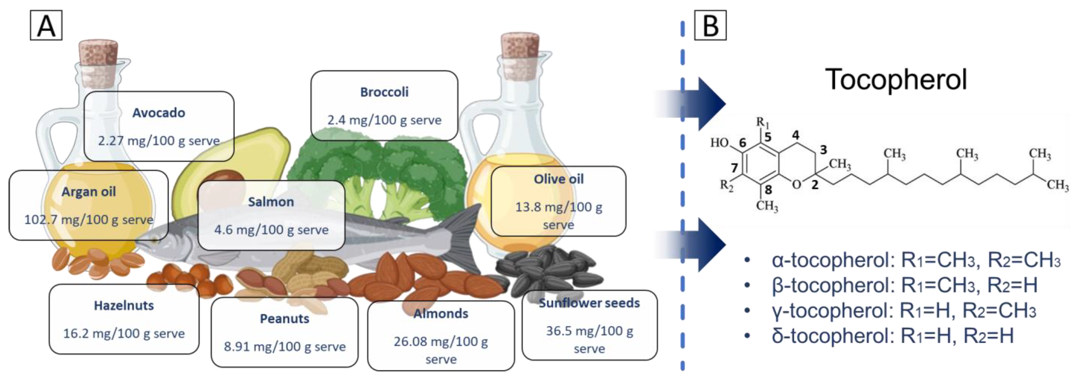
2. Structure and Properties

Tocopherols are a group of lipid-soluble compounds classified under vitamin E [15]. They are recognized for their potent antioxidant properties, which play a crucial role in protecting cell membranes from the oxidative damage caused by free radicals. Structurally, tocopherols are characterized by a chromanol ring attached to a long hydrophobic phytyl tail, making them amphipathic molecules [16]. This allows tocopherols to embed within biological membranes, where they stabilize lipid bilayers and prevent lipid peroxidation. There are four main types of tocopherols [17], alpha (α), beta (β), gamma (γ), and delta (δ), which differ in terms of the number and position of methyl groups on the chromanol ring (Figure 1B).
  • α-tocopherol: the chromanol ring has methyl groups at positions R1 = CH₃ and R2 = CH₃. This fully methylated structure allows α-tocopherol to have the highest antioxidant activity among tocopherols and makes it the most bioavailable form.
  • β-tocopherol: structurally, β-tocopherol has one methyl group located at R1 = CH₃ and one hydrogen atom in R2 = H. Its activity is intermediate, between that of α-tocopherol and γ-tocopherol, due to this arrangement.
  • γ-tocopherol: in this variant, the hydrogen atom is located at R1 = H, while the methyl group is located at R2 = CH₃. The absence of a methyl group at R1 alters its antioxidant properties. While its radical-scavenging activity is slightly lower than α-tocopherol, γ-tocopherol has a unique ability to trap RNS, such as peroxynitrite, which may confer additional protective effects.
  • δ-tocopherol: the chromanol ring in δ-tocopherol has hydrogen atoms in both substituents, which are positioned at R1 = H and R2 = H. This structure results in δ-tocopherol having the highest ability to neutralize ROS among the tocopherol isomers.
γ-tocopherol is distinguished by its unique chemical structure and physical properties, which together underpin its essential biological roles [1]. Its structure features a chromanol ring as the aromatic head, containing a hydroxyl group (-OH) at the 6th position (Figure 1B). This hydroxyl group is critical for its antioxidant activity, as it donates hydrogen atoms to neutralize free radicals and prevent oxidative damage [18]. γ-tocopherol’s substituent pattern, with methyl groups at the 7th and 8th positions, differs from α-tocopherol, which also has a methyl group at the 5th position (Figure 1B). This structural variation influences γ-tocopherol’s antioxidant function and biological activity. Additionally, the molecule possesses a long hydrophobic phytyl tail attached to the chromanol ring [19]. This tail anchors the molecule within lipid bilayers of cell membranes, where it protects membrane lipids from peroxidation and maintains cellular integrity [19]. Its molecular formula is C28H48O2, with a molecular weight of 416.68 g/mol. γ-tocopherol is lipophilic, making it soluble in fats and oils but insoluble in water [20].
Physically, γ-tocopherol appears as a viscous oil with a yellow to amber color. It remains stable under normal physiological conditions but can decompose at high temperatures. While it is insoluble in water, it dissolves readily in organic solvents such as ethanol, acetone, and lipids, facilitating its use in lipid-based applications [11]. γ-tocopherol is relatively stable when exposed to heat and light but is prone to degradation upon prolonged exposure to ultraviolet radiation or oxygen [21,22]. This susceptibility can diminish its antioxidant efficacy over time.

3. Antioxidant Effects

Oxidative stress reflects an imbalance between the systemic manifestation of the reactivity of oxygen species and the ability of biological systems to detoxify the body from free radicals, which leads to the accumulation of ROS in cells, resulting cellular toxicity and subsequently leading to tissue damage [3]. Antioxidants play a crucial role in neutralizing ROS and, among them, γ-tocopherol stands out due to its potent radical-scavenging capabilities [23,24]. Its antioxidant activity is attributed to its unique chemical structure, particularly the chromanol ring, which facilitates the donation of hydrogen atoms and stabilization of the resulting radical species [23,24].
Density functional theory (DFT) has been employed to explore the thermodynamic parameters governing the antioxidant activity of γ-tocopherol [25], suggesting that γ-tocopherol possesses a lower band dissociation than α-tocopherol [26]. This property may enhance the efficiency of γ-tocopherol in donating a hydrogen atom from its phenolic hydroxyl group, which can facilitate free radical neutralization. In addition, the electronic configuration of γ-tocopherol characterized by one less methyl group compared to α-tocopherol, enables it to trap more effectively with RNS, such as peroxynitrite (ONOO-) [27]. Furthermore, key parameters include the bond dissociation enthalpy (BDE), which measures the ease of hydrogen donation, with a low BDE indicating high reactivity towards free radicals [25,28]. The ionization potential (IP) reflects the compound’s electron-donating capacity, with lower IP values enhancing radical scavenging via electron transfer [29].
Theoretical studies also highlight two dominant mechanisms of action: (i) hydrogen atom transfer (HAT), where the phenolic hydrogen atom neutralizes free radicals [30], and (ii) sequential proton loss electron transfer (SPLET), which involves deprotonation followed by electron transfer [31]. These mechanisms underscore γ-tocopherol’s versatility and effectiveness in mitigating oxidative stress and preventing cellular damage.
Various studies have demonstrated, experimentally, the protective effects of γ-tocopherol against oxidative stress through its impact on key markers of this biological process involved in the apparition of several pathologies (Table 1).

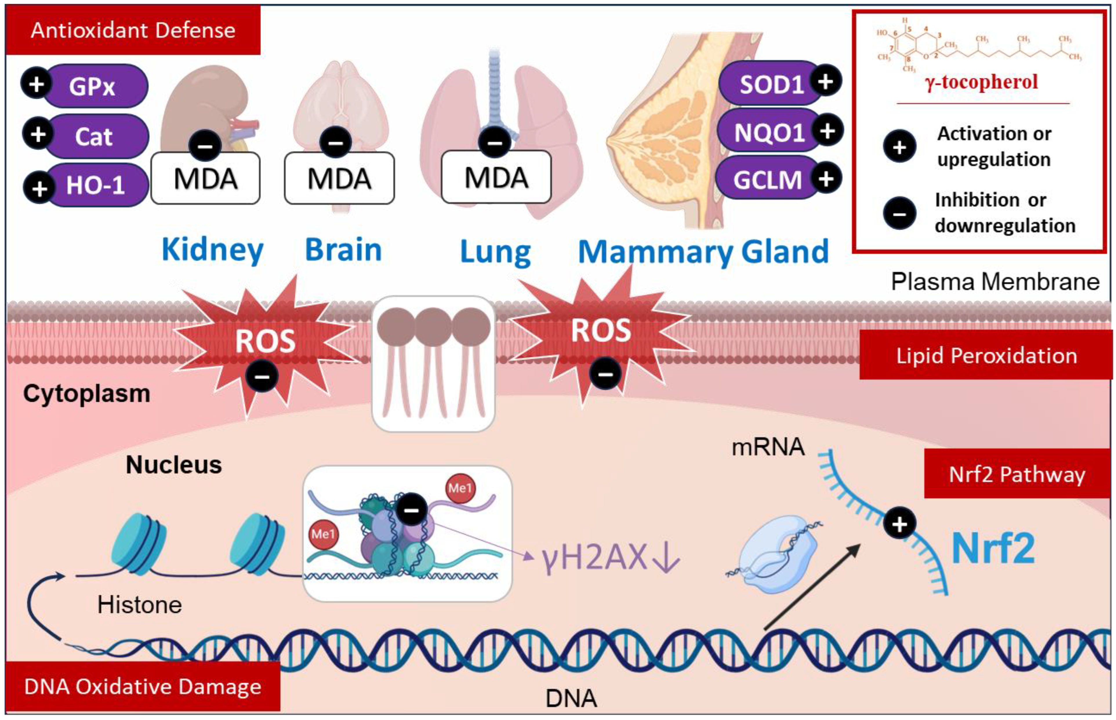
3.1. Effects Against Lipid Peroxidation

ROS cause lipid peroxidation, which is a complex process, renowned for the rearrangement of doubles bonds in polyunsaturated fatty acids, particularly arachidonic acids. This reaction generates an inactivation of receptors, and membrane permeability and fluidity, which leads to apoptosis [44,45].
γ-tocopherol exhibits remarkable antioxidant properties through its ability to modulate lipid peroxidation markers, particularly malondialdehyde (MDA) levels, in diverse biological systems. In diabetic-afflicted mice kidneys, γ-tocopherol was shown to decrease MDA concentrations [32]. Similarly, in normal mice stimulated by lipopolysaccharide (LPS), γ-tocopherol attenuated lipid peroxidation in the liver [33], highlighting its preventive and protective effect across different tissues. In sheep exposed to burn and smoke inhalation, γ-tocopherol nebulization decreased MDA levels in the lungs [34], reflecting its impact in modulating lipid peroxidation even under environmental toxin exposure. Interestingly, when γ-tocopherol is combined with α-tocopherol, MDA levels decreased in the plasma and livers of chicken, suggesting that this interaction might influence specific antioxidant pathways [46]. In light of this, natural sources such as argan oil, abundant in antioxidant compounds like γ-tocopherol [47], are recognized for their restorative properties in cases of oxidative stress [48,49]. Previous studies demonstrate that argan oil can normalize depleted MDA levels in the livers and the brains of mice under oxidative stress challenges, reaffirming the role of diet-derived antioxidants in hepatoprotection [49]. In addition, it was demonstrated that γ-tocopherol attenuated the serum levels of 8-isoprostane in rats implanted with 17β-estradiol (E2), with impacts varying based on the concentration and the period of treatment [33,35] (Figure 2 and Figure 3). Clinically, γ-tocopherol has been explored in conditions like metabolic syndrome, where it regulates lipid peroxidation by decreasing plasmatic MDA+HNE (4-hydroxy 2-nonenal) and lipid peroxides levels [36]. Given the relationship between γ-tocopherol and lipid peroxidation, it has been well-explained how this component can limit this process. One key mechanism involves the formation of metabolites from its oxidation, such as tocopherylquinone, which can scavenge lipid peroxyl radicals (ROO.) and stop lipid peroxidation [2].
This suggests that γ-tocopherol successfully helps to attenuate oxidative stress by regulating lipid peroxidation through reducing and normalizing the markers involved in this process, offering a potential beneficial therapeutic for oxidative stress-related disorders.

3.2. Effects Against DNA Oxidative Damage

ROS, particularly hydroxyl radical (OH•), has the potential to induce DNA damage, which encompasses processes such as base mutation, single- and double-strand breaks, and the formation of DNA adducts [37].
γ-tocopherol has been evaluated in preclinical models, demonstrating its effectiveness in protecting against DNA damage induced by oxidative stress. For instance, in the mammary gland of rats, γ-tocopherol administration significantly attenuated the E2-induced 8-oxo-2′-deoxyguanosine (8-oxo-dG) level, which is a marker of DNA oxidative damage and a product of nitrogenous base oxidation [37], by 72% and 67% at 7 and 14 days of treatments, respectively [35]. Similarly, Chen et al. [38] found that γ-tocopherol lowered the 8-oxo-dG level by 35% in colon tumors and by 42% in the adjacent tissues. On the other hand, the level of 8-oxo-dG was markedly decreased in the wild-type and nuclear factor erythroid 2–related factor 2 (Nrf2) (−/−) epithelial cells of mice, which suggests that the antioxidant potential of γ-tocopherol is independent of Nrf2-regulated antioxidative enzymes [39] (Figure 2). Additionally, it was demonstrated that the supplementation of γ-tocopherol significantly attenuated the levels of the marker of DNA damage phosphorylated histone γH2AX in the colon of 2-amino-1-methyl-6-phenylimidazo(4,5-b) pyridine (PHIP) -treated mice [38]. According to these available studies, γ-tocopherol exerts a protective effect against DNA oxidative damage, which is primarily demonstrated in vivo. These studies suggest an indirect effect of γ-tocopherol is to preserve DNA integrity and stability. However, the direct interactions between γ-tocopherol and DNA have not been conclusively demonstrated. Its role as an antioxidant highlights its preventive effect in maintaining DNA structure. Further research is needed to elucidate the direct interaction between γ-tocopherol and DNA to gain a deeper understanding of the associated biological pathways (Figure 2).

3.3. Effect on Antioxidant Defense

The body has a complex system of antioxidant defenses to protect against the harmful effects of ROS and RNS. There are two distinct levels of antioxidant effects: one is provided by nutrition through vegetable and fruits rich in vitamins C, E, carotenoids, flavonoids, or glutathione, whose role is to scavenging free radicals; the other is endogenous and consists of antioxidant enzymes such as superoxide dismutase (SOD), catalase (Cat), glutathione peroxidase (GPx), and heme oxygenase (HO-1), whose role is to degrade free radicals [50,51,52].
γ-tocopherol treatment is quite successful in improving the oxidative state in the kidneys of diabetic mice by normalizing the protein expression of antioxidant enzymes (catalase, GPx, and HO-1) [32]. At the transcriptional level, Das Gupta et al. found that the supplementation of γ-tocopherol significantly upregulating the mRNA expression of antioxidant enzymes (SOD1, GPx, and catalase) in the mammary glands of rats treated with E2, which can lead to degradation of the ROS produced by E2 [35]. Moreover, an in vitro study revealed that γ-tocopherol neutralizes ROS levels in PBMCs stimulated by LPS [40]. Similarly, the arterial superoxide anion generation, a well-established substrate of SOD enzyme, was significantly decreased in the γ-tocopherol-fed rats [53], suggesting that its degradation is facilitated by the enhancement of this enzyme. In addition, it was demonstrated that there was a notable rise in SOD activity in aortic homogenates as well as in the plasma of rats administered by γ-tocopherol [53]. Furthermore, γ-tocopherol significantly increases the proteins levels of NQO1 (NAD(P)H quinone dehydrogenase 1), GCLM (glutamate-cysteine ligase modifier subunit), and HO-1 in mammary tumors in mice subjected to E2-induced alterations, thereby mitigating oxidative stress, which helps to prevents cellular homeostasis [35] (Figure 2).

3.4. Effect on Nrf2 Pathway

Nrf2 is a transcription factor that is key for the preservation of cellular redox homeostasis and the elimination of reactive and carcinogenic mediators. The connection between Nrf2 and oxidative stress has been thoroughly elucidated by El Kebbaj et al. [54], who highlighted how natural compounds like fatty acids, tocopherols (α, β, γ, and δ), phytosterols (particularly schottenol and spinasterol), and ferulic acid can regulate Nrf2 expression during the oxidative stress process.
In this regard, it was demonstrated that γ-tocopherol-enhanced mammary gland Nrf2 mRNA levels in rats treated with E2 are correlated by increasing the mRNA expression of antioxidant enzyme [35]. Furthermore, a diet enriched with γ-tocopherol mixtures was shown to increase Nrf2 at both the mRNA and protein levels in the livers of mice [41,42]. However, it was also evaluated that Nrf2 did not exhibit a direct correlation with the modulation of MDA levels in the livers of mice [42], suggesting that the mechanism governing these pathways may be independent or may involve additional mediators indirectly linked to Nrf2 modulation. To better understand how γ-tocopherol influences Nrf2, a mechanistic study revealed that a tocopherol-rich mixture containing γ-tocopherol maintains the mRNA and protein levels of Nrf2 by inhibiting CpG hypermethylation in the Nrf2 promoter during prostate carcinogenesis in mice and in TRAMP-C1 cells [43] (Figure 2). These studies suggest that γ-tocopherol inhibits oxidative stress through modulating the Nrf2 signaling pathway to upregulate antioxidant proteins involved in free radical neutralization.

4. Anti-Inflammatory Activity

Inflammation is a key immune response that helps repair tissues after injury, but chronic inflammation can increase the risk of various diseases. γ-tocopherol, a powerful antioxidant, has shown potential in reducing inflammation by limiting neutrophils infiltration and cytokines’ production [55,56]. Several studies have evaluated its effects, revealing that γ-tocopherol may help modulate inflammatory responses, offering therapeutic benefits in conditions associated with acute inflammation and also with chronic inflammation such as diabetes and asthma.

4.1. Carrageenan-Induced Inflammation

γ-tocopherol’s effects in acute inflammation models, where the inflammatory response is more immediate and transient, have been thoroughly evaluated. In the carrageenan-induced inflammation model in the air pouch, γ-tocopherol significantly reduced the pro-inflammatory eicosanoids prostaglandin E2 (PGE2) (46%) and Leukotriene B4 (LTB4) (70%), the latter being a potent chemotactic factor produced by 5-lipoxygenase in neutrophils [57]. At a higher dose (i.e., 100 mg/kg), γ-tocopherol exhibited similar inhibitory effects on PGE2 (51%) and LTB4. Additionally, γ-tocopherol’s effects on TNF-α, a key inflammation mediator, were investigated. At the lower dose (i.e., 33 mg/kg), γ-tocopherol did not significantly inhibit TNF-α (51%), while at the higher dose, γ-tocopherol reduced TNF-α by 65%. These results suggest that γ-tocopherol modulates multiple components of the inflammatory response, offering potential therapeutic benefits in acute inflammation settings (Figure 3).
In a carrageenan-induced inflammation model in rats, a combination of aspirin and γ-tocopherol exhibited enhanced anti-inflammatory effects compared to aspirin alone [58]. The combination reduced PGE2 levels by 40% and decreased exudate volume by 15%, suggesting a prolonged anti-inflammatory action [58]. However, no significant effects were observed on immune cell infiltration. Of note, the combination of aspirin and γ-tocopherol also partially alleviated aspirin-induced gastric lesions and spared the reduction in food intake typically caused by aspirin. Additionally, aspirin and γ-tocopherol reduced gastric PGE2 depletion and decreased 8-isoprostane levels, indicating a protective effect against oxidative stress in the stomach [58]. These findings suggest that γ-tocopherol may improve the therapeutic profile of aspirin by enhancing its anti-inflammatory efficacy and reducing its adverse gastrointestinal effects.
In the IFN-γ/PMA-stimulated Caco-2 intestinal cell model system, γ-tocopherol exhibits significant anti-inflammatory effects, particularly by reducing IL-8 at both mRNA and protein levels [59]. In fact, at concentrations of 10 and 100 μM, γ-tocopherol significantly decreases IFN-γ/PMA-induced IL-8 production without inducing cytotoxicity. Additionally, γ-tocopherol downregulates IL-8 mRNA expression at 8 h, although this effect diminishes by 24 h [59]. Regarding NF-κB activation, γ-tocopherol is an inhibitor with moderate effectiveness, and δ-tocopherol the least effective [59]. Globally, these results highlight γ-tocopherol as a potent anti-inflammatory tocopherol for modulating inflammatory signaling pathways in this model.
In murine RAW264.7 macrophages treated with LPS and human A549 epithelial cancer cells treated with IL-1β, γ-tocopherol and its metabolite gamma-carboxyethyl hydroxychroman (γ-CEHC) effectively inhibit PGE2 synthesis in a dose-dependent manner, with γ-tocopherol demonstrating greater potency (IC50 values: 7.5 ± 2 μM in macrophages and 4 ± 1 μM in A549 cells) [60]. γ-tocopherol also reduces prostaglandin D2 (PGD2) levels in LPS-stimulated macrophages but is slightly less effective than it is on PGE2. While α-tocopherol has minimal or no effect on PGE2 and PGD2 synthesis, γ-CEHC directly inhibits COX-2 activity after short exposures, while γ-tocopherol requires prolonged exposure [60]. However, none of the tocopherols significantly reduce COX-2 protein expression or mRNA levels, despite inflammatory stimulation (Figure 3).
TNF-α stimulation increased Vascular Cell Adhesion Molecule 1 (VCAM-1) expression in HMEC-1 cells, which was significantly reduced by all tocopherols at 20 and 40 μM, with γ-tocopherol showing the strongest effect. At 10 μM, none of the tocopherols affected VCAM-1 expression. Moreover, in lymphatic endothelial cells, tocopherols did not reduce VCAM-1 expression after TNF-α exposure [61] (Figure 4).
γ-tocopherol exhibits potent anti-inflammatory effects by modulating key inflammatory markers such as IL-8 and VCAM-1, with significant inhibition of NF-κB activation and COX-2 activity. The mechanism of action of γ-tocopherol seems to involve both direct inhibition of inflammatory mediators and modulation of the endothelial barrier function, highlighting its therapeutic potential in inflammatory diseases and angiogenesis-related conditions [62]. Its effects were more pronounced than those of α-tocopherol, making γ-tocopherol a promising candidate for further exploration in inflammatory and vascular health management.

4.2. Diabetes-Induced Inflammation

γ-tocopherol demonstrates significant anti-inflammatory effects in diabetic models.
The studies collectively demonstrate that γ-tocopherol exerts anti-inflammatory effects by targeting key molecular pathways, particularly in severe diabetic conditions. γ-tocopherol consistently reduced NF-κB activity across tissues, leading to decreased levels of pro-inflammatory cytokines like IL-1β, TNF-α, and Monocyte Chemoattractant Protein-1 (MCP-1) [32,63]. It also elevated Sirtuin 1 (SIRT1), which inhibits NF-κB signaling and enhances antioxidant and repair mechanisms. The reduction in inflammasome components, such as NOD-like Receptor Pyrin Domain Containing 3 (NLRP3) and caspase-1 in hepatic tissue, further supports γ-tocopherol’s role in mitigating severe inflammation. Additionally, γ-tocopherol’s ability to increase nuclear peroxisome proliferator-activated receptor gamma coactivator 1 alpha (PGC-1α) and p53 in wound healing suggests it improves mitochondrial function and cellular stress responses [63].

4.3. Asthmatic Disease

Asthma is one of the most prevalent chronic childhood illnesses, characterized as a heterogeneous and multifactorial disorder influenced by a complex interplay of genetic and environmental factors [64]. Many in vivo and clinical trials have been conducted to assess the potential of γ-tocopherol in improving this disease.

4.3.1. Non-Allergic Asthma Induced by LPS

A combined animal and clinical study evaluated γ-tocopherol’s effects on LPS-induced airway inflammation [65]. In rats, LPS challenge-induced significant neutrophils accumulation in bronchoalveolar lavage fluid (BALF) and airway tissues, which were mitigated by 4 days of γ-tocopherol supplementation (30 mg/kg/day) [65]. A clinical trial using a double-blind, placebo-controlled crossover design involved 13 non-asthmatic individuals receiving γ-tocopherol supplementation (830 mg/day) or placebo before an LPS challenge. γ-tocopherol significantly reduced neutrophils and eosinophils in sputum and prevented LPS-induced increases in IL-1β levels, although it did not significantly alter IL-6, TNF-α, or markers such as TLR4 and CD11b [65]. These findings underline γ-tocopherol’s potential to reduce neutrophilic and eosinophilic inflammation in both animal and human models (Figure 3). Furthermore, a randomized study examined γ-tocopherol supplementation (1200 mg/day for 14 days) vs. placebo in participants undergoing an LPS challenge [66]. γ-tocopherol significantly reduced post-treatment sputum eosinophils, total mucins, and MUC5AC content when compared to the placebo group. It also attenuated LPS-induced neutrophilia at 6 h and 24 h post-challenge. While mucociliary clearance (MCC) slowed significantly during placebo treatment, no such slowing was observed during γ-tocopherol treatment [66]. Furthermore, γ-tocopherol mitigated LPS-induced increases in IL-1β and IL-8 but did not affect IL-6 or other TH1 cytokines [66]. These results underscore γ-tocopherol’s ability to reduce airway inflammation, neutrophils infiltration, and mucin production while potentially preserving MCC during LPS-induced airway challenges (Figure 3). Additionally, in a phase I open-label study, 16 volunteers (8 asthmatics and 8 healthy controls) were treated with a γ-tocopherol-rich supplement: one gel tab per day for one week, followed by two gel tabs per day for another week, with a washout period between dosing cycles. Blood samples collected before and after each dosing period showed no significant changes in serum cytokines (e.g., IL-1α, IL-1β, IL-12p40, granulocyte-macrophage colony-stimulating factor (GM-CSF), TNF, eotaxin, Rantes, macrophage inflammatory protein-1 alpha (MIP1α), and interferon gamma-inducible protein 10 (IP-10)) [40]. PBMCs collected from 14 participants and exposed to LPS exhibited significantly reduced secretion of pro-inflammatory cytokines (IL-1β, IL-6, and TNF-α) and chemokines (MCP1 and MIP1α) after γ-tocopherol supplementation, without affecting anti-inflammatory cytokines (Interleukin-1 receptor antagonist (IL-1RA) and Interleukin-10 (IL-10)) [40]. No differences in cytokine responses were observed between asthmatic and non-asthmatic participants.

4.3.2. Other Asthma Inducers

In a pilot randomized clinical trial [67], volunteers aged 18–45 underwent a controlled 2 h exposure to 500 mg/m3 of wood smoke particulates (WSPs) to assess γ-tocopherol supplementation’s effects on airway inflammation. Treatment included four doses of 1253 mg over 2 days or daily doses of 1154–1253 mg over 7 days. WSP exposure increased sputum neutrophils at 6 and 24 h, with no significant differences between γ-tocopherol and placebo groups [67]. Unlike its effects on endotoxin-induced inflammation, γ-tocopherol did not mitigate WSP or O3-induced neutrophilia, suggesting it may be more effective against endotoxin-induced airway responses [67].

4.4. Nanoformulation

Nanoformulation strategies have emerged as a promising solution to enhance γ-tocopherol pharmacokinetics and therapeutic efficacy [68]. By incorporating γ-tocopherol into nanoparticles, liposomes, or micelles, its stability, solubility, and targeted delivery to specific tissues are significantly improved [69]. Although studies on the biological effects of γ-tocopherol nanoformulations remain limited, existing research highlights their significant potential. For instance, a study by Kuo et al. demonstrated that γ-tocopherol nanoemulsions enhance anti-inflammatory efficacy by facilitating targeted delivery to inflamed tissues. This targeted approach allows for more efficient suppression of pro-inflammatory cytokines, such as TNF-α and IL-1β. In preclinical models of inflammation induced by 2% croton oil, γ-tocopherol nanoemulsions showed a marked reduction in inflammatory markers, underscoring their therapeutic promise for managing inflammatory conditions [14].

5. Anticancer Activity

The anti-tumoral potential of γ-tocopherol has been extensively studied through a variety of in vitro and in vivo experimental models. The underlying mechanisms by which the molecule exerts its anticancer effects are multifaceted and involve modulation of key signaling pathways and molecular targets that are crucial for tumorigenesis and cell proliferation. In this section, we will discuss the detailed mechanistic role of γ-tocopherol in cancer chemoprevention and destruction.

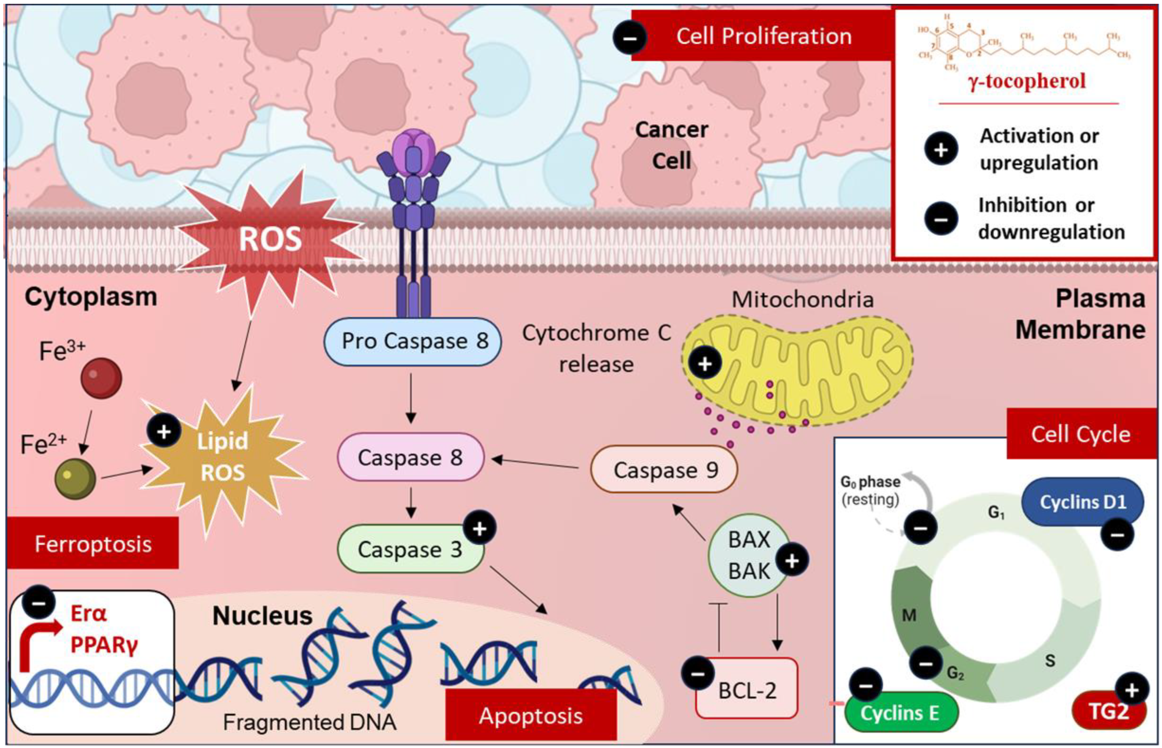
5.1. Modulation of Cell Death

The discovery of novel cell death pathways has significantly expanded our understanding of how cells respond to various forms of cellular damage. Apoptosis and ferroptosis are two critical forms of cell death that are intricately linked in cancer treatment [70,71]. Apoptosis, a regulated process, is often dysregulated in cancer, leading to uncontrolled cell proliferation; therapies that activate apoptotic pathways or inhibit anti-apoptotic proteins hold promise for inducing cancer cell death [72]. Ferroptosis, on the other hand, is a distinct form of programmed cell death that is characterized by the accumulation of lipid hydroperoxides to lethal levels, leading to membrane damage and cell death [73]. It is characterized by iron and lipidic ROS/peroxides’ accumulation due to the Fenton reaction and via the loss of balance in ROS production and cell glutathione (GSH)-dependent antioxidants, which protect cells from lipid peroxidation [74]. Emerging evidence suggests that ferroptosis may have a tumor-suppressive function by removing cells that lack access to critical nutrients or have been compromised by infection or environmental stress [75]. Jiang et al. showed that γ-tocopherol treatment induced apoptosis in prostate cancer cells [76] (Figure 4). γ-tocotrienol was more effective than γ-tocopherol to inhibit prostate tumor growth in vivo [77]. Furthermore, according to Gopalan et al., γ-tocopherol triggers apoptosis and enhances the expression of death receptor-5 in human breast cancer cells [78]. Mechanistic analysis revealed that treatment with γ-tocopherol leads to the intracellular buildup of dihydroceramides and dihydrosphingosine in prostate cancer cells [79], as well as ceramides and dihydroceramides in breast cancer cells [78]. The use of chemical inhibitors to block de novo ceramide biosynthesis significantly reduced γ-tocopherol’s apoptotic effects [78,79]. These results highlight that γ-tocopherol’s anticancer properties are mediated through its influence on the de novo synthesis of sphingolipids. Regarding its metabolites, while γ-tocopherol -13′-COOH has not been specifically investigated, similar metabolites such as δT-13′-COOH and δTE-13′-COOH have been shown to inhibit the proliferation of colon cancer cells, while having minimal effect on normal cells [80].
Moreover, Nrf2 plays a crucial role in protecting against ferroptosis. In fact, Nrf2 upregulate genes involved in antioxidant defense, iron metabolism, and NADPH synthesis. Through this regulation, Nrf2 enhances glutathione synthesis, supporting glutathione peroxidase 4 (GPX4) activity, the primary defense against lipid peroxidation. Nrf2 also regulates iron metabolism by increasing ferroportin expression, which reduces intracellular iron levels and limits ROS generation. Furthermore, it promotes NADPH regeneration via NQO1, an essential process for maintaining GPX4 function [81]. As the downregulation of Nrf2 is generally associated with increased susceptibility to ferroptosis, the importance of maintaining Nrf2 activity becomes apparent. In this regard, it has been demonstrated that a γ-tocopherol-rich mixture of tocopherols (γ-TmT) suppresses prostate tumor development in transgenic adenocarcinoma of the mouse prostate (TRAMP) by epigenetically maintaining Nrf2 expression. Specifically, γ-TmT inhibits DNA methyltransferases (DNMT1, DNMT3A, and DNMT3B), preventing hypermethylation of the Nrf2 promoter and, thus, preserving Nrf2 activity. This sustained Nrf2 expression leads to increased levels of downstream antioxidant and detoxification enzymes, ultimately protecting against prostate tumorigenesis [43] (Figure 4).
In addition to ferroptosis, γ-tocopherols have also been observed to modulate apoptosis, which is particularly relevant in the context of cancer. Apoptosis, a tightly regulated form of programmed cell death, plays a crucial role in maintaining tissue homeostasis by eliminating damaged or unwanted cells. Evasion of apoptosis is recognized as a hallmark of cancer, enabling uncontrolled cell proliferation and tumor development [82]. Mechanistic studies have revealed that the pro-apoptotic effect of γ-tocopherol is mediated through its ability to disrupt the balance between pro- and anti-apoptotic proteins, such as Bcl-2 and Bax [83]. In this study, it has been demonstrated that the combination of γ-tocopherol and 5-fluorouracil (5-FU) modulated the expression of the apoptosis-related genes as it increased the expression of Bax, a pro-apoptotic protein, while decreasing the expression of Bcl-2, an anti-apoptotic protein [83]. This disruption leads to the release of cytochrome c from the mitochondria, triggering the activation of the intrinsic mitochondrial apoptotic pathway. In fact, combining γ-tocopherol and lovastatin in HT-29 cells led to a loss of mitochondrial membrane potential and cytochrome c release, indicating the involvement of the intrinsic apoptotic pathway. Caspase-3 activation, a key executioner of apoptosis, was also observed [84]. In breast cancer cells, it has been demonstrated that γ-tocopherol induces apoptosis by activating the JNK/CHOP/DR5 signaling pathway. In fact, γ-tocopherol treatment activates JNK, a stress-activated protein kinase critical for apoptosis. JNK upregulates CHOP (GADD153), a transcription factor induced by cellular stress, which promotes apoptosis by regulating pro-apoptotic genes. CHOP also increases DR5 expression, a TNF receptor family death receptor, triggering the extrinsic apoptotic pathway and linking intrinsic and extrinsic apoptotic cascades [78].

5.2. Modulation of Cell Cycle

γ-tocopherol has also been shown to modulate the expression and activity of key proteins involved in cell-cycle regulation and survival. A study by Gysin et al. revealed that γ-tocopherol induces cell-cycle arrest at the G0/G1 phase, halting progression to the S phase and subsequent division. This transition, controlled by cyclins D and E, is significantly impaired as γ-tocopherol reduces these proteins’ levels. This cycle arrest is observed in various cancer cell lines, including prostate (DU145 and LNCaP), colon, and bone cancer cells. Furthermore, γ-tocopherol inhibits DNA synthesis by 55% in DU-145 and Caco-2 cells, aligning with its impact on cell-cycle progression [85]. Another study explored the effects of α- and γ-tocopherol on the human androgen-independent PC-3 prostate cancer cell line, revealing that both forms of tocopherol inhibited cell growth, with γ-tocopherol showing significantly higher potency. Also, treatment with γ-tocopherol decreased levels of cyclins D1 and E and increased both the expression and activity of transglutaminase 2 (TG2), which is an enzyme associated with cell differentiation and apoptosis. Moreover, both tocopherols reduced the number of cells entering the S phase of the cell cycle, thereby slowing down cell division, with the effect of γ-tocopherol being more prominent [86]. Interestingly, while Sato et al. explored the combined effects of δ-tocotrienol (δ-T3) and γ-tocopherol on prostate cancer cell growth using the androgen-sensitive LNCaP cell line, they showed that γ-tocopherol-induced arrest at the G2/M phase, suggesting a possible correlation with alterations in the expression of cyclin B, a key regulator of this phase [87] (Figure 4).

5.3. Modulation of Cell Proliferation

In a study investigating the effect of a γ-TmT, containing 58% of γ-tocopherol, on estrogen (E2) -induced mammary hyperplasia in rats, Smolarek et al. showed that γ-TmT inhibits cell proliferation by downregulating estrogen receptor α (ERα) expression, reducing serum estrogen levels, and upregulating peroxisome proliferator-activated receptor (PPARγ) and Nrf2 [42]. Furthermore, Campbell et al. found that γ-tocopherol significantly increased both mRNA and protein expression of PPARγ in SW480 human colon cancer cells in a dose-dependent manner [88]. PPARγ is a ligand-activated transcription factor that regulates gene expression by binding to DNA. It plays a crucial role in carcinogenesis due to its involvement in the normal growth and differentiation of cells. Researchers believe that an agent’s ability to modulate PPARγ could be significant, potentially leading to clinical implications for cancer chemoprevention, particularly in the colon [88]. When compared to α-tocopherol in vitro, γ-tocopherol showed a higher potency in inhibiting the growth and proliferation of MCF-7 breast cancer cells. Furthermore, γ-tocopherol significantly decreased ERα protein levels in these cells, which is a key finding since estrogen, acting through ERα, promotes the growth of many breast cancers. Indeed, by lowering ERα levels, γ-tocopherol disrupts this growth-promoting pathway. In vivo, when included in the diet of mice bearing MCF-7 xenografts, γ-tocopherol also markedly suppresses tumor growth [83] (Figure 4).

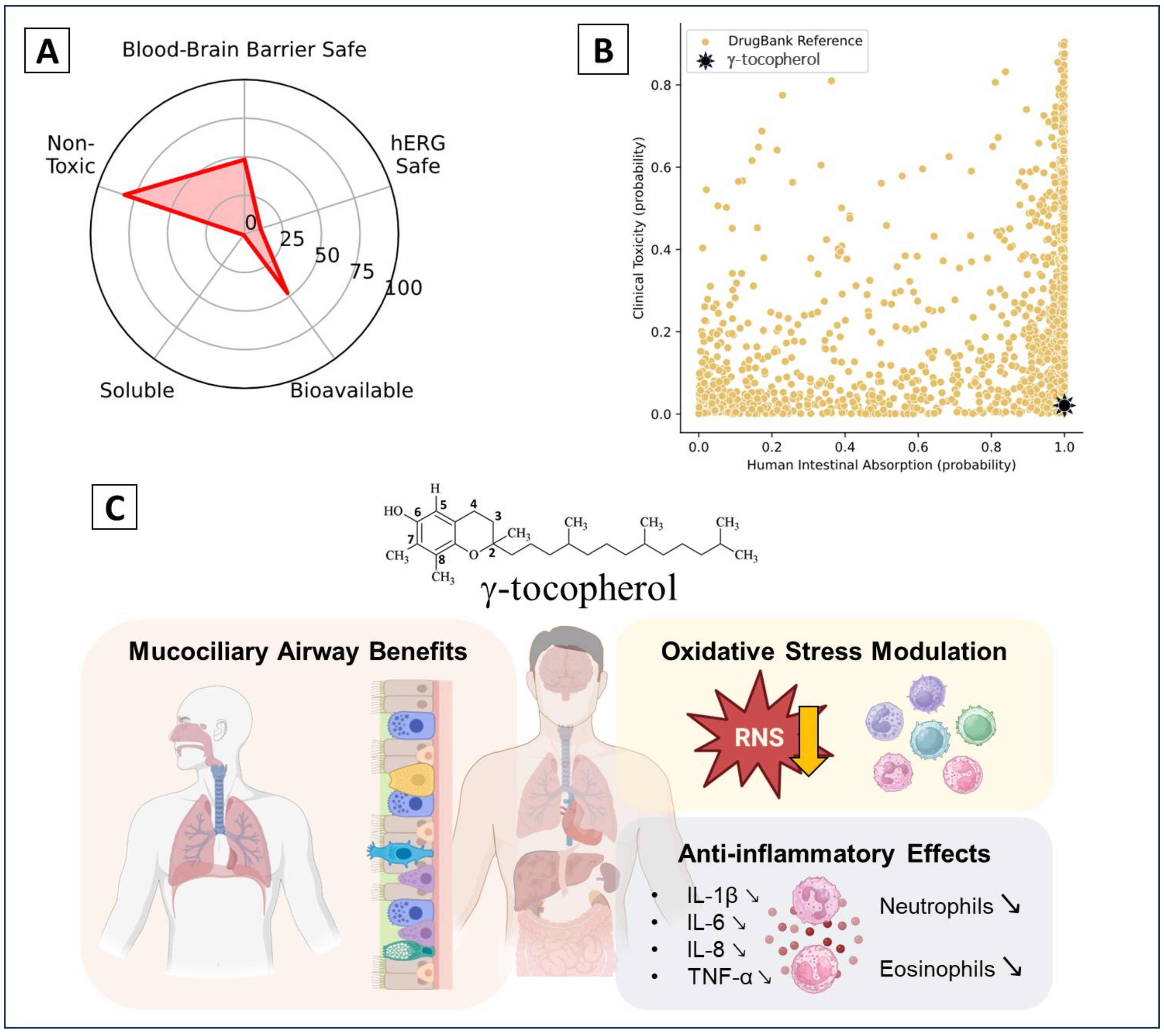
6. Clinical Trials and Therapeutic Potential

γ-tocopherol has been investigated in numerous clinical trials for its potential therapeutic benefits, particularly in managing inflammatory and oxidative stress-related conditions. Trials have demonstrated promising results, including increased serum levels of γ-tocopherol and its metabolite γ-CEHC, reduced nitrosative stress, and inhibition of NF-κB and JNK signaling pathways. γ-tocopherol supplementation also decreased pro-inflammatory cytokines such as IL-1β, IL-6, IL-8, and TNF-α, as well as neutrophil and eosinophil counts elevated by environmental exposures. Moreover, it prevented wood-smoke–induced airway inflammation and improved mucociliary clearance while reducing total sputum mucins. Additionally, γ-tocopherol showed vascular benefits, such as improved flow-mediated dilation, although some trials reported no significant changes in nitric oxide pathways or other oxidative stress markers. These findings highlight its potential role as an anti-inflammatory and antioxidant therapeutic agent. (Figure 5, Table 2).

7. Conclusions

γ-tocopherol, a significant isoform of vitamin E, emerges as a potent antioxidant with diverse protective effects against oxidative stress-induced damage. Its unique structural properties, including the chromanol ring and hydrophobic phytyl tail, enable its integration into lipid membranes, where it plays a pivotal role in neutralizing ROS and RNS. Furthermore, the anti-inflammatory activity of γ-tocopherol is also closely intertwined with its antioxidant properties. By neutralizing reactive oxygen and nitrogen species, γ-tocopherol not only reduces oxidative stress, a key driver of inflammation, but also modulates signaling pathways, such as NF-κB and Nrf2, thereby suppressing the production of pro-inflammatory cytokines and mediators. These combined effects underscore the critical role of its antioxidant capacity in contributing to its anti-inflammatory efficacy. Additionally, γ-tocopherol demonstrates remarkable efficacy in reducing lipid peroxidation markers, such as MDA and 8-isoprostane, across various biological systems, underscoring its potential in preventing cellular and tissue damage. Preclinical studies highlight its protective effects against DNA oxidative damage by reducing markers like 8-oxo-dG and γH2AX in multiple models, indicating its role in safeguarding genetic material. Nevertheless, further investigation into the precise molecular mechanisms and their influence on DNA structure and stability would be valuable. Additionally, γ-tocopherol’s antioxidant effects extend to enhancing the body’s defense systems, potentially offering therapeutic benefits for conditions linked to oxidative stress, including metabolic syndrome, environmental toxin exposure, and chronic inflammation.
Despite these promising findings, the complexity of its mechanisms and interactions with other antioxidants, such as α-tocopherol, necessitates further research. Future studies should focus on elucidating its precise molecular pathways and exploring its clinical applicability as a dietary supplement or therapeutic agent. Overall, γ-tocopherol stands out as a vital natural compound with immense potential to combat oxidative stress and related disorders, reinforcing the importance of diet-derived antioxidants in health promotion and disease prevention. Given the promising antioxidant and protective effects of γ-tocopherol demonstrated in various studies, the exploration of nanoformulation strategies becomes essential. These approaches could enhance its stability, bioavailability, and targeted delivery, addressing the limitations of conventional formulations. Since the existing research on nanoformulated γ-tocopherol is limited but encouraging, further investigations are warranted to optimize its therapeutic potential and broaden its application in oxidative stress-related disorders.

Author Contributions

Conceptualization, R.E.K., Y.L. (Youness Limami) and R.E.D.; data curation, B.E.-S., H.W., S.B., S.R. and Y.L. (Yassir Laaziouez); figures conception, H.W.; writing—original draft preparation, B.E.-S., H.W., S.B., S.R. and Y.L. (Yassir Laaziouez); and writing—review and editing, R.E.K., Y.L. (Youness Limami) and R.E.D. All authors have read and agreed to the published version of the manuscript.

Funding

This research was funded by the Ministry of Higher Education, Scientific Research and Innovation of Morocco (MESRSI), the National Agency for Medicinal and Aromatic Plants (ANPMA), and the National Center for Scientific and Technical Research (CNRST) within the framework of the fourth edition of the research project in the field of Valorization of Medicinal and Aromatic Plants VPMA4 (n°2022/17).

Institutional Review Board Statement

Not applicable.

Informed Consent Statement

Not applicable.

Data Availability Statement

Not applicable.

Acknowledgments

B.E.S gratefully acknowledges the CNRST (Centre National pour la Recherche Scientifique et Technique) in Morocco for the Phd Associate Scholarship PASS (2024–2027).

Conflicts of Interest

The authors declare no conflicts of interest.

References

  1. Rizvi, S.; Raza, S.T.; Ahmed, F.; Ahmad, A.; Abbas, S.; Mahdi, F. The role of vitamin e in human health and some diseases. Sultan Qaboos Univ. Med. J. 2014, 14, e157–e165. [Google Scholar] [PubMed]
  2. Jiang, Q.; Im, S.; Wagner, J.G.; Hernandez, M.L.; Peden, D.B. Gamma-tocopherol, a major form of vitamin E in diets: Insights into antioxidant and anti-inflammatory effects, mechanisms, and roles in disease management. Free Radic. Biol. Med. 2022, 178, 347–359. [Google Scholar] [CrossRef]
  3. Pizzino, G.; Irrera, N.; Cucinotta, M.; Pallio, G.; Mannino, F.; Arcoraci, V.; Squadrito, F.; Altavilla, D.; Bitto, A. Oxidative Stress: Harms and Benefits for Human Health. Oxid. Med. Cell Longev. 2017, 2017, 8416763. [Google Scholar] [CrossRef] [PubMed]
  4. Reddy, V.P. Oxidative Stress in Health and Disease. Biomedicines 2023, 11, 2925. [Google Scholar] [CrossRef] [PubMed]
  5. Hba, S.; Ghaddar, S.; Wahnou, H.; Pinon, A.; El Kebbaj, R.; Pouget, C.; Sol, V.; Liagre, B.; Oudghiri, M.; Limami, Y. Natural Chalcones and Derivatives in Colon Cancer: Pre-Clinical Challenges and the Promise of Chalcone-Based Nanoparticles. Pharmaceutics 2023, 15, 2718. [Google Scholar] [CrossRef] [PubMed]
  6. Zhao, H.; Wu, L.; Yan, G.; Chen, Y.; Zhou, M.; Wu, Y.; Li, Y. Inflammation and tumor progression: Signaling pathways and targeted intervention. Signal Transduct. Target. Ther. 2021, 6, 263. [Google Scholar] [CrossRef]
  7. Reuter, S.; Gupta, S.C.; Chaturvedi, M.M.; Aggarwal, B.B. Oxidative stress, inflammation, and cancer: How are they linked? Free Radic. Biol. Med. 2010, 49, 1603–1616. [Google Scholar] [CrossRef]
  8. Bindu, S.; Mazumder, S.; Bandyopadhyay, U. Non-steroidal anti-inflammatory drugs (NSAIDs) and organ damage: A current perspective. Biochem. Pharmacol. 2020, 180, 114147. [Google Scholar] [CrossRef]
  9. McEvoy, L.; Carr, D.F.; Pirmohamed, M. Pharmacogenomics of NSAID-Induced Upper Gastrointestinal Toxicity. Front. Pharmacol. 2021, 12, 684162. [Google Scholar] [CrossRef]
  10. Wahnou, H.; Ndayambaje, M.; Ouadghiri, Z.; Benayad, S.; Elattar, H.; Chgari, O.; Naya, A.; Zaid, Y.; Oudghiri, M. Artemisia herba-alba: Antioxidant capacity and efficacy in preventing chronic arthritis in vivo. Inflammopharmacology 2024, 32, 1855–1870. [Google Scholar] [CrossRef] [PubMed]
  11. Pahrudin Arrozi, A.; Shukri, S.N.S.; Wan Ngah, W.Z.; Mohd Yusof, Y.A.; Ahmad Damanhuri, M.H.; Jaafar, F.; Makpol, S. Comparative Effects of Alpha- and Gamma-Tocopherol on Mitochondrial Functions in Alzheimer’s Disease In Vitro Model. Sci. Rep. 2020, 10, 8962. [Google Scholar] [CrossRef] [PubMed]
  12. Reiter, E.; Jiang, Q.; Christen, S. Anti-inflammatory properties of alpha- and gamma-tocopherol. Mol. Asp. Med. 2007, 28, 668–691. [Google Scholar] [CrossRef] [PubMed]
  13. Devaraj, S.; Jialal, I. Failure of Vitamin E in Clinical Trials: Is Gamma-Tocopherol the Answer? Nutr. Rev. 2005, 63, 290–293. [Google Scholar] [CrossRef]
  14. Kuo, F.; Subramanian, B.; Kotyla, T.; Wilson, T.A.; Yoganathan, S.; Nicolosi, R.J. Nanoemulsions of an anti-oxidant synergy formulation containing gamma tocopherol have enhanced bioavailability and anti-inflammatory properties. Int. J. Pharm. 2008, 363, 206–213. [Google Scholar] [CrossRef] [PubMed]
  15. Das Gupta, S.; Suh, N. Tocopherols in cancer: An update. Mol. Nutr. Food Res. 2016, 60, 1354–1363. [Google Scholar] [CrossRef] [PubMed]
  16. Colombo, M.L. An Update on Vitamin E, Tocopherol and Tocotrienol—Perspectives. Molecules 2010, 15, 2103–2113. [Google Scholar] [CrossRef] [PubMed]
  17. Xu, Z.; Harvey, K.; Pavlina, T.; Zaloga, G.; Siddiqui, R. Tocopherol and Tocotrienol Homologs in Parenteral Lipid Emulsions. Eur. J. Lipid Sci. Technol. 2015, 117, 15–22. [Google Scholar] [CrossRef] [PubMed]
  18. Charlton, N.C.; Mastyugin, M.; Török, B.; Török, M. Structural Features of Small Molecule Antioxidants and Strategic Modifications to Improve Potential Bioactivity. Molecules 2023, 28, 1057. [Google Scholar] [CrossRef] [PubMed]
  19. Atkinson, J.; Harroun, T.; Wassall, S.R.; Stillwell, W.; Katsaras, J. The location and behavior of alpha-tocopherol in membranes. Mol. Nutr. Food Res. 2010, 54, 641–651. [Google Scholar] [CrossRef] [PubMed]
  20. Shahidi, F.; de Camargo, A.C. Tocopherols and Tocotrienols in Common and Emerging Dietary Sources: Occurrence, Applications, and Health Benefits. Int. J. Mol. Sci. 2016, 17, 1745. [Google Scholar] [CrossRef] [PubMed]
  21. Balakrishnan, G.; Schneider, R.G. Tocopherol degradation and lipid oxidation during storage of Chenopodium quinoa. J. Food Compos. Anal. 2023, 123, 105549. [Google Scholar] [CrossRef]
  22. Bruscatto, M.H.; Zambiazi, R.C.; Sganzerla, M.; Pestana, V.R.; Otero, D.; Lima, R.; Paiva, F. Degradation of Tocopherols in Rice Bran Oil Submitted to Heating at Different Temperatures. J. Chromatogr. Sci. 2009, 47, 762–765. [Google Scholar] [CrossRef]
  23. Aoyama, S.; Nishio, T.; Moriya, D.; Munemasa, S.; Murata, Y.; Nakamura, Y.; Nakamura, T. The Metabolite of γ-Tocopherol, 2,7,8-Trimethyl-2-(2′-Carboxyethyl)-6-Hydroxychroman, Exerts Intracellular Antioxidant Activity via Up-Regulation of Heme Oxygenase-1 in Hepatocytes. Nutraceuticals 2024, 4, 409–416. [Google Scholar] [CrossRef]
  24. Thbayh, D.K.; Mentes, D.; Boros, Z.R.; Palusiak, M.; Farkas, L.; Viskolcz, B.; Fiser, B. α-Tocopherol and Trolox as Effective Natural Additives for Polyurethane Foams: A DFT and Experimental Study. Molecules 2024, 29, 6037. [Google Scholar] [CrossRef]
  25. Amić, A.; Mastil’ák Cagardová, D. A DFT study of the antioxidant potency of α-tocopherol and its derivatives: PMHC, Trolox, and α-CEHC. J. Mol. Liq. 2024, 403, 124796. [Google Scholar] [CrossRef]
  26. Benbrahim, N.; Zeddour-Brahim, K.; Zizi, Z.; Bengharez, Z. Stability and Reactivity of Tocopherols: Theoretical Study. Chem. Proc. 2023, 14, 20. [Google Scholar] [CrossRef]
  27. Wagner, K.H.; Kamal-Eldin, A.; Elmadfa, I. Gamma-tocopherol—an underestimated vitamin? Ann. Nutr. Metab. 2004, 48, 169–188. [Google Scholar] [CrossRef] [PubMed]
  28. Yamabe, S.; Tsuchida, N.; Yamazaki, S. A DFT Study on Transition States of Inhibition of Oxidation by α-Tocopherol. ChemistrySelect 2020, 5, 9184–9194. [Google Scholar] [CrossRef]
  29. Abyar, F.; Farrokhpour, H. Ionization energies and photoelectron spectra of fat-soluble vitamins in the gas phase: A theoretical study. RSC Adv. 2014, 4, 35975–35987. [Google Scholar] [CrossRef]
  30. Warren, J.J.; Mayer, J.M. Predicting organic hydrogen atom transfer rate constants using the Marcus cross relation. Proc. Natl. Acad. Sci. USA 2010, 107, 5282–5287. [Google Scholar] [CrossRef] [PubMed]
  31. Povalishev, V.N.; Polozov, G.I.; Shadyro, O.I. Effects of α-tocopherol and related compounds on reactions involving various organic radicals. Bioorganic Med. Chem. Lett. 2006, 16, 1236–1239. [Google Scholar] [CrossRef] [PubMed]
  32. Shin, H.; Eo, H.; Lim, Y. Similarities and differences between alpha-tocopherol and gamma-tocopherol in amelioration of inflammation, oxidative stress and pre-fibrosis in hyperglycemia induced acute kidney inflammation. Nutr. Res. Pract. 2016, 10, 33–41. [Google Scholar] [CrossRef]
  33. Chung, M.Y.; Yeung, S.F.; Park, H.J.; Volek, J.S.; Bruno, R.S. Dietary α- and γ-tocopherol supplementation attenuates lipopolysaccharide-induced oxidative stress and inflammatory-related responses in an obese mouse model of nonalcoholic steatohepatitis. J. Nutr. Biochem. 2010, 21, 1200–1206. [Google Scholar] [CrossRef] [PubMed]
  34. Yamamoto, Y.; Sousse, L.E.; Enkhbaatar, P.; Kraft, E.R.; Deyo, D.J.; Wright, C.L.; Taylor, A.; Traber, M.G.; Cox, R.A.; Hawkins, H.K.; et al. γ-tocopherol nebulization decreases oxidative stress, arginase activity, and collagen deposition after burn and smoke inhalation in the ovine model. Shock 2012, 38, 671–676. [Google Scholar] [CrossRef] [PubMed]
  35. Das Gupta, S.; So, J.Y.; Wall, B.; Wahler, J.; Smolarek, A.K.; Sae-Tan, S.; Soewono, K.Y.; Yu, H.; Lee, M.J.; Thomas, P.E.; et al. Tocopherols inhibit oxidative and nitrosative stress in estrogen-induced early mammary hyperplasia in ACI rats. Mol. Carcinog. 2015, 54, 916–925. [Google Scholar] [CrossRef]
  36. Devaraj, S.; Leonard, S.; Traber, M.G.; Jialal, I. Gamma-tocopherol supplementation alone and in combination with alpha-tocopherol alters biomarkers of oxidative stress and inflammation in subjects with metabolic syndrome. Free Radic. Biol. Med. 2008, 44, 1203–1208. [Google Scholar] [CrossRef]
  37. Kowalska, M.; Piekut, T.; Prendecki, M.; Sodel, A.; Kozubski, W.; Dorszewska, J. Mitochondrial and Nuclear DNA Oxidative Damage in Physiological and Pathological Aging. DNA Cell Biol. 2020, 39, 1410–1420. [Google Scholar] [CrossRef] [PubMed]
  38. Chen, J.X.; Liu, A.; Lee, M.J.; Wang, H.; Yu, S.; Chi, E.; Reuhl, K.; Suh, N.; Yang, C.S. δ- and γ-tocopherols inhibit phIP/DSS-induced colon carcinogenesis by protection against early cellular and DNA damages. Mol. Carcinog. 2017, 56, 172–183. [Google Scholar] [CrossRef] [PubMed]
  39. Li, G.; Lee, M.J.; Liu, A.B.; Yang, Z.; Lin, Y.; Shih, W.J.; Yang, C.S. The antioxidant and anti-inflammatory activities of tocopherols are independent of Nrf2 in mice. Free Radic. Biol. Med. 2012, 52, 1151–1158. [Google Scholar] [CrossRef]
  40. Wiser, J.; Alexis, N.E.; Jiang, Q.; Wu, W.; Robinette, C.; Roubey, R.; Peden, D.B. In vivo gamma-tocopherol supplementation decreases systemic oxidative stress and cytokine responses of human monocytes in normal and asthmatic subjects. Free Radic. Biol. Med. 2008, 45, 40–49. [Google Scholar] [CrossRef]
  41. Dey, P.; Mah, E.; Li, J.; Jalili, T.; Symons, J.D.; Bruno, R.S. Improved hepatic γ-tocopherol status limits oxidative and inflammatory stress-mediated liver injury in db/db mice with nonalcoholic steatohepatitis. J. Funct. Foods 2018, 40, 670–678. [Google Scholar] [CrossRef]
  42. Smolarek, A.K.; So, J.Y.; Thomas, P.E.; Lee, H.J.; Paul, S.; Dombrowski, A.; Wang, C.X.; Saw, C.L.; Khor, T.O.; Kong, A.N.; et al. Dietary tocopherols inhibit cell proliferation, regulate expression of ERα, PPARγ, and Nrf2, and decrease serum inflammatory markers during the development of mammary hyperplasia. Mol. Carcinog. 2013, 52, 514–525. [Google Scholar] [CrossRef] [PubMed]
  43. Huang, Y.; Khor, T.O.; Shu, L.; Saw, C.L.; Wu, T.Y.; Suh, N.; Yang, C.S.; Kong, A.N. A γ-tocopherol-rich mixture of tocopherols maintains Nrf2 expression in prostate tumors of TRAMP mice via epigenetic inhibition of CpG methylation. J. Nutr. 2012, 142, 818–823. [Google Scholar] [CrossRef] [PubMed]
  44. Gaschler, M.M.; Stockwell, B.R. Lipid peroxidation in cell death. Biochem. Biophys. Res. Commun. 2017, 482, 419–425. [Google Scholar] [CrossRef] [PubMed]
  45. Luo, J.; Mills, K.; le Cessie, S.; Noordam, R.; van Heemst, D. Ageing, age-related diseases and oxidative stress: What to do next? Ageing Res. Rev. 2020, 57, 100982. [Google Scholar] [CrossRef]
  46. Korošec, T.; Tomažin, U.; Horvat, S.; Keber, R.; Salobir, J. The diverse effects of α- and γ-tocopherol on chicken liver transcriptome. Poult. Sci. 2017, 96, 667–680. [Google Scholar] [CrossRef] [PubMed]
  47. Kamal, R.; Kharbach, M.; Vander Heyden, Y.; Doukkali, Z.; Ghchime, R.; Bouklouze, A.; Cherrah, Y.; Alaoui, K. In vivo anti-inflammatory response and bioactive compounds’ profile of polyphenolic extracts from edible Argan oil (Argania spinosa L.), obtained by two extraction methods. J. Food Biochem. 2019, 43, e13066. [Google Scholar] [CrossRef] [PubMed]
  48. Bouchab, H.; Ishaq, A.; El Kebbaj, R.; Nasser, B.; Saretzki, G. Protective effect of argan oil on DNA damage in vivo and in vitro. Biomarkers 2021, 26, 425–433. [Google Scholar] [CrossRef] [PubMed]
  49. Bouchab, H.; Essadek, S.; El Kamouni, S.; Moustaid, K.; Essamadi, A.; Andreoletti, P.; Cherkaoui-Malki, M.; El Kebbaj, R.; Nasser, B. Antioxidant Effects of Argan Oil and Olive Oil against Iron-Induced Oxidative Stress: In Vivo and In Vitro Approaches. Molecules 2023, 28, 5924. [Google Scholar] [CrossRef]
  50. Haleng, J.; Pincemail, J.; Defraigne, J.O.; Charlier, C.; Chapelle, J.P. Oxidative stress. Rev. Medicale Liege 2007, 62, 628–638. [Google Scholar]
  51. El Faqer, O.; Rais, S.; Ouadghiri, Z.; El Faqer, A.; Benchama, Z.; El Ouaddari, A.; Dakir, M.; El Amrani, A.; Mtairag, E.M. Physicochemical properties, GC–MS profiling, and antibacterial potential of Allium sativum essential oil: In vitro and in silico approaches. Sci. Afr. 2024, 26, e02484. [Google Scholar] [CrossRef]
  52. El Faqer, O.; Elkoraichi, I.; Latif, M.; Debierre-Grockiego, F.; Ouadghiri, Z.; Rais, S.; Dimier-Poisson, I.; Mtairag, E.M. Pharmacological insights into Laurus nobilis: HPLC profiling and evaluation of its anti-Toxoplasma, antioxidant, and anti-hemolytic properties. Biochem. Syst. Ecol. 2024, 117, 104891. [Google Scholar] [CrossRef]
  53. Saldeen, T.; Li, D.; Mehta, J.L. Differential effects of alpha- and gamma-tocopherol on low-density lipoprotein oxidation, superoxide activity, platelet aggregation and arterial thrombogenesis. J. Am. Coll. Cardiol. 1999, 34, 1208–1215. [Google Scholar] [CrossRef] [PubMed]
  54. El Kebbaj, R.; Bouchab, H.; Tahri-Joutey, M.; Rabbaa, S.; Limami, Y.; Nasser, B.; Egbujor, M.C.; Tucci, P.; Andreoletti, P.; Saso, L.; et al. The Potential Role of Major Argan Oil Compounds as Nrf2 Regulators and Their Antioxidant Effects. Antioxidants 2024, 13, 344. [Google Scholar] [CrossRef] [PubMed]
  55. Arulselvan, P.; Fard, M.T.; Tan, W.S.; Gothai, S.; Fakurazi, S.; Norhaizan, M.E.; Kumar, S.S. Role of Antioxidants and Natural Products in Inflammation. Oxid. Med. Cell Longev. 2016, 2016, 5276130. [Google Scholar] [CrossRef]
  56. Wahnou, H.; Hmimid, F.; Errami, A.; Nait Irahal, I.; Limami, Y.; Oudghiri, M. Integrating ADMET, enrichment analysis, and molecular docking approach to elucidate the mechanism of Artemisia herba alba for the treatment of inflammatory bowel disease-associated arthritis. J. Toxicol. Environ. Health Part A 2024, 87, 836–854. [Google Scholar] [CrossRef]
  57. Jiang, Q.; Ames, B.N. Gamma-tocopherol, but not alpha-tocopherol, decreases proinflammatory eicosanoids and inflammation damage in rats. FASEB J. 2003, 17, 816–822. [Google Scholar] [CrossRef]
  58. Jiang, Q.; Moreland, M.; Ames, B.N.; Yin, X. A combination of aspirin and gamma-tocopherol is superior to that of aspirin and alpha-tocopherol in anti-inflammatory action and attenuation of aspirin-induced adverse effects. J. Nutr. Biochem. 2009, 20, 894–900. [Google Scholar] [CrossRef]
  59. Elisia, I.; Kitts, D.D. Tocopherol isoforms (α-, γ-, and δ-) show distinct capacities to control Nrf-2 and NfκB signaling pathways that modulate inflammatory response in Caco-2 intestinal cells. Mol. Cell. Biochem. 2015, 404, 123–131. [Google Scholar] [CrossRef] [PubMed]
  60. Jiang, Q.; Elson-Schwab, I.; Courtemanche, C.; Ames, B.N. gamma-tocopherol and its major metabolite, in contrast to alpha-tocopherol, inhibit cyclooxygenase activity in macrophages and epithelial cells. Proc. Natl. Acad. Sci. USA 2000, 97, 11494–11499. [Google Scholar] [CrossRef]
  61. Wells, S.R.; Jennings, M.H.; Rome, C.; Hadjivassiliou, V.; Papas, K.A.; Alexander, J.S. Alpha-, gamma- and delta-tocopherols reduce inflammatory angiogenesis in human microvascular endothelial cells. J. Nutr. Biochem. 2010, 21, 589–597. [Google Scholar] [CrossRef] [PubMed]
  62. Mah, E.; Pei, R.; Guo, Y.; Ballard, K.D.; Barker, T.; Rogers, V.E.; Parker, B.A.; Taylor, A.W.; Traber, M.G.; Volek, J.S.; et al. γ-Tocopherol-rich supplementation additively improves vascular endothelial function during smoking cessation. Free Radic. Biol. Med. 2013, 65, 1291–1299. [Google Scholar] [CrossRef]
  63. Lee, H.; Lim, Y. Gamma-tocopherol ameliorates hyperglycemia-induced hepatic inflammation associated with NLRP3 inflammasome in alloxan-induced diabetic mice. Nutr. Res. Pract. 2019, 13, 377–383. [Google Scholar] [CrossRef] [PubMed]
  64. Gans, M.D.; Gavrilova, T. Understanding the immunology of asthma: Pathophysiology, biomarkers, and treatments for asthma endotypes. Paediatr. Respir. Rev. 2020, 36, 118–127. [Google Scholar] [CrossRef] [PubMed]
  65. Hernandez, M.L.; Wagner, J.G.; Kala, A.; Mills, K.; Wells, H.B.; Alexis, N.E.; Lay, J.C.; Jiang, Q.; Zhang, H.; Zhou, H.; et al. Vitamin E, γ-tocopherol, reduces airway neutrophil recruitment after inhaled endotoxin challenge in rats and in healthy volunteers. Free Radic. Biol. Med. 2013, 60, 56–62. [Google Scholar] [CrossRef]
  66. Burbank, A.J.; Duran, C.G.; Pan, Y.; Burns, P.; Jones, S.; Jiang, Q.; Yang, C.; Jenkins, S.; Wells, H.; Alexis, N.; et al. Gamma tocopherol-enriched supplement reduces sputum eosinophilia and endotoxin-induced sputum neutrophilia in volunteers with asthma. J. Allergy Clin. Immunol. 2018, 141, 1231–1238.e1. [Google Scholar] [CrossRef] [PubMed]
  67. Peden, D.B.; Almond, M.; Brooks, C.; Robinette, C.; Wells, H.; Burbank, A.; Hernandez, M.; Hinderliter, A.; Caughey, M.; Jiang, Q.; et al. A pilot randomized clinical trial of γ-tocopherol supplementation on wood smoke-induced neutrophilic and eosinophilic airway inflammation. J. Allergy Clin. Immunol. Glob. 2023, 2, 100177. [Google Scholar] [CrossRef] [PubMed]
  68. Wahnou, H.; Liagre, B.; Sol, V.; El Attar, H.; Attar, R.; Oudghiri, M.; Duval, R.E.; Limami, Y. Polyphenol-Based Nanoparticles: A Promising Frontier for Enhanced Colorectal Cancer Treatment. Cancers 2023, 15, 3826. [Google Scholar] [CrossRef]
  69. Patra, J.K.; Das, G.; Fraceto, L.F.; Campos, E.V.R.; del Rodriguez-Torres, M.P.; Acosta-Torres, L.S.; Diaz-Torres, L.A.; Grillo, R.; Swamy, M.K.; Sharma, S.; et al. Nano based drug delivery systems: Recent developments and future prospects. J. Nanobiotechnol. 2018, 16, 71. [Google Scholar] [CrossRef] [PubMed]
  70. Nikoletopoulou, V.; Markaki, M.; Palikaras, K.; Tavernarakis, N. Crosstalk between apoptosis, necrosis and autophagy. Biochim. Biophys. Acta—Mol. Cell Res. 2013, 1833, 3448–3459. [Google Scholar] [CrossRef] [PubMed]
  71. Chgari, O.; Wahnou, H.; Ndayambaje, M.; Moukhfi, F.; Benkhnigue, O.; Marnissi, F.; Limami, Y.; Oudghiri, M. Orbea variegata (L.) Haw in skin carcinogenesis: Insights from an in vivo male Swiss mouse model study. J. Toxicol. Environ. Health Part A 2024, 87, 630–645. [Google Scholar] [CrossRef] [PubMed]
  72. Towers, C.G.; Wodetzki, D.; Thorburn, A. Autophagy and cancer: Modulation of cell death pathways and cancer cell adaptations. J. Cell Biol. 2020, 219, e201909033. [Google Scholar] [CrossRef] [PubMed]
  73. Fan, J.; Jiang, T.; He, D. Emerging insights into the role of ferroptosis in the pathogenesis of autoimmune diseases. Front. Immunol. 2023, 14, 1120519. [Google Scholar] [CrossRef] [PubMed]
  74. Benayad, S.; Wahnou, H.; El Kebbaj, R.; Liagre, B.; Sol, V.; Oudghiri, M.; Saad, E.M.; Duval, R.E.; Limami, Y. The Promise of Piperine in Cancer Chemoprevention. Cancers 2023, 15, 5488. [Google Scholar] [CrossRef] [PubMed]
  75. Stockwell, B.R.; Friedmann Angeli, J.P.; Bayir, H.; Bush, A.I.; Conrad, M.; Dixon, S.J.; Fulda, S.; Gascón, S.; Hatzios, S.K.; Kagan, V.E.; et al. Ferroptosis: A Regulated Cell Death Nexus Linking Metabolism, Redox Biology, and Disease. Cell 2017, 171, 273–285. [Google Scholar] [CrossRef] [PubMed]
  76. Di Pardo, A.; Basit, A.; Armirotti, A.; Amico, E.; Castaldo, S.; Pepe, G.; Marracino, F.; Buttari, F.; Digilio, A.F.; Maglione, V. De novo Synthesis of Sphingolipids Is Defective in Experimental Models of Huntington’s Disease. Front. Neurosci. 2017, 11, 698. [Google Scholar] [CrossRef] [PubMed]
  77. Jiang, Q.; Rao, X.; Kim, C.Y.; Freiser, H.; Zhang, Q.; Jiang, Z.; Li, G. Gamma-tocotrienol induces apoptosis and autophagy in prostate cancer cells by increasing intracellular dihydrosphingosine and dihydroceramide. Int. J. Cancer 2012, 130, 685–693. [Google Scholar] [CrossRef] [PubMed]
  78. Gopalan, A.; Yu, W.; Jiang, Q.; Jang, Y.; Sanders, B.G.; Kline, K. Involvement of de novo ceramide synthesis in gamma-tocopherol and gamma-tocotrienol-induced apoptosis in human breast cancer cells. Mol. Nutr. Food Res. 2012, 56, 1803–1811. [Google Scholar] [CrossRef]
  79. Jiang, Q.; Wong, J.; Fyrst, H.; Saba, J.D.; Ames, B.N. γ-Tocopherol or combinations of vitamin E forms induce cell death in human prostate cancer cells by interrupting sphingolipid synthesis. Proc. Natl. Acad. Sci. USA 2004, 101, 17825–17830. [Google Scholar] [CrossRef]
  80. Jang, Y.; Park, N.-Y.; Rostgaard-Hansen, A.L.; Huang, J.; Jiang, Q. Vitamin E metabolite 13′-carboxychromanols inhibit pro-inflammatory enzymes, induce apoptosis and autophagy in human cancer cells by modulating sphingolipids and suppress colon tumor development in mice. Free Radic. Biol. Med. 2016, 95, 190–199. [Google Scholar] [CrossRef]
  81. Yan, R.; Lin, B.; Jin, W.; Tang, L.; Hu, S.; Cai, R. NRF2, a Superstar of Ferroptosis. Antioxidants 2023, 12, 1739. [Google Scholar] [CrossRef] [PubMed]
  82. Plati, J.; Bucur, O.; Khosravi-Far, R. Apoptotic cell signaling in cancer progression and therapy. Integr. Biol. 2011, 3, 279–296. [Google Scholar] [CrossRef] [PubMed]
  83. Bazzaz, R.; Bijanpour, H.; Pirouzpanah, S.M.B.; Yaghmaei, P.; Rashtchizadeh, N. Adjuvant therapy with γ-tocopherol-induce apoptosis in HT-29 colon cancer via cyclin-dependent cell cycle arrest mechanism. J. Biochem. Mol. Toxicol. 2019, 33, e22399. [Google Scholar] [CrossRef] [PubMed]
  84. Rezaei, M.; Zeidooni, L.; Hashemitabar, M.; Razzazzadeh, S.; Mahdavinia, M.; Ghasemi, K. Gamma-tocopherol enhances apoptotic effects of lovastatin in human colorectal carcinoma cell line (HT29). Nutr. Cancer 2014, 66, 1386–1393. [Google Scholar] [CrossRef] [PubMed]
  85. Gysin, R.; Azzi, A.; Visarius, T. Gamma-tocopherol inhibits human cancer cell cycle progression and cell proliferation by down-regulation of cyclins. FASEB J. 2002, 16, 1952–1954. [Google Scholar] [CrossRef] [PubMed]
  86. Torricelli, P.; Caraglia, M.; Abbruzzese, A.; Beninati, S. γ-Tocopherol inhibits human prostate cancer cell proliferation by up-regulation of transglutaminase 2 and down-regulation of cyclins. Amino Acids 2013, 44, 45–51. [Google Scholar] [CrossRef]
  87. Sato, C.; Kaneko, S.; Sato, A.; Virgona, N.; Namiki, K.; Yano, T. Combination Effect of δ-Tocotrienol and γ-Tocopherol on Prostate Cancer Cell Growth. J. Nutr. Sci. Vitaminol. 2017, 63, 349–354. [Google Scholar] [CrossRef] [PubMed]
  88. Campbell, S.E.; Stone, W.L.; Whaley, S.G.; Qui, M.; Krishnan, K. Gamma (γ) tocopherol upregulates peroxisome proliferator activated receptor (PPAR) gamma (γ) expression in SW 480 human colon cancer cell lines. BMC Cancer 2003, 3, 25. [Google Scholar] [CrossRef] [PubMed]
Figure 1. Dietary sources and structural variants of tocopherols: (A) key dietary sources of tocopherols, including argan oil, avocado, broccoli, salmon, hazelnuts, almonds, sunflower seeds, peanuts, and olive oil, with their respective tocopherol content per serving. (B) The chemical structure of tocopherol, highlighting the differences in methyl group positions that distinguish alpha (α), beta (β), gamma (γ), and delta (δ) tocopherols.
Molecules 30 00653 g001
Figure 2. γ-tocopherol’s therapeutic effects in oxidative stress: γ-tocopherol’s role in reducing lipid peroxidation (via suppression of MDA and ROS production), mitigating DNA oxidative damage (indicated by reduced γH2AX, 8-oxo-dG, and PHIP-induced damage), enhancing antioxidant defense systems (elevating levels of GPx, SOD1, Cat, NQO1, HO-1, and GCLM), and its involvement in the Nrf2 pathway (inducing Nrf2 activation and antioxidant gene expression).
Molecules 30 00653 g002
Figure 3. γ-tocopherol’s therapeutic effects in inflammatory and metabolic diseases: γ-tocopherol modulates inflammation and oxidative stress in diabetes (by reducing CRP, MCP-1, IL-1β, TNF-α, and NLRP3, while enhancing p53, PGC-1α, and SIRT1), carrageenan-induced inflammation (by lowering LTB4, TNF-α, PGE2, COX-2, VCAM-1, and NF-κB), and asthmatic disease (by decreasing neutrophilic accumulation and IL-1β levels in LPS-induced inflammation).
Molecules 30 00653 g003
Figure 4. γ-tocopherol’s therapeutic effects in cancer: γ-tocopherol promotes cell death through apoptosis and ferroptosis, evidenced by interactions of Bcl-2, Bax proteins, and activated Caspase 3. It also highlights its impact on the cell cycle, notably inhibiting progression via cyclins D1 and E, which affects the resting (G0) phase and other stages. Lastly, γ-tocopherol influences cell proliferation by downregulating estrogen receptor α (Erα) and PPARγ, thereby reducing cancer cell growth in experimental models.
Molecules 30 00653 g004
Figure 5. γ-tocopherol’s properties and effects. (A) Favorable pharmacokinetic and safety profile (bioavailability, solubility, and non-toxicity)—information obtained from https://admet.ai.greenstonebio.com (accessed on 16 December 2024); (B) high absorption and low toxicity compared to other compounds—information obtained from https://admet.ai.greenstonebio.com (accessed on 16 December 2024); and (C) clinical trial’s key biological effects.
Molecules 30 00653 g005
Table 1. Antioxidant actions and mechanisms across different animal models.
ModelAntioxidant ActionMechanismsReferences
Human (Plasma)
Mice (Kidney, Liver)
Rat (Plasma)
Sheep (Lung)
Chicken (Plasma, Liver)
Anti-lipid peroxidation↓ MDA
↓ MDA+HNE
↓ Lipid peroxide
↓ 8-isoprostane
[32,33,34,35,36]
Mice (Colon tumor and tissue adjacent, Colon)
Rat (Mammary gland)
Anti-DNA damage↓ 8-oxo-dG
↓ γH2AX
[35,37,38,39]
Mice (Kidney, Liver, Prostate, mammary tumor)
Rat (Plasma, Mammary gland, aorta)
PBM Cells
Defense antioxidant↑ mRNA SOD1
↑ SOD activity
↓ Superoxide generation
↓ ROS
↔/↑ Catalase
↔/↑ GPx
↔ HO-1
↑ NQO1
↑ GCLM
↑ HO-1
↔/↑ Nrf2
Inhibition CpG hypermethylation in Nrf2 promoter
[32,35,40,41,42,43]
MDA: malondialdehyde, MDA+HNE: malondialdehyde + 4-hydroxy-2-nonenal, lipid peroxide: lipid peroxidation products, 8-isoprostane: 8-iso-prostaglandin F2α, 8-oxo-dG: 8-Oxo-2′-deoxyguanosine, γH2AX: phosphorylated H2A.X histone variant, mRNA SOD1: messenger RNA for superoxide dismutase 1, SOD activity: superoxide dismutase activity, superoxide generation: generation of superoxide anions (O2•−), ROS: reactive oxygen species, catalase: catalase enzyme activity, GPx: glutathione peroxidase, HO-1: heme oxygenase-1, NQO1: NAD(P)H quinone dehydrogenase 1, GCLM: glutamate-cysteine ligase modifier subunit, Nrf2: nuclear factor erythroid 2-related factor 2, and PBM cells: peripheral blood mononuclear cells; ↑: increase, ↓: decrease, and ↔: standardization.
Table 2. Clinical trials using γ-tocopherol found on https://clinicaltrials.gov (accessed on 25 December 2024).
Intervention/TreatmentPhaseActual EnrollmentIdentifierResponsible PartyResults
3 doses of 1400 mg γ-tocopherol (2 capsules, each is 700 mg), at 12 h intervalsNot
Applicable
10 subjects
18 to 50 years
NCT02610829University of North Carolina, Chapel Hill
  • Increased serum levels of γ-tocopherol and its metabolite γ-CEHC;
  • Reduced levels of nitrosative stress;
  • Inhibit NF-κB and JNK pathways;
  • Reduced cytokine secretion in PBMCs;
  • Reduced reactive nitrogen oxide species (RNOS) generation.
Oral doses 1200 mg of γ-tocopherol (2 capsules of the γ-tocopherol enriched vitamin E preparation)Phase 125 subjects
18 to 50 years
NCT00836368Chapel Hill, North
Carolina, United States
No Results Posted
14 days of daily high dose (1200 mg) γ-tocopherol. Subjects will receive a 14 days supply (28 softgel capsules, approximately 600 g of γ-tocopherol eachPhase 18 subjects
18 to 50 years
NCT00466596University of North Carolina, Chapel HillNo Results Posted
γ-tocopherol 1400 mg, taken as 2700 mg capsules every 12 h for a total of 4 dosesPhase 218 subjects
18 to 45 years
NCT02911688University of North Carolina, Chapel Hill
  • Reduction in neutrophil and eosinophil counts elevated by ozone exposure;
  • Decreased levels of pro-inflammatory cytokines IL-1β, IL-6, IL-8, and TNF-α induced by ozone.
γ-tocopherol Maxi Gamma softgels 1200 mgPhase 118 subjects
18 to 50 years
NCT00631085University of North Carolina, Chapel Hill
  • Decreased LPS-induced neutrophil and eosinophil percentage;
  • Decreased IL-1β levels.
1400 mg of γ-tocopherol -enriched supplement once daily for 7 daysPhase 216 subjects
18 to 45 years
NCT03444298University of North Carolina, Chapel Hill
  • Prevention of wood smoke–induced eosinophilic airway inflammation.
600 mg and 1200 mg of γ-tocopherol safetyPhase 116 subjects
18 to 50 years
NCT00386178University of North Carolina, Chapel HillNo Results Posted
1200 mg of γ-tocopherol daily for 14 daysPhase 1
Phase 2
23 subjects
18 to 50 years
NCT02104505University of North Carolina, Chapel Hill
  • γ-tocopherol treatment prevented the slowdown in mucociliary clearance observed 4 h post-challenge in the placebo group;
  • Total sputum mucins (excluding MUC5AC) were reduced 24 h post-challenge with γ-tocopherol treatment compared to placebo.
One administration of 500 mg γ-tocopherolPhase 1
Phase 2
67 subjects
18 to 60 years
NCT01314443University of
Connecticut
  • Improved flow-mediated dilation;
  • No changes in nitric oxide (NO•)-mediated pathways or other oxidative stress markers (e.g., oxLDL, MDA).
Disclaimer/Publisher’s Note: The statements, opinions and data contained in all publications are solely those of the individual author(s) and contributor(s) and not of MDPI and/or the editor(s). MDPI and/or the editor(s) disclaim responsibility for any injury to people or property resulting from any ideas, methods, instructions or products referred to in the content.

