Review

Modern Approaches in Organic Chromofluorescent Sensor Synthesis for the Detection of Considered First-Row Transition Metal Ions

1
Department of Chemistry, The Women University Multan, Multan 66000, Pakistan
2
Department of Chemistry, University of Massachusetts, Amherst, MA 01003, USA
3
Department of Biology, University of Massachusetts, Amherst, MA 01003, USA
*
Authors to whom correspondence should be addressed.
Molecules 2025, 30(6), 1263; https://doi.org/10.3390/molecules30061263
Submission received: 4 February 2025 / Revised: 4 March 2025 / Accepted: 5 March 2025 / Published: 12 March 2025

Abstract

:
The development of optical chemosensors for the sensitive and selective detection of trace-level metal ions in aqueous solutions has drawn a lot of attention from the scientific community in recent years. Organic sensors offer a number of advantages over traditional identification techniques, including low cost, high sensitivity, selectivity, and simplicity of synthesis. We will focus on colorimetric and fluorometric sensors based on their receptors for the real-time detection of certain first-row transition metal ions like Cr3+, Mn2+, Fe2+, Co2+, Ni2+ Cu2+, and Zn2+. The development of these sensors will aid in the rapid and simple resolution of several problems linked to the detection of potentially hazardous metal ions at trace levels in diverse biological and environmental components. This review article not only gives a comprehensive understanding of the existing techniques, but also encourages more research efforts to address the evolving demands in the field of trace transition metal ion detection.

1. Introduction

Modern research communities are particularly interested in building single organic molecules that can detect a range of metal ions using a number of analytical techniques, as a result of the current surge in chemosensor innovation [1,2,3]. These sensing compounds might become crucial due to their important role in biological systems and very dangerous impact on the environment [4,5]. The development of sensors for electrochemical stripping analysis of heavy metals is an emerging research field due to the increasing need for simple-to-use and cost-effective devices for environmental monitoring and other industrial applications. Many works concerning heavy metal sensing based on the use of carbon nanostructured materials, such as carbon nanotubes(CNTs), have been reported so far. In addition, modifications using nanoparticles, such as cadmium, palladium, lead, bismuth nanoparticles and goldnanoparticles, or other nanostructured materials, have also been reported for their use in heavy metal electrochemical detection [6,7,8,9] A chemosensor is a molecule that combines with an analyte to cause a measurable change. A chemosensor is made up of a receptor and a detector. The signal of the detector changes when the receptor connects with the metal. Fluorescent chemosensors are an important category of chemosensors that monitor hazardous heavy and transition metals in the environment by using fluorescence as the output signal. Over the past few decades, a large number of research publications and review articles have been focused on fluorescent chemosensors for the detection of toxic heavy and transition metals [10]. Various traditional analytical techniques require time-consuming processes, costly equipment, and intricate sample preparation [11]. In contrast to conventional analytical techniques (e.g., spectrophotometric approach [12], colorimetric detection [13], atomic absorption spectrometry [14], flow injection spectrophotometry [15], reversed-phase high performance liquid chromatography [16], electrochemical probe [17]), the approaches based on fluorescent chemosensors have garnered special interest because of their great sensitivity and selectivity, as well as their ease of use and simplicity [18]. The design and synthesis of sensors with excellent selectivity, sensitivity, low detection limit, and immediate response have required a lot of work [19,20]. In recent years, low-cost chemical sensors have become essential for tracking a variety of species that are significant to the environment and human health, including first-row transition metal ions [21]. Due to their sensitive and selective characteristics, fluorescence sensors are regarded as one of the practical instruments among the many sensors for detecting metal ions [22]. Moreover, fluorescent chemosensors could be applied to living organisms for bio-imaging [23]. Multiple analyte monitoring must be performed concurrently and quantitatively since biological and environmental samples typically comprise a variety of chemicals or ions. It appears to be solvable by loading many probes at once. However, combining many probes might result in photobleaching, a bigger invasive impact, and cross-talk, making the situation complex and inappropriate for quantitative study [24]. The challenges of loading several indicators can be solved if a single probe is able to detect multiple analytes with distinct spectral responses. Therefore, even though the fluorescent probe was created for individual analytes, it is imperative to concurrently detect several chemical analytes in both in vivo and environmental systems [25]. In reality, certain single-molecular multi-analyte sensors are quite good, and some of them can detect two metal ions in a single channel at the same time in aqueous circumstances [26]. Transition metal ions are essential to human existence and directly affect cell activity, especially trivalent metal ions like Cr3+ or Fe3+ [27]. Many transition metal ions are found in nature and serve a variety of biological purposes. An excessive buildup of these ions in biosystems can cause illnesses and even death in the worst-case scenario [28,29]. Therefore, it is crucial to understand their temporal and geographical distribution both inside and outside of cells in order to comprehend physiological situations. A sensitive method for accurately detecting and measuring an analyte’s concentration is fluorescence spectroscopy [30]. However, the fundamental structure of the signaling system is made up of a guest-binding (receptor) and signaling (fluorophore) moieties that can be covalently or integrated through a spacer (Figure 1).
The fluorescence signaling unit with a spacer (Figure 1a) is better than the integrated unit (Figure 1b) because it allows one to change the fluorophore, the receptor, or both at once. A chemosensor that can identify a specific metal ion in the presence of several competing ones is perfect. It should be created using either novel recognition principles or improved ones, which are both extremely difficult tasks for chemists. It is necessary to strategically position suitable donor atoms to match the preferred coordination of the metal ions. Emission is efficiently quenched by biologically relevant paramagnetic transition metal ions, and this signaling mechanism has frequently been used to identify the presence of an analyte [32]. For practical and sensitivity reasons, it is preferable to choose a method that increases fluorescence rather than quenching when designing a sensor. In other words, to prevent the quenching of fluorescence, a paramagnetic transition metal ion needs to have a stronger metal ion-receptor (M–R) connection than metal ion–fluorophore (M–F) interaction. The general architecture of the signaling system may be designed with this concept in mind. Despite significant advancements in this area, new techniques for metal ion detection are still required. Compared to conventional identification methods, organic sensors have several benefits, such as cheap cost, high sensitivity, selectivity, and ease of synthesis [33].

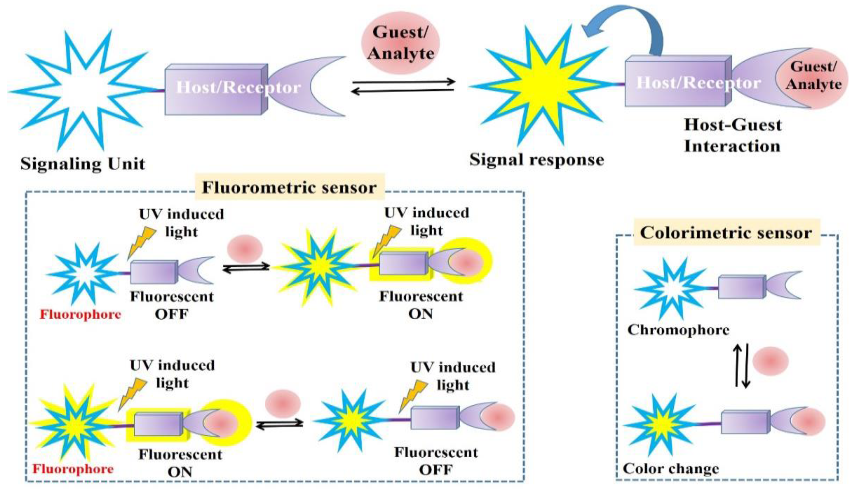
Colorimetric and Fluorometric Detections

In the domain of chemosensors, colorimetric detection is the simplest technique. Using a basic visible spectrophotometer, the colorimetric enabled investigation that can be seen with the naked eye [34]. In the colorimetric approach, a chromophore group serving as the signaling unit in a chemosensor exhibited a distinct color scheme. The chromophore displays its characteristic color in its unbound state as well as the precise color shift that occurs during analyte interaction. Thus, after the analyte is added, the colorimetric method measures the change in absorbance spectrum [35]. The colorimetric approach therefore has limitations such as that it cannot detect background interference or low sensitivity. The fluorometric method is better fitted for practical use because of its higher sensitivity than that of the colorimetric method. A common technique for identifying and measuring cations, anions, or neutral substances is the fluorometric strategic approach. In chemosensor applications, the fluorometric approach employs a chemical moiety with a fluorophore unit [36,37].
Colorimetric chemosensors are used within absorbance spectroscopy to detect a visual color change caused by analyte/guest binding. On the other hand, a fluorometric chemosensor is an analyte/guest that can interact via a variety of sensing methods [38]. Figure 2 shows schematic illustrations for all general sensing mechanisms. The defined phenomena result in distinct pathways for the detection of particular analytes. However, there are two main categories of detection utilizing the fluorometric method: (1) fluorescence “ON” and (2) fluorescence “OFF”. When a specific analyte was added, the free chemosensor agent’s emission intensity was quenched, causing the fluorescence to go “OFF”. In the meantime, fluorescence “ON” increases the emission intensity when the chemosensor agent and analyte interact.
Figure 3 presents schematic illustrations of general fluorometric chemosensing mechanisms, including intramolecular charge transfer (ICT) (Figure 3a) [38], photo-induced electron transfer (PET) (Figure 3b), chelation-enhanced fluorescence (CHEF), chelation-quenched fluorescence (CHQF) (Figure 3c), aggregation-caused quenching (ACQ) (Figure 3d) [39], and aggregation-induced emission (AIE) (Figure 3e) [40]. These defined phenomena lead to distinct pathways for detecting specific analytes [41]. The simplicity and low detection limits that can be attained by employing this technique make sensors based on metal ion-induced changes in fluorescence particularly appealing and the first choice. Therefore, the creation of novel compounds that selectively bind metal ions by fluorescence signal transduction has attracted interest from all around the world. The high ring density of π electrons and the rigid conjugated structure of aromatic compounds are the main causes of fluorescence. Metal ion complexation can lead to either decreased fluorescence-chelation-enhanced quenching (CHEQ) or increased fluorescence, commonly known as chelation-enhanced fluorescence (CHEF). Charge transfer (CT) and electron transfer (ET) are two main mechanisms used to explain how fluorescence sensors react to heavy metals. Photoinduced electron transport (PET), intramolecular charge transfer (ICT), and photoinduced charge transfer (PCT) are examples of these two broad groups. For the detection of heavy metal ions, chemiluminescence and electrochemiluminescence are also used in addition to fluorescence approaches [42].
Various strategies are being employed in order to design the desired binding moiety in a chemosensor for the recognition and detection of the targeted analyte. Organic heterocyclic compounds are helpful in the construction of chemosensors due to their excellent photophysical properties, very low detection limit, high selectivity, synthetic flexibility, reversibility, biocompatibility, and utilization in a wide pH range. These compounds, which have donor atoms like N, O, and S, are highly significant because of their capability of interacting with various metal ions and producing detectable analytical signals [43]. Scientists have been working to build chemosensors using various organic compounds because of their critical need [44]. Among these is the class of colorimetric sensors, which allow metal ion detection with inexpensive equipment [45,46]. Colorimetric sensors that include imine and azide groups are easily synthesized and have a strong binding capacity to metal ions. When aryl-substituted molecukes are bound together, a conjugated system can form and their absorbance is close to or in the visible light spectrum range [47,48]. A common binding group for metal ions chemosensors is the amide group, which also includes thioamide [49]. Protons in thioamide bonds are more potent hydrogen bond donors than those in oxo amide bonds. Compared to an oxo amide, the (C=S)NH proton has a far higher acidity [50]. Taking into account that the thiourea group exhibited an ideal binding moiety and signaling unit [51], several attempts have already been made to design fluorescence sensors that use thiol deprotonation to detect metal ions [52], cyclization [53], and ring-opening [54] in thiourea derivatives. However, each of these chemosensors has a few limitations, such as multiple ion selectivity or a trace level inability to detect in an aqueous media. Thus, there is ongoing interest in the development of fluorescent probes that can selectively remove a certain metal ion without affecting other metal ions [55,56]. Using thiocarbohydrazide as a fluorescent chemosensor, a straightforward Schiff base may selectively identify ions in a semi-aqueous media [57,58]. Chemosensors containing hydrazone–hydrazide moieties are appealing choices due to their high sensitivity and selectivity in detecting a variety of cations and anions [59,60]. Fluorescent sensors have drawn a lot of interest from researchers in the last several years due to their various benefits including nondestructive sample examination, cheap instrument cost, high sensitivity, and outstanding selectivity. Fluorescent sensors typically track their signal as a shift in emission wavelength, fluorescence lifespan, or emission strength in response to minute environmental changes [61]. On the other hand, the development of highly selective ratio metric and “turn-on” fluorescence probes is more important since they can identify metal ions even in living systems, which have been demonstrated to display unique emission characteristics upon metal binding [62]. Schiff base compounds are a novel subclass of chemosensors. These are made up of compounds with a carbonyl group (C=O) and a hydrazone group (N=NCH). Additionally, Schiff bases are crucial to binding chemistry because of their great affinity for metal ions, which are commonly utilized in recognition. This makes them excellent chemosensors. Schiff-based fluorescence chemosensors have received a lot of interest due to their ease of synthesis, cost-effective raw materials, and varied architectures [63]. For many years, the design of novel chemosensors has been of great interest because of their diverse medical aptitude, which includes antibacterial, antiviral, antifungal, anticancer, and anti-inflammatory activity [64].

2. Detection of Selected First-Row Transition Metal Ions

In analytical chemistry, thiocarbohydrazide is utilized for the identification and measurement of inorganic and organic substances. Thiocarbohydrazide may effortlessly be produced in a laboratory by reacting carbon disulfide with hydrazine hydrate [65,66]. Thiocarbohydrazide effectively precipitates aldehydes and ketones quantitatively, generating a large number of derivatives with high melting points that are utilized in gravimetric procedures and for identification or detection. Thermobalance was employed to determine their thermal stability, and their ideal drying temperatures have thus been identified. It was examined that combining carbohydrazide, pyarazole and naphthaldehyde moitieties through suitable linkage would result in intriguing multifunctional sensing compounds for the optical sensing of various metal ions and anions because of the structural properties of these valuable moieties that can be modified to generate effective binding sites [67,68,69]. Several color tests for anions and metal ions rely on the complexing ability of these chemical compounds. More recent reports have included the interaction of carbohydrazide with Cu and of thiocarbohydrazide with first-row transition metal ions such as Cr3+, Mn2+, Fe2+, Co2+, Ni2+ Cu2+, and Zn2+. The study of metal complexes of various thiocarbohydrazide derivatives has been used to examine the binding sites and their molecular structure. Thus, it is evident that thiocarbohydrazide chelates two nitrogen atoms to form complexes with metal ions such as Cr3+, Mn2+, Fe2+, Co2+, Ni2+, Cu2+ and Zn2+ in an acidic medium solution [70]. This mini-review represents a broad overview of organic compounds functioning in colorimetric and fluorescent chemosensors for the identification and detection of first-row transition metal ions. The main focus is on the synthesis of Aryl-substituted organic-based chemosensors, which have shown great promise in detecting a wide range of important first-row transition metal ions including zinc, copper, cobalt, chromium, nickel, manganese, and iron through colorimetric and fluorometric turn-off and turn-on responses. Thiocarbohydrazide 1 was used to add the amine and aldehydes in the presence of ethanol in a schiff-based reaction. At room temperature, the reaction mixture was stirred for three hours following the gradual addition of 8-hydroxy quinoline-2-carbaldehyde 2 and a catalytic amount of acetic acid. The precipitates removed the original ingredients in the reaction mass after they were obtained. Precipitates were collected by filtering, EtOH washing, drying in a hot air oven, cooling, recording, and computation of the percentage yield of synthesized chemosensors (Scheme 1). Using Bene-si-Hildebrand (B-H) plots, and UV–visible spectroscopy titration studies, the detection limit (DL), stoichiometric binding ratio, and recognition process were investigated. Sensor 3 demonstrated a 1:1 binding ratio with an excellent binding constant range of 103 to 105 M−1 with Cu2+ metal ions [71].
M. Yang, J. B. Chae and their coworkers synthesized a novel sensor by reacting 2-furoic hydrazide 4 and di-2-pyridylketone 5 in EtOH. The synthesis procedure involved first dissolving 2-furoic hydrazide in EtOH, and then mixing a few drops of phosphoric acid and di-2-pyridyl ketone into the ethanol solution. After agitating the mixture for a full day at room temperature, a white precipitate formed that was filtered and washed with ether to yield 44% of the desired solid product 6 (Scheme 2). This sensor changed from colorless to blue in the aqueous solution, demonstrating exceptional selectivity and sensitivity for detecting lower concentrations of Fe cations. In addition, the strong binding capability of the sensor in detecting Fe(II), Co(II), and Cu(II) selectively makes it suitable to detect and quantify small quantities of said metal ions in tap water. Sensor 6 shows no visible response to other metal ions such Ca2+, Cr3+, Mn2+, Fe3+, Ni2+, Zn2+, Cd2+, Ag+, Hg2+, and Pb2+. The binding ratio of sensor 6 to Fe2+, Co2+, and Cu2+ is 2(sensors):1 (metal ion). The binding constants of the sensor are as follows: Fe2+: 1.0 × 10⁹ M−2, Co2+: 2 × 10⁹ M−2, and Cu2+: 3.0 × 10⁹ M−2. Sensor 6 works well at a neutral pH, and micromolar concentrations of Fe2+, Co2+, and Cu2+ can be detected in the water samples. The sensor’s color response to Cu2+ is uniquely attenuated by glutathione [47]. In the case of the sensor, the main absorption band originated from the HOMO → LUMO transition, indicating a π → π* transition. In contrast, the Cu–sensor complex showed an intramolecular charge transfer (ICT) [72].
Özlem Özdemir successfully synthesized a novel chemosensor for the identification and detection of metal ions. She designed diimine-Schiff-based 9 by condensing 4-nitro-o-phenylenediamine 7 with 5-chloro or 5-methyl substituted salicylaldehyde 8. Then, nitro-derivative 9 was converted into the amino derivative 10 by adding sodium dithionite. Subsequently, she further condensed 10 with 8 and 2-hydroxy-1-nephthaldehyde 11 to give 12 and 13, respectively. These recently designed Schiff bases, which include ONNO or O3N3 donor atoms, are resistant and soluble in various organic solvents, like DMSO, DMF, and alcohols at low concentrations (Scheme 3). The properties of all Schiff bases under sensor 13 were examined upon addition of the metal ions, such as Cr3+, Fe2+, Fe3+, Co2+, Ni2+, Cu2+, Zn2+, and Pb2+. The interactions between receptors and ions are easily monitored via UV–Vis spectroscopy [73].

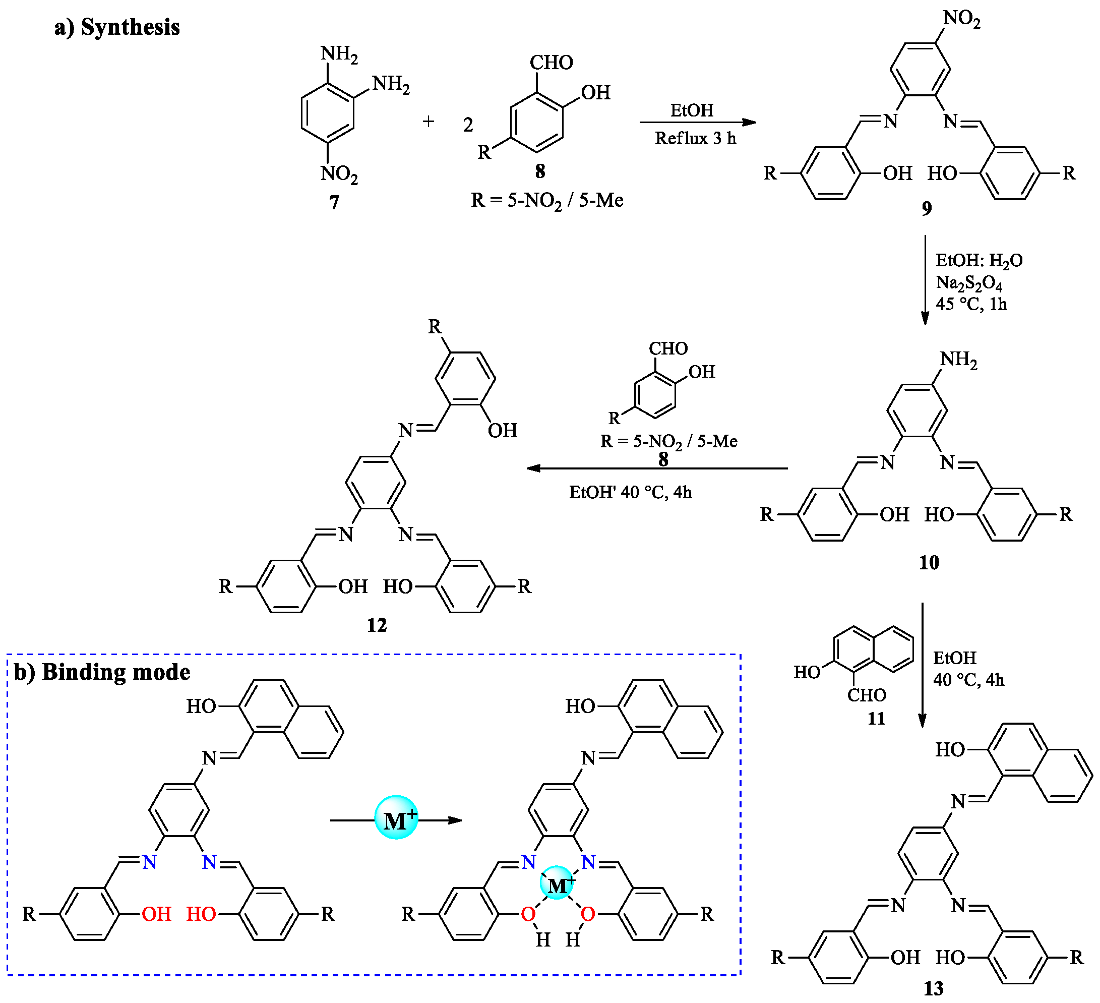
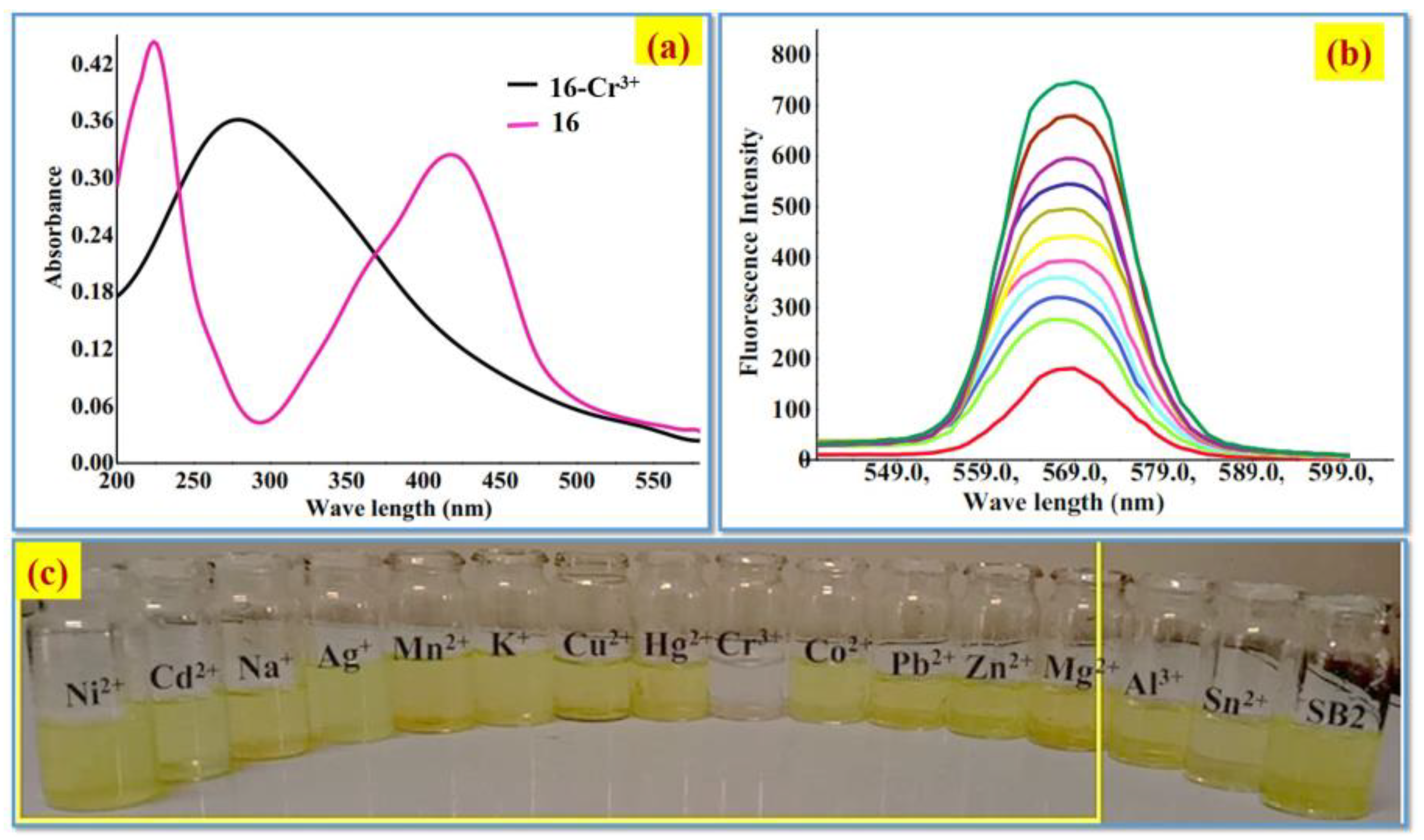
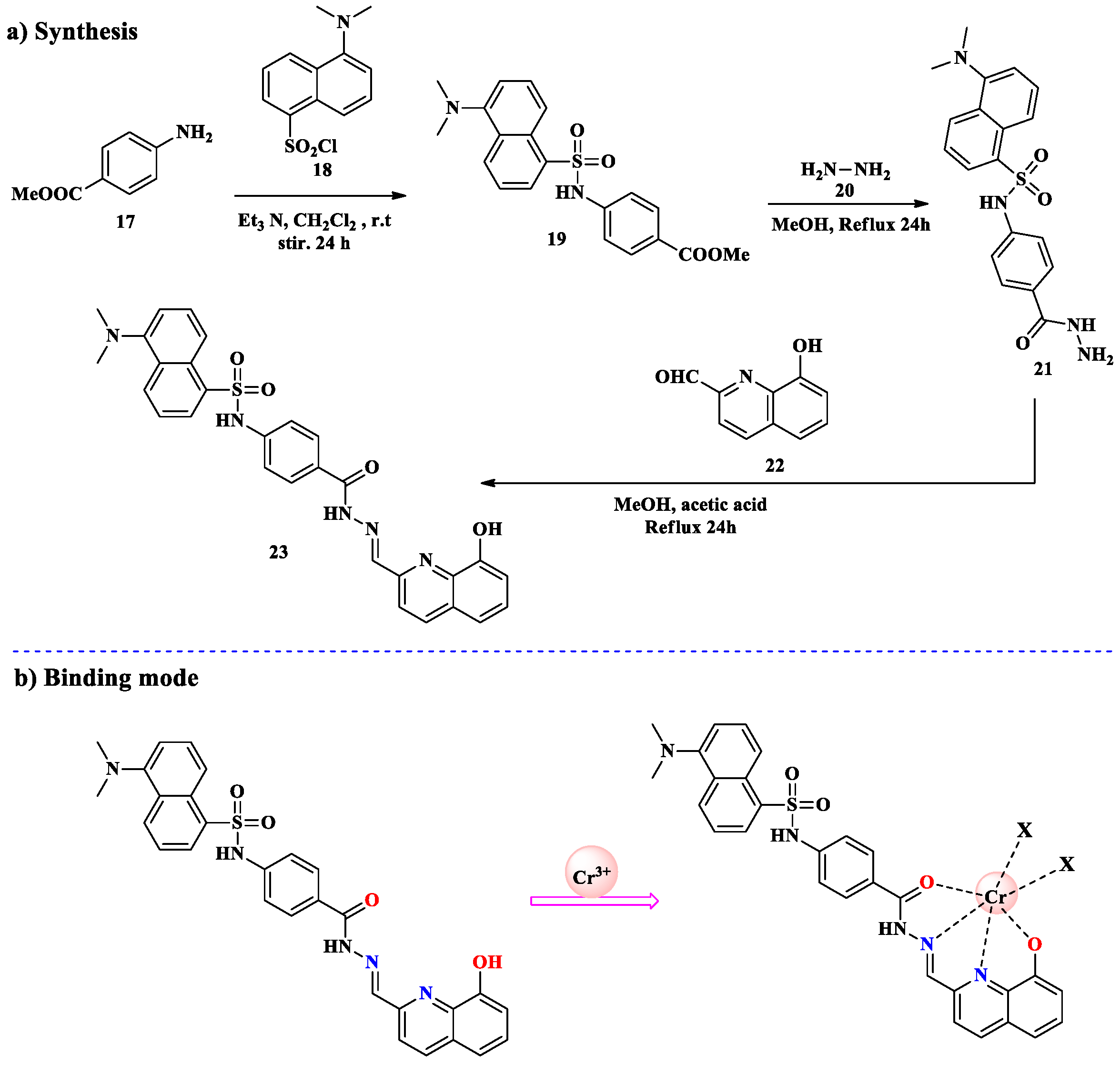
2.1. Cr3+ Ion Detection

Chromium(III) modulates the metabolism of proteins, fats, and carbohydrates, and may boost the activity of specific enzymes. Primarily, it is essential for a variety of metabolic activities [74]. As an important micro-nutrient that is vital for preserving general human health and well-being, chromium(III) ions perform as an insulin potentiator, strengthening its beneficial effects [75]. On the other hand, high concentrations of chromium(III) ions are potentially damaging, disrupting cellular components and cellular structural framework, which might result in genetic abnormalities and even cancer risks [76]. In recent years, chromium(III) has been extensively released into the environment through many industrial processes, such as electroplating, leather tanning, mining, textile dyeing, and the manufacturing of inorganic chemicals. This has resulted in considerable environmental high contamination and has turned into an acute problem [77]. In the meantime, concerns about the harmful environmental impacts of chromium are still prevalent in both agricultural and industrial domains. This underlines the significance of more study concerns and the development of reliable detection approaches, particularly for Cr(III) ions in aqueous systems, with the goal of resolving these persisting environmental problems [78]. Lately, a series of products known as colorimetric and fluorescent chemosensors has gained traction or popularity in the sensing world for their capability to recognize and detect metal ions. These compounds provide a visible and highly sensitive response via color or fluorescence difference. Cr(II) metal ions are also first-row transition metal ions with d4 electronic configuration. They prefer N/O donor ligands. Due to its large size and moderate charge density, ligands that can provide multiple coordination sites help in their stabilization. They are a desirable option for sensing real world applications due to modification [79]. Although colorimetric and fluorescent organic chemosensors have demonstrated enormous potential in the detection of Cr(III) ions, there are still two main obstacles to overcome. Firstly, the waste of the majority of organic chemosensors is not soluble in pure water, which is vital for biological applications. Secondly, the currently available chemosensors often exhibit low selectivity for Cr(III) ions and are susceptible to interference from other metal ion species, potentially leading to false positive results [80]. Due to this, researchers have continuously designed and modified chemosensors with excellent selectivity, sensitivity, and solubility in water samples. Concerning their potential for effectiveness as ligands, tiny organic molecules with N, O, and S atoms, specifically Schiff bases, have appeared as the most promising candidates. These organic compounds are water-soluble due to their very small size and heteroatom existence, and they showed high selectivity, sensitivity, and affordable cost. Schiff bases are emerging choices for chemosensing applications as they effortlessly interact with various metal ions and resulting identifiable analytical signals for study [81]. S. Khan, M. Muhammad, and their colleagues effectively synthesized a chemosensor using a multi-step procedure. 2-hydroxy-1-naphthaldehyde 14 was first dissolved in CH3OH, and then a small amount of glacial acetic acid was added to serve as a catalyst. The reaction mixture was vigorously stirred for 8 min, a 2-amino thiazole methanol 15 solution was introduced to it, and the reaction mixture was refluxed for 12 h. The resultant mixture was cooled down to room temperature, and an 86% yield of the intended product 16 was obtained (Scheme 4). Purification was performed via recrystallization of the crude product from methanol [82].
In the presence of various cations, including Na+, Mg2+, Ag+, Cr3+, Zn2+, Pb2+, Ni2+, K+, Mn2+, Hg2+, Co2+, Sn2+, Cu2+, Cd2+, and Al3+, the UV–Vis absorption studies of 16 were examined. The chemosensor 16 displayed two absorption bands centered at 220 and 422 nm (Figure 4a). Fluorescence spectra of chemosensor 16 were recorded in the presence of various cations in an aqueous medium (MeOH/H2O, 1:9 v/v) in order to examine the sensing capabilities of the chemosensor. Due to photo-induced electron transfer (PET) from the azomethine group (–C=N–) to the naphthalene fluorophore, free chemosensor 16 showed a modest fluorescence emission at 568 nm (λex = 279). A remarkable turn-on response was observed upon the addition of Cr(III) ions to 16 (Figure 4b). Using various monovalent, divalent, and trivalent cations, such as Na+, Mg2+, Ag+, Cr3+, Zn2+, Pb2+, Ni2+, K+, Mn2+, Hg2+, Co2+, Sn2+, Cu2+, Cd2+, and Al3+, the colorimetric analyses of chemosensor 16 were carried out in aqueous media. The aqueous solution of 16 exhibited a yellow color. The addition of various cations did not significantly alter the color of 16 in aqueous environments, with the exception of the Cr(III) ion. When Cr(III) ions were added, the color changed dramatically from yellow to colorless (Figure 4c).
H. Wu and P. Zhou, along with their coworkers, developed a novel chemosensor with a tetradentate metal-binding moiety composed of 8-hydroxyquinoline-carboxhydrazone, which interacts with Cr3+ to form a complex. A solution of methyl 4-aminobenzoate 17 and trimethylamine in dichloromethane was added dropwise to a solution of 5-dimethylamino-1-naphthalenesulfonyl chloride (dansyl chloride, 18) in dichloromethane. The reaction mixture was stirred at room temperature for 24 h. The desired product 19 was washed with water, and the organic layer was dried using MgSO4. The resulting yellow solid was recrystallized from ethyl acetate and used immediately in the subsequent step. The yellow solid product was then combined with hydrazine hydrate 20 in CH3OH and refluxed for 24 h. After solvent evaporation, the product was mixed with appropriate aldehydes 22 dissolved in methanol. Acetic acid was added dropwise to the reaction mixture, which was refluxed for another 24 h. After cooling, the crude mixture was filtered, yielding the desired yellow solid 23 (Scheme 5). A metal-binding moiety of 8-hydroxyquinoline-carboxhydrazone tetradentate is present in Sensor 23 and forms a 1:1 complex with Cr3+. Additionally, 23 showed a considerable increase in fluorescence but a much-decreased quantum yield after Cr3+ binding in an aqueous solution. A Job plot of the 23-Cr3+ fluorescence spectra, with the inflection point at around 0.5, which corresponds to [(23 − 2H) + Cr]+ in the ESI-MS spectrum of a combination of 23 and Cr3+, further verified the 1:1 binding mode [79].
A.Abbasi and M. Shakir synthesized a chemosensor by adding methoxybenzaldehyde 24 with 2-furoic acid hydrazide 25 in EtOH at 25 °C. After about three hours of refluxing the solution mixture, a yellow-colored solution was obtained; from that, gradual solvent evaporation produced an off-white-colored crystalline precipitate including Schiff-based product 26 (Scheme 6). Cr(VI) effectively suppressed the fluorescence of chemosensor 26 through the primary and secondary inner filter effects. Cr(VI) has a detection limit of 0.175 μM and a linear recognition range of 1 μM to 500 μM. The addition of L-ascorbic acid in the concentration range of 10 μM to 390 μM with a LOD of 2.46 μM successfully turned on the chemosensor–Cr(VI) solution’s switched-off fluorescence. The reduction of Cr(VI) to Cr(III) by L-ascorbic acid is the mechanism suggested for the fluorescence turn-on of quenched fluorescence of the Cr(VI)–chemosensor solution of ascorbic acid. This process eliminated both primary and secondary inner filter effects and allowed the fluorescence of chemosensor 26 to return. It was discovered that the technique described in this study was successful in identifying Cr(VI) in groundwater and tap water, ascorbic acid in human serum, and vitamin C tablets [83].

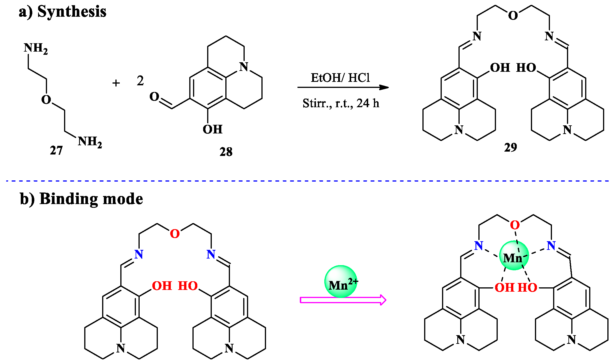
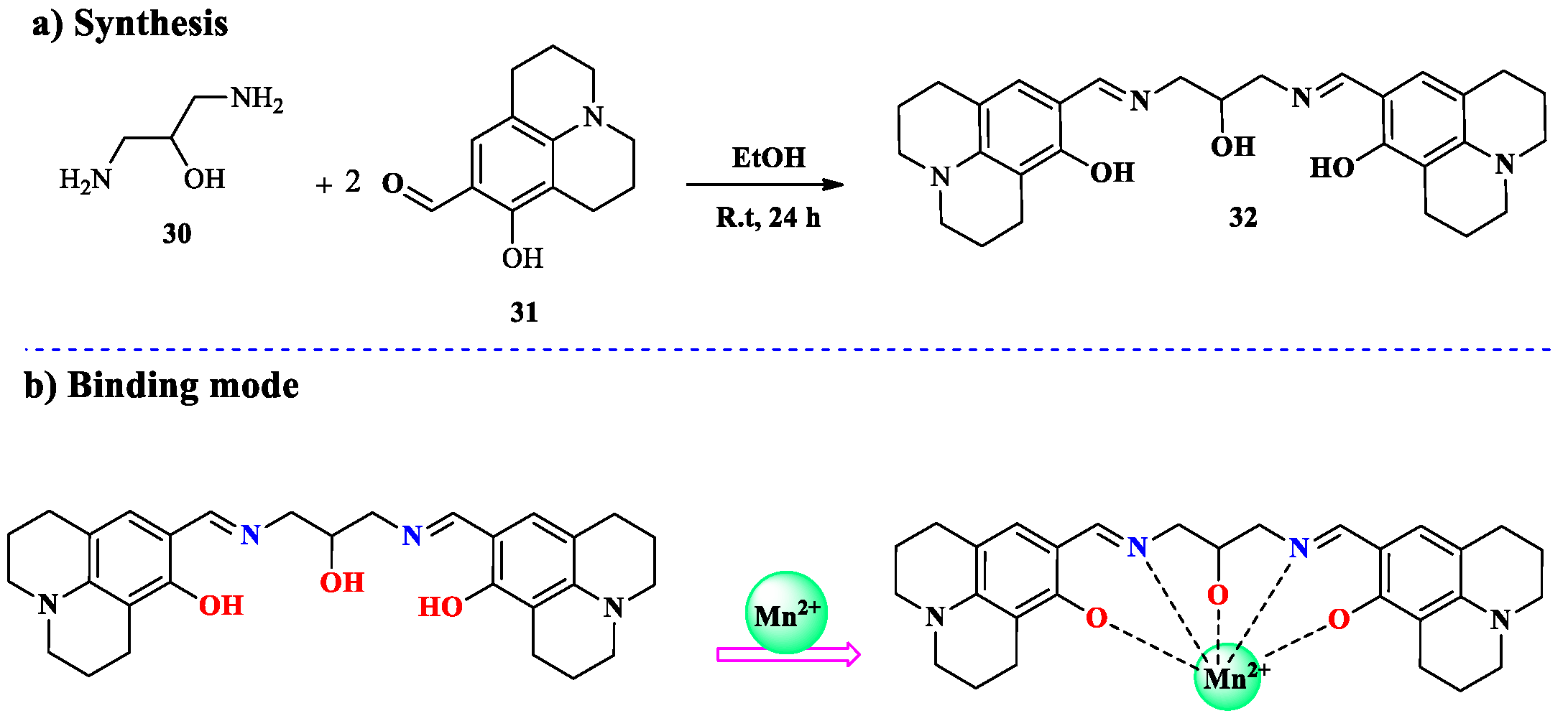
2.2. Mn2+ Ion Detection

The development of chemosensors is greatly facilitated by transition metal ions, such as Fe, Zn, Co, Cu, and Mn, due to their significant biological roles and the harmful environmental effects of their excess presence. Manganese (Mn) is one of the vital trace metals found in all living things and is involved in several important biological processes. Understanding its biological relevance and minimizing its environmental effects relies upon its identification and tracking processes [84,85]. Manganese is frequently employed in biological research, facilitating chemists to explore a broad range of biological processes and mechanisms due to its versatility and significant role as a cofactor in several enzyme groups and the evolution of the photosynthetic oxygen evolution process [86,87]. Manganese(II) ions have demonstrated substantial potential in their high-spin state, as a contrast reagent for magnetic resonance imaging (MRI), sparking passion in the neurobiology research field as a powerful tool for amplifying picture quality and studying brain function [88]. Because Mn2+ ions can be harmful to health, precise stimulation is crucial in neurobiology. Monitoring manganese ions level is important. Despite being somewhat less toxic, long term exposure even for short periods of time can trigger serious neurological consequences like movement or motor disorders, mental health disturbances, memory loss, and indication Parkinson’s-like symptoms [89]. Given their simpler properties, it is essential to design Mn2⁺-selective chemosensors that are capable of distinguishing Mn2⁺ from other transition metal ions, particularly Ca2⁺. This is crucial because Mn2⁺ and Ca2⁺ share the same cellular transport pathways, requiring precise detection and identification of these ions [90,91,92]. In addition to their amazing affinity for transition-heavy metal ions, the N atom in the Schiff bases’ azomethine (C=N) double bonds render them important chelating agents, as well as suitable potential sensors for identification and binding to metal ions [93]. Schiff bases are excellent or ideal choices for designed metal ion complexes and chemosensors because of their well-known strong binding features, with a range of applications, such as anticancer potential. This demonstrates the promising potential of Schiff bases in medicinal chemistry, biological research area [94], antioxidant properties, and appealing electronic and photophysical characteristics, ideally suitable for sensors, and imaging agents [95]. Fluorescent Schiff-based derivatives offer great potential as optical chemosensors for metal ions. Due to their capability to form complexes with metal ions and their unique coordination, stability, fluorescent attributes, and biological activities, symmetrical Schiff bases are attractive for a variety of purposes [96]. One major issue is the controlled synthesis of Schiff-based derivatives along with metal complexes. So many parameters, such as the ligand’s chemical structure, pH, the metal-to-ligand ratio interaction, reaction temperature, solvent medium system, and the metal’s optimal coordination geometry, all influence how well they self-assembled. These parameters affect the complex’s coordination geometry structure; thus, it is crucial to achieve the best synthesis conditions. Therefore, precisely and controllably designing these compounds is a difficult process that requires careful understanding of these aspects [97]. The field of designing fluorescence chemosensors with good selectivity and sensitivity to metal ions is a competitive and important one, with potential use in materials science, healthcare, and environmental monitoring processes [98]. Researchers strive to develop new metal ion chemosensors with greater sensitivity, selectivity, and reliability, even though there are multiple easily accessible commercial strategies available. This ongoing endeavor aims to meet all of the demands resulting from metal ions’ prevalence in living organisms and their huge importance in a range of biological operations [99]. A novel chemosensor was synthesized by Yeon Joo Lee, Chaejin Lim, and their team utilizing the condensation reaction of 2,2-oxybis(ethylamine) and 8-hydroxyjulolidine-9-carboxaldehyde. An ethanolic solution of 8-hydroxyjulolidine-9-carboxaldehyde 27 was made for the sensor by adding two drops of HCl, then gradual addition of 2,2-oxybis(ethylamine) 28 in EtOH (Scheme 7). Methylene chloride in methanol eluent was used in column chromatography to purify the obtained crude product 29 that resulted after 24 h of elimination of solvent under vacuum. This sensor 29 identified Mn2+ ions in MeCN-buffer solution by seeing a noticeable color shift from colorless to yellow [100].
Using nitrate salts of several metal ions, including Ag+, Al3+, Ca2+, Cd2+, Co2+, Cr3+, Cu2+, Fe2+, Fe3+, Hg2+, Ga3+, In3+, K+, Mg2+, Mn2+, Na+, Ni2+, Pb2+, and Zn2+, in MeCN-buffer solution (9:1, v/v, 10 mM HEPES, pH 7.4) at room temperature, the chromogenic sensing ability of 29 was tested. As shown in Figure 5, While other metal ions produced little to no alterations, only the Mn2+ ion caused a noticeable spectrum shift and an immediate color change from colorless to yellow. The second colorimetric chemosensor used to detect Mn2+ in aqueous solution is this one. Significantly, this finding suggests that sensor 29 may be employed as a “naked-eye” Mn2+ ion sensor in aqueous conditions.
Two chemists K. B. Kim and G. J. Park have synthesized a novel Schiff-based chemosensor 32 containing a julolidine moiety. These can detect manganese(II) ions in aqueous solutions by establishing visible color changes. When this novel sensor interacts with Mn2+ ion, it shows a noticeable shift from light yellow to orange color. This color change can be identified with the naked eye. Chemosensor 32 was effectively synthesized by a straightforward coupling reaction of 1,3-diamino-2-propanol 30 and 8-hydroxyjulolidine-9-carboxaldehyde 31 (Scheme 8). The chemosensing characteristics of 32 were further examined by performing a UV–Vis titration of sensor 32 with Mn2+. The highest absorbance at 392 nm was rapidly reduced as increasing quantities of Mn2+ were added to a solution of 32. Three additional absorbances at 348 (ε = 3.4 × 10⁴ M−1 cm−1), 418 (ε = 3.2 × 10⁴ M−1 cm−1), and 532 nm (ε = 4.2 × 10 M−1 cm−1) emerged concurrently. These coefficients are too large to give Mn-based d-d transitions, they must give manganese to ligand-based transitions. Thus, the metal-to-ligand charge transfer (MLCT) may be responsible for the color shift from light yellow to orange in the presence of Mn2+. The development of a single species between sensor 32 and Mn2+ was shown by the obvious observation of the different isosbestic spots at 354 nm and 413 nm [101].
V. Raju, R. S. Kumar, and their associates designed a novel chemosensor 35 by reacting hydrazine 33 with 3,5-dichloro-2-hydroxybenzaldehyde 34 in a simple one-step process. 3,5-dichloro-2-hydroxybenzaldehyde 34 was added in EtOH and then added drop by drop to hydrazine hydrate in a flask. Upon stirring the mixture for about half an hour, a precipitation started to appear. After that, 100% alcohol was used to filter and recrystallize the precipitate to obtain the intended product 35 (Scheme 9). Using a variety of metal ions, the sensing capabilities of sensor 35 were investigated using spectrofluorometric, UV–Vis, and optical tests in THF:H2O (1:1, v/v) media. Thus, when Mn2+ interacted with the original colorless solution of 35, it became yellow, confirming the visual test; however, other ions did not react in natural light. Spectrophotometric techniques are used to examine the photophysical characteristics of 35-Mn2+. When clean 35 interacts with Mn2+, it shows a strong absorption band at 365 nm and a new absorption band at 425 nm. In contrast, other metal ions showed no emission characteristics. Additionally, Job’s plot was used to study the formation 3:2 binding stoichiometry between 35 and Mn2+, and the Benesi–Hilderbrand (BH) technique was used to measure the association constant. The WHO’s recommendations for metal ions in drinking water are significantly higher than the detection limit of 35 for Mn2+, which was determined to be 3.6 × 10⁻⁷ M. There is strong evidence from NMR and theoretical studies that 35 exhibits coordination behavior with Mn2+ [102].

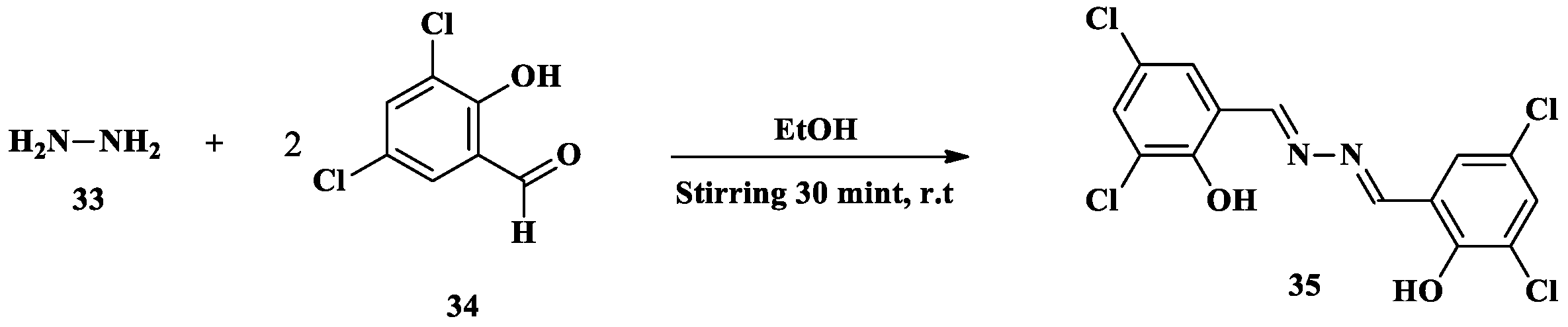
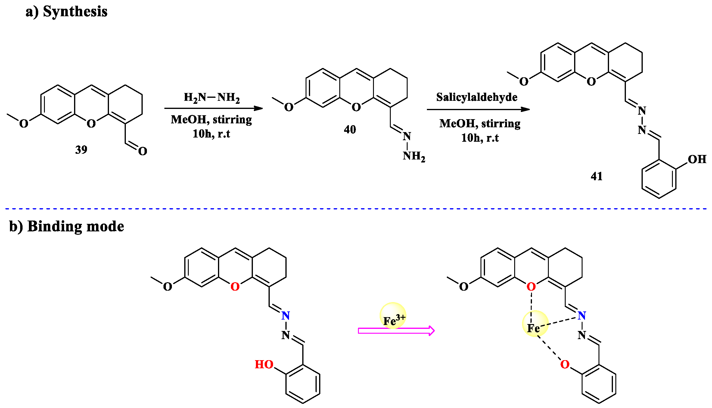
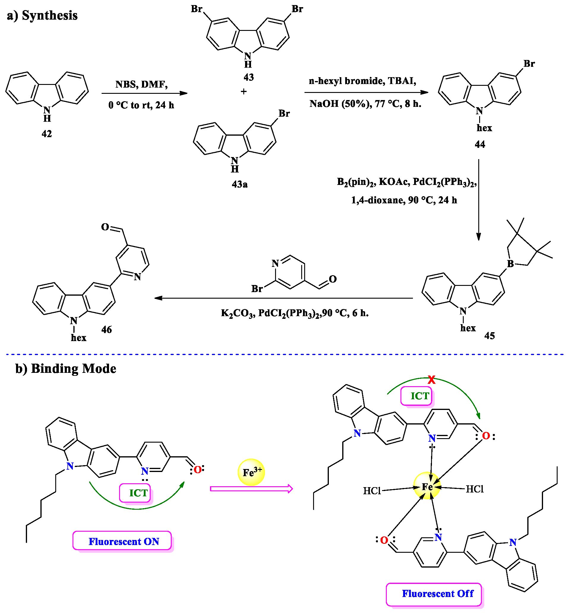
2.3. Fe3+ Ion Detection

Of all the metal ions, iron is an essential component and one of the heavy metals found in the human body. It is believed that iron is essential to several physiological functions [103] as iron’s redox-active form catalyzes the generation of highly reactive oxygen species and can cause a variety of health issues. Excessive iron supplementation may be detrimental to the biological system. Consequently, the identification and study of Fe3+ has emerged as a difficult problem in environmental research and biomedicine in recent years. Now a days, the advanced development of chemosensors for Fe3+ detection has been quite fascinating. Since iron functions as hemoglobin, the primary oxygen carrier in all tissues, and since it supports the movement of electrons as cytochromes, iron is necessary for all biological systems [104]. Both the deficiency and excess of iron would result in various health conditions such as hemochromatosis, Alzheimer’s disease, and anemia (blood insufficiency), which may damage or even kill oxygen-dependent organs [105,106,107,108]. Through numerous kinds of agricultural and industrial processes, Fe2+ ions have major effects on crop yields, water body quality, and industrial operations. Hence, it is now crucial to efficiently track and accurately identify Fe2+ ions in biological, environmental, agricultural, and industrial resources [109,110]. Researchers may find ratiometric analysis more attractive as the ratio of the two emission intensities may be used to calculate the concentration of Fe2+ ions and be eligible for an automatic reimbursement for environmental effects and stability under fluorescence [106,111]. As a reference signal, one of the fluorophores must respond to a fluctuation in Fe3+, whereas the other is intended to be insensitive [112]. The offered chemosensor includes a highly selective ratiometric fluorescent sensor for Fe3+ detection and quantification [113,114,115]. Mohd Mustufa et al. synthesized a novel-N-(4-(dimethylamino) benzylidene) quinolone-3-amine (DBQA) fluorescent chemosensor. A heated ethanolic solution of 3-aminoquinoline 36 was added drop by drop to an ethanolic solution of 4-(dimethylamino)benzaldehyde 37 in order to develop the said chemosensor (DBQA). The reaction mixture was refluxed with agitation for five hours. After cooling, the greenish-black product was isolated by filtration. The product was recrystallized using ethanol, forming needle-shaped crystals that dried to a greenish-black tint. This protocol gave 75% yield of the final product (Scheme 10). By employing the synthesized chemosensor (DBQA) 38, Fe3+ ion was efficiently detected. With the successive addition of the Fe3+ ion, the fluorescence and UV–visible spectra of (DBQA) demonstrated ratiometric activity and a bathochromic shift, exhibiting excellent sensitivity as well as the selectivity of the Fe3+ ion over other transition metal ions [116].
The mechanism of intramolecular charge transfer (ICT) allows DBQA to interact maximally with Fe3+ ions. This makes them an intriguing option for ratiometric fluorescence chemosensors, which are employed in semi-aqueous settings to detect metal ions [117]. Concerning DBQA’s selectivity for various metal ions, in the DBQA fluorescence spectrum, only Fe3+ demonstrates a ratiometric response when compared to other engaging metal ions. In the presence of more competitive metal ions, 38 was added to a DMSO:H2O solution to perform the competition assays. The other analyzed metal ions showed no noticeable ratiometric interaction when Fe³⁺ ions were detected. Figure 6 shows the emission spectra of the chemosensor (BQA). The emission at λem = 413 nm in DMSO:H2O (v/v, 1:4, pH = 7.1) at 25 °C led to the excitation wavelength to be set at 290 nm for the emission investigation. The behavior of the chemosensor (DBQA) towards Fe3+ and NH3 in a semi-aqueous liquid (DMSO:H2O) is demonstrated in this work. When varying amounts of Fe3+ were added, the emission intensity at 413 nm fell as the wavelength increased. A new peak at 463 nm with an isosbestic point at 446 nm was noticed as a result of the apparent ratiometric fluorescence change. When NH3 was added, the ratiometric response of DBQA with Fe3+ could be regained. Fe3+ would most likely be eliminated by NH3 as an ammonium iron complex.
B. Li, X. Gu, and their colleagues successfully synthesized a novel chemosensor for detecting Fe3+ ions. The synthesis procedure involved several steps. Hydrazine hydrate was added after 6-Methoxy-2,3-dihydro-1H-xanthene-4-carbaldehyde 39 had dissolved in MeOH. After stirring the reaction mixture for ten hours, a crimson-red precipitate formed. The precipitate was filtered, and the solid was dissolved in MeOH, with the solvent removed by rotary evaporation. An orange-colored solid 40 was obtained after chilling the residual solution in a refrigerator. The hydrazinolysis product 41 was then dissolved in MeOH, mixed with o-hydroxybenzaldehyde, and stirred for 10 h at room temperature (Scheme 11). This led to the formation of an orange-red precipitate, which was filtered, purified by dissolving in MeOH, and dried by rotary evaporation. Finally, the residual solution was chilled, yielding the desired dark red crystals with a 66% yield. In a solution of MeCN-Tris (v/v, 2:1, 10 mM, pH 7.3), Sensor 41 continually detects Fe3+. Alongside the solution’s color shift from yellow to red, sensor 41 demonstrated a very sensitive and selective fluorescence “turn-on” for Fe3+ over other metal ions. Additionally, only F is detected by the resultant 41-Fe3+ combination among the common different anions. Fluorescence and absorbance titrations, 1 H NMR titrations, HRMS, and theoretical computations were used to identify the bonding processes. Additionally, sensor 41 has been utilized to identify Fe3+ using test paper colorimetry and liquid colorimetry. Furthermore, the fluorescent imaging of Fe3+ in HeLa cells was performed sequentially off-on-off using sensor 41. The photo-induced electron transfer (PET) process in sensor 41 may be activated by the detection of Fe3+. This is because the lone pair electron of the nitrogen atom should be transferred from the –C=N– group to the fluorophore unit, preventing the PET effect [118].
A. Battal, S. B. Kassa and their team developed a chemosensor for detecting Fe3+ ions. The compound was synthesized in four stages starting from carbazole. Firstly, carbazole 42 was brominated with N-bromosuccinimide (NBS) in dimethylformamide, producing a mixture of mono- and dibromo compounds. After crystallization, 3-bromocarbazole 43a was obtained as white crystals in an acceptable yield. Next, 3-bromocarbazole was alkylated with 1-bromohexane in the presence of tetrabutylammonium iodide (TBAI) and 50% aqueous NaOH, yielding 3-bromo-9-hexylcarbazole 44 as a liquid in excellent yield after chromatography. The 3-bromo-9-hexylcarbazole 44 was then reacted with dichlorobis(triphenylphosphine) palladium(II) in 1,4-dioxane and bis(pinacolato)diboron, in the presence of potassium acetate (KOAc), to form the pinacol boronic ester 45. Finally, (9-hexylcarbazole-3-yl)boronic acid pinacol ester underwent a Suzuki–Miyaura reaction with 2-bromopyridine-5-carbaldehyde in the presence of potassium carbonate and bis(triphenylphosphine)palladium(II) dichloride in tetrahydrofuran, yielding the intended compound 46 after multiple purifications by chromatography (Scheme 12). Then, a number of techniques were used to examine its sensing capabilities. The findings demonstrated that sensor 46 detects the Fe3+ ion more strongly than other interfering metal ions. Sensor 46’s sensitivity and selectivity for Fe3+ were excellent for usage in applications. Additionally, it was noted that probe 46’s fluorescence intensity dropped when aqueous Fe3+ ion solutions were applied to it using dimethyl sulfoxide (DMSO). The paramagnetic interaction between sensor 46 and Fe3+ ions is the cause of this situation (turn-off of emissions). Sensor 46’s limit-of-detection (LOD) value was determined to be 1.38 nM. This value is insufficient to compete with its literary counterparts. In a real sample experiment, sensor 46 also showed a greater ability to detect Fe3+ ions than other ions in an actual medium. Consequently, it was determined that sensor 46 is a highly promising option for sensor technology [119].

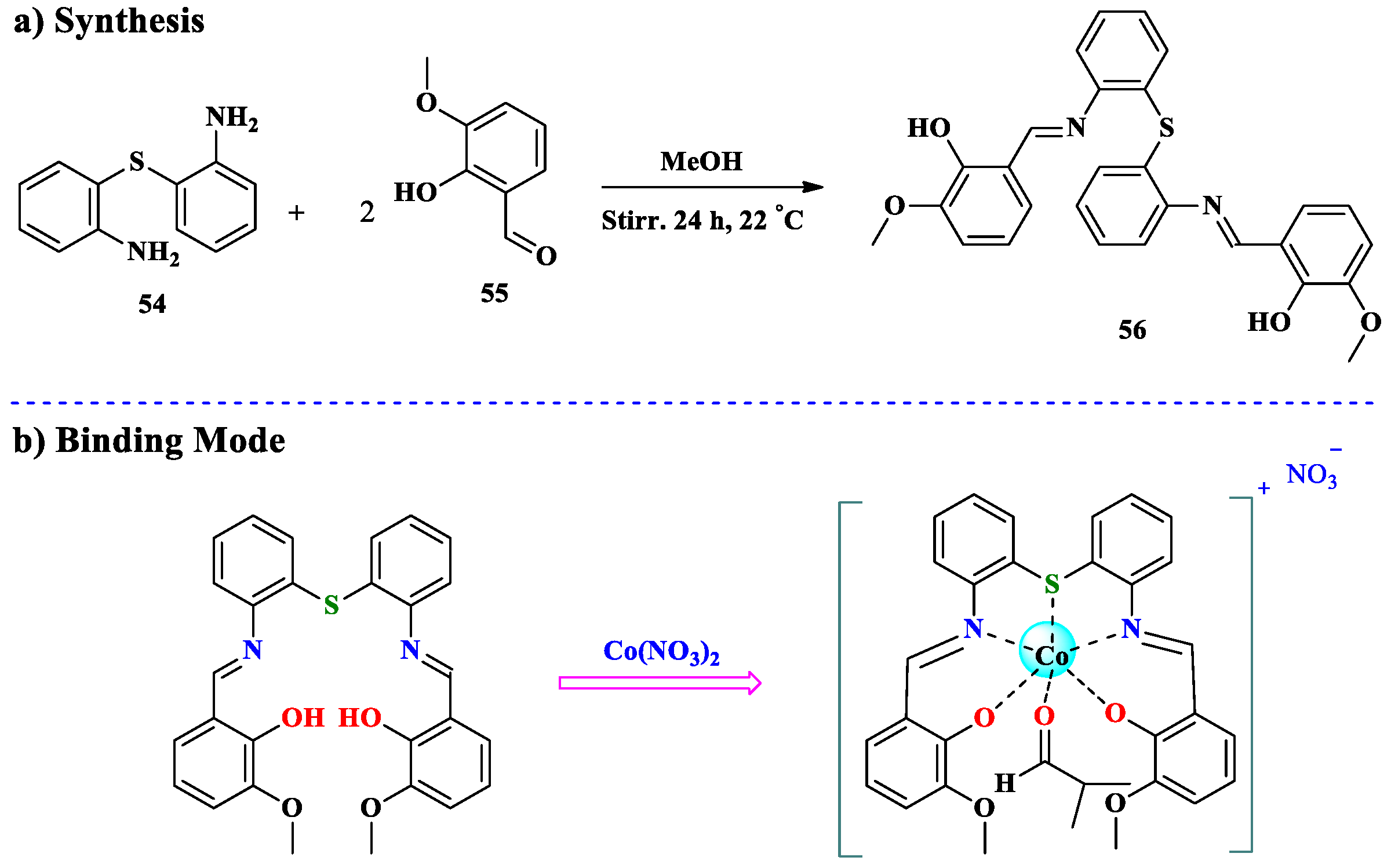
2.4. Co2+ Ion Detection

Metal detection and recognition are essential because of their exclusive applications. In view of the many potential uses for these metals, chemists or researchers in related areas now face a huge difficulty in developing efficient techniques for identifying and detecting metal ions [120]. Among metal cations, cobalt ion (Co2+) comes out as a physiologically needed trace element, fundamental to many biological functions. Its flexibility has resulted in various implementations in a number of domains, like the commercial, industrial, and medical sectors, wherein its special qualities are taken advantage of to encourage advancement [121]. Moreover, cobalt ions require metal of many biological compounds, such as vitamin B12. Co2+ is also vital for the formation of hemoglobin and the metabolism of iron. Its presence is essential for preserving proper biological functioning since it aids in the regulation of iron metabolism and facilitates the formation of hemoglobin, further demonstrating its importance to human health and well-being [122]. On the other hand, long-term contact with cobalt metal can be damaging to human health and result in a number of toxicological side effects, such as digestive problems, damage to the liver, thyroid enlargement, respiratory illnesses like pulmonary disease and asthma, and even a disturbance in cardiac functions. Due to these probable health hazards, it is imperative to keep tabs on cobalt levels to protect people’s health and safety [123]. Therefore, for the purpose of ensuring the safety and quality of water resources and preventing possible adverse effects from cobalt ion contamination, the development of effective techniques for detecting cobalt ions in water samples is essential across a variety of industries and applications, including environmental monitoring, public health, and industrial applications [124]. Metal ions play vital functions in a number of domains such as environmental science, life sciences, and chemistry. There has been a lot of work directed into developing reliable and multifunctional chemosensors for detecting many types of metal ions [125].
A. A. Abd-Elaal, A. Farag, and S. M. Tawfik successfully formed a unique chemosensor using a step-by-step procedure. After mixing 4-(dimethylamino)benzaldehyde 54 and naphthalene-1,5-diamine 47 in EtOH, with a few drops of glacial acetic acid as a catalyst, the reaction mixture was stirred for 12 h at 80 °C. This resulting yellowish solution was permitted to cool at room temperature, and after filtration and washing with cold diethyl ether, the precipitate was collected as a pale yellow powder containing 90% of intended compound 49. After that, compound 49 was mixed in a nitrogen-filled round-bottom flask after being dissolved in EtOH. Upon adding octyl bromide, the reaction mixture was heated for 48 h at 80 °C. Then, the pale green precipitate produced was filtrated, washed with EtOH, and then recrystallized by diethyl ether to create a greenish powder that had a melting point of 312.5 °C and was 75% pure 4,4′-((1Z,1′Z)-(naphthalene-1,5diylbis(azaneylylidene))bis(methaneylylidene))bis(N,N-dimethyl-N octylbenzenaminium) dibromideoctylbenzenaminium) dibromide chemosensor 50 (Scheme 13). After adding the Co2+ ion, the intensity of quenched fluorescence was seen under a UV light, changing from blue to colorless; as a result, it may be used for detection with the naked eye. When sensor 50 interacts with Co2+ ions, its fluorescence intensity decreases, which other ions do not notice, indicating the sensor’s high selectivity. Sensor 50 reduces the LOD of CO2+ to 41 nM, which is below the WHO-approved detection limit. It is interesting to note that sensor 50 effectively demonstrated the utilization of the paper sensor for metal ion detection through color change analysis in each detection zone. Additionally, with sufficient recovery, sensor 50 can identify CO2+ ion contaminants in tap and river water. Their discovery of the application of a naphthalene cationic Schiff-based surfactant in optical sensing creates a research avenue for the development of a portable, low-cost sensor with enhanced sensitivity and specificity. This method detects threats in five minutes and does not involve any costly or intricate steps [126].
To examine the selectivity of sensor 50 towards Co2+ spectrofluorometric titration experiments were performed. The fluorescence properties of 50 solutions with 12 different metal ions, including 1000 µM from each of Fe2+, Cu2+, Mg2+, Co2+, Ni2+, Zn2+, Cd2+, Hg2+, Pb2+, Sn2+, Cr3+ and Mn2+, are shown in Figure 7a. Surprisingly, Co2+ significantly reduced the fluorescence emission of sensor 50, whilst other cations had no effect on the fluorescence intensity of sensor 50. When Co2+ ions were added, sensor 50’s overall fluorescence intensity was quenched to 97.0%. This implied that substantial complexation between sensor 50 and the Co2+ metal ions through the imine (–C=N–) and quaternary ammonium (R4N+) groups is what drives the fluorescence quenching process. The solution for sensor 50 was incubated with 1 equivalent of a Co2+ solution before 1 equivalent of a disodium salt of ethylenediaminetetraacetic acid (Na2EDTA) solution was added to examine if the sensing performance of 50 is reversible. Following the injection of a Na2EDTA solution, sensor 50’s original emission intensity from fluorescent 50-Co2+ was nearly instantly restored (Figure 7b). This work demonstrated that the produced sensor may be easily regenerated and that the binding mechanism of sensor 56 with Co2+ is reversible. Furthermore, as seen in Figure 7c, the blue solution’s color changed to a colorless one with the addition of Co2+.
Similarly, a chemosensor for detecting Co²⁺ ion was synthesized through a condensation reaction between 2-hydroxy-1-naphthaldehyde 51 and 4-phenylthiosemicarbazide 52. In the synthesis procedure, a mixture of the two reactants in absolute ethanol and glacial acetic acid was refluxed for 6 h at 80 °C in the presence of an organic catalyst. The solid product was then cooled, filtered, and purified. After recrystallization from an EtOH-H2O mixture, chemosensor 53 was obtained with a high yield of 95% (Scheme 14) [127].
P. A. Kim, H. Lee, and their team developed a chemosensor for detecting cobalt ions. They dissolved 2,2′-Thiodianiline 54 in methanol and then mixed in 2-hydroxy-3-methoxybenzaldehyde 55. An orange precipitate appeared when the mixture was stirred regularly for the whole day at 22 °C. After filtration and washing with cold EtOH, the precipitates produced 82% of final product 56 (Scheme 15). A chelated-type colorimetric chemosensor 56 was designed for sensing Co2+ in an aqueous solution. Sensor 56 has modest detection limits (0.66 μM for Co2+) and can clearly detect Co2+ by changing color from colorless to yellow. Co2+ could be measured in water samples using TAMS. FT-IR, ESI-mass, UV–Vis measurements, and theoretical computations were used to illustrate the 56 to Co2+ sensing processes [128].

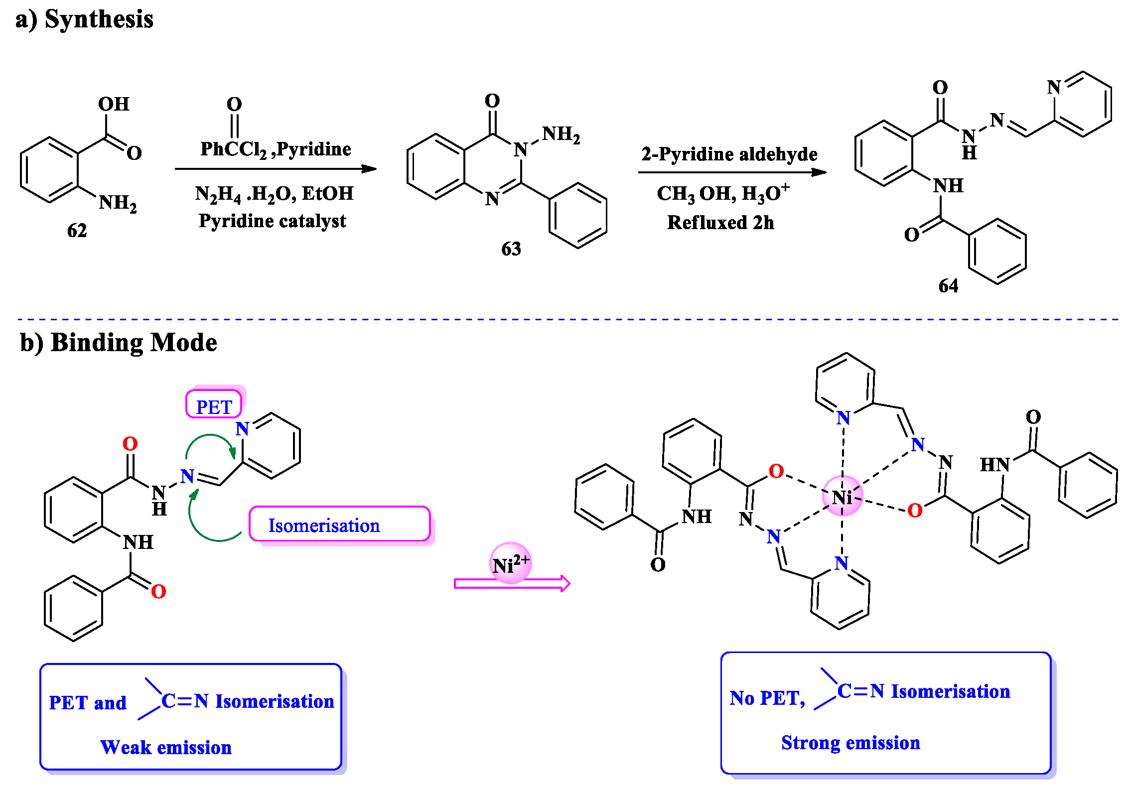
2.5. Ni2+ Ion Detection

Nickel is an incredibly shiny, malleable, and versatile ferromagnetic metal. It is an important component in the production of stainless steel and alloys resistant to corrosion because of its remarkable resistance to environmental corrosion. Moreover, because of its unique properties and resilience, nickel is useful in a variety of applications, such as electroplating, coin manufacturing, petroleum refining industries, textile production, printing, mold-making, and medicinal treatment operators [129]. The extraction and utilization of nickel from ores has grown due to increasing demand for metal. Nevertheless, the techniques used for both its extraction and application are not environmentally friendly. Numerous forms of environmental pollution such as air, water, and soil damage have been identified and recognized as a result of nickel levels exceeding permissible limits. This has adverse impacts on plant, animal, and human health, reinforcing the need for the nickel sector to switch to more environmentally friendly and more sustainable processes [130]. Devastating implications for the environment arise from nickel contamination of the soil and water, notably lower aquatic life production and lesser crop yields. In addition to harming the environment, nickel pollution has serious toxic effects on economic growth and food security, demonstrating the crucial importance of efficient mitigation measurements to safeguard our environment [131]. When airborne nickel metal ions are inhaled, they can enter the body promptly via the respiratory system, producing a prolonged intake and negative consequences later on. These adverse effects include allergic reactions on the skin, pulmonary fibrosis, cardiovascular damage, and a higher risk of cancer, such as kidney and nose cancer. Long-term exposure to nickel metal ions can have detrimental effects on health. That is why it is important to monitor exposure and put safety precautions in action [132]. Considering that nickel contaminants are perilous for the environment, tracking them is a key area of scientific interest. Though there are numerous instrumental procedures for monitoring nickel metal ions, they are sometimes highly expensive, time-consuming, and challenging to perform. In response, scientists have turned to molecular recognition structures as an alternative treatment. These frameworks lead to more accurate and reliable methods of nickel monitoring by supporting the creation of efficient chemosensors that can successfully detect Ni2+ metal ions in aqueous solutions with a high selectivity and sensitivity response [133]. The simplicity, ease of use, high sensitivity, and selectivity of fluorescent chemosensor-based research approaches have garnered much attention, making them a preferred choice over conventional analytical detection techniques [134]. The development of sensors with the best feasible selectivity, sensitivity, and rapid reaction time has been the primary objective of numerous research studies [135]. Multiple fluorescent chemosensors with amplified fluorescence signals have been intended to detect Ni2+ ions with higher sensitivity [136].
W. Lu and J. Chen, along with their coworkers, synthesized a chemosensor for the detection of Ni2+ ions. The experimental results revealed outstanding sensitivity to Ni2+ ions, efficient selectivity, and a significantly low detection limit. In order to design the required chemosensor, reactions of 59 and 2-chloro-6-hydroxybenzaldehyde 60 were refluxed in anhydrous EtOH for 6 h. The reaction mixture was then cooled to room temperature and washed with EtOH to yield the yellow solid product (Scheme 16) [137].
The fluorescence was observed when a number of metal ions (Al3+, Ca2+, Cd2+, Co2+, Cr3+, Cu2+, Fe2+, Fe3+, K+, Mg2+, Mn2+, Na+, Ni2+, Pb2+, and Zn2+) were added to solution 61 to assess the selectivity of 61 towards Ni2+. There is a clear fluorescence response only to Ni2+. When Ni2+ was added, the solution’s color changed from brilliant yellow to dark yellow (Figure 8). Solution 61’s fluorescence intensity was quenched by the addition of Ni2+. As seen in Figure 8a, fluorescence titration profiles of 61 were performed by increasing the concentrations of Ni2+ in the solution to assess the binding capacity of 61 to Ni2+. The fluorescence of 61 at 462.5 nm rapidly lost strength as the quantity of Ni2+ increased. According to LOD = 3σ/k, the concentration range of 1.3 × 10⁻⁶ to 1.6 μM, Ni2+ produced the lowest and most steady fluorescence signal and strong linearity in the fluorescence intensity range (R2 = 0.99238). A standard curve that showed a strong linear relationship between the concentration of Ni2+ ions and the fluorescence intensity was produced. This curve made it feasible to precisely measure Ni2+. The addition of Ni2+ ions resulted in a discernible color change in the chemosensor solution, providing a clue as to sensor 61’s responsiveness or selectivity as well as its visual visibility (Figure 8b) [137].
A. K. Manna, S. Chowdhury, and their collaborators efficiently prepared a novel versatile chemoreceptor that exhibited high potential as a highly sensitive and specific nickel ion sensor by displaying a selective ‘turn-on’ colorimetric and fluorometric response to Ni2+ ions in MeOH. The reaction of 3-amino-2-phenyl-4(3H)-quinazolinone 62 with 2-pyridine carboxaldehyde was performed in dehydrated MeOH. Glacial acetic acid was added as a catalyst to synthesize a Schiff-based chemoreceptor 63. A crystalline solid appeared as the mixture was refluxed for 2 h in a dry air environment and then permitted to stand in the air for one day. After filtration, washing, and air drying, the solid product was obtained with 85% yield (Scheme 17). A colorimetric and fluorometric “turn on” response was displayed by sensor 64 when only Ni2+ ions were present in the methanol–Tris-HCl buffer solution (10 mM, pH 7.2, 1:1 v/v). The ESI-MS study further supported the Job plot analysis’s finding that the receptor forms a 2:1 complex with Ni2+ ions. Additionally, a single crystal of the 64-Ni2+ complex was extracted. According to colorimetric and fluorometric calculations, the detection limit was 1.8 µM and 1.18 µM, respectively. These values are significantly lower than those suggested by the WHO drinking water standards. Chemosensor 64 was used for live cell imaging, smartphone-based analysis, molecular logic gate construction, and the recovery of tainted water samples [134].

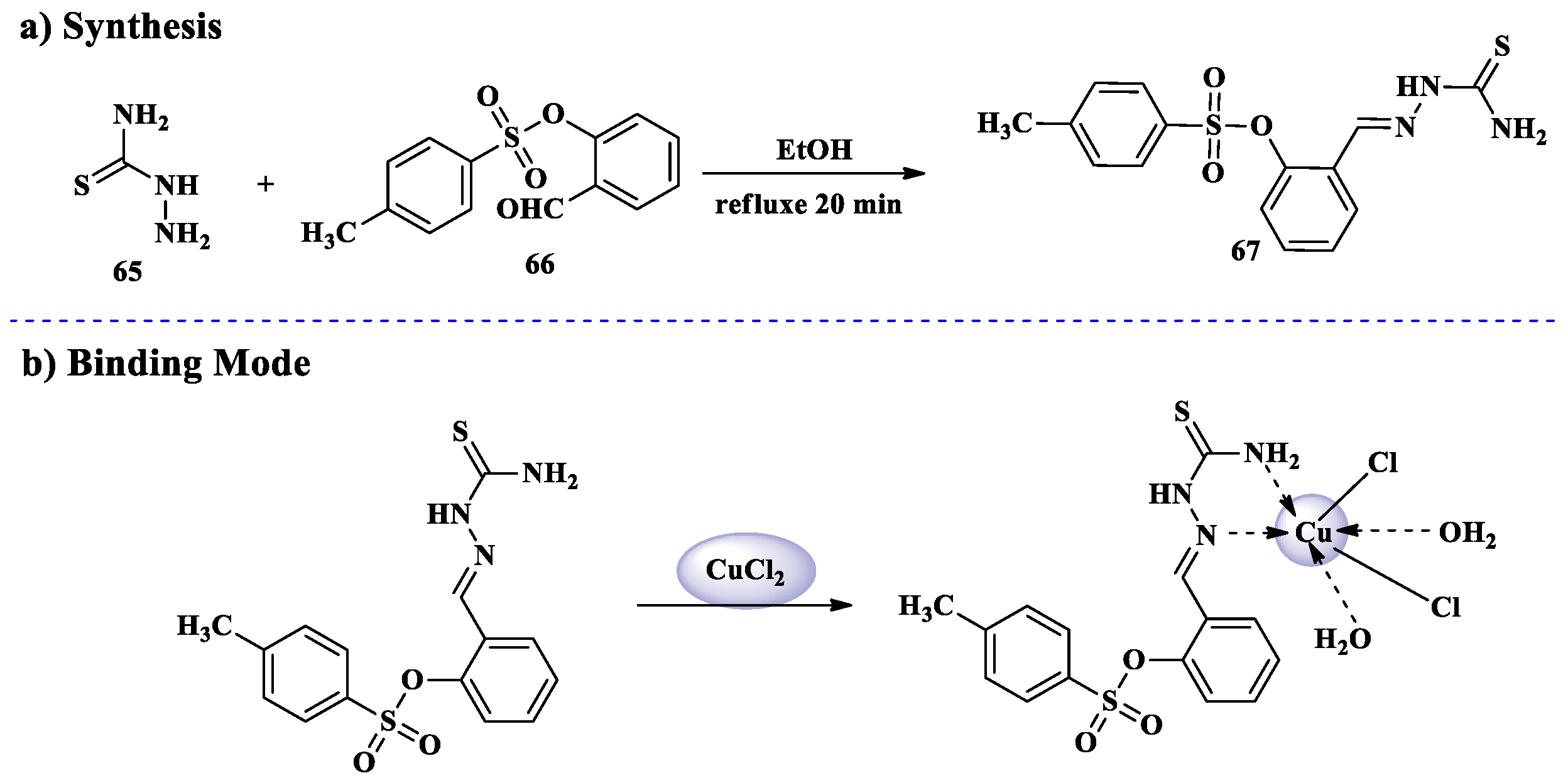
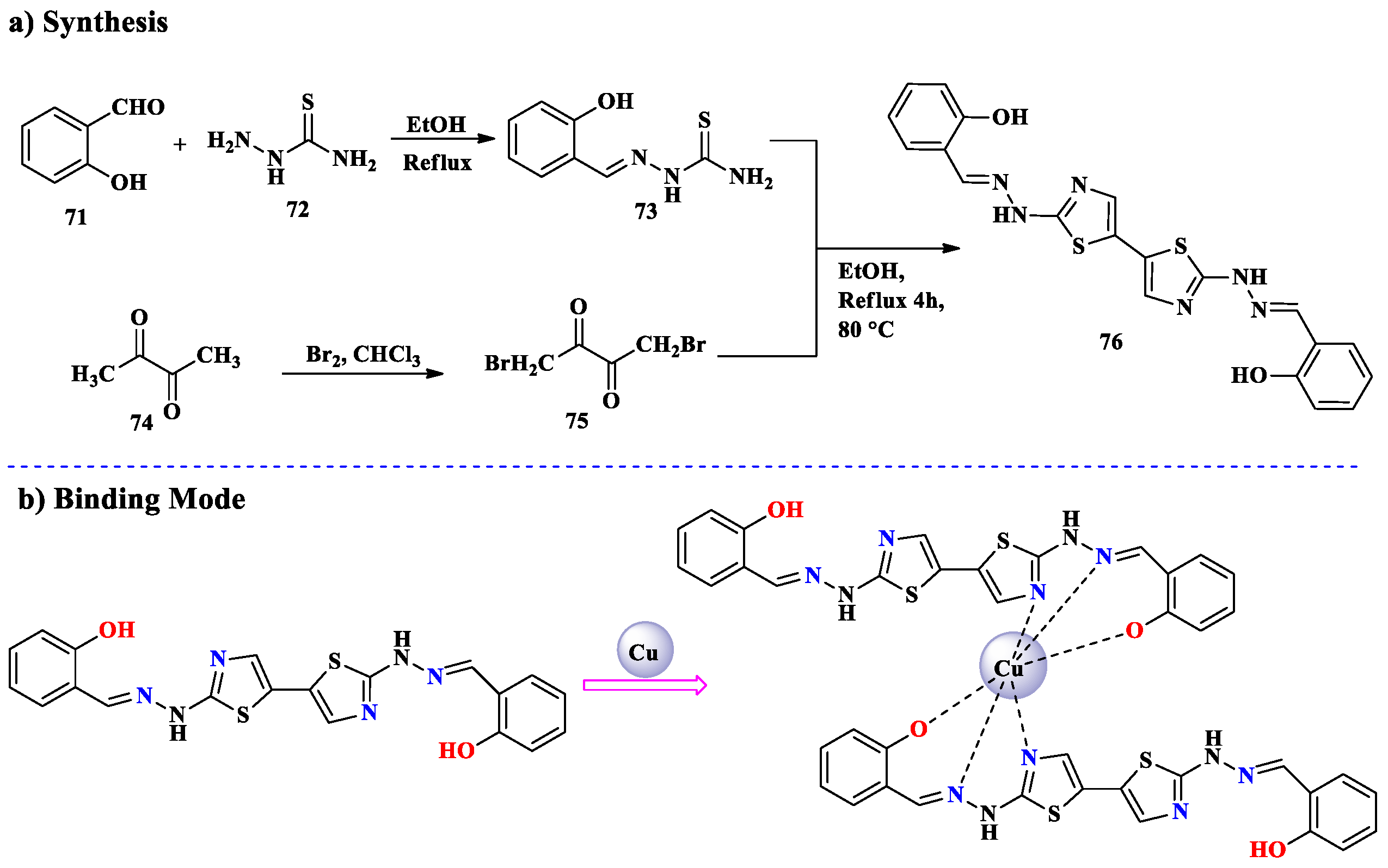
2.6. Cu2+ Ion Detection

Copper, the third most prevalent transition metal in the body, is necessary for a number of physiological functions. It performs a vital role in human digestion. Most of the time, it is probable that Cu2+ is always dangerous to microbes and marine life [138]. Cu2+ pollution has grown increasingly commonplace worldwide as a result of the growing usage of copper in appliances at home and in water pipes, industry, and agriculture. Analytical methods that are rapid, inexpensive, simple, and trustworthy are essential for detecting copper in drinking water and biological specimens, both qualitatively and quantitatively [139,140]. Cu2+ is a d9 ion with a strong preference for N-donor ligands. It often adopts a square planar or distorted octahedral geometry, making selective binding possible through steric and electronic effects. It has been reported that colorimetric and fluorometric chemosensors have been designed to detect Cu2+ ions through changes in fluorescence intensity and color [141]. H. Hrichi and his coworkers synthesized 2-[(carbamothioylhydrazono)methyl-phenyl-4-methylbenzenesulfonate 67, which is identified by NMR and FTIR spectroscopy and has selective colorimetric chemosensor characteristics for Cu2+ ions detection. In an aqueous medium with several competing cations present, such as Na+, K+, Mg2+, Al3+, Ag+, Ca2+, Fe3+, Fe2+, Co2+, Ni2+, Cr3+, Pb2+, Zn2+, Ba2+, and Cd2+, the 67 chemosensor exhibited excellent sensitivity and selectivity toward Cu2+ ions. By combining Cu2+ and CHMPMBS in a 1:1 stoichiometry to form (CHMPMBS:Cu)complex, the chemosensor 67 showed a noticeable color shift from light yellow to green. This compound offers a quick and precise colorimetric test for Cu2+ ion detection with the naked eye. Chemosensor 67 was designed using standard protocol. Thiosemicarbazide 65 and 2-tosyloxybenzaldehyde 66 were added to and dissolved in ethanol and subsequently refluxed (Scheme 18). The precipitate was cooled at room temperature before being filtered and crystallized from the EtOH. The developed chemosensor 67 gave a distinctive color change from pale yellow to pale green. Sensor 67 (CHMPMBS) exhibits a rapid and accurate colorimetric assay for naked-eye detection of Cu2+ ions with a detection limit of 8.12 μM [142].
The cation recognition behavior of CHMPMBS sensor 67 with Na(I), Mg(II), Al(III), K(I), Ca(II), Cr(III), Mn(II), Fe(III), Fe(II), Co(II), Ni(II), Cu(II), Zn(II), Sr(II), Ag(I), Cd(II), Ba(II), and Pb(II) at pH = 6.5 was investigated using UV–Vis absorption spectroscopy. A new charge transfer band appeared, which resulted in the color shift of CHMPMBS. The distinctive CHMPMBS absorption bands vanished as a result of the paramagnetic Cu(II) interaction with CHMPMBS, and clear broadband emerged at about 515 nm (Figure 9a). By mixing 4.0 mL of different cations (50 µM, in H2O) with 1.0 mL of the CHMPMBS solution (50 µM, in DMF-water (1:1 v/v), the colorimetric ability of CHMPMBS was investigated. The absorption spectra of the CHMPMBS in water-DMF (1:1, v/v) solvent showed two distinct absorption bands at 290 nm and 405 nm, respectively, which correspond to the π–π* and n–π* electronic transitions (Figure 9b). The distinctive 67 absorption bands disappeared when the Cu2+ interacted with CHMPMBS, and new broadband, caused by d–d transitions, emerged at about 515 nm. Chemosensor 67 demonstrates the characteristics of being sensitive, selective, and qualitatively capable of detecting Cu2+ ions by displaying a notable color shift from pale yellow to pale green as a result of detecting Cu2+ ions (Figure 9c). There was no change in color in several other samples screened for cations [143].
S. A. Rupa synthesized a new chemosensor 70. A solution of 2-acetyl thiophene 69 in CH3OH was mixed with a solution of 5-bromo-2-hydroxybenzohydrazide 68 in methanol. The resultant mixtures were agitated at 60 °C for six hours with continuous stirring. During the procedure, a yellow precipitate was produced, which was filtered out and vacuum-dried, producing a 47% yield (Scheme 19). Chemosensor 70 was shown to be insensitive to different metal ions; however, its sensing investigations showed a turn-on-enhanced fluorescence and colorimetric response toward Cu2+ ions. The binding stoichiometry of sensor 70 and metal ions is 2:1, according to the Job plots. Furthermore, the results of density functional theory (DFT) clearly suggested that chemosensor 70 might be employed as a potent colorimetric sensor for Cu2+ ion detection. Disk diffusion was used to assess the in vitro antimicrobial properties of sensor 70, and the findings showed excellent antibacterial activity against E. coli [144].
Z. Jia, J. Wei, and their collaborators devised and manufactured a Cu2+-specific chemosensor probe called thiazolyl-hydrazone. Absolute ethanol was used to dissolve 2-(2-hydroxybenzylidene) hydrazine-1-carbothioamide 73 and 1,4-dibromobutanedione 75. The reaction mixture was refluxed for four hours. The precipitate was filtered and recrystallized with DMF and ethanol. A brown-colored solid product produced a 53% yield (Scheme 20). With a binding stoichiometry of 1:1, a Cu2+-specific chemosensor 76 thiazolyl-hydrazone was formed, offering a tridentate chelate NNO coordination mode for Cu2+.When the sensor 76 solution binds to the Cu2+ cation, it quickly changes from colorless to yellow and develops a new, strong absorption band at 414 nm. This change is not induced by other metal cations. The detection limit for the naked eye is as low as 20 μM, whereas the analytical detection limit is 0.75 μM. Furthermore, at a detection limit of 7.2 μM, Cu2+ could quenched the emission signal of sensor 76 [145].

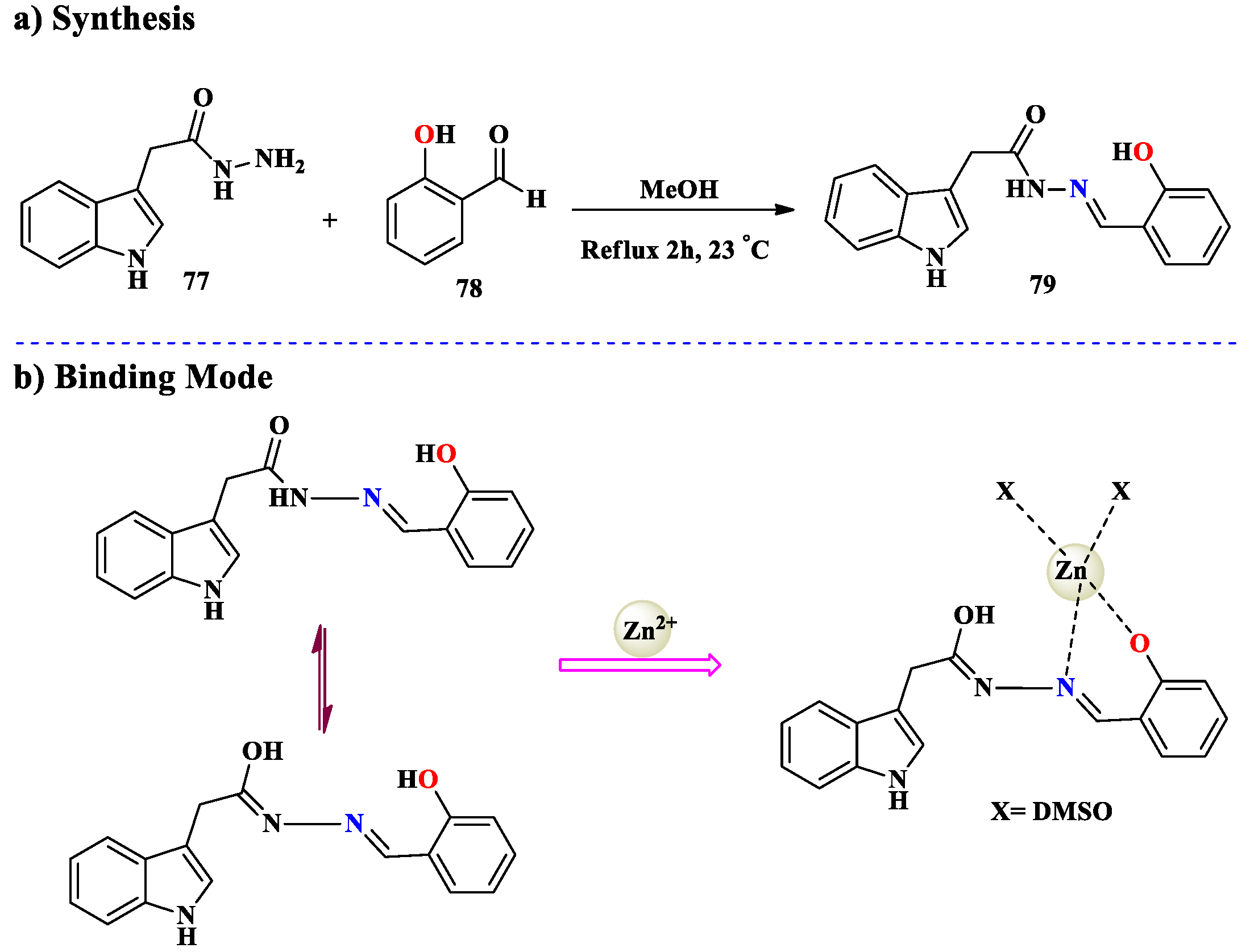
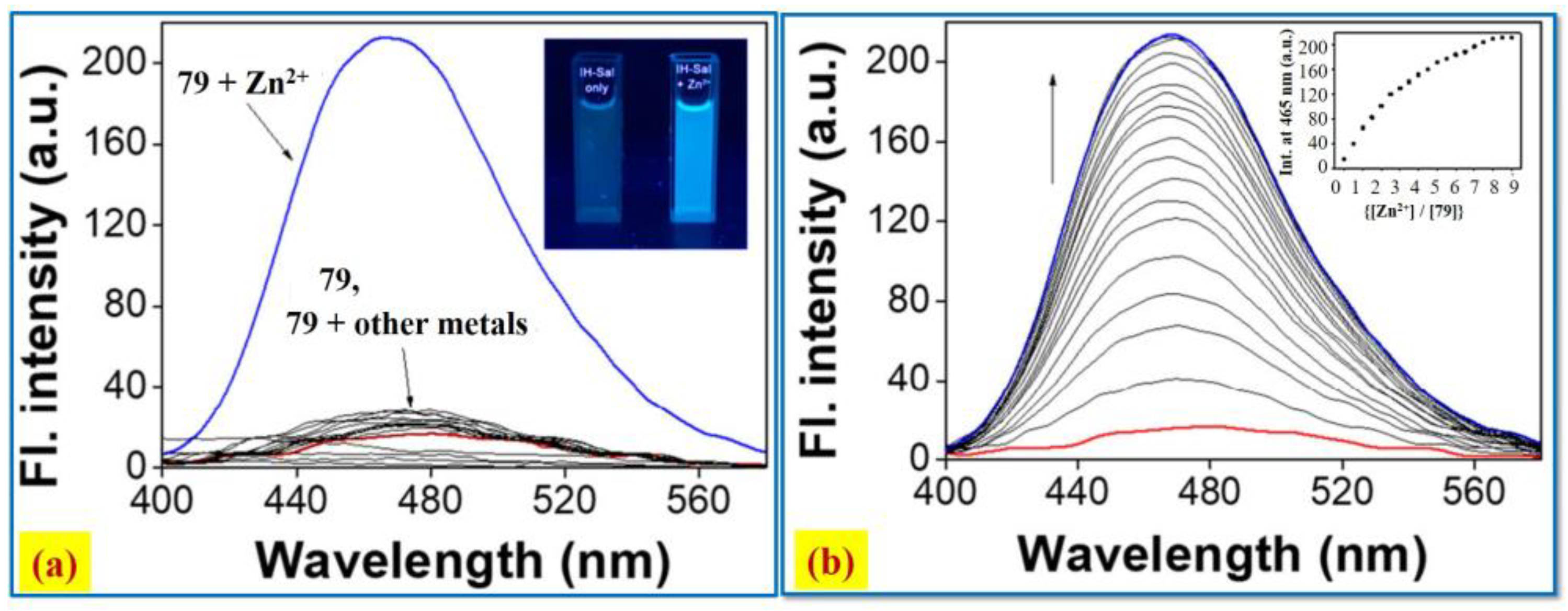
2.7. Zn2+ Ion Detection

Zinc ions are the second most frequent element in the human body. Numerous biological activities, including gene transcription, brain function, and immune system function, depend on the Zn2+ ion [146,147,148]. Even so, an overabundance of Zn2+ ions would upset the equilibrium of cellular functions, leading to neurodegenerative conditions like Parkinson’s and Alzheimer’s disease, diabetes, prostate cancer, Wilson and Menkes, etc. [149,150,151,152,153,154]. Furthermore, it is difficult to identify Zn2+ ions in biological systems because they are colorless in aqueous solutions, do not have unpaired electrons, and remain in the positive two-oxidation state. Zn2+ is a d10 closed-shell ion with no ligand field stabilization energy. It prefers O/S/N donors, typically forming tetrahedral or octahedral complexes. According to this perspective, fluorescent sensors are effective instruments for tracking zinc ions as the selective adsorption of zinc ions on chemosensors can alter the fluorescence characteristics [155]. Consequently, there has been a lot of interest in various research on improving the sensitivity of fluorescent probes for Zn2+ ions. To detect Zn2+ ions, a fluorescent chemosensor based on indole (79) was developed. This exhibited an obvious rise in fluorescence upon interaction with Zn2+ ions. The World Health Organization’s approved criterion for Zn2+ detection using chemosensors was far lower than that chemosensor. This can accurately represent, identify, and measure Zn2+ in actual water samples and living organisms. Fluorescence and UV–visible spectroscopy have been used to show 79-Zn2+ detection approach. Salicylaldehyde 78 and 2-(1H-indol-3-yl)acetohydrazide 77 were refluxed in MeOH for two hours at 23 °C to give [(E)-N0-2-hydroxybenzylidene)-2-(1H-indol-3-yl)acetohydrazide] 79 (Scheme 21). The detection limit (0.41 μM) of sensor 79 can quantify Zn2+ in real water samples. Moreover, it could identify and illustrate the presence of Zn2+ in zebrafish [156].
The fluorescence variance was observed using different metal ions in bis-tris buffer (Figure 10). There was no fluorescence emission from 79 directly. The Zn2+ ion interaction caused a noticeable increase in fluorescence at 465 nm (λex = 369 nm), along with an important Stokes shift. Out of all the chemosensors, 79 + Zn2+ had the biggest Stokes shift.
M. M. Mathew and A. Sreekanth developed a novel chemosensor 82 for detecting Zn2+ ions. They added 2-hydroxynaphthaldehyde 81 in EtOH with a stirred solution of thiphene-2,5-dicarbohydrazide 80, followed by the addition of a few drops of glacial acetic acid as catalyst. The reaction mixture was then refluxed for 6 h, and the reaction progress was monitored using TLC. This resulted in the generation of a dark yellow precipitate (yield = 93%), which was collected via simple filtration and dried under vacuum (Scheme 22). Chemosensor 82 was discovered to be highly emissive upon complexation with the Zn2+ ion. In a DMSO/H2O (6:4 v/v) solution, this turn-on fluorescence recognition was prepared. After calculation, the binding constant was determined to be 1.15 × 10⁴ M−1. The limit of detection (LOD) was determined to be 1.51 × 10⁻⁷ M, which is significantly lower than the allowable amount of Zn2+ (70 μM) in drinking water. Chemosensor 82-Zn2+ complex’s 1:1 binding stoichiometry was predicted by Jobs’ plot. By using a chelation-enhanced fluorescence mechanism, the observed fluorescent enhancement turn-on emission may be eliminated. The 82-Zn2+ combination with EDTA was subjected to the appropriate INHIB-IT/IMPLICATION Logic gate behavior and reversibility tests [157].
X. Peng, X. Tang and others synthesized N-(3,5-di-tert-butylsalicylidene)-2-hydroxybenzoylhydrazine. It was detected as a Zn2+ fluorescence chemosensor. Salicyloyl hydrazide 84 was introduced to a stirred solution of 3,5-di-tert-butyl-2-hydroxybenzaldehyde 83 in EtOH. After eight hours of reflux, the reaction mixture was permitted to settle to room temperature. The resultant yellow precipitates 85 were filtered, cleaned with ethanol, and vacuum-dried. A yield of 80% of product 85 was obtained (Scheme 23). The addition of a single hydroxyl group to chemosensor 85 not only exhibits a robust interaction with Zn2+, but also sets this metal cation apart from its rivals, which include the potent Cd2+, Mg2+, and Ca2+ ions. The development of metal–ligand complexes was responsible for the rise in emission in the presence of Zn2+. Because of the high coordination of Zn2+ that would impose stiffness, the fluorescence response of 85 is improved by Zn2+-selective chelation by around 25-fold. One explanation might be that the substituent hydroxyl group improves the Zn2+ ion’s chelating capacity [158].

3. Conclusions and Future Perspectives

Transition metal ions, particularly the first-row ones, are common and serve a variety of biological purposes. A significant accumulation of these ions, however, may be harmful to one’s health. Therefore, it is crucial to accurately determine their spatial distribution both inside and outside of cells. The amplification of fluorescence by transition metal ions has potential applications beyond biological sensing. However, the majority of transition metal ions are paramagnetic and efficiently quench fluorescence. Optical chemosensors remain a highly active area of research, as evidenced by the papers reviewed. Future efforts should focus on developing and utilizing fluorescent probes that function effectively in pure aqueous solutions at physiological pH levels. This feature article aims to provide readers with a deeper understanding of various synthetic approaches of Aryl-substituted thiocarbohydrazide, thiosemicarbazide, thiazolyl-hydrazone, thiophene diocarbohydrazide, Arylhydrazone, indole, Schiff-based, carbazole, acylhydrazone coumarin chromofluorescent sensors, etc., and their mechanisms of action. We categorize colorimetric and fluorometric sensors for the detection of first-row transition metal ions, including Cr³⁺, Mn²⁺, Fe²⁺, Co²⁺, Ni²⁺, Cu²⁺, and Zn²⁺, while highlighting their ability to simultaneously detect multiple ions. The development of these sensors addresses critical challenges related to the rapid and straightforward detection of hazardous metal ions at trace levels in biological and environmental systems. As we proceed, it is evident that the synthesis of fluorescent chemosensors is far from finished [159]. Their capabilities could be further improved by emerging technologies like fluorescent-based sensors. Furthermore, point-of-care diagnostics and customized healthcare are being revolutionized by the incorporation of these sensors into portable devices [31,160,161]. This study presents a comprehensive overview of current methodologies, and the insights provided here aim to inspire further research to meet the evolving demands in the field of trace transition metal ion detection.

Author Contributions

Conceptualization, S.A. and R.S.; data curation, S.A. and I.K.; analysis, I.K. and S.R.; funding acquisition, R.S.; investigation, S.A., S.R. and I.Z.; supervision, S.A. and R.S.; visualization, S.A.; writing—original draft, S.A., I.K. and S.R.; writing—review and editing, R.S., S.B. and S.A. All authors have read and agreed to the published version of the manuscript.

Funding

This research received no external funding.

Institutional Review Board Statement

Not applicable.

Informed Consent Statement

Not applicable.

Data Availability Statement

The datasets and materials used and/or analyzed during the current study are available from the corresponding authors upon reasonable request.

Conflicts of Interest

The authors declare no conflict of interest.

Abbreviations

CHMPMBS2-[(carbamothioylhydrazono)methyl-phenyl-4-methylbenzenesulfonate
WHOWorld Health Organization
ESI-MSElectrospray Ionization–Mass Spectrometry
µMMicromolar
THFTetrahydrofuran
PBSPhosphate-Buffered Saline
DMSODimethyl Sulfoxide
DMFODimethylformamide
EDTAEthylenediaminetetraacetic acid
LODLimit of detection
DBQAN-(4-(dimethylamino) benzylidene) quinolone-3-amine
HEPESN-(2-Hydroxyethyl)piperazine-N′-2-ethanesulfonic acid

References

  1. Chen, S.-Y.; Li, Z.; Li, K.; Yu, X.-Q. Small molecular fluorescent probes for the detection of lead, cadmium and mercury ions. Coord. Chem. Rev. 2021, 429, 213691. [Google Scholar] [CrossRef]
  2. Gaur, V.K.; Sharma, P.; Gaur, P.; Varjani, S.; Ngo, H.H.; Guo, W.; Chaturvedi, P.; Singhania, R.R. Sustainable mitigation of heavy metals from effluents: Toxicity and fate with recent technological advancements. Bioengineered 2021, 12, 7297–7313. [Google Scholar] [CrossRef]
  3. Khan, S.; Chen, X.; Almahri, A.; Allehyani, E.S.; Alhumaydhi, F.A.; Ibrahim, M.M.; Ali, S. Recent developments in fluorescent and colorimetric chemosensors based on schiff bases for metallic cations detection: A review. J. Environ. Chem. Eng. 2021, 9, 106381. [Google Scholar] [CrossRef]
  4. Mukherjee, S.; Bhattacharyya, S.; Ghosh, K.; Pal, S.; Halder, A.; Naseri, M.; Mohammadniaei, M.; Sarkar, S.; Ghosh, A.; Sun, Y. Sensory development for heavy metal detection: A review on translation from conventional analysis to field-portable sensor. Trends Food Sci. Technol. 2021, 109, 674–689. [Google Scholar] [CrossRef]
  5. Akpor, O.B.; Ohiobor, G.O.; Olaolu, D. Heavy metal pollutants in wastewater effluents: Sources, effects and remediation. Adv. Biosci. Bioeng. 2014, 2, 37–43. [Google Scholar] [CrossRef]
  6. Lopez Marzo, A.M.; Pons, J.; Blake, D.A.; Merkoçi, A. All-integrated and highly sensitive paper based device with sample treatment platform for Cd2+ immunodetection in drinking/tap waters. Anal. Chem. 2013, 85, 3532–3538. [Google Scholar] [CrossRef]
  7. Guerrero, M.; Garcia-Anton, J.; Tristany, M.; Pons, J.; Ros, J.; Philippot, K.; Lecante, P.; Chaudret, B. Design of new N, O hybrid pyrazole derived ligands and their use as stabilizers for the synthesis of Pd nanoparticles. Langmuir 2010, 26, 15532–15540. [Google Scholar] [CrossRef]
  8. Aragay, G.; Pons, J.; Merkoçi, A. Enhanced electrochemical detection of heavy metals at heated graphite nanoparticle-based screen-printed electrodes. J. Mater. Chem. 2011, 21, 4326–4331. [Google Scholar] [CrossRef]
  9. Sun, X.; Ji, J.; Jiang, D.; Li, X.; Zhang, Y.; Li, Z.; Wu, Y. Development of a novel electrochemical sensor using pheochromocytoma cells and its assessment of acrylamide cytotoxicity. Biosens. Bioelectron. 2013, 44, 122–126. [Google Scholar] [CrossRef]
  10. Khan, J. Synthesis and applications of fluorescent chemosensors: A review. J. Fluoresc. 2024, 34, 2485–2494. [Google Scholar] [CrossRef]
  11. Li, W.-T.; Wu, G.-Y.; Qu, W.-J.; Li, Q.; Lou, J.-C.; Lin, Q.; Yao, H.; Zhang, Y.-M.; Wei, T.-B. A colorimetric and reversible fluorescent chemosensor for Ag+ in aqueous solution and its application in IMPLICATION logic gate. Sens. Actuators B Chem. 2017, 239, 671–678. [Google Scholar] [CrossRef]
  12. Francisco, B.B.A.; Caldas, L.F.S.; Brum, D.M.; Cassella, R.J. Novel spectrophotometric method for the determination of aluminum in soda drinks packed in cans and plastic bottles. J. Hazard. Mater. 2010, 181, 485–490. [Google Scholar] [CrossRef]
  13. Tontrong, S.; Khonyoung, S.; Jakmunee, J. Flow injection spectrophotometry using natural reagent from Morinda citrifolia root for determination of aluminium in tea. Food Chem. 2012, 132, 624–629. [Google Scholar] [CrossRef]
  14. Sang, H.; Liang, P.; Du, D. Determination of trace aluminum in biological and water samples by cloud point extraction preconcentration and graphite furnace atomic absorption spectrometry detection. J. Hazard. Mater. 2008, 154, 1127–1132. [Google Scholar] [CrossRef]
  15. Herdan, J.; Feeney, R.; Kounaves, S.P.; Flannery, A.F.; Storment, C.W.; Kovacs, G.T.; Darling, R.B. Field evaluation of an electrochemical probe for in situ screening of heavy metals in groundwater. Environ. Sci. Technol. 1998, 32, 131–136. [Google Scholar] [CrossRef]
  16. Lian, H.; Kang, Y.; Bi, S.; Arkin, Y.; Shao, D.; Li, D.; Chen, Y.; Dai, L.; Gan, N.; Tian, L. Direct determination of trace aluminum with quercetin by reversed-phase high performance liquid chromatography. Talanta 2004, 62, 43–50. [Google Scholar] [CrossRef]
  17. Kim, H.N.; Ren, W.X.; Kim, J.S.; Yoon, J. Fluorescent and colorimetric sensors for detection of lead, cadmium, and mercury ions. Chem. Soc. Rev. 2012, 41, 3210–3244. [Google Scholar] [CrossRef] [PubMed]
  18. Song, F.; Yang, C.; Liu, H.; Gao, Z.; Zhu, J.; Bao, X.; Kan, C. Dual-binding pyridine and rhodamine B conjugate derivatives as fluorescent chemosensors for ferric ions in aqueous media and living cells. Analyst 2019, 144, 3094–3102. [Google Scholar] [CrossRef]
  19. Kumar, G.G.V.; Kesavan, M.P.; Sankarganesh, M.; Sakthipandi, K.; Rajesh, J.; Sivaraman, G. A Schiff base receptor as a fluorescence turn-on sensor for Ni2+ ions in living cells and logic gate application. New J. Chem. 2018, 42, 2865–2873. [Google Scholar] [CrossRef]
  20. Ghazali, S.; Fan, J.; Du, J.; Peng, X. Mito-targeted “turn-on” fluorescent probe for nickel (II) detection. Methods 2019, 168, 24–28. [Google Scholar] [CrossRef]
  21. Li, L.; Wang, J.; Xu, S.; Li, C.; Dong, B. Recent progress in fluorescent probes for metal ion detection. Front. Chem. 2022, 10, 875241. [Google Scholar] [CrossRef]
  22. Hu, L.; Nie, L.; Xu, G.; Shi, H.; Xu, X.; Zhang, X.; Yan, Z. Spectral properties of 4-(4-hydroxy-1-naphthylazo) benzenesulfonic acid and its application for colorimetric determination of trace Fe3+. RSC Adv. 2014, 4, 19370–19374. [Google Scholar] [CrossRef]
  23. Zhao, G.; Guo, B.; Wei, G.; Guang, S.; Gu, Z.; Xu, H. A novel dual-channel Schiff base fluorescent chemo-sensor for Zn2+ and Ca2+ recognition: Synthesis, mechanism and application. Dye. Pigment. 2019, 170, 107614. [Google Scholar] [CrossRef]
  24. Komatsu, H.; Miki, T.; Citterio, D.; Kubota, T.; Shindo, Y.; Kitamura, Y.; Oka, K.; Suzuki, K. Single molecular multianalyte (Ca2+, Mg2+) fluorescent probe and applications to bioimaging. J. Am. Chem. Soc. 2005, 127, 10798–10799. [Google Scholar] [CrossRef] [PubMed]
  25. Kolanowski, J.L.; Liu, F.; New, E.J. Fluorescent probes for the simultaneous detection of multiple analytes in biology. Chem. Soc. Rev. 2018, 47, 195–208. [Google Scholar] [CrossRef] [PubMed]
  26. Khatua, S.; Schmittel, M. A single molecular light-up sensor for quantification of Hg2+ and Ag+ in aqueous medium: High selectivity toward Hg2+ over Ag+ in a mixture. Org. Lett. 2013, 15, 4422–4425. [Google Scholar] [CrossRef]
  27. Zhang, J.F.; Zhou, Y.; Yoon, J.; Kim, J.S. Recent progress in fluorescent and colorimetric chemosensors for detection of precious metal ions (silver, gold and platinum ions). Chem. Soc. Rev. 2011, 40, 3416–3429. [Google Scholar] [CrossRef]
  28. Kaim, W.; Schwederski, B.; Klein, A. Bioinorganic Chemistry—Inorganic Elements in the Chemistry of Life: An Introduction and Guide; John Wiley & Sons: Hoboken, NJ, USA, 2013. [Google Scholar]
  29. Butler, A.; Carrano, C.J. Coordination chemistry of vanadium in biological systems. Coord. Chem. Rev. 1991, 109, 61–105. [Google Scholar] [CrossRef]
  30. Lakowicz, J.R. Principles of Fluorescence Spectroscopy; Springer: Berlin/Heidelberg, Germany, 2006. [Google Scholar]
  31. Pal, S.; Chatterjee, N.; Bharadwaj, P.K. Selectively sensing first-row transition metal ions through fluorescence enhancement. RSC Adv. 2014, 4, 26585–26620. [Google Scholar] [CrossRef]
  32. Fabbrizzi, L.; Poggi, A. Sensors and switches from supramolecular chemistry. Chem. Soc. Rev. 1995, 24, 197–202. [Google Scholar] [CrossRef]
  33. Igiri, B.E.; Okoduwa, S.I.; Idoko, G.O.; Akabuogu, E.P.; Adeyi, A.O.; Ejiogu, I.K. Toxicity and bioremediation of heavy metals contaminated ecosystem from tannery wastewater: A review. J. Toxicol. 2018, 2018, 2568038. [Google Scholar] [CrossRef] [PubMed]
  34. Kaur, B.; Kaur, N.; Kumar, S. Colorimetric metal ion sensors—A comprehensive review of the years 2011–2016. Coord. Chem. Rev. 2018, 358, 13–69. [Google Scholar] [CrossRef]
  35. Bicker, K.L.; Wiskur, S.L.; Lavigne, J.J. Colorimetric sensor design. In Chemosensors: Principles, Strategies, and Applications; John Wiley & Sons: Hoboken, NJ, USA, 2011; pp. 275–295. [Google Scholar]
  36. Wu, D.; Sedgwick, A.C.; Gunnlaugsson, T.; Akkaya, E.U.; Yoon, J.; James, T.D. Fluorescent chemosensors: The past, present and future. Chem. Soc. Rev. 2017, 46, 7105–7123. [Google Scholar] [CrossRef]
  37. Priyangga, K.T.A.; Kurniawan, Y.S.; Ohto, K.; Jumina, J. A review on calixarene fluorescent chemosensor agents for various analytes. J. Multidiscip. Appl. Nat. Sci. 2022, 2, 23–40. [Google Scholar] [CrossRef]
  38. Ali, R.; Razi, S.S.; Shahid, M.; Srivastava, P.; Misra, A. Off–On–Off fluorescence behavior of an intramolecular charge transfer probe toward anions and CO2. Spectrochim. Acta Part A Mol. Biomol. Spectrosc. 2016, 168, 21–28. [Google Scholar] [CrossRef] [PubMed]
  39. Zhang, Z.; Wang, C.; Zhang, Z.; Luo, Y.; Sun, S.; Zhang, G. Cd (II) enhanced fluorescence and Zn (II) quenched fluorescence with phenylenevinylene terpyridine: A theoretical investigation. Spectrochim. Acta Part A Mol. Biomol. Spectrosc. 2019, 209, 40–48. [Google Scholar] [CrossRef] [PubMed]
  40. Samanta, S.; Manna, U.; Ray, T.; Das, G. An aggregation-induced emission (AIE) active probe for multiple targets: A fluorescent sensor for Zn2+ and Al3+ & a colorimetric sensor for Cu2+ and F. Dalton Trans. 2015, 44, 18902–18910. [Google Scholar]
  41. Fukuhara, G. Analytical supramolecular chemistry: Colorimetric and fluorimetric chemosensors. J. Photochem. Photobiol. C Photochem. Rev. 2020, 42, 100340. [Google Scholar] [CrossRef]
  42. Aragay, G.; Pons, J.; Merkoçi, A. Recent trends in macro-, micro-, and nanomaterial-based tools and strategies for heavy-metal detection. Chem. Rev. 2011, 111, 3433–3458. [Google Scholar] [CrossRef]
  43. Darwish, N.B.; Kurdi, A.; Alshihri, S.; Tabbakh, T. Organic heterocyclic-based colorimetric and fluorimetric chemosensors for the detection of different analytes: A review (from 2015 to 2022). Mater. Today Chem. 2023, 27, 101347. [Google Scholar] [CrossRef]
  44. Jo, T.G.; Na, Y.J.; Lee, J.J.; Lee, M.M.; Lee, S.Y.; Kim, C. A multifunctional colorimetric chemosensor for cyanide and copper (II) ions. Sens. Actuators B Chem. 2015, 211, 498–506. [Google Scholar] [CrossRef]
  45. Hu, Y.; Yin, J.; Yoon, J. A multi-responsive cyanine-based colorimetric chemosensor containing dipicolylamine moieties for the detection of Zn (II) and Cu (II) ions. Sens. Actuators B Chem. 2016, 230, 40–45. [Google Scholar] [CrossRef]
  46. Mohammadi, A.; Yaghoubi, S. A new dual colorimetric chemosensor based on quinazolinone for CNˉ, AcOˉ and Cu2+ ions. Sens. Actuators B Chem. 2017, 241, 1069–1075. [Google Scholar] [CrossRef]
  47. Yang, M.; Chae, J.B.; Kim, C.; Harrison, R.G. A visible chemosensor based on carbohydrazide for Fe (II), Co (II) and Cu (II) in aqueous solution. Photochem. Photobiol. Sci. 2019, 18, 1249–1258. [Google Scholar] [CrossRef]
  48. Hu, J.-H.; Li, J.-B.; Qi, J.; Sun, Y. Acylhydrazone based fluorescent chemosensor for zinc in aqueous solution with high selectivity and sensitivity. Sens. Actuators B Chem. 2015, 208, 581–587. [Google Scholar] [CrossRef]
  49. Han, F.; Bao, Y.; Yang, Z.; Fyles, T.M.; Zhao, J.; Peng, X.; Fan, J.; Wu, Y.; Sun, S. Simple bisthiocarbonohydrazones as sensitive, selective, colorimetric, and switch-on fluorescent chemosensors for fluoride anions. Chem.-A Eur. J. 2007, 13, 2880–2892. [Google Scholar] [CrossRef]
  50. Zhou, Y.; You, X.-Y.; Fang, Y.; Li, J.-Y.; Liu, K.; Yao, C. A thiophen-thiooxorhodamine conjugate fluorescent probe for detecting mercury in aqueous media and living cells. Org. Biomol. Chem. 2010, 8, 4819–4822. [Google Scholar] [CrossRef]
  51. Chen, Y.; Wu, L.; Chen, Y.; Bi, N.; Zheng, X.; Qi, H.; Qin, M.; Liao, X.; Zhang, H.; Tian, Y. Determination of mercury (II) by surface-enhanced Raman scattering spectroscopy based on thiol-functionalized silver nanoparticles. Microchim. Acta 2012, 177, 341–348. [Google Scholar] [CrossRef]
  52. Matesanz, A.I.; Cuadrado, I.; Pastor, C.; Souza, P. A Novel Sulfur-bridged Dimeric Zinc (II) Complex with 2, 6-Diacetylpyridine Bis (thiosemicarbazone). Z. Anorg. Allg. Chem. 2005, 631, 780–784. [Google Scholar] [CrossRef]
  53. Casas, J.S.; Castellano, E.E.; Ellena, J.; García-Tasende, M.S.; Sánchez, A.; Sordo, J.; Vázquez-López, E.M.; Vidarte, M.J. Synthesis, Spectroscopic and Structural Aspects of Mono-and Diorganothallium (III) Complexes of 2, 6-Diacetylpyridine Bis (thiosemicarbazone)(H2DAPTSC)—A New Coordination Mode of the HDAPTSC—Anion. Z. Anorg. Allg. Chem. 2003, 629, 261–267. [Google Scholar] [CrossRef]
  54. Paswan, S.; Bharty, M.; Bharati, P.; Sonkar, P.K.; Ganesan, V.; Butcher, R. Square planar Ni (ii) complexes of acetone N 4-phenyl-thiosemicarbazone and in situ generated benzoyl thiosemicarbazide ligands: Synthesis, spectral and structural characterizations, thermal behaviour and electrochemical studies. New J. Chem. 2017, 41, 15466–15474. [Google Scholar] [CrossRef]
  55. Ren, W.; Zhu, C.; Wang, E. Enhanced sensitivity of a direct SERS technique for Hg2+ detection based on the investigation of the interaction between silver nanoparticles and mercury ions. Nanoscale 2012, 4, 5902–5909. [Google Scholar] [CrossRef] [PubMed]
  56. Fang, Y.; Li, X.; Li, J.-Y.; Wang, G.-Y.; Zhou, Y.; Xu, N.-Z.; Hu, Y.; Yao, C. Thiooxo-Rhodamine B hydrazone derivatives bearing bithiophene group as fluorescent chemosensors for detecting mercury (II) in aqueous media and living HeLa cells. Sens. Actuators B Chem. 2018, 255, 1182–1190. [Google Scholar] [CrossRef]
  57. Wang, L.; Zheng, B.; Zhao, Y.; Du, J.; Xiao, D. A Hg2+ selective fluorescent chemosensor based on rhodamine B thiohydrazide and its application in bioimaging. Anal. Methods 2012, 4, 2369–2374. [Google Scholar] [CrossRef]
  58. Liu, Y.; Yang, L.; Li, L.; Liang, X.; Li, S.; Fu, Y. A dual thiourea-appended perylenebisimide “turn-on” fluorescent chemosensor with high selectivity and sensitivity for Hg2+ in living cells. Spectrochim. Acta Part A Mol. Biomol. Spectrosc. 2020, 241, 118678. [Google Scholar] [CrossRef]
  59. Anbazhagan, R.; Sankaran, K. Design, synthesis, computational calculation and biological evaluation of some novel 2-thiazolyl hydrazones. Spectrochim. Acta Part A Mol. Biomol. Spectrosc. 2015, 135, 984–993. [Google Scholar] [CrossRef]
  60. Popiołek, Ł.; Biernasiuk, A. Synthesis and investigation of antimicrobial activities of nitrofurazone analogues containing hydrazide-hydrazone moiety. Saudi Pharm. J. 2017, 25, 1097–1102. [Google Scholar] [CrossRef] [PubMed]
  61. Sharma, S.; Dubey, G.; Sran, B.S.; Bharatam, P.V.; Hundal, G. Fabrication of a Hydrazone-based Al (III)-selective “turn-on” fluorescent chemosensor and ensuing potential recognition of picric acid. ACS Omega 2019, 4, 18520–18529. [Google Scholar] [CrossRef]
  62. Pothulapadu, C.A.S.; Jayaraj, A.; Swathi, N.; Priyanka, R.N.; Sivaraman, G. Novel benzothiazole-based highly selective ratiometric fluorescent turn-on sensors for Zn2+ and colorimetric chemosensors for Zn2+, Cu2+, and Ni2+ ions. ACS Omega 2021, 6, 24473–24483. [Google Scholar] [CrossRef]
  63. Moussa, Z.; Al-Mamary, M.; Al-Juhani, S.; Ahmed, S.A. Preparation and biological assessment of some aromatic hydrazones derived from hydrazides of phenolic acids and aromatic aldehydes. Heliyon 2020, 6, e05019. [Google Scholar] [CrossRef]
  64. Popiołek, Ł. Hydrazide–hydrazones as potential antimicrobial agents: Overview of the literature since 2010. Med. Chem. Res. 2017, 26, 287–301. [Google Scholar] [CrossRef]
  65. Duval, C.; Loc, T.B. Applications variees du thiocarbohydrazide en chimie analytique. Comptes Rendus Hebd. Séances L’académie Sci. 1955, 240, 1097–1099. [Google Scholar]
  66. Alvarez, F.; de Pablos, F.; Ariza, J.L.G. Evaluation of two new asymmetric derivatives of thiocarbohydrazide as spectrophotometric analytical reagents. Talanta 1988, 35, 493–495. [Google Scholar] [CrossRef] [PubMed]
  67. Duval, C.; Xuong, N.D. Emploi des thiocarbohydrazones en analyse organique fonctionelle. Microchim. Acta 1956, 44, 747–749. [Google Scholar] [CrossRef]
  68. Duval, C.; Xuong, N.D. Analyse organique fonctionnelle: Analyse thermogravimétrique des dinitro-2,4 phénylhydrazones. Anal. Chim. Acta 1954, 10, 520–522. [Google Scholar] [CrossRef]
  69. Esteban-Gómez, D.; Fabbrizzi, L.; Licchelli, M. Why, on interaction of urea-based receptors with fluoride, beautiful colors develop. J. Org. Chem. 2005, 70, 5717–5720. [Google Scholar] [CrossRef]
  70. de La Rosa, F.B.; Ariza, J.G.; Pino, F. Derivatives of carbohydrazide, thiocarbohydrazide and diaminoguanidine as photometric analytical reagents—I: 1,3-bis [(2-pyridyl) methyleneamino] thiourea and 1,3-bis [(2-pyridyl) methyleneamino] guanidine. Talanta 1983, 30, 555–564. [Google Scholar] [CrossRef]
  71. Momidi, B.K.; Tekuri, V.; Trivedi, D.R. Multi-signaling thiocarbohydrazide based colorimetric sensors for the selective recognition of heavy metal ions in an aqueous medium. Spectrochim. Acta Part A Mol. Biomol. Spectrosc. 2017, 180, 175–182. [Google Scholar] [CrossRef]
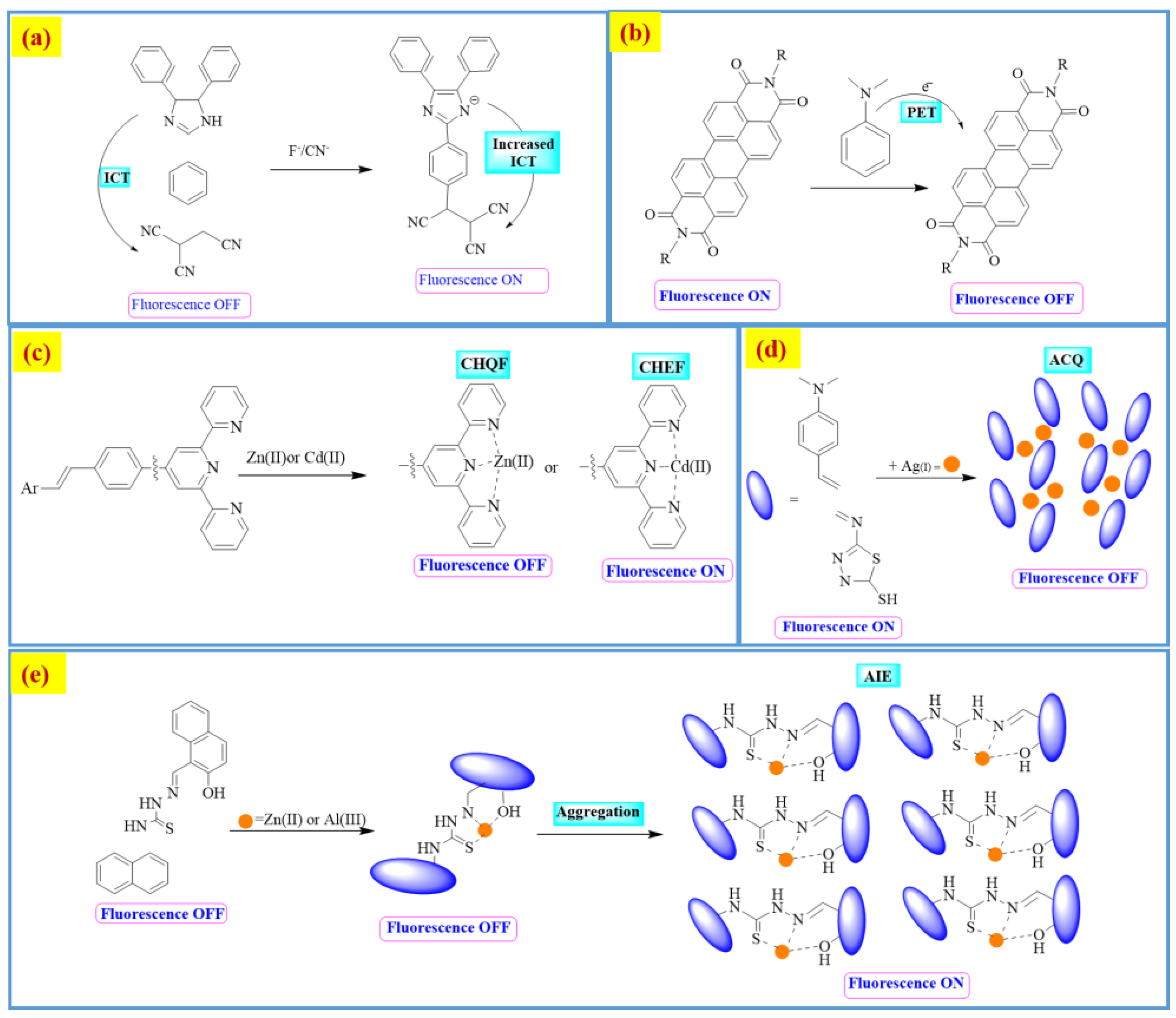
  72. Udhayakumari, D. Mechanistic Innovations in Fluorescent Chemosensors for Detecting Toxic Ions: PET, ICT, ESIPT, FRET and AIE Approaches. J. Fluoresc. 2024, 1–30. [Google Scholar] [CrossRef]
  73. Özdemir, Ö. Novel symmetric diimine-Schiff bases and asymmetric triimine-Schiff bases as chemosensors for the detection of various metal ions. J. Mol. Struct. 2016, 1125, 260–271. [Google Scholar] [CrossRef]
  74. Wang, H.; Liu, S.; Xie, Y.; Bi, J.; Li, Y.; Song, Y.; Cheng, S.; Li, D.; Tan, M. Facile one-step synthesis of highly luminescent N-doped carbon dots as an efficient fluorescent probe for chromium (vi) detection based on the inner filter effect. New J. Chem. 2018, 42, 3729–3735. [Google Scholar] [CrossRef]
  75. Gong, X.; Liu, Y.; Yang, Z.; Shuang, S.; Zhang, Z.; Dong, C. An “on-off-on” fluorescent nanoprobe for recognition of chromium (VI) and ascorbic acid based on phosphorus/nitrogen dual-doped carbon quantum dot. Anal. Chim. Acta 2017, 968, 85–96. [Google Scholar] [CrossRef]
  76. Li, P.; Hong, Y.; Feng, H.; Li, S.F. An efficient “off–on” carbon nanoparticle-based fluorescent sensor for recognition of chromium (VI) and ascorbic acid based on the inner filter effect. J. Mater. Chem. B 2017, 5, 2979–2988. [Google Scholar] [CrossRef] [PubMed]
  77. Bu, L.; Peng, J.; Peng, H.; Liu, S.; Xiao, H.; Liu, D.; Pan, Z.; Chen, Y.; Chen, F.; He, Y. Fluorescent carbon dots for the sensitive detection of Cr (VI) in aqueous media and their application in test papers. RSC Adv. 2016, 6, 95469–95475. [Google Scholar] [CrossRef]
  78. Zheng, M.; Xie, Z.; Qu, D.; Li, D.; Du, P.; Jing, X.; Sun, Z. On–off–on fluorescent carbon dot nanosensor for recognition of chromium (VI) and ascorbic acid based on the inner filter effect. ACS Appl. Mater. Interfaces 2013, 5, 13242–13247. [Google Scholar] [CrossRef]
  79. Wu, H.; Zhou, P.; Wang, J.; Zhao, L.; Duan, C. Dansyl-based fluorescent chemosensors for selective responses of Cr (III). New J. Chem. 2009, 33, 653–658. [Google Scholar] [CrossRef]
  80. Yang, M.; Sun, M.; Zhang, Z.; Wang, S. A novel dansyl-based fluorescent probe for highly selective detection of ferric ions. Talanta 2013, 105, 34–39. [Google Scholar] [CrossRef]
  81. Yu, M.; Du, W.; Zhou, W.; Li, H.; Liu, C.; Wei, L.; Li, Z.; Zhang, H. A 1,8-naphthalimide-based chemosensor with an off-on fluorescence and lifetime imaging response for intracellular Cr3+ and further for S2−. Dye. Pigment. 2016, 126, 279–285. [Google Scholar] [CrossRef]
  82. Khan, S.; Muhammad, M.; Algethami, J.S.; Al-Saidi, H.M.; Almahri, A.; Hassanian, A.A. Synthesis, characterization and applications of schiff base chemosensor for determination of Cr (III) ions. J. Fluoresc. 2022, 32, 1889–1898. [Google Scholar] [CrossRef] [PubMed]
  83. Abbasi, A.; Shakir, M. An inner filter effect based Schiff base chemosensor for recognition of Cr (VI) and ascorbic acid in water matrices. New J. Chem. 2018, 42, 293–300. [Google Scholar] [CrossRef]
  84. Emsley, J. Nature’s Building Blocks: An AZ Guide to the Elements; Oxford University Press: New York, USA, 2011. [Google Scholar]
  85. Gray, T. Elements: A Visual Exploration of Every Known Atom in the Universe; Hachette: London, UK, 2012. [Google Scholar]
  86. Charles Dismukes, G.; van Willigen, R.T. Manganese: The Oxygen-Evolving Complex & Models. In Encyclopedia of Inorganic Chemistry; John Wiley & Sons: Hoboken, NJ, USA, 2006. [Google Scholar]
  87. Raymond, J.; Blankenship, R.E. The origin of the oxygen-evolving complex. Coord. Chem. Rev. 2008, 252, 377–383. [Google Scholar] [CrossRef]
  88. Liang, J.; Canary, J.W. Discrimination between hard metals with soft ligand donor atoms: An on-fluorescence probe for manganese (II). Angew. Chem. (Int. Ed. Engl.) 2010, 49, 7710. [Google Scholar] [CrossRef] [PubMed]
  89. McCreary, J.K. Applications of Manganese-Enhanced Magnetic Resonance Imaging in Neuroscience; University of Lethbridge, Department of Neuroscience: Lethbridge, AB, Canada, 2012. [Google Scholar]
  90. Drapeau, P.; Nachshen, D. Manganese fluxes and manganese-dependent neurotransmitter release in presynaptic nerve endings isolated from rat brain. J. Physiol. 1984, 348, 493–510. [Google Scholar] [CrossRef]
  91. Hunter, D.R.; Haworth, R.A.; Berkoff, H.A. Cellular manganese uptake by the isolated perfused rat heart: A probe for the sarcolemma calcium channel. J. Mol. Cell. Cardiol. 1981, 13, 823–832. [Google Scholar] [CrossRef] [PubMed]
  92. Takeda, A.; Sotogaku, N.; Oku, N. Influence of manganese on the release of neurotransmitters in rat striatum. Brain Res. 2003, 965, 279–282. [Google Scholar] [CrossRef]
  93. Abe, S.; Fujii, K.; Sono, T. Liquid-liquid extraction of manganese (II), copper (II) and zinc (II) with acyclic and macrocyclic Schiff bases containing bisphenol A subunits. Anal. Chim. Acta 1994, 293, 325–330. [Google Scholar] [CrossRef]
  94. Epstein, D.M.; Choudhary, S.; Churchill, M.R.; Keil, K.M.; Eliseev, A.V.; Morrow, J.R. Chloroform-soluble Schiff-base Zn (II) or Cd (II) complexes from a dynamic combinatorial library. Inorg. Chem. 2001, 40, 1591–1596. [Google Scholar] [CrossRef]
  95. Oshima, S.; Hirayama, N.; Kubono, K.; Kokusen, H.; Honjo, T. Structural control of Schiff base ligands for selective extraction of copper (II). Anal. Sci. 2002, 18, 1351–1355. [Google Scholar] [CrossRef]
  96. Zhou, X.-X.; Fang, H.-C.; Ge, Y.-Y.; Zhou, Z.-Y.; Gu, Z.-G.; Gong, X.; Zhao, G.; Zhan, Q.-G.; Zeng, R.-H.; Cai, Y.-P. Assembly of a series of trinuclear zinc (II) compounds with N2O2 donor tetradentate symmetrical Schiff base ligand. Cryst. Growth Des. 2010, 10, 4014–4022. [Google Scholar] [CrossRef]
  97. Hirayama, N.; Taga, J.; Oshima, S.; Honjo, T. Sulfonamide-type di-Schiff base ligands as chelate extraction reagents for divalent metal cations. Anal. Chim. Acta 2002, 466, 295–301. [Google Scholar] [CrossRef]
  98. Hariharan, P.; Anthony, S.P. Substitutional group dependent colori/fluorimetric sensing of Mn2+, Fe3+ and Zn2+ ions by simple Schiff base chemosensor. Spectrochim. Acta Part A Mol. Biomol. Spectrosc. 2015, 136, 1658–1665. [Google Scholar] [CrossRef] [PubMed]
  99. Alharbi, K.H. A review on organic colorimetric and fluorescent chemosensors for the detection of Zn (II) ions. Crit. Rev. Anal. Chem. 2023, 53, 1472–1488. [Google Scholar] [CrossRef] [PubMed]
  100. Lee, Y.J.; Lim, C.; Suh, H.; Song, E.J.; Kim, C. A multifunctional sensor: Chromogenic sensing for Mn2+ and fluorescent sensing for Zn2+ and Al3+. Sens. Actuators B Chem. 2014, 201, 535–544. [Google Scholar] [CrossRef]
  101. Kim, K.B.; Park, G.J.; Kim, H.; Song, E.J.; Bae, J.M.; Kim, C. A novel colorimetric chemosensor for multiple target ions in aqueous solution: Simultaneous detection of Mn (II) and Fe (II). Inorg. Chem. Commun. 2014, 46, 237–240. [Google Scholar] [CrossRef]
  102. Raju, V.; Kumar, R.S.; Tharakeswar, Y.; Kumar, S.A. A multifunctional Schiff-base as chromogenic chemosensor for Mn2+ and fluorescent chemosensor for Zn2+ in semi-aqueous environment. Inorganica Chim. Acta 2019, 493, 49–56. [Google Scholar] [CrossRef]
  103. Qin, J.-c.; Yang, Z.-y.; Wang, G.-q. A novel ratiometric fluorescent probe for detection of Fe3+ by rhodamine–quinoline conjugate. J. Photochem. Photobiol. A Chem. 2015, 310, 122–127. [Google Scholar] [CrossRef]
  104. Liu, S.-R.; Wu, S.-P. New water-soluble highly selective fluorescent chemosensor for Fe (III) ions and its application to living cell imaging. Sens. Actuators B Chem. 2012, 171, 1110–1116. [Google Scholar] [CrossRef]
  105. Sen, S.; Sarkar, S.; Chattopadhyay, B.; Moirangthem, A.; Basu, A.; Dhara, K.; Chattopadhyay, P. A ratiometric fluorescent chemosensor for iron: Discrimination of Fe2+ and Fe3+ and living cell application. Analyst 2012, 137, 3335–3342. [Google Scholar] [CrossRef]
  106. Li, S.; Zhang, D.; Xie, X.; Ma, S.; Liu, Y.; Xu, Z.; Gao, Y.; Ye, Y. A novel solvent-dependently bifunctional NIR absorptive and fluorescent ratiometric probe for detecting Fe3+/Cu2+ and its application in bioimaging. Sens. Actuators B Chem. 2016, 224, 661–667. [Google Scholar] [CrossRef]
  107. Tamil Selvan, G.; Varadaraju, C.; Tamil Selvan, R.; Enoch, I.V.; Mosae Selvakumar, P. On/off fluorescent chemosensor for selective detection of divalent iron and copper ions: Molecular logic operation and protein binding. ACS Omega 2018, 3, 7985–7992. [Google Scholar] [CrossRef]
  108. Nagarajan, R.; Vanjare, B.D.; Lee, K.H. The first tryptophan based turn-off chemosensor for Fe2+ ion detection. Spectrochim. Acta Part A Mol. Biomol. Spectrosc. 2021, 262, 120103. [Google Scholar] [CrossRef] [PubMed]
  109. Luo, A.; Wang, H.; Wang, Y.; Huang, Q.; Zhang, Q. A novel colorimetric and turn-on fluorescent chemosensor for iron (III) ion detection and its application to cellular imaging. Spectrochim. Acta Part A Mol. Biomol. Spectrosc. 2016, 168, 37–44. [Google Scholar] [CrossRef]
  110. Huang, L.; Hou, F.; Cheng, J.; Xi, P.; Chen, F.; Bai, D.; Zeng, Z. Selective off–on fluorescent chemosensor for detection of Fe3+ ions in aqueous media. Org. Biomol. Chem. 2012, 10, 9634–9638. [Google Scholar] [CrossRef] [PubMed]
  111. Liu, J.; Qian, Y. A novel naphthalimide-rhodamine dye: Intramolecular fluorescence resonance energy transfer and ratiometric chemodosimeter for Hg2+ and Fe3+. Dye. Pigment. 2017, 136, 782–790. [Google Scholar] [CrossRef]
  112. Guo, X.; Pan, Q.; Song, X.; Guo, Q.; Zhou, S.; Qiu, J.; Dong, G. Embedding carbon dots in Eu3+-doped metal-organic framework for label-free ratiometric fluorescence detection of Fe3+ ions. J. Am. Ceram. Soc. 2021, 104, 886–895. [Google Scholar] [CrossRef]
  113. Ganesan, J.S.; Sepperumal, M.; Balasubramaniem, A.; Ayyanar, S. A novel pyrazole bearing imidazole frame as ratiometric fluorescent chemosensor for Al3+/Fe3+ ions and its application in HeLa cell imaging. Spectrochim. Acta Part A Mol. Biomol. Spectrosc. 2020, 230, 117993. [Google Scholar] [CrossRef]
  114. Wei, T.-B.; Zhang, P.; Shi, B.-B.; Chen, P.; Lin, Q.; Liu, J.; Zhang, Y.-M. A highly selective chemosensor for colorimetric detection of Fe3+ and fluorescence turn-on response of Zn2+. Dye. Pigment. 2013, 97, 297–302. [Google Scholar] [CrossRef]
  115. Chen, Z.-E.; Zang, X.-F.; Zhang, H. An ethyl thioglycolate-based chemosensor: Spectrophotometric detection of Fe3+ and fluorometric detection of Hg2+ with high selectivity. Spectrochim. Acta Part A Mol. Biomol. Spectrosc. 2021, 260, 119955. [Google Scholar] [CrossRef] [PubMed]
  116. Mustufa, M.; Abbasi, A.; Hanif, S.; Bhat, Z.U.H.; Alam, M.J.; Ahmad, M.; Shakir, M. A combined theoretical and experimental study of ratiometric fluorescent Schiff base chemosensor for detection of Fe3+ ion and its anticancer activity. Chem. Inorg. Mater. 2023, 1, 100011. [Google Scholar] [CrossRef]
  117. Singh, G.; Singh, J.; Singh, J.; Mangat, S.S. Design of selective 8-methylquinolinol based ratiometric Fe2+ and Fe3+/H2PO4 fluorescent chemosensor mimicking NOR and IMPLICATION logic gates. J. Lumin. 2015, 165, 123–129. [Google Scholar] [CrossRef]
  118. Li, B.; Gu, X.; Wang, M.; Liu, X.; Xu, K. A novel “off-on-off” fluorescent probe for sensing of Fe3+ and F successively in aqueous solution and its application in cells. Dye. Pigment. 2021, 194, 109637. [Google Scholar] [CrossRef]
  119. Battal, A.; Kassa, S.B.; Gultekin, N.A.; Tavasli, M.; Onganer, Y. A Carbazole-based Fluorescent Turn-off Chemosensor for Iron (II/III) Detection in a Dimethyl Sulfoxide. J. Fluoresc. 2023, 33, 1421–1429. [Google Scholar] [CrossRef]
  120. Maity, D.; Raj, A.; Karthigeyan, D.; Kundu, T.K.; Govindaraju, T. Reaction-based probes for Co (II) and Cu (I) with dual output modes: Fluorescence live cell imaging. RSC Adv. 2013, 3, 16788–16794. [Google Scholar] [CrossRef]
  121. Wang, Z.-G.; Wang, Y.; Ding, X.-J.; Sun, Y.-X.; Liu, H.-B.; Xie, C.-Z.; Qian, J.; Li, Q.-Z.; Xu, J.-Y. A highly selective colorimetric and fluorescent probe for quantitative detection of Cu2+/Co2+: The unique ON-OFF-ON fluorimetric detection strategy and applications in living cells/zebrafish. Spectrochim. Acta Part A Mol. Biomol. Spectrosc. 2020, 228, 117763. [Google Scholar] [CrossRef] [PubMed]
  122. Lee, J.J.; Choi, Y.W.; You, G.R.; Lee, S.Y.; Kim, C. A phthalazine-based two-in-one chromogenic receptor for detecting Co2+ and Cu2+ in an aqueous environment. Dalton Trans. 2015, 44, 13305–13314. [Google Scholar] [CrossRef] [PubMed]
  123. Maity, D.; Govindaraju, T. Highly selective colorimetric chemosensor for Co2+. Inorg. Chem. 2011, 50, 11282–11284. [Google Scholar] [CrossRef]
  124. Jang, H.J.; Chae, J.B.; Jung, J.M.; So, H.; Kim, C. Colorimetric detection of Co2+, Cu2+, and Zn2+ by a multifunctional chemosensor in aqueous solution. Bull. Korean Chem. Soc. 2019, 40, 650–657. [Google Scholar] [CrossRef]
  125. Min, C.H.; Na, S.; Shin, J.E.; Kim, J.K.; Jo, T.G.; Kim, C. A new Schiff-based chemosensor for chromogenic sensing of Cu2+, Co2+ and S2− in aqueous solution: Experimental and theoretical studies. New J. Chem. 2017, 41, 3991–3999. [Google Scholar] [CrossRef]
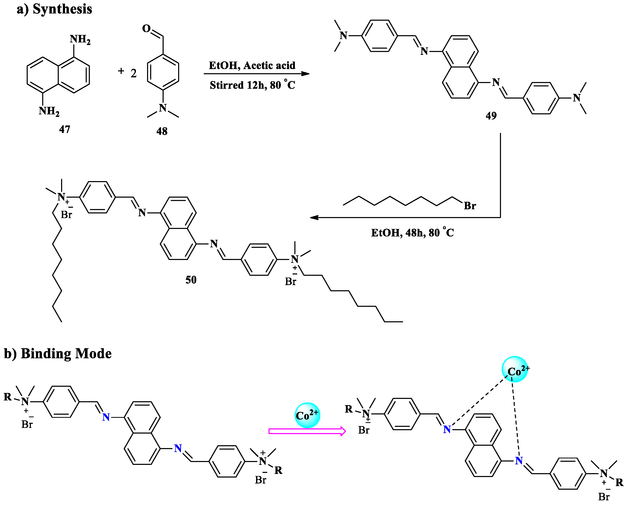
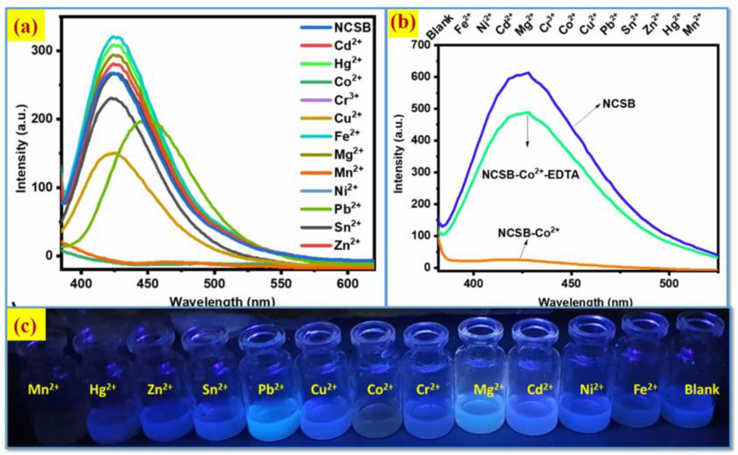
  126. Tawfik, S.M.; Farag, A.A.; Abd-Elaal, A.A. Fluorescence Naphthalene Cationic Schiff Base Reusable Paper as a Sensitive and Selective for Heavy Metals Cations Sensor: RSM, Optimization, and DFT Modelling. J. Fluoresc. 2024, 34, 2139–2155. [Google Scholar] [CrossRef]
  127. Azizi Khereshki, N.; Mohammadi, A.; Zavvar Mousavi, H.; Alizadeh, N. A novel thiosemicarbazide based chemosensor for colorimetric detection of Co2+ in commercial B12 vitamin and Co2+, Ni2+ simultaneously in aqueous media. Supramol. Chem. 2021, 33, 513–526. [Google Scholar] [CrossRef]
  128. Kim, P.A.; Lee, H.; So, H.; Kim, C. A chelated-type colorimetric chemosensor for sensing Co2+ and Cu2+. Inorganica Chim. Acta 2020, 505, 119502. [Google Scholar] [CrossRef]
  129. Liu, X.; Lin, Q.; Wei, T.-B.; Zhang, Y.-M. A highly selective colorimetric chemosensor for detection of nickel ions in aqueous solution. New J. Chem. 2014, 38, 1418–1423. [Google Scholar] [CrossRef]
  130. Kang, J.H.; Lee, S.Y.; Ahn, H.M.; Kim, C. A novel colorimetric chemosensor for the sequential detection of Ni2+ and CN in aqueous solution. Sens. Actuators B Chem. 2017, 242, 25–34. [Google Scholar] [CrossRef]
  131. Goswami, S.; Chakraborty, S.; Das, A.K.; Manna, A.; Bhattacharyya, A.; Quah, C.K.; Fun, H.-K. Selective colorimetric and ratiometric probe for Ni (II) in quinoxaline matrix with the single crystal X-ray structure. RSC Adv. 2014, 4, 20922–20926. [Google Scholar] [CrossRef]
  132. Peralta-Domínguez, D.; Rodríguez, M.; Ramos-Ortíz, G.; Maldonado, J.L.; Meneses-Nava, M.A.; Barbosa-García, O.; Santillan, R.; Farfán, N. A Schiff base derivative from cinnamaldehyde for colorimetric detection of Ni2+ in water. Sens. Actuators B Chem. 2015, 207, 511–517. [Google Scholar] [CrossRef]
  133. Alqasaimeh, M.M.; Abu-Yamin, A.-A.M.; Matar, S.A.; Sarairah, I.A.; Salman, M.M.; Al-As’ad, R.M. Synthesis and characterization of a new Schiff-base derivative as an optical nickel (II) chemosensor and its antimicrobial activity. J. Photochem. Photobiol. A Chem. 2024, 447, 115277. [Google Scholar] [CrossRef]
  134. Manna, A.K.; Chowdhury, S.; Patra, G.K. A novel hydrazide-based selective and sensitive optical chemosensor for the detection of Ni2+ ions: Applications in live cell imaging, molecular logic gates and smart phone-based analysis. Dalton Trans. 2019, 48, 12336–12348. [Google Scholar] [CrossRef]
  135. Korzec, M.; Senkała, S.; Rzycka-Korzec, R.; Kotowicz, S.; Schab-Balcerzak, E.; Polański, J. A highly selective and sensitive sensor with imine and phenyl-ethynyl-phenyl units for the visual and fluorescent detection of copper in water. J. Photochem. Photobiol. A Chem. 2019, 382, 111893. [Google Scholar] [CrossRef]
  136. Jiang, J.; Gou, C.; Luo, J.; Yi, C.; Liu, X. A novel highly selective colorimetric sensor for Ni (II) ion using coumarin derivatives. Inorg. Chem. Commun. 2012, 15, 12–15. [Google Scholar] [CrossRef]
  137. Lu, W.; Chen, J.; Shi, J.; Li, Z.; Xu, L.; Jiang, W.; Yang, S.; Gao, B. An acylhydrazone coumarin as chemosensor for the detection of Ni2+ with excellent sensitivity and low LOD: Synthesis, DFT calculations and application in real water and living cells. Inorganica Chim. Acta 2021, 516, 120144. [Google Scholar] [CrossRef]
  138. Patil, N.; Dhake, R.; Phalak, R.; Fegade, U.; Naushad, M.; Bathula, C.; Kanchi, S.; Govender, K. A colorimetric chemosensor for distinct color change with (E)-2-(1-(3-aminophenyl) ethylideneamino) benzenethiol to detect Cu2+ in real water samples. Anal. Sci. 2023, 39, 1413–1423. [Google Scholar] [CrossRef] [PubMed]
  139. Wang, M.; Leung, K.-H.; Lin, S.; Chan, D.S.-H.; Kwong, D.W.; Leung, C.-H.; Ma, D.-L. A colorimetric chemosensor for Cu2+ ion detection based on an iridium (III) complex. Sci. Rep. 2014, 4, 6794. [Google Scholar] [CrossRef]
  140. Li, G.; Tao, F.; Wang, H.; Li, Y.; Wang, L. A novel reversible colorimetric chemosensor for rapid naked-eye detection of Cu2+ in pure aqueous solution. Sens. Actuators B Chem. 2015, 211, 325–331. [Google Scholar] [CrossRef]
  141. Kowser, Z.; Rayhan, U.; Akther, T.; Redshaw, C.; Yamato, T. A brief review on novel pyrene based fluorometric and colorimetric chemosensors for the detection of Cu2+. Mater. Chem. Front. 2021, 5, 2173–2200. [Google Scholar] [CrossRef]
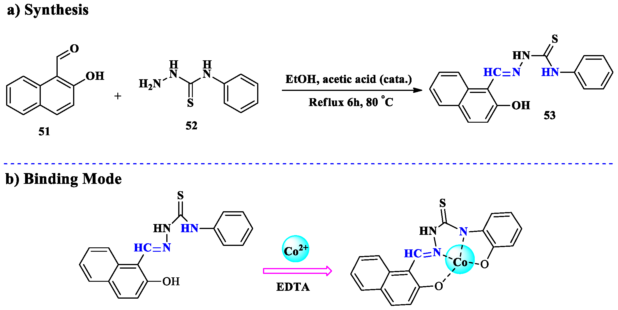
  142. Hrichi, H.; Elkanzi, N.A.; Ali, A.M.; Abdou, A. A novel colorimetric chemosensor based on 2-[(carbamothioylhydrazono) methyl] phenyl 4-methylbenzenesulfonate (CHMPMBS) for the detection of Cu (II) in aqueous medium. Res. Chem. Intermed. 2023, 49, 2257–2276. [Google Scholar] [CrossRef]
  143. Abdou, A.; Mostafa, H.M.; Abdel-Mawgoud, A.-M.M. Seven metal-based bi-dentate NO azocoumarine complexes: Synthesis, physicochemical properties, DFT calculations, drug-likeness, in vitro antimicrobial screening and molecular docking analysis. Inorganica Chim. Acta 2022, 539, 121043. [Google Scholar] [CrossRef]
  144. Rupa, S.A.; Patwary, M.A.M.; Ghann, W.E.; Abdullahi, A.; Uddin, A.R.; Mahmud, M.M.; Haque, M.A.; Uddin, J.; Kazi, M. Synthesis of a novel hydrazone-based compound applied as a fluorescence turn-on chemosensor for iron (iii) and a colorimetric sensor for copper (ii) with antimicrobial, DFT and molecular docking studies. RSC Adv. 2023, 13, 23819–23828. [Google Scholar] [CrossRef] [PubMed]
  145. Jia, Z.; Wei, J.; Ren, Y.; Zhang, N.; Hao, X.-Q.; Zhu, X.; Xu, Y. A new colorimetric and fluorescent chemosensor based on thiazolyl-hydrazone for sensitive detection of copper ions. J. Iran. Chem. Soc. 2020, 17, 609–614. [Google Scholar] [CrossRef]
  146. Kim, Y.S.; Lee, J.J.; Lee, S.Y.; Jo, T.G.; Kim, C. A highly sensitive benzimidazole-based chemosensor for the colorimetric detection of Fe (II) and Fe (III) and the fluorometric detection of Zn (II) in aqueous media. RSC Adv. 2016, 6, 61505–61515. [Google Scholar] [CrossRef]
  147. Liu, D.; Deng, X.; Yin, X.; Wang, Y.; Guo, J.; Chen, J.; Yang, G.; He, H. 1, 8-Naphthalimide-based fluorescent sensor with high selectivity and sensitivity for Zn2+ and its imaging in living cells. Inorg. Chem. Commun. 2019, 101, 117–120. [Google Scholar] [CrossRef]
  148. Burdette, S.C.; Walkup, G.K.; Spingler, B.; Tsien, R.Y.; Lippard, S.J. Fluorescent sensors for Zn2+ based on a fluorescein platform: Synthesis, properties and intracellular distribution. J. Am. Chem. Soc. 2001, 123, 7831–7841. [Google Scholar] [CrossRef] [PubMed]
  149. Liu, D.-Y.; Qi, J.; Liu, X.-Y.; He, H.-R.; Chen, J.-T.; Yang, G.-M. 4-Amino-1,8-naphthalimide-based fluorescent sensor with high selectivity and sensitivity for Zn2+ imaging in living cells. Inorg. Chem. Commun. 2014, 43, 173–178. [Google Scholar] [CrossRef]
  150. Zhang, C.; Liu, Z.; Li, Y.; He, W.; Gao, X.; Guo, Z. In vitro and in vivo imaging application of a 1, 8-naphthalimide-derived Zn2+ fluorescent sensor with nuclear envelope penetrability. Chem. Commun. 2013, 49, 11430–11432. [Google Scholar] [CrossRef] [PubMed]
  151. Liu, D.; Zhao, Y.; Shi, J.; Zhu, H.; Zhang, T.; Qi, P.; Chen, J.; Yang, G.; He, H. A highly selective and sensitive 1, 8-naphthalimide-based fluorescent sensor for Zn2+ imaging in living cells. Bioorganic Med. Chem. Lett. 2019, 29, 2646–2649. [Google Scholar] [CrossRef] [PubMed]
  152. Kwon, J.E.; Lee, S.; You, Y.; Baek, K.-H.; Ohkubo, K.; Cho, J.; Fukuzumi, S.; Shin, I.; Park, S.Y.; Nam, W. Fluorescent zinc sensor with minimized proton-induced interferences: Photophysical mechanism for fluorescence turn-on response and detection of endogenous free zinc ions. Inorg. Chem. 2012, 51, 8760–8774. [Google Scholar] [CrossRef]
  153. Wu, Y.; Peng, X.; Guo, B.; Fan, J.; Zhang, Z.; Wang, J.; Cui, A.; Gao, Y. Boron dipyrromethene fluorophore based fluorescence sensor for the selective imaging of Zn (II) in living cells. Org. Biomol. Chem. 2005, 3, 1387–1392. [Google Scholar] [CrossRef]
  154. Fan, J.; Peng, X.; Wu, Y.; Lu, E.; Hou, J.; Zhang, H.; Zhang, R.; Fu, X. A new PET fluorescent sensor for Zn2+. J. Lumin. 2005, 114, 125–130. [Google Scholar] [CrossRef]
  155. Lee, H.G.; Lee, J.H.; Jang, S.P.; Park, H.M.; Kim, S.-J.; Kim, Y.; Kim, C.; Harrison, R.G. Zinc selective chemosensor based on pyridyl-amide fluorescence. Tetrahedron 2011, 67, 8073–8078. [Google Scholar] [CrossRef]
  156. Choe, D.; So, H.; Park, S.; Lee, H.; Chae, J.B.; Kim, J.; Kim, K.-T.; Kim, C. An indole-based fluorescent chemosensor for detecting Zn2+ in aqueous media and zebrafish. Sensors 2021, 21, 5591. [Google Scholar] [CrossRef]
  157. Mathew, M.M.; Sreekanth, A. Zn2+ ion responsive fluorescent chemosensor probe of Thiophene-diocarbohydrazide derivatives. Inorganica Chim. Acta 2021, 516, 120149. [Google Scholar] [CrossRef]
  158. Peng, X.; Tang, X.; Qin, W.; Dou, W.; Guo, Y.; Zheng, J.; Liu, W.; Wang, D. Aroylhydrazone derivative as fluorescent sensor for highly selective recognition of Zn2+ ions: Syntheses, characterization, crystal structures and spectroscopic properties. Dalton Trans. 2011, 40, 5271–5277. [Google Scholar] [CrossRef] [PubMed]
  159. Kaur, P.; Singh, K. Analyte detection: A decade of progress in the development of optical/fluorescent sensing probes. Chem. Rec. 2023, 23, e202200184. [Google Scholar] [CrossRef] [PubMed]
  160. Ali, Y.S.; Shaw, I.; Liu, Y.; Chen, C. Perspective Chapter: Advanced Nanotechnology Approach for Heavy Metal Toxicity–Analysis. In Heavy Metals in the Environment—Contamination, Risk, and Remediation; IntechOpen Limited: London, UK, 2025. [Google Scholar] [CrossRef]
  161. Khalajiolyaie, A.; Jian, C. Advances in Graphene-Based Materials for Metal Ion Sensing and Wastewater Treatment: A Review. Environments 2025, 12, 43. [Google Scholar] [CrossRef]
Figure 1. Designing of fluorescence signaling unit (a) via spacer signaling unit and via (b) integrated signaling unit. Reproduced from ref. [31].
Molecules 30 01263 g001
Figure 2. The overall detection mechanism of colorimetric and fluorometric chemosensor agents. Reproduced from ref. [37] with permission from Pandawa Institute, copyright 2022.
Molecules 30 01263 g002
Figure 3. Schematic illustrations of general fluorometric chemosensing mechanisms: (a) Intramolecular charge transfer (ICT), where electron movement between donor and acceptor groups alters the fluorescence emission. (b) Photo-induced electron transfer (PET), where electron transfer between the fluorophore and analyte results in fluorescence quenching or enhancement. (c) Chelation-enhanced fluorescence (CHEF) and chelation-quenched fluorescence (CHQF), illustrating the effect of metal ion coordination on the fluorescence intensity. (d) Aggregation-caused quenching (ACQ), where the fluorescence intensity decreases due to the formation of aggregates. (e) Aggregation-induced emission (AIE), where aggregation leads to enhanced fluorescence due to restricted intramolecular motion. Copied from ref. [37] with permission from Pandawa Institute, copyright 2022.
Molecules 30 01263 g003
Scheme 1. (a) Synthesis of Aryl-substituted Thiocarbohydrazide based sensor 3, (b) proposed binding mode with various metal ions.
Molecules 30 01263 sch001
Scheme 2. (a) Synthesis of carbohydrazide-based sensor 6, (b) proposed binding mode.
Molecules 30 01263 sch002
Scheme 3. (a) Synthesis of Schiff-based chemosensor 13, (b) proposed binding mode.
Molecules 30 01263 sch003
Scheme 4. (a) Synthesis of Schiff-based chemosensor 16, (b) proposed binding mode.
Molecules 30 01263 sch004
Figure 4. (a) Color changes in chemosensor 16 (2 µg mL−1) after the addition of 1.0 equivalent of different cations; (b) the fluorescence enhancement of chemosensor 16 (2 µg mL−1) in the MeOH–Water system (1/9 v/v) after the addition of 0.1–3 µg mL−1 of Cr3+ ions; (c) UV–Vis absorption spectra of chemosensor 16 and 16-Cr3+. Copied from ref. [82] with permission from Springer, copyright 2022.
Molecules 30 01263 g004
Scheme 5. (a) Synthesis of Dansyl-based fluorescent chemosensors 23, (b) proposed binding mode.
Molecules 30 01263 sch005
Scheme 6. Synthesis of Schiff-based chemosensor 26.
Molecules 30 01263 sch006
Scheme 7. (a) Synthesis of chromogenic chemosensor 29; (b) proposed binding modes.
Molecules 30 01263 sch007
Figure 5. (a) The chemosensor 29’s absorbance spectrum shifts in response to its exposure to various metal ions; (b) UV–Vis spectra of sensor 29 (30 M) upon the addition of Mn2+ in MeCN buffer solution (10 mM HEPES, pH 7.4, 9:1, v/v); (c) the presence of metal ions triggers an obvious color change in the chemosensor 29. Copied from ref. [100] with permission from Elsevier, copyright 2014.
Molecules 30 01263 g005
Scheme 8. (a) Synthesis of Schiff-based chemosensor 32, (b) proposed binding mode.
Molecules 30 01263 sch008
Scheme 9. Synthesis of Schiff-based chromogenic sensor 35.
Molecules 30 01263 sch009
Scheme 10. (a) Synthesis of DBQA Schiff-based sensor 38; (b) proposed binding mode.
Molecules 30 01263 sch010
Figure 6. Fluorescence spectra of chemosensor 38 (a) DBQA (30 μM) in DMSO:H2O (v/v, 1:4, pH = 7.1) λex = 290 nm simultaneous addition of 0–98 μM of Fe3+; (b) DBQA + Fe3+ (98 μM) in DMSO:H2O (v/v, 1:4, pH = 7.1) λex = 290 nm successive addition of 0–60 μM of NH3. Copied from ref. [116] with permission from Elsevier, copyright 2023.
Molecules 30 01263 g006
Scheme 11. (a) Synthesis of bis-Schiff-based fluorescent sensor 41, (b) proposed binding mode.
Molecules 30 01263 sch011
Scheme 12. (a) Synthesis of carbazole-based chemosensor 46; (b) proposed binding mode.
Molecules 30 01263 sch012
Scheme 13. (a) Synthesis of naphthalene cationic Schiff-based chemosensor 50; (b) proposed binding mode.
Molecules 30 01263 sch013
Figure 7. (a) Fluorescence spectra of 50 in the presence of various metal ion solutions (λex = 350 nm). (b) Fluorescence intensity changes that are reversible at 50 (250 µM) following the successive addition of EDTA and Co2+ ions in aqueous solution (λex = 350 nm). (c) Images of 56 aqueous suspensions (100 µM) with and without the addition of various metal ion solutions taken under 365 nm UV light illumination. Copied from ref. [126] with permission from Springer, copyright 2024.
Molecules 30 01263 g007
Scheme 14. (a) Synthesis of thiosemicarbazide-based Schiff-based chemosensor 53, (b) proposed binding mode.
Molecules 30 01263 sch014
Scheme 15. (a) Synthesis of chelated-type chemosensor 56; (b) proposed binding mode.
Molecules 30 01263 sch015
Scheme 16. (a) Synthesis of acylhydrazone coumarin fluorescent chemosensor 61; (b) proposed binding mode.
Molecules 30 01263 sch016
Figure 8. (a) Fluorescence spectra of sensor 61 in the presence of different metal ions (THF:PBS = 99:1, v/v, λex = 426 nm). (b) Standard curve for fluorescence emission intensity vs. Ni2+ concentration (λex = 426 nm). Insert: Color change induced by the addition of Ni2+ (1 equiv.) to the solution of 61 (61 = 1 × 10⁻⁶ mol/L, THF:PBS = 99:1, v/v). Copied from ref. [137] with permission from Elsevier, copyright 2020.
Molecules 30 01263 g008
Scheme 17. (a) Synthesis of Schiff-based chemosensor 64, (b) proposed binding mode.
Molecules 30 01263 sch017
Scheme 18. (a) Synthesis of CHMPMBS chemosensor 67; (b) proposed binding mode.
Molecules 30 01263 sch018
Figure 9. (a) The physical appearance of a new charge transfer band was the reason underlying the color shift in the 67. The specific CHMPMBS 67 absorption bands vanished as a result of the paramagnetic Cu2+ interaction with CHMPMBS, and an entirely distinct broadband emerged at 515 nm. The concentration of the other ions was approximately double that of the Cu2+ ions. The intensity of the Cu2+-CHMPMBS complex’s absorbance is not substantially influenced by other metal ions; (b) color shift visible to the naked eye when 4 different cations are added to the CHMPMBS solution in DMF-water. (c) Absorption spectra of CHMPMBS. Copied from ref. [142] with permission from Springer, copyright 2023.
Molecules 30 01263 g009
Scheme 19. Synthesis of hydrazone-based sensor 70.
Molecules 30 01263 sch019
Scheme 20. (a) Synthesis of Thiazolyl-hydrazone-based sensor 76; (b) proposed binding mode.
Molecules 30 01263 sch020
Scheme 21. (a) Synthesis of indole-based fluorescent chemosensor 79; (b) proposed binding mode.
Molecules 30 01263 sch021
Figure 10. (a) Fluorescence titration of 79 with varying quantities of Zn2+; (b) fluorescence variations in 79 with variable metal ions and the fluorescence pictures of 79 and 79 + Zn2+ under UV light. Copied from ref. [156] with permission from MDPI, copyright 2021.
Molecules 30 01263 g010
Scheme 22. (a) Synthesis of Thiophene diocarbohydrazide-based sensor 82; (b) proposed binding mode.
Molecules 30 01263 sch022
Scheme 23. Synthesis of Arylhydrazone derivative as fluorescent sensor 85.
Molecules 30 01263 sch023
Disclaimer/Publisher’s Note: The statements, opinions and data contained in all publications are solely those of the individual author(s) and contributor(s) and not of MDPI and/or the editor(s). MDPI and/or the editor(s) disclaim responsibility for any injury to people or property resulting from any ideas, methods, instructions or products referred to in the content.

