Article

Chemical Composition and Toxicity of Achillea millefolium L. Essential Oil Against Acrobasis advenella (Lepidoptera, Pyralidae) Under Laboratory Conditions

by
Edyta Górska-Drabik
1,
Katarzyna Golan
1,*,
Cezary Sempruch
2,
Grzegorz Chrzanowski
3,
Michał P. Dybowski
4 and
Monika Poniewozik
1
1
Department of Plant Protection, Faculty of Horticulture and Landscape Architecture, University of Life Sciences in Lublin, 7 Leszczynskiego St., 20-069 Lublin, Poland
2
Institute of Biological Sciences, Faculty of Sciences, Siedlce University, 14 Bolesława Prusa St., 08-110 Siedlce, Poland
3
Faculty of Biotechnology, Collegium Medicum, University of Rzeszow, 8B Zelwerowicza St., 35-601 Rzeszow, Poland
4
Department of Chromatography, Institute of Chemical Sciences, Faculty of Chemistry, Maria Curie Sklodowska University in Lublin, 3 Maria-Curie Sklodowska Sq., 20-031 Lublin, Poland
*
Author to whom correspondence should be addressed.
Molecules 2025, 30(9), 1927; https://doi.org/10.3390/molecules30091927 (registering DOI)
Submission received: 5 March 2025 / Revised: 18 April 2025 / Accepted: 24 April 2025 / Published: 26 April 2025
(This article belongs to the Topic Natural Products in Crop Pest Management)

Abstract

:
The solutions of essential oil (0.5%, 0.8%, and 1.0% w/v) derived from Achillea millefoilum (EOAM) were evaluated for their repellent and insecticidal activity toward Acrobasis advenella, as well as their effects on the activity of selected larval tissue enzymes. The chemical composition of the EOAM analyzed by GC-MS showed that the main compounds included β-pinene, chamazulene, eucalyptol, and β-caryophyllene. Selection tests showed that larvae avoided inflorescences treated with 0.8% and 1.0% EOAM concentrations. The mortality of larvae increased with concentration and time of exposure to the EO, and the highest mortality rate was observed after 72 h. In addition, the impact of EO was examined on the activity of catalase (CAT), polyphenol oxidase (PPO), peroxidase (POX), α- and β-glucosidases, and acetylcholinesterase (AChE), i.e., important antioxidants, detoxification, digestive, and nervous system enzymes. A significant increase in CAT activity was found in larvae treated with 0.8% and 1.0% EOAM after both 24 and 48 h. Initially, treating larvae with 0.5% EO decreased β-glucosidase activity while increasing α-glucosidase activity. Moreover, 0.8% EOAM inhibited the activity of POX. These findings suggest that EOAM can affect important biochemical processes within the tissues of A. advenella larvae, making it a promising agent for the protection of black chokeberry crops.

1. Introduction

Poland is a leader in the production of chokeberry (Aronia melanocarpa [Michx.] Elliot) fruit and an exporter of finished and semi-finished chokeberry products, i.e., fruit concentrate and frozen foods. The area of chokeberry cultivation in Poland exceeds 14,000 hectares, with annual harvests reaching up to 54,000 tons, which accounts for approximately 70% of global production [1]. Chokeberry fruits are recognized as “superfoods” due to their higher content of phenolic compounds than most other blackberries. Key biologically active components found in these fruits include anthocyanins, carotenoids, flavonoids, tannins, minerals (B, I, Mg, Cu, and Mo), and vitamins (C, B2, B6, E, P, and PP) [2].
Previously, A. melanocarpa was considered a reliable crop, primarily due to its low environmental requirements and low human intervention in pest and disease control. Plantation management was mainly limited to maintenance practices and fertilization and did not require the use of insecticides and fungicides [3]. Over the past two decades, the situation has changed, and chokeberry producers are now forced to use chemical plant protection agents, primarily to control Acrobasis advenella caterpillars (Lepidoptera: Pytalidae, Phycitine) [4,5]. A. advenella is widely distributed throughout Europe. This oligophagous species primarily feeds on plants from the family Rosaceae, particularly those from the genera Sorbus L. and Crataegus L. [6]. Early spring feeding by caterpillars on buds leads to a reduction in yield (on average by 20%) [7] and triggers a follow-up response, resulting in decreased levels of sugars, total flavonoids, and anthocyanins in the fruit. The quality of the raw material is also adversely affected by the feeding of newly hatched caterpillars on immature chokeberries [5]. The adaptation of this species to a new host plant and its widespread occurrence on chokeberry plantations in Poland have made it nearly impossible to achieve both high-quality and satisfactory yields without black chokeberry comprehensive A. advenella control [8]. Chemical insecticides are used to prevent the negative effects of pest feeding on chokeberry buds and fruits. However, these insecticides can impact both the environment and the quality of the fruit [9]. Pyrethroid-based products, such as deltamethrin and lambda-cyhalothrin, are commonly used agents. However, many pyrethroids are known to be toxic to mammals, aquatic organisms, and other wildlife [10,11]. They also contribute to groundwater pollution and disrupt agricultural ecosystems [12,13]. Considering the health benefits of chokeberries and their applications in the pharmaceutical [14] and cosmetic industries [15], it is preferable that the plant material is sourced from organic plantations, free from pesticide use.
An environmentally safe alternative is to replace chemical products with natural substances, such as essential oils (EOs) [16]. As natural compounds, EOs are part of modern trends in plant protection, particularly in the context of organic farming. EOs are well known for their antibacterial, antifungal, antimite, and overall insecticidal properties [17,18,19,20,21]. Their effectiveness is the result of complex interactions between the insect and the host plant [22]. EOs can disrupt the identification of host plants, inhibit egg-laying, and negatively affect the growth and development of insects [23,24,25]. These compounds have documented biocidal, repellent, and antifeedant properties [26,27].
EOs, as aromatic oily extracts, have been one of the most popular alternatives to synthetic chemical insecticides for pest management. EOs show a wide range of effects against insects, acting as repellents, attractants, or antifeedants, or inhibiting respiration and oviposition, impeding host plant identification, and reducing adult emergence by ovicidal and larvicidal activity [23,25,28,29,30,31]. However, the effect of EOs depends strongly on their composition, which is largely determined by genetic variation and environmental conditions [32,33,34].
In recent years, there has been a marked increase in interest in products based on natural plant compounds. This trend may be caused by the gradual withdrawal of active substances used in chemical plant protection products, which are now considered harmful to humans and dangerous to the environment [35]. There is also a noticeable increase in the number of organic farms, driven by the demand for natural raw materials free from pesticide residues [36]. This is the case with black chokeberry, whose fruits from such plantations are sought after for their superior quality, particularly for use in the pharmaceutical and cosmetic industries [2,37].
Achillea species from the family Asteraceae are globally recognized medicinal herbs and have been the focus of numerous pharmacological studies [38,39]. A. millefolium L. shows significant pharmacological activity and has emerged as the most prominent species within the genus [40,41]. The EO of A. millefolium (EOAM) exhibits a broad spectrum of biological activities, including insecticidal properties [42]. The negative (toxic, repellent, antifeedant) effects of EOAM have been documented by many researchers for representatives of various groups of harmful insects: beetles (Coleoptera) [43,44], moth caterpillars (Lepidoptera) [45,46], aphids (Hemiptera, Aphididae) [42,47], and mites (Acari) [48].
The EO can negatively affect different functional systems in insects, usually due to the synergistic interactions that occur in single, natural, and complex mixtures, acting by reducing the resistance of these organisms [49,50]. It is also well known that insects use detoxification enzymes to metabolize deleterious secondary plant metabolites. Antioxidant insect enzymes, such as catalase (CAT), polyphenol oxidase (PPO), and peroxidase (POX), participate in the detoxification of reactive oxygen species (ROS) generated in response to plant allelochemicals, such as phenolics or quinines [51,52,53]. Additionally, various secondary metabolites can affect the digestive processes of insects by altering enzyme activities [54]. Importantly, the reactions are particular and different depending on the EO used and the targeted insect species [55].
While EOAM exhibits a broad range of biological activities, its toxicity, impact on A. advenella biological parameters, and effect on the functioning of enzymatic systems have not been elucidated. Our previous studies on the effect of plant extracts and EOs on the behavior of larvae and females of A. advenella showed that 0.1% EOAM was one of three EOs (besides Tanacetum vulgare and Satureja hortensis) exhibiting repellent properties against larvae and females during the egg-laying period [56].
The main purpose of this study was to determine the effect of various concentrations of A. millefolium essential oil (EOAM) (0.5, 0.8, and 1.0%) on larval mortality, pupation, emergence percentage, longevity, and fertility of A. advenella. Moreover, our research hypothesis posited that the analyzed concentrations of EOAM would disturb the functioning of antioxidant, detoxifying, digestive, and nervous enzymatic systems through changes in the activity of crucial enzymes such as catalase (CAT), peroxidase (POX), polyphenol oxidase (PPO), α- and β-glucosidases, and acetylcholinesterase (AChE). Moreover, the repellent properties of various concentrations of EOAM against A. advenella larvae have been investigated, as well as its potential phytotoxicity in relation to the oil’s chemical composition.

2. Results

2.1. EOAM Chemical Composition

The results of the qualitative and quantitative analysis of A. millefolium EO are presented in Table 1 and Figure 1 (gas chromatogram) and Supplementary Material (MS spectra for unidentified substances). A total of 66 compounds were identified, representing 81.07% of the EO content. The primary compounds included β-pinene (12.84%), eucalyptol (9.15%), chamazulene (9.05%), and β-caryophyllene (7.26%). The percentage of the remaining 62 constituents ranged from 4.47% to 0.06%.

2.2. Settling Inhibitory Activity (SI)

The free-choice bioassays showed that fewer A. advenella larvae settled on inflorescences treated with 0.8% and 1.0% EOAM than on the controls. The SI values ranged from 37.5% to 45.3% (Table 2).

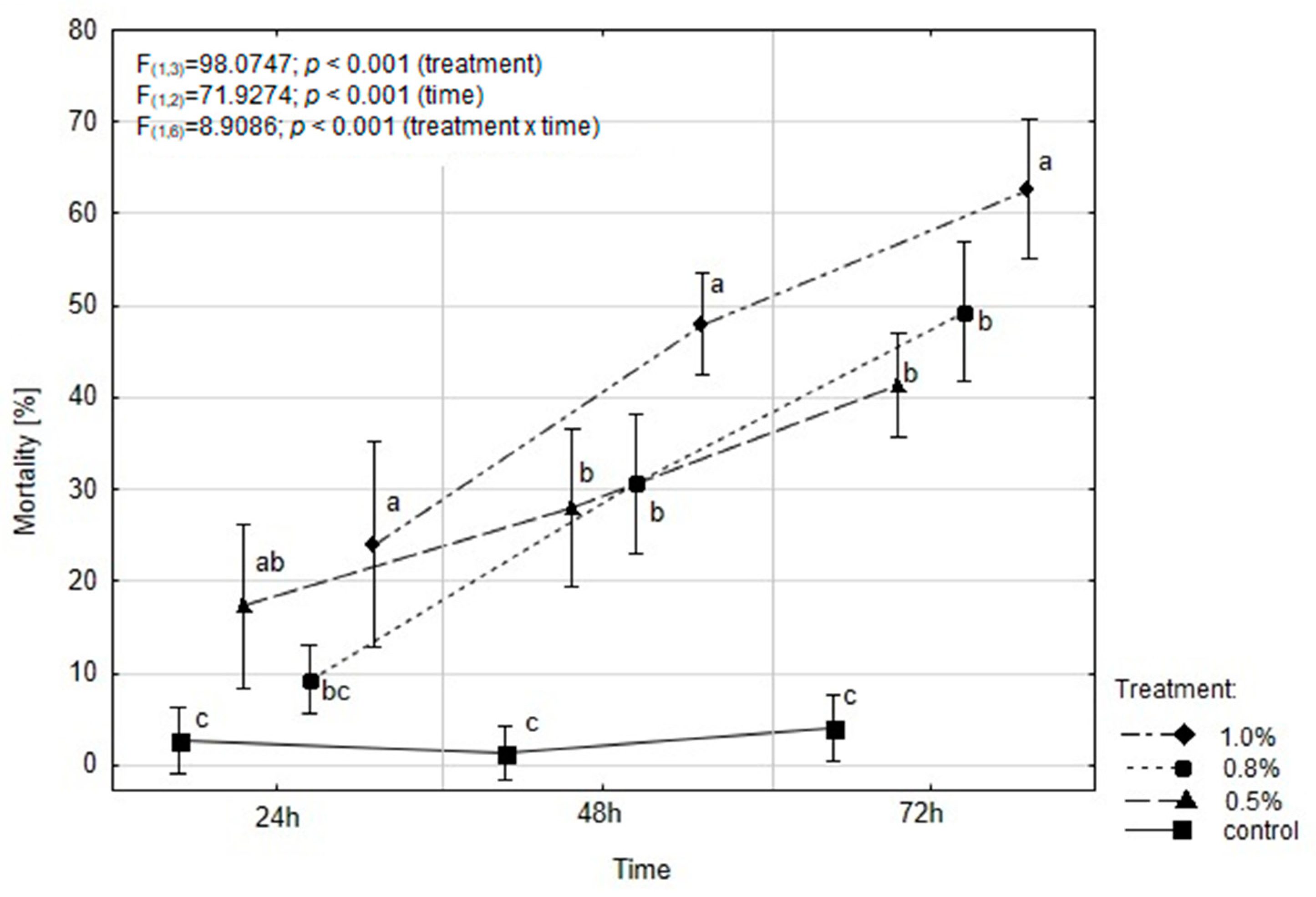
2.3. EOAM Effect on the Mortality of A. advenella larvae

The mortality rate of A. advenella larvae was significantly affected by both the treatment and duration of exposure. Additionally, a significant interaction between treatment and time was observed (Figure 2). After 24 h, dead caterpillars were found in all experimental variants, including the control. The highest mortality rate (24.0%) was observed for 1% oil concentration, and this value doubled after 48 h. A similar proportion of dead caterpillars (49.33%) was recorded for 0.8% oil, but only after 72 h. The application of 1% EOAM resulted in a caterpillar mortality rate of 62.66%. Statistical analysis revealed that all tested concentrations of EOAM significantly affected caterpillar mortality after 48 and 72 h compared to the control.
The estimated lethal concentration values (LC50) with corresponding confidence limits (95%) after 24, 48, and 72 h of exposure to the tested concentrations of EOAM are shown in Table 3. Only the 1.0% concentration showed significant insecticidal properties against A. advenella larvae after 72 h of exposure (LC50 = 0.99%). The LC50 values were significantly different between 24 and 72 h exposure times, as evidenced by the non-overlapping confidence intervals.

2.4. EOAM Effects on A. advenella Demographic Parameters

All concentrations of EOAM tested in the experiment significantly influenced the process of pupation compared to the control, where 93.33% of caterpillars successfully emerged as adults. The lowest number of pupae was observed for 1% EOAM, with only 15.55% of caterpillars pupating (Table 4). However, statistical analysis did not reveal any effect of the tested EOAM concentrations on other demographic parameters of A. advenella, such as adult emergence, longevity, and fertility (Table 4).

2.5. EOAM Phytotoxicity

A thorough examination of sprayed shoots did not reveal any toxic effects on black chokeberry plants sprayed with EOAM solutions. Exposure time did not influence the severity of phytotoxicity. Even at the highest tested concentration (1.0%), the EO did not cause any plant damage. Both 48 and 72 h after application, the percentage of leaf area showing symptoms did not exceed 1% (Table 5).

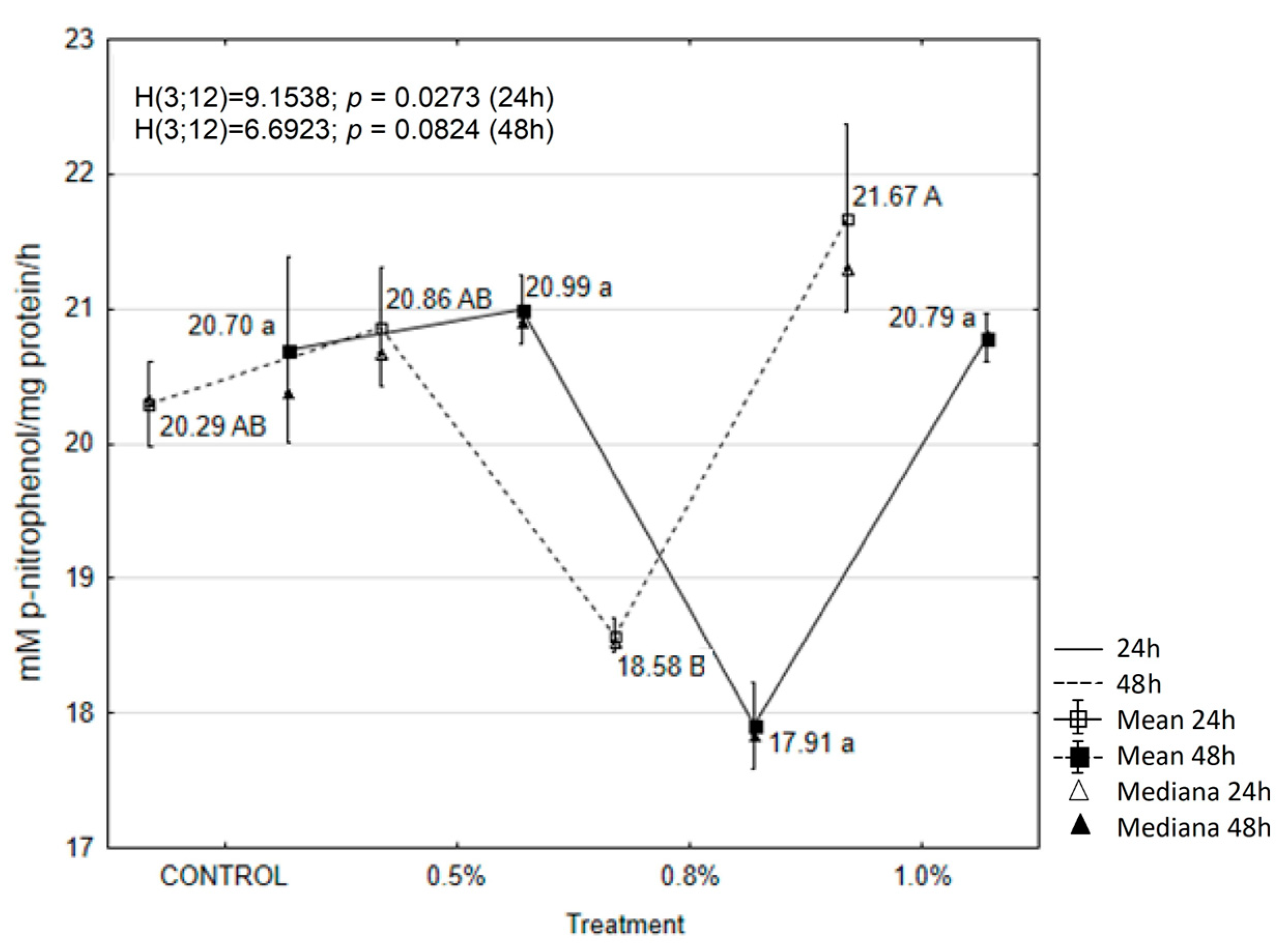
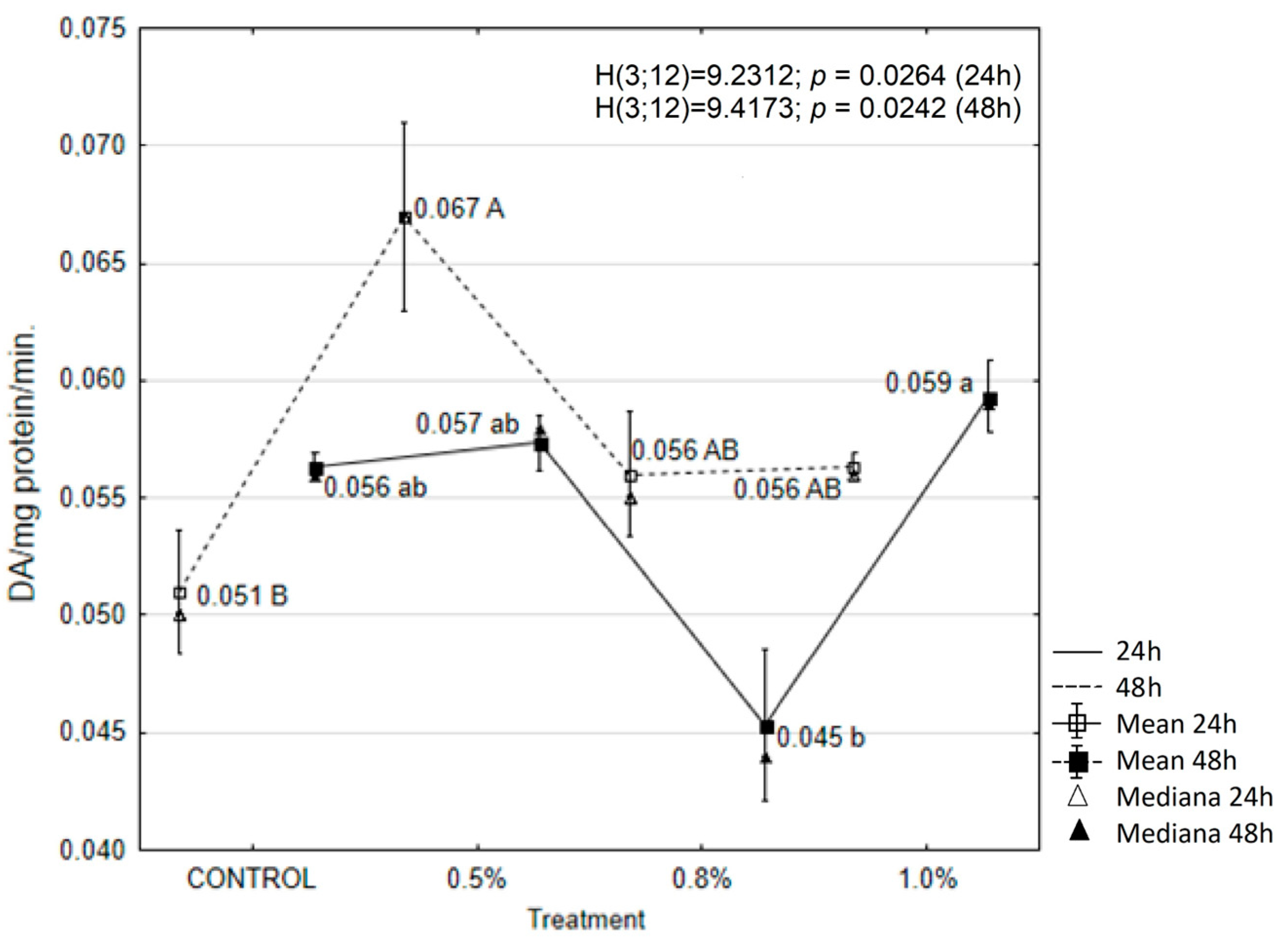
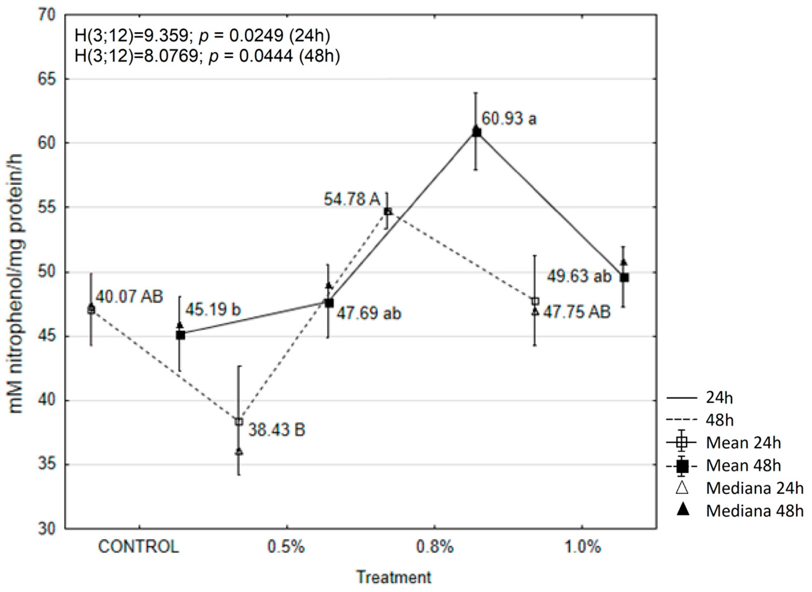
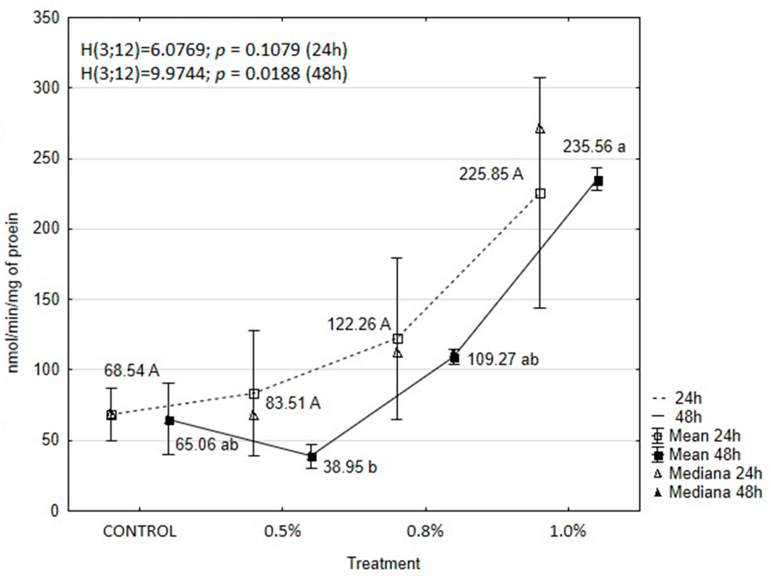
2.6. EOAM Effect on Larval Enzymes

The results showed that treatment with the tested concentrations of EOAM significantly affected the activity of enzymes in the tissues of A. advenella larvae, with the exception of peroxidase (POX) and α-glucosidase. Enzyme activities changed depending on the treatment and exposure time (Figure 3, Figure 4, Figure 5, Figure 6, Figure 7 and Figure 8).
The activity of catalase increased after oil application in all experimental periods. A significant increase in catalase activity was observed for 0.8% and 1.0% EOAM after 24 h, with activity levels reaching 2.40 and 2.14 units, respectively (Figure 3). Overall, a stronger induction was observed 48 h after treatment with 1.0% EOAM, when the activity of this enzyme increased five-fold compared to the control (Figure 3).
Conversely, POX activity decreased significantly after the application of 0.8% EOAM at both 24 and 48 h. However, these changes were not statistically significant compared to the control (Figure 4).
The activity of polyphenol oxidase (PPO) increased during the first experimental period following the application of all tested EOAM concentrations compared to the control (Figure 5). The highest and statistically significant increase in PPO activity occurred 24 h after the application of 0.5% EOAM.
Treatment of A. advenella larvae with 0.5% EOAM resulted in an increase in α-glucosidase activity after 24 h, although these changes were not significantly different (Figure 6). There were also no statistically significant changes in the activity of this enzyme after 48 h.
Regarding β-glucosidase activity, no statistically significant differences were observed 24 h after treatment with any of the tested EOAM concentrations. Nevertheless, a significant increase (1.35 units) in β-glucosidase activity was recorded 48 h after the application of 0.8% EOAM compared to the control (Figure 7).
EOAM treatment also affected the activity of acetylcholinesterase in the tissues of A. advenella larvae. The activity of this enzyme varied with both treatment and time (Figure 8). AChE activity significantly increased after treatment with all tested concentrations throughout the experimental periods, except for 0.5% EOAM at 48 h compared to the control. Among the tested oil concentrations, the strongest effect was observed after applying 1.0% EOAM, with AChE activity showing 3.3-fold and 3.6-fold increases at the respective time points compared to the control. Despite these marked changes in enzyme activity, statistical analysis did not reveal significant differences compared to the control.

3. Discussion

It is well established that the composition of EOs is highly variable, and the content of individual EO components depends not only on the origin of the plant material but also on many other factors, e.g., harvest season, fertilization, extraction method, or plant tissue [22,57,58,59,60]. Previous studies have documented the chemical compositions of A. millefolium EOs from various regions across different countries and continents [42,44,48,60,61,62]. In Poland, research has also indicated variability in both the quantity and composition of the main chemical compounds found in A. millefolium EO. Zawiślak and Nurzyńska-Wierdak [63] reported 95 components in their analyses of the EO from this species. However, the present study identified 29 fewer compounds than those reported by the aforementioned authors. Interestingly, Czerniewicz et al. [42] and Bączek et al. [64] identified even fewer compounds in EOAM compared to the number of components determined in the present work (39 and 40 fewer, respectively).
As shown in the literature, different chemotypes of A. millefolium EO have been identified, including sabinene-type oils [65], 1,8-cineole [66], β-pinene [67,68,69], chamazulene, and p-cymene [70,71]. Judzentiene and Mockute [61] demonstrated significant differences in the chemical composition of EO originating from different A. millefolium forms. For example, the β-pinene content varied from 0.5% to 20.0% in plants within the same populations. The chemical composition of EOAM detected in this study is consistent with previous findings from Poland. Bączek et al. [64] identified three chemotypes of A. millefolium oil: β-pinene, β-pinene + chamazulene, and 1,8-cineole. The EOAM in this study, characterized by a β-pinene + chamazulene chemotype, had a similar chemical profile to that described by Zawiślak and Nurzyńska-Wierdak [63]. The main constituents of the latter EO were β-pinene and chamazulene, both present in higher percentages (respectively, 19.32% and 14.41%) than previously reported. This oil also contained caryophyllene (E-isomer), although eucalyptol was not isolated. On the other hand, a slightly different composition of A. millefolium EO was reported by Czerniewicz et al. [42], where of 27 identified compounds, chamazulene was dominant, and β-pinene was present in lower quantity. Intra-specific genetic divergence may also account for this chemical variability [61].
The present results demonstrated that EOAM effectively inhibited A. advenella larvae from settling on treated inflorescences, with the level of inhibition varying depending on the concentration applied. The highest activity was observed at concentrations of 0.8 and 1.0% EOAM. Our previous research demonstrated the repellent properties of plant extracts and EOs from eight different plant species on the behavior of A. advenella caterpillars and females. Repellent properties have already been demonstrated at a concentration of 0.1% EOAM [56].
The repellent properties of EOAM have been confirmed by many literature reports. For instance, Tampe et al. [44] demonstrated the effectiveness of EOAM in repelling Aegorhinus nodipennis (Hope) (Coleoptera: Curculionidae); additionally, the mixture of metabolites present in EOAM disrupted physiological processes and exhibited neurotoxic effects. These authors highlighted the potential of EO, particularly those containing α- and β-thujone isomers, as effective repellent agents. Their analysis identified the presence of 11 compounds, with β-thujone being the dominant one, accounting for 96.2% of the total content. The toxic properties of β-thujone on insects have been well documented in numerous studies [56,72,73,74]. Additionally, other EOAM components, such as β-pinene, chamazulene, β-caryophyllene, and eucalyptol, identified in our experiments, also demonstrated insect-repellent properties. The repellent potential of β-pinene against Panaphis juglandis (Goeze) and Chromaphis juglandicola (Kalt.) aphid larvae (Hemiptera; Aphidoidea; Drepanosiphinae) has been documented by Krzyżanowski [75]. Meanwhile, Czerniewicz et al. [42] demonstrated the repellent and biocidal properties of EOAM against Myzus persicae (Sulzer). However, the EOAM examined by the aforementioned authors differed from the one tested in the present study, as the 1,8-cineole (9.2%) was the dominant component in that oil; however, β-pinene was also present in a high proportion (6.6%). A study by Conti et al. [69] also confirmed the larvicidal activity of EOAM against the mosquito Aedes albopictus (Skuse) (Diptera: Culicidae), identifying eucalyptol (14.2%) and β-pinene (12.4%) as the main components, which were also dominant in our analysis. Similarly, Nenaah [43] reported EOAM fumigant toxicity against Tribolium castaneum (Herbst) (Coleoptera, Tenebrionidae), with chamazulene (26.2%) and β-pinene (16.6%) being the primary constituents. The author showed a weak effect of EOAM on both the reduction of adult abundance and demographic parameters of this species (larval and pupal development, number of individuals reaching successful adult emergence, total life span, and sex ratio). It should be noted that the toxicity of EOs significantly increased when they were formulated as nano-emulsions.
The repellent and biocidal properties of EOAM have also been demonstrated against other insect species, e.g., Rhopalosiphum maidis (Fitch) (Hemiptera: Aphididae) [47] and Plodia interpunctella (Hübner) (Lepidoptera: Pyralidae) [76]. Halbert et al. [47] conducted an experiment involving 55 substances, including 1.0% oil from A. millefolium, to assess their repellent effects on aphids. The results of the experiment demonstrated a high repellent potential of A. millefolium oil compared to the other substances tested. The mean number of aphids on treated and untreated plates was −1.72 and 3.94 at p = 0.0041, respectively. On the other hand, Ebadollahi and Ashouri [76] showed a strong fumigant activity of EOAM against adults of P. interpunctella (100% mortality when the adults were exposed to 65 μL/l concentrations of A. millefolium after 48 h). However, these authors did not determine the chemical profile of the A. millefolium EOs tested in their study.
Our study showed that caterpillar mortality increased over time, with significant mortality observed only after 72 h (LC50 = 0.99%). In our previous works using 0.5% oils from T. vulgare and S. hortensis against A. advenella larvae, the effects were observed only 72 h after application [28,29]. S. hortensis EO was significantly less effective, with nearly 38% larval mortality after 72 h, compared to T. vulgare EO and the EOAM tested in this study. The varying toxicity of these three EOs in contact with A. advenella larvae suggests a difference in the sensitivity of larvae of this pest to individual EOs, which could be attributed to variations in the molecular weights of individual compounds and their capacity to penetrate larval tissues [77]. Czerniewicz et al. [42] reported a stronger effect of EOAM on M. persicae as early as 24 h (LC50 = 0.34%) and 48 h (LC50 = 0.30%) after treatment, with significant inhibition of aphids’ enzymatic activity also observed during this period. Ebadollahi and Ashouri [76] found that EOAM application resulted in 100% mortality of P. interpunctella within 48 h after treatment, confirming its high potential as a fumigant against stored-product pests. Our research, on the other hand, showed that a significantly higher concentration of EOAM (1.85%) would have to be applied to achieve 50% mortality of caterpillars after 24 h. Naghizadeh et al. [46] also evaluated the effect of EOAM on Phthorimaea operculella Zeller (Lepidoptera: Gelechiidae), showing that treating the food source (potato tubers) with a 0.5% oil concentration reduced egg-laying and shortened insect lifespans. Moreover, sublethal concentrations of A. millefolium oil affected a key pest control parameter, namely the sex ratio, leading to a higher proportion of males in the population.
Since the chemical components of EOs are lipophilic, they can penetrate insect tissues and influence enzyme activity [78]. We stated that the effect of EOAM on A. advenella was connected with the induction of the activity of most of the analyzed enzymes, except for POX and α-glucosidase. The increase in enzymatic activity was usually induced by the higher of the tested concentrations (0.8 or/and 1.0%), with the exception of PPO, whose activity increased after 24 h of exposure to EOAM at 0.5% concentration. In the case of AChE and CAT, the increase in activity occurred after 24 h of exposure to the tested preparations and was maintained in the latter, while in the case of β-glucosidase, this effect was observed only after 48 h. The obtained data partially confirm the results of previous studies, which found that S. hortensis EOs induced PPO and α- and β-glucosidase activity in the tissues of A. advenella larvae, while EOs from T. vulgare induced an increase in CAT and PPO activity in the same species, while inhibiting POX and α- and β-glucosidase [28,29]. Our results and previous data indicate that EOs produced by different plant species are characterized by various chemical compositions and, therefore, may differentially modify insect physiology. Their composition is dominated by one to several components, which often have similar insecticidal activity as the entire mixture. It is widely believed that qualitative and quantitative changes in AChE are responsible for insect resistance to insects. Fournier et al. [79] reported that in Drosophilla melanogaster (Meigen) strains producing different amounts of AChE via P-mediated transformation, resistance to organophosphate pesticides correlates with the amount of the enzyme in the central nervous system. Inhibition of this enzyme disrupts the coordination of the neuromuscular system, finally leading to paralysis and death of the insect. Czerniewicz et al. [42] found that EOs from Asteraceae plants inhibited AChE activity in tissues of female M. persicae, and their concentration correlated with aphicidal potential. Similar activity was also observed for β-phellandrene (the main component of Santolina EOs) against Reticulitermes speratus (Kolbe) and 1,8-cineole (the main component of yarrow and tansy oils) against Aedes aegypthi (L.) [80,81]. It is therefore possible that the increase in AChE activity in A. advenella larvae treated with 0.8 and 1.0% EOAM concentrations indicates a high tolerance of this species to the tested substances. Induction of CAT and PPO activities in A. advenella tissues suggests that the response is at last connected with oxidative stress. Cytotoxic properties of EOs may result from the presence of phenolics, aldehydes, and alcohols in their composition [82]. These biomolecules may affect the fluidity of the membranes, causing the building of radicals, cytochrome C, calcium ions, and proteins as a result of oxidative stress and bioenergetic failure. Forty-eight-hour exposure of the larvae to EOAM also resulted in an increase in β-glucosidase activity. A similar effect in relation to the tested insect species was previously observed in the case of S. hortensis EOs and their main component carvacrol. These substances induced a 194.08% and 214.67% increase in β-glucosidase activity in larval tissues, respectively [28]. On the other hand, Magierowicz et al. [56] and Czerniewicz et al. [83] observed inhibition of this enzyme following treatment of A. advenella with T. vulgare EOs and Rhopalosiphum padi (L.) with EOs from Asteraceae plants. Changes in β-glucosidase activity could affect the metabolism of sugars and osmoregulation in A. advenella tissues due to their participation in the hydrolytic decomposition of some oligosaccharides. On the other hand, this enzyme may participate in plant defense reactions by releasing toxic aglycones from less harmful glycosides [83].
Nevertheless, the phytotoxic properties of EOs have long been a major drawback to their potential broader application, as they can have detrimental effects on cultivated plants [84]. In the present study, EOAM rich in β-pinene and eucalyptol showed no phytotoxicity to A. melanocarpa leaves even when administered at the highest concentrations tested. Neither the exposure time nor the concentration of EOAM had a significant impact on A. melanocarpa leaves and shoots. Both the lowest and highest spray concentrations caused phytotoxic symptoms in less than 1% of the leaf area. Czerniewicz et al. [42] reported different outcomes, where the highest concentration (0.8%) tested led to the development of phytotoxic symptoms on more than 50% of pea leaf surfaces. This discrepancy was likely due to differences in the anatomical structure of the leaves between the two plant species.

4. Materials and Methods

4.1. Insect Culture and Rearing Conditions

The fourth-stage (L4) of A. advenella larvae were collected in May with inflorescences from an organic horticulture farm of black chokeberry in Samoklęski (51.4500 °N, 22.4333 °E) located in southeastern Poland, Lublin Province. Subsequently, they were reared in laboratory conditions at the University of Life Sciences in Lublin (Poland). The larvae (n = 795) were kept separately in Petri dishes (9 cm in diameter) and fed with A. melanocarpa inflorescences. Petri dishes were cleaned, and fresh inflorescences (each approx. 0.7 g) were provided daily. The larvae were used for toxicity tests, as well as demographic and biochemical experiments.

4.2. Plant Material and EOAM Extraction

Dry A. millefolium aerial parts were purchased as an organic product (PL-ECO-01) from Dary Natury Co. (Grodzisk, Poland). For the extraction process, 100 g of plant material was placed in a 1000 mL round-bottom flask with 500 mL of distilled water and subjected to hydrodistillation for 4 h using a Clevenger apparatus. The water extracts of EOs were dried over anhydrous sodium sulfate and stored in amber vials at 4 °C in the dark until GC-MS analysis [85]. The EO yield (expressed as a percentage) was calculated as the ratio of the weight of extracted oil (0.31 g) to the weight of plant material.

4.3. Compound Identification and Estimation by Gas Chromatography–Mass Spectrophotometry

Qualitative analysis of EOs was performed using a GC/MS QP2010 system (Schimadzu, Kyoto, Japan) equipped with a ZB5-MSi fused silica capillary column (30 m × 0.25 mm id. 0.25 μm film thickness, Phenomenex). Helium (grade 5.0) was used as the carrier gas, with a flow rate of 1 mL/min. The injection temperature was set at 310 °C, and 1 μL of the sample was injected in split mode (purge time: 0.7 min). The following temperature program was applied: initial hold at 50 °C for 2 min, followed by a linear temperature increase of 5 °C/min until reaching 310 °C. The mass spectrometer operated in EI mode at 70 eV, with the ion source temperature set at 220 °C. The mass range scanned was from 35 to 450 amu. Qualitative analysis was performed by comparing the retention indices and mass spectra for the obtained peaks with corresponding data from the NIST’14 mass spectrometry library. Quantitative analyses were carried out using a gas chromatograph with a GC/FID GC2010 flame ionization detector (Schimadzu, Kyoto, Japan). Hydrogen was used as the carrier gas at a flow rate of 1 mL/min. The experimental conditions were the same as for GC/MS. Peak identification was performed based on experimentally determined retention indices [86,87].

4.4. Laboratory Bioassays

All bioassays were performed at 22 ± 1 °C, 70 ± 5% relative humidity, and a 16:8 h photoperiod. EOAM concentrations of 0.5%, 0.8%, and 1.0% (w/v) were used for all tests, including demographic and biochemical analyses. The 0.5% concentration (w/v) was prepared by mixing 0.5 mL of EOAM with 2% (v/v) ethanol (99.8%), distilled water, and 0.5 mg of Tween 80 as an emulsifier. The remaining concentrations were prepared in a similar manner, adjusting the volume of EOAM to 0.8 mL for the 0.8% concentration and 1 mL for the 1.0% concentration. A mixture with the same composition but without the EO was used as a control solution [56].

4.4.1. Settling Inhibition Bioassays

The potential of EOAM at concentrations of 0.5%, 0.8%, and 1.0% (w/v) against colonization of A. advenella larvae was evaluated using free-choice tests. Initially, A. melanocarpa inflorescences were immersed in EOAM or control solution for 15 s and dried at room temperature for 15 min. The dried inflorescences were positioned at the periphery of Petri dishes (20 cm diameter) lined with moistened filter paper. Each dish contained four inflorescences treated with EOAM and four untreated control inflorescences. Next, 15 larvae were released in the center of each dish, and after 24 h, the number of insects that settled on each inflorescence was recorded. Each concentration of EO was tested in triplicate. The settling inhibition index (%SI) was calculated as follows: %SI = [1 − (%T/%C)] × 100, where %T is the percentage of larvae on EO-treated inflorescences, and %C is the percentage of larvae on control inflorescences [88].

4.4.2. EOAM Toxicity Test on Larvae

The toxicity assay involved spraying larvae of the same age and black chokeberry inflorescences with the selected EOAM concentrations (w/v) or a control solution. Each treatment was sprayed three times using a 100 cm³ manual plastic sprayer (Puro Cosmetics, Wrocław, Poland). The sprayer’s contents were vigorously mixed before application, with approximately 0.40 cm³ of solution used per treatment performed in triplicate. Each Petri dish contained a single larva [28]. Mortality was recorded at 24, 48, and 72 h after exposure to the EOAM, and LC50 values were calculated using Probit analysis [89]. Larvae were considered dead when they did not respond to stimulation with a camel-hair brush. Mortalities observed in the treatment groups were corrected for natural mortality recorded in the control group. The experiments were conducted using 15 larvae per concentration of EOAM and the control, with five replicates per treatment.

4.4.3. EOAM Influence on Demographic Parameters

Three tested concentrations (0.5%, 0.8%, and 1.0% w/v) were used to evaluate the effects of EO on the population parameters of A. advenella. The experiment was conducted in Petri dishes, where larvae and chokeberry inflorescences were sprayed with the tested EOAM concentrations. After 3–4 days, soil was added to each Petri dish to create suitable conditions for pupation. Daily observations were made, with soil moisture maintained using an atomizer. When the larvae were no longer present on the host plant, the soil was inspected to determine the exact moment the larvae descended for pupation. The study recorded the number of pupae, emerged moths, their longevity, and oviposition of A. advenella. Longevity results were expressed as minimum, maximum, and mean percentages. The percentages of pupation and adult emergence were calculated as follows: pupation (%) = 100 × total number of pupae formed/total number of larvae released and adult emergence (%) = 100 × total number of adults formed/total number of pupae. Each concentration of EOAM, as well as the control, was tested on 15 larvae six times [56].
After the moths emerged, their sex was determined, and then they were paired and placed in rearing cages (31 cm × 21 cm × 21 cm) (TRIXIE Co, Kobylarnia, Poland). Immature infructescences (egg-laying sites) were placed in floral foam to preserve their freshness. One pair of newly emerged A. advenella adults (male and female) were released into each cage. The moths were fed with a 20% honey solution. Immature fruits were checked every day, recording the number of eggs laid on each infructescence. The experiment included 40 pairs of moths (10 pairs per concentration of EOAM and 10 for the control). Insect rearing was carried out until all females died [29].

4.5. EOAM Phytotoxicity Bioassay on A. melanocarpa

Experiments on the reaction of black chokeberry to the tested solutions were carried out under laboratory conditions. To assess the phytotoxicity, 15 cm stem sections were used. A triple application of EOAM solutions was carried out using an atomizer (100 mL). The plants were inspected for phytotoxicity symptoms 48 and 72 h post-application. When phytotoxic effects were observed, the severity was evaluated based on the percentage of leaf surface displaying symptoms, such as necrotic spots or discoloration of the leaf blade. The severity was categorized as follows: none (0–1%), slight (1–25%), medium (25–50%), and high (>50%) [78].

4.6. Enzymatic Analyses

For biochemical analyses, larvae (L4) and black chokeberry inflorescences were placed in plastic 25 × 15 cm insect containers with moistened filter paper to avoid drying [29].
About thirty larvae of A. advenella and ten inflorescences were placed in the center at the bottom of each box. One inflorescence consisted of 10 individual buds of black chokeberry. After two hours, larvae and inflorescences were sprayed with an emulsion of EOAM or a control solution that was prepared as described above. The experiments were conducted for 24 and 48 h. Collected insects were homogenized with 3 mL of cold 0.2 M phosphate buffer pH 5.8 for α- and β-glucosidase assay and with 0.2 M phosphate buffer pH 7.0 for CAT and POX or Tris-HCl buffer pH 7.4 for PPO. Acetylcholinesterase (AChE) was extracted from the insect tissues by homogenization with 1 cm3 of ice-cold 0.1 M Tris-HCl buffer (pH 7.8) containing 20 mM NaCl and 0.5% Triton X-100. The homogenates were filtered through two layers of cheesecloth and centrifuged at 10,000× g for 15 min (centrifuge type Z 323 K, Hermle Labortechnik GmbH, Wehingen, Germany). The supernatants were used in further analyses. All enzymatic analyses were conducted in three independent replicates with the use of a UV–VIS spectrophotometer, type HP 8453 (Hewlett-Packard, Waldbronn, Germany) [29]. All chemical reagents used for enzymatic assays were obtained from Sigma-Aldrich (Poznań, Poland).
CAT activity was measured using Aebi’s [90] method. For this purpose, 0.1 mL of insect homogenate was mixed with 0.1 mL 75 mmol hydrogen peroxide and 0.8 mL extraction buffer. The amount of decomposed H2O2 after 1 min of enzymatic reaction was measured at 240 nm. The activity of the enzyme was calculated as mM H2O2 × min−1 × mg−1 protein.
POX activity was assayed according to Ferman and Dimond [91]. The reaction mixture contained 0.1 mL homogenate of insect tissues, 0.1 mL 0.2 M pyrogallol, 0.5 mL extraction buffer, 0.35 mL distilled water, and 0.05 mL 3% hydrogen peroxide solution. It was incubated at 30 °C for 25 min. The enzymatic reaction was subsequently interrupted by adding 0.25 mL 25% trichloroacetic acid (TCA) solution. The content of purpugallin produced was measured spectrophotometrically at 430 nm. A unit of enzyme activity was defined as mM purpurgallin × min−1 × mg−1 protein.
PPO activity was determined by the method described by Miles [92] with further modifications [93]. To 0.25 mL of insect homogenate, 0.2 mL of extraction buffer and 0.25 mL of 10 mM catechol solution were added. The changes in absorbance were measured at 460 nm after incubation at 30 °C for 1 h. Enzyme activity was expressed as ΔA × min−1 × mg−1 protein.
The α- and β-glucosidase activities were assayed according to Katagiri [94] and Chararas and Chipoulet [95]. p-Nitrophenyl-α-D-glucopyranoside (pNαG) and p-nitrophenyl-β-D-glucopyranoside (pNβG) were used as substrates for α- and β-glucosidase, respectively. The reaction mixture contained 0.2 mL of insect homogenate, 0.1 mL of phosphate buffer pH 5.8, and 0.2 mL of pNαG (15 mM solution in extraction buffer) or pNβG (50 mM solution in extraction buffer). The enzymatic reaction ran for 60 min at 30 °C, and then it was stopped by adding 3 mL of 2% sodium carbonate solution. The content of released p-nitrophenol was measured spectrophotometrically at 405 nm. The activities of both enzymes were expressed as μmol p-nitrophenol × hour−1 × mg−1 protein.
AChE activity was assayed with the use of Ellman et al.’s [96] method. The insect homogenate (0.3 mL) was mixed with 0.4 mL extraction buffer and 0.05 mL of the substrate (75 mM solution of acetylthiocholine iodide—ATChI) and next incubated at 30 °C for 20 min. The enzymatic reaction was stopped by the addition of 0.05 mL 0.644 mM solution of eserine. Then, 0.05 mL of 10 mM 5,5′-dithiobis-2-nitrobenzoic acid (DTNB) solution with 0.018 M of sodium bicarbonate was added to the reaction mixture. The absorbance was measured at 412 nm, and the extinction coefficient (134.6 mM−1 × cm−1) was used to convert absorbance into molarity. The enzyme activity was expressed as nM ATChI hydrolyzed by mg enzymatic protein during one minute of the reaction (nM min−1 × mg−1 of protein).
Protein content in all analyzed enzymatic extracts was assayed using the method of Lowry et al. [97].

4.7. Statistical Analysis

The results in figures and tables are presented as arithmetic means with standard deviation (SD). The Shapiro–Wilk test was used to assess the normality of data distribution. For the free-choice bioassay, differences between the number of larvae on treated inflorescences and those on the control were analyzed using the Student’s t-test. Mortality rates and demographic parameters were analyzed using one-way ANOVA, and differences between means were determined using Tukey’s post hoc test at a significance level of p ≤ 0.05. When the data deviated from a normal distribution, the Kruskal–Wallis test was applied as a non-parametric alternative to ANOVA. In such a case, Dunn’s test was used as the post hoc test (for enzyme activities—CAT, POX, PPO, α- and β-glucosidase, and AChE). Probit analysis was conducted to estimate LC50 values along with their corresponding 95% confidence limits (CLs) using IBM SPSS v. 23 (IBM Corp., Armonk, NY, USA). All statistical analyses were conducted using Statistica v. 13.0 software (Statsoft, Kraków, Poland).

5. Conclusions

The present study is the first report of the insecticidal activity of EOAM against A. advenella. The highest tested concentration of EOAM resulted in the strongest insecticidal effect against A. advenella larvae and adversely affected the pupation process. The application of EOAM to larvae was associated with increased activity of most of the enzymes analyzed. However, the observed increase in AChE activity in A. advenella larvae may indicate a high tolerance of this species to the tested substances. To enhance the efficacy of EOAM, future research should focus on testing higher concentrations of EOAM and consider the different larval growth stages, as individuals in the early developmental stages exhibit increased sensitivity to biologically active compounds. Future studies should also explore synergistic effects between EOAM components and assess the response of A. advenella to individual constituents. This will provide a deeper understanding of EOAM’s potential in managing A. advenella, a major pest in black chokeberry crops.

Supplementary Materials

The following supporting information can be downloaded at: https://www.mdpi.com/article/10.3390/molecules30091927/s1, Figures S1–S7: MS spectra for unidentified substances of EOAM.

Author Contributions

Conceptualization, E.G.-D.; methodology, E.G.-D., C.S., G.C. and M.P.D.; software, M.P; validation, K.G.; formal analysis, K.G.; investigation, E.G.-D., K.G., C.S. and M.P.D.; data curation, E.G.-D., M.P.D. and G.C.; writing—original draft preparation, E.G.-D., C.S. and K.G.; writing—review and editing, E.G.-D., C.S. and K.G.; visualization, M.P.; supervision, E.G.-D. and K.G; project administration, E.G.-D.; funding acquisition, E.G.-D. All authors have read and agreed to the published version of the manuscript.

Funding

This research was funded by the University of Life Sciences in Lublin, grant number SUBB.WOK.19.019.

Institutional Review Board Statement

Not applicable.

Informed Consent Statement

Not applicable.

Data Availability Statement

The data that support the findings of this study are available from the corresponding author upon reasonable request.

Acknowledgments

The authors would like to thank Piotr Nowak, the owner of the organic black chokeberry horticulture farm in Samoklęski, and MSc Anna Król for support in insect rearing.

Conflicts of Interest

The authors declare no conflicts of interest.

References

  1. Statistics Poland. Table 93. 2023. Available online: https://stat.gov.pl/en/topics/agriculture-forestry/agricultural-and-horticultural-crops/production-of-agricultural-and-horticultural-crops-in-2023,2,8.html (accessed on 23 July 2024).
  2. Wawer, I.; Egert, P.; Hołub, B. Aronia Superowoc; Wyd. Wektor: Warszawa, Poland, 2015; p. 221. [Google Scholar]
  3. Rusnak, J. Uprawa Aronii; Małopolski Ośrodek Doradztwa Rolniczego: Karnowice, Poland, 2013; p. 20. [Google Scholar]
  4. Górska-Drabik, E. Trachycera advenella (Zinck.) (Lepidoptera, Pyralidae)—A new pest on black chokeberry (Aronia melanocarpa). Prog. Plant Prot. 2009, 49, 531–534. [Google Scholar]
  5. Górska-Drabik, E. Occurrence of Acrobasis advenella (Zinck.) (Lepidoptera, Pyralidae, Phycitinae) on Black Chokeberry in Poland and Its Ciochemical Interaction with Host Plants; University of Life Science in Lublin: Lublin, Poland, 2013; Volume 382, p. 121. [Google Scholar]
  6. Slamka, F. Die Zünslerartigen (Pyraloidea) Mitteleuropas; Verlag nicht ermittelbar: Bratislava, Slovakia, 1997; p. 112. [Google Scholar]
  7. Górska-Drabik, E. Owady i roztocze zagrażające aronii czarnoowocowej. Insects and mites occurring in black chokeberry. In Proceedings of the 9th Fruit Conference ‘Trends in the Cultivation of Berry and Stone Fruit Species’, Kraśnik, Poland, 20–23 February 2013; pp. 30–32. [Google Scholar]
  8. Buler, Z.; Doruchowski, G.; Filipczak, J.; Głos, H.; Hołdaj, M.; Lisek, J.; Łabanowska, B.; Meszka, B.; Morgaś, H.; Poniatowska, A.; et al. Metodyka: Integrowana Produkcja Aronia; Instytut Ogrodnictwa: Skierniewice, Poland, 2016; p. 49. [Google Scholar]
  9. Chauhan, S.S.; Agrawal, S.; Srivastava, A. Effect of imidacloprid insecticide residue on biochemical parametersin potatoes and its estimation by HPLC. Asian J. Pharm. Clin. Res. 2013, 6, 114–117. [Google Scholar]
  10. Zhu, Q.; Yang, Y.; Zhong, Y.; Lao, Z.; O’Neill, P.; Hong, D.; Zhang, K.; Zhao, S. Synthesis, Insecticidal Activity, Resistance, Photodegradation and Toxicity of Pyrethroids (A Review). Chemosphere 2020, 254, 126779. [Google Scholar] [CrossRef] [PubMed]
  11. Frank, D.F.; Miller, G.W.; Harvey, D.J.; Brander, S.M.; Geist, J.; Connon, R.E.; Lein, P.J. Bifenthrin causes transcriptomic alterations in mTOR and ryanodine receptor-dependent signaling and delayed hyperactivity in developing zebrafish (Danio rerio). Aquat. Toxicol. 2018, 200, 50–61. [Google Scholar] [CrossRef]
  12. Wang, R.; Zhang, S.; Xiao, K.; Cai, M.; Liu, H. Occurrence, sources, and risk assessment of pyrethroid insecticides in surface water and tap water from Taihu Lake, China. J. Environ. Manag. 2023, 325, 116565. [Google Scholar] [CrossRef]
  13. Tang, T.; Wu, R.; Zhang, L.; Wang, Y.; Ling, J.; Du, W.; Shen, G.; Chen, Y.; Zhao, M. Distribution and partitioning of pyrethroid insecticides in agricultural lands: Critical influencing factors. Environ. Int. 2021, 156, 106736. [Google Scholar] [CrossRef] [PubMed]
  14. Broncel, M.; Koziróg, M.; Duchnowicz, P.; Koter-Michalak, M.; Sikora, J.; Chojnowska-Jezierska, J. Aronia melanocarpa extract reduces blood pressure, serum endothelin, lipid, and oxidative stress marker levels in patients with metabolic syndrome. Med. Sci. Monit. 2010, 16, CR28–CR34. [Google Scholar]
  15. Ćujić, N.; Žugić, A.; Živković, J.; Zdunić, G. Preliminary safety estimate of cosmetic anti-age creams with chokeberry extract, using in vivo bioengineering techniques. Lek. Sirovine 2017, 37, 41–44. [Google Scholar] [CrossRef]
  16. Dey, D.; Gupta, M.K. Use of essential oils for insect pest management-a review. Innov. Farming. 2016, 1, 21–29. [Google Scholar]
  17. Chang, S.-T.; Chen, P.-F.; Chang, S.-C. Antibacterial Activity of Leaf Essential Oils and Their Constituents from Cinnamomum Osmophloeum. J. Ethnopharmacol. 2001, 77, 123–127. [Google Scholar] [CrossRef]
  18. Chen, F.; Chang, S.T.; Wu, H.H. Antimite activity of essential oils and their components from Cinnamomum osmophloeum. Quart. J. Chin. For. 2002, 35, 397–404. [Google Scholar]
  19. Cheng, S.; Chang, H.; Chang, S. Bioactivity of selected plant essential oils against the yellow fever mosquito Aedes aegypti larvae. Bioresour. Technol. 2003, 89, 99–102. [Google Scholar] [CrossRef] [PubMed]
  20. Mssillou, I.; Agour, A.; Allali, A.; Saghrouchni, H.; Bourhia, M.; El Moussaoui, A.; Salamatullah, A.M.; Alzahrani, A.; Aboul-Soud, M.A.M.; Giesy, J.P.; et al. Antioxidant, Antimicrobial, and Insecticidal Properties of a Chemically Characterized Essential Oil from the Leaves of Dittrichia viscosa L. Molecules 2022, 27, 2282. [Google Scholar] [CrossRef]
  21. Benmeddour, T.; Messaoudi, K.; Flamini, G. First investigation of the chemical composition, antioxidant, antimicrobial and larvicidal activities of the essential oil of the subspecies Ononis angustissima Lam. subsp. filifolia Murb. Nat. Prod. Res. 2024, 1–16. [Google Scholar] [CrossRef] [PubMed]
  22. Regnault-Roger, C. The potential of botanical essential oils for insect pest control. Integr. Pest. Manag. Rev. 1997, 2, 25–34. [Google Scholar] [CrossRef]
  23. Sarwar, M.; Salman, M. Toxicity of oils formulation as a new useful tool in crop protection for insect pests control. Int. J. Chem. Biomol. Sci. 2015, 1, 297–302. [Google Scholar]
  24. Osman, S.E.I.; Swidan, M.H.; Kheirallah, D.A.; Nour, F.E. Histological effects of essential oils, their monoterpenoids and insect growth regulators on midgut, integument of larvae and ovaries of khapra beetle, Trogoderma granarium everts. J. Biol. Sci. 2016, 16, 93–101. [Google Scholar] [CrossRef]
  25. Ali, M.A.; Doaa, S.M.; El-Sayed, H.S.; Asmaa, M.E. Antifeedant activity and some biochemical effects of garlic and lemon essential oils on Spodoptera littoralis (Boisduval) (Lepidoptera: Noctuidae). J. Entomol. Zool. 2017, 5, 1476–1482. [Google Scholar]
  26. Shaaya, E.; Rafaeli, A. Essential oils as biorational insecticides—Potency and mode of action. In Insecticide Design Using Advanced Technologies, 1st ed.; Ishaaya, I., Nauen, R., Horowitz, A.R., Eds.; Springer: Berlin/Heidelberg, Germany, 2007; pp. 249–261. [Google Scholar]
  27. Koul, O.; Walia, S.; Dhaliwal, G.S. Essential oils as green pesticides: Potential and constraints. Biopestic. Int. 2008, 4, 63–84. [Google Scholar]
  28. Magierowicz, K.; Górska-Drabik, E.; Sempruch, C. The insecticidal activity of Satureja hortensis essential oil and its active ingredient carvacrol against Acrobasis advenella (Zinck.) (Lepidoptera, Pyralidae). Pestic. Biochem. Physiol. 2019, 153, 122–128. [Google Scholar] [CrossRef]
  29. Magierowicz, K.; Górska-Drabik, E.; Golan, K. Effects of plant extracts and essential oils on the behavior of Acrobasis advenella (Zinck.) caterpillars and females. J. Plant Dis. Prot. 2020, 127, 63–71. [Google Scholar] [CrossRef]
  30. Amini, S.; Khalaj, H.; Ahvazi, M.; Labbafi, M. Composition and biological effects of different populations of Cupressus to control adult weevil (Tribolium castaneum Herbst). J. Med. Plants 2023, 22, 113–126. [Google Scholar] [CrossRef]
  31. Zouirech, O.; El Moussaoui, A.; Saghrouchni, H.; Gaafar, A.R.Z.; Nafidi, H.A.; Bourhia, M.; Khallouki, F.; Lyoussi, B.; Derwich, E. Prefatory in silico studies and in vitro insecticidal effect of Nigella sativa (L.) essential oil and its active compound (carvacrol) against the Callosobruchus maculatus adults (Fab), a major pest of chickpea. Open Chem. 2023, 21, 20230133. [Google Scholar] [CrossRef]
  32. Mockute, D.; Judzentiene, A. Chemotypes of the essential oils of Achillea millefolium L. ssp. millefolium growing wild in Eastern Lithuania. Chemija 2002, 13, 168–173. [Google Scholar]
  33. Mazandarani, M.; Mirdeilami, S.Z.; Pessarakli, M. Essential oil composition and antibacterial activity of Achillea millefolium L. from different regions in North east of Iran. J. Med. Plant Res. 2013, 7, 1063–1069. [Google Scholar]
  34. Długokęcka, A. Zaprzyjaźnij się z ziołami. WMODR, Olsztyn, Poland. 2020, p. 17. Available online: https://wmodr.pl/files/bv1jQI81GI42ujDprjOGQmN6AQJ7FrzTGYW6tWPP.pdf (accessed on 20 July 2024).
  35. Perry, T.; Batterham, P.; Daborn, P.J. The biology of insecticidal activity and resistance. Insect Biochem. Mol. Biol. 2011, 41, 411–422. [Google Scholar] [CrossRef] [PubMed]
  36. Zieliński, M.; Wrzaszcz, W.; Sobierajewska, J.; Adamski, M. Development and Effects of Organic Farms in Poland, Taking into Account Their Location in Areas Facing Natural or Other Specific Constraints. Agriculture 2024, 14, 297. [Google Scholar] [CrossRef]
  37. Kokotkiewicz, A.; Jaremicz, Z.; Luczkiewicz, M. Aronia plants: A review of traditional use, biological activities, and perspectives for modern medicine. J. Med. Food 2010, 13, 255–269. [Google Scholar] [CrossRef]
  38. Nemeth, E. Essential oil composition of species in the genus Achillea. J. Essent. Oil Res. 2005, 17, 501–512. [Google Scholar] [CrossRef]
  39. Nemeth, E.; Bernath, J. Biological activities of Yarrow Species (Achillea spp.). Curr. Pharm. Des. 2008, 14, 3151–3167. [Google Scholar] [CrossRef]
  40. Csupor-Löffler, B.; Hajdú, Z.; Zupkó, I.; Réthy, B.; Falkay, G.; Forgo, P.; Hohmann, J. Antiproliferative effect of flavonoids and sesquiterpenoids from Achillea millefolium s.l. on cultured human tumour cell lines. Phytother. Res. 2009, 23, 672–676. [Google Scholar] [CrossRef] [PubMed]
  41. Barda, C.; Grafakou, M.-E.; Tomou, E.-M.; Skaltsa, H. Phytochemistry and evidence-based traditional uses of the genus Achillea L.: An Update (2011–2021). Sci. Pharm. 2021, 89, 50. [Google Scholar] [CrossRef]
  42. Czerniewicz, P.; Chrzanowski, G.; Sprawka, I.; Sytykiewicz, H. Aphicidal activity of selected Asteraceae essential oils and their effect on enzyme activities of the green peach aphid, Myzus persicae (Sulzer). Pestic. Biochem. Phys. 2018, 145, 84–92. [Google Scholar] [CrossRef] [PubMed]
  43. Nenaah, G.E. Chemical composition, toxicity and growth inhibitory activities of essential oils of three Achillea species and their nano-emulsions against Tribolium castaneum (Herbst). Ind. Crop. Prod. 2014, 53, 252–260. [Google Scholar] [CrossRef]
  44. Tampe, J.; Parra, L.; Huaiquil, K.; Mutis, A.; Quiroz, A. Repellent effect and metabolite volatile profile of the essential oil of Achillea millefolium against Aegorhinus nodipennis (Hope) (Coleoptera: Curculionide). Neotrop. Entomol. 2015, 44, 279–285. [Google Scholar] [CrossRef]
  45. Karahroodi, Z.R.; Moharramipour, S.; Rahbarpour, A. Investigated repellency effect of some essential oils of 17 native medicinal plants on adults Plodia interpunctella. Am.-Eurasian. J. Sustain. Agric. 2009, 3, 181–184. [Google Scholar]
  46. Naghizadeh, S.; Rafiee-Dastjerdi, H.; Golizadeh, A.; Esmaielpour, B.; Mahdavi, V. The effect of essential oils of Artemisia absinthum L., Achillea millefolium L. and Artemisia dracunculus L. against potato tuber moth, Phthorimaea operculella Zeller (Lepidoptera: Gelechiidae). Jordan J. Agric. Sci. 2016, 12, 1115–1123. [Google Scholar] [CrossRef]
  47. Halbert, S.E.; Corsini, D.; Wiebe, M.; Vaughn, S.F. Plant-derived compounds and extracts with potential as aphid repellents. Ann. Appl. Biol. 2009, 154, 303–307. [Google Scholar] [CrossRef]
  48. Ahmadi, Z.; Saber, M.; Bagheri, M.; Mahdavinia, G.R. Achillea millefolium essential oil and chitosan nanocapsules with enhanced activity against Tetranychus urticae. J. Pest. Sci. 2018, 91, 837–848. [Google Scholar] [CrossRef]
  49. Cetin, H.; Erler, F.; Yanikoglu, A. A comparative evaluation of Origanum onites essential oil and its four major components as larvicides against the pine processionary moth, Thaumetopoea wilkinsoni. Pest. Manag. Sci. 2007, 63, 830–833. [Google Scholar] [CrossRef]
  50. Akhtar, Y.; Pages, E.; Stevens, A.; Bradbury, R.; da Camara, C.A.G.; Isman, M.B. Effect of chemical complexity of essential oils onfeeding deterrence in larvae of the cabbage looper. Physiol. Entomol. 2012, 37, 81–89. [Google Scholar] [CrossRef]
  51. Felton, G.W.; Duffey, S.S. Protective action of midgut catalase in lepidopteran larvae against oxidative plant defenses. J. Chem. Ecol. 1991, 17, 1715–1732. [Google Scholar] [CrossRef]
  52. Kolawole, A.O.; Olajuyigbe, F.M.; Ajele, J.O.; Adedire, C.O. Activity of the antioxidant defense system in a typical bioinsecticide- and synthetic insecticide-treated bowpea storage beetle Callosobrochus maculatus F. (Coleoptera: Chrysomelidae). Int. J. Insect Sci. 2014, 6, IJIS-S19434. [Google Scholar] [CrossRef]
  53. Mittler, R. Oxidative stress, antioxidants and stress tolerance. Trends Plant Sci. 2002, 7, 405–410. [Google Scholar] [CrossRef] [PubMed]
  54. Mardani-Talaee, M.; Rahimi, V.; Zibaee, A. Effects of host plants on digestive enzymatic activities and some components involved in intermediary metabolism of Chrysodeixis chalcites (Lepidoptera: Noctuidae). J. Entomol. Acarol. Res. 2014, 46, 96–101. [Google Scholar] [CrossRef]
  55. Popescu, I.E.; Gostin, I.N.; Blidar, C.F. An overview of the mechanisms of action and administration technologies of the essential oils used as green insecticides. Agri. Eng. 2024, 6, 1195–1217. [Google Scholar] [CrossRef]
  56. Magierowicz, K.; Górska-Drabik, E.; Sempruch, C. The effect of Tanacetum vulgare essential oil and its main components on some ecological and physiological parameters of Acrobasis advenella (Zinck.) (Lepidoptera: Pyralidae). Phestic. Biochem. Phys. 2020, 162, 105–112. [Google Scholar] [CrossRef] [PubMed]
  57. Orav, A.; Arak, E.; Raal, A. Phytochemical analysis of the essential oil of Achillea millefolium L. from various European Countries. Nat. Prod. Res. 2006, 20, 1082–1088. [Google Scholar] [CrossRef]
  58. Tuberoso, C.I.G.; Kowalczyk, A. Chemical composition of the essential oils of Achillea millefolium L. isolated by different distillation methods. J. Essent. Oil Res. 2009, 21, 108–111. [Google Scholar] [CrossRef]
  59. Farajpour, M.; Ebrahimi, M.; Baghizadeh, A.; Aalifar, M. Phytochemical and yield variation among iranian Achillea millefolium accessions. Hort. Sci. 2017, 52, 827–830. [Google Scholar] [CrossRef]
  60. Aziz, E.E.; Badawy, E.M.; Zheljazkov, V.D.; Nicola, S.M.; Fouad, H. Yield and chemical composition of essential oil of Achillea millefolium L. as affected by harvest time. Egypt. J. Chem. 2019, 62, 533–540. [Google Scholar] [CrossRef]
  61. Judzentiene, A.; Mockute, D. Essential oil composition of two yarrow taxonomic forms. Cent. Europ. J. of Biol. 2010, 5, 346–352. [Google Scholar] [CrossRef]
  62. Naghizadeh, S.; Rafiee-Dastjerdi, H.; Naseri, B.; Golizadeh, A.; Esmaielpour, B. Insecticidal activity of essential oils from Artemisia absinthium L., Artemisia dracunculus L. and Achillea millefolium L. against Phthorimaea operculella Zeller (Lepidoptera: Gelechiidae). J. Crop Prot. 2019, 8, 479–489. [Google Scholar]
  63. Zawiślak, G.; Nurzyńska-Wierdak, R. Rozwój roślin oraz skład chemiczny olejku eterycznego krwawnika pospolitego (Achillea millefolium L.) uprawianego w warunkach klimatu umiarkowanego. Ann. Hortic. 2017, 27, 31–38. [Google Scholar] [CrossRef]
  64. Bączek, K.; Kosakowska, O.; Przybył, L.J.; Kuźma, P.; Ejdys, M.; Obiedziński, M.; Węglarz, Z. Intraspecific variability of yarrow (Achillea millefolium L. s.l.) in respect of developmental and chemical traits. Herba Pol. 2015, 61, 37–52. [Google Scholar] [CrossRef]
  65. Rohloff, J.; Skagen, E.B.; Steen, A.H.; Iversen, T.H. Production of yarrow (Achillea millefolium L.) in Norway: Essential oil content and quality. J. Agric. Food Chem. 2000, 48, 6205–6209. [Google Scholar] [CrossRef] [PubMed]
  66. Figueiredo, A.C.; Barroso, J.G.; Pais, M.S.S.; Scheffer, J.J.C. Composition of the essential oils from leaves and flowers of Achillea millefolium L. ssp. millefolium. Flavour. Fragr. J. 1992, 7, 219–222. [Google Scholar] [CrossRef]
  67. Orav, A.; Kailas, T.; Ivask, K. Composition of the essential oil from Achillea millefolium L. from Estonia. J. Essent. Oil Res. 2001, 13, 290–294. [Google Scholar] [CrossRef]
  68. Mockute, D.; Judzentiene, A. Variability of the essential oils composition of Achillea millefolium L. ssp. millefolium growing wild in Lithuania. J. Biochem. Syst. Ecol. 2003, 31, 1033–1045. [Google Scholar] [CrossRef]
  69. Conti, B.; Canale, A.; Bertoli, A.; Gozzini, F.; Pistelli, L. Essential oil composition and larvicidal activity of six Mediterranean aromatic plants against the mosquito Aedes albopictus (Diptera: Culicidae). Parasitol. Res. 2010, 107, 1455–1461. [Google Scholar] [CrossRef]
  70. Jaimand, K.; Rezaee, M.B. Investigation on the chemical constituents of essential oils from Achillea millefolium L. subsp. millefolium by distillation methods. Iranian J. Med. Aromat. Plant Res. 2004, 20, 181–190. [Google Scholar]
  71. Jaimand, K.; Rezaee, M.B.; Mozaffarian, V. Chemical constituents of the leaf and flower oils from Achillea millefolium ssp. elbursensis Hub.-Mor. from Iran rich in chamazulene. J. Essent. Oil Res. 2006, 18, 293–295. [Google Scholar] [CrossRef]
  72. Hwang, Y.-S.; Wu, K.-H.; Kumamoto, J.; Axelrod, H.; Mulla, M.S. Isolation and identification of mosquito repellents in Artemisia vulgaris. J. Chem. Ecol. 1985, 11, 1297–1306. [Google Scholar] [CrossRef]
  73. Rojht, H.; Meško, A.; Vidrih, M.; Trdan, S. Insecticidal activity of four different substances against larvae and adults of sycamore lace bug (Corythucha ciliata Say, Heteroptera, Tingidae). Acta Agric. Slov. 2009, 93, 31–36. [Google Scholar] [CrossRef]
  74. Szołyga, B.; Gniłka, R.; Szczepanik, M.; Szumny, A. Chemical composition and insecticidal activity of Thuja occidentalis and Tanacetum vulgare essential oils against larvae of the lesser mealworm, Alphitobius diaperinus. Entomol. Exp. Appl. 2014, 151, 1–10. [Google Scholar] [CrossRef]
  75. Krzyżanowski, R. Wpływ lotnych związków na zachowanie mszyc związane z żerowaniem. Kosmos 2017, 66, 413–420. [Google Scholar]
  76. Ebadollahi, A.; Ashouri, S. Toxicity of essential oils isolated from Achillea millefolium L., Artemisia dracunculus L. and Heracleum persicum Desf. against adults of Plodia interpunctella (Hübner) (Lepidoptera: Pyralidae) in Islamic Republic of Iran. Ecol. Balk. 2011, 3, 41–48. [Google Scholar]
  77. Stock, D.; Holloway, P.J. Possible mechanisms for surfactant-induced foliar uptake of agrochemicals. Pestic. Sci. 1993, 38, 165–177. [Google Scholar] [CrossRef]
  78. Karamaouna, F.; Kimbaris, A.; Michaelakis, A.; Papachristos, D.; Polissiou, M.; Papatsakona, P.; Tsora, E. Insecticidal activity of plant essential oils against the vine mealybug, Planococcus ficus. J. Insect Sci. 2013, 13, 142. [Google Scholar] [CrossRef]
  79. Fournier, D.; Bride, J.M.; Hoffmann, F.; Karch, F. Acetylcholiesterase. Two types of modifications confer resistance to insecticide. J. Biol. Chem. 1992, 267, 14270–14274. [Google Scholar] [CrossRef]
  80. Khanikor, B.; Parida, P.; Yadav, R.N.S.; Bora, D. Comparative mode of action of some terpene compounds agains octopamine receptor and acetyl cholinesterase of mosquito and human system by the help of homology modelling and docking studies. J. Appl. Pharm. Sci. 2013, 3, 6–12. [Google Scholar] [CrossRef]
  81. Seo, S.M.; Kim, J.; Kang, J.; Koh, S.A.; Ahn, Y.J.; Park, I.K. Fumigant toxicity and acetylcholinesterase inhibitory activity of 4 Asteraceae plant essential oils and their constituents against Japanese termite (Reticulitermes speratus Kolbe). Pestic. Biochem. Physiol. 2014, 113, 55–61. [Google Scholar] [CrossRef]
  82. Bakkali, F.; Averbeck, S.; Averbeck, D.; Idaomar, M. Biological effects of essential oils—A review. Food Chem. Toxicol. 2008, 46, 446–475. [Google Scholar] [CrossRef]
  83. Czerniewicz, P.; Sytykiewicz, H.; Chrzanowski, G. The Effect of Essential Oils from Asteraceae Plants on Behavior and Selected Physiological Parameters of the Bird Cherry-Oat Aphid. Molecules 2024, 29, 1673. [Google Scholar] [CrossRef] [PubMed]
  84. Górska-Drabik, E.; Golan, K.; Kot, I.; Kmieć, K.; Poniewozik, M.; Dzida, K.; Bochniak, A. The Effect of pre-harvest treatments with Tanacetum vulgare L. and Satureja montana L. essential oils (EOs) on the yield and chemical composition of Aronia melanocarpa (Michx.) Elliot fruit. Agriculture 2024, 14, 12. [Google Scholar] [CrossRef]
  85. Angourani, H.R.; Zarei, A.; Moghadam, M.M.; Ramazani, A.; Mastinu, A. Investigation on the Essential Oils of the Achillea Species: From Chemical Analysis to the In Silico Uptake against SARS-CoV-2 Main Protease. Life 2023, 13, 378. [Google Scholar] [CrossRef]
  86. Sun, Y.; Zhang, R.; Wang, Q.; Xu, B. Programmed-temperature gaschromatographic retention index. J. Chromatogr. 1993, 657, 1–15. [Google Scholar] [CrossRef]
  87. Babushok, V.I.; Zenkevich, I.G. Retention Indices for Most Frequently Reported Essential Oil Compounds in GC. Chromatographia 2009, 69, 257–269. [Google Scholar] [CrossRef]
  88. Gutiérrez, C.; Fereres, A.; Reina, M.; Cabrera, R.; Gonzáles-Coloma, A. Behavioral and sublethal effects of structurally related lower terpenes on Myzus persicae, J. Chem.Ecol. 1997, 23, 1641–1650. [Google Scholar] [CrossRef]
  89. Finney, D.J. Probit Analysis; Cambridge University Press: Cambridge, UK, 1971. [Google Scholar]
  90. Aebi, H. Catalase In Vitro. Methods Enzymol. 1984, 105, 121–126. [Google Scholar] [CrossRef]
  91. Ferman, H.; Dimond, A.E. Peroxidase activity and phytophthora resistance in different organs of the potato plant. Phytopathology 1967, 57, 69–72. [Google Scholar]
  92. Miles, P.W. Studies on the salivary physiology of plant bugs: Oxidase activity in the salivary apparatus and saliva. J. Insect Physiol. 1964, 10, 121–129. [Google Scholar] [CrossRef]
  93. Laurema, S.; Varis, A.L.; Miettinen, H. Studies on enzymes in the salivary glands of Lygus rugulipennis (Hemiptera, Miridae). Insect Biochem. 1985, 15, 211–224. [Google Scholar] [CrossRef]
  94. Katagiri, C. α-d-Glucosidase in the serum of the american cockroach, Periplaneta americana. Insect Biochem. 1979, 9, 199–204. [Google Scholar] [CrossRef]
  95. Chararas, C.; Chipoulet, J.M. Purification by chromatography and properties of a β-glucosidase from the larvae of Phoracantha semipunctata. Comp. Biochem. Physiol. Part B 1982, 72, 559–564. [Google Scholar] [CrossRef]
  96. Ellman, G.L.; Courtney, K.D.; Andres, V.; Featherstone, R.M. A new and rapid colorimetric determination of acetylcholinesterase activity. Biochem. Pharmacol. 1961, 7, 88–95. [Google Scholar] [CrossRef]
  97. Lowry, J.O.H.; Rosebrough, N.J.; Farr, A.L.; Randal, R.J. Protein measurement with the Folin phenol reagent. J. Biol.Chem. 1951, 193, 256–277. [Google Scholar] [CrossRef]
Figure 1. GC-MS chromatogram of the essential oil isolated from Achillea millefolium L.
Molecules 30 01927 g001
Figure 2. Effect of EOAM on mortality (%) of Acrobasis advenella larvae. The data in the figure are mean ± SD (standard deviation) of three replicates. Points marked by different letters are significantly different (separately for each time period) (Tukey’s HSD test p ≤ 0.05).
Molecules 30 01927 g002
Figure 3. The effects of EOAM at different concentrations on catalase (CAT) in Acrobasis advenella larvae. Results are reported as mean ± SD (standard deviation), calculated from three replicates. Points marked by different letters are significantly different (Kruskal–Wallis, Dunn’s test, p ≤ 0.05).
Molecules 30 01927 g003
Figure 4. The effects of EOAM at different concentrations on peroxidase (POX) in Acrobasis advenella larvae. Results are reported as mean ± SD (standard deviation), calculated from three replicates. Points marked by different letters are significantly different (Kruskal–Wallis, Dunn’s test, p ≤ 0.05).
Molecules 30 01927 g004
Figure 5. The effects of EOAM at different concentrations on polyphenol oxidase (PPO) in Acrobasis advenella larvae. Results are reported as mean ± SD (standard deviation), calculated from three replicates. Points marked by different letters are significantly different (Kruskal–Wallis, Dunn’s test, p ≤ 0.05).
Molecules 30 01927 g005
Figure 6. The effects of EOAM at different concentrations on α-glucosidase in Acrobasis advenella larvae. Results are reported as mean ± SD (standard deviation), calculated from three replicates. Points marked by different letters are significantly different (Kruskal–Wallis, Dunn’s test, p ≤ 0.05).
Molecules 30 01927 g006
Figure 7. The effects of EOAM at different concentrations on β-glucosidase in Acrobasis advenella larvae. Results are reported as mean ± SD (standard deviation), calculated from three replicates. Points marked by different letters are significantly different (Kruskal–Wallis, Dunn’s test, p ≤ 0.05).
Molecules 30 01927 g007
Figure 8. The effects of EOAM at different concentrations on acetylcholinesterase (AChE) in Acrobasis advenella larvae. Results are reported as mean ± SD (standard deviation), calculated from three replicates. Points marked by different letters are significantly different (Kruskal–Wallis, Dunn’s test, p ≤ 0.05).
Molecules 30 01927 g008
Table 1. The composition of the essential oil of Achillea millefolium (Nanga, Złotów, Poland).
No.CompoundRetention Time
[min]
CAS no.Retention IndexQuantity
[%]
1.α-Thujene4.9712867-05-29280.06
2.α-Pinene5.2137785-70-89341.87
3.Camphene5.50779-92-59480.33
4.Sabinen6.0653387-41-59742.28
5.β-Pinene6.177127-91-397912.84
6.β-Myrcene6.700123-35-39940.22
7.2-Carene7.2914497-92-110170.16
8.β-Cymene 7.485535-77-310260.54
9.Eucalyptol7.593470-82-610319.15
10.γ-Terpinene8.38999-85-410591.29
11.Linalool9.21778-70-611020.40
12.α-Campholenal9.6684501-58-011300.30
13.trans-Pinocarveol10.5351674-08-411410.21
14.Camphor10.63276-22-211462.74
15.Menthone10.98914073-97-311540.34
16.Isoborneol11.188124-76-511560.44
17.Pinocarvone11.28830460-92-511652.88
18.Terpinen-4-ol11.62420126-76-511792.35
19.α-Terpineol12.02010482-56-111942.39
20.trans-p-Menth-1-en-3-ol12.62116721-39-412140.08
21.Piperitone13.77689-81-612590.20
22.4-Thujen-2-α-yl acetate14.37095875-05-112740.08
23.Bornyl acetate14.5725655-61-812881.45
24.Lavandulyl acetate14.86925905-14-012920.12
25.Thymol15.06489-83-812990.06
26.Eugenol15.25297-53-013630.20
27.Capric acid methyl ester15.777110-42-913710.08
28.α-Ylangene16.28314912-44-813740.14
29.Copaene16.9303856-25-513770.31
30.β-Bourbonene 17.1475208-59-313850.31
31.β-Longipinene17.36339703-25-814150.38
32.β-Caryophyllene18.01587-44-514227.26
33.β-Copaene18.24918252-44-314311.02
34.Isogermacrene D18.633317819-80-014830.45
35.γ-Humulene18.8376753-98-614841.39
36.Isocaryophillene19.028118-65-014850.31
37.γ-Amorphene 19.4366980-46-714860.70
38.γ-Curcumene19.54128976-68-314870.58
39.Ar-Curcumene19.625644-30-414890.85
40.epi-Cubebol19.92538230-60-314980.71
41.Guaia-6,9-diene20.01636577-33-014420.29
42.α-Muurolene20.33310208-80-715030.71
43.Δ-Cadinene 20.561483-76-115271.42
44.Alloaromadendrene oxide-(2)20.88585710-39-015360.23
45.α-Calacorene21.01321391-99-115480.12
46.Unknown21.22315540.28
47.Unknown21.30415620.16
48.Nerolidol21.5702306-78-715673.79
49.Spathulenol21.82177171-55-215841.23
50.Caryophyllene oxide21.9201139-30-615894.47
51.Viridiflorol22.140552-02-315981.54
52.Humulene epoxide II22.42619888-34-716150.76
53.γ-Eudesmol22.517473-15-416290.40
54.Unknown22.61716340.40
55.Unknown22.99116400.72
56.Caryophylla-4(12),8(13)-dien-5α-ol23.15019431-79-916430.73
57.τ-Cadinol23.2595937-11-116470.66
58.α-Cadinol23.554481-34-516622.23
59.Unknown23.94416791.68
60.Aromadendrane-4,10-diol24.12070051-38-616910.27
61.Eudesma-4(15),7-dien-1β-ol24.255119120-23-917111.32
62.Unknown24.95917360.30
63.Chamazulene25.205529-05-517419.05
64.Bisabolone25.57872441-71-517420.48
65.α-Springene25.90977898-97-617810.32
66.Hexahydrofarnesyl acetone27.614502-69-218301.58
67.Hexadecanoic acid, methyl ester29.229112-39-019271.97
68.Phytol30.336150-86-719510.22
69.Unknown32.34220571.82
70.9,12-Octadecadienoic acid, ethyl ester32.342112-63-020781.25
71.18-Octadec-9-enolide32.69720990.56
72.Linolenic acid methyl ester33.148463-40-121050.49
73.Unknown33.66521250.88
74.Unknown34.60921590.20
Oil yield (%) 0.31
The percentage of the analyte was determined based on the peak normalization method.
Table 2. Settling inhibition activity (SI) of EOAM toward Acrobasis advenella larvae.
Concentration (%)No. of Larvae Per Inflorescence ± SDpSI (%)
TreatedControl
0.56.3 ± 0.5775.6 ± 0.5770.2337.5
0.84.0 ± 1.07.33 ± 1.1550.019445.3
1.04.33 ± 0.5777.00 ± 1.000.01638.6
Numbers represent the mean number (±SD) of larvae that settled on the treated and control inflorescences; p ≤ 0.05 denotes statistically significant differences (Student t-test).
Table 3. Toxicity (LC50) of the EOAM to A. advenella larvae after 24, 48, and 72 h of exposure time.
Exposure Time (In Hours)LC50 a95% CL bRegression Curve ± SEχ2p
Slope
241.851.38–3.700.923 ± 0.28819.110.385
481.221.07–1.491.507 ± 0.25612.290.832
720.990.88–1.151.542 ± 0.22210.740.905
a—lethal concentration (% w/v); b—confidence limit, which has been calculated with 95% confidence.
Table 4. Concentration effect of EOAM on demographic parameters of Acrobasis advenella.
EOAM Concentration
(%)
PupaAdultsFemale Fertility
Pupation
(%) ± SD
Emergence
(%) ± SD
Longevity (Days)
Min.Max.Mean ± SDMin.Max.Mean ± SD
0.535.56 ± 3.85 b75.57 ± 21.42 a 5118.20 ± 1.620 a87149118.30 ± 20.47 a
0.822.22 ± 13.88 b86.67 ± 23.09 a 5107.50 ± 1.65 a65156115.60 ± 27.35 a
1.015.55 ± 3.85 b88.90 ± 19.23 a5119.00 ± 1.41 a16153107.10 ± 44.38 a
Control93.33± 6.67 a100.00 ± 0 a5128.00 ± 1.83 a107150128.20 ± 13.55 a
F(1,3) = 56.5156
p = 0.000010
F(1,3) = 0.8841
p = 0.489306
F(1,3) = 0.4462
p = 0.721488
F(1,3) = 0.9196
p = 0.441190
The data in the table are mean ± SD (standard deviation) of six replicates; means followed by different letters in columns are significantly different at p ≤ 0.05 (Tukey’s HSD test).
Table 5. Phytotoxicity of EOAM on A. melanocarpa leaves.
Exposure Time (In Hours)Concentration of EOAM (% w/v)
0.50.81.0
48
72
Evaluation of phytotoxicity severity was assessed as leaf surface percentage with symptoms: none “−” (0–1%).
Disclaimer/Publisher’s Note: The statements, opinions and data contained in all publications are solely those of the individual author(s) and contributor(s) and not of MDPI and/or the editor(s). MDPI and/or the editor(s) disclaim responsibility for any injury to people or property resulting from any ideas, methods, instructions or products referred to in the content.

