Article

Enhancement of Immune Activation Activities of Spirulina maxima Grown in Deep-Sea Water

1
Department of Medical Biomaterials Engineering, Kangwon National University, Chuncheon 200-701, Korea
2
Korea Institute of Ocean Science and Technology (KIOST), Ansan 426-744, Korea
3
Department of Food Science and Engineering, Seowon University, Cheongju, Chungbuk 361-742, Korea
*
Author to whom correspondence should be addressed.
Int. J. Mol. Sci. 2013, 14(6), 12205-12221; https://doi.org/10.3390/ijms140612205
Submission received: 19 April 2013 / Revised: 22 May 2013 / Accepted: 29 May 2013 / Published: 6 June 2013
(This article belongs to the Section Biochemistry)

Abstract

:
In this study, the immuno-modulatory and anticancer activities of marine algae, Spirulina maxima grown in deep-sea water (DSW), were investigated. It was found that the extract of S. maxima, cultured in DSW, effectively suppressed the expression of Bcl2 in A549 cells as well as inhibiting various human cancer cells with concentration dependency, which possibly implies that the extracts may play more important roles in controlling cancer cell growth. The secretion of cytokines IL-6 and TNF-α from human B cells was also greatly increased, compared to those of the extract grown in conventional sea-water. The growth of Human Natural Killer (NK) cells in the presence of the extracts from DSW was significantly higher (12.2 × 10⁴ viable cells/mL) when compared to the control (1.1 × 10⁴ viable cells/mL). Based on HPLC analysis, the increase in the biological activities of the extracts from DSW was caused by considerably high amounts of β-carotene and ascorbic acid because the DSW contained high concentrations and good ratios of several key minerals for biosynthesizing β-carotene and ascorbic acid, as well as maintaining high cell growth.

1. Introduction

Spirulina sp. is 300 μm to 500 μm long and eight meters in diameter, and lives in salt lakes in tropical regions [1]. About 35 different strains of Spirulina have been identified, among them, the cyanobacterium, Spirulina maxima, inhabits carbonate-rich lakes in torrid zones [2]. Many studies have examined the effects of Spirulina on modulation of the host immune system, and their anti-cancer activities [38] because Spirulina platensis contains high quality proteins, iron, gamma-linoleic fatty acids, carotenoids, and vitamins, etc. [1,9]. Due to its unique structural properties, lack of cellulose, low nucleic acid content, 56% protein content, 1% fat content, 16% carbohydrate content, and large mineral, vitamin, fiber, and pigment content, S. platensis is easily digested and absorbed in the body [1]. Many researchers have examined the efficacy of S. platensis in enhancing fat digestion and inhibiting lipid oxidation due to its phycocyanin, vitamin B12, phenoic acid, and tocopherol content [10,11]. In addition, it has been reported that carotenoid, phycocyanin, and novel sulfated polysaccharide, display anti-aging, antitumor, and antioxidant activities [1215]. Therefore, Spirulina has been widely used as food and nutritional health supplements in over 100 countries, and its beneficial health effects have been recognized by several international organizations including WHO, FAO, UNIDO, and UNICEF [16].
Compared to S. platensis, however, relatively few studies have examined the biological activities of S. maxima, even though it has a number of advantages, including its easy scale-up due to its fast cell growth, and rich protein and essential amino acid content, which are comparable to S. platensis [1]. Therefore, it is necessary to develop the proper cultivation conditions for the large-scale production of S. maxima biomass. With this goal in mind, we examined the potential of using deep-sea water as a culture medium to grow S. maxima. Deep-sea water has a relatively constant temperature, abundant nutrients such as calcium, magnesium, nitrates, and phosphates, etc., and stable water quality, even though there might be some variations of their compositions according to collection places [1623]. Thus, deep-sea water would be a good substrate for algal growth and biomass production since it contains various nutrients, including a fluorescent red pigment, and β-carotene, etc. [1,24]. Therefore, the objective of this study was to investigate the potential of using deep-sea water to enhance the anticancer and immuno-modulatory activities of S. maxima through a relatively simple and cheap medium, compared with those grown in conventional sea-water enforced with essential nutritive medium.

2. Results and Discussion

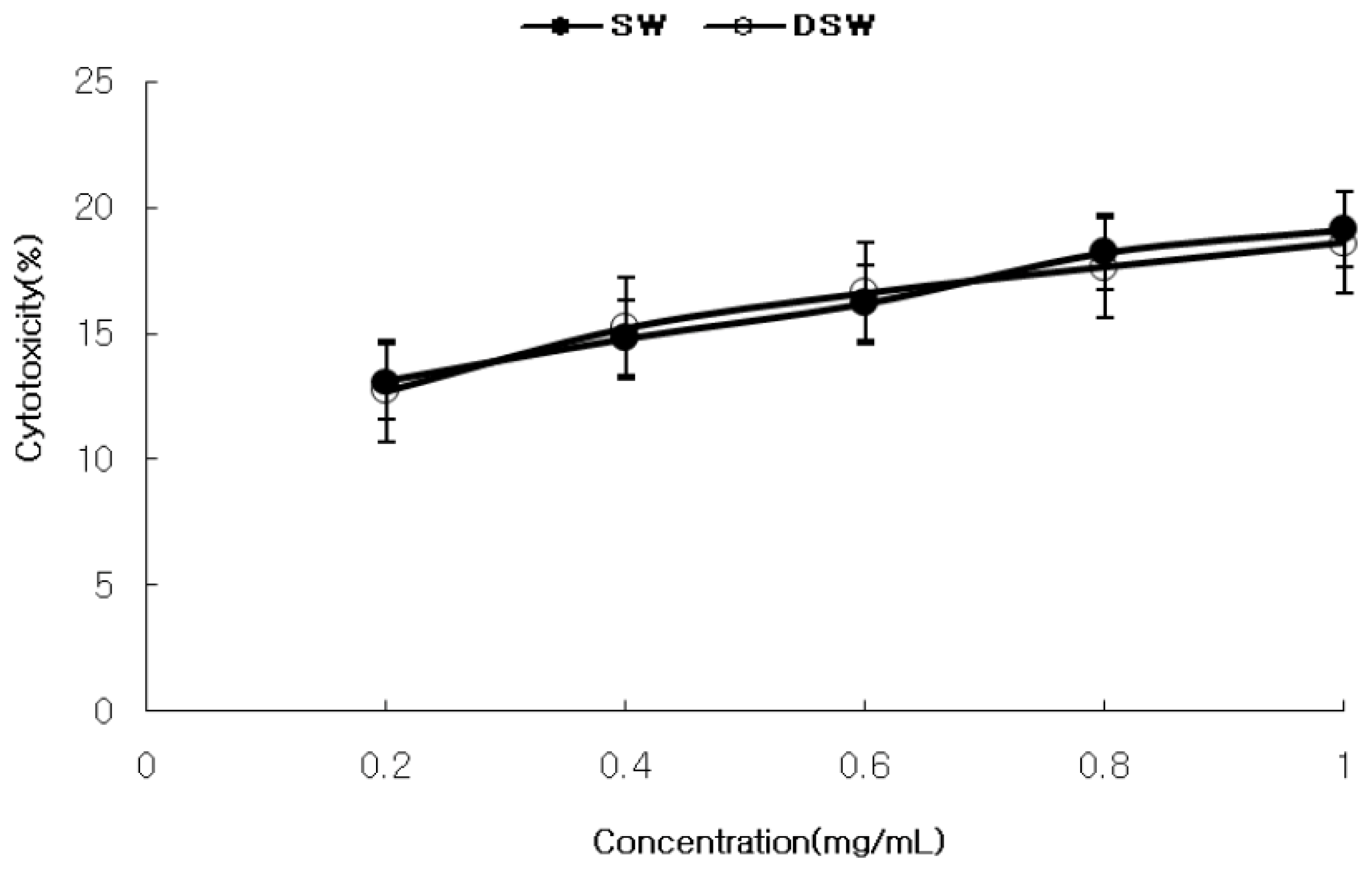
2.1. Measurement of Cytotoxicity and Anticancer Activity

Figure 1 shows the cytotoxicity of S. maxima against human normal kidney cells, which increased in a concentration dependent manner (0.2~1.0 mg/mL). The water extracts from seawater (SW) had a higher cytotoxicity (19.1%) than the water extract from the deep-sea water (DSW) (17.5%) at the highest extract concentration (1.0 mg/mL). As shown in Table 1 and Figure 2, DSW showed a stronger cytotoxicity against most cancer cell lines than SW, as well as a higher selectivity that implies the ratio of selectively inhibiting the cancer cells compared to normal cell. Interestingly, DSW showed a stronger inhibition (85%–93%) at higher concentrations than SW at concentrations lower than 0.4 mg/mL for most cancer cells, especially in MCF-7 and Hep3B, respectively. It is worth noting that this inhibition level was similar to, or even higher than, those reported for the addition of S. plantesis extracts [5,25], this result of depressing various cancer cells implies that the extracts could play a role in inhibiting cancer cell growth by improving immune activation [26]. Therefore, the results of investigating immune activation activities of DSW extracts were shown for the following experiments.

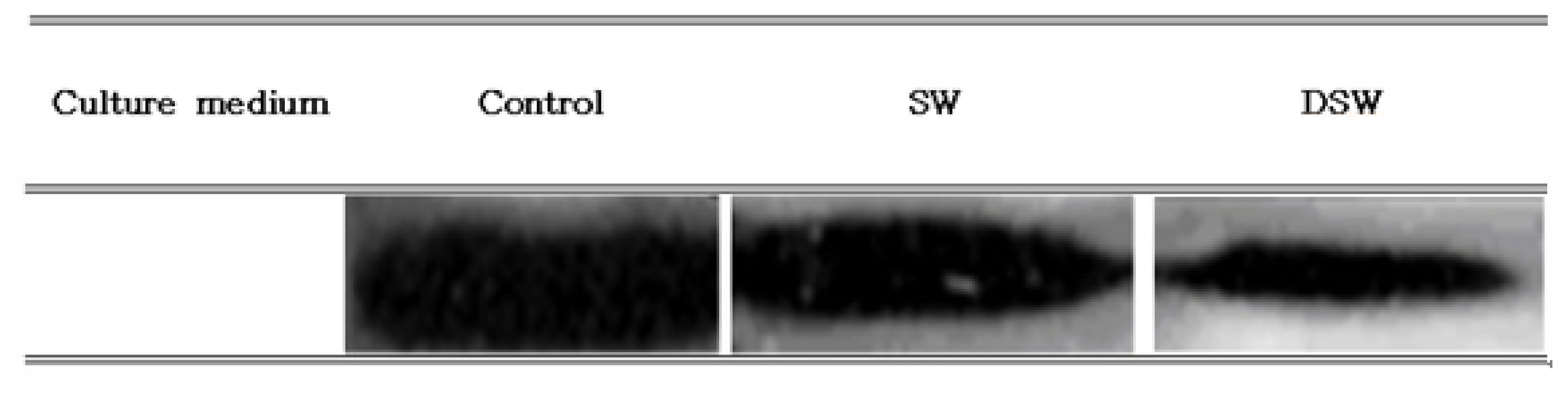
2.2. Measurement of Bcl-2 Expression in Human Cancer Cell

To examine the anticancer activities of the samples at the molecular level, the downregulation of Bcl-2 expression in A549 cells was determined since A549 cell growth was also similarly inhibited in treating the DSW extract (data not shown). Bcl-2 was selected for this purpose because it is an anti-apoptosis gene that indirectly inhibits apoptosis by inhibiting the opening of PT channels on the mitochondrion, which lowers the mitochondrial potential and blocks the release of cytochrome C from the mitochondrion [27]. As shown in Figure 3, the control, which did not contain any samples, had a higher level of Bcl-2 protein expression than the two other cases, and the Bcl-2 expression level was lowest in cell treated with 1.0 mg/mL of DSW. This result clearly indicates that the extract from DSW could effectively inhibit the proliferation of cancer cell growth by reducing the expression of Bcl-2 in human stomach cancer cell, which was well correlated to the results shown in Figure 3.

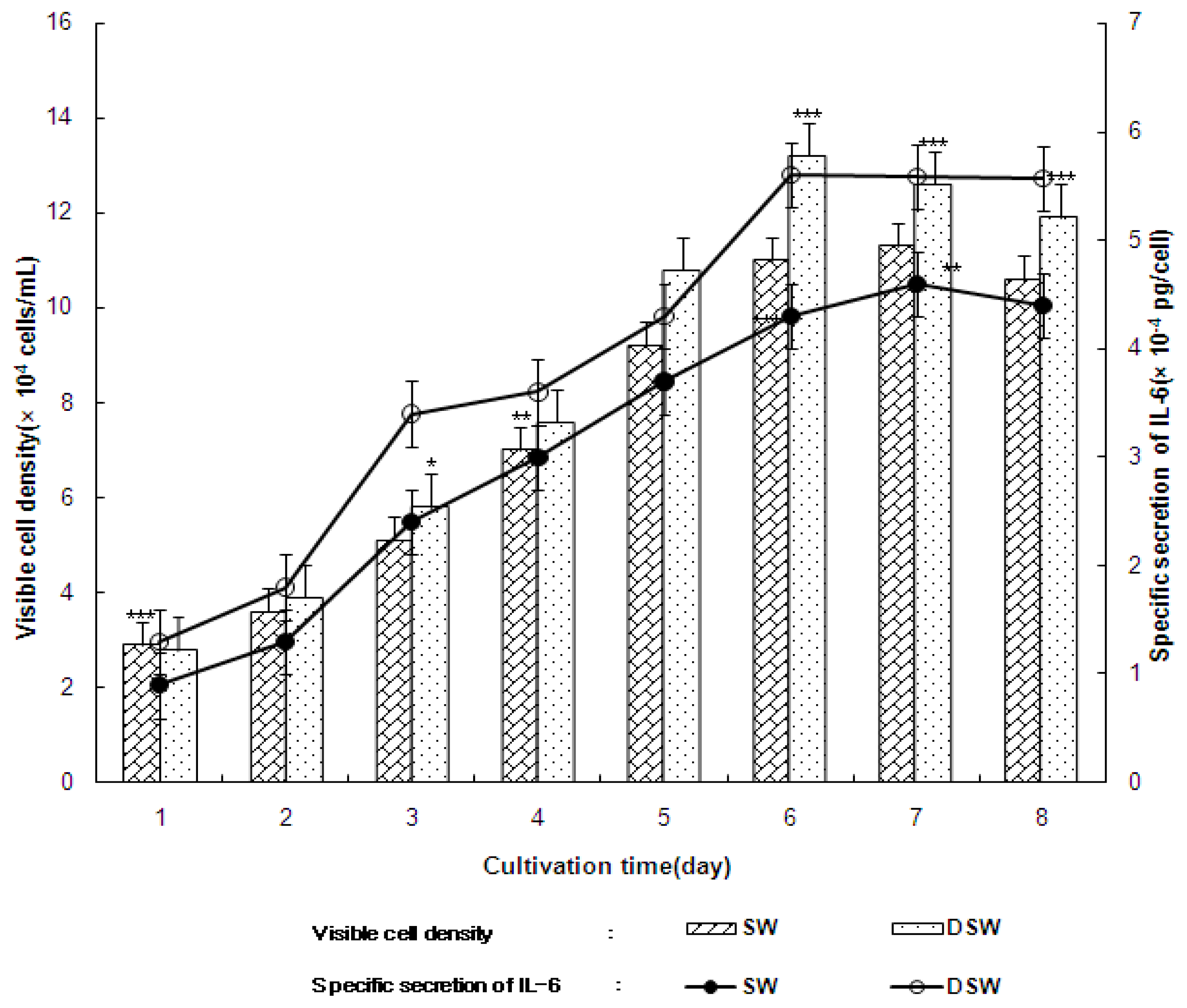
2.3. Secretion of Cytokine, Interleukin-6 (IL-6), and Tumor Nucreosis Factor-α (TNF-α) from the Growth of Human B and T Cells

The above data demonstrated that the extracts from DSW had strong anticancer activities, which indicates that they may also have immuno-modulatory activities since many studies have reported that there is a close relationship between anticancer and immune activation activities in natural extracts [28,29]. In Figure 4, the growth of human B cell and secretion of Interleukin-6 (Il-6) was compared between the addition of 1.0 (mg/mL) of SW or DSW. In these experiments, it was apparent that the extract from deep-sea water enhanced both cell growth and secretion of IL-6 from B cells. In addition, cell growth was maintained during the latter periods of the cultivation in the presence of DSW, while cell growth decreased in the presence of SW. This may have been the reason why the extract obtained from deep-sea water could improve cell growth, which also resulted in the constant specific production of IL-6, even during the latter period of cultivation. Similar results were also observed for natural products that displayed immune activation activity since the secretion of specific cytokines, such as IL-6 or Tumor Nucreosis Factor-α (TNF-α), was closely related to immune-modulatory activities within the cells [1,30]. Table 2 also clearly demonstrates that DSW enhanced immune functions by increasing both cell growth and IL-6 and TNF-α secretion, compared to the addition of SW. It should also be noted that the extracts had a stronger positive effect on T cell growth than B cell growth. As shown in Table 2, cell growth after the addition of the S. maxima extract continually increased from one to six days. Particularly, the growth of human T cells increased by over 2.4 × 10⁵ viable cells/mL in the presence of 1.0 mg/mL of DSW. In general, T cell growth (24 × 104 viable cells/mL) in the presence of S. maxima cultivated in deep-sea water was significantly higher than that of SW (6.4 × 10⁴ viable cells/mL). The DSW also increased B cell growth (18 × 104 viable cells/mL) compared to seawater culture mediums. The secretion of interleukin-6 and TNF-α by human T cells were also increased up to over 5.30 × 10⁻⁴ pg/cell and 5.60 × 10⁻⁴ pg/cell, respectively through the addition DSW. The secretion of IL-6 in the presence of deep-sea water was higher than IL-6 secretion in the presence of seawater (Table 2). A similar pattern as seen with the cytokine secretion from T cells was also observed for B cells, where the secretion of Interleukin-6 and TNF-α were increased to over 5.30 × 10⁻⁴ pg/cell and 5.60 × 10⁻⁴ pg/cell, respectively, through the addition DSW.

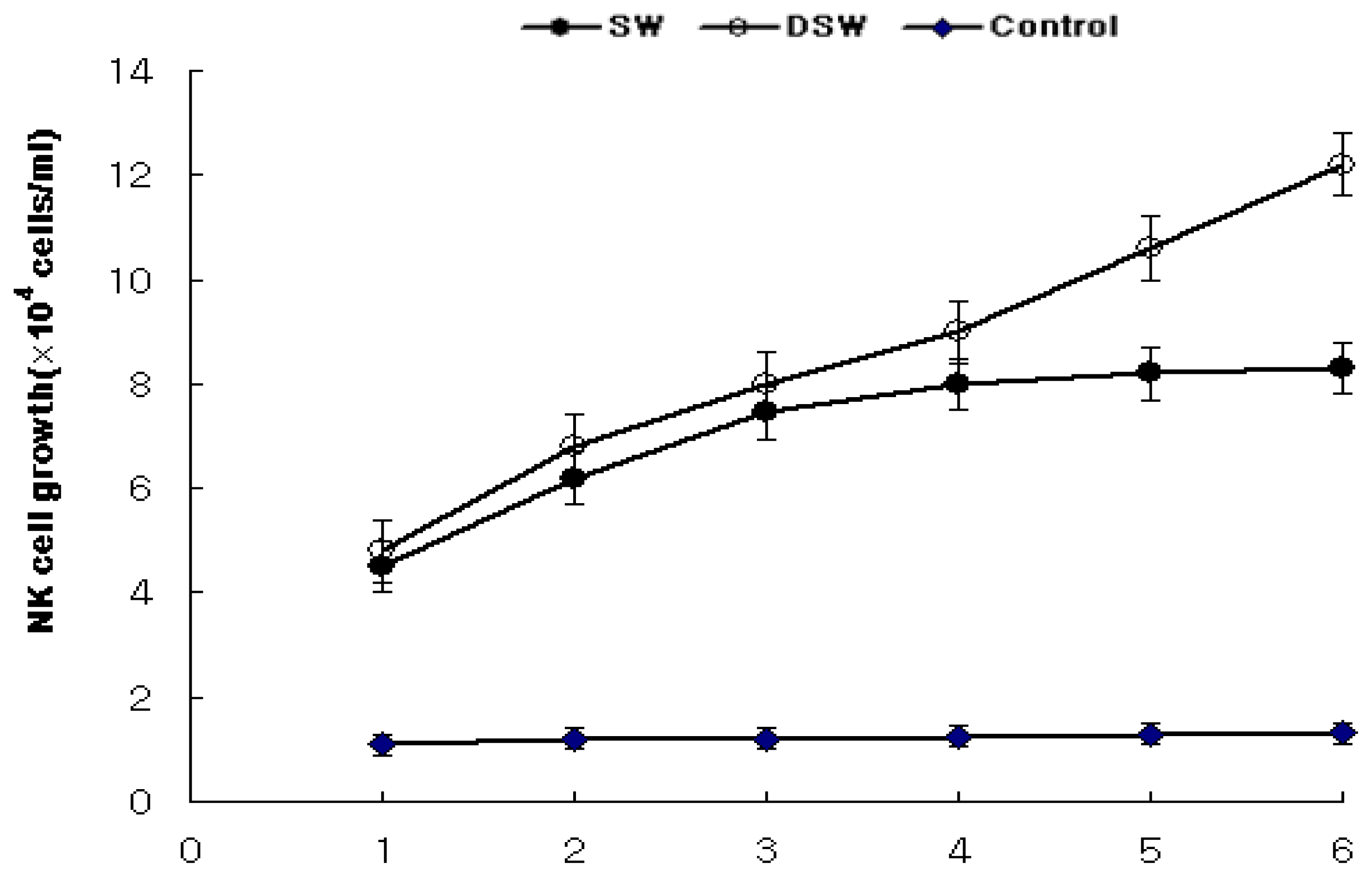
2.4. Enhancement of NK Cells Associated with Human T Cell Growth

In addition to measuring the increase in the secretion of cytokines from B or T cells, the immune activation activities of the extracts must also be confirmed by observing NK cell growth after the addition of stimulated B and T cells since the cytokines released from these cells could affect NK-92MI cell growth promotion [31,32]. In general, NK cell growth gradually increased according to the cultivation time after the addition of the samples, as shown in Figure 5. The extracts of DSW significantly increased NK cell growth to over 12.2 × 10⁴ viable cells/mL, while the extracts of SW only increased cell growth by approximately 8.3 × 10⁴ viable cells/mL. The increase in NK cell growth during the latter periods of cultivation, in the presence of the seawater extract, was significantly lower than in the presence of the deep-sea water extract. However, all the extracts of S. maxima appeared to improve NK cell growth relative to the control (not adding any extract), which could not maintain the growth of NK cells. This result clearly demonstrates that NK cell growth was significantly promoted due to the high amount of cytokines released after the addition of the deep-sea water S. maxima extract, relative to the extract of S. maxima cultivated in a seawater medium.

2.5. HPLC Analysis

The results presented above indicate that the S. maxima extracts, grown from deep-sea water, had a stronger physiological effect than the extracts grown in seawater, particularly in regards to the immune activation activities associated with anticancer activities. To better understand the biological activities of the extracts, the HPLC profiles of the extracts grown in the two different medium was determined to identify the key biologically active components in S. maxima (Figure 6). This was done because previous studies have reported that S. maxima contains a high content of β-carotene, ascorbic acid, thiamin, and γ-linolenic acid, iron etc., all of which have strong anticancer and immune activities [3337]. Particularly, β-carotene and vitamin C have multiple biological activities such as anticarcinogenic [33,34], antimutagenic [35], antioxidant, anti-inflamnatory, antiprolifertive [33], and antiatherogenic properties [38]. In addition, these compounds have been used as chemo-preventive agents against cancer diseases in various organs such as the lung, stomach, colon, breast, and prostate [37]. The extract cultivated under deep-sea water conditions had higher amounts of all the components than the extracts cultivated in seawater (SW). Using the absorbance of the standard compounds at 254 nm, the S. maxima extracts grown in deep-sea water (DSW) were found to contain 1100 μg/g of β-Carotene (peak retention time six minutes as a standard), 12.4 μg/g of ascorbic acid (peak retention time five minutes) and 14 μg/g of thiamin (peak retention time three minutes). These concentrations were higher than those grown in conventional seawater (520 μg/g of β-Carotene, 7.3 μg/g of ascorbic acid and 8 μg/g of thiamin). Moreover, in comparing these values to those reported in other studies (800 μg/g of β-Carotene, 6.7 μg/g of ascorbic acid, and 10 μg/g of thiamin) [39], it appears that the deep-sea water improved the biosynthesis of all three components and the extracts cultivated in seawater only displayed an increase in the ascorbic acid concentration. From various reported data, 2500 ug/g of pure carotene or 60 ug/g of ascorbic acid inhibited cancer cell lines and also enhanced immune activation activities by improving 20% [26,40]: however, the efficacy of each single compound was somewhat lower than those of the extracts containing both compounds, possibly by synergic activities between them, as well high treatment concentrations. Therefore, these results clearly indicate why the DSW extract displayed a higher immune activation activity and a higher anticancer activity [28,29].

2.6. Discussion

There have been several reports on the biological activities of S. platensis; however, there have only been a comparably small number of studies on the biological activities of S. maxima. In addition, the effects of cultivation in deep-sea water on the biological activities of the extracts have received little attention, even though deep-sea water is a clean resource that contains high concentrations of several important minerals for micro algal cultivations. There have been several studies that reported an increase in cell growth and better physiological performance when samples were cultivated in deep-sea water [18,41,42], however, an increase in the biological functions of the extracts after cultivation in deep-sea water has not been examined, which could explain why deep-sea water increases the immune activation activities of S. maxima.
Therefore, in this work, the effect of a higher content of β-Carotene and ascorbic acid on the biological activity of the extract was examined by growing them in deep-sea water. This was done because these compounds are known to play an important role in multiple biological activities such as anticarcinogenic [33,34,43], antimutagenic [35], antioxidant [36], and anti-inflamnatory activity, etc., In general, the extracts grown from deep-sea water (DSW) had less cytotoxicity against normal human cell lines than the extracts cultivated in conventional seawater (SW) (19% vs. 23%). This result was comparable to the results reported for the S. platensis extract, which had a cytotoxicity of approximately 26% [5]. The extracts inhibited cancer cell growth in the range of 75% to 91%, after the addition of 1.0 mg/mL of the extract. This was in good agreement with a previous study, which reported that the S. platensis extracts inhibited the growth of cancer cells by more than 75% [5]. The anticancer activities of the extracts may have been caused by an increase in human immune cell growth associated with the increase of cytokines such as tumor necrosis factor (TNF)-α and IL-2 because tumor cells can acquire resistance to (TNF)-α related apoptosis-inducing ligand (TRAIL), an innate immune molecule that selectively induces apoptosis in tumor cells [44,45]. The results of our work indicate that DSW extracts may enhance TRAIL-induced apoptosis, which can sensitize several cancer cells [4651]. This hypothesis was also supported by the result that Bcl-2 expression level was significantly decreased in the presence of the DSW extracts. Overall, the extracts obtained from deep-sea water displayed stronger immuno-modulatory activities than the extracts from seawater, which also resulted in higher anticancer activities because of the significantly higher β-Carotene and ascorbic acid contents of the extracts grown in deep-sea water.
It was first reported that S. maxima grown in deep-sea water contained high amounts of β-carotene and ascorbic acid because deep-sea water has high calcium and magnesium contents, as well as nitrate and phosphate contents [41]. Because of this higher nutritive mineral content, cell growth was activated, which could result in a higher β-carotene and vitamin C content [41,42]. Deep-sea water is used in numerous fields ranging from foods to medicines, and health products, and the results of this study indicate that deep-sea water could be used to develop bioactive substances from S. maxima. However, further studies on identifying other new active components in DSW and more detailed immune activation mechanisms should be carried out for widely expanding the use of its extracts.

3. Experimental Section

3.1. Cells and Chemicals

The strain of S. maxima (CY-023) was obtained from Korea Marine Microalgae Culture Center (Pukyoung National University, Pusan, Korea), and all cell lines (human breast adenocarcinoma MCF-7, human gastric cancer cell line AGS, human hepatocillular carcinoma Hep3B, human embryonic kidney cell HEK 293, human lung carcinoma A549, NK cell and human T cell, B cell) were obtained from American Types of Culture Collection (ATCC, Manassas, VA, USA). RPMI 1640 and alpha minimum essential medium (α-MEM) were purchased from GIBCO (Invitrogen, Carlsbad, CA, USA). HEPES buffer was purchased from SIGMA (St. Louis, MO, USA). Fetal bovine serum and horse serum was purchased from GIBCO (Invitrogen). Gentamycin sulfate and sulforthodamine B (SRB) were purchased from Sigma. Deep-sea water was collected at a 300 m-depth from the sea surface at the Deep Ocean Water Application Research Center of the Korea Ocean Research & Development Institute (Goseong, Korea). The concentrations of key minerals in the seawater (SW) and the deep-sea water (DSW) are compared in Table 3.

3.2. Culture Condition and Extraction of S. maxima

S. maxima was cultivated in two types of media: Seawater medium containing NaHCO3, K2HPO4, NaNO3, K2SO4, NaCl, MgSO4·7H2O, CaCl2, FeSO4·7H2O, EDTA, Solution A5 (A5 contained: H3BO3, MnCl2, ZnSO4, CUSO4, MoO2), and Solution B6 (NH4VO3, K2Cr(SO4)4, NiSO4, Na2WO4, Ti(SO4)3, Co(NO3)2), and Deep sea water medium containing only NaHCO3 as an inorganic carbon source [52]. Five percent (w/v) of the cells were grown in a 1 L Erlenmeyer flask (working volume 500 mL) for batch cultivation at 28 ± 2 °C, and continuously illuminated with white fluorescent lamps (40 W). Air containing 0.3% CO2 (v/v) was constantly supplied at a rate of 250 (mL/min) and cells were cultivated at an agitation speed of 150 rpm [52,53]. The cells were then collected by centrifuge at 3000 rpm from a culture flask. The collected cells were dried by ambient air at 20 °C before extraction. The powdered samples of S. maxima were extracted with water at 100 °C for 12 h. The water extract was concentrated in a rotary vacuum evaporator (Eyela, Tokyo, Japan), then freeze dried, and stored at −20 °C before use.

3.3. Measurement of Cytotoxicity and Anticancer Activity

In vitro cytotoxicity against several human cancer cell lines including MCF-7, Hep-3B, AGS, and normal human embryonic kidney cell (HEK 293) was measured using the modified Sulforhodamine B (SRB) assay [5,54] as follows: All cell lines were incubated in tissue culture flasks in the desired media with 5% CO2 at 37 °C. Trypsinized (trypsin-EDTA, Gibco (invitrogen, Carlsbad, CA, USA)) cell cultures were washed with media and diluted to 1 × 10⁴ viable cells/mL in each well of a 96 well plate. The plates were incubated at 37 °C for 48 h with the addition of several concentrations of the extracts. A hundred microliters of 20% ice-cold trichloroacetic acid (TCA) solution was then gently layered on top of the medium overlaying the cells. The plates were incubated for 60 min at 4 °C. The plates were rinsed several times with tap water and then the cells were stained with a 0.4% SRB solution (50 μL stain/well) for 15 min at room temperature. The SRB staining solution was removed, wells were then rinsed five times with 1% acetic acid to remove unbound dye, and left to air-dry. The bound SRB dye was solubilized by adding unbuffered Tris-base solution (100 μL/well), and the plates were placed on a plate shaker for one hour at room temperature. Plates were then read at 540 nm, using a microplate reader (Thermo max, Molecular Devices, Sunnyvale, CA, USA) and the results were expressed as the percentage of the control values.
The selectivity of extracts on each cancer cell line was estimated using the following equation;
Selectivity = AA / CT
where AA and CT indicate the anticancer activity ratio and the extract-induced cytotoxicity on HEK 293 cells, respectively.

3.4. Measurement of Bcl2 Protein Level

The expression levels of Bcl-2 in AGS cells cultivated with, and without, the samples (control) were measured as follows [55]. The cells were lysed directly into a solution containing 1% Triton X-100, 20 mM Tris (pH 8.0), 137 mM NaCl, 15% glycerol, 5 mM EDTA, 1 mM Pefablock, 0.1 mg/mL aprotinin, and 0.1 mg/mL leupeptin. The debris was removed by centrifugation for 15 min at 15,000 × g and 4 °C, and equivalent amounts of protein were separated by 10% SDS-PAGE [56,57] and transferred onto nitrocellulose filters. The filters were firstly stained to confirm uniform transfer of all samples, and then incubated in blocking solution for two hours at room temperature. The filters were reacted first with the anti-Bcl-2 at a dilution of 1:1000 for two hours, followed by two washes with PBS and two washes with TBST. Filters were then incubated with 1:1000 horseradish peroxidase-conjugated anti mouse secondary antibodies for one hour, then washed with TBST and developed using the Super Signal West Pico Kit.

3.5. Measurement of Human T and B Cell Growth and Secretion of Cytokines

Human immune T cell (Jurkat; ATCC) and B cell (Raji; ATCC) cells were cultured at 37 °C in RPMI 1640 medium containing 10% FBS in 5% CO2. Each sample of 0.5 g/L was added and cell growth was measured by the number of cells in 24 well plates that initially contained 1.0 × 10⁴ viable cells/mL using trypan dye in a hemacytometer [58]. Secretion of cytokines, IL-6 and TNF-α were also quantified using IL-6 and TNF-α kits (Chemicon, Billerica, MA, USA) as follows [59]. After adjusting the cell concentration to 1~2 × 10⁴ viable cells/mL, 900 L the cell concentrate was placed into 24 well plates and cultured for 24 h (37 °C, 5% CO2). 100 L of the cells with 0.5 g/L extract was cultured again. The cell cultures were centrifuged to obtain the supernatant and the absorbance of the supernatant at 450 nm was read using a microplate reader (Thermo max, Molecular Devices). The amounts of cytokines were measured using the O.D. values of the standards.

3.6. Enhancement of Human Natural Killer (NK) Cell Growth

The NK-92MI cell line (CRL-2408, ATCC, USA) was diluted to 2 × 10⁷ viable cells/mL using 2 mM l-glutamine, 0.2 mM myoinositol, 20 mM folic acid, 0.1 mM 2-mercaptoethanol, 12.5% fetal bovine serum (FBS) and 12.5% horse serum (Myelocult) in α-MEM medium. Cell proliferation during culture inT-25 flasks in the presence of samples at a concentration of 0.5 g/L was determined. The cells cultures were then centrifuged to obtain the supernatant. 900 L of the NK-92MI cell line was aliquoted into 24 well plates at 4~5 × 10⁴ viable cells/mL, and 100 L of the supernatant from T cells was then placed into the well and cultured for 48 h. The growth of NK-92MI cells was measured over six days using a cell counter [60,61].

3.7. HPLC Analysis

The extracts of S. maxima grown in seawater or deep-sea water were analyzed by HPLC (Hewlett Packard HP1100, USA) after filtering with 4.5 m filter paper. The samples were separated at 27 °C on Phenomenex (250 × 4.60 mm, 5 micron) column. The mobile phase consisted of acetone, acetonitrile, and orthophosphoric acid in the ratio of 70:20:10 and a flow rate of 1 mL/min was used. The HPLC peaks were monitored at 254 nm for 30 min and β-carotene, ascorbic acid, and thiamin were used as standards to assess the retention time of these compounds under the same conditions [62].

3.8. Statistical Analysis

All experiments were conducted in three duplicates, and expressed as mean ± S.D. Data were analyzed using the Statistical Analysis System software. Fisher’s Least Significant Difference (LSD) was used to determine significant difference among the treatments at p < 0.05.

4. Conclusions

There have been several promising results of having better cell growth and physiological performance of micro algae grown in deep-sea water; however, there are very few data related to the biological functions of the extracts, as well as a lack of scientific explanation. In this work, it was clearly shown that the amounts of β-Carotene and ascorbic acid of the extract were significantly increased by cultivating S. maxima in deep-sea water, possibly due to rich important minerals for algal cell growth and relatively low pathogens. It was interesting that the extracts grown from deep-sea water (DSW) had less cytotoxicity and better anticancer activities than those from conventional seawater (SW), which would imply that the mineral compositions of deep-sea water plays a role in constituting cell materials to affect the immune functions. The selective anticancer activities of DSW could be caused by improving immuno-modulatory activities associated with the increase of key cytokines, TNF-α and IL-2. Our work demonstrates that DSW increased TRAIL-induced apoptosis, which can sensitize several cancer cells, by the result that Bcl-2 expression level was significantly decreased in the presence of the DSW extracts. We believe this is the first report that S. maxima grown in deep-sea water has better immune activation activities than that in surface seawater because the extracts contained high amounts of β-carotene and ascorbic acid, even though new biologically active components in the extract should be investigated.

Acknowledgments

This research was supported by a grant from Technology Development for Bioenergy Production from Marine Biomass Funded by Ministry of Land, Transport and Maritime Affairs of Korean Government (PM57210).

Conflict of Interest

The authors declare no conflict of interest.

References

  1. Oh, S.H.; Han, J.G.; Ha, J.H.; Kim, Y.; Jeong, M.H.; Kim, S.S.; Jeong, H.S.; Choi, G.P.; Park, U.Y.; Kang, D.H.; et al. Enhancement of immune activity of Spirulina maxima by low temperature ultrasonification extraction. Korean J. Food Sci. Technol 2009, 41, 313–319. [Google Scholar]
  2. Jassby, A. Spirulina: A Model for Microalgae as Human Food. In Algae and Human Affair; Cambridge University Press: Cambridge, UK, 1998; pp. 65–67. [Google Scholar]
  3. Ayehunie, S.; Belay, A.; Hu, Y.; Baba, T.; Ruprecht, R. Inhibition of HIV-1 replication by an aqueous extract of Spirulina platensis (Arthrospira platensis). J. Acquir. Immune. Defic. Syndr. Hum. Retrovirol 1996, 18, 7–12. [Google Scholar]
  4. Babu, M. Evaluation of chemoprevention of oral cancer with Spirulina fusiformis. Nutr. Cancer 1995, 24, 197–202. [Google Scholar]
  5. Kim, H.S.; Kim, C.H.; Kwon, M.C.; Song, Y.K.; Cho, J.H.; Gwak, H.G.; Hwang, B.Y.; Kim, J.C.; Lee, H.Y. Anticancer activity of ultrasonified extracts from seawater-based culture of the microalga Spirulina platensis. Korean J. Fish. Soc 2006, 39, 318–325. [Google Scholar]
  6. Tomohiro, H.; Misako, M.; Kaoru, H.; Yoshiko, S.; Michio, U.; Tsukasa, S. Activation of the human innate immune system by Spirulina: Augmentation of interferon production and NK cytotoxicity by oral administration of hot water extract of Spirulina platensis. Int. Immunopharmacol 2002, 2, 423–434. [Google Scholar]
  7. Mohamed, F.I.; Doaa, A.A.; Augusta, F.; Mohamed, E.A.; Rajiv, L.G.; Wael, M.I.; Madhwa, H.R.; Allal, O. Chemoprevention of rat liver toxicity and carcinogenesis by Spirulina. Int. J. Biol. Sci 2009, 5, 377–387. [Google Scholar]
  8. Rathore, N.K.; Singh, S.; Singh, V. Spirulina as immuno-enhancer and biomodulator. J. Immunol. Immunopathol 2004, 6, 1–10. [Google Scholar]
  9. Mazo, V.K.; Gmoshinshii, I.V.; Zilova, I.S. Microalgae Spirulina in human nutrition. Vopr. Pitan 2004, 73, 45–53. [Google Scholar]
  10. Kay, R.A. Microalgae as food and supplement. Crit. Rev. Food Sci. Nutr 1991, 30, 555–573. [Google Scholar]
  11. Yang, H.N.; Lee, E.H.; Kim, H.M. Spirulina platensis inhibits anaphylactic reaction. Life Sci 1997, 61, 1237–1244. [Google Scholar]
  12. Hernandez, A.C.; Nieves, I.; Meckes, M.; Chamorro, G.; Barron, B.L. Antiviral activity of Spirulina maxima against Herpes simplex virus type 2. Antiviral Res 2002, 56, 297–285. [Google Scholar]
  13. Jeong, S.W.; Lee, N.K.; Kim, S.J.; Han, D.S. Screening of tyrosinase inhibitor from plants. Korean J. Food Sci. Technol 1995, 27, 891–896. [Google Scholar]
  14. Kim, I.S; Cho, Z.S. Modulation of human fibroblast proliferation and collegan production by Prostaglandin E2. Korean Biochem. J 1993, 26, 40–52. [Google Scholar]
  15. Cho, J.H.; Lee, K.M.; Kim, N.S; Kang, W.H. The effects of whitening components on human melanocytes in vitro. Korean Cosmet. Sci 1997, 23, 115–121. [Google Scholar]
  16. Fukami, K.; Nishijima, T.; Hata, Y. Availability of deep-sea water and effects of bacteria isolated from deep-sea water on the mass culture of food microalga Chaetoceros ceratosporum. Nippon Suisan Gakkaishi 1992, 58, 931–936. [Google Scholar]
  17. Nakasone, T.; Akeda, S. The application of deep-sea water in Japan. Nakasone Akeda. 1997, 69–75. [Google Scholar]
  18. Fukami, K.; Nishimura, S.; Ogusa, M.; Asada, M.; Nishijima, T. Continuous culture with deep-sea water of a benthic food diatom Nitzschia sp. Hydrobiologia 1997, 358, 245–249. [Google Scholar]
  19. Roger, H. Chemical exchange between sea water and deep ocean basalts. Earth Planet. Sci. Lett 1970, 9, 269–279. [Google Scholar]
  20. Quinby-Hunt, M.S.; Turehian, K.K. Distribution of elements in sea water. Eos Trans. Am. Geophys. Union 1983, 64, 130–135. [Google Scholar]
  21. Berger, W.H. Deep-sea carbonate: Pteropod distribution and the aragonite compensation depth. Deep Sea Res 1978, 25, 447–452. [Google Scholar]
  22. Hwang, H.S.; Kim, S.H.; Yoo, Y.G.; Chu, Y.S.; Shon, Y.H.; Nam, K.S.; Yun, J.W. Inhibitory effect of deep-sea water on differentiation of 3T3-L1 adipocytes. Mar. Biotechnol 2009, 11, 161–168. [Google Scholar]
  23. Hataguchi, Y.; Tai, H.; Nakajima, H.; Kimata, H. Drinking deep-sea water restores mineral imbalance in atopic eczema/dermatitis syndrome. Eur. J. Clin. Nutr 2005, 59, 1093–1096. [Google Scholar]
  24. Fusako, M.; Kyoko, M.; Taeko, N.; Nao, M.; Yuhsuke, S. Effects of red algae cultivated with deep-sea water on the oxidation-reduction status of liver, lung, brain, and bone metabolism in SAMP1 and SAMR1. Int. Congr. Ser 2003, 1260, 413–416. [Google Scholar]
  25. Lingling, Y.; Yao, W.; Qingjun, Z.; Peng, C.; Yiqiang, W.; Ye, W.; Ting, L.; Lixin, X. Inhibitory effects of polysaccharide extract from Spirulina platensis on corneal neovascularization. Mol. Vis 1991, 15, 1951–1961. [Google Scholar]
  26. Joel, S.; Gerald, S. Regression of experimental hamster cancer by beta carotene and algae extracts. J. Oral. Maxillofac. Surg 1987, 45, 510–515. [Google Scholar]
  27. Yang, J.; Liu, X.; Bhalla, K.; Kim, C.N.; Ibrado, A.M.; Cai, J.; Peng, T.I.; Jones, D.P.; Wang, X. Prevention of apoptosis by Bcl-2: Release of cytochrome c from mitochondria blocked. Science 1997, 275, 1129–1132. [Google Scholar]
  28. Youn, C.K.; Cho, H.J.; Kim, S.H.; Kim, H.B.; Kim, M.H.; Chang, I.Y.; Lee, J.S.; Chung, M.H.; Hahm, K.S.; You, H.J. Bcl-2 expression suppresses mismatch repair activity through inhibition of E2F transcriptional activity. Nat. Cell Biol 2005, 7, 137–147. [Google Scholar]
  29. Hwang, C.S.; Choi, J.O.; Kim, I.S. Detection of bcl-2 & bax Expression in the Head & Neck Cancers. Cancer Res. Treat 1995, 27, 570–577. [Google Scholar]
  30. Yim, J.H.; Son, E.; Pyo, S.; Lee, H.K. Novel sulfated polysaccharide derived from red-tide microalgae Gyrodinium impudicum strain KG03 with immunostimulating activity in vivo. Mar. Biotechnol 2005, 7, 1436–2228. [Google Scholar]
  31. Liu, R.; van-Kaer, L.; La-Cava, La.; Price, M.; Campagnolo, D.I.; Collins, M.; Young, D.A.; Vollmer, T.L.; Shp, F.D. Autoreactive T cells mediate NK cell degeneration in autoimmune disease. J. Immunol 2006, 176, 5247–5254. [Google Scholar]
  32. See, D.M.; Broumand, N.; Sahl, L.; Tilles, J.G. In vitro effects of echinacea and ginseng on natural killer and antibody-dependent cell cytotoxicity in healthy subjects and chronic fatigue syndrome or acquired immunodeficiency syndrome patients. Immunopharmacology 1997, 35, 229–235. [Google Scholar]
  33. Abd El-Baky, H.H.; El Baz, F.K.; El-Baroty, G.S. Spirulina Species as a source of carotenoids and α-tocopherol and its anticarcinoma factors. Biotechnol 2003, 2, 222–240. [Google Scholar]
  34. Abd El-Baky, H.H.; El Baz, F.K.; El-Baroty, G.S. Production of antioxidant by the green alga Dunaliella salina. Int. J. Agric. Biol 2004, 6, 49–57. [Google Scholar]
  35. Bendich, A. Non vitamin A activity of carotenoids: Immuno enhancement. Trends in Food Sci. Technol. 1991, 127–129. [Google Scholar]
  36. Aboul-Enein, A.M.; El Baz, F.K.; El-Baroty, G.S.; Youssef, A.M.; Abd El-Baky, H.H. Antioxidant activity of algal extracts on lipid peroxidation. J. Med. Sci 2003, 3, 87–98. [Google Scholar]
  37. Poppel, G.V.; Goldbohm, R.A. Epidemiological evidences for β-carotene and cancer prevention. Am. J. Clin. Nut 1995, 62, 1393S–1402S. [Google Scholar]
  38. Schwartz, J.L. The dual role of nutrients as antioxidants and peroxidants: Their effects on tumor cell growth. J. Nutr 1996, 126, 12215–12275. [Google Scholar]
  39. Packer, L.; Glazer, A.N. Methods in Enzymology. In Cyanobacteria; Academic Press: San Diego, CA, USA, 1988. [Google Scholar]
  40. Aristo, V.; Mamdooh, G. In vivo effect of ascorbic acid on enhancement of human natural killer cell activity. Nutr. Res 1993, 13, 753–764. [Google Scholar]
  41. Abd El-Baky, H.H.; El Baz, F.K.; El-Baroty, G.S. Production of carotenoids from marine microalgae and its evaluation as food colorant and lowering cholesterol agents. Am. Eurasian J. Agric. Environ. Sci 2007, 2, 792–800. [Google Scholar]
  42. Fukami, K.; Nishijima, T.; Ishida, Y. Stimulative and inhibitory effects of bacteria on the growth of microalgae. Hydrobiologia 1997, 358, 185–191. [Google Scholar]
  43. Henson, D.E.; Block, G.; Levine, M. Ascorbic acids: Biologic functions and relation to cancer. J. Natl. Cancer Inst 1991, 83, 547–550. [Google Scholar]
  44. Lee, J.J.; Shin, D.H.; Park, S.E.; Kim, W.I.; Park, D.I.; Choi, Y.H.; Hong, S.H. Euphorbiae humifusae sensitizes apoptosis of TRAIL-resistant human gastric adenocarcinoma AGS cells. J. Life Sci 2008, 18, 120–128. [Google Scholar]
  45. Thomas, W.D.; Hersey, P. TNF-related apoptosis-inducing ligand (TRAIL) induces apoptosis in Fas ligand-resistant melanoma cells and mediates CD4 T cell killing of target cells. J. Immunol 1998, 161, 2195–2200. [Google Scholar]
  46. Qadir, S.A.; Kwon, M.C.; Han, J.G.; Ha, J.H.; Chung, H.S.; Ahnm, J.; Lee, H.Y. Effect of different extraction protocols on anticancer and antioxidant activities of Berberis koreana bark extracts. J. Biosci. Bioeng 2009, 107, 331–338. [Google Scholar]
  47. Femenia, A.; Garcia-Marin, M.; Simal, S.; Rossello, C.; Blasco, M. Effects of supercritical carbon dioxide (SC-CO2) oil extraction on the cell wall composition of almond fruits. J. Agric. Food Chem 2001, 49, 5828–5834. [Google Scholar]
  48. Hamburger, M.; Baumann, D.; Adler, S. Supercritical carbon dioxide extraction of selected medical plants—Effects of high pressure and added ethanol on yield of extracted substances. Phytochem. Anal 2004, 15, 46–54. [Google Scholar]
  49. Kaufmann, B.; Christen, P. Recent extraction techniques for natural products: Microwave-assisted extraction and pressurised solvent extraction. Phytochem. Anal 2002, 13, 105–113. [Google Scholar]
  50. Sharma, U.K.; Sharma, K.; Sharma, N.; Sharma, A.; Singh, H.P.; Sinha, A.K. Microwave-assisted efficient extraction of different parts of Hippophae rhamnoides for the comparative evaluation of antioxidant activity and quantification of Its phenolic constituents by reverse-phase high-performance liquid chromatography (RP-HPLC). J. Agric. Food Chem 2008, 56, 374–379. [Google Scholar]
  51. Zhang, S.; Zhang, J.; Wang, C. Extraction of steroid saponins from Paris polyphylla Sm. var. yunnanensis using novel ultrahigh pressure extraction technology. Pharm. Chem. J 2007, 41, 424–429. [Google Scholar]
  52. Samson, R.; Leduy, A. Multistage continuous cultivation of blue-green alga Spirulina maxima in the flat tank photobioreactors with recycle. Can. J. Chem. Eng 1985, 63, 105–112. [Google Scholar]
  53. Abd El-Baky, H.H.; El Baz, F.K.; El-Baroty, G.S. Production of phenolic compounds from Spirulina maxima microalgae and its protective effects in vitro toward hepatotoxicity model. Afr. J. Pharm. Pharmacol 2009, 3, 133–139. [Google Scholar]
  54. Doll, R.; Peto, R. The causes of cancer, quantitative estimates of avoidable risks of cancer in the United States today. J. Natl. Cancer Inst 1981, 66, 1192–1308. [Google Scholar]
  55. Li, H.L.; Chen, D.D.; Li, X.H.; Zhang, H.W.; Lu, Y.Q.; Ye, C.L.; Ren, X.D. Changes of NF-κB, p53, Bcl-2 and caspase in apoptosis induced by JTE-522 in human gastric adenocarcinoma cell line AGS cells: role of reactive oxygen species. World J. Gastroenterol 2002, 8, 431–435. [Google Scholar]
  56. Laemmli, U.K. Cleavage if structural proteins during assembly of the head of bacteriophasfe T4. Nature 1970, 227, 680–685. [Google Scholar]
  57. Towbin, H.; Staehelin, T.; Gordon, J. Electrophoretic transfer of proteins from polyacrylamide fels to nitrocellulose sheets: Procedures and some applications. Proc. Natl. Acad. Sci. USA 1976, 76, 4350–4354. [Google Scholar]
  58. Lee, S.H.; Lee, H.S.; Park, Y.S.; Hwang, B.; Kim, J.H.; Lee, H.Y. Screening of immune activation activities in the leaves of Dendropanax morbifera Lev. Hanguk Yakyong Changmul Hakhoe Chi 2002, 10, 109–115. [Google Scholar]
  59. Ha, J.H.; Kwon, M.C.; Han, J.G.; Jin, L.; Jeong, H.S.; Choi, G.P.; Park, U.Y.; You, S.G.; Lee, H.Y. Enhancement of immunomodulatory activities of low molecular weight fucoidan isolated from Hizikia fusiforme. Korean J. Food Sci. Technol 2008, 40, 545–550. [Google Scholar]
  60. Yueran, Z.; Rui, S.; You, L.; Chunyi, G.; Zhigang, T. Expression of leptin receptors and response to leptin stimulation of human natural killer cell lines. Biochem. Biophys. Res. Commun 2003, 300, 247–252. [Google Scholar]
  61. Limdbolum, C.K. IL-2 receptor signaling through the Shb adapter protein in T and NK cells. Biochem. Biophys. Res. Commun 2002, 296, 929–936. [Google Scholar]
  62. Driskell, W.J.; Bashor, M.M.; Neese, J.W. Beta-carotene determined in serum by liquid chromatography with an internal standard. Clin. Chem 1983, 29, 1042–1044. [Google Scholar]
Figure 1. Cytotoxicity of the extracts of S. maxima cultivated in SW and DSW on the normal cell line, HEK 293. SW and DSW extracts were extracted at 100 °C. Mean ± SD from triplicate separated experiments are shown. (SW: seawater, DSW: deep-sea water).
Ijms 14 12205f1
Figure 2. Inhibition of human stomach adenocarcinoma (AGS) (bar chart, percent) and selectivity (line chart) after adding 1.0 mg/mL of the extract of S. maxima cultivated in SW and DSW. Mean ± S.D. from triplicate separated experiments are shown. Each value were compared with the control at * p < 0.01, ** p < 0.005, *** p < 0.001 by Student t-test. (SW: seawater, DSW: deep-sea water).
Ijms 14 12205f2
Figure 3. The expression of Bcl-2 in A549 cells after adding 1.0 mg/mL of the extract of S. maxima grown in different media. (SW: seawater, DSW: deep-sea water).
Ijms 14 12205f3
Figure 4. The growth (bar chart, percent) and secretion (line chart) of Interleukin-6 (IL-6) from human B cell after the adding 1.0 mg/mL of the water extract of S. maxima cultivated in SW and DSW. Mean ± S.D. from triplicate separated experiments are shown. Each value were compared with the control at * p < 0.01, ** p < 0.005, *** p < 0.001 by Student t-test. (SW: seawater, DSW: deep-sea water).
Ijms 14 12205f4
Figure 5. The NK cell growth after the addition of supernatants from T cells that were treated with the water extract of S. maxima cultivated in SW and DSW. Mean ± S.D. from triplicate separated experiments are shown. (SW: seawater, DSW: deep-sea water).
Ijms 14 12205f5
Figure 6. Comparison of the HPLC chromatograms for the water extract of S. maxima cultivated in seawater (a) and deep-sea water (b).
Ijms 14 12205f6
Table 1. Inhibition of cell growth of MCF-7 and Hep3B cancer cell lines after addition of the extract under various cultivation conditions.
Concentration (mg/mL)Seawater (SW)Deep-sea water (DSW)


MCF-7Hep3BMCF-7Hep3B
0.243.5 ± 0.2244.2 ± 0.22 ***46.5 ± 0.23 ***45.3 ± 0.40 **
0.452.1 ± 0.23 *55.6 ± 0.2456.1 ± 0.4359.4 ± 0.47
0.661.7 ± 0.2062.4 ± 0.23 *68.2 ± 0.26 **74.4 ± 0.44 *
0.873.4 ± 0.27 **75.1 ± 0.2877.5 ± 0.3982.1 ± 0.41 ***
1.080.5 ± 0.41 ***83.4 ± 0.31 **89.6 ± 0.42 *93.5 ± 0.43 **
Mean ± SD from triplicate separated experiments are shown. Each value were compared with control at
*p < 0.01,
**p < 0.005,
***p < 0.001 by Student t-test.
Table 2. Cell growth and IL-6 and Tumor Nucreosis Factor-α (TNF-α) secretion from human immune T cells after adding 1.0 mg/mL of S. maxima extracted under various cultivation conditions.
SampleCultivation time (Day)T cellB cell


Viable cell density (×104 cells/mL)Cytokine secretion (×10−4 pg/cell)Viable cell density (×104 cells/mL)Cytokine secretion (×10−4 pg/cell)


IL-6TNF-αIL-6TNF-α
DSW12.9 ± 0.02 *1.2 ± 0.16 ***1.3 ± 0.11 *2.8 ± 0.07 **1.3 ± 0.01 **1.4 ± 0.06 *
24.2 ± 0.04 **1.6 ± 0.07 ***1.5 ± 0.03 ***3.9 ± 0.06 *1.8 ± 0.15 ***1.7 ± 0.04 ***
36.5 ± 0.05 *3.2 ± 0.06 **2.3 ± 0.07 **5.8 ± 0.02 **3.4 ± 0.23 **2.6 ± 0.01 ***
48.8 ± 0.06 ***3.7 ± 0.03 *3.3 ± 0.05 *7.6 ± 0.10 *3.6 ± 0.16 *3.4 ± 0.08 *
516.0 ± 0.08 ***5.3 ± 0.04 ***4.8 ± 0.15 **12.0 ± 0.06 *4.0 ± 0.07 **5 ± 0.02 **
624.0 ± 0.09 *6.4 ± 0.11 *5.6 ± 0.11 *18.0 ± 0.08 ***5.9 ± 0.18 **5.7 ± 0.13 *

SW13.1 ± 0.06 *1.1 ± 0.15 ***1.3 ± 0.16 **2.9 ± 0.03 ***0.9 ± 0.12 **1.1 ± 0.15 *
23.8 ± 0.09 ***1.6 ± 0.20 *1.7 ± 0.03 ***3.6 ± 0.05 *1.3 ± 0.09 *1.9 ± 0.06 **
35.4 ± 0.07 *2.6 ± 0.18 *2.6 ± 0.04 *5.1 ± 0.02 *2.4 ± 0.16 *2.6 ± 0.03 **
47.3 ± 0.05 ***3.2 ± 0.07 ***3.7 ± 0.01 ***7.0 ± 0.12 ***3 ± 0.13 ***3.8 ± 0.11 ***
59.6 ± 0.10 *3.8 ± 0.06 *4.3 ± 0.20 *9.2 ± 0.09 *3.7 ± 0.02 **4.2 ± 0.13 *
611.2 ± 0.09 **5.3 ± 0.04 *5.6 ± 0.1811.0 ± 0.08 **5.6 ± 0.05 *5.7 ± 0.13 ***
Mean ± SD from triplicate separated experiments are shown. Each value were compared with control at
*p < 0.01,
**p < 0.005,
***p < 0.001 by Student t-test.
(SW: seawater, DSW: deep-sea water).
Table 3. Comparison of the mineral compositions of deep-sea water and seawater.
CompositionSea waterDeep seawater


mg/kgmg/kg
Main elementK-380
Na890010780
Ca393403
Mg10801320
SiO2.02.8
Cl1200019350
SO4−22648898
F11.3
Li0.160.18
Sr7.814.3

Trace elementFe0.020.09
Mn0.030.16
Cu-0.26
Pb-0.11
Zn-0.45
Cd-0.05
Ni-0.36

Nutritive saltsNO3−0.040.28
PO43−0.0120.06
Si0.442.8

