1. Introduction
Ocular neovascularization (NV) is a main cause of blindness associated with ischemic retinal disorders, including proliferative diabetic retinopathy (PDR), retinopathy of prematurity (ROP) and age-related macular degeneration (AMD) [
1,
2]. Retinal neovascularization is largely the result of angiogenesis, in which cells of endothelial origin from the existing blood vessels start to proliferate out of control [
3]. Although a lot of energy and effort have been paid to clarifying the molecular mechanism involved, few breakthroughs have been acquired in the past few decades [
4]. The exact mechanism underlying the pathogenesis remains unknown; hence, there is no satisfactory therapy.
Vascular endothelial growth factor (VEGF) is the most potent cytokine that stimulates angiogenesis in retina [
5,
6]. Among the five subtypes, VEGF-A has more affinity for angiogenic tissue. VEGF-A realizes its functions through binding to vascular endothelium expressing receptors VEGFR-1 (tyrosine kinase receptor) and VEGFR-2 (KDR/kinase insert domain-containing receptor) [
7]. It has been reported that injection of exogenous VEGF to the eyes of monkeys can cause NV of retina [
8,
9]. On the other side, the application of anti-VEGF antibodies was proven to alleviate the severity of NV [
10]. Up to now, a great number of agents aimed at anti-VEGF have been recorded, and this has been regarded as one of the most effective strategies [
11,
12].
Emodin is an anthraquinone derivative from the rhizome of
Rheum palmatum L., a plant widely used in traditional Chinese medicine as a laxative [
13,
14]. Previous research showed that emodin possesses antifungal, antibacterial, antiviral and anti-VEGF activities [
15]. It was shown that emodin inhibits endothelial cell proliferation and has different effects on endothelial and tumor cell angiogenesis. In the context of the eye, emodin lessened inflammation and scarring in a mouse ocular alkali burn model [
16], so it is regarded as a promising candidate for ocular neovascularization. However, like many other anti-VEGF agents, the application of emodin is limited due to its hydrophobicity. Therefore, it is important and necessary to discover new methods for efficient delivery.
In recent years, the development of nanomaterials has drawn considerable attention for its diverse applications. One of these is the construction of a drug delivery system [
17] that can exceedingly reduce harmful nonspecific side-effects and toxicity compared to other types of carriers. More importantly, nanocarriers can commonly overcome low concentrations of drugs and modify the characteristic of the drug [
18]. Due to the large fraction of voids in their inner space, hollow micro- and nano-structures have emerged as powerful candidates in a wide range of applications, including drug delivery, efficient catalysis, waste removal, gas sensors, as well as lithium-ion batteries [
19,
20]. In particular, the use of silica-based nanoparticles has shown ever-increasing advances in biocompatibility and has achieved great success in the fields of bioengineering [
21,
22]. Therefore, the aim of the current research is to construct a system by employing hollow nanoparticles as the carriers for better emodin delivery.
2. Results and Discussion
Known as angiogenesis, the development of new blood vessels from preexisting vasculature has a causal role in ocular neovascularization. VEGF plays the role of a positive regulator of angiogenesis, and its proven role in angiogenesis has provided evidence for the use of anti-VEGF agents as potential therapies [
23]. As a result, VEGF inhibitors have been applied as the mainstay in the treatment of ocular neovascularization. Most anti-VEGF drugs for ocular disease treatment are applied as eye drop formulations or invasive injections. The former drug delivery system is regarded as low efficiency, and the latter one usually brings fear to patients. Furthermore, the concentration of drug in retina hardly turns out to be effective, because of ocular barriers [
24]. Therefore, it is meaningful to invite a new system for efficient and comfortable drug delivery. Up to now, nanoparticles have been regarded as a new approach to achieve this goal. In the present study, we showed that hollow MgSiO
3 nanoparticles are potent carriers of emodin and inhibit the function of retinal capillary endothelial cells
in vitro.
As described previously, the formation of SiO
2 colloidal spheres in this system heavily depends on the classical Stöber method [
25]. Then, the facile hydrothermal approach was used to acquire the growing of MgSiO
3 hollow structures on the basis of a chemical-template etching mechanism. Specifically, silica colloidal spheres were incubated with an alkaline solution, which contains magnesium ions, ammonium ions and ammonia. Then, the solution was transferred to a Teflon-lined stainless-steel autoclave. Through high-temperature treatment, the silica chains were broken by hydroxide ions, and silicate-ion groups were generated. These silicate ions released from SiO
2 colloidal spheres react with magnesium ions, and magnesium silicate was constructed
in situ around the SiO
2 cores. Followed by the slow release of silicate ions from the SiO
2 cores, well-structured magnesium silicate hollow spheres are formed gradually.
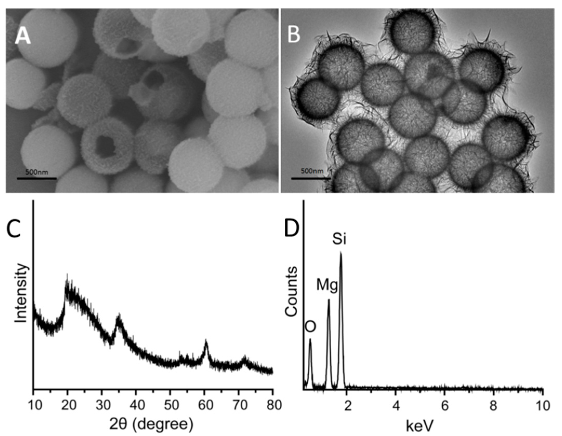
The morphological and structural analyses of the MgSiO
3 hollow spheres were carried via scanning electron microscopy (SEM) and transmission electron microscopy (TEM) at first. As exhibited in
Figure 1A,B, MgSiO
3 hollow spheres with a uniform and divergent nature revealed a relatively coarse surface and a mean diameter of ~400 nm. Since the average diameter of the SiO
2 precursor we used was 380 nm, the average diameter of our sample is nearly 400 nm. TEM image shown in
Figure 1B indicates that the magnesium silicate spheres possess a hollow structure with an obviously darkish center. Furthermore, the edge is black, and thickness of the shell of the hollow structures was about 30 nm. Actually, needle-like structures could be observed at first glance through the SEM and TEM images. The hollow structures were constituted of large, narrow and nanoscale lamellae. Phase purity and crystalline order are two of the most important parameters affecting the properties of materials. Wide-angle x-ray diffraction (XRD) patterns shown in
Figure 1C demonstrate that all of the well-defined diffraction peaks present the features of MgSiO
3. Energy-dispersive spectroscopy (EDS) analysis further confirmed that our product consists of magnesium, silicon and oxygen elements (
Figure 1D).
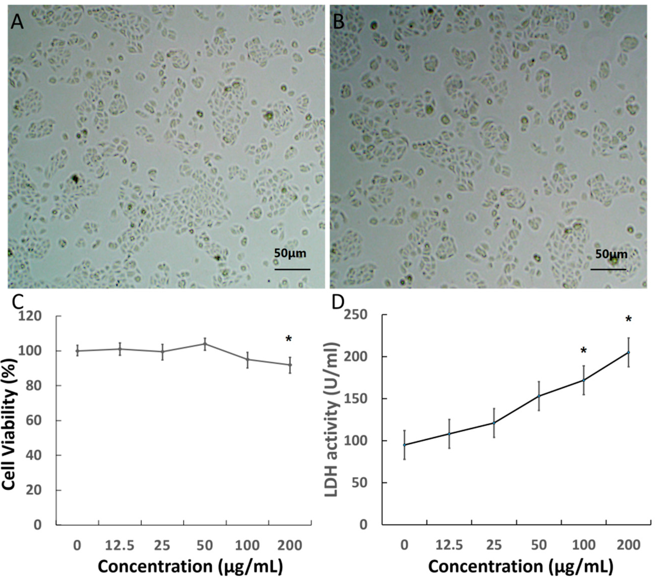
Prior to using nanomaterials for bio-systems applications, we investigated the cytotoxicity of the MgSiO
3 hollow spheres. The MTT assay associated with retina capillary endothelial cells was applied to evaluate the cytotoxicity of MgSiO
3 hollow spheres. We investigated the behavior of the MgSiO
3 hollow spheres in living cells via observing the morphological changes of cells using a microscope. As shown in
Figure 2A,B, microscopy images illustrated no obvious difference in the cell morphology for the cells treated with hollow spheres compared to the control groups after incubation with nanoparticles. As illustrated in
Figure 2C, cell viability was not hindered by nanoparticles up to a concentration of 200 μg/mL after 48 h of incubation. Otherwise, lactate dehydrogenase (LDH) activity induced by nanoparticles at various concentrations was also tested to evaluate cytotoxicity. This obviously shows that LDH activity became significant until the concentration was higher than 100 μg/mL (
Figure 2D), thereby indicating satisfactory results that supported the biocompatibility of the hollow spheres in the applied dosages.
Figure 1.
(A) Scanning electron microscope (SEM) image; (B) transmission electron microscope (TEM) images of MgSiO3 nanoparticles; (C) wide-angle XRD (X-ray diffraction) pattern of uniform MgSiO3 hollow spheres; and (D) the EDS (energy-dispersive spectroscopy) spectrum of uniform MgSiO3 hollow spheres.
Figure 2.
(A,B) Visible light microscopy images of retinal capillary endothelial cells incubated with nanoparticles and control; (C) the effect of MgSiO3 hollow spheres on the viability of cells; and (D) LDH (lactate dehydrogenase) activity induced by MgSiO3 nanoparticles. (* p < 0.05).
Figure 2.
(A,B) Visible light microscopy images of retinal capillary endothelial cells incubated with nanoparticles and control; (C) the effect of MgSiO3 hollow spheres on the viability of cells; and (D) LDH (lactate dehydrogenase) activity induced by MgSiO3 nanoparticles. (* p < 0.05).
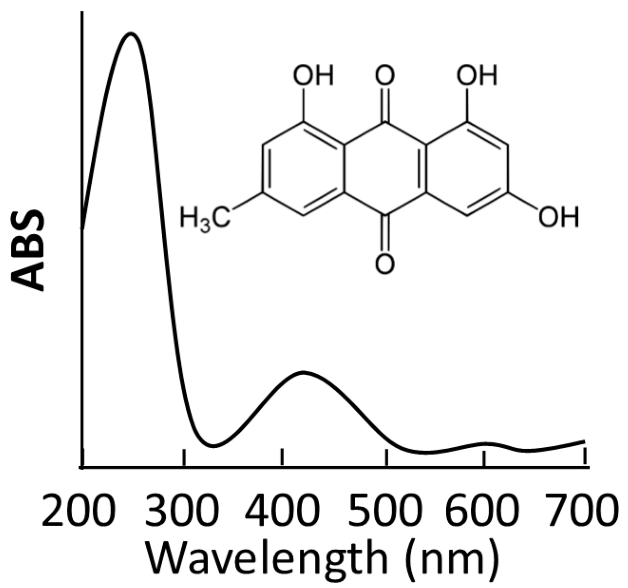
Up to now, the lack of aqueous solubility has always limited practical application for anti-VEGF treatment in clinics. To overcome this bottleneck, hollow materials have held more potential for the development of novel delivery systems [
25,
26]. These are characterized by a large space for storing guest molecules, which is an excellent feature for efficient nano-carriers. Emodin was employed as a model of a hydrophobic anti-VEGF drug in our current study. The MgSiO
3 hollow spheres were loaded with emodin molecules by soaking them in dimethyl sulfoxide (DMSO) containing emodin for 24 h. After emodin–MgSiO
3 was removed via careful centrifugation, emodin–MgSiO
3 was washed twice with phosphate buffer saline (PBS) to remove additional adsorbed drug on the surface of the MgSiO
3 hollow spheres. Detected via UV-Vis spectroscopy, approximately 130 μg of the emodin molecules were stored inside 1 mg of the hollow spheres, demonstrating the high loading capacity of the MgSiO
3 hollow spheres (
Figure 3).
Figure 3.
UV-Vis spectrum and chemical structure of the emodin molecules.
Figure 3.
UV-Vis spectrum and chemical structure of the emodin molecules.
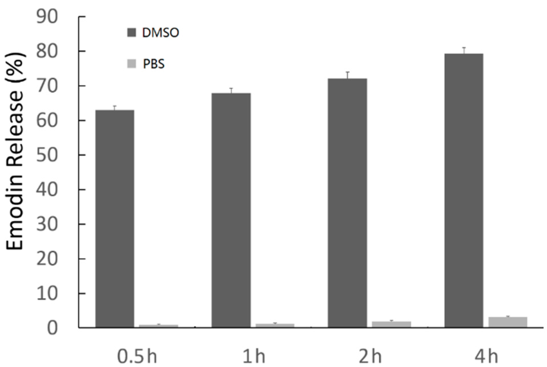
Figure 4 shows the amount of emodin released when emodin–MgSiO
3 was dispersed in PBS (pH = 7.4) and DMSO with different incubation periods. Less than 4% of the stored emodin could be released into the supernatant when emodin–MgSiO
3 was dispersed in PBS and left in suspension for up to 4 h. However, once emodin–MgSiO
3 was dispersed in DMSO for 0.5 h, most of the emodin could be released and detected in the supernatant. The above results suggest that the negligible drug leakage of emodin–MgSiO
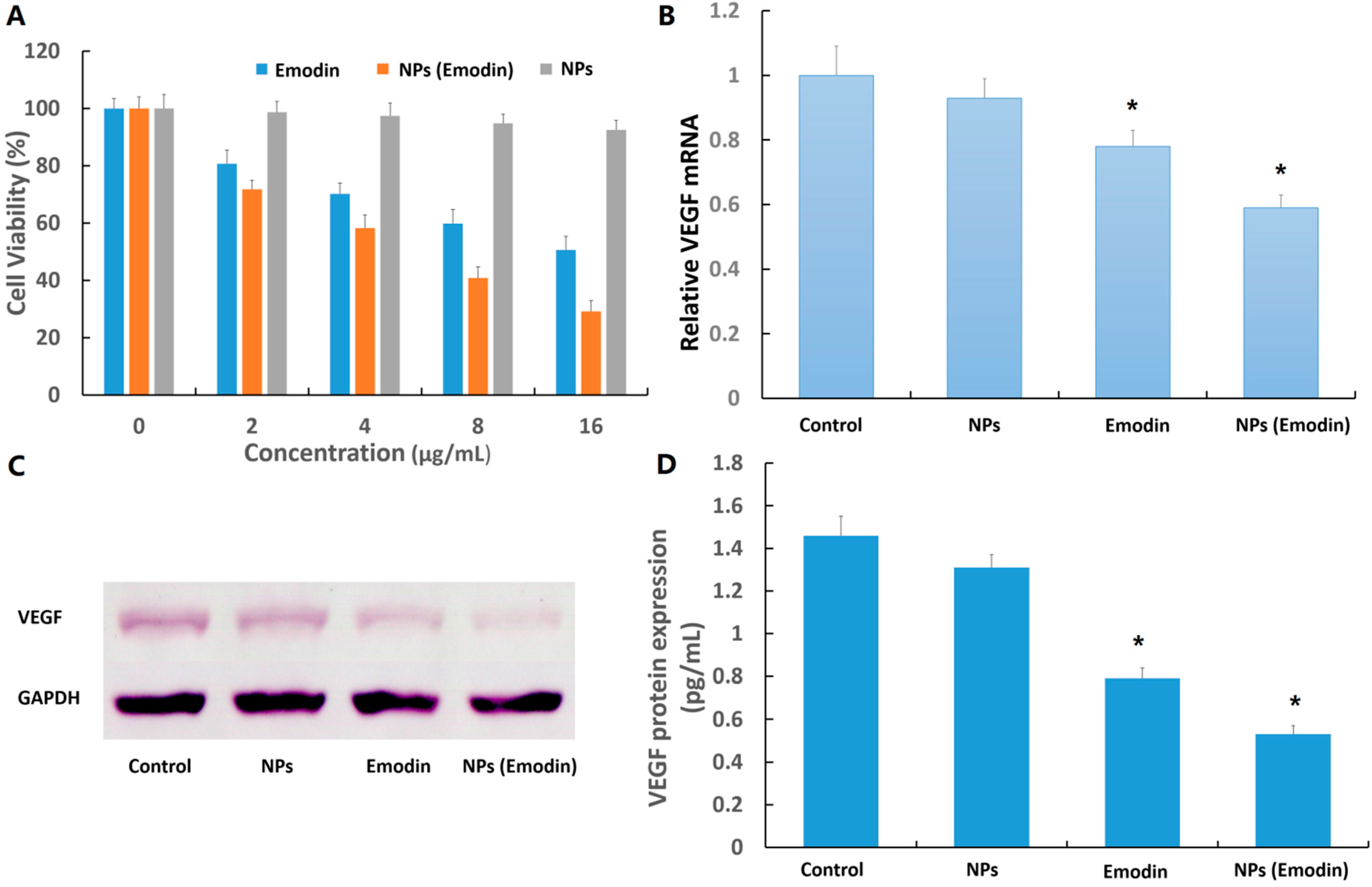
3 in PBS has great significance in the minimization of side effects. The viabilities of retina capillary endothelial cells against emodin and emodin–MgSiO
3 after incubation for 24 h are present in
Figure 5A. Compared with the control group, both emodin and emodin–MgSiO
3 exhibited toxicity towards retina capillary endothelial cells. The slight cytotoxicity of free emodin was probably in accordance with the low solubility of emodin in the hydrophilic medium. The enhanced cytotoxicity of emodin–MgSiO
3 further made out the efficient drug-delivery capability by using carriers. Due to the presence of hydrophobic sections in the cells, emodin–MgSiO
3 could rationally release emodin at these active sites, which plays an important role in inhibiting the proliferation of cells.
Figure 4.
The amounts of emodin released from the emodin–MgSiO3 complex measured via UV-Vis spectroscopy.
Figure 4.
The amounts of emodin released from the emodin–MgSiO3 complex measured via UV-Vis spectroscopy.
Figure 5.
(A) Viability of cells incubated with emodin and emodin–MgSiO3 under different incubation concentrations; (B) VEGF (vascular endothelial growth factor) mRNA expression of cells; (C) VEGF protein expression measured by western blot; and (D) enzyme-linked immunosorbent assay (ELISA) analysis for VEGF protein expression.(* p < 0.05).
To evaluate emodin and emodin–MgSiO
3 hollow spheres for
in vitro anti-VEGF therapy, the expression of VEGF gene and protein was studied. Transcripts for VEGF in the presence of emodin and emodin–MgSiO
3 were measured after 24 h of incubation. As shown in
Figure 5B, the presence of free emodin could downregulate the expression in accordance with a previous report [
27]. Otherwise, VEGF mRNA expression was significantly inhibited when incorporated with emodin–MgSiO
3, suggesting the effectiveness of our nano-carrier. Furthermore, we examined the expression of VEGF protein by two methods. As a critical agent on angiogenesis, VEGF is well known for its distinct effect on capillary endothelial cell proliferation and action. As shown in
Figure 5C,D, VEGF expression induced by emodin and emodin–MgSiO
3 is significantly downregulated, while no change is present in the presence of single MgSiO
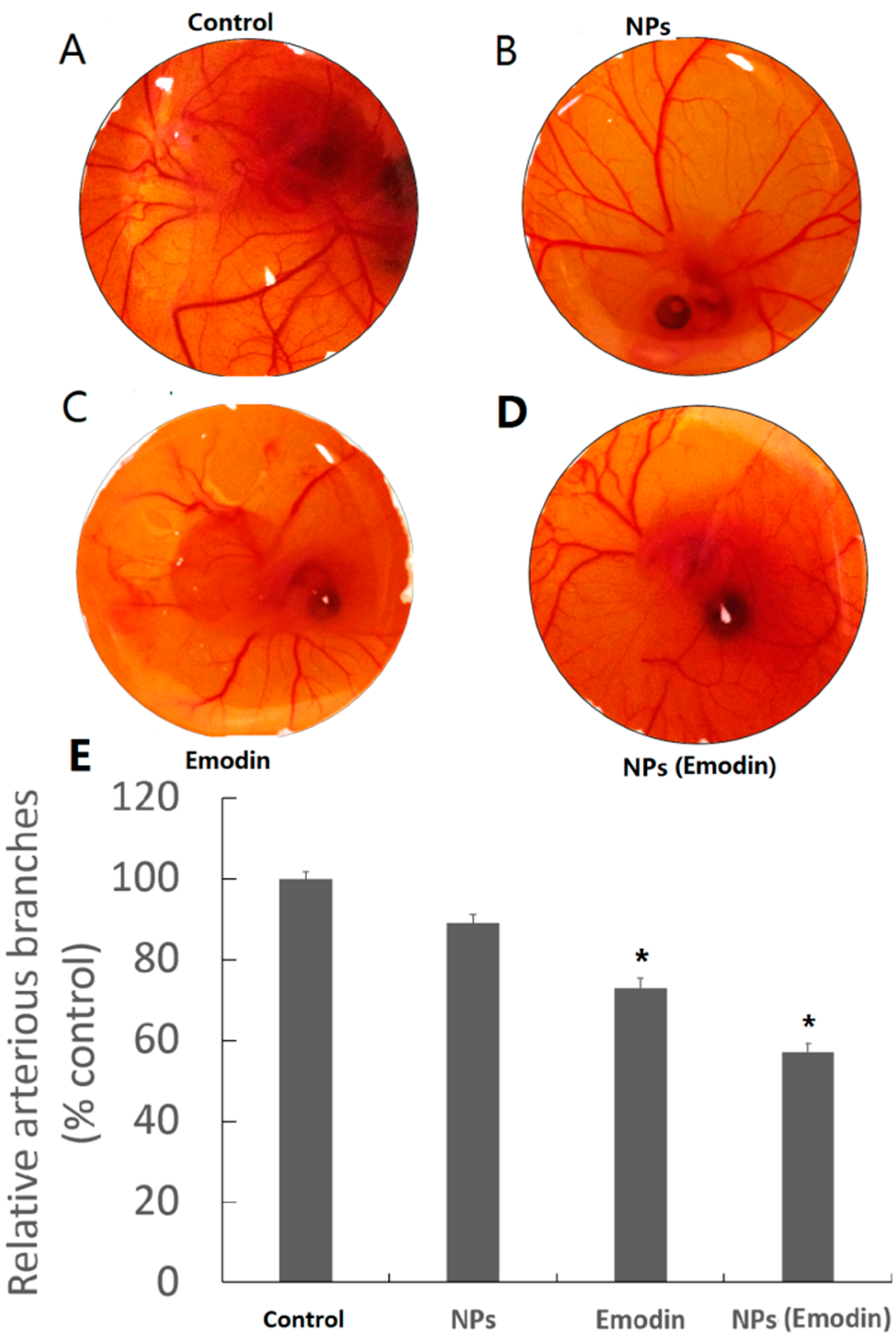
3 nanoparticles. To demonstrate the robust features of emodin-loaded MgSiO
3 hollow spheres, the
in vivo inhibitory effect on angiogenesis was then demonstrated on fertilized chicken eggs. Blood vessel formation was compared among a single dose of PBS, free MgSiO
3 nanoparticles, free emodin, as well as emodin-loaded nanoparticles, respectively. Illustrated by
Figure 6, less and slenderer vessels were observed in the presence of emodin and emodin–MgSiO
3, indicating the validity of our nanoparticles.
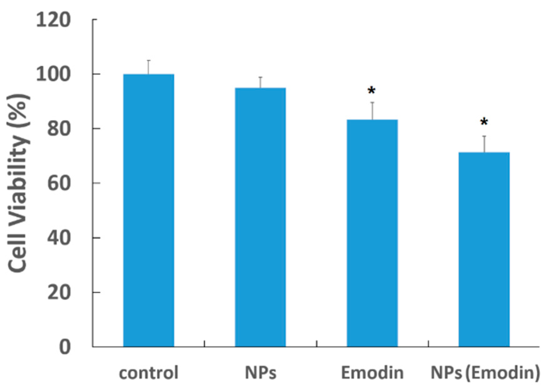
Since the anti-angiogenesis effect of single emodin or emodin–MgSiO
3 has been proven in retinal capillary endothelial cells, here we further tested the cytotoxicity of our emodin-loaded nanoparticles by employing human umbilical vein endothelial cells (HUVECs). As presented in
Figure 7, a similar inhibitory effect on proliferation was observed in HUVECs treated by emodin or emodin–MgSiO
3, while single nanoparticles (NPs) were less effect. This result raised the potential application of our nanoparticles in the treatment of other angiogenesis-related diseases, but not ocular neovascularization only. However, more studies, like cell migration and
in vitro tube formation, which were not carried out in current research, are still needed to clarify before further applications.
Figure 6.
Blood vessel formation of fertilized chicken eggs incubated with different agents (A–D) and quantitative analysis (E). (* p < 0.05).
Figure 6.
Blood vessel formation of fertilized chicken eggs incubated with different agents (A–D) and quantitative analysis (E). (* p < 0.05).
Figure 7.
Viability of HUVECs incubated with emodin and emodin–MgSiO3. (* p < 0.05).
Figure 7.
Viability of HUVECs incubated with emodin and emodin–MgSiO3. (* p < 0.05).
3. Experimental Section
3.1. Chemicals
Magnesium chloride hexahydrate (MgCl2·6H2O), ammonia chloride, ammonium hydroxide aqueous solution (28%) and ethanol were purchased from Beijing Chemicals (Beijing, China). Tetraethyl orthosilicate (TEOS) and (3-aminopropyl) trimethoxysilane (APTES) were purchased from Sigma-Aldrich (Shanghai, China). All of the chemicals were applied as received without further purification.
3.3. Cell Cultures
Retina capillary endothelial cells were supplied by American Type Culture Collection. Retina capillary endothelial cells were cultured in dulbecco’s modified eagle medium (DMEM) supplemented with 10% fetal calf serum. The culture media contained 100 UI/mL penicillin and 100 UI/mL streptomycin. The human umbilical vein endothelial cells (HUVECs) were cultured in M199 media (Gibco, Cleveland, TN, USA) containing 10% fetal bovine serum (Gibco), 2 mM glutamine (Gibco) and 1% penicillin/streptomycin (Sigma, Shanghai, China). All of the two cell lines were cultured in a humidified atmosphere containing 5% CO2 at 37 °C. Cells were harvested by the use of trypsin and were re-suspended in fresh complete medium before plating.
3.4. In Vitro Cytotoxicity Assay of NPs
MTT reduction assays were carried out to quantify the cytotoxicity of MgSiO3 hollow spheres. In a typical procedure, retina capillary endothelial cells were cultured in 96-well plates at a density of 5000 per well for 12 h to allow the cells to attach. After cell attachment, the media were changed to α-MEM with MgSiO3 at the indicated concentrations. After 48 h incubation, 10 μL MTT (5 g/L) were added to each well and cultured for another 4 h. Then, the supernatant was removed, and 150 μL DMSO were added. This was shaken for 15 min for crystal dissolution. The absorbance at 570 nm was measured with a micro-ELISA reader (Synergy2, Biotek, Winooski, VT, USA). The proliferation rate was calculated and compared to the control group.
3.5. Lactate Dehydrogenase (LDH) Release Assay
After the exposure of cells to MgSiO3 hollow spheres for 24 h, 50 μL of cell culture media were used to detect LDH activity by the LDH kit (Jiancheng, Nanjing, China), according to the manufacturer’s instruction. Cells incubated with 0.25% Triton for 15 min served as the positive control. The absorbance at 440 nm was measured by the Beckman DU-640B UV-visible spectrophotometer (Beckman Coulter Company, Changchun, China).
3.6. Emodin Releasing
The release of emodin under different solvent conditions was performed in vitro. In detail, 1 mg of emodin–MgSiO3 was re-suspended in a centrifuge tube (10 mL) containing 5 mL PBS or DMSO. At certain time intervals, the MgSiO3 hollow spheres were centrifuged, and 0.5 mL of release medium containing the free drug were transferred out to determine the concentration of emodin.
3.7. In Vitro Cytotoxicity Assays of Free Emodin and Emodin-Loaded NPs
In a typical procedure, retina capillary endothelial cells or HUVECs were cultured in 96-well plates at a density of 5000 per well for 12 h to allow the cells to attach. Subsequently, free emodin and emodin-loaded NPs with the same drug amount were added to the culture medium. After incubation for 8 h, the medium-containing nanoparticles were removed and fresh medium was placed. At the end of the incubation time (48 h), cell samples were treated with MTT for another 4 h, which was followed by the addition of dimethyl sulfoxide (DMSO) to dissolve the formazan crystals. A Bio-Rad model-680 microplate reader (Bio-Rad, Changchun, China) was applied to measure the absorbance at a wavelength of 570 nm.
3.8. Real Time PCR Analysis
In detail, cells were seeded in a 24-well plate at 104/well to determine gene expression. Free emodin and emodin-loaded NPs were added to the culture medium. After 8 h, the medium was replaced by fresh medium. Followed by another 72 h-culture, total RNA was isolated using EASY spin Plus (BLKW, Beijing, China), according to the manufacturer’s instructions. RNA was quantified spectrophotometrically at 260 nm (Nanodrop ND1000, Wilmington, DE, USA), and the quality was examined using the ratio of absorption at 260 and 280 nm, with a ratio between 1.9 and 2.1 as acceptable. Reverse transcription was performed with 2 μg of total RNA using PrimeScript RT Reagent Kit (Takara Co., Otsu, Japan). Each sample was then analyzed by quantitative real-time PCR (qPCR) (Stratagene MX3000P, La Jolla, CA, USA) in the SYBR Premix Ex TapII (Takara Co.), setting the cycles as follows: 10 s/95 °C PCR initial activation step; 40 cycles of denaturation for 20 s/95 °C and annealing step for 20 s/60 °C. The change in VEGF mRNA levels was determined by the formula 2−ΔΔCt, where ΔCt is the value from the threshold cycle (Ct) of the treated sample subtracted from the Ct value of the untreated or zero time-point control sample. The relative amount of mRNA in the sample was normalized to GAPDH mRNA. VEGF primers (Invitrogen, Carlsbad, CA, USA) used in this research are as follows: forward primer GAGCAAGACAAGAAAATCCC-3, reverse primer CCTCGGCTTGTCACATCTG.
3.9. Western Blot
After incubation with free emodin and emodin-loaded NPs for 72 h, 40 μg of total protein were separated by sodium dodecyl sulfate polyacrylamide gel electrophoresis (SDS-PAGE), using a gradient gel ((10%–12%), Bio-Rad Laboratories), transferred to nitrocellulose membrane and analyzed by immunoblotting using chemiluminescence (Santa Cruz, CA, USA). The primary antibodies used were VEGF-A (Abcam, MA, USA, 1:500) or GAPDH (Santa Cruz, CA, USA, 1:1000) and peroxidase-conjugated anti-mouse IgG (Santa Cruz, CA, USA, 1:1000).
3.10. ELISA Analysis for VEGF Expression
Cells were cultured under the indicated condition for 24 h. Culture medium was collected and prepared for VEGF ELISA assay. The assays were performed according to the protocol of the manufacturer using a specific ELISA kit (BlueGene Biotech, Shanghai, China). Each protein sample was analyzed in triplicate with parallel 3-well culture plates to ensure accurate results.
3.11. Angiogenesis Assay in Fertilized Chicken Eggs
The effect of free emodin and emodin-loaded NPs on ex vivo angiogenesis was determined by the angiogenesis assay. Briefly, fertile leghorn chicken eggs were candled on embryonic Day 8; a small opening was made at the top of the live eggs. Different types of emodin for treatment were mixed with 0.5% methyl cellulose in water and gently placed on the egg. The eggs were incubated for 48 h and photographed. The antiangiogenic analysis was indicated by the relative numbers of arterious branches. The assay was performed three times to ensure reproducibility.
3.12. Statistical Analysis
All experiments were performed thrice, and the data are expressed as the means ± SD. The difference between mean values was evaluated by using the ANOVA and considered to be statistically significant when p < 0.05.


