Article

“Biological Adhesion” is a Significantly Regulated Molecular Process during Long-Term Primary In Vitro Culture of Oviductal Epithelial Cells (Oecs): A Transcriptomic and Proteomic Study

by
Joanna Budna-Tukan
1,*,
Agata Światły-Błaszkiewicz
2,
Piotr Celichowski
1,
Sandra Kałużna
1,
Aneta Konwerska
1,
Patrycja Sujka-Kordowska
1,
Maurycy Jankowski
3,
Magdalena Kulus
4,
Michal Jeseta
5,
Hanna Piotrowska-Kempisty
6,
Małgorzata Józkowiak
6,
Paweł Antosik
4,
Dorota Bukowska
4,
Mariusz T. Skowroński
4,
Jan Matysiak
2,
Michał Nowicki
1 and
Bartosz Kempisty
1,3,5
1
Department of Histology and Embryology, Poznan University of Medical Sciences, 60-781 Poznan, Poland
2
Department of Inorganic and Analytical Chemistry, Poznan University of Medical Sciences, 60-870 Poznań, Poland
3
Department of Anatomy, Poznan University of Medical Sciences, 60-781 Poznan, Poland
4
Veterinary Center, Nicolaus Copernicus University in Toruń, 87-100 Torun, Poland
5
Department of Obstetrics and Gynecology, University Hospital and Masaryk University, 601 77 Brno, Czech Republic
6
Department of Toxicology, Poznan University of Medical Sciences, 61-631 Poznan, Poland
*
Author to whom correspondence should be addressed.
Int. J. Mol. Sci. 2019, 20(14), 3387; https://doi.org/10.3390/ijms20143387
Submission received: 23 January 2019 / Revised: 25 June 2019 / Accepted: 8 July 2019 / Published: 10 July 2019
(This article belongs to the Section Molecular Biology)

Abstract

:
Oviductal epithelial cells (OECs) actively produce stimulating and protecting factors, favoring survival and viability of gametes and early embryos. The oviduct participates in the initial reproductive events, which strongly depends on adhesion. The analysis of differential gene expression in OECs, during long-term in vitro culture, enables recognition of new molecular markers regulating several processes, including “biological adhesion”. Porcine oviducts were stained with hematoxylin and eosin, as well as with antibodies against epithelial markers. Then, OECs were long-term in vitro cultured and after 24 h, 7, 15, and 30 days of culture were subjected to transcriptomic and proteomic assays. Microarrays were employed to evaluate gene expression, with Matrix-assisted laser desorption/ionization-time of light (MALDI-TOF) mass spectrometry applied to determine the proteome. The results revealed proper morphology of the oviducts and typical epithelial structure of OECs during the culture. From the set of differentially expressed genes (DEGs), we have selected the 130 that encoded proteins detected by MALDI-TOF MS analysis. From this gene pool, 18 significantly enriched gene ontology biological processes (GO BP) terms were extracted. Among them we focused on genes belonging to “biological adhesion” GO BP. It is suggested that increased expression of studied genes can be attributed to the process of intensive secretion of substances that exhibit favorable influence on oviductal environment, which prime gametes adhesion and viability, fertilization, and early embryo journey.

1. Introduction

The oviduct is a cylindrical structure extending between the ovary and the uterus. It is organized into three segments (the infundibulum, the ampulla, and the isthmus) and four histological layers (mucosa, submucosa, muscularis, and serosa). The innermost layer of mucosa presents variable character, depending on the region. In the infundibulum, it forms finger-like folds, which become highly developed and branched in the ampulla, to finally decrease and adopt simplified structure in the isthmus [1]. Regardless of the region, mucosa is lined with simple columnar epithelium, comprised of ciliated and non-ciliated cells, presenting secretory and motor function. i.e., oviductal epithelial cells (OECs) [2,3]. During the estrus cycle, under hormonal changes, proportions of OEC types and their morphology undergo substantial alterations, with prevalence of ciliated cells in the follicular phase and secretory cells in the luteal phase [1].
The abovementioned morphology and location of the oviduct justify its participation in the initial reproductive events. Many of them involve formation or disruption of tight connections between somatic cells, gametes, embryos, and reproductive organs, and thus it strongly depends on adhesion abilities. The process of biological adhesion is indispensable for intercellular communication, signal transduction, proliferation, or apoptosis [4]. Cellular connections include: cytoskeleton-involving adherens junctions (AJs), formed by integrins, cadherins, and desmosomes [5]; selective tight junctions, controlling the molecular, and ionic flow, made up of claudins and occludins [6]; and lastly, gap junctions, based on connexins, allowing small substance flow between adjacent cells [7].
Oocytes, in cumulus-oocyte complexes (COCs), form tight junction connections with cumulus cells (CCs), allowing the flow of stimulating factors necessary for oocyte maturation [7]. In the majority of mammalian species, oocyte maturation takes place in the ovarian follicle, and is followed by disruption of follicular wall and ovulation. As a result, compact structures of the COCs loosen. Ovulated COCs transiently adhere to OECs of oviductal ampulla before they start to flow towards uterus. The oviduct promotes the oocyte’s journey with the use of three mechanisms: movement of the fimbriae of the infundibulum, beating movement of OEC cilia, and peristaltic contractions of the muscularis layer [8]. Simultaneously, counter-current movement of the sperm occurs. However, before this may happen the oviduct modifies the spermatozoa, after their passage from the cervix through utero-tubal junction, making them capable of fertilization. Some sperm migrate directly to the ampulla, while the majority adhere to the OECs, forming reservoir pending for capacitation at the time of ovulation [9]. Transient adhesion of COCs to OECs is most likely the signal indicating the occurrence of ovulation, triggering sperm hyperactivation and release [10]. This passage is crucial for successful fertilization and ensures that only a low number of selected spermatozoa reaches the ampullar-isthmic junction where fertilization takes place [11]. The latter is highly dependent on E-cadherin assured by intercellular adhesion [12]. Following fertilization, ciliary action eases the transport of the zygote towards uterus [8].
The precise mechanism of gamete and embryo interactions with OECs has not yet been described. Although intrauterine implantation is a definite sign of pregnancy, it is known that communication processes begin much earlier. Large preovulatory follicles, characterized by intense hormonal production, were shown to alter OEC gene expression differently than small preovulatory follicles, presumably creating an optimal oviductal environment for the future embryo [13]. Moreover, non-fertilized oocytes stay in the oviduct longer, while fertilized ones are directly transferred to the uterus [14]. The presence of embryos in the oviduct of pigs, cows, and horses was shown to change the transcriptome of the OECs, especially involving genes related to immune response, thus protecting the embryo from rejection [15]. All this points to a conclusion that OECs actively produce stimulating, protecting, and growth factors, favoring survival and viability of gametes and early embryos [16,17,18].
Thus, many co-culture systems are implemented, including those employing the porcine model, to mimic in vivo COC-OEC interactions and to improve the efficiency of COCs’ in vitro maturation. Among them, co-culture with OECs significantly improved nuclear maturation of canine oocytes [19], blastocyst formation frequency, total cell number, and subsequent embryo development in pigs [20] and humans [21]. It is defined that co-cultures with OECs are beneficial, since they enrich culture medium with growth factors and cytokines, as well as work as medium detoxifiers and developmental block inhibitors [20].
The biggest clinical opportunity associated with OECs is the possible application of their co-cultures in improvement of the in vitro fertilization (IVF) outcomes. Hence, there is a large need to analyze the changes of their function outside of the physiological conditions. The long-term in vitro cultures of OECs could bring their specific ex vivo features, as well as the ability to secrete stimulating factors, into light. The analysis of differential gene and protein expression in OECs during vitro long-term cultivation represents a good and stable model for recognition of molecular markers of several processes, including “biological adhesion”, which could indicate long-term viability of those cells for potential clinical applications.
Therefore, the main aim of this study is to analyze the changes in the transcriptomic and proteomic profile of cultured OECs, in relation to that detected closely to the point of collection (24 h of culture). The authors presume that the changes in gene and protein expression will, to some extent, mirror the effects of the ex vivo culture conditions on the physiological properties of the OECs. The knowledge gained could then be used to better understand the processes that might also occur during OEC-containing co-cultures. This, in turn, could serve to improve the methodology of OEC-gamete/embryo co-cultures, which ultimately serves in maximizing the positive outcomes of the IVF procedures.

2. Results

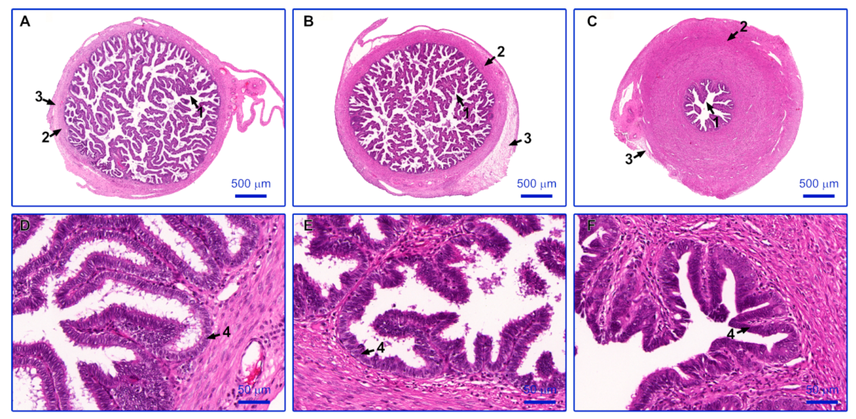
2.1. Histological Evaluation

Observation of paraffin sections stained with the hematoxylin and eosin (H+E) method allowed to assess the morphological structure of the various parts of the oviduct. Each of the analyzed sections showed normal histological structure. Particular segments of oviduct vary in the thickness of the muscle layer and the length and number of mucosal folds. The infundibulum contains long, finger-like mucosal folds and a thin muscle layer. In the ampulla, mucosa forms numerous, branched folds, while the muscle layer is thicker compared to the infundibulum. The isthmus is characterized by a thick layer of smooth muscles distributed in circular and longitudinal layers, while the folds of the mucosa are poorly developed. In each segment of the oviduct, the mucosal folds are lined with the simple columnar ciliated epithelium. Non-prominent cilia indicate anestrous period of the ovarian cycle. The photographed results were presented in Figure 1.

2.2. Oviductal Epithelial Cell (OEC) Culture

The OECs subjected to long-term cell culture did not significantly change their morphology during its period. The pictures, depicting cells in all of the timeframes from which the material for molecular analyses was collected, present the same epithelial-like shape throughout the culture time (Figure 2).

2.3. Microarray Expression Analysis

The investigation of OEC transcriptome following 24 h, 7, 15, and 30 days of long-term in vitro culture yielded 14,789 porcine transcripts. From this group, after seven days of culture, 1146 genes met the criteria of significantly changed expression; 733 of them were up-regulated and 413 were down-regulated. After 15 days of culture, 938 genes were up-regulated and 562 were down-regulated, giving 1500 differentially expressed genes in total. Finally, after 30 days of culture, from the 1970 genes that significantly altered their transcript levels, 1444 were up-regulated and 526 were down-regulated.

2.4. nanoLC-MALDI-TOF/TOF MS/MS Analysis

The nano liquid chromatography-matrix-assisted laser desorption/ionization-time of light tandem mass spectrometry (nanoLC-MALDI-TOF/TOF MS/MS) analysis allowed for the identification of the total number of 3240 unique tryptic peptides, which correspond to 389 proteins. The representative example of the mass spectrum (m/z 1417.6834), which was identified as one of the fascin peptides, is shown in Figure 3. In the database used, there was no possibility to restrict the search to the Sus scrofa species. Hence, the “Other mammals” database was searched, which yielded some proteins assigned to species other than pig. The list of those proteins was presented in Supplementary Table S1. As they were detected in porcine samples, it can be assumed that these proteins are homologues of those found in Sus scrofa. Moreover, some additional searches were made including “Homo sapiens” and “Rodentia” datasets, in order to complete obtained molecular pattern. Results were presented in Tables S2 and S3.

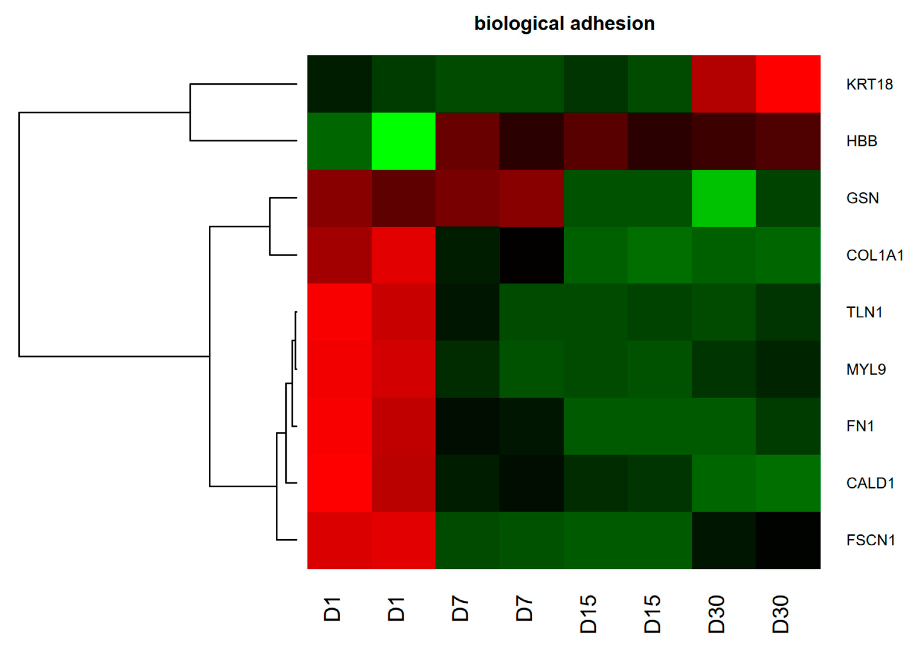
2.5. Genes and Proteins of Interest

From the differently regulated genes, we chose the 130 porcine analogs of genes encoding proteins detected by MALDI-TOF analysis. From this pool, genes belonging to the 20 significantly enriched Gene Ontology Biological Processes (GO BP) terms were provided by the DAVID software. The list of enriched GO BP terms and the amount of differently expressed genes were shown in Figure 4.
In this manuscript, we focused on the genes that belong to “biological adhesion” GO BP. The hierarchical clustering of selected genes was carried out and result was presented as a heatmap (Figure 5).

2.6. RT-qPCR Analysis of Genes of Interest

The results of the microarray analysis were validated using the real-time quantitative polymerase chain reaction (RT-qPCR) method. Most of the patterns of expression changes were consistent between both of the methods. Keratin 18 (KRT18) was an exception, showing opposite direction of change on day seven of primary culture. Furthermore, the scale of the detected alterations was bigger in RT-qPCR than in microarrays. As this pattern is consistent across all of the analyzed genes, it can be assumed that it results from the difference in sensitivity of both methods. All of the data obtained was presented and compared on a bar graph (Figure 6).

2.7. Western Blot Analysis of Proteins of Interest

The results of MALDI-TOF analysis were validated using the Western blot method. It was performed to indicate changes in Collagen Type I Alpha 1 Chain (COL1A1), Fibronectin 1 (FN1), and Talin 1 (TLN1) protein levels during OEC primary long-term in vitro culture (Figure 7) as genes encoding them presented the most substantial changes in RNA expression. The protein analysis showed an increased expression of COL1A1, FN1, and TLN1 proteins in 7, 15, and 30 days of OEC culture by ~300%. However, an increase in FN1 protein level was more pronounced in 15 and 30 days of culture (~500%) as compared to those in 24 h.

3. Discussion

Previous transcriptomic studies on bovine OECs revealed groups of genes exhibiting altered expression dependent on the phase of estrous cycle [22] and specific for either the ampulla or the isthmus [23]. Among others, the most prominent changes in OECs isolated from the ampulla were related to genes associated with cell migration and motility, ciliary movement, and beat frequency. In the isthmus, similar findings indicated genes associated with: nitrogen, lipid, cholesterol, and nucleotide synthesis, cell cycle, vesicle-mediated transport, and apoptosis [24]. In our study, we only discuss genes that were differentially expressed during 30 days of OEC culture, protein products of which were also found in cells on the last day of culture. Presence of protein on the 30th day of culture was not always reflected by the change of its gene expression during the course of cultivation, suggesting the protein has stable expression during culture periods. Selected genes and proteins were found robustly in several ontology groups related to crucial cellular processes. From the group of those belonging to the “cellular adhesion” GO BP, the genes which attracted our attention were Keratin 18 (KRT18), Gelsolin (GSN), Fibronectin 1 (FN1), Collagen Type I Alpha 1 Chain (COL1A1), Talin 1 (TLN1), Fascin Actin Bundling Protein 1 (FASCN1), Myosin Light Chain 1 (MYL9), Hemoglobin Subunit Beta (CALD1), and Hemoglobin Subunit Beta (HBB).
The process of adhesion plays an important role in reproductive events associated with both gametes and embryos. During their maturation, the oocytes form tight gap junctions connections with cumulus cells (CCs), enabling achievement of the MII stage of the female gamete [7]. Additionally, recent studies show that adhesion has a great impact on post-ovulatory journey of the oocyte through the oviduct. It was found in cows that interactions between COCs and OECs start immediately after the COCs’ entry to the oviductal ampulla [8], promoting hyperactivation of sperm bound in the isthmus and its counter-current movement towards ampulla [10]. Specifics of the events facilitating transport of the oocyte, as well as its interactions with the OECs, remain unclear. Nevertheless, in vitro co-cultures of COCs with ampullar and isthmic OECs was found to be beneficial, increasing the time of zona pellucida digestion and monospermic penetration rate [25].
Sperm interactions with the OECs are certainly better described [26]. The mechanism of sperm detention in the caudal isthmus is based on their adhesion to OECs, and is assured by binding of Arg-Gly-Asp amino acid sequence on the spermatozoa with integrins on the OECs [27]. After capacitation and hyperactivation, sperm loses its binding affinity, presumably due to modification of surface proteins and physical disruption of connections [3]. It is assumed that this interaction affects changes in oviductal genes’ expression, mainly related to ciliary motility [28], muscularis contractions [29], intercellular transport, and communication [30]. Moreover, adhesion of male and female gametes to the epithelial cells lining the oviduct promotes alterations in their proteome [31]. Additionally, OECs-provided maintenance of embryonal growth was associated with synthesis of various growth factors [25]. Therefore, it is assumed that OECs are programmed to support fertilization and further embryo development [32].
Keratin 18 is a protein regarded as a marker of epithelial cells. Its high expression is found in mammalian single-layered epithelia, such as single columnar epithelium lining the oviduct. Its main role, together with keratin 8, is the formation of intermediate filaments (IF), creating structural framework of epithelial cells [33]. Although keratins are mainly known for their ability to strengthen and stabilize cells, they can indirectly influence intercellular adhesion. The latter can be facilitated by clusters of desmosomal cadherins, supported by IF-made desmoplakins and plakophilins [34]. However, it is worth keeping in mind that the confluence of cultured cells, and thus number of cell junctions, increases along the culture time [35], which may prove to be an alternative explanation for increased KRT18 expression. In microarray results, an exception was noted on day 30, when KRT18 expression dropped. However, the RT-qPCR validation did not confirm this result. Thus, increased expression of KRT18 and the presence of keratin 18 during the culture may reflect not only in vitro induced changes of the OEC but also their enhanced adhesiveness, possibly promoting interactions with gametes.
The oviduct promotes gamete and early embryo transport [3]. The dominant role in this passage is attributed to action of cilia that cover the simple columnar epithelium lining the oviduct [36]. One of the proteins involved in ciliogenesis is gelsolin. This calcium-regulated protein is responsible for assembly and disassembly of actin filaments. Its ability to bind to “plus” ends of actin monomers and filaments prevents monomer exchange and three-dimensional structure formation. Experiments have shown that small interfering ribonucleic acid (siRNA)-mediated depletion of GSN causes decreased count of ciliated cells, indicating that actin filaments severing plays a role in ciliogenesis [37]. However, it is widely known that cells grown in in vitro monolayers are prone to dedifferentiation and lose their morphological features, including cilia [38]. Thus, we speculate that the increase in GSN expression in our culture was associated with other processes. It is known that Gelsolin is related to many other pathways in mammals, especially adhesion. Analysis of extracellular vesicles isolated from the oviductal fluid (oEVs) revealed transcripts and proteins belonging to few GO terms, including cell adhesion. Gelsolin was one of the members of the group of proteins most abundant across the estrous cycle, with profound impact on sperm–oviduct interaction and early embryonic development [39]. It was found in uterine epithelial cells that actin binding proteins, including Gelsolin, are involved in regulation of endometrial skeleton, as well as in embryo attachment to the epithelium via focal adhesion kinase interaction [40].
Adhesion is a process strictly linked to membrane-associated proteins called integrins, which can bind to neighboring cell membrane or elements of extracellular matrix (ECM) [41]. They are known receptors of an adhesive glycoprotein: fibronectin 1 [42]. So far, reported functions of FN1 in mammalian reproduction are related to sperm-oocyte binding, implantation and placentation, while none of them attributed to the oviductal journey [43,44]. In silico protein interaction studies, related to oocytes maturation, fertilization, and embryo growth in sheep, showed that FN1, together with other proteins, favors cell growth in in vitro conditions [45]. Goossens et al. observed in cattle that FN1 expression level increased from the morula to the blastocyst stage, even before embryo implantation. It raises the possibility that embryonic FN1 matrix gains adhesive properties to keep cells together, as well as to enable following migration of embryonal cell layers [46]. Although the developing zygote travels passively towards the uterus, where maternal recognition of pregnancy takes place, it can exert signals “informing” about its presence and modifying expression of specific genes in oviductal cells. As the result, oviduct secretes factors with highly beneficial influence on embryo development and/or decreases activity of the immune system [47,48,49,50].
Another ECM component, collagen type I alpha 1 chain(COL1A1), is an element of fibril-forming collagen, a crucial component of connective tissue, abundantly present in bone, tendons, and dermis. Its high expression was also found in other tissues and organs, including endometrium and placenta [51,52]. However, there were no reports, until know, about its presence in oviducts. It is possible that the synthesis of FN1 and COL1A1 extracellular proteins can influence the microenvironment of oviducts in some manner. However, results of our experiments, conducted without hormonal supplementation, cannot be referred to changes occurring during the pregnancy and are more likely attributed to increasing confluency of cultured cells.
The involvement of focal adhesion, linking cellular integrins with ECM, has been already noted in reproductive events [40,46,53,54,55]. Talin 1 (TLN1) couples integrins to actin cytoskeleton, playing part in integrin-mediated events [56]. Compact structure of COCs is guaranteed by focal adhesion complexes composed, among others, by TLN1. Kawashima et al. revealed in mice that, in response to LH surge, COCs undergo the process of expansion and CC movement caused by disruption of focal adhesion proteins, such as TLN1 [57]. Subsequently, events triggered by fertilization in mammals are integrin-dependent and thus, indirectly TLN1-dependent. As the result, oocytes become activated through cytoskeletal structures [58]. Furthermore, the forming blastocyst, to complete the implantation process, interacts with uterine epithelial cells through integrins. It was presented in mice that downregulation of TLN1 resulted in impaired adhesion and implantation rate, while its disruption inhibited formation of focal adhesion and subsequently led to embryo death [59]. This outcome emphasizes the crucial role of cytoskeletal proteins and their ligands for proper adhesion and implantation of blastocyst [60]. Lack of information about the role of TLN1 in the mammalian oviduct directed our attention towards epithelial cultures. Experiments showed that different culture conditions and coatings influence TLN1 expression [61,62]. Increased expression of TLN1 in our culture, similarly like FN1 and COL1A1, can reflect high confluency and stable cultivation of porcine OECs, even on the 30th day of in vitro culture. However, we cannot exclude the possibility that these genes exert functions stimulating oviductal environment, as well as benefit gamete and embryo passage.
Apart from the abovementioned genes and their products, we also selected a group with known function in adhesion process but without proven role in reproduction, including: fascin actin bundling protein 1 (FASCN1) [63,64,65,66,67], myosin light chain 1 (MYL9) [68], caldesmon 1 (CALD1) [69], and hemoglobin subunit beta (HBB) [70]. Further examination may explain their yet unrevealed potential in reproduction.
In summary, high expression of some of the adhesion-related genes and proteins can be explained by their role during reproduction in processes such as oocyte or spermatozoa binding to the epithelium lining the oviduct, while for some of them there are no reports describing mentioned relations. OECs in vitro culture is stable and widely used model, that brings some of the physiological features of those cells into light. However, despite numerous advantages, cells cultured ex vivo, deprived of their usual microenvironment, may undergo transcriptomic and proteomic changes that reflect in their phenotype. Thus, direct translation of in vitro culture results to pathways related to reproduction in vivo is not a simple task and requires a large amount of further studies. However, beneficial effect of co-culture of gametes and embryos with OECs has been already proven. Therefore, we can speculate that increased expression of genes and proteins may reflect the extent of physiological properties of the OECs that is maintained during the process of long-term in vitro culture. This in turn can be attributed to intensive secretion of substances with favorable influence on oviductal environment, which prime gamete adhesion and viability, fertilization, and early embryo journey. Hence, functional tests are needed to precisely designate their share in mimicking in vivo conditions.

4. Material and Methods

Data availability: All raw data generated during the research and used to obtain results presented in this manuscript is available from the corresponding author on a reasonable request.

4.1. Tissue Collection

Oviducts were collected from 45 crossbred gilts from a commercial herd. The animals at the age of approximately nine months, which displayed two regular estrous cycles, were slaughtered after reaching the anestrus phase of the cycle. Collected tissues were immediately transported to the laboratory, kept in an isolated container. The research related to animal use has been complied with all the relevant national regulations and institutional policies for the care and use of animals. Bioethical Committee approval no. 32/2012 from 1 June 2012.

4.2. Histological Evaluation

Shortly after the collection of tissues, oviducts from 10 gilts were sectioned to separate the infundibulum, ampulla and isthmus regions. Subsequently, tissues were fixed for 48 h in Bouin’s solution, dehydrated, embedded in paraffin blocks and cut with a rotary microtome into sections of 3–4 µm. Obtained paraffin sections were stained using a hematoxylin and eosin (H + E) staining technique, following a previously described protocol [71]. Analysis of all sections was performed under a light microscope, with pictures taken using high-resolution scanning technique (Olympus BX61VS microscope scanner, Olympus, Tokyo, Japan).

4.3. Primary Long-Term Culture of OECs

The method used for OEC harvesting and culture was based on the available literature sources [72,73]. Collected oviducts were washed twice in Dulbecco’s phosphate buffered saline (PBS) and sectioned longitudinally. OECs were gently scratched with the use of sterile surgical blades and subsequently enzymatically digested for 1 h at 37 °C with 1mg/mL collagenase I (Sigma Aldrich, St. Louis, MO, USA) in Dulbecco’s modified Eagle’s medium (DMEM; Sigma Aldrich). Cell suspension was filtered through 40 µm pore size strainer and centrifuged for 10 min at 200× g. After rinsing with PBS, OECs were incubated for another 10 min at 37 °C with 0.5% Trypsin/EDTA (Sigma Aldrich), filtered and centrifuged as described above, and suspended in DMEM supplemented with 10% FCS, 100 U/mL penicillin, 100 µg/mL streptomycin, and 1 µg/mL amphotericin B. The cells were cultured for up to 30 days at 37 °C, in a humidified atmosphere of 5% CO2. The culture medium was changed every three days. Once OECs reached confluency of around 80%, they were passaged to another culture dish at the seeding density of 2 × 10⁴ cells/cm2. Throughout the time of the culture, pictures of cell morphology were taken using an inverted microscope employing relief phase contrast (Olympus IX73, Olympus).

4.4. RNA Isolation from Oviductal Epithelial Cells (OECs)

OECs were collected, pooled and suspended in TRI Reagent (Sigma Aldrich) after 24 h, 7, 15, and 30 days of culture. Total mRNA was isolated from samples using RNeasy MinElute cleanup Kit (Qiagen, Hilden, Germany). The amount, purity, integrity and quality of total mRNA were determined as described previously [74]. Each mRNA sample was diluted to 100 ng/µL concentration.

4.5. Microarray Expression Analysis and Statistics

The 100 ng of mRNA from each pooled sample was transferred to succeeding microarray procedure described in details in our recent paper [75]. Briefly, primary cDNA was obtained and amplified (Ambion® WT Expression Kit, Ambion Inc., Thermo Fisher Scientific, Waltham, MA, USA). Then, cDNA was labelled with biotin and fragmented (Affymetrix GeneChip® WT Terminal Labeling and Hybridization, Affymetrix, Santa Clara, CA, USA). Resulting fragments were hybridized to the microarray strips (Affymetrix® Porcine Gene 1.1 ST Array Strip, Affymetrix), which were afterwards washed, stained according to the manufacturer’s protocol (Affymetrix GeneAtlas Fluidics Station, Affymetrix), scanned (Imaging Station of the GeneAtlas System, Affymetrix) and analyzed (Affymetrix GeneAtlasTM Operating Software, Affymetrix), resulting in generation of raw CEL files containing all of the data.
Downstream analyses were performed, and graphs were obtained using Bioconductor and R programming languages, as well as the Robust Multiarray Averaging (RMA) algorithm. Statistical significance of analyzed genes was determined with the use of moderated t-statistics from the empirical Bayes method, with obtained p-value corrected for multiple comparisons using Benjamini and Hochberg’s false discovery rate. Genes regarded as differentially expressed presented p-value beneath 0.05 and expression higher than 2-fold.

4.6. Protein Digestion

Cells harvested from culture flasks were centrifuged and rinsed twice with 50mM ammonium bicarbonate solution to remove residues of the culture medium. Subsequently, the sample was suspended in 1 mL of 50% methanol in a solution of 0.1% trifluoroacetic acid (TFA) in water, mixed for 1 min and incubated at −20 °C for 5 min. The procedure of mixing and incubation was repeated three times. Then, the sample was sonicated for 15 min and centrifuged. The supernatant was divided into three tubes and subjected to the protocol for in-solution tryptic digestion, according to modified Pierce Kit procedure (Thermo Scientific, USA). After overnight digestion, samples were purified and concentrated using C18 ZipTip micropipette tips (Millipore, Burlington, MA, USA) according to the protocol provided by the manufacturer.

4.7. nanoLC-MALDI-TOF/TOF MS/MS Analysis

Three purified samples were fractionated using nano-liquid chromatography (nanoLC): EASY-nano LC II connected with Proteineer-fc II fraction collector (Bruker Daltonics, Billerica, MA, USA). Detailed conditions of the performed separation, as well as specifications of LC columns were described in our previous research [76]. 384 fractions were obtained, with each automatically mixed with HCCA matrix solution and spotted onto MALDI target plate (MTP AnchorChip 384, Bruker Daltonics, Billerica, MA, USA). Subsequently, MALDI-TOF/TOF MS/MS analysis was performed using the UltrafleXtreme instrument (Bruker Daltonics, Billerica, MA, USA). Typical settings of the apparatus for MS and MS/MS mode were presented in our previous study [76]. MS spectra were externally calibrated with the use of peptide calibration mixture in the 0.7–3.5 kDa mass range (Bruker Daltonics). WARP-LC 1.3 platform (Bruker Daltonics, Billerica, MA, USA) was used to select precursor ions with signal-to-noise threshold above 5 for MS/MS analysis. The following platforms and software were used for analysis, data acquisition and evaluation: WARP LC 1.3, ProteinScape 3.1, FlexControl 3.4, and FlexAnalysis 3.4. (Bruker Daltonics, Billerica, MA, USA).
Protein search was conducted using the National Center for Biotechnology Information (NCBI) database with Mascot 2.4.1 search engine (Matrix Science, London, UK) using the following parameters: trypsin digestion, 1 missed cleavage, peptide mass tolerance: 25 ppm; MS/MS fragment mass tolerance: 0.7 Da; peptide charge: 1+; monoisotopic mass; and carbamidomethylation of cysteine as fixed modification. The searches were restricted to “Other mammals”. Additional searches were made including “Homo sapiens” and “Rodentia” datasets. Only proteins with at least two unique peptides, score above 80 and false discovery rate below 1% were included. Results obtained from three analysis were combined into one list of the proteins generated by the ProteinScape software.

4.8. Selection of Genes of Interest

Since not every gene encodes a protein, changes in gene expression are not always correlated with protein changes. To select differentially expressed genes accompanied by their protein products, NCBI eFetch tool was used to match obtained genes with their corresponding protein products detected in MALDI-TOF MS analysis. Subsequently, pig analogues of these genes were found, with only the differentially expressed genes from these pairs used in following analysis.
Selected genes were analyzed according to an already published sequence [77]. Briefly, the genes were uploaded to the Database for Annotation, Visualization, and Integrated Discovery software (DAVID, Laboratory of Immunopathogenesis and Bioinformatics, Frederick, MD, USA) [78] to extract enriched GO BPs. Only GO BPs containing at least five genes and with a p-value lower than 0.05 were chosen.

4.9. RT-qPCR Validation

The relative changes in gene expression results obtained from the microarray analysis was validated with the use of real-time quantitative polymerase chain reaction (RT-qPCR). Primers specific to genes of interest were designed using the Primer3 software. Exon-exon design method was used as an additional measure to avoid amplification of genomic DNA fragments. The primers were also designed to allow amplification of all of the transcription variants of genes of interest found in the Ensembl database. The sequences of primers used were presented in Table 1. The reaction mix included cDNA samples, forward and reverse primers, and SYBR Green I as a detection dye. Negative controls were processes with primers replaced with highly purified water to ensure lack of non-specific amplification. HPRT, ACTB, and GAPDH were used as housekeeping genes. Water samples were also included as blanks, to ensure its purity and eliminate background noise. The reactions were processed, each in three technical repetitions, in the LightCycler 96 Real-Time PCR system (Roche Diagnostics, Mannheim, Germany). The specificity of amplification was estimated based on the dissociation curves provided by the software provided by the equipment manufacturer. The transcript levels, as well as the fold changes in expression were determined using the 2^-ΔΔCT method.

4.10. Sodium Dodecyl Sulphate-Polyacrylamide Gel Electrophoresis (SDS-PAGE) and Western Blot Analysis

The expression levels of several proteins of interest were validated using Western blot analysis. Three proteins were selected based on the most substantial changes in their RNA expression. Total protein of OECs was isolated with RIPA lysis buffer. A total of 15 µg protein was re-suspended in sample buffer and separated on 8% Tris-glycine gel with the aid of SDS-PAGE. The proteins were transferred to PVDF membrane, which was blocked with 5% milk in Tris buffered saline/Tween. Immunodetection was performed with rabbit polyclonal anti-COL1A1 Ab (ab34710), rabbit polyclonal anti-Fibronectin 1 (FN1) Ab (ab2413), and rabbit polyclonal anti-Talin 1 (TLN1) Ab (ab71333) followed by incubation with goat anti-rabbit HRP Ab (ab205718). The membranes were also incubated with anti-actin (ACTB) HRP Ab (sc-1616) to equalize protein loading of the lanes. SuperSignal West Femto maximum sensitivity substrate Pierce Biotechnology Inc. (Rockford, IL, USA) was used to reveal bands.

Supplementary Materials

The following are available online at https://www.mdpi.com/1422-0067/20/14/3387/s1, Table S1. The list of 389 proteins identified by the nanoLC-MALDI-TOF/TOF MS/MS analysis. Table S2. The list of 220 proteins identified by the nanoLC-MALDI-TOF/TOF MS/MS analysis using “Homo sapiens” dataset. Table S3. The list of 225 proteins identified by the nanoLC-MALDI-TOF/TOF MS/MS analysis using “Rodentia” dataset.

Author Contributions

Conceptualization, J.B.-T. and B.K.; Methodology, J.B.-T. and P.S.-K.; Software, P.C.; Validation, J.B.-T., and A.Ś.-B.; Formal Analysis, J.B.-T., B.K., J.M., and A.K.; Investigation, A.Ś.-B., S.K., M.J. (Małgorzata Józkowiak), and M.K.; Resources, P.A. and D.B.; Data Curation, P.C., M.J. (Michal Jeseta), and H.P.-K.; Writing—Original Draft Preparation, J.B.-T., A.Ś.-B., and M.J. (Maurycy Jankowsk); Writing—Review and Editing, J.B.-T. and M.J. (Maurycy Jankowsk); Visualization, P.C., A.K., and P.S.-K.; Supervision, M.T.S. and J.M.; Project Administration, M.N. and B.K.; Funding Acquisition, M.T.S. and B.K.

Funding

This research was funded by Polish National Science Centre, grant number UMO-2016/21/B/NZ9/03535.

Conflicts of Interest

The authors declare no conflict of interest.

References

  1. Abe, H. The mammalian oviductal epithelium: Regional variations in cytological and functional aspects of the oviductal secretory cells. Histol. Histopathol. 1996, 11, 743–768. [Google Scholar] [PubMed]
  2. Buhi, W.C.; Alvarez, I.M.; Kouba, A.J. Secreted proteins of the oviduct. Cells Tissues Organs 2000, 166, 165–179. [Google Scholar] [CrossRef] [PubMed]
  3. Lyons, R.A.; Saridogan, E.; Djahanbakhch, O. The reproductive significance of human Fallopian tube cilia. Hum. Reprod. Update 2006, 12, 363–372. [Google Scholar] [CrossRef] [PubMed]
  4. Cal, S.; Freije, J.M.P.; López, J.M.; Takada, Y.; López-Otín, C. ADAM 23/MDC3, a Human disintegrin That promotes cell adhesion via interaction with the αvβ3 Integrin through an RGD-independent Mechanism. Mol. Biol. Cell 2000, 11, 1457–1469. [Google Scholar] [CrossRef] [PubMed]
  5. Tsanou, E.; Peschos, D.; Batistatou, A.; Charalabopoulos, A.; Charalabopoulos, K. The E-cadherin adhesion molecule and colorectal cancer. A global literature approach. Anticancer Res. 2008, 28, 3815–3826. [Google Scholar] [PubMed]
  6. Wang, X.; Tully, O.; Ngo, B.; Zitin, M.; Mullin, J.M. Epithelial tight junctional changes in colorectal cancer tissues. Sci. World J. 2011, 11, 826–841. [Google Scholar] [CrossRef] [PubMed]
  7. Kempisty, B.; Ziółkowska, A.; Ciesiółka, S.; Piotrowska, H.; Antosik, P.; Bukowska, D.; Nowicki, M.; Brussow, K.P.; Zabel, M. Study on connexin gene and protein expression and cellular distribution in relation to real-time proliferation of porcine granulosa cells. J. Biol. Regul. Homeost. Agents 2014, 28, 625–635. [Google Scholar] [PubMed]
  8. Kölle, S.; Dubielzig, S.; Reese, S.; Wehrend, A.; König, P.; Kummer, W. Ciliary transport, gamete interaction, and effects of the early embryo in the oviduct: Ex vivo analyses using a new digital videomicroscopic system in the cow. Biol. Reprod. 2009, 81, 267–274. [Google Scholar] [CrossRef] [PubMed]
  9. Coy, P.; García-Vázquez, F.A.; Visconti, P.E.; Avilés, M. Roles of the oviduct in mammalian fertilization. Reproduction 2012, 144, 649–660. [Google Scholar] [CrossRef] [PubMed] [Green Version]
  10. Maillo, V.; Sánchez-Calabuig, M.J.; Lopera-Vasquez, R.; Hamdi, M.; Gutierrez-Adan, A.; Lonergan, P.; Rizos, D. Oviductal response to gametes and early embryos in mammals. Reproduction 2016, 152, R127–R141. [Google Scholar] [CrossRef] [PubMed] [Green Version]
  11. Tokuhiro, K.; Ikawa, M.; Benham, A.M.; Okabe, M. Protein disulfide isomerase homolog PDILT is required for quality control of sperm membrane protein ADAM3 and male fertility [corrected]. Proc. Natl. Acad. Sci. USA 2012, 109, 3850–3855. [Google Scholar] [CrossRef] [PubMed]
  12. Caballero, J.N.; Gervasi, M.G.; Veiga, M.F.; Dalvit, G.C.; Perez-Martínez, S.; Cetica, P.D.; Vazquez-Levin, M.H. Epithelial cadherin is present in bovine oviduct epithelial cells and gametes, and is involved in fertilization-related events. Theriogenology 2014, 81, 1189–1206. [Google Scholar] [CrossRef] [PubMed]
  13. Gonella-Diaza, A.M.; Andrade, S.C.d.S.; Sponchiado, M.; Pugliesi, G.; Mesquita, F.S.; Van Hoeck, V.; de Francisco, R.S.; Gasparin, G.R.; Coutinho, L.L.; Binelli, M. Size of the ovulatory follicle dictates spatial differences in the oviductal transcriptome in cattle. PLoS ONE 2015, 10, e0145321. [Google Scholar] [CrossRef] [PubMed]
  14. Weber, J.A.; Freeman, D.A.; Vanderwall, D.K.; Woods, G.L. Prostaglandin E2 secretion by oviductal transport-stage equine embryos. Biol. Reprod. 1991, 45, 540–543. [Google Scholar] [CrossRef] [PubMed]
  15. Smits, K.; De Coninck, D.I.M.; Van Nieuwerburgh, F.; Govaere, J.; Van Poucke, M.; Peelman, L.; Deforce, D.; Van Soom, A. The equine embryo influences immune-related gene expression in the oviduct. Biol. Reprod. 2016, 94, 36. [Google Scholar] [CrossRef] [PubMed]
  16. Hugentobler, S.A.; Diskin, M.G.; Leese, H.J.; Humpherson, P.G.; Watson, T.; Sreenan, J.M.; Morris, D.G. Amino acids in oviduct and uterine fluid and blood plasma during the estrous cycle in the bovine. Mol. Reprod. Dev. 2007, 74, 445–454. [Google Scholar] [CrossRef]
  17. Hugentobler, S.A.; Morris, D.G.; Sreenan, J.M.; Diskin, M.G. Ion concentrations in oviduct and uterine fluid and blood serum during the estrous cycle in the bovine. Theriogenology 2007, 68, 538–548. [Google Scholar] [CrossRef]
  18. Hugentobler, S.A.; Humpherson, P.G.; Leese, H.J.; Sreenan, J.M.; Morris, D.G. Energy substrates in bovine oviduct and uterine fluid and blood plasma during the oestrous cycle. Mol. Reprod. Dev. 2008, 75, 496–503. [Google Scholar] [CrossRef]
  19. Lee, S.H.; Oh, H.J.; Kim, M.J.; Kim, G.A.; Choi, Y.B.; Jo, Y.K.; Nugraha, S.M.E.; Lee, C.B. Oocyte maturation-related gene expression in the canine oviduct, cumulus cells, and oocytes and effect of co-culture with oviduct cells on in vitro maturation of oocytes. J. Assist. Reprod. Genet. 2017, 34, 929–938. [Google Scholar] [CrossRef]
  20. Lee, S.H.; Oh, H.J.; Kim, M.J.; Setyawan, E.; Maha, N.; Lee, B.C. Interaction of the EGFR signaling pathway with porcine cumulus oocyte complexes and oviduct cells in a coculture system. J. Cell Physiol. 2018, 234, 4030–4043. [Google Scholar] [CrossRef]
  21. Kervancioglu, M.E.; Saridogan, E.; Atasü, T.; Camlibel, T.; Demircan, A.; Sarikamis, B.; Djahanbakhch, O. Human Fallopian tube epithelial cell co-culture increases fertilization rates in male factor infertility but not in tubal or unexplained infertility. Hum. Reprod. 1997, 12, 1253–1258. [Google Scholar] [CrossRef] [PubMed]
  22. Bauersachs, S.; Rehfeld, S.; Ulbrich, S.E.; Mallok, S.; Prelle, K.; Wenigerkind, H.; Einspanier, R.; Blum, H.; Wolf, E. Monitoring gene expression changes in bovine oviduct epithelial cells during the oestrous cycle. J. Mol. Endocrinol. 2004, 32, 449–466. [Google Scholar] [CrossRef] [PubMed]
  23. Cerny, K.L.; Garrett, E.; Walton, A.J.; Anderson, L.H.; Bridges, P.J. A transcriptomal analysis of bovine oviductal epithelial cells collected during the follicular phase versus the luteal phase of the estrous cycle. Reprod. Biol. Endocrinol. 2015, 13, 84. [Google Scholar] [CrossRef] [PubMed]
  24. Maillo, V.; de Frutos, C.; O’Gaora, P.; Forde, N.; Burns, G.W.; Spencer, T.E.; Gutierez, A.A.; Lonergan, P.; Rizos, D. Spatial differences in gene expression in the bovine oviduct. Reproduction 2016, 152, 37–46. [Google Scholar] [CrossRef] [PubMed] [Green Version]
  25. Gandolfi, F. Functions of proteins secreted by oviduct epithelial cells. Microsc. Res. Tech. 1995, 32, 1–12. [Google Scholar] [CrossRef] [PubMed]
  26. Miller, D.J. Regulation of sperm function by oviduct fluid and the epithelium: Insight into the role of glycans. Reprod. Domest. Anim. 2015, 50 (Suppl. 2), 31–39. [Google Scholar] [CrossRef]
  27. Reeve, L.; Ledger, W.L.; Pacey, A.A. Does the Arg-Gly-Asp (RGD) adhesion sequence play a role in mediating sperm interaction with the human endosalpinx? Hum. Reprod. 2003, 18, 1461–1468. [Google Scholar] [CrossRef] [Green Version]
  28. Li, H.W.R.; Liao, S.B.; Chiu, P.C.N.; Tam, W.W.; Ho, J.C.; Ng, E.H.Y.; Ho, P.C.; Yeung, W.S.B.; Tang, F.; Sum, O.W. Expression of adrenomedullin in human oviduct, its regulation by the hormonal cycle and contact with spermatozoa, and its effect on ciliary beat frequency of the oviductal epithelium. J. Clin. Endocrinol. Metab. 2010, 95, E18–E25. [Google Scholar] [CrossRef] [PubMed]
  29. Fazeli, A.; Affara, N.A.; Hubank, M.; Holt, W.V. Sperm-induced modification of the oviductal gene expression profile after natural insemination in mice. Biol. Reprod. 2004, 71, 60–65. [Google Scholar] [CrossRef] [PubMed]
  30. López-Úbeda, R.; García-Vázquez, F.A.; Romar, R.; Gadea, J.; Muñoz, M.; Hunter, R.H.F.; Coy, P. Oviductal transcriptome is modified after insemination during spontaneous ovulation in the sow. PLoS ONE 2015, 10, e0130128. [Google Scholar] [CrossRef]
  31. Georgiou, A.S.; Snijders, A.P.L.; Sostaric, E.; Aflatoonian, R.; Vazquez, J.L.; Vazquez, J.M.; Roca, J.; Martinez, E.A.; Wright, P.C.; Fazeli, A. Modulation of the oviductal environment by gametes. J. Proteome Res. 2007, 6, 4656–4666. [Google Scholar] [CrossRef] [PubMed]
  32. Dadashpour Davachi, N.; Zare Shahneh, A.; Kohram, H.; Zhandi, M.; Shamsi, H.; Hajiyavand, A.M.; Saadat, M. Differential influence of ampullary and isthmic derived epithelial cells on zona pellucida hardening and in vitro fertilization in ovine. Reprod. Biol. 2016, 16, 61–69. [Google Scholar] [CrossRef] [PubMed] [Green Version]
  33. Nishimura, R.; Kato, K.; Fujiwara, S.; Ohashi, K.; Mizuno, K. Solo and keratin filaments regulate epithelial tubule morphology. Cell Struct. Funct. 2018, 43, 95–105. [Google Scholar] [CrossRef] [PubMed]
  34. Roxas, J.L.; Vedantam, G.; Viswanathan, V.K. Epithelial maturity influences EPEC-induced desmosomal alterations. Gut Microb. 2018, 10, 1–5. [Google Scholar] [CrossRef]
  35. Schlegel, N.; Meir, M.; Heupel, W.-M.; Holthöfer, B.; Leube, R.E.; Waschke, J. Desmoglein 2-mediated adhesion is required for intestinal epithelial barrier integrity. Am. J. Physiol. Gastrointest. Liver Physiol. 2010, 298, G774–G783. [Google Scholar] [CrossRef] [PubMed] [Green Version]
  36. Jansen, R.P. Endocrine response in the fallopian tube. Endocr. Rev. 1984, 5, 525–551. [Google Scholar] [CrossRef] [PubMed]
  37. Kim, J.; Lee, J.E.; Heynen-Genel, S.; Suyama, E.; Ono, K.; Lee, K.; Ideker, T.; Blanc, P.A.; Gleeson, G.K. Functional genomic screen for modulators of ciliogenesis and cilium length. Nature 2010, 464, 1048–1051. [Google Scholar] [CrossRef] [PubMed] [Green Version]
  38. Ulbrich, S.E.; Zitta, K.; Hiendleder, S.; Wolf, E. In vitro systems for intercepting early embryo-maternal cross-talk in the bovine oviduct. Theriogenology 2010, 73, 802–816. [Google Scholar] [CrossRef] [PubMed]
  39. Almiñana, C.; Tsikis, G.; Labas, V.; Uzbekov, R.; da Silveira, J.C.; Bauersachs, S.; Mermillod, P. Deciphering the oviductal extracellular vesicles content across the estrous cycle: Implications for the gametes-oviduct interactions and the environment of the potential embryo. BMC Genom. 2018, 19, 622. [Google Scholar] [CrossRef] [PubMed]
  40. Jalali, B.M.; Likszo, P.; Andronowska, A.; Skarzynski, D.J. Alterations in the distribution of actin and its binding proteins in the porcine endometrium during early pregnancy: Possible role in epithelial remodeling and embryo adhesion. Theriogenology 2018, 116, 17–27. [Google Scholar] [CrossRef]
  41. Schwartz, M.A. Integrins and extracellular matrix in mechanotransduction. Cold Spring Harb. Perspect. Biol. 2010, 2, a005066. [Google Scholar] [CrossRef] [PubMed]
  42. Johansson, S.; Svineng, G.; Wennerberg, K.; Armulik, A.; Lohikangas, L. Fibronectin-integrin interactions. Front. Biosci. 1997, 2, d126–d146. [Google Scholar] [CrossRef] [PubMed]
  43. Bronson, R.A.; Fusi, F.M. Integrins and human reproduction. Mol. Hum. Reprod. 1996, 2, 153–168. [Google Scholar] [CrossRef] [PubMed] [Green Version]
  44. Wang, J.; Armant, D.R. Integrin-mediated adhesion and signaling during blastocyst implantation. Cells Tissues Organs 2002, 172, 190–201. [Google Scholar] [CrossRef] [PubMed]
  45. Sreenivas, D.; Kaladhar, D.S.; Samy, A.P.; Kumar, R.S. Understanding mechanism of in vitro maturation, fertilization and culture of sheep embryoes through in silico analysis. Bioinformation 2012, 8, 1030–1034. [Google Scholar] [CrossRef] [PubMed] [Green Version]
  46. Goossens, K.; Van Soom, A.; Van Zeveren, A.; Favoreel, H.; Peelman, L.J. Quantification of fibronectin 1 (FN1) splice variants, including two novel ones, and analysis of integrins as candidate FN1 receptors in bovine preimplantation embryos. BMC Dev. Biol. 2009, 9, 1. [Google Scholar] [CrossRef] [PubMed]
  47. Lee, K.-F.; Yao, Y.-Q.; Kwok, K.-L.; Xu, J.-S.; Yeung, W.S.B. Early developing embryos affect the gene expression patterns in the mouse oviduct. Biochem. Biophys. Res. Commun. 2002, 292, 564–570. [Google Scholar] [CrossRef]
  48. Chang, H.-S.; Cheng, W.T.K.; Wu, H.-K.; Choo, K.-B. Identification of genes expressed in the epithelium of porcine oviduct containing early embryos at various stages of development. Mol. Reprod. Dev. 2000, 56, 331–335. [Google Scholar] [CrossRef]
  49. Almiñana, C.; Heath, P.R.; Wilkinson, S.; Sanchez-Osorio, J.; Cuello, C.; Parrilla, I.; Gil, M.A.; Vazquez, J.L.; Vazquez, J.M.; Roca, J.; et al. Early developing pig embryos mediate their own environment in the maternal tract. PLoS ONE 2012, 7, e33625. [Google Scholar] [CrossRef]
  50. Lee, K.-F.; Yeung, W.S.B. Gamete/embryo—Oviduct interactions: Implications on in vitro culture. Hum. Fertil. 2006, 9, 137–143. [Google Scholar] [CrossRef]
  51. Szóstek-Mioduchowska, A.Z.; Lukasik, K.; Skarzynski, D.J.; Okuda, K. Effect of transforming growth factor -β1 on α-smooth muscle actin and collagen expression in equine endometrial fibroblasts. Theriogenology 2019, 124, 9–17. [Google Scholar] [CrossRef] [PubMed]
  52. Arai, K.Y.; Nishiyama, T. Developmental changes in extracellular matrix messenger RNAs in the mouse placenta during the second half of pregnancy: Possible factors involved in the regulation of placental extracellular matrix expression. Biol. Reprod. 2007, 77, 923–933. [Google Scholar] [CrossRef] [PubMed]
  53. Kitasaka, H.; Kawai, T.; Hoque, S.A.M.; Umehara, T.; Fujita, Y.; Shimada, M. Inductions of granulosa cell luteinization and cumulus expansion are dependent on the fibronectin-integrin pathway during ovulation process in mice. PLoS ONE 2018, 13, e0192458. [Google Scholar] [CrossRef] [PubMed]
  54. Chan, D.; Thomas, C.J.; Taylor, V.J.; Burke, R.D. Integrins on eggs: focal adhesion kinase is activated at fertilization, forms a complex with integrins, and is necessary for cortex formation and cell cycle initiation. Mol. Biol. Cell. 2013, 24, 3472–3481. [Google Scholar] [CrossRef] [PubMed]
  55. Kaczynski, P.; Baryla, M.; Goryszewska, E.; Bauersachs, S.; Waclawik, A. Prostaglandin F2α promotes embryo implantation and development in the pig. Reproduction 2018, 156, 405–419. [Google Scholar] [CrossRef] [PubMed]
  56. Critchley, D.R.; Gingras, A.R. Talin at a glance. J. Cell Sci. 2008, 121, 1345–1347. [Google Scholar] [CrossRef] [PubMed] [Green Version]
  57. Kawashima, I.; Liu, Z.; Mullany, L.K.; Mihara, T.; Richards, J.S.; Shimada, M. EGF-like factors induce expansion of the cumulus cell-oocyte complexes by activating calpain-mediated cell movement. Endocrinology 2012, 153, 3949–3959. [Google Scholar] [CrossRef] [PubMed]
  58. De Nadai, C.; Fenichel, P.; Donzeau, M.; Epel, D.; Ciapa, B. Characterisation and role of integrins during gametic interaction and egg activation. Zygote 1996, 4, 31–40. [Google Scholar] [CrossRef] [PubMed]
  59. Priddle, H.; Hemmings, L.; Monkley, S.; Woods, A.; Patel, B.; Sutton, D.; Dunn, G.A.; Zicha, D.; Critchley, D.R. Disruption of the talin gene compromises focal adhesion assembly in undifferentiated but not differentiated embryonic stem cells. J. Cell Biol. 1998, 142, 1121–1133. [Google Scholar] [CrossRef] [PubMed]
  60. Zhang, S.; Yu, L.; Qu, T.; Hu, Y.; Yuan, D.; Zhang, S.; Xu, Q.; Zhao, Y.-B.; Zhang, J.-H.; Yue, L.-M. The changes of cytoskeletal proteins induced by the fast effect of estrogen in mouse blastocysts and its roles in implantation. Reprod. Sci. 2017, 24, 1639–1646. [Google Scholar] [CrossRef] [PubMed]
  61. Semich, R.; Robenek, H. Organization of the cytoskeleton and the focal contacts of bovine aortic endothelial cells cultured on type I and III collagen. J. Histochem. Cytochem. 1990, 38, 59–67. [Google Scholar] [CrossRef] [PubMed]
  62. Bacáková, L.; Mares, V.; Lisá, V.; Svorcík, V. Molecular mechanisms of improved adhesion and growth of an endothelial cell line cultured on polystyrene implanted with fluorine ions. Biomaterials 2000, 21, 1173–1179. [Google Scholar] [CrossRef]
  63. Bi, J.; Zhu, Y.; Chen, X.; Yu, M.; Zhang, Y.; Li, B.; Sun, J.; Shen, H.; Kong, C. The role of fascin in migration and invasion of urothelial carcinoma of the bladder. Urol. Int. 2013, 91, 227–235. [Google Scholar] [CrossRef] [PubMed]
  64. Shen, T.-Y.; Mei, L.-L.; Qiu, Y.-T.; Shi, Z.-Z. Identification of candidate target genes of genomic aberrations in esophageal squamous cell carcinoma. Oncol. Lett. 2016, 12, 2956–2961. [Google Scholar] [CrossRef] [PubMed] [Green Version]
  65. Adams, J.C. Fascin-1 as a biomarker and prospective therapeutic target in colorectal cancer. Expert Rev. Mol. Diagn. 2015, 15, 41–48. [Google Scholar] [CrossRef]
  66. Wang, C.; Huang, B.; Wu, Z.; Sun, X.; Zeng, Y.; Wang, Y. Expression of Fascin-1 protein in breast cancer and its clinicopathologic correlation. Zhonghua Bing Li Xue Za Zhi 2014, 43, 451–454. [Google Scholar]
  67. Stewart, C.J.R.; Crook, M.; Loi, S. Fascin expression in endocervical neoplasia: Correlation with tumour morphology and growth pattern. J. Clin. Pathol. 2012, 65, 213–217. [Google Scholar] [CrossRef]
  68. Park, I.; Han, C.; Jin, S.; Lee, B.; Choi, H.; Kwon, J.T.; Kim, D.; Kim, J.; Lifirsu, E.; Park, J.W.; et al. Myosin regulatory light chains are required to maintain the stability of myosin II and cellular integrity. Biochem. J. 2011, 434, 171–180. [Google Scholar] [CrossRef] [Green Version]
  69. Cuomo, M.E.; Knebel, A.; Platt, G.; Morrice, N.; Cohen, P.; Mittnacht, S. Regulation of microfilament organization by kaposi sarcoma-associated herpes virus-cyclin·CDK6 phosphorylation of caldesmon. J. Biol. Chem. 2005, 280, 35844–35858. [Google Scholar] [CrossRef]
  70. Cao, A.; Galanello, R. Beta-thalassemia. Genet. Med. 2010, 12, 61–76. [Google Scholar] [CrossRef] [Green Version]
  71. Chermuła, B.; Brązert, M.; Jeseta, M.; Ożegowska, K.; Sujka-Kordowska, P.; Konwerska, A.; Bryja, A.; Kranc, W.; Jankowski, M.; Nawrocki, M.J.; et al. The unique mechanisms of cellular proliferation, migration and apoptosis are regulated through oocyte maturational development—A complete transcriptomic and histochemical study. Int. J. Mol. Sci. 2018, 20, 84. [Google Scholar] [CrossRef]
  72. Aldarmahi, A. Establishment and characterization of female reproductive tract epithelial cell culture. J. Microsc. Ultrastruct. 2017, 5, 105–110. [Google Scholar] [CrossRef] [PubMed] [Green Version]
  73. Kidson, A.; Schoevers, E.; Langendijk, P.; Verheijden, J.; Colenbrander, B.; Bevers, M. The effect of oviductal epithelial cell co-culture during in vitro maturation on sow oocyte morphology, fertilization and embryo development. Theriogenology 2003, 59, 1889–1903. [Google Scholar] [CrossRef]
  74. Kranc, W.; Jankowski, M.; Budna, J.; Celichowski, P.; Khozmi, R.; Bryja, A.; Borys, S.; Dyszkiewicz-Konwinska, M.; Jeseta, M.; Magas, M.; et al. Amino acids metabolism and degradation is regulated during porcine oviductal epithelial cells (OECs) primary culture in vitro—A signaling pathways activation approach. Med. J. Cell. Biol. 2018, 6, 18–26. [Google Scholar] [CrossRef]
  75. Kranc, W.; Brązert, M.; Ożegowska, K.; Budna-Tukan, J.; Celichowski, P.; Jankowski, M.; Bryja, A.; Nawrocki, M.J.; Popis, M.; Jeseta, M.; et al. Response to abiotic and organic substances stimulation belongs to ontologic groups significantly up-regulated in porcine immature oocytes. Med. J. Cell. Biol. 2018, 6, 91–100. [Google Scholar] [CrossRef] [Green Version]
  76. Matysiak, J.; Hajduk, J.; Mayer, F.; Hebeler, R.; Kokot, Z.J. Hyphenated LC–MALDI–ToF/ToF and LC–ESI–QToF approach in proteomic characterization of honeybee venom. J. Pharm. Biomed. Anal. 2016, 121, 69–76. [Google Scholar] [CrossRef] [PubMed]
  77. Bryja, A.; Dyszkiewicz-Konwińska, M.; Jankowski, M.; Celichowski, P.; Stefańska, K.; Chamier-Gliszczyńska, A.; Popis, M.; Mehr, K.; Bukowska, D.; Antosik, P.; et al. Ion homeostasis and transport are regulated by genes differentially expressed in porcine buccal pouch mucosal cells during long-term culture in vitro-a microarray approach. Bryja Al Med. J. Cell. Biol. 2018. [Google Scholar] [CrossRef]
  78. Huang, D.W.; Sherman, B.T.; Tan, Q.; Collins, J.R.; Alvord, W.G.; Roayaei, J.; Stephens, R.; Baseler, W.M.; Clifford, H.L.; Lempicki, R.A. The DAVID Gene functional classification tool: A novel biological module-centric algorithm to functionally analyze large gene lists. Genome Biol. 2007, 8, R183. [Google Scholar] [CrossRef] [PubMed]
Figure 1. Representative sections of porcine oviducts stained with hematoxylin and eosin. (A) the infundibulum, (B) the ampulla, (C) the isthmus, (DF) epithelium lining the mucosal folds, in the infundibulum, the ampulla and the isthmus, respectively. Arrows: 1: mucosal folds, 2: muscle layer, 3: serosa, and 4: simple columnar ciliated epithelium.
Ijms 20 03387 g001
Figure 2. The representative images of oviductal epithelial cells (OECs) primary long-term in vitro culture taken after 24 h, 7, 15, and 30 days of culture using an inverted microscope with relief phase contrast.
Ijms 20 03387 g002
Figure 3. The representative example of the mass spectrum of m/z 1417.6834, identified as one of the fascin peptides.
Ijms 20 03387 g003
Figure 4. The plot showing the list of enriched gene ontology biological processes (GO BP) terms. The size of the circle corresponds to the number of genes. The color intensity indicates the fold enrichment of the terms.
Ijms 20 03387 g004
Figure 5. Heatmap representation of differentially expressed genes belonging to “biological adhesion” GO BP term. Microarray signal intensity values for each gene were adapted to Row Z-Score scale ranging from −2 (the lowest expression) to +2 (the highest expression) and reflected by red and green colors, respectively.
Ijms 20 03387 g005
Figure 6. The results of the microarray and real-time quantitative polymerase chain reaction (RT-qPCR) analyses presented in a form of a bar graph.
Ijms 20 03387 g006
Figure 7. The changes in Collagen Type I Alpha 1 Chain (COL1A1), Fibronectin 1 (FN1), Talin 1 (TLN1), and Actin Beta (ACTB) proteins expression in 24 h, 7, 15, and 30 days of OEC culture.
Ijms 20 03387 g007
Table 1. Sequences of primers used for the real-time quantitative polymerase chain reaction (RT-qPCR). analysis.
NameForward Primer (5’–3’)Reverse Primer (5’–3’)
FN1TGAGCCTGAAGAGACCTGCTCAGCTCCAATGCAGGTACAG
KRT18GGGCTCAGATCTTTGCAAGTGTCTCATACTTGACTCTGAAGTCATC
GSNAGAAACAGATCTGGAGAATCGAACGCCCTGCCAGTTGTAGAT
COL1A1GGTCTCCCAGGTCCTAAGGGCTAGGACCAGTTTCACCCT
TLN1AGGCCAGAAAGAGTGTGACACCGTTCTTGGCATTTTGGGA
FSCN1CGCAGGTCAACATCTACAGCCTCGAGAGTGTAGCCTGTGG
MYL9GGCCTTCAACATGATCGACCGCTTCTCCCCAAACATGGTG
CALD1CACAAGCTCAAACACACCGATCAGCTCCTCCAGTTCCAAG
HBBGTGACGGCCTGAAACATCTCCTGGCCCACAAGTACCACTA

