Structure Determination by Single-Particle Cryo-Electron Microscopy: Only the Sky (and Intrinsic Disorder) is the Limit


Abstract
:1. Introduction: Brief Historic Overview of Methods of Structural Biology
2. Cryo-EM: Single-Particle Analysis (SPA)
2.1. Specimen Preparation for Single Particle Cryo-EM Analysis
2.2. Data Acquisition and Image Processing
2.3. 3D-Reconstruction and Structure Validation
3. Single-Particle Cryo-EM Analysis of Membrane Proteins
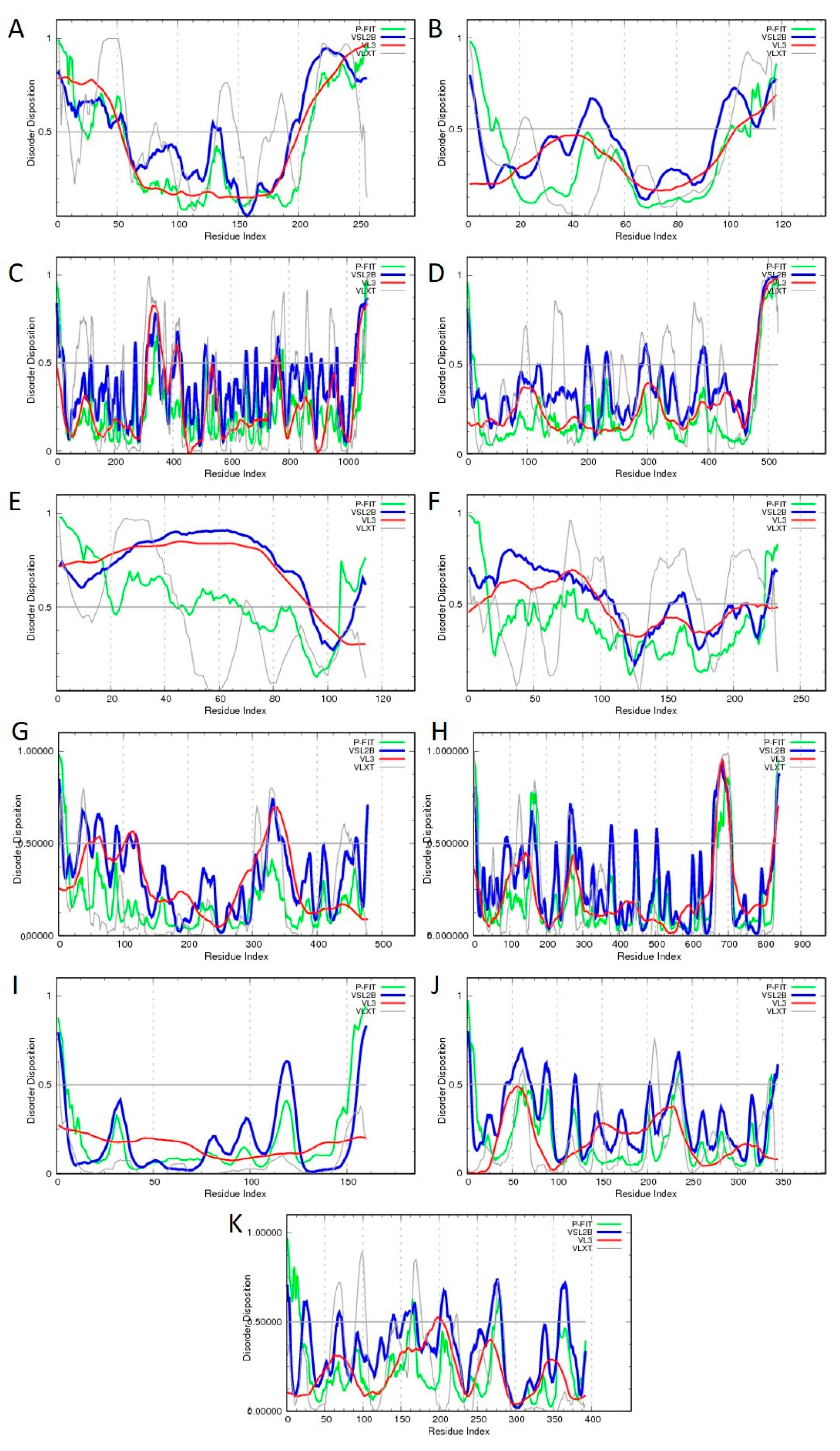
3.1. Ryanodine Receptor (RyR) Channels
3.2. ATPases/V-ATPases
3.3. Transient Receptor Potential (TRP) Channels
3.4. Potassium Channels
4. Conclusions
Author Contributions
Funding
Conflicts of Interest
References
- Egelman, E.H. The current revolution in cryo-em. Biophys. J. 2016, 110, 1008–1012. [Google Scholar] [CrossRef] [PubMed]
- Babu, M.M. The contribution of intrinsically disordered regions to protein function, cellular complexity, and human disease. Biochem. Soc. Trans. 2016, 44, 1185–1200. [Google Scholar] [CrossRef] [PubMed] [Green Version]
- Uversky, V.N. P53 proteoforms and intrinsic disorder: An illustration of the protein structure-function continuum concept. Int. J. Mol. Sci. 2016, 17, 1874. [Google Scholar] [CrossRef] [PubMed]
- DeForte, S.; Uversky, V.N. Order, disorder, and everything in between. Molecules 2016, 21, 1090. [Google Scholar] [CrossRef] [PubMed]
- Uversky, V.N. Dancing protein clouds: The strange biology and chaotic physics of intrinsically disordered proteins. J. Biol. Chem. 2016, 291, 6681–6688. [Google Scholar] [CrossRef] [PubMed]
- Uversky, V.N. Unusual biophysics of intrinsically disordered proteins. Biochim. Biophys. Acta 2013, 1834, 932–951. [Google Scholar] [CrossRef] [PubMed]
- van der Lee, R.; Buljan, M.; Lang, B.; Weatheritt, R.J.; Daughdrill, G.W.; Dunker, A.K.; Fuxreiter, M.; Gough, J.; Gsponer, J.; Jones, D.T.; et al. Classification of intrinsically disordered regions and proteins. Chem. Rev. 2014, 114, 6589–6631. [Google Scholar] [CrossRef]
- Oldfield, C.J.; Dunker, A.K. Intrinsically disordered proteins and intrinsically disordered protein regions. Annu. Rev. Biochem. 2014, 83, 553–584. [Google Scholar] [CrossRef]
- Heather, J.M.; Chain, B. The sequence of sequencers: The history of sequencing DNA. Genomics 2016, 107, 1–8. [Google Scholar] [CrossRef]
- Marion, D. An introduction to biological nmr spectroscopy. Mol. Cell. Proteom. 2013, 12, 3006–3025. [Google Scholar] [CrossRef]
- Sugiki, T.; Kobayashi, N.; Fujiwara, T. Modern technologies of solution nuclear magnetic resonance spectroscopy for three-dimensional structure determination of proteins open avenues for life scientists. Comput. Struct. Biotechnol. J. 2017, 15, 328–339. [Google Scholar] [CrossRef]
- Schneider, R.; Blackledge, M.; Jensen, M.R. Elucidating binding mechanisms and dynamics of intrinsically disordered protein complexes using nmr spectroscopy. Curr. Opin. Struct. Biol. 2019, 54, 10–18. [Google Scholar] [CrossRef]
- Salvi, N.; Abyzov, A.; Blackledge, M. Atomic resolution conformational dynamics of intrinsically disordered proteins from nmr spin relaxation. Prog. Nucl. Magn. Reson. Spectrosc. 2017, 102–103, 43–60. [Google Scholar] [CrossRef]
- Gibbs, E.B.; Cook, E.C.; Showalter, S.A. Application of nmr to studies of intrinsically disordered proteins. Arch. Biochem. Biophys. 2017, 628, 57–70. [Google Scholar] [CrossRef]
- Kurzbach, D.; Kontaxis, G.; Coudevylle, N.; Konrat, R. Nmr spectroscopic studies of the conformational ensembles of intrinsically disordered proteins. Adv. Exp. Med. Biol. 2015, 870, 149–185. [Google Scholar]
- Kragelj, J.; Blackledge, M.; Jensen, M.R. Ensemble calculation for intrinsically disordered proteins using nmr parameters. Adv. Exp. Med. Biol. 2015, 870, 123–147. [Google Scholar]
- Brutscher, B.; Felli, I.C.; Gil-Caballero, S.; Hosek, T.; Kummerle, R.; Piai, A.; Pierattelli, R.; Solyom, Z. Nmr methods for the study of instrinsically disordered proteins structure, dynamics, and interactions: General overview and practical guidelines. Adv. Exp. Med. Biol. 2015, 870, 49–122. [Google Scholar]
- Dunker, A.K.; Oldfield, C.J. Back to the future: Nuclear magnetic resonance and bioinformatics studies on intrinsically disordered proteins. Adv. Exp. Med. Biol. 2015, 870, 1–34. [Google Scholar]
- Jensen, M.R.; Zweckstetter, M.; Huang, J.R.; Blackledge, M. Exploring free-energy landscapes of intrinsically disordered proteins at atomic resolution using nmr spectroscopy. Chem. Rev. 2014, 114, 6632–6660. [Google Scholar] [CrossRef]
- Felli, I.C.; Pierattelli, R. Novel methods based on (13)c detection to study intrinsically disordered proteins. J. Magn. Reson. 2014, 241, 115–125. [Google Scholar] [CrossRef]
- Konrat, R. Nmr contributions to structural dynamics studies of intrinsically disordered proteins. J. Magn. Reson. 2014, 241, 74–85. [Google Scholar] [CrossRef]
- Kosol, S.; Contreras-Martos, S.; Cedeno, C.; Tompa, P. Structural characterization of intrinsically disordered proteins by nmr spectroscopy. Molecules 2013, 18, 10802–10828. [Google Scholar] [CrossRef]
- Felli, I.C.; Pierattelli, R. Recent progress in nmr spectroscopy: Toward the study of intrinsically disordered proteins of increasing size and complexity. IUBMB Life 2012, 64, 473–481. [Google Scholar] [CrossRef]
- Mittag, T.; Forman-Kay, J.D. Atomic-level characterization of disordered protein ensembles. Curr. Opin. Struct. Biol. 2007, 17, 3–14. [Google Scholar] [CrossRef]
- Eliezer, D. Characterizing residual structure in disordered protein states using nuclear magnetic resonance. Methods Mol. Biol. 2007, 350, 49–67. [Google Scholar]
- Dyson, H.J.; Wright, P.E. Unfolded proteins and protein folding studied by nmr. Chem. Rev. 2004, 104, 3607–3622. [Google Scholar] [CrossRef]
- Liu, J.; Perumal, N.B.; Oldfield, C.J.; Su, E.W.; Uversky, V.N.; Dunker, A.K. Intrinsic disorder in transcription factors. Biochemistry 2006, 45, 6873–6888. [Google Scholar] [CrossRef]
- Wider, G.; Wuthrich, K. Nmr spectroscopy of large molecules and multimolecular assemblies in solution. Curr. Opin. Struct. Biol. 1999, 9, 594–601. [Google Scholar] [CrossRef]
- Jiang, Y.; Kalodimos, C.G. Nmr studies of large proteins. J. Mol. Biol. 2017, 429, 2667–2676. [Google Scholar] [CrossRef]
- Sprangers, R.; Kay, L.E. Quantitative dynamics and binding studies of the 20s proteasome by nmr. Nature 2007, 445, 618–622. [Google Scholar] [CrossRef]
- Pervushin, K.; Riek, R.; Wider, G.; Wuthrich, K. Attenuated t2 relaxation by mutual cancellation of dipole-dipole coupling and chemical shift anisotropy indicates an avenue to nmr structures of very large biological macromolecules in solution. Proc. Natl. Acad. Sci. USA 1997, 94, 12366–12371. [Google Scholar] [CrossRef]
- Lundstrom, P.; Vallurupalli, P.; Hansen, D.F.; Kay, L.E. Isotope labeling methods for studies of excited protein states by relaxation dispersion nmr spectroscopy. Nat. Protoc. 2009, 4, 1641–1648. [Google Scholar] [CrossRef]
- Rosen, M.K.; Gardner, K.H.; Willis, R.C.; Parris, W.E.; Pawson, T.; Kay, L.E. Selective methyl group protonation of perdeuterated proteins. J. Mol. Biol. 1996, 263, 627–636. [Google Scholar] [CrossRef]
- Goto, N.K.; Gardner, K.H.; Mueller, G.A.; Willis, R.C.; Kay, L.E. A robust and cost-effective method for the production of val, leu, ile (delta 1) methyl-protonated 15n-, 13c-, 2h-labeled proteins. J. Biomol. NMR 1999, 13, 369–374. [Google Scholar] [CrossRef]
- Chavan, T.S.; Abraham, S.; Gaponenko, V. Application of reductive (1)(3)c-methylation of lysines to enhance the sensitivity of conventional nmr methods. Molecules 2013, 18, 7103–7119. [Google Scholar] [CrossRef]
- De Zorzi, R.; Mi, W.; Liao, M.; Walz, T. Single-particle electron microscopy in the study of membrane protein structure. J. Electron Microsc. 2016, 65, 81–96. [Google Scholar] [CrossRef]
- Cheng, Y.; Grigorieff, N.; Penczek, P.A.; Walz, T. A primer to single-particle cryo-electron microscopy. Cell 2015, 161, 438–449. [Google Scholar] [CrossRef]
- Cressey, D.; Callaway, E. Cryo-electron microscopy wins chemistry nobel. Nature 2017, 550, 167. [Google Scholar] [CrossRef]
- Shoemaker, S.C.; Ando, N. X-rays in the cryo-electron microscopy era: Structural biology’s dynamic future. Biochemistry 2018, 57, 277–285. [Google Scholar] [CrossRef]
- Chiu, W.; Downing, K.H. Editorial overview: Cryo electron microscopy: Exciting advances in cryoem herald a new era in structural biology. Curr. Opin. Struct. Biol. 2017, 46, iv–viii. [Google Scholar] [CrossRef]
- Hanske, J.; Sadian, Y.; Muller, C.W. The cryo-em resolution revolution and transcription complexes. Curr. Opin. Struct. Biol. 2018, 52, 8–15. [Google Scholar] [CrossRef]
- Moreau, M.; Cochard, P. [nobel prize in chemistry 2014—From microscopy to nanoscopy: A revolution in resolution]. Med. Sci. 2014, 30, 1169–1176. [Google Scholar]
- Kuhlbrandt, W. Biochemistry. The resolution revolution. Science 2014, 343, 1443–1444. [Google Scholar] [CrossRef]
- Bharat, T.A.M.; Russo, C.J.; Lowe, J.; Passmore, L.A.; Scheres, S.H.W. Advances in single-particle electron cryomicroscopy structure determination applied to sub-tomogram averaging. Structure 2015, 23, 1743–1753. [Google Scholar] [CrossRef]
- Scarff, C.A.; Fuller, M.J.G.; Thompson, R.F.; Iadaza, M.G. Variations on negative stain electron microscopy methods: Tools for tackling challenging systems. J. Vis. Exp. 2018, 132, e57199. [Google Scholar] [CrossRef]
- Takizawa, Y.; Binshtein, E.; Erwin, A.L.; Pyburn, T.M.; Mittendorf, K.F.; Ohi, M.D. While the revolution will not be crystallized, biochemistry reigns supreme. Protein Sci. 2017, 26, 69–81. [Google Scholar] [CrossRef]
- Adamus, K.; Le, S.N.; Elmlund, H.; Boudes, M.; Elmlund, D. Agarfix: Simple and accessible stabilization of challenging single-particle cryo-em specimens through crosslinking in a matrix of agar. J. Struct. Biol. 2019. [Google Scholar] [CrossRef]
- Uversky, V.N. The most important thing is the tail: Multitudinous functionalities of intrinsically disordered protein termini. Febs. Lett. 2013, 587, 1891–1901. [Google Scholar] [CrossRef] [Green Version]
- Arteni, A.A.; Ajlani, G.; Boekema, E.J. Structural organisation of phycobilisomes from Synechocystis sp. Strain pcc6803 and their interaction with the membrane. Biochim. Biophys. Acta 2009, 1787, 272–279. [Google Scholar] [CrossRef]
- Stark, H. Grafix: Stabilization of fragile macromolecular complexes for single particle cryo-em. Methods Enzym. 2010, 481, 109–126. [Google Scholar]
- Nguyen, V.Q.; Ranjan, A.; Stengel, F.; Wei, D.; Aebersold, R.; Wu, C.; Leschziner, A.E. Molecular architecture of the atp-dependent chromatin-remodeling complex swr1. Cell 2013, 154, 1220–1231. [Google Scholar] [CrossRef]
- Du, D.; Wang, Z.; James, N.R.; Voss, J.E.; Klimont, E.; Ohene-Agyei, T.; Venter, H.; Chiu, W.; Luisi, B.F. Structure of the acrab-tolc multidrug efflux pump. Nature 2014, 509, 512–515. [Google Scholar] [CrossRef]
- Hauer, F.; Gerle, C.; Fischer, N.; Oshima, A.; Shinzawa-Itoh, K.; Shimada, S.; Yokoyama, K.; Fujiyoshi, Y.; Stark, H. Grader: Membrane protein complex preparation for single-particle cryo-em. Structure 2015, 23, 1769–1775. [Google Scholar] [CrossRef]
- Engel, C.; Plitzko, J.; Cramer, P. Rna polymerase i-rrn3 complex at 4.8 a resolution. Nat. Commun. 2016, 7, 12129. [Google Scholar] [CrossRef]
- Kang, J.Y.; Olinares, P.D.; Chen, J.; Campbell, E.A.; Mustaev, A.; Chait, B.T.; Gottesman, M.E.; Darst, S.A. Structural basis of transcription arrest by coliphage hk022 nun in an escherichia coli rna polymerase elongation complex. Elife 2017, 6, e25478. [Google Scholar] [CrossRef]
- Drulyte, I.; Johnson, R.M.; Hesketh, E.L.; Hurdiss, D.L.; Scarff, C.A.; Porav, S.A.; Ranson, N.A.; Muench, S.P.; Thompson, R.F. Approaches to altering particle distributions in cryo-electron microscopy sample preparation. Acta Cryst. D Struct. Biol. 2018, 74, 560–571. [Google Scholar] [CrossRef]
- Uversky, V.N.; Narizhneva, N.V. Effect of natural ligands on the structural properties and conformational stability of proteins. Biochemistry 1998, 63, 420–433. [Google Scholar]
- Ferreira de Freitas, R.; Schapira, M. A systematic analysis of atomic protein-ligand interactions in the pdb. Medchemcomm 2017, 8, 1970–1981. [Google Scholar] [CrossRef]
- Muller, I. Guidelines for the successful generation of protein-ligand complex crystals. Acta. Cryst. D Struct. Biol. 2017, 73, 79–92. [Google Scholar] [CrossRef]
- Deller, M.C.; Kong, L.; Rupp, B. Protein stability: A crystallographer’s perspective. Acta. Cryst. F Struct. Biol. Commun. 2016, 72, 72–95. [Google Scholar] [CrossRef]
- Du, X.; Li, Y.; Xia, Y.L.; Ai, S.M.; Liang, J.; Sang, P.; Ji, X.L.; Liu, S.Q. Insights into protein-ligand interactions: Mechanisms, models, and methods. Int. J. Mol. Sci. 2016, 17, 144. [Google Scholar] [CrossRef]
- Hozjan, V.; Guo, K.; Wu, X.; Oppermann, U. Ligand supplementation as a method to increase soluble heterologous protein production. Expert Rev. Proteom. 2008, 5, 137–143. [Google Scholar] [CrossRef]
- Hassell, A.M.; An, G.; Bledsoe, R.K.; Bynum, J.M.; Carter, H.L.; Deng, S.J.; Gampe, R.T.; Grisard, T.E.; Madauss, K.P.; Nolte, R.T.; et al. Crystallization of protein-ligand complexes. Acta Cryst. D Biol. Cryst. 2007, 63, 72–79. [Google Scholar] [CrossRef]
- Celej, M.S.; Montich, G.G.; Fidelio, G.D. Protein stability induced by ligand binding correlates with changes in protein flexibility. Protein Sci. 2003, 12, 1496–1506. [Google Scholar] [CrossRef] [Green Version]
- Waldron, T.T.; Murphy, K.P. Stabilization of proteins by ligand binding: Application to drug screening and determination of unfolding energetics. Biochemistry 2003, 42, 5058–5064. [Google Scholar] [CrossRef]
- Murata, K.; Wolf, M. Cryo-electron microscopy for structural analysis of dynamic biological macromolecules. Biochim. Biophys. Acta Gen. Subj. 2018, 1862, 324–334. [Google Scholar] [CrossRef]
- Thurmer, K.; Nie, S. Formation of hexagonal and cubic ice during low-temperature growth. Proc. Natl. Acad. Sci. USA 2013, 110, 11757–11762. [Google Scholar] [CrossRef] [Green Version]
- Al-Amoudi, A.; Dubochet, J.; Studer, D. Amorphous solid water produced by cryosectioning of crystalline ice at 113 k. J. Microsc. 2002, 207, 146–153. [Google Scholar] [CrossRef]
- Thompson, R.F.; Iadanza, M.G.; Hesketh, E.L.; Rawson, S.; Ranson, N.A. Collection, pre-processing and on-the-fly analysis of data for high-resolution, single-particle cryo-electron microscopy. Nat. Protoc. 2019, 14, 100–118. [Google Scholar] [CrossRef]
- Lyumkis, D. Challenges and opportunities in cryo-em single-particle analysis. J. Biol. Chem. 2019, 294, 5181–5197. [Google Scholar] [CrossRef]
- Tan, Y.Z.; Cheng, A.; Potter, C.S.; Carragher, B. Automated data collection in single particle electron microscopy. Microscopy 2016, 65, 43–56. [Google Scholar] [CrossRef]
- Glaeser, R.M.; Typke, D.; Tiemeijer, P.C.; Pulokas, J.; Cheng, A. Precise beam-tilt alignment and collimation are required to minimize the phase error associated with coma in high-resolution cryo-em. J. Struct. Biol. 2011, 174, 1–10. [Google Scholar] [CrossRef]
- Zivanov, J.; Nakane, T.; Forsberg, B.O.; Kimanius, D.; Hagen, W.J.; Lindahl, E.; Scheres, S.H. New tools for automated high-resolution cryo-em structure determination in relion-3. Elife 2018, 7, e42166. [Google Scholar] [CrossRef]
- Kim, L.Y.; Rice, W.J.; Eng, E.T.; Kopylov, M.; Cheng, A.; Raczkowski, A.M.; Jordan, K.D.; Bobe, D.; Potter, C.S.; Carragher, B. Benchmarking cryo-em single particle analysis workflow. Front. Mol. Biosci. 2018, 5, 50. [Google Scholar] [CrossRef]
- Zhang, X.; Zhou, Z.H. Limiting factors in atomic resolution cryo electron microscopy: No simple tricks. J. Struct. Biol. 2011, 175, 253–263. [Google Scholar] [CrossRef] [Green Version]
- Orlova, E.V.; Saibil, H.R. Structural analysis of macromolecular assemblies by electron microscopy. Chem. Rev. 2011, 111, 7710–7748. [Google Scholar] [CrossRef]
- Yoshioka, C.; Pulokas, J.; Fellmann, D.; Potter, C.S.; Milligan, R.A.; Carragher, B. Automation of random conical tilt and orthogonal tilt data collection using feature-based correlation. J. Struct. Biol. 2007, 159, 335–346. [Google Scholar] [CrossRef] [Green Version]
- Khoshouei, M.; Radjainia, M.; Baumeister, W.; Danev, R. Cryo-em structure of haemoglobin at 3.2 a determined with the volta phase plate. Nat. Commun. 2017, 8, 16099. [Google Scholar] [CrossRef]
- Liang, Y.L.; Khoshouei, M.; Radjainia, M.; Zhang, Y.; Glukhova, A.; Tarrasch, J.; Thal, D.M.; Furness, S.G.B.; Christopoulos, G.; Coudrat, T.; et al. Phase-plate cryo-em structure of a class b gpcr-g-protein complex. Nature 2017, 546, 118–123. [Google Scholar] [CrossRef]
- Fan, X.; Wang, J.; Zhang, X.; Yang, Z.; Zhang, J.C.; Zhao, L.; Peng, H.L.; Lei, J.; Wang, H.W. Single particle cryo-em reconstruction of 52 kda streptavidin at 3.2 angstrom resolution. Nat. Commun. 2019, 10, 2386. [Google Scholar] [CrossRef]
- Chua, E.Y.; Vogirala, V.K.; Inian, O.; Wong, A.S.; Nordenskiold, L.; Plitzko, J.M.; Danev, R.; Sandin, S. 3.9 a structure of the nucleosome core particle determined by phase-plate cryo-em. Nucleic Acids Res. 2016, 44, 8013–8019. [Google Scholar] [CrossRef]
- Danev, R.; Yanagisawa, H.; Kikkawa, M. Cryo-electron microscopy methodology: Current aspects and future directions. Trends Biochem. Sci. 2019. [Google Scholar] [CrossRef]
- Scheres, S.H. Processing of structurally heterogeneous cryo-em data in relion. Methods Enzym. 2016, 579, 125–157. [Google Scholar]
- Wang, F.; Gong, H.; Liu, G.; Li, M.; Yan, C.; Xia, T.; Li, X.; Zeng, J. Deeppicker: A deep learning approach for fully automated particle picking in cryo-em. J. Struct. Biol. 2016, 195, 325–336. [Google Scholar] [CrossRef]
- Al-Azzawi, A.; Ouadou, A.; Tanner, J.J.; Cheng, J. Autocryopicker: An unsupervised learning approach for fully automated single particle picking in cryo-em images. BMC Bioinform. 2019, 20, 326. [Google Scholar] [CrossRef]
- Wagner, T.; Merino, F.; Stabrin, M.; Moriya, T.; Antoni, C.; Apelbaum, A.; Hagel, P.; Sitsel, O.; Raisch, T.; Prumbaum, D.; et al. Sphire-cryolo is a fast and accurate fully automated particle picker for cryo-em. Commun. Biol. 2019, 2, 218. [Google Scholar] [CrossRef]
- Scheres, S.H. Classification of structural heterogeneity by maximum-likelihood methods. Methods Enzym. 2010, 482, 295–320. [Google Scholar]
- Sigworth, F.J.; Doerschuk, P.C.; Carazo, J.M.; Scheres, S.H. An introduction to maximum-likelihood methods in cryo-em. Methods Enzym. 2010, 482, 263–294. [Google Scholar]
- Nogales, E.; Scheres, S.H. Cryo-em: A unique tool for the visualization of macromolecular complexity. Mol. Cell 2015, 58, 677–689. [Google Scholar] [CrossRef]
- Sigworth, F.J. Principles of cryo-em single-particle image processing. Microscopy 2016, 65, 57–67. [Google Scholar] [CrossRef]
- Heymann, J.B. Validation of 3d em reconstructions: The phantom in the noise. AIMS Biophys. 2015, 2, 21–35. [Google Scholar] [CrossRef]
- Leschziner, A.E.; Nogales, E. Visualizing flexibility at molecular resolution: Analysis of heterogeneity in single-particle electron microscopy reconstructions. Annu. Rev. Biophys. Biomol. Struct. 2007, 36, 43–62. [Google Scholar] [CrossRef]
- Skiniotis, G.; Southworth, D.R. Single-particle cryo-electron microscopy of macromolecular complexes. Microscopy 2016, 65, 9–22. [Google Scholar] [CrossRef]
- Scheres, S.H.; Nunez-Ramirez, R.; Sorzano, C.O.; Carazo, J.M.; Marabini, R. Image processing for electron microscopy single-particle analysis using xmipp. Nat. Protoc. 2008, 3, 977–990. [Google Scholar] [CrossRef]
- Afanasyev, P.; Seer-Linnemayr, C.; Ravelli, R.B.G.; Matadeen, R.; De Carlo, S.; Alewijnse, B.; Portugal, R.V.; Pannu, N.S.; Schatz, M.; van Heel, M. Single-particle cryo-em using alignment by classification (abc): The structure of lumbricus terrestris haemoglobin. IUCrJ 2017, 4, 678–694. [Google Scholar] [CrossRef]
- Baker, M.R.; Fan, G.; Serysheva, I.I. Single-particle cryo-em of the ryanodine receptor channel in an aqueous environment. Eur. J. Transl. Myol. 2015, 25, 35–48. [Google Scholar] [CrossRef]
- Lanner, J.T.; Georgiou, D.K.; Joshi, A.D.; Hamilton, S.L. Ryanodine receptors: Structure, expression, molecular details, and function in calcium release. Cold Spring Harb. Perspect. Biol. 2010, 2, a003996. [Google Scholar] [CrossRef]
- Pessah, I.N.; Waterhouse, A.L.; Casida, J.E. The calcium-ryanodine receptor complex of skeletal and cardiac muscle. Biochem. Biophys. Res. Commun. 1985, 128, 449–456. [Google Scholar] [CrossRef]
- Inui, M.; Saito, A.; Fleischer, S. Purification of the ryanodine receptor and identity with feet structures of junctional terminal cisternae of sarcoplasmic reticulum from fast skeletal muscle. J. Biol. Chem. 1987, 262, 1740–1747. [Google Scholar]
- Lai, F.A.; Erickson, H.P.; Rousseau, E.; Liu, Q.Y.; Meissner, G. Purification and reconstitution of the calcium release channel from skeletal muscle. Nature 1988, 331, 315–319. [Google Scholar]
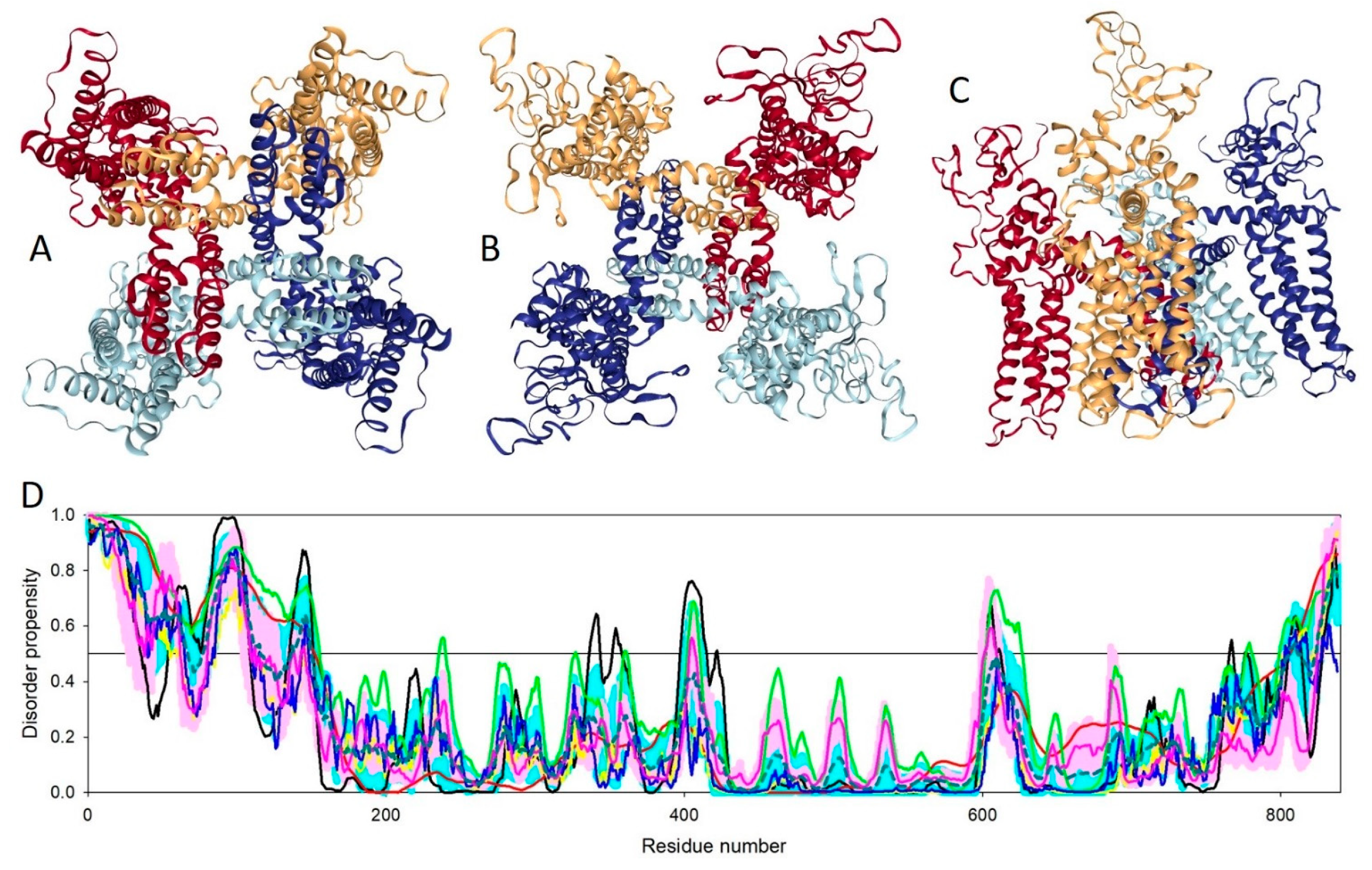
- Yan, Z.; Bai, X.; Yan, C.; Wu, J.; Li, Z.; Xie, T.; Peng, W.; Yin, C.; Li, X.; Scheres, S.H.W.; et al. Structure of the rabbit ryanodine receptor ryr1 at near-atomic resolution. Nature 2015, 517, 50–55. [Google Scholar] [CrossRef]
- Takeshima, H.; Nishimura, S.; Matsumoto, T.; Ishida, H.; Kangawa, K.; Minamino, N.; Matsuo, H.; Ueda, M.; Hanaoka, M.; Hirose, T.; et al. Primary structure and expression from complementary DNA of skeletal muscle ryanodine receptor. Nature 1989, 339, 439–445. [Google Scholar] [CrossRef]
- Rossi, D.; Sorrentino, V. Molecular genetics of ryanodine receptors Ca2+-release channels. Cell Calcium 2002, 32, 307–319. [Google Scholar] [CrossRef]
- Radermacher, M.; Rao, V.; Grassucci, R.; Frank, J.; Timerman, A.P.; Fleischer, S.; Wagenknecht, T. Cryo-electron microscopy and three-dimensional reconstruction of the calcium release channel/ryanodine receptor from skeletal muscle. J. Cell Biol. 1994, 127, 411–423. [Google Scholar] [CrossRef]
- des Georges, A.; Clarke, O.B.; Zalk, R.; Yuan, Q.; Condon, K.J.; Grassucci, R.A.; Hendrickson, W.A.; Marks, A.R.; Frank, J. Structural basis for gating and activation of ryr1. Cell 2016, 167, 145–157. [Google Scholar] [CrossRef]
- Romero, P.; Obradovic, Z.; Li, X.; Garner, E.C.; Brown, C.J.; Dunker, A.K. Sequence complexity of disordered protein. Proteins 2001, 42, 38–48. [Google Scholar] [CrossRef]
- Li, X.; Romero, P.; Rani, M.; Dunker, A.K.; Obradovic, Z. Predicting protein disorder for n-, c-, and internal regions. Genome Inform. 1999, 10, 30–40. [Google Scholar]
- Xue, B.; Dunbrack, R.L.; Williams, R.W.; Dunker, A.K.; Uversky, V.N. Pondr-fit: A meta-predictor of intrinsically disordered amino acids. Biochim. Biophys. Acta 2010, 1804, 996–1010. [Google Scholar] [CrossRef]
- Peng, K.; Vucetic, S.; Radivojac, P.; Brown, C.J.; Dunker, A.K.; Obradovic, Z. Optimizing long intrinsic disorder predictors with protein evolutionary information. J. Bioinform. Comput. Biol. 2005, 3, 35–60. [Google Scholar] [CrossRef]
- Obradovic, Z.; Peng, K.; Vucetic, S.; Radivojac, P.; Dunker, A.K. Exploiting heterogeneous sequence properties improves prediction of protein disorder. Proteins 2005, 61 (Suppl. 7), 176–182. [Google Scholar] [CrossRef]
- Peng, K.; Radivojac, P.; Vucetic, S.; Dunker, A.K.; Obradovic, Z. Length-dependent prediction of protein intrinsic disorder. BMC Bioinform. 2006, 7, 208. [Google Scholar] [CrossRef]
- Dosztanyi, Z.; Csizmok, V.; Tompa, P.; Simon, I. Iupred: Web server for the prediction of intrinsically unstructured regions of proteins based on estimated energy content. Bioinformatics 2005, 21, 3433–3434. [Google Scholar] [CrossRef]
- Cross, R.L.; Muller, V. The evolution of a-, f-, and v-type atp synthases and atpases: Reversals in function and changes in the h+/atp coupling ratio. Febs Lett. 2004, 576, 1–4. [Google Scholar] [CrossRef]
- Gruber, G.; Marshansky, V. New insights into structure-function relationships between archeal atp synthase (a1a0) and vacuolar type atpase (v1v0). Bioessays 2008, 30, 1096–1109. [Google Scholar] [CrossRef]
- Schafer, G.; Meyering-Vos, M. F-type or v-type? The chimeric nature of the archaebacterial atp synthase. Biochim. Biophys. Acta 1992, 1101, 232–235. [Google Scholar] [CrossRef]
- Mendoza-Hoffmann, F.; Zarco-Zavala, M.; Ortega, R.; Garcia-Trejo, J.J. Control of rotation of the f1fo-atp synthase nanomotor by an inhibitory alpha-helix from unfolded epsilon or intrinsically disordered zeta and if1 proteins. J. Bioenerg. Biomembr. 2018, 50, 403–424. [Google Scholar] [CrossRef]
- Stransky, L.; Cotter, K.; Forgac, M. The function of v-atpases in cancer. Physiol. Rev. 2016, 96, 1071–1091. [Google Scholar] [CrossRef]
- Toei, M.; Saum, R.; Forgac, M. Regulation and isoform function of the v-atpases. Biochemistry 2010, 49, 4715–4723. [Google Scholar] [CrossRef]
- Radax, C.; Sigurdsson, O.; Hreggvidsson, G.O.; Aichinger, N.; Gruber, C.; Kristjansson, J.K.; Stan-Lotter, H. F-and v-atpases in the genus thermus and related species. Syst. Appl. Microbiol. 1998, 21, 12–22. [Google Scholar] [CrossRef]
- Forgac, M. Vacuolar atpases: Rotary proton pumps in physiology and pathophysiology. Nat. Rev. Mol. Cell Biol. 2007, 8, 917–929. [Google Scholar] [CrossRef]
- Lau, W.C.; Rubinstein, J.L. Structure of intact thermus thermophilus v-atpase by cryo-em reveals organization of the membrane-bound v(o) motor. Proc. Natl. Acad. Sci. USA 2010, 107, 1367–1372. [Google Scholar] [CrossRef]
- Marshansky, V.; Rubinstein, J.L.; Gruber, G. Eukaryotic v-atpase: Novel structural findings and functional insights. Biochim. Biophys. Acta 2014, 1837, 857–879. [Google Scholar] [CrossRef]
- Schep, D.G.; Zhao, J.; Rubinstein, J.L. Models for the a subunits of the thermus thermophilus v/a-atpase and saccharomyces cerevisiae v-atpase enzymes by cryo-em and evolutionary covariance. Proc. Natl. Acad. Sci. USA 2016, 113, 3245–3250. [Google Scholar] [CrossRef]
- Zhao, J.; Benlekbir, S.; Rubinstein, J.L. Electron cryomicroscopy observation of rotational states in a eukaryotic v-atpase. Nature 2015, 521, 241–245. [Google Scholar] [CrossRef]
- Rajagopalan, K.; Mooney, S.M.; Parekh, N.; Getzenberg, R.H.; Kulkarni, P. A majority of the cancer/testis antigens are intrinsically disordered proteins. J. Cell Biochem. 2011, 112, 3256–3267. [Google Scholar] [CrossRef] [Green Version]
- Venkatachalam, K.; Montell, C. Trp channels. Annu. Rev. Biochem. 2007, 76, 387–417. [Google Scholar] [CrossRef]
- Ramsey, I.S.; Delling, M.; Clapham, D.E. An introduction to trp channels. Annu. Rev. Physiol. 2006, 68, 619–647. [Google Scholar] [CrossRef]
- Liao, M.; Cao, E.; Julius, D.; Cheng, Y. Structure of the trpv1 ion channel determined by electron cryo-microscopy. Nature 2013, 504, 107–112. [Google Scholar] [CrossRef]
- Bevan, S.; Quallo, T.; Andersson, D.A. Trpv1. Handb. Exp. Pharm. 2014, 222, 207–245. [Google Scholar]
- Julius, D. Trp channels and pain. Annu. Rev. Cell Dev. Biol. 2013, 29, 355–384. [Google Scholar] [CrossRef]
- Hellmich, U.A.; Gaudet, R. Structural biology of trp channels. Handb. Exp. Pharm. 2014, 223, 963–990. [Google Scholar]
- Li, M.; Yu, Y.; Yang, J. Structural biology of trp channels. Adv. Exp. Med. Biol. 2011, 704, 1–23. [Google Scholar]
- Gao, Y.; Cao, E.; Julius, D.; Cheng, Y. Trpv1 structures in nanodiscs reveal mechanisms of ligand and lipid action. Nature 2016, 534, 347–351. [Google Scholar] [CrossRef]
- Popot, J.L.; Althoff, T.; Bagnard, D.; Baneres, J.L.; Bazzacco, P.; Billon-Denis, E.; Catoire, L.J.; Champeil, P.; Charvolin, D.; Cocco, M.J.; et al. Amphipols from a to z. Annu. Rev. Biophys. 2011, 40, 379–408. [Google Scholar] [CrossRef]
- Kuang, Q.; Purhonen, P.; Hebert, H. Structure of potassium channels. Cell Mol. Life Sci. 2015, 72, 3677–3693. [Google Scholar] [CrossRef] [Green Version]
- Bocksteins, E. Kv5, kv6, kv8, and kv9 subunits: No simple silent bystanders. J. Gen. Physiol. 2016, 147, 105–125. [Google Scholar] [CrossRef]
- Jan, L.Y.; Jan, Y.N. Voltage-gated potassium channels and the diversity of electrical signalling. J. Physiol. 2012, 590, 2591–2599. [Google Scholar] [CrossRef]
- Swartz, K.J. Sensing voltage across lipid membranes. Nature 2008, 456, 891–897. [Google Scholar] [CrossRef] [Green Version]
- Bezanilla, F. How membrane proteins sense voltage. Nat. Rev. Mol. Cell Biol. 2008, 9, 323–332. [Google Scholar] [CrossRef]
- Attali, B.; Gao, Z.B. Ion channels research in the post-genomic era. Acta Pharm. Sin. 2016, 37, 1–3. [Google Scholar] [CrossRef] [Green Version]
- Wang, L.; Sigworth, F.J. Structure of the bk potassium channel in a lipid membrane from electron cryomicroscopy. Nature 2009, 461, 292–295. [Google Scholar] [CrossRef]
- Kaczmarek, L.K. Slack, slick and sodium-activated potassium channels. ISRN Neurosci. 2013, 2013, 354262. [Google Scholar] [CrossRef]
- Dryer, S.E. Na(+)-activated k+ channels: A new family of large-conductance ion channels. Trends Neurosci. 1994, 17, 155–160. [Google Scholar] [CrossRef]
- Hite, R.K.; Yuan, P.; Li, Z.; Hsuing, Y.; Walz, T.; MacKinnon, R. Cryo-electron microscopy structure of the slo2.2 na(+)-activated k(+) channel. Nature 2015, 527, 198–203. [Google Scholar] [CrossRef]


© 2019 by the authors. Licensee MDPI, Basel, Switzerland. This article is an open access article distributed under the terms and conditions of the Creative Commons Attribution (CC BY) license (http://creativecommons.org/licenses/by/4.0/).
Share and Cite
Nwanochie, E.; Uversky, V.N. Structure Determination by Single-Particle Cryo-Electron Microscopy: Only the Sky (and Intrinsic Disorder) is the Limit. Int. J. Mol. Sci. 2019, 20, 4186. https://doi.org/10.3390/ijms20174186
Nwanochie E, Uversky VN. Structure Determination by Single-Particle Cryo-Electron Microscopy: Only the Sky (and Intrinsic Disorder) is the Limit. International Journal of Molecular Sciences. 2019; 20(17):4186. https://doi.org/10.3390/ijms20174186
Chicago/Turabian StyleNwanochie, Emeka, and Vladimir N. Uversky. 2019. "Structure Determination by Single-Particle Cryo-Electron Microscopy: Only the Sky (and Intrinsic Disorder) is the Limit" International Journal of Molecular Sciences 20, no. 17: 4186. https://doi.org/10.3390/ijms20174186