Next Article in Journal
Spike Activator 1, Encoding a bHLH, Mediates Axillary Bud Development and Spike Initiation in Phalaenopsis aphrodite
Next Article in Special Issue
Oxidative Stress Markers to Investigate the Effects of Hyperoxia in Anesthesia
Previous Article in Journal
Modelling Neurotropic Flavivirus Infection in Human Induced Pluripotent Stem Cell-Derived Systems
Previous Article in Special Issue
The Fast Lane of Hypoxic Adaptation: Glucose Transport Is Modulated via A HIF-Hydroxylase-AMPK-Axis in Jejunum Epithelium
 
 
Retraction published on 7 May 2024, see Int. J. Mol. Sci. 2024, 25(9), 5039.
Article

RETRACTED: Intermittent Hypoxia-Hyperoxia Training Improves Cognitive Function and Decreases Circulating Biomarkers of Alzheimer’s Disease in Patients with Mild Cognitive Impairment: A Pilot Study

1
Department of General and Molecular Pathophysiology, Bogomoletz Institute of Physiology, 01024 Kyiv, Ukraine
2
Department of Hypoxia, Bogomoletz Institute of Physiology, 01024 Kyiv, Ukraine
3
Department of Age Physiology and Pathology of Nervous System, Chebotarev Institute of Gerontology NAMS of Ukraine, 04114 Kyiv, Ukraine
4
Department of Clinical Physiology and Pathology of Internal Organs, Chebotarev Institute of Gerontology NAMS of Ukraine, 04114 Kyiv, Ukraine
5
CellAir Constructions GmbH, 73614 Schorndorf, Germany
6
Department of Internal Medicine, Virginia Commonwealth University, Richmond, VA 23298-0204, USA
*
Author to whom correspondence should be addressed.
Deceased in April 2021.
Int. J. Mol. Sci. 2019, 20(21), 5405; https://doi.org/10.3390/ijms20215405
Submission received: 9 October 2019 / Revised: 23 October 2019 / Accepted: 24 October 2019 / Published: 30 October 2019 / Retracted: 7 May 2024
(This article belongs to the Special Issue Adaptation to Hypoxia: A Chimera?)

Abstract

:
Alzheimer’s disease (AD) affects not only the central nervous system, but also peripheral blood cells including neutrophils and platelets, which actively participate in pathogenesis of AD through a vicious cycle between platelets aggregation and production of excessive amyloid beta (Aβ). Platelets adhesion on amyloid plaques also increases the risk of cerebral microcirculation disorders. Moreover, activated platelets release soluble adhesion molecules that cause migration, adhesion/activation of neutrophils and formation of neutrophil extracellular traps (NETs), which may damage blood brain barrier and destroy brain parenchyma. The present study examined the effects of intermittent hypoxic-hyperoxic training (IHHT) on elderly patients with mild cognitive impairment (MCI), a precursor of AD. Twenty-one participants (age 51–74 years) were divided into three groups: Healthy Control (n = 7), MCI+Sham (n = 6), and MCI+IHHT (n = 8). IHHT was carried out five times per week for three weeks (total 15 sessions). Each IHHT session consisted of four cycles of 5-min hypoxia (12% FIO2) and 3-min hyperoxia (33% FIO2). Cognitive parameters, Aβ and amyloid precursor protein (APP) expression, microRNA 29, and long non-coding RNA in isolated platelets as well as NETs in peripheral blood were investigated. We found an initial decline in cognitive function indices in both MCI+Sham and MCI+IHHT groups and significant correlations between cognitive test scores and the levels of circulating biomarkers of AD. Whereas sham training led to no change in these parameters, IHHT resulted in the improvement in cognitive test scores, along with significant increase in APP ratio and decrease in Aβ expression and NETs formation one day after the end of three-week IHHT. Such effects on Aβ expression and NETs formation remained more pronounced one month after IHHT. In conclusion, our results from this pilot study suggested a potential utility of IHHT as a new non-pharmacological therapy to improve cognitive function in pre-AD patients and slow down the development of AD.

Graphical Abstract

1. Introduction

Alzheimer’s disease (AD) is a complex of mental disorder involving the accumulation of amyloid beta (Aβ) polymers in the brain, which affects not only the central nervous system, but also other body organs [1,2,3]. As an important contributing factor of AD, amyloid precursor protein (APP) is concentrated in synapses and takes part in cell-matrix and cell–cell interaction in neurons [4]. This adhesion molecule also participates in various processes in different tissues, for example, APP is involved in hemostasis and thrombosis in platelets [5] and in sperm motility and sperm-oocyte interaction [6]. Although there was little evidence of blood clotting or fertility impairments in individuals with AD, abnormal APP expression in the patients’ platelets was similar to those found in their neurons [7,8]. Nevertheless, APP expression may serve as one of the peripheral biomarkers for AD and may be useful for evaluating the AD pathology as well as therapy effectiveness. For instance, platelets isolated from blood of AD patients contain predominantly shorter forms APP, which increase the probability of Aβ 1–42 formation. Therefore, the use of APP ratio in platelets has been proposed as an early peripheral biomarker of AD [7]. Platelets may actively participate in pathogenesis of AD through two ways. First, the aggregated platelets produce extra levels of Aβ and the increased expression of APP on cell surface can trigger release of Aβ into plasma [2,3]. Second, Aβ is a direct activator of platelets aggregation in brain tissue [9], whereas platelets may adhere on amyloid plaques and increase the risk of microcirculation disorder in the brain [10].
Moreover, activated platelets can release soluble adhesion molecules that cause migration, adhesion, and activation of neutrophils. In addition to secretory degranulation, reactive oxygen species (ROS) production, and phagocytosis, neutrophils can also produce neutrophil extracellular traps (NETs), which were characterized as a fundamental host innate immune defense mechanism [11]. NETs represent chromatin with inserted proteases released by activated cell DNA network that serves as a trap for bacteria and other antigen carriers. Platelets can initiate NETosis in different pathologies such as [12,13]. Similar to other nonspecific neutrophil reactions, excessive NETosis may damage its own cell structures. The presence of proteases in NETs initiate blood vessel inflammation, which leads to blood-brain-barrier destruction and neuronal degradation [14,15]. NETs were found in vessels and brain parenchyma of individuals with AD [16]. Moreover, NET-associated enzymatic system digests amyloid fibrils into short species that are cytotoxic [17]. Thus, the combined effects of platelets and neutrophils may play a role in the AD-related tissue destruction [18,19].
Another important factor affecting the rate of APP cleavage and Aβ formation is Beta-secretase 1, also known as beta-site amyloid precursor protein cleaving enzyme 1 (BACE1). BACE1 activity is elevated in platelet membranes of patients with mild cognitive impairments (MCI) and AD [2]. BACE1 translation is regulated by non-coding RNAs, such as microRNA 29a (miR-29a), which blocks beta-secretase translation [20,21,22,23,24,25], whereas lncRNA BACE1-AS, binds complementarily to mRNA and protects it from the inhibitory effect of miRNAs [26].
Considering the importance of prevention and treatment of AD, there is a constant call for new therapies. There is growing evidence supporting the notion that sessions of brief repeated, controllable exposures to moderate levels of hypoxia, termed as intermittent hypoxia-normoxia training (IHT) or intermittent hypoxia-hyperoxia training (IHHT), may improve cerebral vascular function and mitochondrial energy production, same as those that have been demonstrated in sports training and/or rehabilitative medicine. For instance, IHT has beneficial effects on elderly health [27,28,29]. During the past decade, a modified intermittent hypoxia training method, i.e., IHHT, was introduced and applied in several human studies or clinical trials in several European countries [27,30,31]. The underlying cellular mechanisms of IHT or IHHT may include the improvement of mitochondrial function, regulation of glucose transport, activation of antioxidant enzyme system, as well as anti-inflammatory effects. These effects can be beneficial in the prevention and treatment of cardiovascular and brain diseases, such as AD [32,33,34,35,36]. Exercise combined with IHT additionally enhanced exercise performance and improved cognitive function in elderly individuals [37,38,39]. IHT also improved walking ability after chronic spinal cord injury [40]. Moreover, there is evidence showing that hypoxia caused significant reduction of spontaneous and phorbol myristate acetate (PMA)-stimulated NET formation in cultured neutrophils [11]. Under this context, our present study was designed to investigate the effects of IHHT on cognitive function, along with the circulating biomarkers of AD in patients with MCI, a pathological condition found in approximately 70% of AD cases as the first symptomatic sign of AD.

2. Results

2.1. Cognitive Function

At baseline, all studied parameters of cognitive function were similar among both MCI groups, but significantly different as compared with the Healthy Control group. In particular, MoCA-test scores were significantly declined in MCI+Sham and MCI+IHHT groups as compared with Healthy Control group (by 27% or 32%, respectively), indicating mild cognitive impairments (Table 1). The long latency cognitive evoked potentials were significantly higher in both MCI groups (i.e., by 23% and 27%, respectively in P300 and by 21% in N200; Table 1).
The course of normoxic sham training did not change the cognitive parameters in MCI+Sham group, whereas a significant enhancement of MoCA-test score after IHHT was observed in MCI+IHHT group one day after the end of IHHT (by 11%, p < 0.05). This effect was less pronounced at one month after IHHT (by 7%, no statistical significance). There was a downward trend in P300 and N200 latency in MCI+IHHT group one day as well as one month after IHHT termination (Table 1). Thus, we have observed improvement of parameters of cognitive function in the elderly MCI patients after IHHT.

2.2. NETs Formation

At pre-IHHT baseline, in both MCI-groups the non-stimulated NETs exceeded more than three times the values of healthy people (Table 2, Figure 1). The course of sham IHHT did not change NETns in MCI+Sham group, but actual IHHT decreased their formation by 53% in MCI+IHHT group. In one month after IHHT termination this decrease was expressed even more (by 56%) approaching the healthy people indices. Unlike non-stimulated NETs, the formation of PMA-stimulated neutrophil extracellular traps did not differ significantly in all three groups. Sham IHHT did not influence NETst, meanwhile we registered a tendency to decrease in MCI+IHHT group by 31% immediately after IHHT which turned into a significant difference a month after the end of the course (a decrease of 52% compared to baseline).

2.3. Platelets Amyloid Precursor Proteins (APP) and Amyloid Beta (Aβ)

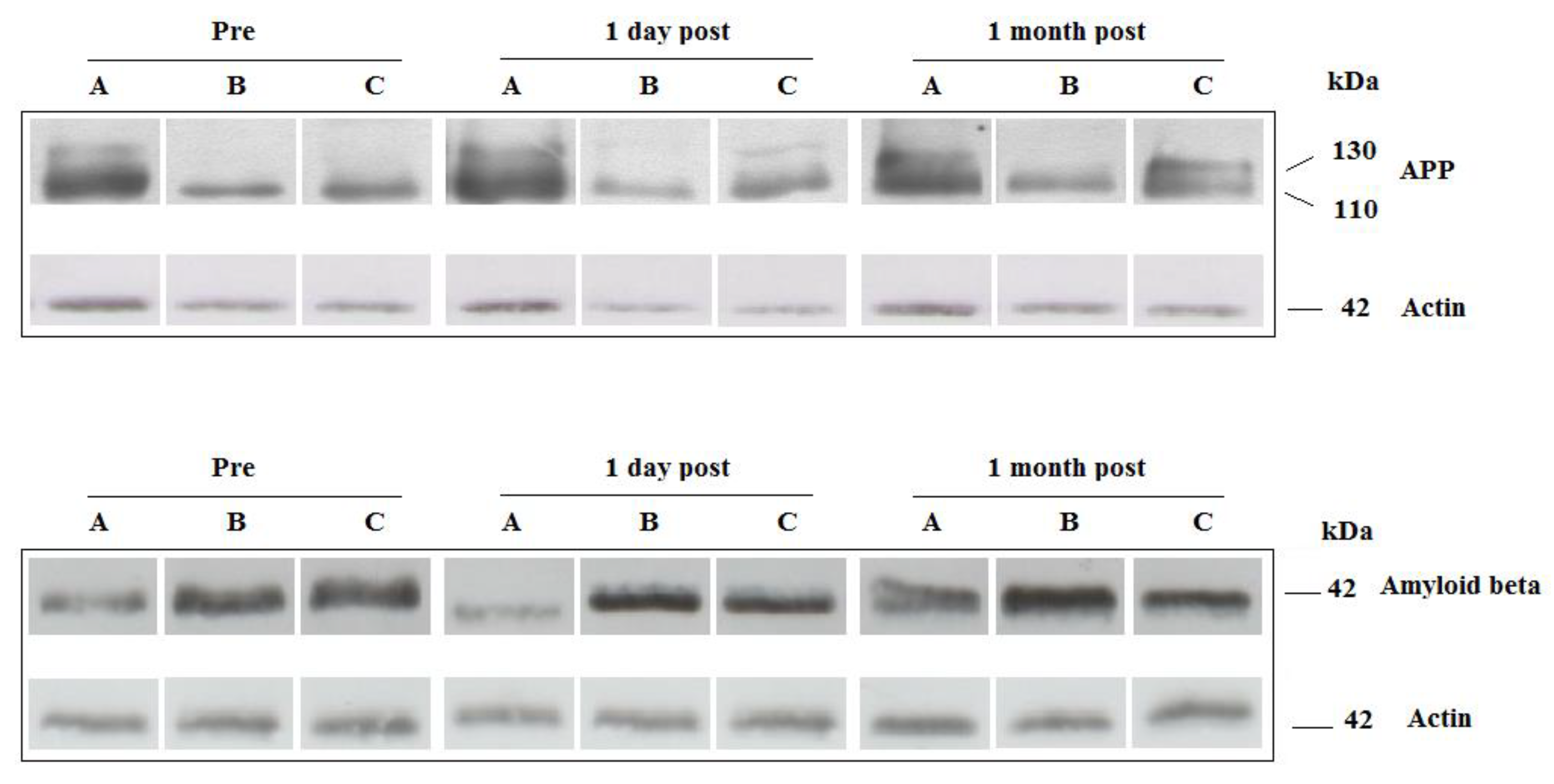
At pre-IHHT baseline, both investigated fractions (110 and 130) of APP were significantly reduced in both MCI groups as compared to Healthy Control group, i.e., APP130 and APP110 decreased by 62% and 55%, respectively in MCI+Sham group, and by 61% and 47%, respectively in MCI+IHHT group (Table 3, Figure 2). The APP ratio was shifted towards higher level of APP110. Neither APP130 nor APP110 changed in MCI+Sham group one day and one month after IHHT termination. To the contrary, IHHT resulted in 60% and 30% increase in APP130 and APP110, respectively one day after the end of IHHT and these values are significantly different from MCI+Sham group. At the later time point (one month after IHHT), the elevated APP130 slightly diminished but remained significantly higher than pre-IHHT baseline (42% increase). However, the delayed effect of APP110 was even more pronounced at one month after IHHT (36% increase versus pre-IHHT baseline), which also led to a significantly higher level than those of MCI+Sham group. Consequently, the APP130/110 ratio had 21% increase at one day after IHHT termination, but returned to the baseline level after one month.
At Pre-baseline, Aβ expression in both MCI-groups exceeded in 2.5 times this parameter in healthy people. Sham training did not influence significantly Aβ in Group II, whereas IHHT led to 15% and 28% decrease one day and one month after IHHT termination, respectively.

2.4. Expression of miR-29a and lncRNA BACE-AS in Platelets

Surprisingly, we did not observe significant difference in miR-29a expression among the treatment groups with or without IHHT. However, expression of lncRNA BACE-AS, the fast regulator of beta-site amyloid precursor protein cleaving enzyme (BACE1) translation, was significantly upregulated (>6 folds) at baseline in both MCI groups as compared with the healthy controls (Table 4). Whereas Sham training did not affect its expression, lncRNA BACE-AS was significantly downregulated by 57% in MCI+IHHT group one day after the termination of IHHT.

2.5. Correlation Analysis

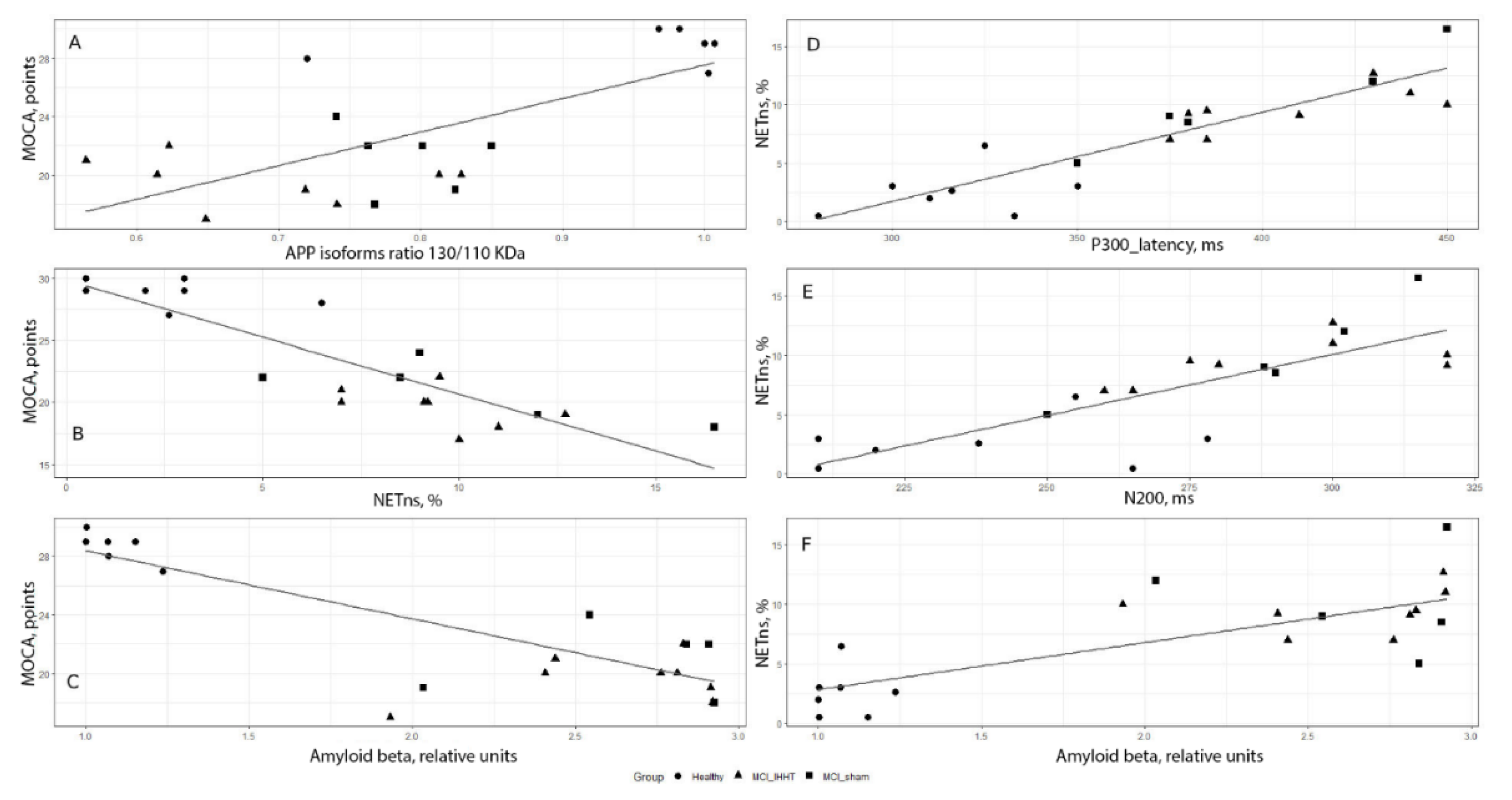
In order to evaluate the relationships between cognitive function indices and the circulating biomarkers measured, we conducted correlation analysis using the combined baseline data from all three groups. As shown in Figure 3, a strong positive correlation between MoCA score and APP110/APP130 ratio was observed: r = 0.72, p < 0.05. We also found that MoCA negatively correlated with both NETns (r = −0.87, p < 0.01) and Aβ expression (r = −0.79, p = 0.02) (Figure 3, left column). On the other hand, NETns were positively correlated with long latency cognitive event-related potentials P300 (r = 0.90, p < 0.01) and N200 (r = 0.79, p = 0.02), as well as Aβ expression (r = 0.81, p = 0.03) (Figure 3, right column).

3. Discussion

Weakening of cognitive functions is the prominent clinical symptom during the progression of cognitive decline from MCI to AD. The cognitive state of MCI patients was also correlated with neurophysiological markers of cognitive evoked potentials (by more than 25% increased latency of N200 and P300 peaks). The decline of cognitive function in MCI groups was associated with a significant decrease in APP130 and APP110 isoforms of platelet amyloid precursor proteins and decreased ratio of APP130/APP110, as well as 2.5 times increase in Aβ expression as compared with the age-matched healthy subjects. We also observed a 6-fold increase in lncRNA BACE-AS, which is a regulator of beta-site APP cleaving enzyme translation. The peripheral blood of MCI patients also had three times excessive level of NETs formation. There were a strong positive correlation between total MoCA test scores and both APP forms as well as a negative correlation with amyloid beta expression in platelets. We also found a positive correlation between the latency of cognitive evoked potentials and non-stimulated NETs. NETns were in turn positively associated with Aβ expression in platelets.
A key finding of our present study is that three-week IHHT resulted in significant improvement in the subject’s cognitive performance, i.e., increase total MoCA-test scores and downward trend in latency of cognitive evoked potentials one day after the end of IHHT session. This was accompanied by changes in circulating biomarkers in peripheral blood of the subjects. For example, there were a pronounced increase in APP130 and APP110 fractions in platelets and a significant decrease in Aβ expression in the MCI patients one day after the termination of IHHT sessions and became even more pronounced one month after the end of IHHT. A significant downregulation of lncRNA BACE-AS and NETs formation were also found in peripheral blood one day after the end of IHHT.
There have been extensive investigations on the cellular and molecular mechanisms of AD development as well as therapeutic approaches to control this serious disease [18,19,41,42,43,44]. Among the contributing factors, chronic reduction in cerebral blood flow and excessive Aβ deposition are primarily linked to neurodegeneration [45,46]. Previous studies have demonstrated that IHT could potentially ameliorate the development and progression of AD and dementia [47,48,49]. In particular, in a rat model of experimental AD, researchers found that the decrease in cerebral blood flow following Aβ injection was blunted by IHT [50]. Similarly, IHT augmented cerebral blood flow and endothelial nitric oxide production, decreased oxidative stress, prevented neuronal degeneration, and stimulated neurogenesis and neuroregeneration [33,51,52,53,54,55].
Several recent works in humans confirmed our data on the improved cognitive function with IHT or IHHT. In particular, it was demonstrated that IHT augmented the cognitive benefits of strength-endurance training in a randomized controlled study in 60 to 70 years old subjects [39]. It was shown that hypoxic treadmill training (three times/week for 1 h over four weeks) in multiple sclerosis patients improved fitness level, mood, and mobility [56]. Similar results were also reported by Burtscher et al. [57,58]. A recent study that evaluated the effects of IHHT on patients with MCI (64–92 years of age) has shown significant improvements in the cognitive state and physical endurance [37]. These findings were also in accordance with the study results from another research group [27,30].
Furthermore, our present study observed an association between the changes in cognitive function and the changes in platelet amyloid precursor protein (APP), Aβ expression in peripheral blood. The mechanisms through which Aβ provokes cognitive decline have been extensively explored [59], including the pathologic increase in processing and transformation of APP into Aβ. lncRna BACE 1-AS, a non-coding RNA, may increase transcription of beta-secretase and in turn contribute to the accumulation of Aβ. Whereas the MCI patients showed a significant increase in NETosis, a positive correlation was observed between the elevated level of NETosis and the content of Aβ. Possible explanations for this phenomenon include neutrophil and platelet interactions. For example, the increase in BACE1 activity by 24% in platelet membranes of patients with MCI and by 17% in those with AD [2]. Our present study also demonstrated that the completion of 15 sessions of IHHT resulted in declined lncRna BACE 1-AS levels, along with decreased Aβ and increased APP expression. Thus, we would postulate a possible association between Aβ reduction and BACE 1 downregulation. On the other hand, we did not find changes in miR29-a expression, which blocks beta-secretase translation and could serve as a marker for amyloid accumulation (21,22). Previous studies showed that certain microRNAs may play a negative role under intermittent hypoxia load in the patients with obstructive sleep apnea (OSA) [60].
It is notable that certain degrees of concern or even confusion exist on the divergent effects of IHHT versus OSA, a common and pathological form of intermittent hypoxia that occurs in human population including elderly. We should consider several fundamental differences between these two intermittent hypoxia-related phenomena. First, OSA is characterized by brief, recurrent cycles of hypoxia-reoxygenation (<60 s per cycle for up to 30 cycles per h overnight), whereas our IHHT program had much fewer hypoxia episodes (four cycles of 5 min hypoxia-hyperoxia per day). Another major difference is that CO2 accumulation in circulation in each of the OSA asphyxiation episodes may lead to acidemia, whereas IHHT-activated hyperventilation usually results in hypocapnea and alkalemia. Thirdly, OSA-led asphyxiation causes arousals that result in fragmented and unproductive sleeping patterns that lead to neurological defects in the OSA patients. To the contrary, our subjects underwent all sessions of IHHT during their regular awake hours and therefore no sleep disruption occurred. It is also conceivable that OSA-induced intensive ROS overproduction plays a pathogenic role in various diseases, whereas a moderate amount of ROS generated in a well-controlled, therapeutic IHHT protocol may serve as signaling trigger for increasing cytoprotective antioxidant enzyme expression/activities.
Special attention should be paid to the changes in APP ratio. Initially, the APP ratio of MCI patients shifted toward APP110. The ratio of APP isoforms was reported to be significantly lower in patients with AD or MCI and it paralleled with cognitive decline and can predict the transition from MCI to AD [2]. IHHT resulted in downregulation of APP110 and favorably shifted the APP ratio, which is one of the putative biomarkers of AD, towards a better prognostic sign. It was demonstrated that APP participates in formation of synapses and synaptic plasticity [61]. Assuming APP expression in platelets serves as a valid peripheral biomarker of APP expression in the brain, an IHHT-induced improvement in cognitive function may be associated with not only a reduced toxic effect of amyloids, but also increased APP content. Along with the changes in platelet biomarkers, IHHT also led to a decrease in NETosis, which could be a sequential or parallel phenomenon. In brief, a decrease in Aβ should diminish both platelets and neutrophils activation. It was previously shown that short term sojourn in mountains or obstructive sleep apnea may increase platelet aggregation and reactivity [62,63], whereas prolonged stay on high altitude resulted in significant decline in platelets aggregation [64].
Nevertheless, there are several limitations in the present study, including very small sample size of the elderly subjects, due to the recruitment difficulty for the cooperative subjects who were willing to complete the entire duration of the two month-long studies as well as the unexpected dropouts. However, our present work on the limited numbers of patients did produce some statistically significant results and may serve well as a good starting point for designing and conducting future studies in larger scale of participants and various pre-AD and AD patient cohorts. In addition, this investigation was conducted in a single-blinded fashion, since the researchers needed to know the actual inspired oxygen concentration in order to set up the equipment for sham training or IHHT. The patients did not know the exact types of gas mixtures (sham or IHHT) they were receiving. In addition, anti-inflammatory effect of IHHT in both blood and the brain was not studied in the current work. This may be an interesting topic for future investigations that aim at uncovering the beneficial mechanisms of IHHT in AD prevention and treatment.

4. Materials and Methods

4.1. Characteristics of Participants

The human study was conducted according to the principles of Helsinki Declaration of Human Rights. The research protocol, patient information, and their informed consent forms were reviewed and approved by the Ethics Committee of Chebotarev Institute of Gerontology, Kiev, Ukraine (protocol #9, 11 June 2017). Twenty-three subjects (age 51 to 74 years) were initially enrolled into this pilot clinical study by the Chebotarev Institute of Gerontology, Kiev, Ukraine. All subjects received detailed information of the study process and a written informed consent was obtained from each of the participants. They were nonsmokers and did not take any medication (including cholesterol-lowering drugs, statins, or fibrates) two weeks before and during the study sessions. There were no infections during the past month and no major cardiovascular or respiratory complications in any of the subjects. These subjects had no family history of AD nor medical history of Type I/ Type II diabetes, myocardium infarction, or stroke. The blood levels of low density lipoprotein cholesterol (LDL-C) ranged from 2.31 to 3.43 mmol/L and the average values did not differ between the three groups.
The participants consisted of seven healthy subjects without signs of cognitive impairment syndrome (Healthy Control group) and 16 patients diagnosed with MCI, who were recruited from the Department of Aging Physiology and Pathology of Nervous System of the Chebotarev Institute of Gerontology. The diagnosis of MCI was according to the revised Petersen criteria [65]. The criteria of the MCI diagnosis include subjective cognitive complaint, objective evidence of cognitive impairment, absence of difficulties in functional activities of daily life, and no dementia. Objective cognitive decline was evaluated using the Montreal Cognitive Assessment (MoCA) test [66,67] with the cut off score of 25 to 19 [68]. The Clinical Dementia Rating scale used was 0.5 points [69]. The key exclusion criteria for the subjects were: Signs of cerebrovascular pathology-related cognitive decline, metabolic/endocrine disease, alcohol consumption and intake of any psychotropic drugs, abnormalities in liver, kidney, lungs and heart, infection, neurologic and psychiatric diseases that can interfere with cognitive decline. Healthy group subjects were also checked by a neurologist for excluding any signs of cognitive pathology. The selected patients with MCI were randomly assigned to either MCI+Sham or MCI+IHHT group. Two initially enrolled MCI patients did not complete the entire study, because of one death and another had flu. A total of seven healthy subjects (Health Control) and 14 patients with MCI (six patients in MCI+Sham and eight patients in MCI+IHHT) completed the planned sessions of the study. All participants were informed about the strict observance of lifestyle required during the period of examination and reported any lifestyle changes if occurred. The patients had no knowledge about which training condition (sham versus hypoxic exposures) actually performed. Anthropometric characteristics of the participants are summarized in Table 5 below.

4.2. Protocol of IHHT

All sessions of the present study were conducted in a quiet room at comfortable temperature (21 to 23 °C). Venous blood samples were collected at three time points, i.e., 1) The day before beginning of IHHT to measure base line parameters; 2) The day after completion of the actual or sham IHHT course to evaluate the immediate effects of IHHT; and 3) One month after the end of IHHT for determining the delayed effects of IHHT. Cell isolation of the collected blood samples was conducted immediately. Cognitive functions were investigated in all groups in the same day. Same examinations were repeated at the next day and one month after the end of IHHT.
Sham or IHHT treatment started the next morning. The Healthy Control subjects did not undergo either sham or actual IHHT. The MCI patients received sessions of sham or actual IHHT, which underwent five times a week for the subsequent three weeks, i.e., each subject received total of 15 sessions. Each session consisted of four cycles of 5-min hypoxia (12% inspired O2) followed by 3-min hyperoxia (IHHT, breathing with 33% inspired O2) [28]. The IHHT sessions were performed using a computer-controlled apparatus of “CellAir One” (Cellgym Technologies GmbH, Berlin, Germany). During each session, we continuously monitored the subjects’ systolic and diastolic blood pressure, heart rate, and peripheral oxygen saturation.

4.3. Cognitive Function Assessment

We used both MoCA test and long latency cognitive event-related potentials (ERPs) in add ball paradigm as the non-invasive tools for assessment of brain function and cognitive status. These parameters have also served as the objective markers of treatment efficiency [70,71,72].
Long latency cognitive evoked potentials were recorded in a soundproof room using a 19-channel electroencephalography (EEG) equipment (Neurocom, XAI-MEDICA). During the EEG test, the subjects seated comfortably on an armchair and remained awake throughout the test. They were instructed to keep eyes open for avoiding eye movement and to count the rare random hearing stimuli (with 20% probability) and the similar frequent stimuli (with 80% probability). The sound stimuli were delivered via speakers with 80 dB HL (decibels Hearing Level) sound intensity. A minimum of 30 rare stimuli events were recorded per patient with a 1000 ms time window. Latency cognitive evoked potentials were recorded at the maximum wave amplitude. Latency of N200 and P300 peaks of cognitive evoked potential in Cz electrode position were taken for analysis.

4.4. Neutrophil Isolation and NETs Evaluation

Polymorphonuclear leukocyte (PMN) isolation and detection of NETs were performed as previously described [73]. In brief, to obtain PMN, the blood sample was collected and stabilized by EDTA in a tube (Sarstedt) mixed with 0.9% NaCl in 1:1 ratio and carefully laminated on the Percoll gradient with layer density of 45%, 54%, and 63%. After a gradient centrifugation of the blood sample (3000× rpm for 15 min), PMNs were detected between the second and third layers. The cells were collected and purified by erythrocyte lysis with ACK (Ammonium-Chloride-Potassium) buffer. The granulocytes were then washed with HBSS (Hank’s Balanced Salt Solution) buffer and diluted in RPMI (Roswell Park Memorial Institute) medium with 10% (v/v) heat-inactivated fetal bovine serum. The PMNs were plated in a 24-well dish or Petri dish with a cell density of 100,000 cells/cm2.
To stimulate NET formation, 20 nM of phorbol myristate acetate (PMA) was added to the cultured cells for 3 h (NETst group) and another set of non-stimulated neutrophils (NETns) served as the controls. NET was detected using a fluorescence microscope with fluorogenic dyes (propidium iodide and Hoechst 33342 biz-benzamide), which directly stain chromatin.

4.5. Platelets Isolation

Blood sample was collected from each of the subjects by venipuncture into a standard 6 mL tube containing EDTA. Platelet-rich plasma was prepared by repeated centrifugation at room temperature. After pelleting, the platelets were washed with phosphate buffered saline (PBS) and counted. After additional centrifugation at 3000× g for 10 min, the platelets were resuspended in 0.7 mL PBS for further analysis.

4.6. Western Blot Analysis

The platelets were incubated in an ice-cold RIPA-buffer (1:3) containing 20 mM Tris–HCl, 150 mM NaCl, 1 mM EDTA, 1% NP-40, 1% sodium deoxycholate, 0.1% SDS, 1 µM leupeptin, and 1 mM protease inhibitor PMSF (pH 7.5) for 30 min and subsequently centrifuged for 15 min at 11,000× g at 4 °C and the supernatant was collected. After measurement of protein concentration, the protein fraction of platelet lysate was separated using 10% polyacrylamide gel with 0.1% SDS and then transferred onto a nitrocellulose membrane (90 min at 200 mA). The membrane was incubated overnight at 4 °С with a purified mouse monoclonal primary antibody for APP (1:1000, MAB348, Sigma-Aldrich, St. Louis, MO, USA) or a rabbit monoclonal primary antibody for Aβ 1–42 (1:1000, ab201060, Abcam, Cambridge, MA, USA). In addition, a mouse monoclonal primary antibody for β-actin was used as an internal control for protein loading (1:2000, 2 h incubation, A1978, Sigma-Aldrich, St. Louis, MO, USA). Anti-rabbit or anti-mouse secondary antibodies were added to the membranes, which were visualized with chemiluminescence reagents (ECL kit, Amersham Pharmacia Biotech, Little Chalfont, United Kingdom) and exposed to X-ray film and the scanned Western blot bands were analysed with densitometry. Protein levels of APP and Aβ 1–42 were normalized against the corresponding β-actin values and their relative expression ratio was calculated.

4.7. RNA Isolation and Real-Time Polymerase Chain Reaction (PCR)

Total RNA was extracted from the isolated platelets using phenol–chloroform extraction method. RNA concentration was determined using a spectrophotometer (Model ND1000, NanoDrop Technologies, Wilmington, DE, USA). Reverse transcription of miR-29a and snRNA U6 was performed in 6 µg of total RNA using MicroRNA Reverse Transcription Kit (Applied Biosystems, Foster City, CA, USA) with Assay ID 002112 and 001973, respectively. Reverse transcription of lncRNA BACE1 was performed on 6 µg of total RNA using a RevertAidTM H Minus First Strand cDNA Synthesis Kit (Fermentas, Vilnius, Lithuania) with a random hexamer primer. The obtained single-stranded DNA was used for further PCR using TaqMan MicroRNA Assay (Applied Biosystems, Foster City, CA, USA). The amplification of lncRNA BACE1 AS was performed using SYBR Green PCR Master Mix with following primers: Up 5′-GCA AAC GAA GGT TGG TGG TG-3′ Dw 5′-CCC AGC AGT AAC CCC CTA CT-3′. The results were normalized with snRNA U6.

4.8. Statistical Analysis

All data were analyzed using SPSS software version 21.0 (SPSS Inc., Chicago, IL, USA). One-way analysis of variance (ANOVA) with repeated measures followed by Bonferroni post hoc test was used to determine statistical significance of the differences between the groups. Two-way ANOVA with repeated measures followed by Bonferroni post hoc correction was performed to determine the group main effect and time effect (i.e., among three time-points: Baseline, one day and one month after sham or actual IHHT). Training effects within one group (pre- versus after sham or actual IHHT) were analyzed using paired t test. Correlation analysis was performed to determine the relationships between numerical features. p < 0.05 was defined as statistical significance.

5. Conclusions

Our present pilot study has demonstrated a month-long-lasting therapeutic effect of IHHT on cognitive function in elderly patients with MCI. Such positive changes at neuropsychological, electrophysiological, and biomarker levels may indicate a whole brain benefit afforded by IHHT, which offers its unique clinical applicability as a non-pharmacological therapy. In particular, a strong association between activation of BACE1-dependent synthesis of Aβ in platelets, increased formation of extracellular traps by neutrophils, and decline in cognitive function in MCI patients as well as the modifications following the three-week IHHT sessions were revealed for the first time. We propose that the panel of peripheral blood biomarkers may serve as a diagnostic tool and treatment efficacy marker, which permit us to not only predict the pathological development of AD at its early stage, but also guide our therapeutic intervention with IHHT. Nevertheless, much larger scale of investigations in pre-AD and AD patients would be warranted to further validate these preliminary findings.

Author Contributions

Conceptualization, Z.O.S., T.V.S., L.V.T., and V.E.D.; Methodology, Z.O.S., T.V.S., V.B.S., and V.A.K.; Formal analysis, Z.O.S., S.V.G., D.S., and V.E.D.; Investigation, O.N.G., E.E., N.Y.B., V.A.K., A.M.S., L.V.T., and D.A.P.; Writing—original draft preparation, Z.O.S. and T.V.S.; Writing—review and editing, L.X.; Project administration, V.E.D.; Funding acquisition, E.E.

Funding

This research work was partially funded by the Ukrainian State Budget Program “Support for the Development of Priority Areas of Scientific Research” (Code: 6541230) and CellAir Constructions GmbH, Schorndorf, Germany. The APC was funded by CellAir Constructions GmbH.

Acknowledgments

The authors are grateful to M. Kramer (ImmBioMed GmbH & Co. KG, Germany) for helping in the organization of this research.

Conflicts of Interest

E.E. is an owner of CellAir Constructions GmbH, Schorndorf, Germany; L.X. is a co-founder of Xiamen Innovo Medical Technology Co. Ltd., Xiamen, China. These firms had no role in the design of the study; in the collection, analyses, or interpretation of data; in the writing of the manuscript, or in the decision to publish the results. All other authors declare no conflict of interests in conducting and publishing this work.

Abbreviations

Amyloid beta
ACKAmmonium-Chloride-Potassium
ADAlzheimer’s disease
APPAmyloid precursor protein
BACE1Beta-site amyloid precursor protein cleaving enzyme 1
dB HLdecibels Hearing Level
EEGElectroencephalography
ERPsEvent-Related Potentials
FIO2Fractional concentration of oxygen in the inspired gas
HBSSHank’s Balanced Salt Solution
IHHTIntermittent Hypoxia-Hyperoxia Training
IHTIntermittent Hypoxia-Normoxia Training
lncRNALong noncoding RNA
MCIMild Cognitive Impairment
MoCAMontreal Cognitive Assessment
NETnsNon-stimulated neutrophil extracellular traps
NETsNeutrophil extracellular traps
OSAObstructive Sleep Apnea
PBSPhosphate Buffered Saline
PCRPolymerase Chain Reaction
PMAPhorbol myristate acetate
PMNPolymorphonuclear leukocyte
ROSReactive oxygen species

References

  1. Dong, Y.; Lagarde, J.; Xicota, L.; Corne, H.; Chantran, Y.; Chaigneau, T.; Crestani, B.; Bottlaender, M.; Potier, M.-C.; Aucouturier, P.; et al. Neutrophil hyperactivation correlates with Alzheimer’s disease progression. Ann. Neurol. 2018, 83, 387–405. [Google Scholar] [CrossRef] [PubMed]
  2. Evin, G.; Li, Q.X. Platelets and Alzheimer’s disease: Potential of APP as a biomarker. World J. Psychiatry 2012, 2, 102–113. [Google Scholar] [CrossRef] [PubMed]
  3. Kucheryavykh, L.Y.; Davila-Rodriguez, J.; Rivera-Aponte, D.E.; Zueva, L.V.; Washington, A.V.; Sanabria, P.; Inyushin, M.Y. Platelets are responsible for the accumulation of beta-amyloid in blood clots inside and around blood vessels in mouse brain after thrombosis. Brain Res. Bull. 2017, 128, 98–105. [Google Scholar] [CrossRef] [PubMed]
  4. Muller, U.C.; Zheng, H. Physiological functions of APP family proteins. Cold Spring Harb. Perspect. Med. 2012, 2, a006288. [Google Scholar] [CrossRef] [PubMed]
  5. Visconte, C.; Canino, J.; Guidetti, G.F.; Zarà, M.; Seppi, C.; Abubaker, A.A.; Pula, G.; Torti, M.; Canobbio, I. Amyloid precursor protein is required for in vitro platelet adhesion to amyloid peptides and potentiation of thrombus formation. Cell Signal. 2018, 52, 95–102. [Google Scholar] [CrossRef]
  6. Silva, J.V.; Yoon, S.; Domingues, S.; Guimarães, S.C.; Goltsev, A.V.; Silva, E.F.D.C.E.; Mendes, J.F.F.; Silva, O.A.B.D.C.E.; Fardilha, M. Amyloid precursor protein interaction network in human testis: Sentinel proteins for male reproduction. BMC Bioinform. 2015, 16, 12. [Google Scholar] [CrossRef]
  7. Borroni, B.; Agosti, C.; Marcello, E.; Di Luca, M.; Padovani, A. Blood cell markers in Alzheimer Disease: Amyloid Precursor Protein form ratio in platelets. Exp. Gerontol. 2010, 45, 53–56. [Google Scholar] [CrossRef]
  8. Pluta, R.; Ułamek-Kozioł, M.; Januszewski, S.; Czuczwar, S.J. Platelets, lymphocytes and erythrocytes from Alzheimer’s disease patients: The quest for blood cell-based biomarkers. Folia Neuropathol. 2018, 56, 14–20. [Google Scholar] [CrossRef]
  9. Canobbio, I.; Guidetti, G.F.; Oliviero, B.; Manganaro, D.; Vara, D.; Torti, M.; Pula, G. Amyloid beta-peptide-dependent activation of human platelets: Essential role for Ca2+ and ADP in aggregation and thrombus formation. Biochem. J. 2014, 462, 513–523. [Google Scholar] [CrossRef]
  10. Gowert, N.S.; Donner, L.; Chatterjee, M.; Eisele, Y.S.; Towhid, S.T.; Munzer, P.; Walker, B.; Ogorek, I.; Borst, O.; Grandoch, M.; et al. Blood platelets in the progression of Alzheimer’s disease. PLoS ONE 2014, 9, e90523. [Google Scholar] [CrossRef]
  11. Branitzki-Heinemann, K.; Mollerherm, H.; Vollger, L.; Husein, D.M.; de Buhr, N.; Blodkamp, S.; Reuner, F.; Brogden, G.; Naim, H.; von Kockritz-Blickwede, M. Formation of Neutrophil Extracellular Traps under Low Oxygen Level. Front. Immunol. 2016, 7, 518. [Google Scholar] [CrossRef] [PubMed]
  12. Kazzaz, N.M.; Sule, G.; Knight, J.S. Intercellular Interactions as Regulators of NETosis. Front. Immunol. 2016, 7, 1532. [Google Scholar] [CrossRef] [PubMed]
  13. Lisman, T. Platelet-neutrophil interactions as drivers of inflammatory and thrombotic disease. Cell Tissue Res. 2018, 371, 567–576. [Google Scholar] [CrossRef] [PubMed]
  14. Kniewallner, K.M.; Foidl, B.M.; Humpel, C. Platelets isolated from an Alzheimer mouse damage healthy cortical vessels and cause inflammation in an organotypic ex vivo brain slice model. Sci. Rep. 2018, 8, 15483. [Google Scholar] [CrossRef]
  15. Zenaro, E.; Piacentino, G.; Constantin, G. The blood-brain barrier in Alzheimer’s disease. Neurobiol. Dis. 2017, 107, 41–56. [Google Scholar] [CrossRef]
  16. Pietronigro, E.C.; Della Bianca, V.; Zenaro, E.; Constantin, G. NETosis in Alzheimer’s Disease. Front. Immunol. 2017, 8, 159. [Google Scholar] [CrossRef]
  17. Azevedo, E.P.C.; Guimarães-Costa, A.B.; Torezani, G.S.; Braga, C.A.; Palhano, F.L.; Kelly, J.W.; Saraiva, E.M.; Foguel, D. Amyloid Fibrils Trigger the Release of Neutrophil Extracellular Traps (NETs), Causing Fibril Fragmentation by NET-associated Elastase. J. Boil. Chem. 2012, 287, 37206–37218. [Google Scholar] [CrossRef]
  18. Duyckaerts, C.; Delatour, B.; Potier, M.-C. Classification and basic pathology of Alzheimer disease. Acta Neuropathol. 2009, 118, 5–36. [Google Scholar] [CrossRef]
  19. Gomez-Isla, T.; Spires, T.; De Calignon, A.; Hyman, B.T. Neuropathology of Alzheimer’s Disease. Dementias 2008, 89, 233–243. [Google Scholar]
  20. Basavaraju, M.; de Lencastre, A. Alzheimer’s disease: Presence and role of microRNAs. Biomol. Concepts 2016, 7, 241–252. [Google Scholar] [CrossRef]
  21. Gupta, P.; Bhattacharjee, S.; Sharma, A.R.; Sharma, G.; Lee, S.-S.; Chakraborty, C.; Gupta, S.B.P. miRNAs in Alzheimer Disease-A Therapeutic Perspective. Curr. Alzheimer Res. 2017, 14, 1198–1206. [Google Scholar] [CrossRef] [PubMed]
  22. Jia, L.H.; Liu, Y.N. Downregulated serum miR-223 servers as biomarker in Alzheimer’s disease. Cell Biochem. Funct. 2016, 34, 233–237. [Google Scholar] [CrossRef] [PubMed]
  23. Kiko, T.; Nakagawa, K.; Tsuduki, T.; Furukawa, K.; Arai, H.; Miyazawa, T. MicroRNAs in Plasma and Cerebrospinal Fluid as Potential Markers for Alzheimer’s Disease. J. Alzheimer’s Dis. 2014, 39, 253–259. [Google Scholar] [CrossRef] [PubMed]
  24. Wang, W.X.; Huang, Q.; Hu, Y.; Stromberg, A.J.; Nelson, P.T. Patterns of microRNA expression in normal and early Alzheimer’s disease human temporal cortex: White matter versus gray matter. Acta. Neuropathol. 2011, 121, 193–205. [Google Scholar] [CrossRef]
  25. Yang, G.; Song, Y.; Zhou, X.; Deng, Y.; Liu, T.; Weng, G.; Yu, D.; Pan, S. MicroRNA-29c targets β-site amyloid precursor protein-cleaving enzyme 1 and has a neuroprotective role in vitro and in vivo. Mol. Med. Rep. 2015, 12, 3081–3088. [Google Scholar] [CrossRef]
  26. Feng, L.; Liao, Y.-T.; He, J.-C.; Xie, C.-L.; Chen, S.-Y.; Fan, H.-H.; Su, Z.-P.; Wang, Z. Plasma long non-coding RNA BACE1 as a novel biomarker for diagnosis of Alzheimer disease. BMC Neurol. 2018, 18, 4. [Google Scholar] [CrossRef]
  27. Dudnik, E.; Zagaynaya, E.; Glazachev, O.S.; Susta, D. Intermittent Hypoxia–Hyperoxia Conditioning Improves Cardiorespiratory Fitness in Older Comorbid Cardiac Outpatients without Hematological Changes: A Randomized Controlled Trial. High Alt. Med. Boil. 2018, 19, 339–343. [Google Scholar] [CrossRef]
  28. Serebrovska, T.V.; Portnychenko, A.G.; I Drevytska, T.; I Portnichenko, V.; Xi, L.; Egorov, E.; Gavalko, A.V.; Naskalova, S.; Chizhova, V.; Shatylo, V.B. Intermittent hypoxia training in prediabetes patients: Beneficial effects on glucose homeostasis, hypoxia tolerance and gene expression. Exp. Boil. Med. 2017, 242, 1542–1552. [Google Scholar] [CrossRef]
  29. Shatilo, V.B.; Korkushko, O.V.; Ischuk, V.A.; Downey, H.F.; Serebrovskaya, T.V. Effects of Intermittent Hypoxia Training on Exercise Performance, Hemodynamics, and Ventilation in Healthy Senior Men. High Alt. Med. Boil. 2008, 9, 43–52. [Google Scholar] [CrossRef]
  30. Glazachev, O.; Kopylov, P.; Susta, D.; Dudnik, E.; Zagaynaya, E. Adaptations following an intermittent hypoxia-hyperoxia training in coronary artery disease patients: A controlled study. Clin. Cardiol. 2017, 40, 370–376. [Google Scholar] [CrossRef]
  31. Susta, D.; Dudnik, E.; Glazachev, O.S. A programme based on repeated hypoxia-hyperoxia exposure and light exercise enhances performance in athletes with overtraining syndrome: A pilot study. Clin. Physiol. Funct. Imaging 2017, 37, 276–281. [Google Scholar] [CrossRef] [PubMed]
  32. Mallet, R.T.; Manukhina, E.B.; Ruelas, S.S.; Caffrey, J.L.; Downey, H.F. Cardioprotection by intermittent hypoxia conditioning: Evidence, mechanisms, and therapeutic potential. Am. J. Physiol. Circ. Physiol. 2018, 315, H216–H232. [Google Scholar] [CrossRef] [PubMed]
  33. Manukhina, E.B.; Downey, H.F.; Shi, X.; Mallet, R.T. Intermittent hypoxia training protects cerebrovascular function in Alzheimer’s disease. Exp. Boil. Med. 2016, 241, 1351–1363. [Google Scholar] [CrossRef] [PubMed]
  34. Mateika, J.H.; Komnenov, D. Intermittent hypoxia initiated plasticity in humans: A multipronged therapeutic approach to treat sleep apnea and overlapping co-morbidities. Exp. Neurol. 2017, 287, 113–129. [Google Scholar] [CrossRef] [PubMed]
  35. Serebrovskaya, T.V.; Manukhina, E.B.; Smith, M.L.; Downey, H.F.; Mallet, R.T. Intermittent Hypoxia: Cause of or Therapy for Systemic Hypertension? Exp. Boil. Med. 2008, 233, 627–650. [Google Scholar] [CrossRef]
  36. Serebrovskaya, T.V.; Xi, L. Intermittent hypoxia training as non-pharmacologic therapy for cardiovascular diseases: Practical analysis on methods and equipment. Exp. Biol. Med. 2016, 241, 1708–1723. [Google Scholar] [CrossRef]
  37. Bayer, U.; Likar, R.; Pinter, G.; Stettner, H.; Demschar, S.; Trummer, B.; Neuwersch, S.; Glazachev, O.; Burtscher, M. Intermittent hypoxic-hyperoxic training on cognitive performance in geriatric patients. Alzheimer’s Dement. : Transl. Res. Clin. Interv. 2017, 3, 114–122. [Google Scholar] [CrossRef]
  38. Bayer, U.; Likar, R.; Pinter, G.; Stettner, H.; Demschar, S.; Trummer, B.; Neuwersch, S.; Glazachev, O.; Burtscher, M. Effects of intermittent hypoxia-hyperoxia on mobility and perceived health in geriatric patients performing a multimodal training intervention: A randomized controlled trial. BMC Geriatr. 2019, 19, 167. [Google Scholar] [CrossRef]
  39. Schega, L.; Peter, B.; Törpel, A.; Mutschler, H.; Isermann, B.; Hamacher, D. Effects of Intermittent Hypoxia on Cognitive Performance and Quality of Life in Elderly Adults: A Pilot Study. Gerontol 2013, 59, 316–323. [Google Scholar] [CrossRef]
  40. Dougherty, B.; Terada, J.; Springborn, S.; Vinit, S.; Macfarlane, P.; Mitchell, G. Daily acute intermittent hypoxia improves breathing function with acute and chronic spinal injury via distinct mechanisms. Respir. Physiol. Neurobiol. 2018, 256, 50–57. [Google Scholar] [CrossRef]
  41. Ju, Y.-E.S.; Ooms, S.J.; Sutphen, C.; Macauley, S.L.; Zangrilli, M.A.; Jerome, G.; Fagan, A.M.; Mignot, E.; Zempel, J.M.; Claassen, J.A.; et al. Slow wave sleep disruption increases cerebrospinal fluid amyloid-β levels. Brain 2017, 140, 2104–2111. [Google Scholar] [CrossRef] [PubMed]
  42. Liguori, C.; Mercuri, N.B.; Izzi, F.; Romigi, A.; Cordella, A.; Sancesario, G.; Placidi, F. Obstructive Sleep Apnea is Associated With Early but Possibly Modifiable Alzheimer’s Disease Biomarkers Changes. Sleep 2017, 40. [Google Scholar] [CrossRef] [PubMed]
  43. Nelson, P.T.; Alafuzoff, I.; Bigio, E.H.; Bouras, C.; Braak, H.; Cairns, N.J.; Castellani, R.J.; Crain, B.J.; Davies, P.; Del Tredici, K.; et al. Correlation of Alzheimer disease neuropathologic changes with cognitive status: A review of the literature. J. Neuropathol. Exp. Neurol. 2012, 71, 362–381. [Google Scholar] [CrossRef] [PubMed]
  44. Struble, R.G.; Ala, T.; Patrylo, P.R.; Brewer, G.J.; Yan, X.-X. Is brain amyloid production a cause or a result of dementia of the Alzheimer’s type? J. Alzheimer’s Dis. 2010, 22, 393–399. [Google Scholar] [CrossRef] [PubMed]
  45. Lourenço, C.F.; Ledo, A.; Barbosa, R.M.; Laranjinha, J. Neurovascular uncoupling in the triple transgenic model of Alzheimer’s disease: Impaired cerebral blood flow response to neuronal-derived nitric oxide signaling. Exp. Neurol. 2017, 291, 36–43. [Google Scholar] [CrossRef] [PubMed]
  46. Toth, P.; Tarantini, S.; Csiszar, A.; Ungvari, Z. Functional vascular contributions to cognitive impairment and dementia: Mechanisms and consequences of cerebral autoregulatory dysfunction, endothelial impairment, and neurovascular uncoupling in aging. Am. J. Physiol. Heart Circ. Physiol. 2017, 312, H1–H20. [Google Scholar] [CrossRef] [PubMed]
  47. Arkhipenko, Y.V.; Sazontova, T.G.; Zhukova, A.G. Adaptation to periodic hypoxia and hyperoxia improves resistance of membrane structures in heart, liver, and brain. Bull. Exp. Boil. Med. 2005, 140, 278–281. [Google Scholar] [CrossRef]
  48. Lukyanova, L.D.; Dudchenko, A.M.; Tsybina, T.A.; Germanova, E.L.; Tkachuk, E.N.; Erenburg, I.V. Effect of intermittent normobaric hypoxia on kinetic properties of mitochondrial enzymes. Bull. Exp. Boil. Med. 2007, 144, 795–801. [Google Scholar] [CrossRef]
  49. Lukyanova, L.D.; Kirova, Y.I. Mitochondria-controlled signaling mechanisms of brain protection in hypoxia. Front. Neurosci. 2015, 9, 320. [Google Scholar] [CrossRef]
  50. Mashina, S.Y.; Aleksandrin, V.V.; Goryacheva, A.V.; Vlasova, M.A.; Vanin, A.F.; Malyshev, I.Y.; Manukhina, E.B. Adaptation to hypoxia prevents disturbances in cerebral blood flow during neurodegenerative process. Bull. Exp. Boil. Med. 2006, 142, 169–172. [Google Scholar] [CrossRef]
  51. Haider, T.; Casucci, G.; Linser, T.; Faulhaber, M.; Gatterer, H.; Ott, G.; Linser, A.; Ehrenbourg, I.; Tkatchouk, E.; Burtscher, M.; et al. Interval hypoxic training improves autonomic cardiovascular and respiratory control in patients with mild chronic obstructive pulmonary disease. J. Hypertens. 2009, 27, 1648–1654. [Google Scholar] [CrossRef] [PubMed]
  52. Manukhina, E.B.; Belkina, L.M.; Terekhina, O.L.; Abramochkin, D.V.; A Smirnova, E.; Budanova, O.P.; Mallet, R.T.; Downey, H.F. Normobaric, intermittent hypoxia conditioning is cardio- and vasoprotective in rats. Exp. Boil. Med. 2013, 238, 1413–1420. [Google Scholar] [CrossRef] [PubMed]
  53. Manukhina, E.B.; Tseilikman, V.E.; Tseilikman, O.B.; Komelkova, M.V.; Kondashevskaya, M.V.; Goryacheva, A.V.; Lapshin, M.S.; Platkovskii, P.O.; Alliluev, A.V.; Downey, H.F. Intermittent hypoxia improves behavioral and adrenal gland dysfunction induced by posttraumatic stress disorder in rats. J. Appl. Physiol. 2018, 125, 931–937. [Google Scholar] [CrossRef] [PubMed]
  54. Sazontova, T.G.; Glazachev, O.S.; Bolotova, A.V.; Dudnik, E.N.; Striapko, N.V.; Bedareva, I.V.; A Anchishkina, N.; Arkhipenko, I.V. Adaptation to hypoxia and hyperoxia improves physical endurance: The role of reactive oxygen species and redox-signaling. Ross. Fiziol. zhurnal Im. IM Sechenova 2012, 98, 793–807. [Google Scholar]
  55. Zhu, X.-H.; Yan, H.-C.; Zhang, J.; Qu, H.-D.; Qiu, X.-S.; Chen, L.; Li, S.-J.; Cao, X.; Bean, J.C.; Qin, X.-H.; et al. Intermittent Hypoxia Promotes Hippocampal Neurogenesis and Produces Antidepressant-Like Effects in Adult Rats. J. Neurosci. 2010, 30, 12653–12663. [Google Scholar] [CrossRef]
  56. Mähler, A.; Balogh, A.; Csizmadia, I.; Klug, L.; Kleinewietfeld, M.; Steiniger, J.; Šušnjar, U.; Müller, D.N.; Boschmann, M.; Paul, F. Metabolic, Mental and Immunological Effects of Normoxic and Hypoxic Training in Multiple Sclerosis Patients: A Pilot Study. Front. Immunol. 2018, 9. [Google Scholar] [CrossRef]
  57. Burtscher, M. High-altitude cerebral effects: Risks and mechanisms. Lancet Neurol. 2009, 8, 604–605. [Google Scholar] [CrossRef]
  58. Burtscher, M.; Pachinger, O.; Ehrenbourg, I.; Mitterbauer, G.; Faulhaber, M.; Pühringer, R.; Tkatchouk, E. Intermittent hypoxia increases exercise tolerance in elderly men with and without coronary artery disease. Int. J. Cardiol. 2004, 96, 247–254. [Google Scholar] [CrossRef]
  59. Findlay, M.D.; Dawson, J.; Dickie, D.A.; Forbes, K.P.; McGlynn, D.; Quinn, T.; Mark, P.B. Investigating the Relationship between Cerebral Blood Flow and Cognitive Function in Hemodialysis Patients. J. Am. Soc. Nephrol. 2019, 30, 147–158. [Google Scholar] [CrossRef]
  60. Gao, H.; Han, Z.; Huang, S.; Bai, R.; Ge, X.; Chen, F.; Lei, P. Intermittent hypoxia caused cognitive dysfunction relate to miRNAs dysregulation in hippocampus. Behav. Brain Res. 2017, 335, 80–87. [Google Scholar] [CrossRef]
  61. Montagna, E.; Dorostkar, M.M.; Herms, J. The Role of APP in Structural Spine Plasticity. Front. Mol. Neurosci. 2017, 10, 79. [Google Scholar] [CrossRef] [PubMed]
  62. Rahangdale, S.; Yeh, S.Y.; Novack, V.; Stevenson, K.; Barnard, M.R.; Furman, M.I.; Frelinger, A.L.; Michelson, A.D.; Malhotra, A. The Influence of Intermittent Hypoxemia on Platelet Activation in Obese Patients with Obstructive Sleep Apnea. J. Clin. Sleep Med. 2011, 7, 172–178. [Google Scholar] [PubMed]
  63. Rocke, A.S.; Paterson, G.G.; Barber, M.T.; Jackson, A.I.R.; Main, S.; Stannett, C.; Schnopp, M.F.; Baillie, J.K.; Horne, E.H.; Moores, C.; et al. Thromboelastometry and Platelet Function during Acclimatization to High Altitude. Thromb. Haemost. 2018, 118, 63–71. [Google Scholar] [CrossRef] [PubMed]
  64. Vij, A.G. Effect of prolonged stay at high altitude on platelet aggregation and fibrinogen levels. Platelets 2009, 20, 421–427. [Google Scholar] [CrossRef] [PubMed]
  65. Albert, M.S.; DeKosky, S.T.; Dickson, D.W.; Dubois, B.; Feldman, H.H.; Fox, N.C.; Gamst, A.; Holtzman, D.M.; Jagust, W.J.; Petersen, R.C.; et al. The diagnosis of mild cognitive impairment due to Alzheimer’s disease: Recommendations from the National Institute on Aging-Alzheimer’s Association workgroups on diagnostic guidelines for Alzheimer’s disease. Alzheimer’s Dement. 2011, 7, 270–279. [Google Scholar] [CrossRef]
  66. Dawes, P.; Pye, A.; Reeves, D.; Yeung, W.K.; Sheikh, S.; Thodi, C.; Charalambous, A.P.; Gallant, K.; Nasreddine, Z.; Leroi, I. Protocol for the development of versions of the Montreal Cognitive Assessment (MoCA) for people with hearing or vision impairment. BMJ Open 2019, 9, e026246. [Google Scholar] [CrossRef]
  67. Nasreddine, Z.S.; Phillips, N.A.; Bedirian, V.; Charbonneau, S.; Whitehead, V.; Collin, I.; Cummings, J.L.; Chertkow, H.; Bédirian, V. The Montreal Cognitive Assessment, MoCA: A Brief Screening Tool for Mild Cognitive Impairment. J. Am. Geriatr. Soc. 2005, 53, 695–699. [Google Scholar] [CrossRef]
  68. Milani, S.A.; Marsiske, M.; Cottler, L.B.; Chen, X.; Striley, C.W. Optimal cutoffs for the Montreal Cognitive Assessment vary by race and ethnicity. Alzheimer’s Dement. Diagn. Assess. Dis. Monit. 2018, 10, 773–781. [Google Scholar] [CrossRef]
  69. Morris, J.C. Clinical Dementia Rating: A Reliable and Valid Diagnostic and Staging Measure for Dementia of the Alzheimer Type. Int. Psychogeriatr 1997, 9, 173–176. [Google Scholar] [CrossRef]
  70. Hünerli, D.; Emek-Savaş, D.D.; Çavuşoğlu, B.; Çolakoğlu, B.D.; Ada, E.; Yener, G.G. Mild cognitive impairment in Parkinson’s disease is associated with decreased P300 amplitude and reduced putamen volume. Clin. Neurophysiol. 2019, 130, 1208–1217. [Google Scholar] [CrossRef]
  71. Lai, C.-L.; Lin, R.-T.; Liou, L.-M.; Liu, C.-K. The role of event-related potentials in cognitive decline in Alzheimer’s disease. Clin. Neurophysiol. 2010, 121, 194–199. [Google Scholar] [CrossRef] [PubMed]
  72. Vaitkevicius, A.; Kaubrys, G.; Audronytė, E. Distinctive Effect of Donepezil Treatment on P300 and N200 Subcomponents of Auditory Event-Related Evoked Potentials in Alzheimer Disease Patients. Med. Sci. Monit. 2015, 21, 1920–1927. [Google Scholar] [CrossRef] [PubMed]
  73. Pashevin, D.O.; Nagibin, V.S.; Tumanovska, L.V.; Moibenko, A.A.; Dosenko, V.E. Proteasome Inhibition Diminishes the Formation of Neutrophil Extracellular Traps and Prevents the Death of Cardiomyocytes in Coculture with Activated Neutrophils during Anoxia-Reoxygenation. Pathobiol 2015, 82, 290–298. [Google Scholar] [CrossRef] [PubMed]
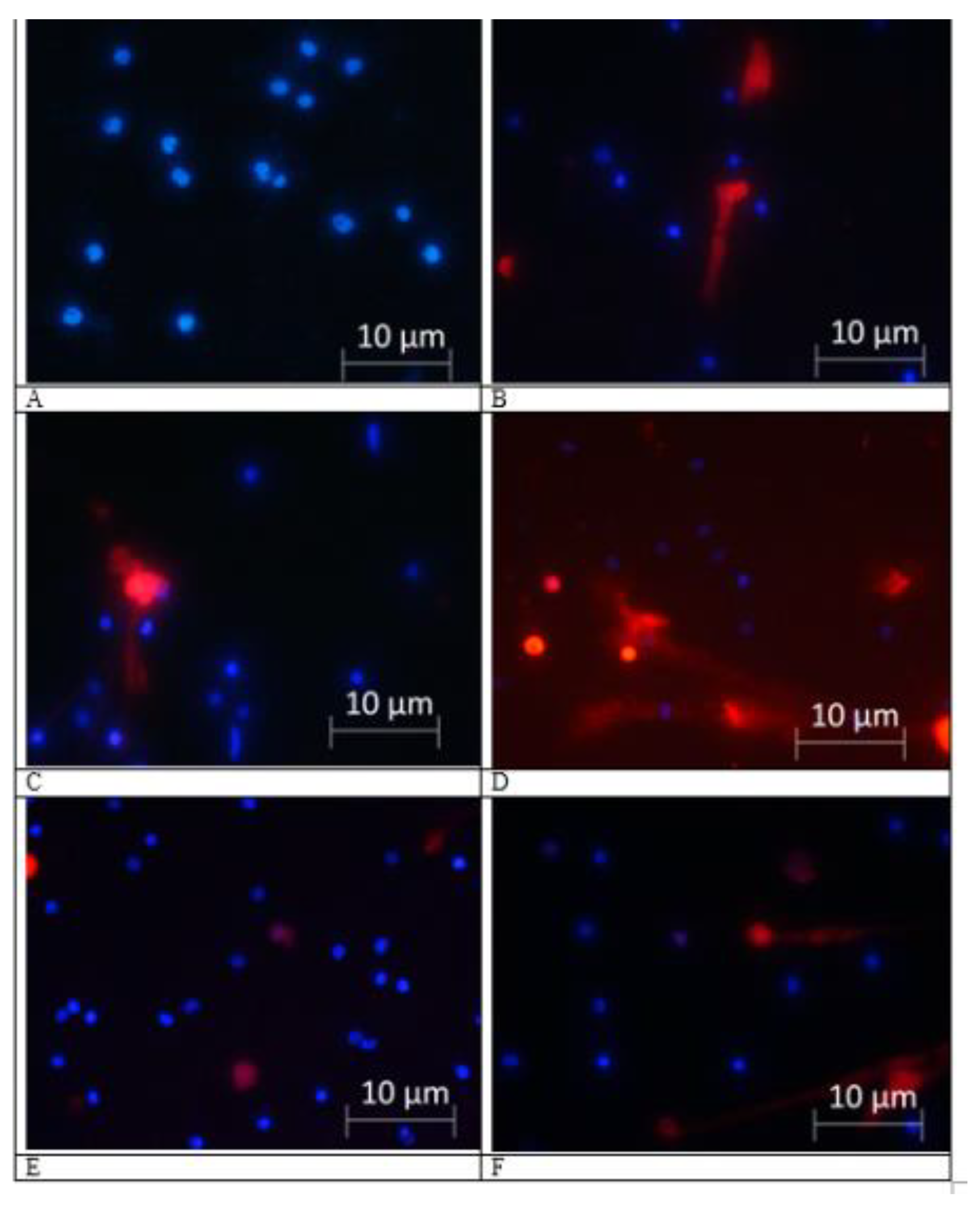
Figure 1. Fluorescence microscopy (40× magnification) of isolated polymorphonuclear leukocyte (PMN): Spontaneous (left column) and phorbol myristate acetate (PMA)-induced (right column) neutrophil extracellular traps in mild cognitive impairments (MCI) patients. Viable neutrophil nuclear DNA was stained with membrane permeable Hoechst 33342 (blue), and neutrophil extracellular traps (NETs) were visualized by membrane impermeable Propidium Iodide (red) staining extracellular DNA. (A,B) PMN from Healthy Control group at baseline time point; (C,D) PMN from patient of MCI+IHHT group at pre-IHHT baseline time point; and (E,F) PMN from the same patient of MCI+IHHT group one day after the termination of intermittent hypoxia-hyperoxia training (IHHT).
Ijms 20 05405 g001
Figure 2. Western blot analysis of amyloid precursor protein (APP) forms (APP130 and APP110) and amyloid beta (Aβ) protein expression levels in the platelets collected from Healthy Control (A), MCI+Sham (B), and MCI+IHHT (C) groups at three time points. Beta-actin served as loading control. Representative pictures of Western blots of APP and beta-actin. Abbreviations: Pre (Pre-IHHT baseline); 1 day post (one-day after the end of IHHT sessions); and 1 month post (one-month after the end of IHHT); MCI, mild cognitive impairment; IHHT, intermittent hypoxia-hyperoxia training.
Ijms 20 05405 g002
Figure 3. Correlation relationships between cognitive function indices and some circulating biomarkers of Alzheimer’s disease in all three groups of patients with mild cognitive impairments at pre-baseline. Left column: (A) Montreal Cognitive Assessment (MoCA) score and amyloid precursor protein (APP) isoforms ratio 130/110 (r = 0.72, p < 0.05), (B) MoCA score and non-stimulated neutrophil extracellular traps (NETns) (r = −0.87, p < 0.01); (C) MoCA score and amyloid beta (Aβ) expression (r = −0.79, p = 0.02). Right column: (D) NETns and long latency cognitive event-related potential P300 (r = 0.90, p < 0.01); (E) NETns and long latency cognitive event-related potential N200 (r = 0.79, p = 0.02), (F) NETns and Aβ expression (r = 0.81, p = 0.03).
Ijms 20 05405 g003
Table 1. Сognitive functions before and after the three-week sessions of sham training or intermittent hypoxia-hyperoxia training (IHHT).
GroupHealthy Control
(n = 7)
MCI + Sham
(n = 6)
MCI + IHHT (n = 8)Group Main EffectTime Effect
3 Time-Points
Group × Time Interaction
MoCA, score
Pre-28.9 ± 1.0621.2 ± 2.12 #19.6 ±1.59 #F = 50.031F = 13.207F = 5.029
1-day Post29.3 ± 0.7520.3 ± 2.1 #22.1 ± 1.68 #,*P = 0.000P = 0.000P = 0.03
1-month Post29.4 ± 1.1320.0 ± 1.75 #21.3 ± 1.57 #
P300, ms
Pre-319 ± 24.5398 ± 27.7 #407 ± 29.7 #F = 15.233F = 5.535F = 3.904
1-day Post317 ± 19.5396 ± 29.73 #391 ± 29.7 #P = 0.000P = 0.008P = 0.01
1-month Post318 ± 22.8398 ± 27.97 #392 ± 28.4 #
N200, ms
Pre-239 ± 23.3280 ± 21.7 #290 ± 21.4 #F = 7.285F = 11.087F = 7.254
1-day Post242 ± 24.3284 ± 21.9 #273 ± 20.2 #P = 0.005P = 0.000P = 0.000
1-month Post238 ± 26.6285 ± 22.4 #275 ± 20.1 #
Data are mean ± SD and analyzed with two-way ANOVA with repeated measures. Abbreviations: MoCA: The Montreal Cognitive Assessment; P300 and N200 are components of event-related potentials; Pre: Pre-IHHT baseline; One-day Post: One day after IHHT termination; One-month Post: One-month after IHHT termination. Symbols: # p < 0.05 versus Healthy Control group; * p < 0.05 versus Pre-baseline.
Table 2. Neutrophil extracellular traps (NETs) formation in neutrophils culture under three-week sessions of sham training or IHHT.
GroupHealthy Control
(n = 7)
MCI + Sham
(n = 6)
MCI + IHHT
(n = 8)
Group Main Effect Time Effect
3 time- points
Group × Time Interaction
APP130, relative unit
Pre-1.04 ± 0.050.40 ± 0.07 #0.41 ± 0.11 #F = 90.897F = 5.843F = 7.814
1-day Post1.04 ± 0.040.37 ± 0.03 #0.65 ± 0.12 #,*,^P = 0.000P = 0.007P = 0.000
1-month Post1.05 ± 0.050.36 ± 0.04 #0.58 ± 0.13 #,*,^
APP110, relative unit
Pre-1.02 ± 0.040.47 ± 0.17 #0.56 ± 0.14 #F = 83.45F = 4.063F = 2.137
1-day Post1.02 ± 0.030.49 ± 0.09 #0.73 ± 0.14 #,*,^P = 0.000P = 0.026P = 0.098
1-month Post1.05 ± 0.060.48 ± 0.08 #0.76 ± 0.11 #,*,^
APP ratio
Pre-1,01 ± 0.100.82 ± 0.05 #0.73 ± 0.09 #F = 14.622F = 0.857F = 1.426
1-day Post1.01 ± 0.110.77 ± 0.13 #0.89 ± 0.09 #,*,^P = 0.000P = 0.435P = 0.249
1-month Post1.00 ± 0.110.76 ± 0.06 #0.76 ± 0.1 #
Aβ, relative unit
Pre-1.08 ± 0.12.69 ± 0.35 #2.63 ± 0.34 #F = 924.103F = 1.238F = 4.723
1-day Post1.17 ± 0.212.76 ± 0.35 #2.24 ± 0.43 #,*,^P = 0.000P = 0.302P = 0.004
1-month Post1.29 ± 0.52.82 ± 0.22 #2.08 ± 0.44 #,*,^
Data are mean ± SD and were analyzed with two-way ANOVA with repeated measures. Abbreviations: NETns: Neutrophil extracellular traps without stimulation; NETst: Neutrophil extracellular traps in cells stimulated by phorbol myristate acetate (PMA); Pre: Pre-IHHT baseline; 1-day Post: One day after IHHT termination; 1-month Post: One month after IHHT termination. Symbols: # p < 0.01 versus Healthy Control group; * p < 0.05 versus Pre-baseline; ^ p < 0.05 versus MCI+Sham group.
Table 3. Platelets amyloid precursor proteins (APP) and amyloid beta (Aβ) under three-week sessions of sham training or IHHT.
GroupHealthy Control
(n = 7)
MCI +Sham
(n = 6)
MCI+ IHHT
(n = 8)
Group Main EffectTime Effect
3 Time-Points
Group × Time Interaction
NETns, %
Pre-2.58 ± 2.229.62 ± 4.83 #9.47 ± 2.06 #F = 119.799F = 3.689F = 4.170
1-day Post2.05 ± 1.799.22 ± 3.94 #4.48 ± 1.09 #,*,^P = 0.001P = 0.04P = 0.011
1-month Post3.02 ± 1.518.25 ± 2.04 #4.21 ± 1.28 *,^
NETst, %
Pre-10.3 ± 4.6812.4 ± 6.0712.7 ± 6.18F = 0.573F = 5.316F = 0.803
1-day Post7.18 ± 3.0410.20 ± 4.868.76 ± 3.34P = 0.577P = 0.012P = 0.534
1-month Post9.18 ± 2.8011.16 ± 3.636.11 ± 3.50 *,^
Data are mean ± SD and analyzed with two-way ANOVA with repeated measures. Abbreviations: Pre: Pre-IHHT baseline; One-day Post: One day after IHHT termination; One-month Post: One month after IHHT termination; APP130: Amyloid precursor protein form of 130 kDa; APP110: Amyloid precursor protein form of 110 kDa; Aβ: Amyloid beta peptide of 42 amino acid lengths. Symbols: # p < 0.01 versus Healthy Control group; * p < 0.05 versus Pre-baseline; ^ p < 0.05 versus MCI+Sham group.
Table 4. Expression of lncRNA BACE-AS, the fast regulator of beta-site amyloid precursor protein cleaving enzyme (BACE1) under three-week sessions of sham training or IHHT.
GroupHealthy Control
(n = 7)
MCI + Sham
(n = 6)
MCI + IHHT
(n = 8)
Group Main EffectTime Effect
3 Time-Points
Group × Time Interaction
lncRNA BACE1-AS, relative unit
Pre-13.6 ± 10.892.5 ± 45.5 #85.3 ± 55.6 #F = 11.708F = 1.288F = 0.729
1-day Post12.6 ± 5.975.4 ± 57.3 #36.8 ± 34.6 *P = 0.001P = 0.288P = 0.075
1-month Post10.6 ± 7.799.3 ± 70.4 #45.6 ± 32.8 #
Data are mean ± SD and analyzed with two-way ANOVA with repeated measures. Abbreviations: MCI: mild cognitive impairment; IHHT: intermittent hypoxia-hyperoxia training; Pre: Pre-IHHT baseline; 1-day Post: One day after IHHT termination; 1-month Post: One month after IHHT termination. Symbols: # p < 0.01 versus Healthy Control group; * p < 0.05 versus Pre-baseline.
Table 5. Anthropometric characteristics of the participants.
GroupsGender (Female/Male)Age
(Year)
BMI
(kg/m2)
SBP
(mmHg)
DBP
(mmHg)
Healthy Control (n = 7)6/163.0 ± 10.026.5 ± 3.6125.8 ± 15.881.0 ± 11.0
MCI + Sham (n = 6)6/072.6 ± 6.926.3 ± 5.5135.8 ± 18.681.4 ± 14.0
MCI+ IHHT (n = 8)6/268.2 ± 7.227.7 ± 2.0137.3 ± 13.483.7 ± 9.8
Data are presented as mean ± Standard Deviation (SD). Abbreviations: MCI: mild cognitive impairment; IHHT: intermittent hypoxia-hyperoxia training; BMI: body mass index; SBP: systolic blood pressure; DBP: diastolic blood pressure. One-way analysis of variance (ANOVA) followed by Bonferroni post-hoc test were used to evaluate statistical significance of the difference among three groups. No significant difference was found between any pair of the three treatment groups for all reported anthropometric parameters.