Article

The Role of EjSVPs in Flower Initiation in Eriobotrya japonica

1
State Key Laboratory for Conservation and Utilization of Subtropical Agro-bioresources, College of Horticulture, South China Agricultural University, Wushan Road 483, Tianhe District, Guangzhou 510642, China
2
Key Laboratory of Loquat Germplasm Innovation and Utilization (Putian University), Fujian Province University, Putian 351100, China
*
Authors to whom correspondence should be addressed.
These authors contributed equally to this work.
Int. J. Mol. Sci. 2019, 20(23), 5933; https://doi.org/10.3390/ijms20235933
Submission received: 17 October 2019 / Revised: 16 November 2019 / Accepted: 22 November 2019 / Published: 26 November 2019
(This article belongs to the Section Molecular Plant Sciences)

Abstract

:
Flowering plants have evolved different flowering habits to sustain long-term reproduction. Most woody trees experience dormancy and then bloom in the warm spring, but loquat blooms in the cold autumn and winter. To explore its mechanism of flowering regulation, we cloned two SHORT VEGETATIVE PHASE (SVP) homologous genes from ‘Jiefanzhong’ loquat (Eriobotrya japonica Lindl.), namely, EjSVP1 and EjSVP2. Sequence analysis revealed that the EjSVPs were typical MADS-box transcription factors and exhibited a close genetic relationship with other plant SVP/DORMANCY-ASSOCIATED MADS-BOX (DAM) proteins. The temporal and spatial expression patterns showed that EjSVP1 and EjSVP2 were mainly expressed in the shoot apical meristem (SAM) after the initiation of flowering; after reaching their highest level, they gradually decreased with the development of the flower until they could not be detected. EjSVP1 expression levels were relatively high in young tissues, and EjSVP2 expression levels were relatively high in young to mature transformed tissues. Interestingly, EjSVP2 showed relatively high expression levels in various flower tissues. We analyzed the EjSVP promoter regions and found that they did not contain the C-repeat/dehydration-responsive element. Finally, we overexpressed the EjSVPs in wild-type Arabidopsis thaliana Col-0 and found no significant changes in the number of rosette leaves of Arabidopsis thaliana; however, overexpression of EjSVP2 affected the formation of Arabidopsis thaliana flower organs. In conclusion, EjSVPs were found to play an active role in the development of loquat flowering. These findings may provide a reference for exploring the regulation mechanisms of loquat flowering and the dormancy mechanisms of other plants.

1. Introduction

The transformation of flowering plants from the vegetative stage to the reproductive stage results from coordinating the endogenous and exogenous environments. For successful reproduction, plants have evolved complex regulatory networks to ensure that plants bloom at the right time; these regulatory elements include the photoperiod, age, autonomic, vernalization, and gibberellin pathways [1,2,3,4,5]. These pathways are not independent but are linked to collectively regulate plant flowering transitions by a number of integrators such as FLOWERING LOCUS T (FT), SUPPRESSOR OF OVEREXPRESSION OF CONSTANS 1 (SOC1), and SHORT VEGETATIVE PHASE (SVP).
MIKC-type MADS-box transcription factors play an important role in the regulation of flower development and plant architecture. It contains the highly conserved MADS-box and K-box, and a relatively low degree of conserved I-domain and C-domain [6,7,8,9,10]. SVP is a member of the MIKC-type gene family and encodes a type II MADS-box protein containing the highly conserved MADS-box and K-box [8]. The svp mutant enables early flowering, and SVP is highly expressed during vegetative growth, delaying Arabidopsis thaliana flowering by inhibiting the expression of floral homeotic genes [11,12]. Studies in tomato plants have found that svp mutations inhibit tomato inflorescence growth [13].
In fruit trees, the MADS-box transcription factor DORMANCY-ASSOCIATED MADS-BOX (DAM) gene is involved in the regulation of dormancy. The DAM gene in fruit trees has the same sequence homology as SVP and is sometimes called the SVP-like gene [14]. In the study of Rosaceae fruit trees, such as apple [15,16,17,18,19,20,21], pear [22,23,24,25,26,27], peach [28,29,30,31,32], apricot [33,34,35], and sweet cherry [36,37], SVP-like/DAM genes were shown to maintain plant dormancy and have the highest expression levels during dormancy. SVP-like/DAM genes have also been identified in other plants, such as leafy spurge [38,39], kiwifruit [40], and aspen [41].
Loquat (Eriobotrya japonica Lindl.) belongs to the Rosaceae family and is an evergreen fruit tree planted in subtropical regions. Rosaceae contains the most abundant fruit crop species, such as apple, pear, peach, strawberry and loquat. In the Rosaceae family, the initiation and flowering of flower buds usually occur in different years, and flower buds need to undergo dormancy, such as for apples and pears [42]. Different from its close Rosaceae relatives such as apple and pear, the cultivated loquat (Eriobotrya japonica) blossoms from late fall into winter, and fruits ripen from late spring to summer. Its flower buds are continuously differentiated and do not undergo dormancy until flowering [43]. However, there are few reports on the regulation of loquat flowering. To date, EjTFL1 [44], EjLFY [45], EjAP1 [46], EjFT [47], and EjSOC1 [43] have been identified from cultivated loquat; and EdGI, EdCO, EdFT, and EdFD have been cloned from wild loquat Eriobotrya deflexa Nakai forma koshunensis [48,49]. However, the cause of this unique flowering phenomenon remains unclear. More importantly, the function of the SVP/DAM genes in loquat has not been reported.
In this study, two SVP homologues were isolated from loquat, namely, EjSVP1 and EjSVP2. To explore their role in the growth and development of loquat, we examined their expression patterns in different periods and different tissues of loquat and examined the effects of GA3 and short-day treatment on the expression of EjSVPs. Finally, we overexpressed EjSVPs in wild-type Arabidopsis thaliana Col-0 for functional analysis.

2. Results

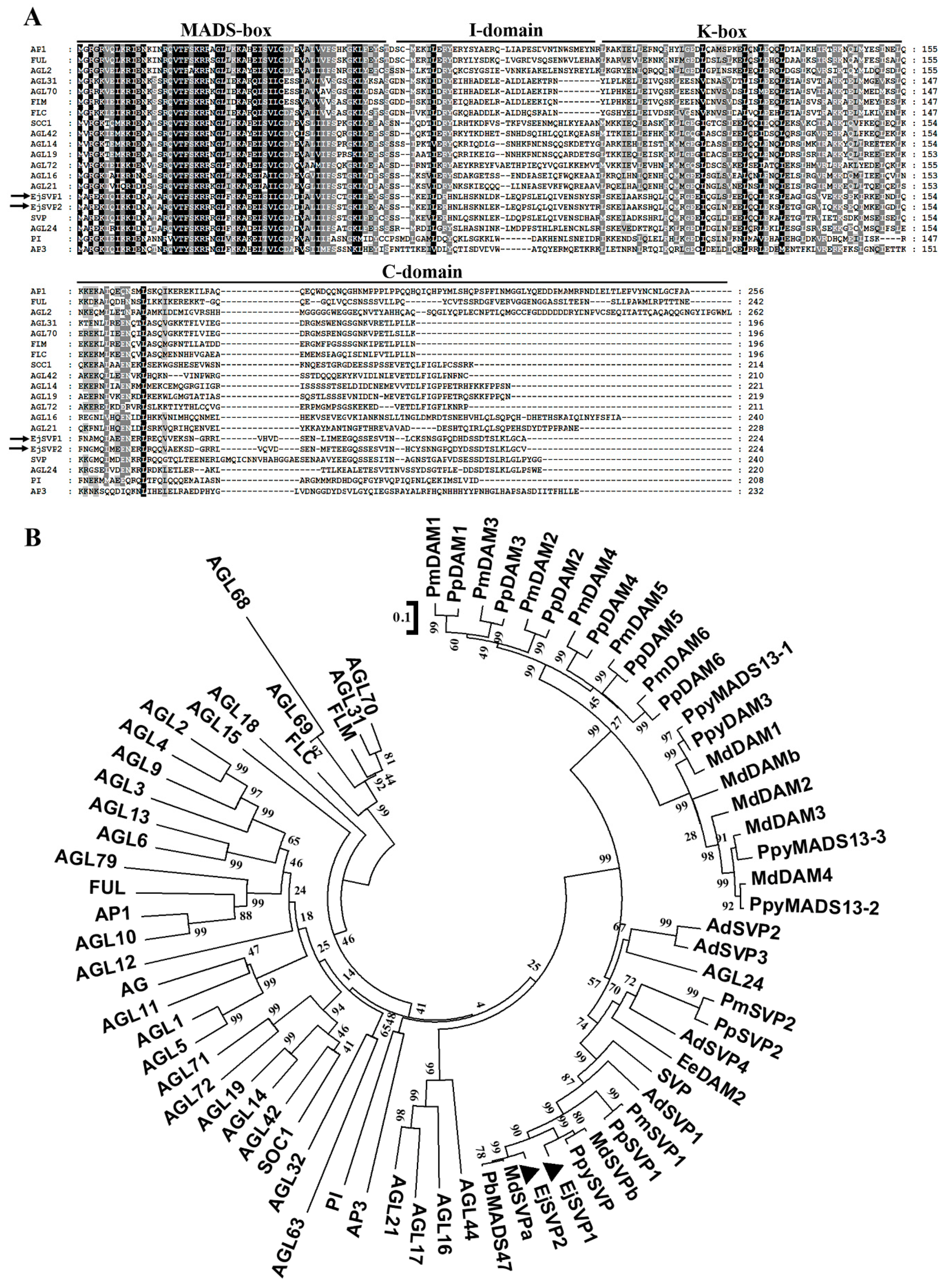
2.1. Cloning and Identification of Loquat EjSVPs

Two SVP homologous genes were isolated from loquat: EjSVP1 and EjSVP2. Their full-length coding sequences are 675 bp and they encode 224 amino acids (Supplementary Sequence S1 and S2). Their sequences are highly similar, with a nucleotide sequence identity of 92.74% and a predicted amino acid sequence identity of 86.61%.
As described above, the MIKC-type MADS-box transcription factor includes four structures: the MADS-box, I-domain, K-box and C-domain. Sequence analysis showed that both EjSVP proteins have four typical structures: the MADS-box, I-domain, K-box and C-domain (Figure 1A).
According to the results of the phylogenetic tree, EjSVP1 and EjSVP2 are clustered with the SVP-like/DAM proteins of other plants. More importantly, EjSVPs and the Maloideae SVP-like/DAM protein clustered in a small clade (Figure 1B).

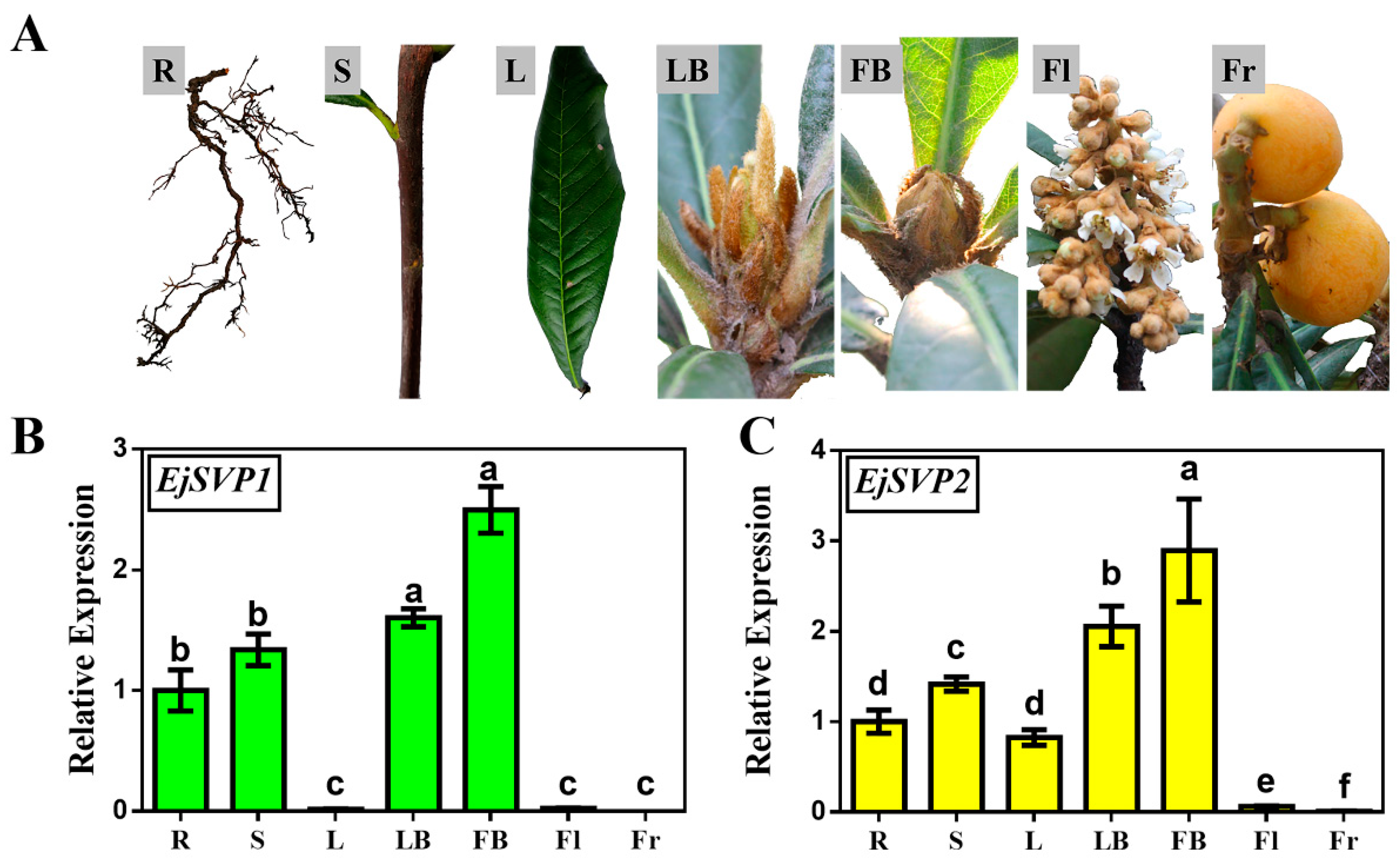
2.2. Tissue-Specific Expression Patterns of EjSVPs in Loquat

To explore the potential functions of EjSVP1 and EjSVP2 in loquat, the transcription levels of EjSVP1 and EjSVP2 in various tissues of ‘Jiefangzhong’ were investigated, including the roots, stems, leaves, leaf buds, flower buds, flowers, and fruit (Figure 2A).
qRT-PCR results showed that EjSVP1 and EjSVP2 were mainly expressed in the roots, stems, leaf buds and flower buds (Figure 2B, C). Both genes were slightly expressed in the inflorescences. EjSVP1 was hardly expressed in mature leaves (Figure 2B), while the expression level of EjSVP2 was relatively high (Figure 2C). The expression patterns of EjSVP1 and EjSVP2 in mature leaves were significantly different, indicating that the two genes may perform different functions during the growth and development of loquat.

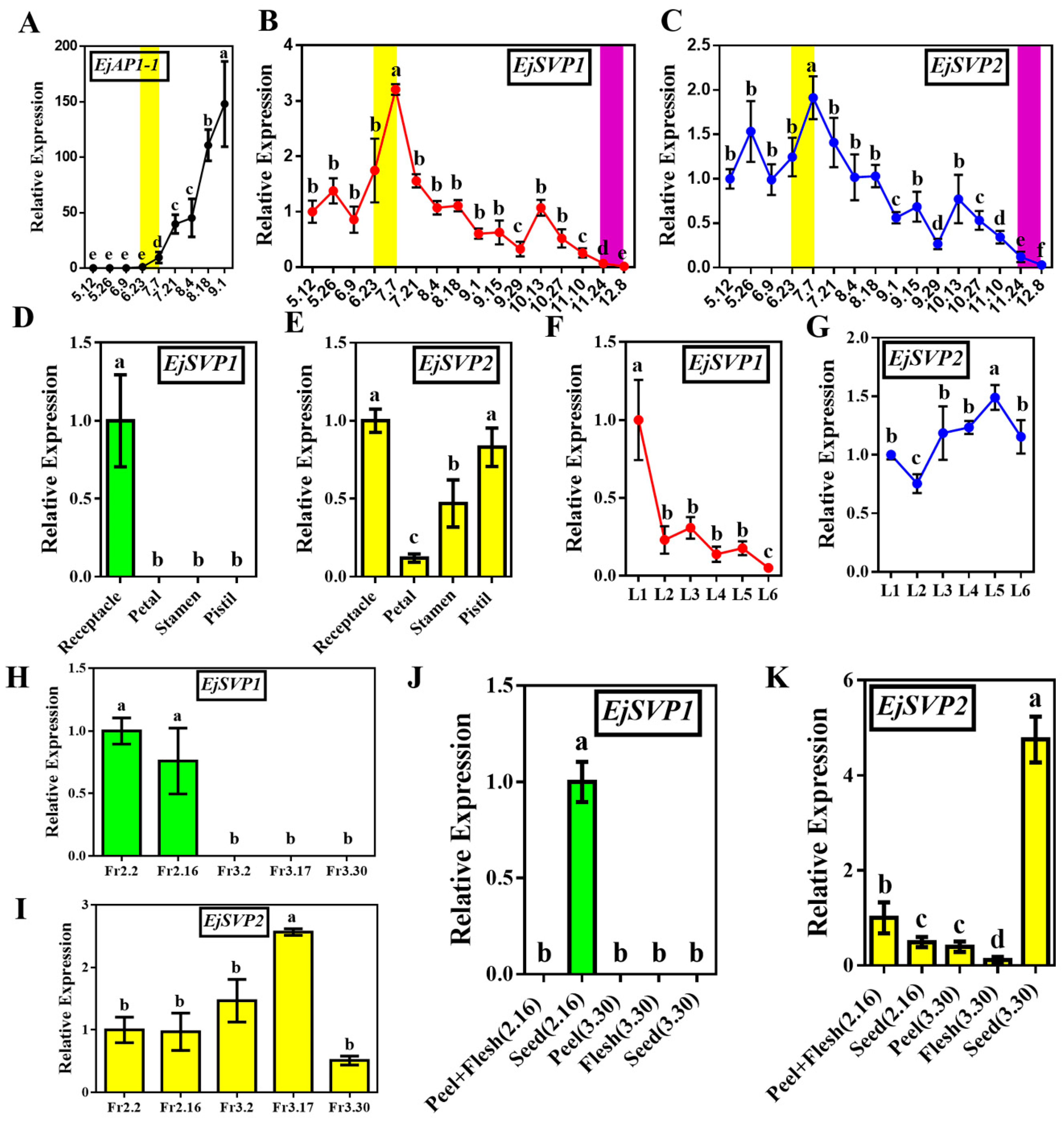
2.3. Temporal and Spatial Expression Patterns of EjSVPs in Loquat

To further clarify the roles of EjSVP1 and EjSVP2 in loquat, we analyzed the expression levels of EjSVPs in the shoot apical meristems (SAM) (different developmental stages), fruit (different developmental stages), flower (different tissues) and leaves (different maturities in the same period; see Supplementary Figure S1).
The ortholog gene EjAP1-1 of the floral meristem identity gene AP1 has been confirmed as a marker gene for identifying loquat floral bud differentiation [43]. In this experiment, we found a high level of EjAP1-1 expression after 23 June (Figure 3A), which is consistent with the results of Jiang et al. [43]. This finding indicates that the flower bud differentiation of loquat occurs towards the end of June and in early July.
The expression trends of EjSVP1 and EjSVP2 were similar during flower development in SAM. The expression levels of the two genes were relatively high after the initiation of flower bud differentiation. With the development of flower buds, their expression levels gradually decreased until they were almost undetectable in open flowers (Figure 3B,C). In different tissues of the flowers (not opened), EjSVP1 was expressed only in the receptacle, while EjSVP2 was expressed in various tissues (Figure 3D,E).
EjSVP1 was mainly expressed in L1–L3 (the leaves at different maturities in the same period) and was almost undetectable after leaf maturity (L6) (Figure 3F), while EjSVP2 was expressed in several stages of leaf development (Figure 3G). Moreover, as the leaves developed, the expression level of EjSVP2 gradually increased and then slightly decreased after maturity (Figure 3G). Similarly, during fruit development, EjSVP1 was mainly expressed in the young fruit stage (Fr2.2–Fr2.16) but only in the seeds (Figure 3H,J), while EjSVP2 was relatively highly expressed during the whole developmental period, and the peak expression occurred during the fruit expansion period (Fr3.2–Fr3.17) (Figure 3I). Regarding different fruit tissues and different periods, EjSVP2 was mainly expressed in the flesh and peel of young fruit (the peel could not be separated at this stage), and expressed in the seeds of mature fruits (Figure 3K). Previous studies have shown that the leaves undergo intense cell division during the L1–L2 period [50,51].

2.4. Effects of Exogenous GA3 Treatment and Short-Day Treatment on the Expression of EjSVPs

Loquat could not undergo flower bud differentiation after GA3 and short-day (SD) treatments. To explore the effects of two treatments on EjSVPs, the transcription levels of EjAP1-1, EjSVP1 and EjSVP2 were detected. EjAP1-1 was strongly inhibited after GA3 and SD treatment (Figure 4A,D), and this result is consistent with Jiang et al. [43]. Compared with the control group, the expression levels of EjSVP1 and EjSVP2 were higher in the days after GA3 treatment but returned to the control group when stopping treatment (Figure 4B,C). The expression levels of EjSVP1 were decreased after SD treatment, while EjSVP2 expression levels did not show significant changes (Figure 4E,F).

2.5. Subcellular Localization of EjSVPs

To determine the cellular localization of the encoded EjSVP proteins, two fusion expression vectors, 35S:EjSVP1-GFP and 35S:EjSVP2-GFP, were constructed and transferred into the epidermis of tobacco plant cells to observe the fluorescence signal. The green fluorescence of the 35S:EjSVP1-GFP and 35S:EjSVP2-GFP fusion proteins were detected in the nucleus (Figure 5), indicating that EjSVP1 and EjSVP2 are localized in the nucleus. This result is consistent with the characteristics of general transcription factors.

2.6. Promoter Analysis of EjSVPs

Previous studies have shown that the C-repeat/dehydration-responsive element (CRT/DRE) is a cold response element found in leafy spurge [39], Japanese pear [27,52], apple [16,17] and Japanese apricot [35]. C-repeat binding factors (CBFs) increase rapidly at low temperatures [16,17,27,34,35,53,54]. CBF recognizes and binds to the CRT/DRE motif to regulate the expression of SVP-like/DAM genes.
To explore why EjSVPs possess this expression pattern that is different from other dormant plants, their promoter (sequence information was obtained from the de novo genome sequencing project of loquat) structural elements were analyzed. No CCGAC structural elements (CRT/DRE) were found in the promoter regions of the two EjSVPs (Supplementary Sequence S3, S4). Interestingly, one CCAAT structural element was found in the promoter region of EjSVP1 (Supplementary Sequence S3), and four CCAAT structural elements were found in the promoter region of EjSVP2 (Supplementary Sequence S4). The cis-element CCAAT DNA-binding motif is a structural element that responds to the key gene CO of the photoperiod pathway and is called CORE (CO response element) [55,56]. Interestingly, EjSVPs began to be highly expressed after 23 June (Figure 3B,C), which is the longest day of sunshine in Guangzhou.
In addition, AuxRR-core (GGTCCAT) was also found in the promoter region of EjSVP1 but was not found in the promoter region of EjSVP2 (Supplementary Sequence S3), which may be the reason for the relatively high expression of EjSVP1 in tender tissues.

2.7. Functional Analysis of EjSVPs in Arabidopsis thaliana

We overexpressed the EjSVP genes in wild-type Arabidopsis thaliana Col-0 and found that the number of rosettes of T3 generation 35S:EjSVP1 and 35S:EjSVP2 transgenic lines were similar to that of wild-type Col-0 (Figure 6A,B). We then performed qRT-PCR and RT-PCR analysis of EjSVP in the transgenic lines and found that EjSVP were abundantly expressed in the transgenic lines (Figure 6C–E). In addition, we performed qRT-PCR analysis of AtSOC1 and the floral meristem identity gene AtAP1 in Arabidopsis thaliana, and the results showed that the AtSOC1 expression levels in the transgenic lines were not significantly different from that in Col-0 (Figure 6F). The expression level of AtAP1 was not significantly different between the 35S:EjSVP1 transgenic lines and the Col-0 line. The 35S:EjSVP2 transgenic line #4 exhibited significant higher expression of AtAP1 compared to wild type, while no difference could be observed in line #10 (Figure 6A,B,G).
Interestingly, compared to the wild type Arabidopsis thaliana Col-0 phenotype (Figure 7G,H), in the 35S:EjSVP2 transgenic line, various types of floral organ variations were produced, including flowers that had bract-like sepals (Figure 7A–D,F,J), stunted stamens (Figure 7B–E,J), the formation of a secondary flower (Figure 7B–F), and petals that accumulated pigment (Figure 7I). However, the 35S:EjSVP1 transgenic line did not have any of these features.

3. Discussion

Rosaceae woody fruit trees are extremely important in peoples’ lives and bring great benefits to growers every year. Loquat belongs to the Maloideae family, and its flowering habit is unique for woody fruit trees. It is interesting and meaningful to explore the reasons for its different flowering habits compared to apples and pears. Moreover, the flowering times among the 26 species of Eriobotrya are different [57]. For example, Eriobotrya deflexa Nakai blooms in spring, similar to apples [48,49,58]. The flowering habits and varying germplasms in Eriobotrya have demonstrated that loquat could be an ideal model material for the study of Rosaceae flowering shifts to adapt to climate changes. Studying the regulation mechanism of loquat flowering could provide a favorable reference for the flowering regulation of other woody plants.
Our results show that EjSVPs and MdSVPs form a small clade with a high genetic relationship (Figure 1B), suggesting that they are more similar in structure and closer in genetic relationship, and thus suggest that EjSVPs could have similar functions to the SVP-like/DAM proteins of Maloideae. However, the SVP/DAM gene in apples is mainly involved in dormancy [15,16,17,18,19,20,21]. Jiang et al. [43] showed that loquat did not undergo dormancy, but rather its flower bud differentiation was continuous and blossomed in autumn and winter. The expression pattern of EjSVPs is also different from the orthologous genes in apples [21]. Additionally, analysis of promoter cis-structural elements found that the promotor of EjSVPs is different from other plants and does not contain CRT/DRE (Supplementary Sequence S3, S4). These findings indicate that loquat produces a unique pattern of flowering regulation during natural selection. However, the specific reasons for these flowering characteristics, as well as the functional changes of EjSVPs, require further progress.
EjSVP1 expression levels were relatively high in young tissues, and EjSVP2 expression levels were relatively high in young to mature transformed tissues (Figure 3). Furthermore, exogenous GA3 could strongly inhibit the expression of EjSOC1s and EjAP1s, and the SAM showed strong vegetative growth, while the stem tip of the short-day treatment group almost stopped growing, and the loquat could not undergo flower bud differentiation [43]. Our results show that GA3 treatment promotes the expression of EjSVPs, whereas SD treatment has some inhibitory effects on the expression of EjSVP1 (Figure 4). Based on these results, we speculated that EjSVP1 may regulate cell proliferation, and EjSVP2 may be involved in cell expansion.
In Arabidopsis thaliana, SVP genes are expressed at relatively high levels in the roots and leaves during vegetative growth, while they are expressed in low amounts in inflorescences, and hardly expressed in flowers and siliques [11]. However, in this experiment, EjSVPs were found to be highly expressed in flower buds (Figure 2B,C). In the 35S:EjSVP2 transgenic line, the flowers and siliques produced variations, such as flowers that had bract-like sepals, stunted stamens, the formation of secondary flowers, and petals that accumulated pigment (Figure 7). However, these phenotypes were not found in the 35S:EjSVP1 transgenic line. Combined with the qRT-PCR results from loquat flowers, EjSVP1 was found to be only expressed in receptacles, while EjSVP2 was expressed in receptacles, petals, and stamens, especially in the pistils. These findings suggest that EjSVP2 actively participates in the morphogenesis of flowers in loquats.
In this study, the 35S:EjSVP2 transgenic line showed a phenotype similar to the 35S:AGL24 transgenic line (Figure 7A–F) [59,60]. In addition, phylogenetic tree analysis found that EjSVPs and AtAGL24 were clustered together (Figure 1B). Interestingly, in Arabidopsis thaliana, the heterodimerization of SOC1 and AGL24 activates the expression of the downstream floral meristem identity gene LFY and promotes flowering [60,61]. More importantly, in a study of Japanese apricot, PmDAM6 (SVP-like) was found to interact with PmSOC1 [62]. Moreover, EjSOC1 in loquat has also been verified to play an active role in flower regulation, and the expression pattern in SAM was similar to that of EjSVPs (Figure 2B,C) [43]. We hypothesize that in loquat, EjSVP2 may be more similar to the regulation pattern of AGL24 in Arabidopsis thaliana, where it interacts with EjSOC1 to regulate the development of loquat flowering.
The SVP gene also plays an important role in response to temperature signals, which inhibits flowering by negatively regulating FT expression [63]. SVP can also promote EARLY FLOWERING MYB PROTEIN (EFM) expression, and EFM can regulate flowering in response to temperature-suppressed FT expression [64]. In addition, when the temperature is changed from 16 °C to 27 °C, more FLOWERING LOCUS M (FLM)-δ-SVP complexes are formed, which hinders FLM-β and SVP from inhibiting flowering. Higher temperatures can degrade SVP protein, and then SVP-FLM-β complexes are reduced, thereby activating downstream target genes such as FT and SOC1 expression, promoting flowering [65]. Interestingly, from May to July, the temperature rose (Supplementary Figure S2A), and the expression of EjSVPs began to rise sharply and peak (Figure 3B,C). On the other hand, the phytohormone abscisic acid (ABA) plays a key role in response to stress such as drought. Under long-days and drought conditions, GIGANTEA (GI) can positively regulate the expression of FT and SOC1 to promote flowering, and requires the participation of ABA, independent of CO; in short-days, SVP can prevent ABA from activating SOC1 [66]. Importantly, there was more rain in May–June, and the rain was greatly reduced in July (Supplementary Figure S2B). These results suggested that EjSVPs may be involved in the regulation of loquat flowering by temperature signal or drought stress.

4. Materials and Methods

4.1. Plant Materials and Growth Conditions

Twelve-year-old ‘Jiefanzhong’ loquat (Eriobotrya japonica Lindl.) were used in each experiment. The trees were located at the loquat germplasm resource preservation garden (South China Agricultural University, Guangzhou, China), and showed normal flowering. Selected disease-free plant tissues (roots, stems, leaves, buds, flowers and fruits) with consistent phenotypes were collected at each sampling (tissues were taken at 4 p.m., every two weeks). Samples for qRT-PCR analysis were frozen in liquid nitrogen and then stored in an ultralow temperature freezer at −80 °C until use. Arabidopsis thaliana wild-type Col-0 and Nicotiana benthamiana were used in this study and grown under long-day conditions (16 h light/8 h dark cycle) at 22 °C.

4.2. RNA Extraction, Reverse-Transcription, Gene Isolation, and Sequence Analysis

Total RNA was extracted using an EASY Spin Plus Plant RNA Extraction kit (Aidlab, Beijing, China). First-strand cDNA was generated from loquat bud RNA using a PrimeScriptTM RT reagent kit with gDNA Eraser (TaKaRa, shiga, Japan), following the manufacturer’s instructions.
The full-length coding sequences of EjSVP1 and EjSVP2 were isolated from first-strand cDNA using Prime STAR® Max DNA Polymerase (TaKaRa, shiga, Japan). The two primers for EjSVP1 were 5′-ATGGCGAGGGAGAAGATTCAGAT-3′ and 5′-TTAAGCGCACCCCAATTTTAGAGA-3′; and the two for EjSVP2 were 5′-ATGGCGAGGGAGAAAATTCAGAT-3′ and 5′-TTAAACGCACCCCAATTTTAGAGA-3′. The PCR conditions were carried out in strict accordance with the reagent instructions. Sequence information was obtained from the de novo genome sequencing project of loquat, which has not yet been published. Sequence analysis was carried out according to a previous method [43]. Predictive analysis of cis-acting elements of the promoter of EjSVPs was performed online at and PLACE (https://www.dna.affrc.go.jp/PLACE/?action=newplace) and PlantCARE (http://bioinformatics.psb.ugent.be/webtools/plantcare/html/).

4.3. Gene Expression Analysis

Quantitative real time polymerase chain reaction (qRT-PCR) using iTaqTM universal SYBR Green Super mix kit (Bio-Rad, Hercules, CA, USA) was carried out in triplicate using a LightCyclerR 480 system (Roche, Basel, Switzerland). The relative expression levels were all evaluated by the 2−ΔΔCt (cycle threshold) method [67]. Ejβ-Actin was used as an internal control for loquat [68], and AtTUB2 (AT5G62690) for Arabidopsis thaliana [64]. The relative expression levels were expressed as the ratio of the expression levels between the genes and the reference gene, so as to indicate the relative transcript levels of the genes compared the levels of Ejβ-actin or AtTUB2. The primer sequences used in qRT-PCR for EjAP1-1 have been described by Jiang et al. [43]. Two primers were used for EjSVP1, 5′-GAAGTCCCTTGAAGCTGGCT-3′ and 5′-CTCTTCCGCCAATTGCATCG-3′; and two for EjSVP2, 5′-TCCCTTGAAACTGGCTTGGG-3′ and 5′-CTCCGCCACTTGCTGTCTTA-3′. The primer sequences used in RT-PCR for EjSVPs were the same as the cloning primers (stop codon removed). The PCR conditions were carried out in strict accordance with the reagent instructions. The RT-PCR product was detected by agarose gel electrophoresis (10 mg mL−1). Data were collected from three biological replicates.

4.4. Short-Day and GA3 Treatments

For short-day (SD) treatment, the natural light was set for 8 h (10 a.m.–6 p.m.), with full darkness for 16 h (6 p.m.–10 a.m. the following day) using shading. The control plants were grown naturally. In the GA3 treatment, 300 mg L−1 of GA3 aqueous solution (Dingguo, Beijing, China) was sprayed onto the plants every two weeks, and 0.1% (v/v) phosphoric acid and 0.025% (v/v) Triton X-100 were added as surfactants. The control plants were sprayed with an aqueous solution containing the surfactants. The experimental treatment time was from 18 May 2018 to 10 August 2018. The SAM of the treatment groups and the control groups was used for qRT-PCR analysis, and the sampling method was the same as in Section 4.1.

4.5. Subcellular Localization and Arabidopsis thaliana Transformation

EjSVP1 and EjSVP2 were cloned into pGreen-35S-GFP [69] for subcellular localization analysis and into pGreen-35S [69] for Arabidopsis thaliana transformation. Each construct was introduced into Agrobacterium tumefaciens GV3101::pSoup and then transformed into Arabidopsis thaliana Col-0 using the floral dip method [70] or the transient transformation of N. benthamiana leaves [71]. Transgenic seedlings were selected on soil using Basta (3.5 mg L-1). Fluorescence microscopy (Observer D1, Zeiss, Jena, Germany) was used to detect the fluorescence signal of the EjSVP-GFP fusion proteins.

4.6. Statistics and Analysis

Microsoft Office Excel was used to analyze the data, and the difference between the data was assessed by student’s t test. Graph production was performed using Microsoft Office PowerPoint and GraphPad Prism 6 software.

5. Conclusions

In this study, we identified and named two EjSVP genes from loquat. Through expression analysis during flower development, we found that two EjSVP genes play an active role in flower development. At the same time, temporal and spatial expression analysis and GA3 and SD treatment results suggest that EjSVPs may be involved in cell division or growth. In addition, overexpression of EjSVPs to wild type Arabidopsis thaliana Col-0 had no significant effect on the change of rosette leaves. The promoter region of EjSVP genes in loquat does not contain CRT/DRE, which may be the main reason that the flowering habits of loquat differ from other fruit trees in the Rosaceae family, which also provides an important clue for the study of loquat flower regulation.

Supplementary Materials

Supplementary materials can be found at https://www.mdpi.com/1422-0067/20/23/5933/s1.

Author Contributions

Y.J., J.P. and X.Y. mainly performed the research. Y.J., J.P. and Z.Z. wrote the manuscript. S.L. (Shoukai Lin), S.L. (Shunquan Lin), X.Y. and Z.Z. revised and approved the manuscript. All authors read and approved the final manuscript.

Funding

This study was supported by Key Realm R&D Program of GuangDong Provide (2018B020202011), Open Fund of Key laboratory of Loquat Germplasm Innovation and Utilization (Putian University), Fujian Province University (2019005).

Acknowledgments

We are grateful for the experimental instruments provided by the Key Laboratory of Innovation and Utilization of Horticultural Crop Resources in South China (Ministry of Agriculture). We wish to thank American Journal Experts for their language editing services for this manuscript.

Conflicts of Interest

The authors declare no conflict of interest.

References

  1. Moon, J.; Lee, H.; Kim, M.; Lee, I. Analysis of flowering pathway integrators in Arabidopsis. Plant Cell Physiol. 2005, 46, 292–299. [Google Scholar] [CrossRef] [PubMed]
  2. Amasino, R.M.; Michaels, S.D. The timing of flowering. Plant Physiol. 2010, 154, 516–520. [Google Scholar] [CrossRef] [PubMed]
  3. Srikanth, A.; Schmid, M. Regulation of flowering time: All roads lead to Rome. Cell Mol. Life Sci. 2011, 68, 2013–2037. [Google Scholar] [CrossRef] [PubMed]
  4. Song, Y.H.; Ito, S.; Imaizumi, T. Flowering time regulation: Photoperiod- and temperature-sensing in leaves. Trends Plant Sci. 2013, 18, 575–583. [Google Scholar] [CrossRef]
  5. Teotia, S.; Tang, G. To bloom or not to bloom: Role of microRNAs in plant flowering. Mol. Plant 2015, 8, 359–377. [Google Scholar] [CrossRef]
  6. Theissen, G.; Kim, J.T.; Saedler, H. Classification and phylogeny of the MADS-box multigene family suggest defined roles of MADS-box gene subfamilies in the morphological evolution of eukaryotes. J. Mol. Evol. 1996, 43, 484–516. [Google Scholar] [CrossRef]
  7. Parenicova, L.; de Folter, S.; Kieffer, M.; Horner, D.S.; Favalli, C.; Busscher, J.; Cook, H.E.; Ingram, R.M.; Kater, M.M.; Davies, B.; et al. Molecular and phylogenetic analyses of the complete MADS-box transcription factor family in Arabidopsis: New openings to the MADS world. Plant Cell 2003, 15, 1538–1551. [Google Scholar] [CrossRef]
  8. Vandenbussche, M.; Theissen, G.; Van de Peer, Y.; Gerats, T. Structural diversification and neo-functionalization during floral MADS-box gene evolution by C-terminal frameshift mutations. Nucleic. Acids Res. 2003, 31, 4401–4409. [Google Scholar] [CrossRef]
  9. Smaczniak, C.; Immink, R.G.; Angenent, G.C.; Kaufmann, K. Developmental and evolutionary diversity of plant MADS-domain factors: Insights from recent studies. Development 2012, 139, 3081–3098. [Google Scholar] [CrossRef]
  10. Chen, F.; Zhang, X.; Liu, X.; Zhang, L. Evolutionary Analysis of MIKC(c)-Type MADS-Box Genes in Gymnosperms and Angiosperms. Front. Plant. Sci 2017, 8, 895. [Google Scholar] [CrossRef]
  11. Hartmann, U.; Hohmann, S.; Nettesheim, K.; Wisman, E.; Saedler, H.; Huijser, P. Molecular cloning of SVP: A negative regulator of the floral transition in Arabidopsis. Plant J. 2000, 21, 351–360. [Google Scholar] [CrossRef] [PubMed]
  12. Gregis, V.; Sessa, A.; Dorca-Fornell, C.; Kater, M.M. The Arabidopsis floral meristem identity genes AP1, AGL24 and SVP directly repress class B and C floral homeotic genes. Plant J. 2009, 60, 626–637. [Google Scholar] [CrossRef] [PubMed]
  13. Thouet, J.; Quinet, M.; Lutts, S.; Kinet, J.M.; Perilleux, C. Repression of floral meristem fate is crucial in shaping tomato inflorescence. PLoS ONE 2012, 7, e31096. [Google Scholar] [CrossRef] [PubMed]
  14. Falavigna, V.d.S.; Guitton, B.; Costes, E.; Andres, F. I Want to (Bud) Break Free: The Potential Role of DAM and SVP-Like Genes in Regulating Dormancy Cycle in Temperate Fruit Trees. Front. Plant Sci. 2019, 9. [Google Scholar] [CrossRef] [PubMed]
  15. Falavigna, V.d.S.; Porto, D.D.; Buffon, V.; Margis-Pinheiro, M.; Pasquali, G.; Revers, L.F. Differential Transcriptional Profiles of Dormancy-Related Genes in Apple Buds. Plant Mol. Biol. Report. 2014, 32, 796–813. [Google Scholar] [CrossRef]
  16. Mimida, N.; Saito, T.; Moriguchi, T.; Suzuki, A.; Komori, S.; Wada, M. Expression of DORMANCY-ASSOCIATED MADS-BOX (DAM)-like genes in apple. Biol. Plant 2015, 59, 237–244. [Google Scholar] [CrossRef]
  17. Wisniewski, M.; Norelli, J.; Artlip, T. Overexpression of a peach CBF gene in apple: A model for understanding the integration of growth, dormancy, and cold hardiness in woody plants. Front. Plant Sci. 2015, 6, 85. [Google Scholar] [CrossRef]
  18. Kumar, G.; Arya, P.; Gupta, K.; Randhawa, V.; Acharya, V.; Singh, A.K. Comparative phylogenetic analysis and transcriptional profiling of MADS-box gene family identified DAM and FLC-like genes in apple (Malus x domestica). Sci. Rep. 2016, 6. [Google Scholar] [CrossRef]
  19. Kumar, G.; Gupta, K.; Pathania, S.; Swarnkar, M.K.; Rattan, U.K.; Singh, G.; Sharma, R.K.; Singh, A.K. Chilling Affects Phytohormone and Post-Embryonic Development Pathways during Bud Break and Fruit Set in Apple (Malus domestica Borkh.). Sci. Rep. 2017, 7. [Google Scholar] [CrossRef]
  20. Porto, D.D.; Falavigna, V.D.; Arenhart, R.A.; Perini, P.; Buffon, V.; Anzanello, R.; dos Santos, H.P.; Fialho, F.B.; de Oliveira, P.R.D.; Revers, L.F. Structural genomics and transcriptional characterization of the Dormancy-Associated MADS-box genes during bud dormancy progression in apple. Tree Genet. Genomes 2016, 12. [Google Scholar] [CrossRef]
  21. Wu, R.; Tomes, S.; Karunairetnam, S.; Tustin, S.D.; Hellens, R.P.; Allan, A.C.; Macknight, R.C.; Varkonyi-Gasic, E. SVP-like MADS Box Genes Control Dormancy and Budbreak in Apple. Front. Plant Sci. 2017, 8. [Google Scholar] [CrossRef] [PubMed]
  22. Ubi, B.E.; Sakamoto, D.; Ban, Y.; Shimada, T.; Ito, A.; Nakajima, I.; Takemura, Y.; Tamura, F.; Saito, T.; Moriguchi, T. Molecular Cloning of Dormancy-associated MADS-box Gene Homologs and Their Characterization during Seasonal Endodormancy Transitional Phases of Japanese Pear. J. Am. Soc. Hortic. Sci. 2010, 135, 174–182. [Google Scholar] [CrossRef]
  23. Liu, G.; Li, W.; Zheng, P.; Xu, T.; Chen, L.; Liu, D.; Hussain, S.; Teng, Y. Transcriptomic analysis of ‘Suli’ pear (Pyrus pyrifolia white pear group) buds during the dormancy by RNA-Seq. Bmc Genom. 2012, 13. [Google Scholar] [CrossRef] [PubMed]
  24. Bai, S.; Saito, T.; Sakamoto, D.; Ito, A.; Fujii, H.; Moriguchi, T. Transcriptome Analysis of Japanese Pear (Pyrus pyrifolia Nakai) Flower Buds Transitioning Through Endodormancy. Plant Cell Physiol. 2013, 54, 1132–1151. [Google Scholar] [CrossRef] [PubMed]
  25. Saito, T.; Bai, S.; Ito, A.; Sakamoto, D.; Saito, T.; Ubi, B.E.; Imai, T.; Moriguchi, T. Expression and genomic structure of the dormancy-associated MADS box genes MADS13 in Japanese pears (Pyrus pyrifolia Nakai) that differ in their chilling requirement for endodormancy release. Tree Physiol. 2013, 33, 654–667. [Google Scholar] [CrossRef] [PubMed]
  26. Ito, A.; Saito, T.; Sakamoto, D.; Sugiura, T.; Bai, S.; Moriguchi, T. Physiological differences between bud breaking and flowering after dormancy completion revealed by DAM and FT/TFL1 expression in Japanese pear (Pyrus pyrifolia). Tree Physiol. 2016, 36, 109–120. [Google Scholar] [CrossRef]
  27. Niu, Q.; Li, J.; Cai, D.; Qian, M.; Jia, H.; Bai, S.; Hussain, S.; Liu, G.; Teng, Y.; Zheng, X. Dormancy-associated MADS-box genes and microRNAs jointly control dormancy transition in pear (Pyrus pyrifolia white pear group) flower bud. J. Exp. Bot. 2016, 67, 239–257. [Google Scholar] [CrossRef]
  28. Li, Z.; Reighard, G.L.; Abbott, A.G.; Bielenberg, D.G. Dormancy-associated MADS genes from the EVG locus of peach Prunus persica (L.) Batsch have distinct seasonal and photoperiodic expression patterns. J. Exp. Bot. 2009, 60, 3521–3530. [Google Scholar] [CrossRef]
  29. Jimenez, S.; Reighard, G.L.; Bielenberg, D.G. Gene expression of DAM5 and DAM6 is suppressed by chilling temperatures and inversely correlated with bud break rate. Plant Mol. Biol. 2010, 73, 157–167. [Google Scholar] [CrossRef]
  30. Yamane, H.; Ooka, T.; Jotatsu, H.; Hosaka, Y.; Sasaki, R.; Tao, R. Expressional regulation of PpDAM5 and PpDAM6, peach (Prunus persica) dormancy-associated MADS-box genes, by low temperature and dormancy-breaking reagent treatment. J. Exp. Bot. 2011, 62, 3481–3488. [Google Scholar] [CrossRef]
  31. Yamane, H.; Tao, R.; Ooka, T.; Jotatsu, H.; Sasaki, R.; Yonemori, K. Comparative Analyses of Dormancy-associated MADS-box Genes, PpDAM5 and PpDAM6, in Low- and High-chill Peaches (Prunus persica L.). J. Jpn. Soc. Hortic. Sci. 2011, 80, 276–283. [Google Scholar] [CrossRef] [Green Version]
  32. Yamane, H.; Ooka, T.; Jotatsu, H.; Sasaki, R.; Tao, R. Expression analysis of PpDAM5 and PpDAM6 during flower bud development in peach (Prunus persica). Sci Hortic. (Amst.) 2011, 129, 844–848. [Google Scholar] [CrossRef]
  33. Sasaki, R.; Yamane, H.; Ooka, T.; Jotatsu, H.; Kitamura, Y.; Akagi, T.; Tao, R. Functional and Expressional Analyses of PmDAM Genes Associated with Endodormancy in Japanese Apricot. Plant Physiol. 2011, 157, 485–497. [Google Scholar] [CrossRef] [PubMed] [Green Version]
  34. Zhao, K.; Zhou, Y.; Li, Y.; Zhuo, X.; Ahmad, S.; Han, Y.; Yong, X.; Zhang, Q. Crosstalk of PmCBFs and PmDAMs Based on the Changes of Phytohormones under Seasonal Cold Stress in the Stem of Prunus mume. Int. J. Mol. Sci. 2018, 19, 15. [Google Scholar] [CrossRef] [PubMed] [Green Version]
  35. Zhao, K.; Zhou, Y.; Ahmad, S.; Yong, X.; Xie, X.; Han, Y.; Li, Y.; Sun, L.; Zhang, Q. PmCBFs synthetically affect PmDAM6 by alternative promoter binding and protein complexes towards the dormancy of bud for Prunus mume. Sci. Rep. 2018, 8. [Google Scholar] [CrossRef] [PubMed] [Green Version]
  36. Zhu, Y.; Li, Y.; Xin, D.; Chen, W.; Shao, X.; Wang, Y.; Guo, W. RNA-Seq-based transcriptome analysis of dormant flower buds of Chinese cherry (Prunus pseudocerasus). Gene 2015, 555, 362–376. [Google Scholar] [CrossRef]
  37. Rothkegel, K.; Sanchez, E.; Montes, C.; Greve, M.; Tapia, S.; Bravo, S.; Prieto, H.; Miyasaka Almeida, A. DNA methylation and small interference RNAs participate in the regulation of MADS-box genes involved in dormancy in sweet cherry (Prunus avium L.). Tree Physiol. 2017, 37, 1739–1751. [Google Scholar] [CrossRef]
  38. Horvath, D.P.; Chao, W.S.; Suttle, J.C.; Thimmapuram, J.; Anderson, J.V. Transcriptome analysis identifies novel responses and potential regulatory genes involved in seasonal dormancy transitions of leafy spurge (Euphorbia esula L.). Bmc Genom. 2008, 9, 536. [Google Scholar] [CrossRef] [Green Version]
  39. Horvath, D.P.; Sung, S.; Kim, D.; Chao, W.; Anderson, J. Characterization, expression and function of DORMANCY ASSOCIATED MADS-BOX genes from leafy spurge. Plant Mol. Biol. 2010, 73, 169–179. [Google Scholar] [CrossRef]
  40. Wu, R.-M.; Walton, E.F.; Richardson, A.C.; Wood, M.; Hellens, R.P.; Varkonyi-Gasic, E. Conservation and divergence of four kiwifruit SVP-like MADS-box genes suggest distinct roles in kiwifruit bud dormancy and flowering. J. Exp. Bot. 2012, 63, 797–807. [Google Scholar] [CrossRef] [Green Version]
  41. Singh, R.K.; Maurya, J.P.; Azeez, A.; Miskolczi, P.; Tylewicz, S.; Stojkovic, K.; Delhomme, N.; Busov, V.; Bhalerao, R.P. A genetic network mediating the control of bud break in hybrid aspen. Nat. Commun. 2018, 9. [Google Scholar] [CrossRef] [PubMed] [Green Version]
  42. Kurokura, T.; Mimida, N.; Battey, N.H.; Hytonen, T. The regulation of seasonal flowering in the Rosaceae. J. Exp. Bot. 2013, 64, 4131–4141. [Google Scholar] [CrossRef] [PubMed] [Green Version]
  43. Jiang, Y.; Peng, J.; Zhu, Y.; Su, W.; Zhang, L.; Jing, Y.; Lin, S.; Gao, Y. The Role of EjSOC1s in Flower Initiation in Eriobotrya japonica. Front. Plant Sci 2019, 10, 253. [Google Scholar] [CrossRef] [PubMed] [Green Version]
  44. Esumi, T.; Tao, R.; Yonemori, K. Isolation of LEAFY and TERMINAL FLOWER 1 homologues from six fruit tree species in the subfamily Maloideae of the Rosaceae. Sex. Plant Reprod. 2005, 17, 277–287. [Google Scholar] [CrossRef]
  45. Liu, Y.; Zhao, Q.; Meng, N.; Song, H.; Li, C.; Hu, G.; Wu, J.; Lin, S.; Zhang, Z. Over-expression of EjLFY-1 Leads to an Early Flowering Habit in Strawberry (Fragaria x ananassa) and Its Asexual Progeny. Front. Plant Sci. 2017, 8. [Google Scholar] [CrossRef] [PubMed] [Green Version]
  46. Liu, Y.; Song, H.; Liu, Z.; Hu, G.; Lin, S. Molecular characterization of loquat EjAP1 gene in relation to flowering. Plant Growth Regul. 2013, 70, 287–296. [Google Scholar] [CrossRef]
  47. Reig, C.; Gil-Munoz, F.; Vera-Sirera, F.; Garcia-Lorca, A.; Martinez-Fuentes, A.; Mesejo, C.; Perez-Amador, M.A.; Agusti, M. Bud sprouting and floral induction and expression of FT in loquat [Eriobotrya japonica (Thunb.) Lindl.]. Planta 2017, 246, 915–925. [Google Scholar] [CrossRef]
  48. Zhang, L.; Yu, H.; Lin, S.; Gao, Y. Molecular Characterization of FT and FD Homologs from Eriobotrya deflexa Nakai forma koshunensis. Front. Plant Sci. 2016, 7, 8. [Google Scholar] [CrossRef] [Green Version]
  49. Zhang, L.; Jiang, Y.; Zhu, Y.; Su, W.; Long, T.; Huang, T.; Peng, J.; Yu, H.; Lin, S.; Gao, Y. Functional characterization of GI and CO homologs from Eriobotrya deflexa Nakai forma koshunensis. Plant Cell Rep. 2019. [Google Scholar] [CrossRef]
  50. Gonzalez, N.; Vanhaeren, H.; Inze, D. Leaf size control: Complex coordination of cell division and expansion. Trends Plant Sci. 2012, 17, 332–340. [Google Scholar] [CrossRef]
  51. Nelissen, H.; Gonzalez, N.; Inze, D. Leaf growth in dicots and monocots: So different yet so alike. Curr Opin Plant Biol. 2016, 33, 72–76. [Google Scholar] [CrossRef] [PubMed] [Green Version]
  52. Saito, T.; Bai, S.; Imai, T.; Ito, A.; Nakajima, I.; Moriguchi, T. Histone modification and signalling cascade of the dormancy-associated MADS-box gene, PpMADS13-1, in Japanese pear (Pyrus pyrifolia) during endodormancy. Plant Cell Environ. 2015, 38, 1157–1166. [Google Scholar] [CrossRef] [PubMed]
  53. Akhtar, M.; Jaiswal, A.; Taj, G.; Jaiswal, J.P.; Qureshi, M.I.; Singh, N.K. DREB1/CBF transcription factors: Their structure, function and role in abiotic stress tolerance in plants. J. Genet. 2012, 91, 385–395. [Google Scholar] [CrossRef] [PubMed]
  54. Zhao, C.; Lang, Z.; Zhu, J.-K. Cold responsive gene transcription becomes more complex. Trends Plant Sci. 2015, 20, 466–468. [Google Scholar] [CrossRef] [PubMed] [Green Version]
  55. Tiwari, S.B.; Shen, Y.; Chang, H.C.; Hou, Y.; Harris, A.; Ma, S.F.; McPartland, M.; Hymus, G.J.; Adam, L.; Marion, C.; et al. The flowering time regulator CONSTANS is recruited to the FLOWERING LOCUS T promoter via a unique cis-element. New Phytol 2010, 187, 57–66. [Google Scholar] [CrossRef]
  56. Adrian, J.; Farrona, S.; Reimer, J.J.; Albani, M.C.; Coupland, G.; Turck, F. cis-Regulatory elements and chromatin state coordinately control temporal and spatial expression of FLOWERING LOCUS T in Arabidopsis. Plant Cell 2010, 22, 1425–1440. [Google Scholar] [CrossRef] [Green Version]
  57. Lin, S.Q. A Review on Research of the Wild Species in Genus Eriobotrya Germplasm and Their Innovative Utilization. Acta Hortic. Sin. 2017, 44, 1704–1716. [Google Scholar] [CrossRef]
  58. Gu, C.; Spongberg, S.A. ERIOBOTRYA Lindley. In Flora of China; Zhengyi, W., Raven, P.H., Hong, D.Y., Eds.; Missouri Botanical Garden Press: Jefferson, MO, USA, 2003; Volume 9, pp. 138–140. [Google Scholar]
  59. Yu, H.; Ito, T.; Wellmer, F.; Meyerowitz, E.M. Repression of AGAMOUS-LIKE 24 is a crucial step in promoting flower development. Nat. Genet. 2004, 36, 157–161. [Google Scholar] [CrossRef] [Green Version]
  60. Liu, C.; Chen, H.; Er, H.L.; Soo, H.M.; Kumar, P.P.; Han, J.H.; Liou, Y.C.; Yu, H. Direct interaction of AGL24 and SOC1 integrates flowering signals in Arabidopsis. Development 2008, 135, 1481–1491. [Google Scholar] [CrossRef] [Green Version]
  61. Lee, J.; Oh, M.; Park, H.; Lee, I. SOC1 translocated to the nucleus by interaction with AGL24 directly regulates leafy. Plant J. 2008, 55, 832–843. [Google Scholar] [CrossRef]
  62. Kitamura, Y.; Takanori, T.; Yamane, H.; Tao, R. Simultaneous down-regulation of DORMANCY-ASSOCIATED MADS-box6 and SOC1 during dormancy release in Japanese apricot (Prunus mume) flower buds. Hortic. Sci. Biotechnol. 2016, 91, 476–482. [Google Scholar] [CrossRef]
  63. Jeong Hwan, L.; Seong Jeon, Y.; Soo Hyun, P.; Ildoo, H.; Jong Seob, L.; Hoon, A.J.J.G.D. Role of SVP in the control of flowering time by ambient temperature in Arabidopsis. Genes Dev. 2007, 21, 397–402. [Google Scholar]
  64. Yan, Y.; Shen, L.; Chen, Y.; Bao, S.; Thong, Z.; Yu, H. A MYB-domain protein EFM mediates flowering responses to environmental cues in Arabidopsis. Dev. Cell 2014, 30, 437–448. [Google Scholar] [CrossRef] [PubMed] [Green Version]
  65. Jeong Hwan, L.; Hak-Seung, R.; Kyung Sook, C.; David, P.; Soonkap, K.; Markus, S.; Hoon, A.J.J.S. Regulation of temperature-responsive flowering by MADS-box transcription factor repressors. Science 2013, 342, 628–632. [Google Scholar]
  66. Riboni, M.; Galbiati, M.; Tonelli, C.; Conti, L. GIGANTEA enables drought escape response via abscisic acid-dependent activation of the florigens and suppressor of overexpression of constans. Plant Physiol. 2013, 162, 1706–1719. [Google Scholar] [CrossRef] [PubMed] [Green Version]
  67. Livak, K.J.; Schmittgen, T.D. Analysis of relative gene expression data using real-time quantitative PCR and the 2(-Delta Delta C(T)) Method. Methods 2001, 25, 402–408. [Google Scholar] [CrossRef]
  68. Shan, L.L.; Li, X.; Wang, P.; Cai, C.; Zhang, B.; Sun, C.D.; Zhang, W.S.; Xu, C.J.; Ferguson, I.; Chen, K.S. Characterization of cDNAs associated with lignification and their expression profiles in loquat fruit with different lignin accumulation. Planta 2008, 227, 1243–1254. [Google Scholar] [CrossRef]
  69. Lee, L.Y.; Hou, X.; Fang, L.; Fan, S.; Kumar, P.P.; Yu, H. STUNTED mediates the control of cell proliferation by GA in Arabidopsis. Development 2012, 139, 1568–1576. [Google Scholar] [CrossRef] [Green Version]
  70. Zhang, X.; Henriques, R.; Lin, S.S.; Niu, Q.W.; Chua, N.H. Agrobacterium-mediated transformation of Arabidopsis thaliana using the floral dip method. Nat. Protoc. 2006, 1, 641–646. [Google Scholar] [CrossRef]
  71. Sparkes, I.A.; Runions, J.; Kearns, A.; Hawes, C. Rapid, transient expression of fluorescent fusion proteins in tobacco plants and generation of stably transformed plants. Nat. Protoc. 2006, 1, 2019–2025. [Google Scholar] [CrossRef]
Figure 1. Amino acid sequence alignment and phylogenetic analysis of EjSVP proteins. (A) Alignment of the amino acid sequences of the EjSVP proteins with the members of the Arabidopsis thaliana MIKC-type transcription factor family. The black line marks the highly conserved MADS-box and K-box domains. The I-domain is behind the MADS-box, and the C-domain is behind the K-box. (B) Phylogenetic analysis of MIKC-type transcription factors from Arabidopsis thaliana, SVP-like/DAM, and EjSVPs. The protein sequences were retrieved from NCBI (for accession IDs, see the Supplementary ID information).
Ijms 20 05933 g001
Figure 2. Tissue-specific expression levels of EjSVP1 and EjSVP2. (A) Different tissues from loquat. (B,C) Relative expression levels of EjSVP1 and EjSVP2 in the different tissues shown in (A) were measured by qRT-PCR (error bars indicate Standard Error(SE) from three biological replicates). The Ejβ-actin gene served as an internal control. The relative expression levels were expressed as the ratio of the expression levels between the EjSVP genes and the reference gene, so as to indicate the relative transcript levels of the genes compared the levels of Ejβ-actin. An expression value of 1 was assigned to the first sample. R, root; S, shoot; L, leaf (May 26); LB, leaf bud (May 26); FB, flower bud (August 18); Fl, flower (December 8); Fr, fruit (March 30). Different letters on bar indicate significant difference (p < 0.05) by student’s t-test.
Ijms 20 05933 g002
Figure 3. Expression patterns of EjSVPs during loquat growth and development. (A) Expression trend of EjAP1-1 in the SAM from May to September. (B,C) Expression trend of EjSVP1 and EjSVP2 in the SAM from May to December. (D,E) Relative expression levels of EjSVP1 and EjSVP2 in different flower tissues. (F,G) Relative expression levels of EjSVP1 and EjSVP2 in leaves at different levels of maturity in the same period. (H,I) Expression trend of EjSVP1 and EjSVP2 during fruit development. (J,K) Relative expression levels of EjSVP1 and EjSVP2 in different fruit tissues at different developmental stages. The Ejβ-actin gene served as an internal control. The relative expression levels were expressed as the ratio of the expression levels between the EjSVP genes and the reference gene, so as to indicate the relative transcript levels of the genes compared the levels of Ejβ-actin. An expression value of 1 was assigned to the first sample. The yellow background (in A, B, and C) indicates the initiation period of flower budding and the purple background indicates flower opening. Error bars indicates ± SE from three biological replicates. Different letters on bar indicate significant difference (p < 0.05) by student’s t-test.
Ijms 20 05933 g003
Figure 4. Effect of GA3 treatment and SD treatment on EjSVP1 and EjSVP2 expression in loquat SAM. (A) The expression trends of EjAP1-1 in the GA3 treatment and control groups. (B) and (C) The expression trends of EjSVP1 and EjSVP2 in the gibberellin treatment and control groups. (D) The expression trends of EjAP1-1 in the short-day treatment and control groups. (E) and (F) The expression trends of EjSVP1 and EjSVP2 in the short-day treatment and control groups. The Ejβ-actin gene served as an internal control. The relative expression levels were expressed as the ratio of the expression levels between the EjSVP genes and the reference gene, so as to indicate the relative transcript levels of the genes compared the levels of Ejβ-actin. An expression value of 1 was assigned to the first sample. The experimental treatment time was from 18 May to 10 August. The yellow background (in A, B, and C) indicates the initiation period of flower budding. Error bars indicates ± SE from three biological replicates. ‘ns’ indicates that the difference is not significant between treatment groups and control (or normal) groups; asterisks (*) indicate significant between treatment groups and control (or normal) groups, **** p < 0.0001, *** p < 0.001, ** p < 0.01, * p < 0.05, by student’s t-test.
Ijms 20 05933 g004
Figure 5. Subcellular localization of EjSVPs. GFP, GFP fluorescence; 4,6-diamidino-2-phenylindole (DAPI) staining indicates nuclear localization; bright-field (BF); merged image of GFP, BF and DAPI (Merged). Scale bars = 50 µm. 35S: GFP as a control.
Ijms 20 05933 g005
Figure 6. Effect of EjSVP overexpression on Arabidopsis thaliana flowering. (A) Forty-day-old 35S:EjSVP transgenic lines and Col-0 phenotype. (B) The number of rosette leaves of Col-0 and 35S:EjSVP transgenic plants. n = 20; the error bars represent the SD from n = 20, by student’s t-test. (C) and (D) EjSVP expression in twenty-day-old Col-0 and 35S:EjSVP transgenic plants, error bars indicate ± SE from three biological replicates. (E) RT-PCR results of EjSVP in twenty-day-old transgenic plants. (F,G) Expression of AtSOC1 and AtAP1 in twenty-day-old Col-0 and 35S:EjSVP transgenic plants. AtTUB2 (AT5G62690) served as an internal control. The relative expression levels were expressed as the ratio of the expression levels between the genes and the reference gene, so as to indicate the relative transcript levels of the genes compared the levels of AtTUB2. An expression value of 1 was assigned to the first sample. Error bars indicate ± SE from three biological replicates. ‘ns’ indicates that the difference is not significant between transgenic lines and Col-0; asterisks (*) indicate significant between transgenic lines and Col-0, **** p < 0.0001, *** p < 0.001, ** p < 0.01, by student’s t-test.
Ijms 20 05933 g006
Figure 7. Phenotypes of flowers and siliques of Col-0 and 35S:EjSVP transgenic plants. (AD,F,I,J) Variations in the flowering of EjSVP2-overexpressing transgenic lines. (E) Cross-sectional view of the secondary flowers in (D,F). (G,H) Flowering traits of wild-type Arabidopsis thaliana Col-0. Scale bars = 5 mm.
Ijms 20 05933 g007

