Article

Interactions between Membrane Resistance, GABA-A Receptor Properties, Bicarbonate Dynamics and Cl-Transport Shape Activity-Dependent Changes of Intracellular Cl Concentration

1
Institute of Physiology, University Medical Center Mainz, Johannes Gutenberg University, Duesbergweg 6, 55128 Mainz, Germany
2
ICAR3R - Interdisciplinary Centre for 3Rs in Animal Research, Faculty of Medicine, Justus-Liebig-University, Rudolf-Buchheim-Str. 6, 35392 Giessen, Germany
3
Institute of Clinical Neuroanatomy, Neuroscience Center, Goethe University, 60590 Frankfurt am Main, Germany
4
Frankfurt Institute for Advanced Studies, 60438 Frankfurt am Main, Germany
*
Author to whom correspondence should be addressed.
Int. J. Mol. Sci. 2019, 20(6), 1416; https://doi.org/10.3390/ijms20061416
Submission received: 13 February 2019 / Revised: 15 March 2019 / Accepted: 18 March 2019 / Published: 20 March 2019
(This article belongs to the Special Issue Rhine-Main Neuroscience Network: rmn^2-Oberwesel 2018)

Abstract

:
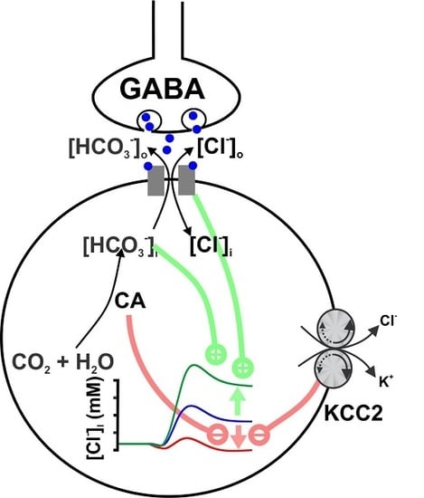
The effects of ionotropic γ-aminobutyric acid receptor (GABA-A, GABAA) activation depends critically on the Cl-gradient across neuronal membranes. Previous studies demonstrated that the intracellular Cl-concentration ([Cl]i) is not stable but shows a considerable amount of activity-dependent plasticity. To characterize how membrane properties and different molecules that are directly or indirectly involved in GABAergic synaptic transmission affect GABA-induced [Cl]i changes, we performed compartmental modeling in the NEURON environment. These simulations demonstrate that GABA-induced [Cl]i changes decrease at higher membrane resistance, revealing a sigmoidal dependency between both parameters. Increase in GABAergic conductivity enhances [Cl]i with a logarithmic dependency, while increasing the decay time of GABAA receptors leads to a nearly linear enhancement of the [Cl]i changes. Implementing physiological levels of HCO3-conductivity to GABAA receptors enhances the [Cl]i changes over a wide range of [Cl]i, but this effect depends on the stability of the HCO3 gradient and the intracellular pH. Finally, these simulations show that pure diffusional Cl-elimination from dendrites is slow and that a high activity of Cl-transport is required to improve the spatiotemporal restriction of GABA-induced [Cl]i changes. In summary, these simulations revealed a complex interplay between several key factors that influence GABA-induced [Cl]i changes. The results suggest that some of these factors, including high resting [Cl]i, high input resistance, slow decay time of GABAA receptors and dynamic HCO3 gradient, are specifically adapted in early postnatal neurons to facilitate limited activity-dependent [Cl]i decreases.

Graphical Abstract

1. Introduction

GABA (γ-aminobutyric acid) is the main inhibitory neurotransmitter in the mature brain and acts via ionotropic GABAA/GABAC receptors and via metabotropic GABAB receptors [1]. In the adult brain, GABA mediates its inhibitory effect by hyperpolarizing the membrane and by shunting excitatory inputs. GABAA receptors are ligand-gated anion-channels with a high permeability for Cl ions and a considerable additional permeability for HCO3 ions [1]. In the mature brain the activity of a K+-Cl-Cotransporter (KCC, mainly in its isoform KCC2) establishes a low intracellular Cl concentration ([Cl]i) [2,3], which accounts for a Cl influx and thus a membrane hyperpolarization upon activation of GABAA receptors [1]. Due to this Cl-flux, activation of GABAA receptors can influence [Cl]i on a time scale of seconds to minutes [4,5,6,7,8,9], a process termed “ionic plasticity” [3,10,11]. The magnitude of activity-dependent [Cl]i-transients depends on the Cl influx, dendritic volume and morphology, as well as on the capacity of Cl extrusion systems [12,13,14,15,16,17]. In addition, the membrane potential and the HCO3 permeability of GABAA receptors (PHCO3) contribute to the size of [Cl]i changes [6,18,19,20]. Therefore recent concepts of inhibition considered neuronal [Cl]i as a state- and compartment-dependent parameter of individual cells [14,20]. Detectable activity-dependent [Cl]i changes can occur in the adult nervous system under massive GABAergic stimulation [12,21]. However, already small alterations in [Cl]i or in the dynamics of the [Cl]i homeostasis critically influence information processing in neurons [20,22]. As the proper function in the adult nervous system relies on adequate inhibition [1,23,24], these activity-dependent [Cl]i changes play important roles in physiological and pathophysiological processes [10,11,17].
In the immature nervous system GABA typically induces depolarizing membrane responses [25,26,27,28,29,30]. These depolarizing GABAergic responses are caused by an elevated intracellular Cl concentration ([Cl]i), which is maintained by a Cl accumulation via the isoform 1 of the Na+-dependent K+-Cl-cotransporter (NKCC1) [3,29,31,32]. Recent studies suggest that at least in the postnatal neocortex, these depolarizing GABAergic responses mainly mediate inhibition [30,33], likely by increasing membrane shunting [1,34]. Several results indicate that depolarizing GABAergic neurotransmission is of specific relevance for immature spontaneous activity and for the maturation of the central nervous system [35,36,37]. Giant depolarizing potentials (GDPs) are a well-described network phenomenon in the immature hippocampus and the neocortex that represent spontaneous GABA-dependent activity [25,38,39]. In line with the high [Cl]i and the depolarizing responses, activation of GABAA receptors causes a decline in [Cl]i of immature neurons [29,40,41,42]. This attenuation of the [Cl] gradient reduces possible excitatory effects of GABA [29,42,43] and may serve to limit GABAergic excitation and/or to stabilize recurrent network events [10,13,40]. A recent study demonstrated that GDPs, which are associated with a high amount of GABAergic activity [25,44], induce long-lasting [Cl]i transients and influence the steady-state [Cl]i of CA3 pyramidal neurons in hippocampal slices from early postnatal mice neurons [45], making it a suitable model for ionic plasticity in the immature brain.
However, while the existence of ionic plasticity is well accepted and several factors influencing activity-dependent [Cl]i transients have been described, the role of biophysical membrane characteristics, molecular properties of GABAA-receptors or Cl-transporters, and the stability of HCO3 homeostasis on ionic plasticity has not yet been systematically investigated. Here we used a detailed biophysical compartmental model in the NEURON environment to demonstrate how cellular and molecular properties such as input resistance, pH, HCO3-selectivity, kinetics of GABAA receptors, the kinetics of NKCC1 mediated Cl transmembrane transport, and the activity of carbonic anhydrases influence activity-dependent [Cl]i transients. While most modeling is performed in isolated dendritic compartments, here we also replicate the well-described GDP-induced [Cl]i transients of immature hippocampal CA3 neurons [45].

2. Results

In order to study the question how various membrane parameters and the properties of different molecules involved in GABAergic transmission influence activity-dependent [Cl]i transients, we first computed the GABA-induced [Cl]i changes in an isolated dendrite, which allows a better mechanistic understanding of the underlying processes. Subsequently we also used a model of a reconstructed CA3 pyramidal neuron [45] to compare the results of our computational models with the GDP-dependent [Cl]i transients recorded in immature hippocampal CA3 neurons [45]. For the latter model, we implemented experimentally derived parameters of GABAergic synapses and GDP-activity provided by Lombardi et al. [45].

2.1. Influence of Membrane Conductance

First, we analyzed the influence of the membrane conductance on the [Cl]i changes induced by a single GABAergic input in an isolated dendrite. In this model, the experimentally determined conductance underlying single spontaneous GABAergic postsynaptic responses (gGABA) was implemented in an isolated dendrite equipped with passive conductances (gpas) varying between 10−6 S/cm2 and 0.1 S/cm2. These passive conductances correspond to input resistances (RInput) between ca. 670 MΩ and 0.67 kΩ, respectively, when they were implemented in a reconstructed CA3 pyramidal neuron. The results of this experiment demonstrated that upon stimulation of a single GABAergic input (gGABA = 0.789 nS, τ = 37 ms, PHCO3 = 0, [Cl]i = 30 mM) not only the depolarization, but also the GABA-induced [Cl]i transient depended on the passive conductances. A detailed analysis revealed a strong, sigmoidal dependency between RInput and peak [Cl]i changes (Figure 1a) or depolarization (Figure 1b) upon a single GABA stimulus. This effect of gpas on the GABA-induced [Cl]i transients was caused by the fact that at lower gpas the GABAergic currents induced a substantial depolarization, which attenuated the electromotive force on Cl ions (DFCl) during GABA stimulation (Figure 1c). At a low gpas of 10−7 S/cm2 (corresponding to a Rinput of ca. 4 GΩ in the reconstructed neuron) the GABAergic depolarization reached ECl (Figure 1c, solid lines). Therefore, DFCl approximated 0 and no persistent Cl fluxes occurred. In contrast, at a gpas of 0.018 S/cm2 (corresponding to Rinput of ca. 41 MΩ) Em remained negative to ECl, thus enabling permanent Cl fluxes (Figure 1c, dashed lines).
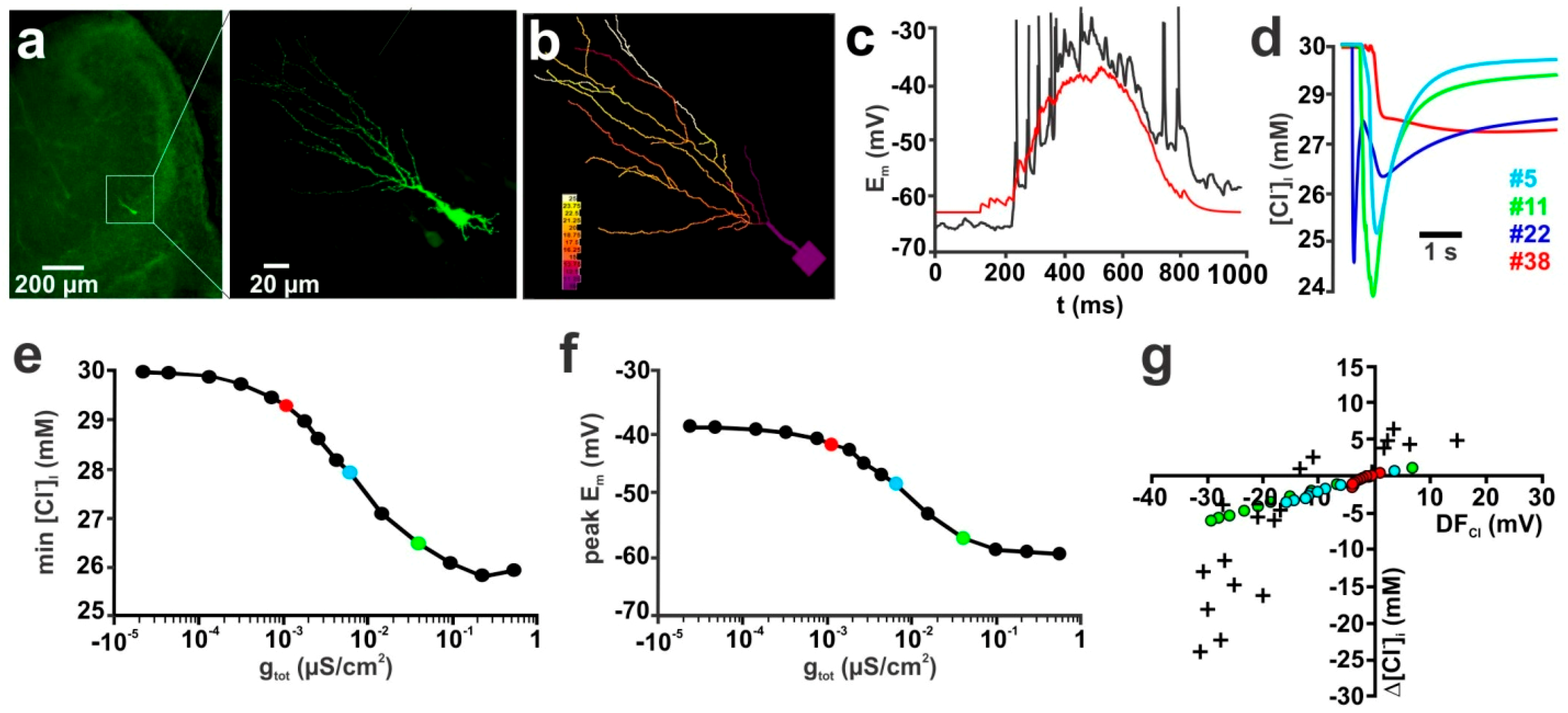
Next we simulated how gpas influences [Cl]i in a reconstructed neuron (Figure 2a,b), which receives complex GABAergic inputs that typically occur during GDP activity [45] (Figure 2c,d). For these experiments we initially equipped the dendrite with 101 GABAergic synapses (g = 0.789 nS, τ = 37 ms; all values from Lombardi et al. [45]), set PHCO3 to 0 and used an initial [Cl]i to 30 mM. Each of these 101 GABAergic synapses was randomly distributed within the dendrites of the reconstructed neuron. The time points for the stimulation of every synapse follows a normal distribution (µ = 600ms, σ = 900 ms). These values were derived from in-vitro experiments and resemble the distribution of GABAergic inputs during a GDP [45] (Figure 2c). In order to reduce the complexity of the analysis and to mimic the procedures of [Cl]i estimation used by Lombardi et al. [45] (which estimated [Cl]i changes from changes in ERev determined by focal GABA application within the dendritic compartment) we use for all further analyses the average [Cl]i of all dendrites.
Using this model, we investigated how different gpas between 10−6 S/cm2 and 0.1 S/cm2 affect the GDP-induced [Cl]i transients. This simulation demonstrated that also in a complex dendritic compartment gpas critically influenced the amount of [Cl]i changes (Figure 2e). Also, under these conditions the GABAergic depolarization during a GDP approached ECl at low gpas (Figure 2f), which minimized DFCl and the remaining Cl fluxes. One particular result of this computational study was that the GDP-induced [Cl]i transient amounts to less than 1 mM in a reconstructed CA3 pyramidal neuron equipped with the passive membrane conductance determined experimentally in these neurons (red symbols in Figure 2e–g), which is lower than the experimentally determined [Cl]i changes of 10.3 ± 3.3 mM (n = 4) in a real CA3 pyramidal neuron at comparable conditions [45]. To further specify the influence of gpas on the GDP-induced [Cl]i transients, we simulated the peak dendritic [Cl]i change for different initial [Cl]i at three different gpas. For this purpose we used values of 0.049 mS/cm2 (corresponding to a RInput of 901 MΩ, typical for immature hippocampal neurons [45]), 0.28 mS/cm2 (189 MΩ, adult neuron in whole-cell patch-clamp configuration [46]), and 1.8 mS/cm2 (41 MΩ, adult neuron with sharp electrode [47]). These simulations demonstrated that, if mature properties of gpas were implemented in the simulated neuron, the GDP-induced [Cl]i changes were roughly comparable to the values observed in real CA3 pyramidal neurons (Figure 2g), while at gpas typical for immature CA3 pyramidal neurons only marginal GDP-induced [Cl]i changes occurred.
In order to adapt the simulation of GDP-induced responses to the physiological properties of CA3 pyramidal neurons we incorporated an inward rectification in the background conductance (Supplementary Figure S1a,b). In addition, we had to increase the number of GABAergic synaptic inputs (nGABA) to compensate the influence of massive space clamp problems on the experimental determination of this parameter (Supplementary Figure S1c–f). For all further simulations in the reconstructed neurons we used the inward rectifying background conductance and implemented 302, 395, and 523 GABAergic synapses for PHCO3 values of 0.0, 0.18, and 0.44, respectively. However, even with the inward rectifying conductance and 302 synaptic inputs the GDP-induced [Cl]i changes were smaller than observed under in-vitro conditions (Supplementary Figure S1e).

2.2. Influence of GABA Receptor Conductivity and Kinetics

Next we analyzed the influence of the GABAergic conductance (gGABA) on [Cl]i transients. Initial experiments in an isolated dendrite showed that initially the [Cl]i transient was localized underneath the synapse, and within 3 s a diffusional equilibration throughout the dendrite occurred (Supplementary Figure S2a,b). Therefore, we estimated the total amount of GABA-evoked [Cl]i changes by averaging the [Cl]i over all nodes of the dendrite 3 s after the GABAergic stimulus. To analyze the relation between total gGABA and the [Cl]i changes, we first systematically increase gGABA from 0.789 nS to 78.9 nS (Figure 3a). These simulations demonstrated that the GABA-evoked [Cl]i changes rose with increasing gGABA, but did not depend linearly on gGABA (Figure 3b, black line). This nonlinear effect was due to the larger membrane depolarization upon stronger GABAergic stimulation, which reduced DFCl under this condition (data not shown). In an additional set of simulations, we enhanced the level of GABAergic stimulation by increasing the number of GABAergic synapses (nGABA) from 1 to 100, with gGABA of 0.789 nS for each synapse. The synapses were for each nGABA evenly distributed across the isolated dendrite. These simulations revealed that this distributed stimulation led to a reduced relative [Cl]i decrease at higher nGABA (Figure 3b, red line), as compared to the previous simulation paradigm (Figure 3b, black line). This observation is most probably due to the fact that with distributed synapses Em reaches more depolarized values close to ECl (−56.9 mV at 1 × 78.9 nS vs. −40.7 mV at 100 × 0.789 nS, data not shown).
To investigate whether a similar dependency between the amount of GABAergic inputs and [Cl]i could also be observed during a simulated GDP in a CA3 pyramidal neuron we increased gGABA from 0.789 nS (Figure 3c, blue line) to 7.89 nS (red line) at each of the 302 synapses used to simulate a GDP. This 10× increase in gGABA augmented the maximal GDP-induced [Cl]i decrease from 4.3 mM to 6.8 mM (Figure 3d). This surprisingly small effect was due to the fact that the increased gGABA also reduced the average DFCl from −8.6 mV to −5 mV (Figure 3d). When a similar increase in the amount of GABAergic stimulation was implemented by a 10× increase in nGABA (from 301 to 3010) a slightly larger maximal [Cl]i decrease by 7.1 mM was observed (Figure 3c,d, green line/symbols). This result indicates that the GDP-induced [Cl]i changes were close to saturation values when realistic values for nGABA, gGABA and RInput were implemented in a simulated CA3 pyramidal neuron.
In addition, we simulated how changes in the decay kinetics of GABAA receptor-mediated currents (τGABA) influence the [Cl]i transients (Supplementary Figure S2c,d). Systematic variation of τGABA between 10 ms and 1000 ms for a single synapse (gGABA = 0.789 nS) in an isolated dendrite revealed that the average [Cl]i showed a nearly linear dependency on τGABA (Figure 4a, black line). If gGABA was increased by a factor of 20 (gGABA = 15.78 nS) the average [Cl]i concentration still showed a nearly linear dependency on τGABA (Figure 4a, red line). Since these responses suggested a strong influence of τGABA on the GABA-induced Cl fluxes, we also varied τGABA of all GABAergic synapses that were implemented on the reconstructed CA3 pyramidal neurons. These simulations revealed that an increase in τGABA indeed increased the GDP-induced [Cl]i changes (Figure 4b). The maximal GDP-induced decline in [Cl]i increases from 1.3 mM to 4.3 mM and 10.4 mM for τGABA of 3.7 ms, 37 ms and 370 ms, respectively (Figure 4c).

2.3. Contribution of the HCO3 Conductance of GABA Receptors

In all previous experiments, we simulated GABAA mediated responses under the simplified consideration that GABAA receptors are ligand-gated Cl channels. However, GABAA receptors are anion channels with a considerable HCO3 permeability [1]. The relative HCO3-permeability of GABAA receptors (PHCO3) ranges between 0.18 (determined in spinal cord neurons [48]) and 0.44 (determined in adult hippocampal neurons [49]), although also higher values have been suggested [1]. Therefore, we next simulated how PHCO3 affects GABAergic Em and [Cl]i responses upon stimulation of a single synapse in an isolated dendrite. Addition of a HCO3 conductance to GABAergic currents induce a depolarizing shift in the peak depolarizations induced by GABAergic stimulation (Supplementary Figure S3a,b). Since this additional depolarization affected the DFCl, the GABAergic [Cl]i changes were also influenced by PHCO3. Under particular conditions, i.e. when Em crossed ECl during synaptic responses, the GABAergic activation lead to biphasic [Cl]i changes (Figure 5a). For further analysis we plotted for such biphasic responses the maximal and minimal [Cl]i upon GABAergic stimulation (e.g. Figure 5b, blue lines). A systematic analysis of the effect of GABAergic inputs on the [Cl]i changes revealed that the [Cl]i changes were shifted towards more outward fluxes at higher PHCO3 (Figure 5b), indicating that with increasing PHCO3 a substantial [Cl]i increase is induced by GABAergic stimulation.
However, these initial assumptions neglect the fact that the HCO3 fluxes will also affect [HCO3]i. Rapid regeneration of [HCO3]i levels by carbonic anhydrases, which stabilize [HCO3]i, is absent in immature neurons [50]. Therefore, we first simulated the GABA-induced Em and [Cl]i changes under the assumption that HCO3 will not be replenished (by implementing a HCO3 relaxation time constant (τHCO3) of 10 min) and is only redistributed by diffusion. These simulations revealed that the activation of GABAA receptors induced a rapid decline in [HCO3]i (Supplementary Figure S3c). The [HCO3]i decline depended on both PHCO3 and [Cl]i and was maximal at low [Cl]i with values of 1.8 mM and 2.3 mM for PHCO3 of 0.18 and 0.44, respectively (Figure 5c). In line with this [HCO3]i decline, the GABAergic depolarization was drastically decreased (Figure 5d) under dynamic [HCO3]i conditions. The attenuation of activity-dependent [HCO3] gradients also reduced the size of associated [Cl]i changes (Figure 5e) and at intermediate [Cl]i even reversed the effect (Supplementary Figure S3d).
As suggested from the results in isolated dendrites, the GDP-induced depolarization simulated in the reconstructed neuron was augmented if PHCO3 was increased from 0.0 to 0.18 and 0.44 under the assumption of stable [HCO3] gradients (Figure 6a,c, shaded symbols). And because under these conditions Em could become positive to ECl, the Cl fluxes were enhanced and GDP-induced [Cl]i transients increased (Figure 6a,d, shaded symbols). Increasing PHCO3 shifted the [Cl]i level at which GDP-induced [Cl]i transients change from influx to efflux, reflecting the impact of EHCO3 on the DFCL. The maximal influence of PHCO3 on the [Cl]i changes was observed at low [Cl]i (Figure 6d, shaded symbols), because at these conditions, the depolarizing effect of HCO3 fluxes opposed the hyperpolarizing effects of ECl.
Implementation of a model that allowed dynamic [HCO3]i changes (using a τHCO3 of 10 min) in the reconstructed CA3 pyramidal neuron showed that the GDP-induced GABAergic currents induced massive changes in [HCO3]i, depending on PHCO3 (Supplementary Figure S3e). This GDP-induced [HCO3]i decrease during a GDP diminished the membrane depolarization (Figure 6b,c, plain lines/symbols), which in turn caused a drastic reduction in the GDP-induced [Cl]i transients (Figure 6b,d, plain lines/symbols). In summary, addition of PHCO3 to GABAergic currents augmented the DFCl and thus the GDP-induced [Cl]i transients (Figure 6e). Using these parameters, the simulated GDP-induced [Cl]i transients resembled the size of [Cl]i transients observed in real cells, however, only in the quadrant with positive DFCl values (Figure 6e).

2.4. The Stability of HCO3 Gradients Influences Activity-Dependent [Cl]i Transients

The previous results clearly demonstrate that GABAA receptor-mediated [HCO3]i transients massively influence the Em and [Cl]i changes under these conditions. However, the two conditions used in these experiments (stable [HCO3]i or negligible [HCO3]i regeneration at τHCO3 of 10 min) are obviously not physiological in immature neurons, which lack carbonic anhydrases, but in which spontaneous CO2 hydration and/or transmembrane transport of HCO3 can occur [50]. Therefore, we next investigated how τHCO3 influences the stability of [HCO3] gradients and GABA induced [Cl]i transients. For that we systematically changed the decay-time of [HCO3]i relaxation (τHCO3) implemented in the NEURON model (Supplementary Figure S4a,b). A systematic simulation in isolated dendrites revealed that [Cl]i changes remained rather constant at τHCO3 ≥ 90 ms (Figure 7a). The half-maximal [Cl]i changes occurred at a τHCO3 around 10 ms, which is substantially shorter than the τHCO3 of ca. 70 ms for half-maximal [HCO3]i changes (Supplementary Figure S4b). More than 85% of the maximal [Cl]i changes took place at τHCO3 below 100 ms (Figure 7a). However, it must also be considered that a decreased temporal stability of the [HCO3] gradient will also influence the lateral diffusion of HCO3. Indeed, a systematic simulation of the spatial aspects of the activity-dependent [HCO3]i transients revealed that the stability of HCO3 massively influenced the [HCO3] gradient along the isolated dendrite (Figure 7b), although the maximal [HCO3]i change at the synaptic site was nearly saturated already at a τHCO3 ≥ 90 ms (Figure 7b). In accordance with the results obtained in isolated dendrites, also in the reconstructed CA3 pyramidal neuron τHCO3 had a large effect on the GDP-induced [HCO3]i transients (Supplementary Figure S4c,d), but only a minor effect on the associated [Cl]i changes (Figure 7c). For τHCO3 ≥ 90 ms the GDP-induced [Cl]i changes were only marginally affected (Figure 7c, Supplementary Figure S4c) by changes in τHCO3. At τHCO3 of 1 ms, the maximal GDP-induced [Cl]i decrease amounted to 5.2 mM, while it was 4.6 mM, 4.5 mM and 4.4 mM for τHCO3 values of 90 ms, 518 ms and 3 s, respectively. This small effect was also reflected by the minimal changes in the relation between DFCl and GDP-induced [Cl]i transients (Figure 7d).
GABAergic [51] and glutamatergic [52,53] synaptic transmission is accompanied by substantial pH changes. These pH changes, however, indirectly affect GABAergic transmission, since they alter the [HCO3]i. To estimate, how such pH changes influence activity-dependent [Cl]i transients, we first simulated the effect of such pH shifts by constantly altering the pH value from 7.2 to 7.0 or 7.4 in an isolated dendrite. According to the Henderson-Hasselbalch equation, these pH shifts alter [HCO3]i from 14.1 mM to 9 mM or 22.7 mM, respectively. Because this pH-dependent differences in [HCO3]i affect DFGABA, the membrane depolarization upon GABAA receptor activation was reduced at pH 7.0 and enhanced at a more alkaline pH of 7.4 (Figure 8a). In line with this altered GABAergic membrane depolarization, the DFCl during GABAergic stimulation was also affected, shifting the resulting Cl fluxes. This can be exemplified at intermediate [Cl]i, where the biphasic Cl fluxes at a normal pH of 7.2, were transformed to Cl efflux at a pH of 7.0 and to a Cl influx at a pH of 7.4 (Figure 8a). A systematic analysis of Cl fluxes at different initial [Cl]i demonstrated that, in comparison to pH 7.2, the Cl influx at low initial [Cl]i was decreased at pH 7.0, while it was enhanced at pH 7.4 (Figure 8b). In contrast, the Cl efflux at high [Cl]i was enhanced at pH 7.0 and reduced at pH 7.4 (Figure 8d). In consequence, intracellular acidification shifted the [Cl]i range at which Cl efflux occurs to lower initial [Cl]i, whereas intracellular alkalinization shifted this range to higher initial [Cl]i (Figure 8b).
Simulations in the reconstructed CA3 pyramidal neuron revealed that a lower pH of 7.0 led to smaller GDP-induced membrane depolarizations, as compared to the standard pH of 7.2 (Figure 8c, Supplementary Figure S4e, red line/symbols). This reduced depolarization resulted in a reduced GDP-associated Cl influx (4.6 mM vs. 7.6 mM) at low initial [Cl]i and in an increased Cl efflux (−6.1 mM vs. −4.3 mM) at high initial [Cl]i concentration (Figure 8d, red symbols). Conversely, at a higher pH of 7.4 the membrane responses during a GDP were more depolarized (Figure 8c, Supplementary Figure S4e green lines/symbols), which resulted in enhanced Cl influx of 11.2 mM at low initial [Cl]i and a decreased Cl efflux of −2.3 mM at low [Cl]i (Figure 8d, green symbols). In summary, these results demonstrate that the acidification associated with synaptic transmission reduced the activity-dependent [Cl]i transients at low [Cl]i, while the activity-dependent Cl efflux at high [Cl]i was enhanced by such acidic shifts.

2.5. Influence of Transmembrane Cl Transport

Finally, we analyzed how the kinetics of the [Cl]i homeostasis influenced the temporal and spatial constrains of the activity-dependent [Cl]i changes. For that we systematically changed the decay-time of the [Cl]i relaxation (τCl) implemented in the NEURON model. Initial simulations in isolated and soma-attached dendrites revealed that for τCl of ≥ 10 s the decay of [Cl]i response was dominated by diffusional exchange with the soma (Supplementary Figure S4f–h). At a τCl of 321 s 99.2% of Cl fluxes were depleted by diffusional exchange with the soma, while at faster τCl a substantial smaller fraction of only 83.5 % (τCl = 10 s), 33.2 % (τCl = 1 s) and 2.4% (τCl = 100 ms) of the Cl fluxes was eliminated by diffusion from the dendrite to the soma. Analysis of the spatial distribution of [Cl]i along the dendrite revealed that τCl also affects the size of activity-dependent [Cl]i changes at distant dendritic sites (Supplementary Figure S4i–k). The dominance of diffusional elimination of Cl was also reflected by the observation that at slow τCl ≤ 10s the [Cl]i was substantial lower at the proximal than at the distal end of the dendrite (Supplementary Figure S4i–k).
To analyze the influence of τCl on the spatial aspects of the [Cl]i transients we implemented two simultaneous GABAergic inputs that were located equidistant to the [Cl]i recording site at distances of 10 µm, 30 µm, 100 µm and 300 µm and systematically increased τCl from 1 ms to 220 s (Figure 9a). These simulations revealed not only that the maximal [Cl]i depended on the distance between GABAergic stimulation sites and the node of [Cl]i determination, but also that τCl critically influenced the [Cl]i change at a given distance to the stimulation sites (Figure 9a). This dependency between spatial restrictions of activity-dependent [Cl]i changes and τCl was quantified by the τCl at which half-maximal [Cl]i changes occur (τCl50). If the distance of the GABAergic synapses was 10 µm τCl50 amounted to 12 ms, and this τCl50 increased to 60.5 ms, 726 ms and 4.6 s at synaptic distances of 30 µm, 100 µm and 300 µm, respectively. To analyze the temporal aspects of [Cl]i summation we simulated five consecutive GABA stimulations at frequencies (fGABA) of 0.3 Hz, 1 Hz, 3 Hz and 10 Hz and determine the [Cl]i at the stimulation site, while systematically varying τCl (Figure 9b). These simulations revealed a sigmoidal dependency between τCl and the temporal summation of [Cl]i. A larger amount of [Cl]i summation and a lower τCl50 was observed at higher frequencies. The τCl50 amounted to 1.9 s for fGABA of 0.1 Hz, 931 ms for fGABA of 1 Hz, 268 ms for fGABA of 3 Hz, and 53 ms for fGABA of 10 Hz. In summary, these results demonstrated that τCl values of less than 1 s are required to prevent substantial activity-dependent [Cl]i changes in the spatial and/or temporal domain at fGABA ≥ 1Hz and less than 100 µm distance between synaptic sites.
In accordance with these results in single dendrites, all previous simulations of GDP-induced [Cl]i transients in the reconstructed CA3 pyramidal cells revealed substantial [Cl]i changes, because in these simulations the experimentally determined τCl of 174 s for NKCC1-mediated active Cl re-accumulation and of 321 s for passive Cl reduction were implemented and during a GDP stimulation a high frequency of GABAergic input was applied. In order to get more insights into how the capacity of [Cl]i regulation systems can influence activity-dependent [Cl]i transients within a realistic dendritic compartment, we finally simulated how different τCl influenced the GDP-induced [Cl]i transients in the reconstructed CA3 neuron (Figure 9c,d). This simulation revealed that decreasing τCl from the experimentally determined values >100 s to 10 s or 1 s had only a minimal impact of the GDP-induced [Cl]i transients (Figure 9c). The maximal GDP-induced [Cl]i change amounted to 4.41 mM at a τCl of 10 s and to 4.21 mM at a τCl of 1 s, but were reduced to 2.99 mM at a τCl of 0.1 s and to 0.88 mM at a τCl of 10 ms. These results demonstrate that fast and efficient [Cl]i homeostatic processes are required to limit GDP-induced [Cl]i transients. Accordingly, the Δ[Cl]i vs. DFCl plot also revealed comparable GDP-induced [Cl]i changes at τCl of 10 s and 1 s, and smaller [Cl]i changes at a τCl of 100 ms (Figure 9d). Only a further reduction in τCl to 10 ms substantially suppressed GDP-induced [Cl]i changes. In summary, these results indicate that τCl influences the temporal and spatial properties of activity-dependent [Cl]i changes, but that τCl values that are substantially smaller than the experimentally determined values are required to suppress activity-dependent [Cl]i changes.

3. Discussion

In the present study we used a detailed biophysical compartmental modeling in the NEURON environment to systematically investigate how several cellular and molecular neuronal parameters influence the GABAA receptor-mediated [Cl]i changes. The main observations of this study can be summarized as follows: (i) A high Rinput reduces activity-dependent [Cl]i transients, while at low Rinput considerable activity-dependent [Cl]i transients can be observed. (ii) The activity-dependent [Cl]i transients show a logarithmic impact of gGABA, while τGABA has in a wide gGABA range a nearly linear influence on [Cl]i. (iii) The PHCO3 of GABAA-receptors enhances activity-dependent [Cl]i transients, but with instable [HCO3] gradients this effect is largely diminished. (iv) Activity-dependent Cl fluxes where shifted toward efflux at acidic and towards influx at alkaline pH. (v) τCl has a major impact on the spatiotemporal aspects of activity-dependent [Cl]i transients, but unrealistically fast τCl values are required to prevent [Cl]i transients at physiologically-relevant activity levels.
By 1990, it was suggested by Qian and Sejnowski [54] that the Cl fluxes via activated GABAA receptors will dissipate the Cl gradient in small compartments and thus mediate potentially instable inhibitory responses. This theoretical assumption was proven by experimental studies, which demonstrated that massive GABAergic activation can shift hyperpolarizing responses toward depolarization [12,21] and induce [Cl]i transients [55]. In the past the physiological and pathophysiological consequences of such activity-dependent [Cl]i changes have been investigated and discussed [13,17,20,36,56] and the basic principles of activity-dependent [Cl]i changes and their implications for neuronal information processing have been modeled [7,15,16,22,57,58]. However, the complex interplay and contribution of passive membrane leak, GABAA conductance, Cl diffusion/ transport and stability of [HCO3] gradients to these activity-dependent [Cl]i changes have not yet been systematically investigated.
Our simulations revealed a strong dependence between RInput and the GABAA receptor induced [Cl]i transients. While at high RInput GABA-induced [Cl]i changes were minimal, they increased in a nonlinear relation with decreasing RInput (Figure 1c). This relation between RInput and the [Cl]i changes is due to the fact that at high RInput even small GABAergic currents bring Em close to ECl, which minimizes DFCl and thus the Cl fluxes (Figure 1c). At low RInput the passive membrane conductance stabilizes Em and thus DFCl. In consequence, larger Cl fluxes can be expected. Accordingly, implementation of “adult like” membrane properties [47] in a reconstructed immature neuron massively enhanced activity-dependent [Cl]i changes (Figure 1c). In contrast, it seems obvious that immature neurons, with their high RInput [59], are less susceptible to activity-dependent [Cl]i changes.
However, in this respect, it is important to consider that in immature neurons [Cl]i is high and GABAergic responses are depolarizing [30,60,61], therefore activity-dependent Cl fluxes are directed outward and are leading to [Cl]i decrease [42,44]. In addition, the HCO3-permeability of GABAA receptors also needs to be considered. The high RInput in immature neurons causes Em to approach EGABA, which normally is positive to ECl due to the HCO3-permeability of GABAA-receptors [1,4]. Therefore, stable Cl influx would be expected under this condition and [Cl]i should ultimately approach the value defined by EHCO3, which at steady-state HCO3 gradients ([HCO3]i = 14.1 mM and [HCO3]e = 26 mM) amounts to 72.4 mM. This estimation suggests that under certain conditions GABAergic activity can even increase [Cl]i from the already high [Cl]i-levels in immature neurons (see Supplementary Figure S3d). But further properties of activity-dependent [Cl]i changes observed in our simulations protect immature neurons from excessive [Cl]i increases. In particular, we found that the influence of PHCO3 is relatively small at high [Cl]i (Figure 6d), due to the fact that under this condition the contribution of EHCO3 to EGABA is small (as described by the Goldman-Hodgkin-Katz-Equation [1]). In addition, in immature neurons the HCO3 gradient is instable because they lack carbonic anhydrases [50], which additionally attenuates the depolarizing effect of HCO3 on Em and thus reduces DFCl.
Under the assumption of a stable HCO3 gradient, EGABA is in a wide range positive to ECl, thereby permitting Cl influx during GABAergic stimulation, unless the thermodynamics equilibrium at 72.4 mM is reached. This is supported by the observation that no stable steady-state [Cl]i is reached with realistic PHCO3 values of 0.18 or 0.44 in our simulation (Supplementary Figure S3d, shaded lines). Experimental studies indeed demonstrated that massive GABAergic stimulation can shift EGABA from hyperpolarizing towards depolarizing and even excitation [12,62,63,64]. On the other hand, if we implemented in our model that [HCO3]i can be altered by GABAA receptors, the activity-dependent [Cl]i changes were massively reduced (Figure 5e). Our simulations also demonstrate that the switch from static [HCO3] to dynamic [HCO3] condition shifts the [Cl]i setpoint at which activity-dependent Cl influx was replaced by Cl efflux to considerably lower values (Figure 5e). This observation is due to the fact that the depolarizing HCO3 fluxes through the GABAA receptor are attenuated by the dissipating HCO3 gradient, which reduces EGABA and thus DFCl. Similar conclusions were drawn from experiments in which a block of carbonic anhydrases with acetazolamide, which provides a pharmacological destabilization of [HCO3], also reduces EGABA shifts ([18], but see [62]). We conclude from these observations that the lack of carbonic anhydrase VII in immature neurons [50,65] may serve to limit the activity-dependent [Cl]i changes in these neurons.
On the other hand, our simulations in a reconstructed CA3 pyramidal neuron revealed that although massive [HCO3]i changes are induced under this condition, the total GDP-induced [Cl]i change was only marginally affected by variations in τHCO3 (Figure 7d). This lack of effect was most probably due to the fact that the activity-dependent [HCO3]i change was already maximal at a τHCO3 of 90 ms at the synaptic site. This local saturated [HCO3]i change at the subsynaptic site is the only determinant for the synaptic effects of the [HCO3]i. Our simulations also suggest that for an effective, physiologically relevant control of [HCO3]i during GABAergic activity τHCO3 should be less than 70 ms (Supplementary Figure S4b). This fast relaxation time requires fast molecular processes that allow effective elimination of HCO3. Indeed, carbonic anhydrases, the enzymes that mediate the degradation or regeneration of HCO3 into/from H2O and CO2, are among the fastest enzymes known. The kcat of murine carbonic anhydrase VII for the hydration oh CO2 is 4.5 × 10⁵ s−1 at physiological pH [66]. From the assumption that the ca. 2mM [HCO3]i change in the dendrite corresponds at a dendritic volume of 16 pL to ca. 0.3 fmol HCO3 ions, it can be estimated that about 4000 molecules of carbonic anhydrase VII are required to replenish the lost HCO3 within 100 ms. This estimation suggests that it is reasonable that sufficient carbonic anhydrase activity can be located in the dendritic compartment to reliably stabilize [HCO3]i. However, as the reaction mediated by carbonic anhydrases includes H+ ions, the kinetics and thermodynamics of this process depends on the intracellular pH [67]. Thus dendritic H+-buffering and handling indirectly also affects activity-dependent [Cl]i changes [15]. The acidification associated with neuronal activity [52,53] will slow down the kinetics of carbonic anhydrases [66]. However, this effect is negligible in comparison to the effect of the intracellular pH on [HCO3]i. The intracellular pH is an essential parameter that determines [HCO3]i [67]. Thus the intracellular acidification observed upon activation of GABAergic and glutamatergic synapses [51,52,53] will alter [HCO3]i and subsequently influence GABAergic transmission. Our simulation revealed that an intracellular acidification will reduce the activity-dependent [Cl]i changes at low [Cl]i. This result, which is in accordance with a previous simulation [15], indicate that in adult neurons a parallel acidification will limit Cl influx and thus stabilize inhibitory transmission In contrast, at a high [Cl]i typical for immature neuron the intracellular acidification enhanced the activity-dependent Cl efflux and may contribute to the loss of depolarizing drive and putative excitatory effects after strong GABAergic stimulation.
Another factor that has a stringent effect on activity-dependent [Cl]i changes in our simulations is τGABA. This confirmed and extended previous computational analyses (c.f. Figure 4d in [7]). It has been found that in general the decay kinetics of GABAergic transmission get faster during development [68]. Therefore the slow decay kinetics of GABAergic transmission in immature neurons [68,69] may be a factor that enables activity-dependent [Cl]i transients, while the faster GABAergic postsynaptic currents in mature neurons not only improve the temporal precision of GABAergic transmission [68], but also the stability of inhibition. While a stable inhibition is a prerequisite for the proper function of mature neuronal networks, dynamic changes in [Cl]i can be mandatory for physiological relevant functional features of the immature central nervous system. It has been suggested that activity-dependent changes in [Cl]i, and the resulting switch from GABAergic inhibition to excitation, can underlie oscillatory activity [13]. In addition, in the immature nervous system the resting [Cl]i is decreased by GABAergic activity, which will result in a diminished excitatory drive and/or a dominance of shunting inhibition and may thus serve to limit a possible excitatory effect of GABA [40]. Therefore, for immature neurons an unstable [Cl]i homeostasis may be functionally relevant, as it allows activity-dependent scaling of [Cl]i-dependent synaptic transmission [42,43].
In consequence, the molecular configuration of immature neurons (high [Cl]i, long τGABA and missing CA-VII) will generate conditions that allow limited activity-dependent [Cl]i decreases. This, in addition to the aforementioned effect of the high input resistance, may be an explanation why the [Cl]i homeostasis of immature neurons is maintained by a relatively ineffective transmembrane Cl transport [29]. In contrast, in mature neurons the situation is different. In the adult brain, the low [Cl]i is needed to maintain hyperpolarizing inhibition [1] and an activity-dependent [Cl]i increase will attenuate membrane hyperpolarization. While it is obvious that massive changes in [Cl]i will impair GABAergic inhibition and can lead to hyperexcitability [18], recent modeling experiments demonstrate that even minimal changes in the capacity of Cl-extrusion can have strong effects on information processing and storage in neurons [22]. Although the low RInput and the fast τGABA counteracts activity-dependent [Cl]i increase in adult neurons, their low [Cl]i and their effective carbonic anhydrases can lead to substantial [Cl]i changes in their dendrite. The adverse effect of such local activity-dependent [Cl]i increases is enhanced by the more elaborated dendritic compartment in mature neurons, which limits diffusional elimination of Cl-ions [13].
Our simulations reveal that the connection of an isolated dendrite to the soma drastically reduces the equilibrium [Cl]i after synaptic stimulation (Supplementary Figure S4f–k), demonstrating the important role of diffusional Cl elimination under this condition. The large volume to surface ratio (and thus volume to conductance ratio) of the soma enables this compartment to serve as Cl sink in these in-silico experiments. Also in-vitro it has been demonstrated that activation of dendritic GABAA receptors induced massive shifts in EGABA, whereas only minimal changes were observed upon perisomatic stimulation [10,12,26]. The dominance of perisomatic GABAergic terminals [70] may be related to the requirement of stable [Cl] gradients to maintain stable inhibition over a wide range of activity levels. However, the diffusion of [Cl]i through dendrite is a relatively slow and inefficient process, due to the small diameter in distal dendrites [7,55]. Addition of spines to dendrites drastically slow down diffusion along dendrites [16], suggesting that the complexity of the dendritic compartment (i.e. the number of arborizations that enhance tortuosity in the dendritic compartment) hinders Cl-elimination by diffusion to the soma. Therefore, active elimination of Cl from the cytoplasm is required to prevent or minimize activity-dependent [Cl]i changes in the elaborated dendritic compartment of adult neurons.
The elementary role of transmembrane Cl transporters for neuronal [Cl]i homeostasis has been shown by a variety of studies [2,29,32,58,71]. Modeling studies revealed that slightly altered rates of transmembrane Cl-transport, which does only marginally affect resting [Cl]i levels, have a strong effect on the spatiotemporal distribution of activity-dependent [Cl]i-transients in dendrites [15]. Therefore it is not surprising that the simulation of an enhanced capacity of transmembrane Cl transport by increasing τCl attenuates activity-dependent [Cl]i transients. However, to minimize these [Cl]i transients a rather low τCl of < 100 ms is required. These low τCl values are several orders of magnitude below the experimentally determined kinetics of the NKCC1-mediated Cl-accumulation (τCl = 158 s) in immature neurons [29]. Because of this slow kinetic of transmembrane transport of Cl in immature neurons, we also consider that a Cl/HCO3 exchange mediated by the anion exchanger in immature neurons [72] has only a marginal effect on both activity-dependent [Cl]i and [HCO3]i transients in these neurons. In the mature situation (low [Cl]i and effective transmembrane [Cl]i transport), modeling studies suggest that an interference between [Cl]i and [HCO3]i by this mechanism can reduce the activity-dependent [Cl]i changes [15].
While it is generally assumed that the neuron-specific Cl-extruder KCC2 mediates more efficient Cl transport than NKCC1, only few experimental studies addressed the kinetics of KCC2-dependent Cl-extrusion. Experiments in brain stem neurons demonstrated that KCC2 mediated Cl-extrusion after [Cl]i increase by ca. 10 mM requires several minutes [73]. In contrast, in-vivo experiments revealed that the activity-dependent [Cl]i increase after an epileptic seizure recovered within less than 30 s [9] and in hippocampal slices GABA-induced [Cl]i transients recovered back to low steady-stale levels with a time constant of 3.3 s [12]. However, it is not clear how diffusional processes and/or the kinetics of the used Cl sensor contribute to these kinetic properties. Simulations suggest that with realistic KCC2 levels τCl in the distal dendrites (≥ 200 µm from the soma) is between 100 ms and 200 ms [15], and thus probably lower than estimated from experimental data. Even this time constant is higher than the τCl required in our simulations to prevent local activity-dependent [Cl]i changes, suggesting that considerable [Cl]i changes can occur at GABAergic synaptic sites. While our and other simulations [15] suggest these transients may be restricted to local dendritic domains, it must be emphasized that subtle changes in the efficacy of KCC2 mediated Cl-transport can already enhance the excitability in single neurons because activity-dependent [Cl]i transients may superimpose these effects [22]. In consequence, impairments of KCC2 mediated Cl transport can led to a breakdown of sufficient inhibition in neuronal networks and contribute to hyperexcitability [15,17,20,56,74]. In this respect it is also relevant to consider that the activity of both NKCC1 and KCC2 are regulated by a variety of processes [75,76,77,78]. This indicates that the spatiotemporal [Cl]i dynamics in the dendritic compartment may be adapted to the functional states.
The limitation of our model to fully describe GDP-induced [Cl]i transients in CA3 pyramidal neurons is obvious from the fact, that we massively underestimate the [Cl]i decrease observed in real CA3 pyramidal neurons at high [Cl]i (e.g. Figure 6e). Therefore, additional factors must be proposed, which enhance the GABAA-receptor-mediated Cl efflux. Possible mechanisms that improve Cl efflux are e.g. an inhomogeneous distribution of voltage-activated K+ channels in the dendritic compartment, an underestimation of nGABA in our in-vitro experiments due to voltage-clamp errors in the elaborated dendrite [79], or the effect of glutamatergic transmission during a GDP [20,80]. In addition, we also found that τGABA has a major impact on the GDP-induced [Cl]i transients, and it might well be that the decay kinetics of spontaneous GABAergic PSCs of 37 ms [45] reflect the kinetic properties of a subpopulation of GABAergic inputs, that is less involved in the generation of GDPs. Finally, in our simulations the dendrite was implemented as a hollow tube with a diameter determined from the histological reconstruction. Under realistic assumptions the neuron is, however, filled with cytoplasm that contains large proteins, particles of different sizes and vesicles and tubes of intracellular organelles. Thus the free, “unexcluded” volume in the cytoplasm is restricted to an estimated fraction of ca. 60%, a principle termed cytoplasmic crowding [81]. This restricted free cytoplasmic water volume will increase the size of [Cl]i transients upon identical Cl fluxes.

4. Materials and Methods

Compartmental Modeling

The biophysically realistic compartmental modelling was performed using the NEURON environment (neuron.yale.edu). The source code of models and stimulation files used in the present paper can be found in ModelDB [82] at http://modeldb.yale.edu/253369 (access date 14 March 2019) and was included in the supplementary material of this publication. For compartmental modelling we used either a simple ball and stick model (soma with d = 20 µm, linear dendrite with l = 200 µm and 103 nodes) or a reconstructed CA3 pyramidal cell (from Lombardi et al. [45]). Except where noted the dendrite was detached from the soma to analyse dendritic [Cl]i transients. The reconstructed neuron resembled the somatodendritic morphology of a typical immature CA3 pyramidal neuron (see Figure 2a,b). For this purpose images of a biocytin-filled neuron [83,84] were taken with 60× oil-immersion objectives and the somatodendritic morphology was reconstructed using Fiji (www.fiji.sc). It contained a soma (d = 15 µm), a dendritic trunk (d = 2 µm, l = 32 µm, 9 segments) and 56 dendrites (d = 0.36 µm, 9 segments each). In all of these compartments a specific axial resistance (Ra) of 34.5 Ωcm and a specific membrane capacitance (Cm) of 1 µF cm−2; were implemented. The specific membrane conductance (gpas) varied (see Figure 1a and Figure 2e) and in the majority of the experiments was modeled by a voltage dependent process given by a Boltzmann-like equation:
g pas = g min + g max ( 1 + exp ( ( E m E 50 ) s ) )
with gmax = 0.002800 S/cm2 (experimentally determined gInput at depolarized potentials, see Supplementary Figure S1a), gmin = 0.000660 S/cm2 (experimentally determined minimal gInput at hyperpolarized potentials, see Figure 3a), e50 = −31 mV (half-maximal voltage), s = −6 (slope of the voltage-dependency). The reversal potential of this voltage-depended gpas was set to −60 mV.
GABAA synapses were simulated as a postsynaptic parallel Cl and HCO3 conductance with exponential rise and exponential decay [7]:
IGABA = ICl + IHCO3 = 1/(1+P) gGABA (V–ECl) + P/(1+P) gGABA (V–EHCO3)
where P is a fractional ionic conductance that was used to split the GABAA conductance (gGABA) into Cl and HCO3 conductance. ECl and EHCO3 were calculated from Nernst equation. The GABAA conductance was modeled using a two-term exponential function, using separate values of rise time (0.5 ms) and decay time (variable, mostly 37 ms) [45]. Parameters used in our simulations were as follows: [Cl]o = 133.5 mM, [HCO3]i = 14.1 mM, [HCO3]o = 24 mM, temperature = 31 °C, P = 0.44 [49]. For the ball and stick model a single GABAA synapse was placed in the middle of the dendrite, except where noted. For the simulation of a GDP in the reconstructed CA3 neuron 101–3020 GABAergic synapses were randomly distributed within the dendrites of the reconstructed neuron. GABA inputs were activated stochastically using a normal distribution (µ = 600ms, σ = 900 ms) that emulates the distribution of GABAergic PSCs during a GDP observed in immature hippocampal CA3 pyramidal neurons [45]. The properties of these synapses were always given in the results part and/or the corresponding figure legends.
From the quotient between the charge transfer of a GDP and of spontaneous GABAergic postsynaptic currents at a holding potential (VHold) of 0 mV it was estimated that 101 GABAergic inputs underlie a GDP [45]. To compensate for the space-clamp problems during a GDP, that were not considered by Lombardi et al. [45], we simulated the charge transfer during a GDP under their experimental conditions ([Cl]i = 10 mM, VHold = 0 mV) and determined that 302, 395, and 523 (for PHCO3 values of 0.0, 0.18, and 0.44, respectively) GABAergic synapses are required to generate the observed GDP-induced charge transfer (Supplementary Figure S1c–f). For these experiments we implement the single-electrode voltage clamp procedure provided by NEURON, using an access resistance of 5 MΩ. The charge transfer was calculated from the integral of the holding currents (IHold) during the GDP.
For the modeling of the GABAA receptor-induced [Cl]i and [HCO3]i changes, we calculated ion diffusion and uptake by standard compartmental diffusion modeling [16,85,86,87]. To simulate intracellular Cl and HCO3 dynamics, we adapted our previously published model [7]. Longitudinal Cl and HCO3 diffusion along dendrites was modeled as the exchange of anions between adjacent compartments. For radial diffusion, the volume was discretized into a series of 4 concentric shells around a cylindrical core [85] and Cl or HCO3 was allowed to flow between adjacent shells [88]. The free diffusion coefficient of Cl inside neurons was set to 2 µm2/ms [55,89]. Since the cytoplasmatic diffusion constant for HCO3 is, to our knowledge, unknown, we also used a value of 2 µm2/ms. To simulate transmembrane transport of Cl and HCO3, we implemented an exponential relaxation process for [Cl]i and [HCO3]i to resting levels [Cl]irest or [HCO3]irest with a time constant τIon.
d [ Ion ] i dt = [ Ion ] i rest e [ Ion ] i τ Ion
Cl transport was in most experiments (if not otherwise noted) modeled as bimodal process, for [Cl]i < [Cl]irest τIon was set to 174 s to emulate an NKCC1-like Cl transport mechanism. For [Cl]i > [Cl]irest τIon was set to 321 s to emulate passive Cl efflux (both values obtained from unpublished experiments on immature rat CA3 hippocampal neurons).
The impact of GABAergic Cl currents on [Cl]i and [HCO3]i was calculated as:
d [ Ion ] i dt = 1 F I Ion volume
To simulate the GABAergic activity during a GDP, a unitary peak conductance of 0.789 nS and a decay of 37 ms were applied to each GABAergic synapse. These values resulted in a unitary currents of pA, which was in accordance with the mean amplitude of spontaneous GABAergic postsynaptic currents in CA3 paramidal neurons [45].
For the isolated neurons the [Cl]i and [HCO3]i concentration was averaged over all segments of the dendrite, except where noted. For the simulated neurons we analyzed mean [Cl]i and [HCO3]i of all dendrites:
[ C l ] i = 1 n d e n d × j = 1 n d e n d [ C l ] i D e n d ( j )   @   0.5   o f   t o t a l   l e n g t h
This procedure mimics the experimental procedure of Lombardi et al [45], who determined EGABA by focal application in the dendritic compartment.
For the calculation of Δ[Cl]i the maximal deviation of [Cl]i upon a GABAergic stimulus ([Cl]iS) was subtracted from the resting [Cl]i before the stimulus ([Cl]iR). For biphasic responses both minimal and maximal [Cl]iR were determined and Δ[Cl]i was calculated as:
Δ [ C l ] i =   [ C l ] i S ,   min [ C l ] i R     i f   a b s ( [ C l ] i S ,   min ) > a b s ( [ C l ] i S ,   max )
Δ [ C l ] i = [ C l ] i S ,   max [ C l ] i R     i f   a b s ( [ C l ] i S ,   min )   a b s ( [ C l ] i S ,   max )
The driving-force of Cl (DFCl) was calculated from the difference between the average Em during a GDP and ECl (DFCl = Em – ECl). To calculate the ratio between transmembrane [Cl]i transport and diffusional [Cl]i depletion into the soma, we normalized the diffusional exchange between the last somatic node and the soma (as calculated from Fick’s law) to conditions were transmembrane [Cl]i loss was absent (τCl = 109 ms) and diffusional dendrite to soma transport was allowed to equilibrate for 2 min.
All electrophysiological data were taken from our previous publication [45]. However, for a comparison of these results with the simulations, we had to take different PHCO3 into account. Therefore the GDP-induced [Cl]i changes were recalculated using PHCO3 values of 0.0, 0.18 (determined in spinal cord neurons [48]) and 0.44 (determined in adult hippocampal neurons [49]). The [Cl]i was calculated from EGABA with the Goldman-Hodgkin-Katz equation:
E GGABA = RT ZF × ln ( P C l [ C l ] e + P HCO 3 [ HCO 3 ] e P C l [ C l ] i + P HCO 3 [ HCO 3 ] i )
For the calculation of [Cl]i from EGABA we used a [Cl]e of 133.5 mM, an extracellular HCO3 concentration ([HCO3]e) of 24 mM and assumed a constant [HCO3]i of 14.1 mM (calculated from an intracellular pH of 7.2 [90], a CO2 pressure (pCO2) of 38 mmHg, a Henry coefficient (α) of 0.0318 mM/mmHg and a pKs of 6.128 [91] with the Henderson-Hasselbalch equation), if not otherwise mentioned.
[ HCO 3 ] i = 10 ( pH pKs + log ( α × pCO 2 ) )
RInput was calculated from the Em response upon a simulated current injection (IInj) according to Ohms law:
R Input = E m I I n j

Supplementary Materials

Supplementary materials can be found at https://www.mdpi.com/1422-0067/20/6/1416/s1.

Author Contributions

Conceptualization, P.J. and W.K.; Investigation, A.L. and W.K.; Coding, A.L., P.J. and W.K.; Writing—original draft, A.L., P.J., H.J.L. and W.K.; Writing—review and editing, P.J., H.J.L. and W.K.

Funding

This research was funded by grants of the Deutsche Forschungsgemeinschaft to WK (KI-835/3) and to HJL (CRC 1080) and by grants of the University Medical Center Giessen and Marburg (UKGM) to P.J.

Acknowledgments

The authors thank Beate Krumm for her excellent technical support. W.K. thanks Kristina and Benjamin for their patience.

Conflicts of Interest

The authors declare no conflict of interest.

Abbreviations

DFClElectromotive driving force on Cl ions
EClEquilibrium potential for Cl
EGABAReversal potential of GABAergic currents
EHCO3Equilibrium potential for HCO3
GABAγ-Amino butyric acid
GDPGiant depolarizing potential
gGABAConductance of GABAergic synapse
gpasPassive membrane conductance
KCCK+-Cl-Cotransporter
NKCC1Na+-K+-Cl-Cotransporter, Isoform 1
nGABANumber of GABAergic synapses
PHCO3Relative HCO3 permeability of GABAA receptors
VHoldHolding potential
τCl Time constant of [Cl] relaxation
τGABADecay time constant of GABAA receptors
τHCO3Time constant of [HCO3] relaxation

References

  1. Farrant, M.; Kaila, K. The cellular, molecular and ionic basis of GABA(A) receptor signaling. Prog. Brain Res. 2007, 160, 59–87. [Google Scholar] [PubMed]
  2. Rivera, C.; Voipio, J.; Payne, J.A.; Ruusuvuori, E.; Lahtinen, H.; Lamsa, K.; Pirvola, U.; Saarma, M.; Kaila, K. The K+/Cl co-transporter KCC2 renders GABA hyperpolarizing during neuronal maturation. Nature 1999, 397, 251–255. [Google Scholar] [CrossRef] [PubMed]
  3. Blaesse, P.; Airaksinen, M.S.; Rivera, C.; Kaila, K. Cation-chloride cotransporters and neuronal function. Neuron 2009, 61, 820–838. [Google Scholar] [CrossRef]
  4. Kaila, K.; Pasternack, M.; Saarikoski, J.; Voipio, J. Influence of GABA-gated bicarbonate conductance on potential, current and intracellular chloride in crayfish muscle fibres. J. Physiol. 1989, 416, 161–181. [Google Scholar] [CrossRef] [PubMed]
  5. Bracci, E.; Vreugdenhil, M.; Hack, S.P.; Jefferys, J.G.R. Dynamic modulation of excitation and inhibition during stimulation at gamma and beta frequencies in the CA1 hippocampal region. J. Neurophysiol. 2001, 85, 2412–2422. [Google Scholar] [CrossRef]
  6. Isomura, Y.; Sugimoto, M.; Fujiwara-Tsukamoto, Y.; Yamamoto-Muraki, S.; Yamada, J.; Fukuda, A. Synaptically activated Cl accumulation responsible for depolarizing GABAergic responses in mature hippocampal neurons. J. Neurophysiol. 2003, 90, 2752–2756. [Google Scholar] [CrossRef]
  7. Jedlicka, P.; Deller, T.; Gutkin, B.S.; Backus, K.H. Activity-Dependent Intracellular Chloride Accumulation and Diffusion Controls GABA(A) Receptor-Mediated Synaptic Transmission. Hippocampus 2011, 21, 885–898. [Google Scholar]
  8. Lillis, K.P.; Kramer, M.A.; Mertz, J.; Staley, K.J.; White, J.A. Pyramidal cells accumulate chloride at seizure onset. Neurobiol. Dis. 2012, 47, 358–366. [Google Scholar] [CrossRef] [Green Version]
  9. Sato, S.S.; Artoni, P.; Landi, S.; Cozzolino, O.; Parra, R.; Pracucci, E.; Trovato, F.; Szczurkowska, J.; Luin, S.; Arosio, D.; et al. Simultaneous two-photon imaging of intracellular chloride concentration and pH in mouse pyramidal neurons in vivo. Proc. Natl. Acad. Sci. USA 2017, 114, E8770–E8779. [Google Scholar] [CrossRef] [Green Version]
  10. Raimondo, J.V.; Markram, H.; Akerman, C.J. Short-term ionic plasticity at GABAergic synapses. Front. Synaptic Neurosci. 2012, 4, 5. [Google Scholar] [CrossRef]
  11. Kaila, K.; Price, T.J.; Payne, J.A.; Puskarjov, M.; Voipio, J. Cation-chloride cotransporters in neuronal development, plasticity and disease. Nat. Rev. Neurosci. 2014, 15, 637–654. [Google Scholar] [CrossRef] [PubMed] [Green Version]
  12. Staley, K.J.; Proctor, W.R. Modulation of mammalian dendritic GABAA receptor function by the kinetics of Cl and HCO3 transport. J. Physiol. 1999, 519, 693–712. [Google Scholar] [CrossRef] [PubMed]
  13. Jedlicka, P.; Backus, K.H. Inhibitory transmission, activity-dependent ionic changes and neuronal network oscillations. Physiol. Res. 2006, 55, 139–149. [Google Scholar] [PubMed]
  14. Wright, R.; Raimondo, J.V.; Akerman, C.J. Spatial and Temporal Dynamics in the Ionic Driving Force for GABA(A) Receptors. Neural Plast. 2011, 278395. [Google Scholar] [CrossRef]
  15. Doyon, N.; Prescott, S.A.; Castonguay, A.; Godin, A.G.; Kroger, H.; De Koninck, Y. Efficacy of Synaptic Inhibition Depends on Multiple, Dynamically Interacting Mechanisms Implicated in Chloride Homeostasis. PLoS Comput. Biol. 2011, 7, 9. [Google Scholar] [CrossRef]
  16. Mohapatra, N.; Tonnesen, J.; Vlachos, A.; Kuner, T.; Deller, T.; Nagerl, U.V.; Santamaria, F.; Jedlicka, P. Spines slow down dendritic chloride diffusion and affect short-term ionic plasticity of GABAergic inhibition. Sci. Rep. 2016, 6, 23196. [Google Scholar] [CrossRef] [PubMed]
  17. Buchin, A.; Chizhov, A.; Huberfeld, G.; Miles, R.; Gutkin, B.S. Reduced Efficacy of the KCC2 Cotransporter Promotes Epileptic Oscillations in a Subiculum Network Model. J. Neurosci. 2016, 36, 11619–11633. [Google Scholar] [CrossRef] [PubMed]
  18. Staley, K.J.; Soldo, B.L.; Proctor, W.R. Ionic mechanisms of neuronal excitation by inhibitory GABAA receptors. Science 1995, 269, 977–981. [Google Scholar] [CrossRef]
  19. Sun, M.K.; Zhao, W.Q.; Nelson, T.J.; Alkon, D.L. Theta rhythm of hippocampal CA1 neuron activity: Gating by GABAergic synaptic depolarization. J. Neurophysiol. 2001, 85, 269–279. [Google Scholar] [CrossRef]
  20. Doyon, N.; Vinay, L.; Prescott, S.A.; De Koninck, Y. Chloride Regulation: A Dynamic Equilibrium Crucial for Synaptic Inhibition. Neuron 2016, 89, 1157–1172. [Google Scholar] [CrossRef]
  21. Thompson, S.M.; Gähwiler, B.H. Activity-dependent disinhibition. I. Repetitive stimulation reduces IPSP driving force and conductance in the hippocampus in vitro. J. Neurophysiol. 1989, 61, 501–511. [Google Scholar] [CrossRef]
  22. Doyon, N.; Prescott, S.A.; De Koninck, Y. Mild KCC2 Hypofunction Causes Inconspicuous Chloride Dysregulation that Degrades Neural Coding. Front. Cell. Neurosci. 2016, 9, 516. [Google Scholar] [CrossRef]
  23. Bernard, C.; Cossart, R.; Hirsch, J.C.; Esclapez, M.; Ben Ari, Y. What is GABAergic inhibition? How is it modified in epilepsy? Epilepsia 2000, 41, S90–S95. [Google Scholar] [CrossRef]
  24. Birke, G.; Draguhn, A. No Simple Brake—The Complex Functions of Inhibitory Synapses. Pharmacopsychiatry 2010, 43, S21–S31. [Google Scholar] [CrossRef]
  25. Ben-Ari, Y.; Cherubini, E.; Corradetti, R.; Gaiarsa, J.-L. Giant synaptic potentials in immature rat CA3 hippocampal neurones. J. Physiol. 1989, 416, 303–325. [Google Scholar] [CrossRef]
  26. Luhmann, H.J.; Prince, D.A. Postnatal maturation of the GABAergic system in rat neocortex. J. Neurophysiol. 1991, 65, 247–263. [Google Scholar] [CrossRef]
  27. Owens, D.F.; Boyce, L.H.; Davis, M.B.; Kriegstein, A.R. Excitatory GABA responses in embryonic and neonatal cortical slices demonstrated by gramicidin perforated-patch recordings and calcium imaging. J. Neurosci. 1996, 16, 6414–6423. [Google Scholar] [CrossRef]
  28. Hanganu, I.L.; Kilb, W.; Luhmann, H.J. Functional Synaptic Projections onto Subplate Neurons in Neonatal Rat Somatosensory Cortex. J. Neurosci. 2002, 22, 7165–7176. [Google Scholar] [CrossRef]
  29. Achilles, K.; Okabe, A.; Ikeda, M.; Shimizu-Okabe, C.; Yamada, J.; Fukuda, A.; Luhmann, H.J.; Kilb, W. Kinetic properties of Cl uptake mediated by Na+-dependent K+-2Cl cotransport in immature rat neocortical neurons. J. Neurosci. 2007, 27, 8616–8627. [Google Scholar] [CrossRef]
  30. Kirmse, K.; Kummer, M.; Kovalchuk, Y.; Witte, O.W.; Garaschuk, O.; Holthoff, K. GABA depolarizes immature neurons and inhibits network activity in the neonatal neocortex in vivo. Nat. Commun. 2015, 6, 7750. [Google Scholar] [CrossRef] [Green Version]
  31. Rohrbough, J.; Spitzer, N.C. Regulation of intracellular Cl levels by Na(+)-dependent Cl cotransport distinguishes depolarizing from hyperpolarizing GABAA receptor-mediated responses in spinal neurons. J. Neurosci. 1996, 16, 82–91. [Google Scholar] [CrossRef] [PubMed]
  32. Yamada, J.; Okabe, A.; Toyoda, H.; Kilb, W.; Luhmann, H.J.; Fukuda, A. Cl uptake promoting depolarizing GABA actions in immature rat neocortical neurones is mediated by NKCC1. J. Physiol. 2004, 557, 829–841. [Google Scholar] [CrossRef] [PubMed]
  33. Valeeva, G.; Tressard, T.; Mukhtarov, M.; Baude, A.; Khazipov, R. An Optogenetic Approach for Investigation of Excitatory and Inhibitory Network GABA Actions in Mice Expressing Channelrhodopsin-2 in GABAergic Neurons. J. Neurosci. 2016, 36, 5961–5973. [Google Scholar] [CrossRef] [PubMed] [Green Version]
  34. Kolbaev, S.N.; Achilles, K.; Luhmann, H.J.; Kilb, W. Effect of depolarizing GABA(A)-mediated membrane responses on excitability of Cajal-Retzius cells in the immature rat neocortex. J. Neurophysiol. 2011, 106, 2034–2044. [Google Scholar] [CrossRef] [PubMed]
  35. Owens, D.F.; Kriegstein, A.R. Is there more to GABA than synaptic inhibition? Nat. Rev. Neurosci. 2002, 3, 715–727. [Google Scholar] [CrossRef] [PubMed]
  36. Ben Ari, Y.; Khalilov, I.; Kahle, K.T.; Cherubini, E. The GABA Excitatory/Inhibitory Shift in Brain Maturation and Neurological Disorders. Neuroscientist 2012, 18, 467–486. [Google Scholar] [CrossRef] [PubMed]
  37. Luhmann, H.J.; Kirischuk, S.; Sinning, A.; Kilb, W. Early GABAergic circuitry in the cerebral cortex. Curr. Opin. Neurobiol. 2014, 26, 72–78. [Google Scholar] [CrossRef]
  38. Sipila, S.T.; Huttu, K.; Soltesz, I.; Voipio, J.; Kaila, K. Depolarizing GABA acts on intrinsically bursting pyramidal neurons to drive giant depolarizing potentials in the immature hippocampus. J. Neurosci. 2005, 25, 5280–5289. [Google Scholar] [CrossRef] [PubMed]
  39. Allene, C.; Cattani, A.; Ackman, J.B.; Bonifazi, P.; Aniksztejn, L.; Ben Ari, Y.; Cossart, R. Sequential Generation of Two Distinct Synapse-Driven Network Patterns in Developing Neocortex. J. Neurosci. 2008, 28, 12851–12863. [Google Scholar] [CrossRef] [Green Version]
  40. Chub, N.; O’Donovan, M.J. Post-episode depression of GABAergic transmission in spinal neurons of the chick embryo. J. Neurophysiol. 2001, 85, 2166–2176. [Google Scholar] [CrossRef]
  41. Lindsly, C.; Gonzalez-Islas, C.; Wenner, P. Activity Blockade and GABA(A) Receptor Blockade Produce Synaptic Scaling through Chloride Accumulation in Embryonic Spinal Motoneurons and Interneurons. PLoS ONE 2014, 9, e94559. [Google Scholar] [CrossRef] [PubMed]
  42. Kolbaev, S.N.; Luhmann, H.J.; Kilb, W. Activity-dependent scaling of GABAergic excitation by dynamic Cl changes in Cajal-Retzius cells. Pflugers Arch. 2011, 461, 557–565. [Google Scholar] [CrossRef] [PubMed]
  43. Gonzalez-Islas, C.; Chub, N.; Garcia-Bereguiain, M.A.; Wenner, P. GABAergic synaptic scaling in embryonic motoneurons is mediated by a shift in the chloride reversal potential. J. Neurosci. 2010, 30, 13016–13020. [Google Scholar] [CrossRef] [Green Version]
  44. Khalilov, I.; Minlebaev, M.; Mukhtarov, M.; Khazipov, R. Dynamic Changes from Depolarizing to Hyperpolarizing GABAergic Actions during Giant Depolarizing Potentials in the Neonatal Rat Hippocampus. J. Neurosci. 2015, 35, 12635–12642. [Google Scholar] [CrossRef] [PubMed] [Green Version]
  45. Lombardi, A.; Jedlicka, P.; Luhmann, H.J.; Kilb, W. Giant Depolarizing Potentials Trigger Transient Changes in the Intracellular Cl Concentration in CA3 Pyramidal Neurons of the Immature Mouse Hippocampus. Front. Cell. Neurosci. 2018, 12, 420. [Google Scholar] [CrossRef]
  46. Kowalski, J.; Gan, J.; Jonas, P.; Pernia-Andrade, A.J. Intrinsic membrane properties determine hippocampal differential firing pattern in vivo in anesthetized rats. Hippocampus 2016, 26, 668–682. [Google Scholar] [CrossRef]
  47. Behrens, C.J.; Ul Haq, R.; Liotta, A.; Anderson, M.L.; Heinemann, U. Nonspecific effects of the gap junction blocker mefloquine on fast hippocampal network oscillations in the adult rat in vitro. Neuroscience 2011, 192, 11–19. [Google Scholar] [CrossRef] [PubMed]
  48. Bormann, J.; Hamill, O.P.; Sakmann, B. Mechanism of anion permeation through channels gated by glycine and gamma-aminobutyric acid in mouse cultured spinal neurones. J. Physiol. 1987, 385, 243–286. [Google Scholar] [CrossRef]
  49. Fatima-Shad, K.; Barry, P.H. Anion Permeation in GABA- and Glycine-Gated Channels of Mammalian Cultured Hippocampal Neurons. Proc. Biol. Sci. 1993, 253, 69–75. [Google Scholar]
  50. Ruusuvuori, E.; Li, H.; Huttu, K.; Palva, J.M.; Smirnov, S.; Rivera, C.; Kaila, K.; Voipio, J. Carbonic Anhydrase Isoform VII Acts as a Molecular Switch in the Development of Synchronous Gamma-Frequency Firing of Hippocampal CA1 Pyramidal Cells. J. Neurosci. 2004, 24, 2699–2707. [Google Scholar] [CrossRef] [Green Version]
  51. Kaila, K.; Voipio, J. Postsynaptic fall in intracellular pH induced by GABA-activated bicarbonate conductance. Nature 1987, 330, 163–165. [Google Scholar] [CrossRef]
  52. Wang, G.J.; Randall, R.D.; Thayer, S.A. Glutamate-Induced Intracellular Acidification of Cultured Hippocampal-Neurons Demonstrates Altered Energy-Metabolism Resulting from Ca2+ Loads. J. Neurophysiol. 1994, 72, 2563–2569. [Google Scholar] [CrossRef]
  53. Kilb, W.; Schlue, W.R. Mechanism of the kainate-induced intracellular acidification in leech Retzius neurons. Brain Res. 1999, 824, 168–182. [Google Scholar] [CrossRef]
  54. Qian, N.; Sejnowski, T.J. When Is an Inhibitory Synapse Effective. Proc. Natl. Acad. Sci. USA 1990, 87, 8145–8149. [Google Scholar] [CrossRef]
  55. Kuner, T.; Augustine, G.J. A genetically encoded ratiometric indicator for chloride: Capturing chloride transients in cultured hippocampal neurons. Neuron 2000, 27, 447–459. [Google Scholar] [CrossRef]
  56. Kaila, K.; Ruusuvuori, E.; Seja, P.; Voipio, J.; Puskarjov, M. GABA actions and ionic plasticity in epilepsy. Curr. Opin. Neurobiol. 2014, 26, 34–41. [Google Scholar] [CrossRef] [PubMed] [Green Version]
  57. Lewin, N.; Aksay, E.; Clancy, C.E. Computational modeling reveals dendritic origins of GABA(a)-mediated excitation in ca1 pyramidal neurons. PLoS ONE 2012, 7, e47250. [Google Scholar] [CrossRef]
  58. Dusterwald, K.M.; Currin, C.B.; Burman, R.J.; Akerman, C.J.; Kay, A.R.; Raimondo, J.V. Biophysical models reveal the relative importance of transporter proteins and impermeant anions in chloride homeostasis. Elife 2018, 7, e39575. [Google Scholar] [CrossRef]
  59. Luhmann, H.J.; Reiprich, R.A.; Hanganu, I.; Kilb, W. Cellular physiology of the neonatal rat cerebral cortex: Intrinsic membrane properties, sodium and calcium currents. J. Neurosci. Res. 2000, 62, 574–584. [Google Scholar] [CrossRef]
  60. Rheims, S.; Minlebaev, M.; Ivanov, A.; Represa, A.; Khazipov, R.; Holmes, G.L.; Ben-Ari, Y.; Zilberter, Y. Excitatory GABA in rodent developing neocortex in vitro. J. Neurophysiol. 2008, 100, 609–619. [Google Scholar] [CrossRef]
  61. Ben-Ari, Y.; Woodin, M.A.; Sernagor, E.; Cancedda, L.; Vinay, L.; Rivera, C.; Legendre, P.; Luhmann, H.J.; Bordey, A.; Wenner, P.; et al. Refuting the challenges of the developmental shift of polarity of GABA actions: GABA more exciting than ever! Front. Cell. Neurosci. 2012, 6, 35. [Google Scholar] [CrossRef]
  62. Kaila, K.; Lamsa, K.; Smirnov, S.; Taira, T.; Voipio, J. Long-lasting GABA-mediated depolarization evoked by high-frequency stimulation in pyramidal neurons of rat hippocampal slice is attributable to a network-driven, bicarbonate-dependent K+ transient. J. Neurosci. 1997, 17, 7662–7672. [Google Scholar] [CrossRef] [PubMed]
  63. Dallwig, R.; Deitmer, J.W.; Backus, K.H. On the mechanism of GABA-induced currents in cultured rat cortical neurons. Pflugers Arch. 1999, 437, 289–297. [Google Scholar] [CrossRef]
  64. Sun, M.; Dahl, D.; Alkon, D.L. Heterosynaptic transformation of GABAergic gating in the hippocampus and effects of carbonic anhydrase inhibition. J. Pharmacol. Exp. Ther. 2001, 296, 811–817. [Google Scholar] [PubMed]
  65. Rivera, C.; Voipio, J.; Kaila, K. Two developmental switches in GABAergic signalling: The K+- Cl cotransporter KCC2 and carbonic anhydrase CAVII. J. Physiol. 2005, 562, 27–36. [Google Scholar] [CrossRef] [PubMed]
  66. Earnhardt, J.N.; Qian, M.Z.; Tu, C.K.; Lakkis, M.M.; Bergenhem, N.C.H.; Laipis, P.J.; Tashian, R.E.; Silverman, D.N. The catalytic properties of murine carbonic anhydrase VII. Biochemistry 1998, 37, 10837–10845. [Google Scholar] [CrossRef] [PubMed]
  67. Kaila, K. Ionic basis of GABAA receptor channel function in the nervous system. Prog. Neurobiol. 1994, 42, 489–537. [Google Scholar] [CrossRef]
  68. Doischer, D.; Hosp, J.A.; Yanagawa, Y.; Obata, K.; Jonas, P.; Vida, I.; Bartos, M. Postnatal Differentiation of Basket Cells from Slow to Fast Signaling Devices. J. Neurosci. 2008, 28, 12956–12968. [Google Scholar] [CrossRef] [Green Version]
  69. Kilb, W.; Luhmann, H.J. Spontaneous GABAergic postsynaptic currents in Cajal-Retzius cells in neonatal rat cerebral cortex. Eur. J. Neurosci. 2001, 13, 1387–1390. [Google Scholar] [CrossRef]
  70. Kubota, Y. Untangling GABAergic wiring in the cortical microcircuit. Curr. Opin. Neurobiol. 2014, 26, 7–14. [Google Scholar] [CrossRef]
  71. Misgeld, U.; Deisz, R.A.; Dodt, H.U.; Lux, H.D. The role of chloride transport in postsynaptic inhibition of hippocampal neurons. Science 1986, 232, 1413–1415. [Google Scholar] [CrossRef] [PubMed]
  72. Gonzalez-Islas, C.; Chub, N.; Wenner, P. NKCC1 and AE3 appear to accumulate chloride in embryonic motoneurons. J. Neurophysiol. 2009, 101, 507–518. [Google Scholar] [CrossRef] [PubMed]
  73. Kakazu, Y.; Uchida, S.; Nakagawa, T.; Akaike, N.; Nabekura, J. Reversibility and cation selectivity of the K+-Cl cotransport in rat central neurons. J. Neurophysiol. 2000, 84, 281–288. [Google Scholar] [CrossRef] [PubMed]
  74. Kahle, K.T.; Staley, K.J.; Nahed, B.V.; Gamba, G.; Hebert, S.C.; Lifton, R.P.; Mount, D.B. Roles of the cation-chloride cotransporters in neurological disease. Nat. Clin. Pract. Neurol. 2008, 4, 490–503. [Google Scholar] [CrossRef] [PubMed]
  75. Khirug, S.; Ahmad, F.; Puskarjov, M.; Afzalov, R.; Kaila, K.; Blaesse, P. A single seizure episode leads to rapid functional activation of KCC2 in the neonatal rat hippocampus. J. Neurosci. 2010, 30, 12028–12035. [Google Scholar] [CrossRef] [PubMed]
  76. Inoue, K.; Furukawa, T.; Kumada, T.; Yamada, J.; Wang, T.Y.; Inoue, R.; Fukuda, A. Taurine Inhibits K+-Cl Cotransporter KCC2 to Regulate Embryonic Cl Homeostasis via With-no-lysine (WNK) Protein Kinase Signaling Pathway. J. Biol. Chem. 2012, 287, 20839–20850. [Google Scholar] [CrossRef]
  77. Russell, J.M. Sodium-potassium-chloride cotransport. Physiol. Rev. 2000, 80, 211–276. [Google Scholar] [CrossRef] [PubMed]
  78. Delpire, E.; Austin, T.M. Kinase regulation of Na+-K+-2Cl cotransport in primary afferent neurons. J. Physiol. 2010, 588, 3365–3373. [Google Scholar] [CrossRef]
  79. Bar-Yehuda, D.; Korngreen, A. Space-clamp problems when voltage clamping neurons expressing voltage-gated conductances. J. Neurophysiol. 2008, 99, 1127–1136. [Google Scholar] [CrossRef]
  80. Backus, K.H.; Deitmer, J.W.; Friauf, E. Glycine-activated currents are changed by coincident membrane depolarization in developing rat auditory brainstem neurones. J. Physiol. 1998, 507, 783–794. [Google Scholar] [CrossRef] [Green Version]
  81. Ellis, R.J.; Minton, A.P. Cell biology—Join the crowd. Nature 2003, 425, 27–28. [Google Scholar] [CrossRef] [PubMed]
  82. McDougal, R.A.; Morse, T.M.; Carnevale, T.; Marenco, L.; Wang, R.; Migliore, M.; Miller, P.L.; Shepherd, G.M.; Hines, M.L. Twenty years of ModelDB and beyond: Building essential modeling tools for the future of neuroscience. J. Comput. Neurosci. 2017, 42, 1–10. [Google Scholar] [CrossRef] [PubMed]
  83. Horikawa, K.; Armstrong, W.E. A versatile means of intracellular labeling: Injection of biocytin and its detection with avidin conjugates. J. Neurosci. Meth. 1988, 25, 1–11. [Google Scholar] [CrossRef]
  84. Schröder, R.; Luhmann, H.J. Morphology, electrophysiology and pathophysiology of supragranular neurons in rat primary somatosensory cortex. Eur. J. Neurosci. 1997, 9, 163–176. [Google Scholar] [CrossRef] [PubMed]
  85. De Schutter, E.; Smolen, P. Calcium Dynamics in Large Neuronal Models. In Methods in Neuronal Modeling; Koch, C., Segev, I., Eds.; MIT Press: Cambridge, MA, USA, 1998; pp. 211–250. [Google Scholar]
  86. De Schutter, E. Modeling intracellular calcium dynamics. In Computational Modeling Methods for Neuroscientists; MIT Press: Cambridge, MA, USA, 2010; pp. 93–105. [Google Scholar]
  87. Mohapatra, N.; Deans, H.; Santamaria, F.; Jedlicka, P. Modeling Ion Concentrations. In Encyclopedia of Computational Neuroscience; Jaeger, D., Jung, R., Eds.; Springer: New York, NY, USA, 2015. [Google Scholar] [CrossRef]
  88. Hines, M.L.; Carnevale, N.T. Expanding NEURON’s repertoire of mechanisms with NMODL. Neural Comput. 2000, 12, 995–1007. [Google Scholar] [CrossRef]
  89. Santhakumar, V.; Aradi, I.; Soltesz, I. Role of mossy fiber sprouting and mossy cell loss in hyperexcitability: A network model of the dentate gyrus incorporating cell types and axonal topography. J. Neurophysiol. 2005, 93, 437–453. [Google Scholar] [CrossRef] [PubMed]
  90. Ruusuvuori, E.; Kirilkin, I.; Pandya, N.; Kaila, K. Spontaneous network events driven by depolarizing GABA action in neonatal hippocampal slices are not attributable to deficient mitochondrial energy metabolism. J. Neurosci. 2010, 30, 15638–15642. [Google Scholar] [CrossRef]
  91. Mitchell, R.A.; Herbert, D.A.; Carman, C.T. Acid-Base Constants and Temperature Coefficients for Cerebrospinal Fluid. J. Appl. Physiol. 1965, 20, 27–30. [Google Scholar] [CrossRef] [PubMed]
Figure 1. Passive membrane conductance (gpas) influences GABA-induced [Cl]i transients. At low gpas values, GABAergic currents induce strong depolarization, attenuating the driving force for Cl ions and thereby decreasing Cl fluxes. (a) The [Cl]i transients induced by a single GABAergic stimulation (g = 0.789 nS, τ = 37 ms, PHCO3 = 0, [Cl]i = 30 mM) show a strong dependency on gpas. Three typical traces are displayed as inset. (b) The GABA-induced membrane depolarization also shows a sigmoidal dependency on gpas. (c) Effect of gpas on Em (black lines), ECl (red lines) and [Cl]i (blue lines) in an isolated dendrite using constant GABAergic currents (gGABA = 0.1 µS). Note that at low gpas values (0.1 nS/cm2, solid lines) Em approximates ECl, while at high gpas (18 mS/cm2, dashed lines) Em stays below ECl. Accordingly [Cl]i shows only a small transient change at low gpas, while a steady decline in [Cl]i occurs at high gpas.
Ijms 20 01416 g001
Figure 2. Passive membrane conductance (gpas) influences GABA-induced [Cl]i transients in a reconstructed CA3 pyramidal neuron. Similar to the simulations in the isolated dendrite (Figure 1), GABAergic depolarization during a GDP approaches ECl at low gpas values, thereby minimizing the driving force for Cl fluxes. (a) Immunofluorescence image of a biocytin labeled CA3 pyramidal neuron. (b) Reconstruction of this CA3 neuron as instrumented for NEURON simulation, with the colors representing the [Cl]i during an exemplary GDP. (c) Typical Em trace of a GDP recorded in a real CA3 pyramidal neuron (black trace) and a simulated Em trace of the reconstructed neuron upon stimulation with GDP-derived parameters (red trace). (d) Representative [Cl]i transients during a GDP displayed for 4 arbitrary dendrites. Note the asynchronous onset of individual [Cl]i transients and that [Cl]i transients are composed of synaptic Cl influx and diffusion from adjacent elements. (e) The average dendritic [Cl]i depends on the total conductance (gtot) of the simulated cell. Please note that a cell that resembles the passive conductance of an immature hippocampal neurons (red symbol: RInput = 901 MΩ) shows only a marginal [Cl]i decrease, while in cells equipped with a mature gpas (cyan symbol: RInput = 189 MΩ, green symbol: RInput = 41 MΩ) larger GPD-induced [Cl]i transients occur. (f) Effect of gtot on the peak depolarization during a GDP. Symbols are marked as indicated in (e). (g) Relationship between GABAergic driving force (DFCl) and GDP-induced [Cl]i transients. The crosses mark values determined experimentally in real CA3 pyramidal neurons. The colored cycles displays the [Cl]i changes computed for the three given RInput values as indicated in (e). Note that for the immature RInput only negligible GPD-induced [Cl]i changes are generated (a, b and c modified and used with permission from [45]).
Ijms 20 01416 g002
Figure 3. Influence of the GABAergic conductance (gGABA) on GABA-induced [Cl]i transients. (a) Time course of average [Cl]i in an isolated dendrite upon single synaptic stimulation using gGABA between 3.945 nS and 39.45 nS. (b) Average [Cl]i in an isolated dendrite stimulated at a single synapse with gGABA between 0.789 nS (red trace) and 78.9 nS (black trace). Note the non-linear dependency between [Cl]i changes and gGABA. In an additional set of simulations, the total GABAergic current was varied by increasing the number (nGABA) of evenly distributed single synapses (with gGABA = 0.789 nS) from 1 to 100 (red trace). Note that under these conditions, smaller [Cl]i changes occur. (c) GDP-induced average [Cl]i and Em changes in a reconstructed CA3 pyramidal neuron under control conditions (τ = 37 ms, PHCO3 = 0, gGABA = 0.789 nS, nGABA = 302, blue trace) and upon enhanced stimulation by either increasing the conductance (gGABA = 7.89 nS, nGABA = 302, red trace) or the number of synapses (gGABA = 0.789 nS, nGABA = 3020, green trace). (d) Dependency between DFCl and the GDP-induced [Cl]i transients obtained with different stimulation conditions.
Ijms 20 01416 g003
Figure 4. Influence of the decay time constant of GABA receptors (τGABA) on GABA-induced [Cl]i transients. (a) Relationship between average [Cl]i and τGABA at gGABA of 0.789 nS (black trace) or 15.78 nS (red trace) upon a single synaptic stimulation (PHCO3 = 0, [Cl]i = 30 mM) in an isolated dendrite. (b) GDP-induced average [Cl]i and Em changes (nGABA = 302, gGABA = 0.789 nS, PHCO3 = 0) using τGABA of 37 ms (red trace) and 370 ms (blue trace) in a reconstructed CA3 pyramidal neuron. (c) Relationship between DFCl and the GDP-induced [Cl]i transients obtained with different τGABA of 3.7 ms (green), 37 ms (blue) and 370 ms (red).
Ijms 20 01416 g004
Figure 5. Influence of the relative HCO3 conductivity (PHCO3) on GABA-induced membrane depolarization and [Cl]i transients in an isolated dendrite. Activity-dependent decline in [HCO3]i reduces GABAergic depolarization and affects [Cl]i changes. (a) Time course of Em and [Cl]i changes (Δ[Cl]i) upon a single synaptic stimulation (gGABA = 7.89 nS, τ = 37 ms, PHCO3 = 0.18, [HCO3]i = 14.1 mM) at initial [Cl]i of 30 mM (dark blue), 40 mM (middle) and 50 mM (light blue). Note that at intermediate [Cl]i, a synaptic stimulus can induce biphasic [Cl]i responses. (b) Dependency between Δ[Cl]i and [Cl]i upon a single synaptic stimulation (gGABA = 7.89 nS, τ = 37 ms, [HCO3]i = 14.1 mM) for different PHCO3. Note the biphasic responses for PHCO3 of 0.18 (represented by the two blue lines) and that at higher PHCO3 the [Cl]i fluxes are shifted towards influx even for high initial [Cl]i. (c) Dependency between [HCO3]i and [Cl]i upon a single synaptic stimulation using a model with dynamic [HCO3]i (gGABA = 7.89 nS, τ = 37 ms, initial [Cl]i = 30 mM, initial [HCO3]i = 14.1 mM). (d) Dependency between peak depolarization and [Cl]i upon a single synaptic stimulation (conditions as in c) at different PHCO3. Note that the implementation of dynamic [HCO3]i (plain lines) massively reduces peak depolarization as compared to conditions with static [HCO3]i (shaded lines). (e) Dependency between [Cl]i changes and [Cl]i upon a single synaptic stimulation (conditions as in c). Dual lines with identical colors represent biphasic responses. Note the reduced [Cl]i changes with dynamic [HCO3]i as compared to static [HCO3]i conditions (shown in b) and that the [Cl]i at which Cl influx changes to Cl efflux was shifted to lower [Cl]i.
Ijms 20 01416 g005
Figure 6. Influence of PHCO3 on GABA-induced [Cl]i transients in a reconstructed CA3 pyramidal neuron. (a) Time course of Em and average [Cl]i during a simulated GDP at different PHCO3 (gGABA = 0.789 nS, initial [Cl]i = 30 mM, [HCO3]i = 14.1) using a model with a constant [HCO3]i. (b) Em, average [Cl]i and [HCO3]i during a simulated GDP at different PHCO3 (gGABA = 0.789 nS, initial [Cl]i = 30 mM,) using a model that implements dynamic [HCO3]i. Note that membrane depolarization and [Cl]i transients are diminished upon implementation of dynamic [HCO3]i. (c) Maximal Em during a GDP at different initial [Cl]i and PHCO3 using static (shaded lines) or dynamic [HCO3]i (plain lines). (d) GDP-induced [Cl]i changes at different initial [Cl]i and PHCO3 using static (shaded lines) or dynamic [HCO3]i (plain lines). (e) Dependency between DFCl and the GDP-induced [Cl]i transients obtained with different PHCO3 under dynamic [HCO3]i conditions at PHCO3 of 0 (red), 0.18 ms (blue) and 0.44 (green).
Ijms 20 01416 g006
Figure 7. Influence of the stability of HCO3 gradients (via variations in τHCO3) on GABA-induced membrane depolarization and [Cl]i transients. (a) Dependency between [Cl]i changes (determined 1 s after stimulus) and τGABA at PHCO3 of 0.18 and 0.44 upon a single synaptic stimulation (gGABA = 7.89 nS, τ = 37 ms, PHCO3 = 0.18, initial [Cl]i = 30 mM) in an isolated dendrite. Note that at τHCO3 of ca. 1 s the maximal [Cl]i changes are reached. (b) Spatial profile of maximal [HCO3]i changes upon the single synaptic stimulation (parameters as in a) at different τHCO3. Note that τHCO3 influences the spatial profile of [HCO3]i, although the peak [HCO3]i values are mainly comparable. (c) Dependency between maximal GDP-induced [Cl]i changes (gGABA = 0.789 nS, τ = 37 ms, PHCO3 = 0.18, nGABA = 395, initial [Cl]i = 30 mM) and initial [Cl]i for different τHCO3 in the reconstructed CA3 pyramidal neuron. Note that the influence of τHCO3 on [HCO3]i changes is largest at low [Cl]i, but that overall τHCO3 has only a minimal impact on the [Cl]i changes. (d) Dependency between DFCl and the GDP-induced [Cl]i transients obtained with different τHCO3 (shadings as in c).
Ijms 20 01416 g007
Figure 8. Influence of pH on GABA-induced membrane depolarization and [Cl]i transients. (a) Time course of Em and [Cl]i upon a single synaptic stimulation (gGABA = 7.89 nS, τ = 37 ms, PHCO3 = 0.18, initial [Cl]i = 30 mM) in an isolated dendrite at different pH. Note the effect of pH on the depolarizations and that the biphasic [Cl]i at pH 7.2 was transformed into Cl efflux at pH 7.0 and to Cl influx at pH 7.4. (b) Dependency between GDP-induced [Cl]i changes and initial [Cl]i for different pH. For each pH the two lines represent maximal and minimal [Cl]i changes. Note that pH 7.0 shifts [Cl]i changes towards Cl efflux, whereas pH 7.4 shifts [Cl]i changes towards Cl influx. (c) Time course of GDP-induced depolarization and [Cl]i changes in the reconstructed CA3 pyramidal neuron (gGABA = 0.789 nS, τ = 37 ms, PHCO3 = 0.18, nGABA = 395, initial [Cl]i = 30 mM) at different pH (color code as in a). Note that the GDP-induced [Cl]i changes are diminished at pH 7.0 and enhanced at pH 7.4. (d) Dependency between DFCl and the GDP-induced [Cl]i transients obtained at different pH. Note that at pH 7.0 the GDP-induced [Cl]i increase was diminished, while the [Cl]i decrease was slightly enhanced.
Ijms 20 01416 g008
Figure 9. Influence of Cl diffusion and the kinetics of transmembrane Cl transport on GABA-induced [Cl]i transients. (a) Dependency between τCl and [Cl]i determined in the middle between 2 simultaneously stimulated synapses (parameters as in a) located 10 µM, 30 µM, 100 µM and 100 µm from the node of [Cl]i recording. The inset represents a schematic illustration of the spatial arrangement. (b) Analysis of temporal summation of activity-dependent [Cl]i transients upon 5 consecutive GABA stimuli (parameters as in a) provided at frequencies of 0.3 Hz, 1 Hz, 3 Hz and 10 Hz in the dendrite + soma arrangement. The inset illustrates typical [Cl]i traces obtained at 3 Hz with τCl of 41 ms and 4.1 s. The ratio in the [Cl]i between the first and fifth stimulus (rel. [Cl]i5/1) shows a sigmoidal dependency on τCl. Note that with higher stimulus frequencies faster τCl are required to prevent summation of [Cl]i transients. (c) Dependency between maximal GDP-induced [Cl]i changes (gGABA = 0.789 nS, τGABA = 37 ms, PHCO3 = 0.18, τHCO3 = 1 s, nGABA = 395) and initial [Cl]i for different τCl in the reconstructed CA3 neuron (d) Dependency between DFCl and the GDP-induced [Cl]i transients obtained with different τCl.
Ijms 20 01416 g009