Article

MiMIF-2 Effector of Meloidogyne incognita Exhibited Enzyme Activities and Potential Roles in Plant Salicylic Acid Synthesis

1
Institute of Vegetables and Flowers, Chinese Academy of Agricultural Science, Beijing 100081, China
2
Department of Plant Pathology and Key Laboratory of Plant Pathology of the Ministry of Agriculture, China Agricultural University, Beijing 100193, China
*
Authors to whom correspondence should be addressed.
Int. J. Mol. Sci. 2020, 21(10), 3507; https://doi.org/10.3390/ijms21103507
Submission received: 15 April 2020 / Revised: 6 May 2020 / Accepted: 13 May 2020 / Published: 15 May 2020
(This article belongs to the Section Molecular Plant Sciences)

Abstract

:
Plant-parasitic nematodes secrete a series of effectors to promote parasitism by modulating host immunity, but the detailed molecular mechanism is ambiguous. Animal parasites secrete macrophage migration inhibitory factor (MIF)-like proteins for evasion of host immune systems, in which their biochemical activities play essential roles. Previous research demonstrated that MiMIF-2 effector was secreted by Meloidogyne incognita and modulated host immunity by interacting with annexins. In this study, we show that MiMIF-2 had tautomerase activity and protected nematodes against H2O2 damage. MiMIF-2 expression not only decreased the amount of H2O2 generation during nematode infection in Arabidopsis, but also suppressed Bax-induced cell death by inhibiting reactive oxygen species burst in Nicotiana benthamiana. Further, RNA-seq transcriptome analysis and RT-qPCR showed that the expression of some heat-shock proteins was down regulated in MiMIF-2 transgenic Arabidopsis. After treatment with flg22, RNA-seq transcriptome analysis indicated that the differentially expressed genes in MiMIF-2 expressing Arabidopsis were pointed to plant hormone signal transduction, compound metabolism and plant defense. RT-qPCR and metabolomic results confirmed that salicylic acid (SA) related marker genes and SA content were significantly decreased. Our results provide a comprehensive understanding of how MiMIF-2 modulates plant immunity and broaden knowledge of the intricate relationship between M. incognita and host plants.

1. Introduction

Root-knot nematodes (RKNs; Meloidogyne spp.) are one of the most destructive obligate parasites of plants, infecting more than 5500 plant species and leading to over 70 billion dollars losses annually [1,2]. After hatching, pre-parasitic juveniles are attracted by the roots, infect, parasitic juveniles enter the vascular organization, choose feeding sites, induce to form ‘giant cells’ and molt three times [3] and complete a generation in some weeks. While plants are never passive, they have evolved to possess innate immunity to survive from various attacks. In recent decades, there are many thought-provoking researches, which give us inspirations on plant and RKNs interaction. In 2006, researchers put forward the ‘zigzag’ model to elucidate the interaction between pathogens and plants, which indicating the competitive state between pathogens and hosts [4]. In plant cells, the first layer immunity is pathogen-associated molecular pattern triggered immunity (PTI) that based on plant cell surface receptors. The second layer immunity is the recognition of pathogen effectors by plant resistance proteins, which is called effector-triggered immunity (ETI) [5]. Recently, researchers proposed to define plant immunities based on microbe recognition—either extracellular or intracellular, which is called “spatial immunity model” [6]. This model is widely accepted as depicting immune signaling during plant–microbe interactions. As successful biotrophic pathogens, RKNs have adapted to fine-tune host immune responses in an evolutionary arms race, of which a mass of secretions play essential roles in modulating plant immunity [7,8,9,10].
In the light of research evidence, plants recruit complex phytohormone signaling networks to defend pathogens, especially during ETI [11,12]. Although it is not clear what is the criterion of a nematode feeding site (NFS) and how to form giant cells (GCs) or syncytium, plant parasitic nematodes manipulate phytohormone pathways for NFS construction and GCs/syncytium formation [13]. Evidence showed that the level of plant SA is elevated in reaction with PTI and ETI [14,15]. Molecular mechanism investigation provided evidence that the expression of some PR (pathogenesis-related) genes depended on SA, which encoded proteins with anti-microbial activities [16]. Regarding this, fungal and oomycete pathogens secreted effectors (for example, chorismate mutase and isochorismatase) to promote infection by modulating SA biosynthesis [17,18]. Likewise, plants accumulated lower SA levels showed more susceptible to plant-parasitic nematodes [19,20], whereas enhanced SA levels showed less nematode infections [13,21]. Although large quantities of RKNs effectors were shown to suppress plant immune responses [10], only few effectors have potential links involving in modulating SA-mediated defense [12].
Reactive oxygen species (ROS) are involved in many biologic processes. For example, they modulate signal transduction in cells and plant development, response to biotic and abiotic stresses and relate to programmed cell death (PCD) [22,23,24,25]. The ROS signaling network is very conserved in plants, which integrates ROS producing pathways and ROS scavenging mechanisms [26]. Evidence showed that ROS burst were triggered when bacterial, fungal or viral pathogens recognized by plant host [25]. Likewise, ROS burst was also triggered by RKNs and cyst nematodes infection, which was modulated by plant NADPH oxidases to limit plant cell death and promote parasitism [27]. Moreover, evidence showed that RKNs secreted effectors to fine-tune ROS burst. MjTTL5 was an effector of Meloidogyne javanica. MjTTL5 interacted with Arabidopsis ferredoxin: thioredoxin reductase catalytic subunit (AtFTRc), which was involved in host antioxidant system [28]. Accumulated evidence suggested that ROS was integrated with plant hormone signaling pathways to regulate plant processes and response to environmental factors, of which ROS triggered SA increase and SA subsequently enhanced ROS accumulation for plant immunity [29,30].
Macrophage migration inhibitory factor (MIF) like proteins are multi-functional proteins, which is regarded as a major regulator of innate and adaptive immune responses [31,32]. Evidence showed that biologic and enzymatic activities of MIF-like proteins played vital roles [32]. Previous research confirmed that Human MIF recombinant protein possessed phenylpyruvate tautomerase in vitro, which ρ-hydroxyphenylpyruvate and phenylpyruvate were substrates [33]. However, there was no report about the endogenous substrates for MIF tautomerase activity. MIF-like proteins from Helminths and Protozoa were shown to play crucial roles in modulating host immune responses and their enzyme activities were investigated. For instance, BmMIF was proved to secret by the filarial parasite Brugia malayi, which modulated host immune system and possessed tautomerase activity [34]. There were four MIF-like proteins in the free-living nematode Caenorhabditis elegans, they all had tautomerase activity and three of four showed oxidoreductase activity [35]. Parasites usually use sophistic means by which they successfully escape from host immune attack. Trichinella was reported to produce MIF that evaded host immunity and exhibited tautomerase and oxidoreductase activities [36,37]. Plasmodium falciparum is known for infamous malaria. PMIF was secreted by Plasmodium, which possessed tautomerase and oxidoreductase activities [38]. Recent report confirmed that the MiMIF-2 was a MIF-like protein and expressed in the M. incognita hypodermis and secreted into plant tissues. Moreover, MiMIF-2 modulated host immunity by interacting with two Arabidopsis annexins [39]. However, the enzyme activities of MiMIF-2 were not investigated. And it is fascinating to study whether MiMIF-2 possesses tautomerase and oxidoreductase activity during interaction with plant host cells.
In this study, we showed that MiMIF-2 exhibited tautomerase activity and played roles in protecting nematodes from oxidative stress, which was in accordance with the function of mammalian MIF-like proteins [40]. Transcriptomic profiling and hormone quantification revealed that MiMIF-2 expression in Arabidopsis not only affected SA-mediated defense pathway, which was an essential signaling pathway in plants [14,30,41,42], but also interfered phenylpropanoid pathway after treatment with flg22. Together, these results provided a comprehensive interpretation that how MiMIF-2 effector affected plant immunity after entering host cells. Moreover, this work could broaden our knowledge to the multiple functions of M. incognita effector MiMIF-2, which integrated with host ROS, SA and compound metabolism for promoting parasitism.

2. Results

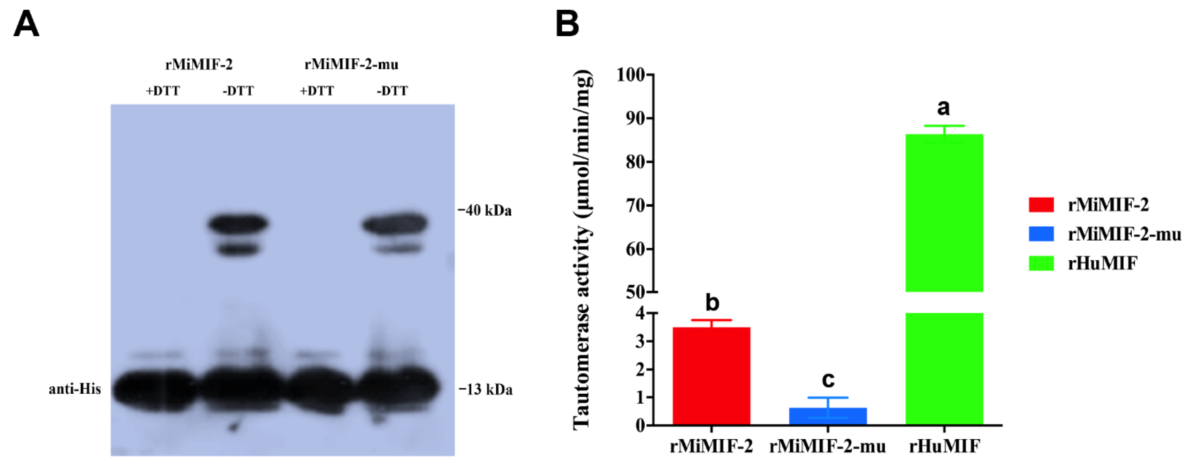
2.1. Tautomerase Activity of MiMIF-2

To identify the possible tautomerase activity of MiMIF-2, the recombinant MiMIF-2 with a N-terminal His tag (rMiMIF-2) and a mutated version with a glycine in place of the first proline residue (rMiMIF-2-mu) were used. These proteins were purified from the cells and checked by western blotting. In reducing conditions (+DTT, Dithiothreitol), the His tag antibody recognized the soluble purified rMiMIF-2 and rMiMIF-2-mu proteins at the expected size (13 kDa). After non-reducing (-DTT) electrophoresis, the molecular mass of the purified recombinant proteins was approximately 40 kDa, indicating the formation of a trimer (Figure 1A). The rMiMIF-2 catalyzed the tautomerization of the p-hydroxyphenylpyruvate substrate, but its activity was much lower than that of human MIF (Figure 1B). Moreover, rMiMIF-2-mu had only residual activity toward the substrate (Figure 1B). These results indicated that rMiMIF-2 had tautomerase activity.

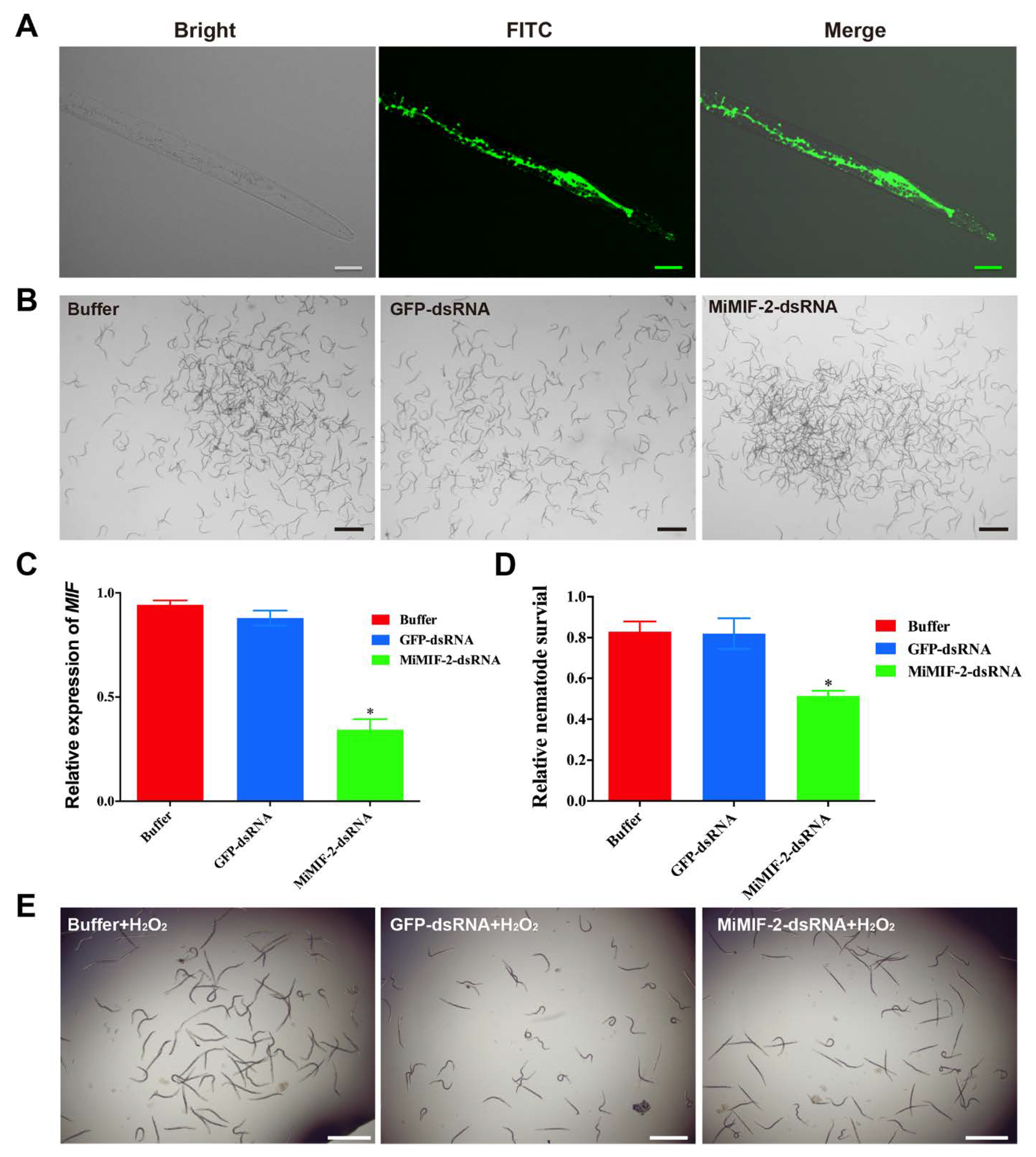
2.2. MiMIF-2 Has Antioxidant Activity

To examine whether MiMIF-2 exhibited redox activity, Escherichia coli (E. coli) expressing rMiMIF-2 was challenged with organic peroxides (cumene hydroperoxide, CH and t-butyl hydroperoxide, t-BOOH). As shown in the result, MiMIF-2 expression significantly increased the tolerance of E. coli to CH and t-BOOH (Figure 2A), as measured by a 65% and 60% reduction in the diameter of the dead bacteria, respectively, relative to cells transformed with empty vector controls (Figure 2B). Likewise, rMiMIF-2-mu also increased the tolerance of E. coli, but to a less extent than rMiMIF-2 (Figure 2).
Further, in vitro RNAi experiment was conducted to investigate the role of MiMIFs in relieving oxidative stress. Compared with the treatment with buffer, there were no phenotypic changes in nematodes viability after treatment with MiMIF-2 dsRNA or GFP dsRNA (Figure 3A,B). RT-qPCR performed using MiMIF specific primers (Table S1) demonstrated that MiMIFs transcript levels were decreased by 80%, whereas treatment with control GFP dsRNA had no effect (Figure 3C). Further, following treatment with 10-mM H2O2, nematodes viability was 30% lower in J2 s in which MiMIF genes silenced by RNAi than in those treated with GFP dsRNA or buffer (Figure 3D,E). Together, these results showed that MiMIF-2 was involved in antioxidant activity.

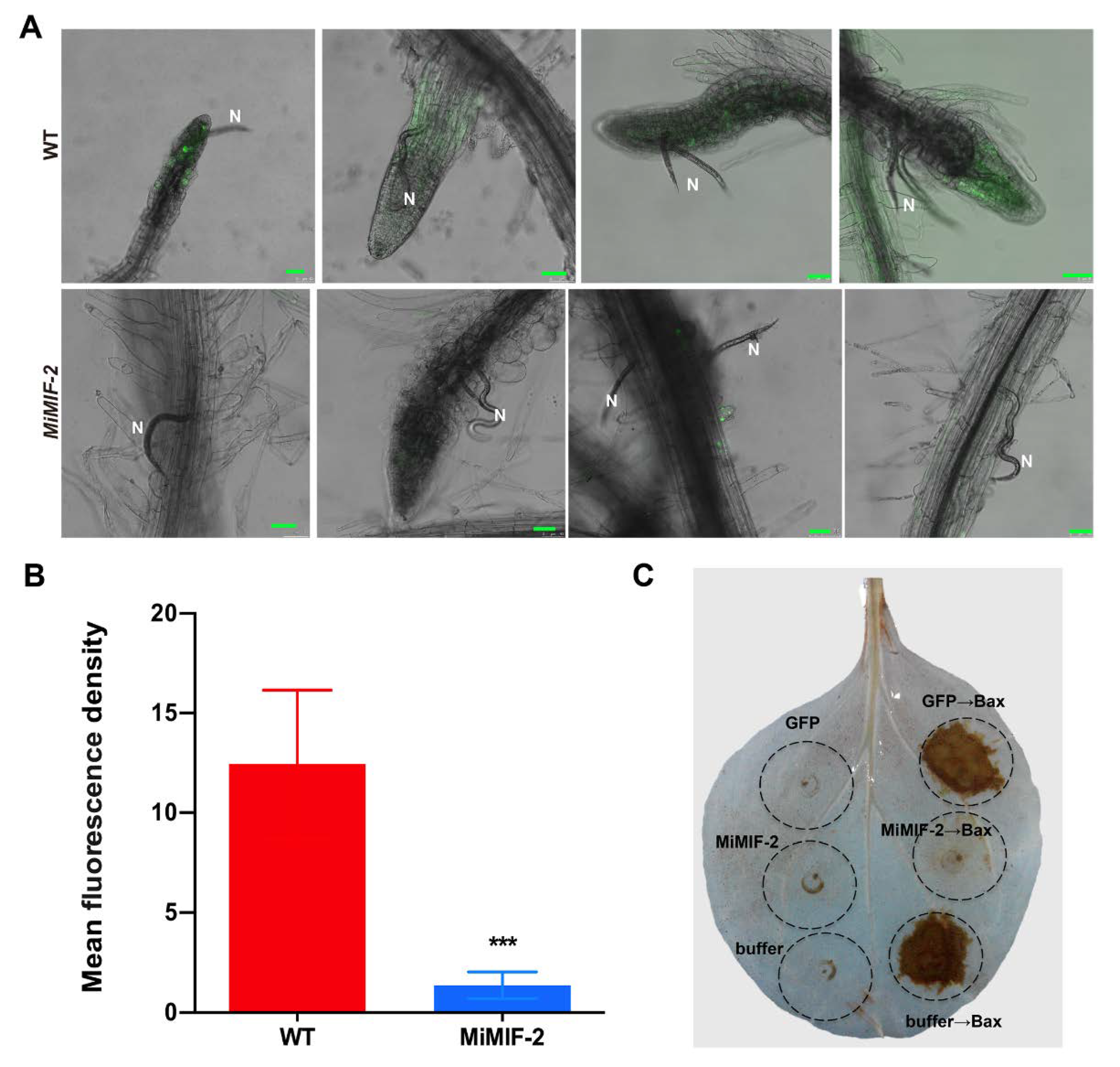
2.3. MiMIF-2 Suppresses Reactive Oxygen Species (ROS) Burst

To investigate ROS production in plant roots upon nematode infection, Arabidopsis roots were labeled with 5-(and-6)-chloromethyl-2′,7′-dichlorofluorescein diacetate (CM-H2DCFDA) after M. incognita inoculation. Confocal microscopy observation results showed that, 1 day post infection (dpi) of M. incognita, there was significant difference (p < 0.001) of CM-H2DCFDA fluorescence intensity between wild type Arabidopsis and MiMIF-2 expressing lines (Figure 4A and Figure S1), which decreased more than ten-fold in MiMIF-2 expressing lines (Figure 4B).
To detect ROS generation during programmed cell death (PCD), MiMIF-2 and Bax were co-expressed in N. benthamiana leaves. A. tumefaciens cells carrying the MiMIF-2 gene were infiltrated 24 h prior to infiltration with Bax, and two days post infiltration DAB staining was conducted to determine the amount of H2O2. The results showed that H2O2 production was significantly weak at the positions where MiMIF-2 was expressed (Figure 4C and Figure S2). Together, these results indicated that MiMIF-2 played roles in suppressing ROS burst in planta.

2.4. Identification of Differentially Expressed Genes (DEGs) in MiMIF-2 Expressing Arabidopsis Lines and Wild Type Plants

We previously showed that ectopic expressing MiMIF-2 in Arabidopsis showed identical phenotypes with wild type, but increased susceptibility to M. incognita [39]. To further investigate potential gene expression changes and signaling pathway modifications regulated by the MiMIF-2 effector, we conducted an RNA-seq experiment to compare wild type Arabidopsis and the MiMIF-2 transgenic lines. The FPKM method was used to calculate DEGs between WT and MiMIF-2 ectopic expressing Arabidopsis lines. Under the criterion of |log2FC| ≥ 1 and FDR ≤ 0.1, compared with wild type, 26 DEGs were identified in MiMIF-2 expressing Arabidopsis through pairwise comparisons (Data S1). Among them, 5 genes were upregulated and 21 genes were downregulated. Interestingly, the expression levels of 8 genes encoding heat-shock protein (HSP) associated proteins were downregulated (Data S1). In order to confirm the transcriptomic data, DEGs of all HSP-associated genes and two upregulated genes (AT4G08950 and AT1G13609) were verified by RT-qPCR. On the whole, RT-qPCR showed that these 10 genes exhibited similar expression trends with the annotations of transcriptomic data, indicating that differential gene expression was due to the effect of the MiMIF-2 expression (Figure 5).

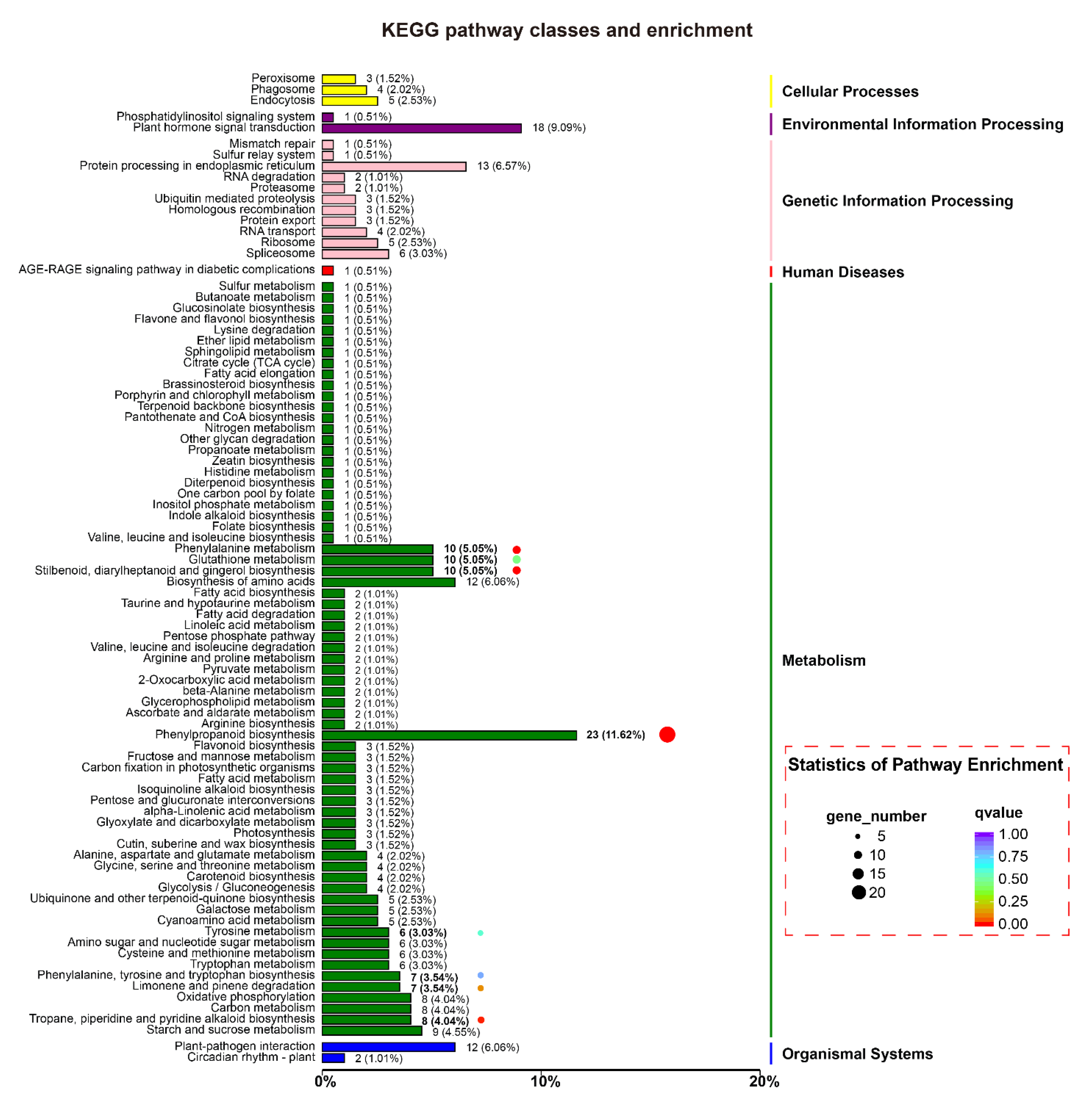
2.5. Plant Response Pathways and Metabolism Pathways Were Interfered in MiMIF-2 Ectopic Expressing Arabidopsis after Treatment with flg22

Nematode infection induced ROS burst was significantly suppressed in MiMIF-2 expressing Arabidopsis, indicating that MiMIF-2 may interfere plant immunity pathways. In this study, transcriptomic profile on wild type and MiMIF-2 ectopic expressing Arabidopsis was investigated after treatment with flg22 for 24 h. A total of 1204 DEGs were identified through pairwise comparisons in MiMIF-2 ectopic expressing Arabidopsis, of which 438 genes were upregulated and 766 genes were downregulated (Data S2). COG function classification of these identified DEGs showed that they were categorized into 21 function classes. Among them, signal transduction mechanisms (14.89%), secondary metabolites biosynthesis, transport and catabolism (10.51%), carbohydrate transport and metabolism (8.23%), posttranslational modification, protein turnover, chaperones (7.53%), and defense mechanisms (6.65%) were relatively abundant (Figure S3A). Based on the three GO categories, the majority classifications of the DEGs were cellular process, metabolic process and single-organism process in “biologic process category”, cell part, cell and organelle in “cellular component category”, binding and catalytic activity in “molecular function category” (Figure S3B). And Kyoto Encyclopedia of Genes and Genomes (KEGG) enrichment analysis showed that 327 DEGs were mostly involved in metabolism, in which phenylpropanoid biosynthesis, phenylalanine metabolism, stilbenoid, diarylheptanoid and gingerol biosynthesis, tropane, piperidine and pyridine alkaloid biosynthesis were the main enriched pathways (Figure 6). This information suggested that MiMIF-2 expression in planta mainly affected plant response pathways and metabolism pathways.

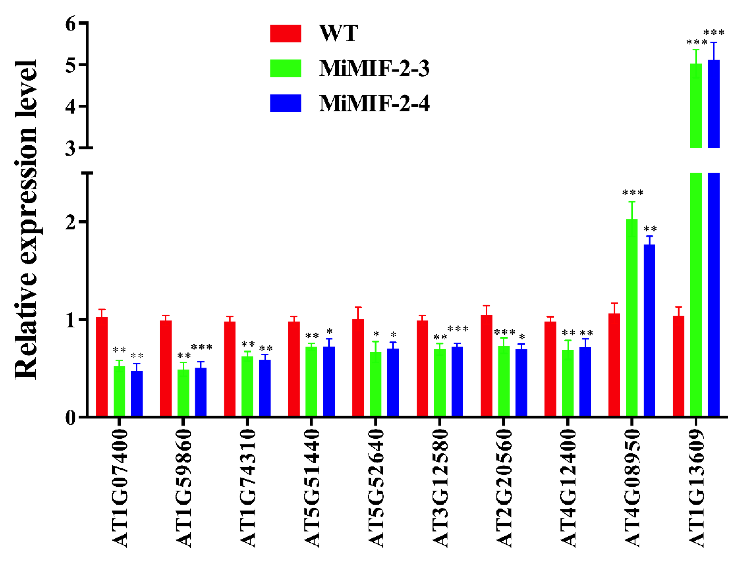
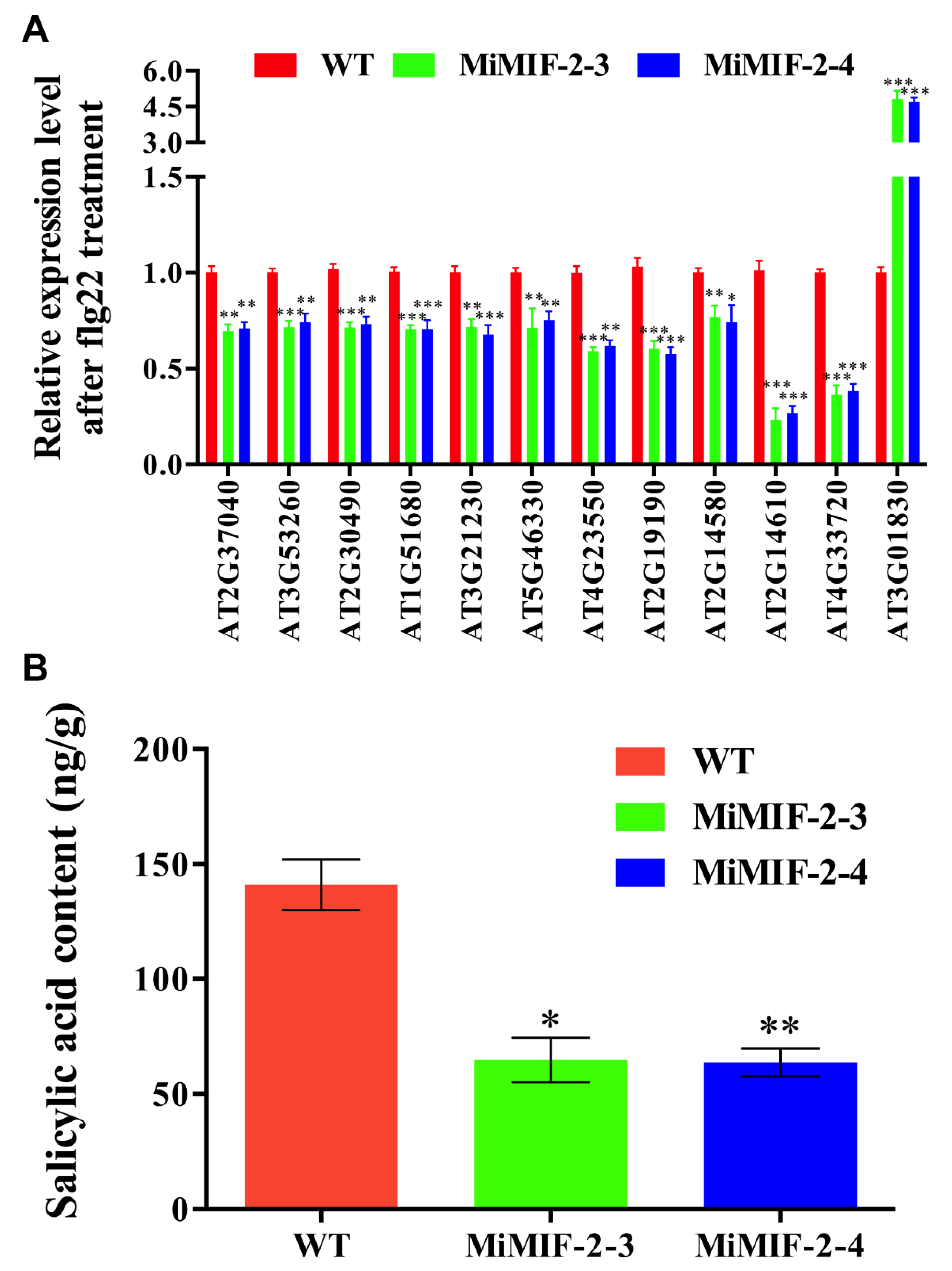
2.6. Defense-Associated Genes Were Down Regulated and Salicylic Acid (SA) Content Was Decreased in MiMIF-2 Expressing Arabidopsis after Treatment with flg22

By analyzing the data of differentially expressed genes that involved in the pathways of plant–pathogen interaction, plant hormone signal transduction and phenylalanine metabolism, some defense-associated genes were down regulated, for example PR1, FRK1, WRK29 and PAL. Further, RT-qPCR was conducted to verify the expressions of key genes involved in defense pathways, phenylpropanoid biosynthesis and phenylalanine metabolism. And the results were in accordance with the transcriptome data, indicating that differential gene expression was due to the effect of the MiMIF-2 expression (Figure 7A). Moreover, metabolic analyses revealed a dramatic decrease of SA content in MiMIF-2 expressing Arabidopsis lines (Figure 7B), which suggested a potential effect of MiMIF-2 on the SA synthesis.

3. Discussion

MiMIFs are homologous to the human MIF and contain the conserved amino acids Pro1, Lys32 and Ile64, involved in tautomerase activity. Previous report demonstrated that the X-ray crystal structure of MIF was a homotrimer [43], which was related structurally to the small bacterial isomerases 4-oxalocrotonate tautomerase, 5-carboxy- methyl-2-hydroxymuconate and chorismate mutase [44]. Meanwhile, mammalian and helminth MIFs both have tautomerase activity [45]. In this study, our results showed that rMiMIF-2 formed a trimer in non-denaturing western blotting and catalyzed the tautomerization of p-hydroxyphenylpyruvate, but the activity was much weaker than that of HuMIF. A previous study indicated that the initiator methionine of MIF was cleaved, exposing the proline residue, which was essential for MIF tautomerase activity [46]. Plasmodium falciparum-derived MIF-like protein [47] and a MIF-like protein from the filarial parasite O. volvulus (OvMIF-2) had low levels of activity with the substrates p-hydroxyphenylpyruvate and L-dopamethylester [48]. Our findings confirmed the requirement for the N-terminal proline residue for tautomerase activity and raised the possibility that MIF may exert its biologic action via an enzymatic reaction.
MIF-like proteins have thiol–protein oxidoreductase activity in animals and this activity is dependent on a C-X-X-C motif. For instance, the human MIF can modulate immune responses through a cysteine-mediated redox mechanism [49]. Sequence analyses have shown that the C-X-X-C motif is not present in all helminth orthologs [45], but there is growing evidence for the involvement of MIFs in immune processes through a number of subtle mechanisms. Plasmodium-derived MIFs have two of the four cysteine residues of the CC motif and CXCR2 and CXCR4 have been identified as functional chemokine receptors [50]. In this study, the rMiMIF-2 protein could protect E. coli against organic peroxide and MiMIF-2 dsRNA-treated nematodes were less viable in H2O2 solution than nematodes subjected to a control treatment. Interestingly, our result is similar with clade B M. incognita cuticle-secreted peroxiredoxin (PRX) proteins, which has protective anti-oxidant activity [51]. We speculate that MiMIF-2 may have evolved sophisticated functions in the redox system that may contribute to M. incognita parasitism.
ROS burst is a symbol of plant cell response to microbe infection. Bax is a death-promoting member of the Bcl-2 family of proteins, which triggers cell death when expressed in N. benthamiana. Previous evidence showed that Bax-induced cell death was similar to hypersensitive response and correlated with accumulation of the defense-related protein PR1 [52]. In this work, we co-expressed Bax with MiMIF-2 in N. benthamiana leaves. Results showed that H2O2 production was significantly alleviated after MiMIF-2 expression. Moreover, our nematode infection assay showed that CM-H2DCFDA fluorescence was weaker in MiMIF-2 ectopic expressing lines than in the wild-type control. Together, these results indicated that MiMIF-2 probably facilitated parasitism by suppressing the host ROS burst.
Ectopic expression of MiMIF-2 in Arabidopsis allowed us to identify the potential functions of MiMIF-2 by assessing its effect on plant cells. We identified a total of 26 DEGs in response to MiMIF-2 expression in Arabidopsis according to RNA-seq data. Interestingly, 8 DEGs encoded heat-shock proteins (HSPs) were all downregulated. These included HSP20, HSP70, HSP90 and some HSP family proteins. Only 5 DEGs were upregulated, of which three only had functional predication. Among them, AT4G08950 encoded an EXO (EXORDIUM) gene, which was response to brassinosteroid (BR). Evidence showed that EXO overexpression promoted shoot and root growth, while exo knock out mutant lines showed reduced BR-triggered cotyledon and hypocotyl growth. Moreover, expression-profiling results indicated that EXO controlled a growth-related gene expansion 5 [53]. Interestingly, plant expansins were involved in the establishment of feeding cells by plant-parasitic nematodes [54,55,56,57]. It would be meaningful to explore the interaction between EXO expression and M. incognita parasitism. Together, the transcriptome profile on wild type and MiMIF-2 expressing Arabidopsis indicated that MiMIF-2 potentially involved in modulating HSP-like genes expression in plant cell, which could be an explanation for the abolished H2O2 production during nematode infection in MiMIF-2 expressing Arabidopsis roots and MiMIF-2/Bax co-expression in N. benthamiana leaves.
Flg22 is a conserved peptide derived from Pseudomonas syringae flagellin. It is perceived by the receptor kinase FLS2 in Arabidopsis, leading to the induction of plant immune responses, such as callose deposition, the expression of defense marker genes, ROS burst and the activation of MAPK cascades [58,59,60,61]. Previous report showed that ectopic expressing MiMIF-2 in Arabidopsis affected PAMP-triggered immunity, which MAPK cascade was interfered, some defense-related marker genes were significantly repressed and callose deposition was reduced after treatment with flg22 [39]. To further investigate the effect of MiMIF-2 expression in plant cell in response to flg22, transcriptomic profile on wild type and MiMIF-2 expressing Arabidopsis was investigated after treatment with flg22 for 24 h. Previous report indicated that transcriptional response to 1 µM flg22 was terminated within 24 Hours [62]. However, we identified 1204 DEGs, in which many genes were involved in plant–pathogen interaction. For instance, AT5G46330 encodes FLS2, which is an essential leucine-rich repeat serine/threonine protein kinase involving plant innate immunity [63]. Strong evidence confirmed that flg22 was recognized by FLS2-BAK1 complex (BAK1 is a co-receptor) and activated downstream immune responses [64]. The FLS2 expression was inhibited in flg22 treated MiMIF-2 expressing plants, which could affect downstream signal transduction and finally led to the suppression of immune-related marker genes expression. In accordance with the expression level of FLS2, AT4G23550 encodes WRKY DNA-binding protein 29, which was shown to interact with the heat-shock factor f1b (hsf1b) involving abiotic stress tolerance [65]. Likewise, genes encoding FRK1 (AT2G19190) and pathogenesis-related proteins (AT2G14580, AT2G14610 and AT4G33720) were all downregulated, of which AT2G14610 encodes PR1 is essential for plant immunity and cell-to-cell signal transduction. Further metabolic analyses revealed that SA content was significantly decreased in MiMIF-2 expressing Arabidopsis after treatment with flg22. Together, these results indicated that MiMIF-2 expression may have a role in dampening plant responses to PAMP.
Based on the KEGG pathways of related DEGs, we noticed that phenylpropanoid biosynthesis and phenylalanine metabolism pathways were suppressed. Plant phenylpropanoids are involved in many cellular processes and functional pathways, especially plant growth and development, protection against biotic or abiotic stresses [66,67]. Starting from phenylalanine, phenylpropanoids pathway also produces a variety of different kinds of compounds, including SA. SA is an essential plant hormone against various biotic and abiotic stress, including restrict plant-parasitic nematode infection [13,30,41]. Although SA is predominantly synthesized via the chorismate/isochorismate pathway in Arabidopsis (about 90%), mutations in phenylalanine ammonia–lyase (PAL) genes resulted in a 90% reduction of basal PAL activity and 50% decrease in pathogen-induced SA accumulation, indicating the importance of PAL for SA biosynthesis [68]. A previous research demonstrated that the transcription of PAL gene was increased in resistant soybean cultivars after M. incognita infections [69]. Moreover, the expression of PAL gene was also activated in tomato when the plant was infected with M. incognita [70]. Phenylpropanoid biosynthetic pathway was also induced by pathogen infection [71,72]. AT2G30490 encodes a cinnamate-4-hydroxylase (C4H), which catalyzes key reactions of phenylpropanoid pathways and modulates salicylic acid accumulation [73]. Recently, the biosynthetic pathways for phenylalanine and phenylalanine-derived compounds were activated for the induction of ETI [74], which gives inspirations for analyzing the role of phenylpropanoids during the interaction between plant-parasitic nematodes and hosts. In this study, we found some genes involved in phenylpropanoid pathways were downregulated, especially AT2G37040 encodes PAL1, AT3G53260 encodes PAL2, AT2G30490 encodes C4H, and the decreased SA content in MiMIF-2 expressing Arabidopsis. Thus, we speculate that MiMIF-2 expression in plant may interfere with host cell metabolism, especially the phenylpropanoid metabolic pathway, leading to endogenous hormone levels decreased.
In this study, we confirmed that MiMIF-2 had tautomerase activity and antioxidant activity. MiMIF-2 expression in Arabidopsis may affect plant defense pathways by suppressing SA synthesis. Altogether, our data suggested, to our knowledge, a possible molecular explanation for M. incognita MiMIF-2 effector, which interfered with plant cell metabolism and SA-mediated immune responses through enzymatic activities.

4. Materials and Methods

4.1. Nematode Collection and Plant Materials

M. incognita was reproduced on tomato plants (Solanum lycopersicum var. ‘Baiguo’). Infective pre-parasitic juveniles were collected after eggs hatching 24 h.
Two homozygous MiMIF-2 expressing Arabidopsis lines (MiMIF-2-T3-3 and MiMIF-2-T3-4) were used in this study, described previously [39]. Surface-sterilized Arabidopsis seeds were seeded on MS medium (Sigma, St. Louis, MO, USA) and placed in a plant growth incubator with a 16-h light and 8-h dark photoperiod at 23 °C. Tobacco (Nicotiana benthamiana) plants were grown in pots under the same condition with Arabidopsis.

4.2. SDS–PAGE, Immunoblotting and Biochemical Analyses of rMiMIF-2

Recombinant MiMIF-2 (rMiMIF-2, with an N-terminal His tag) was reported before [39]. SDS-PAGE was performed on a separating gel of 15% (w/v) acrylamide and a stacking gel of 4% (w/v) acrylamide. Protein was transferred to PVDF membrane using semi-dry transfer (Bio-Rad, Hercules, CA, USA). Western blot analysis was performed for the detection of rMiMIF-2 with His-tag antibody (Sigma, St. Louis, MO, USA).
The MiMIF-2 tautomerase assay was conducted as previously described [47]. Briefly, the substrate, p-hydroxyphenylpyruvate (Sigma, St. Louis, MO, USA), was dissolved in 50-mM ammonium acetate (pH 6.0) and the recombinant MIF protein was dissolved in boric acid (pH 6.2). The enzymatic activity was determined at 25 °C by addition of 50 µL of 0.2-mM p-hydroxyphenylpyruvate to a 96-well plate containing 50-µL MIF solution and measuring the increase in absorbance at 330 nm. Human MIF recombinant protein (AmyJet Scientific, Inc., Wuhan, China) was used as a positive control. This experiment consisted of three technical replicates; results were represented by the mean plus SD of three biologic replicates. The data were subjected to one-way ANOVA followed by Dunnett’s multiple comparisons test using Prism 6.0 (Alpha 0.05, GraphPad Software, Inc., La Jolla, CA, USA).
Bacteria viability assays were conducted as described previously [51]. Bacteria expressing rMiMIF-2 and rMiMIF-2-mu were transferred to Top agar (Luria–Bertani broth (LB) medium supplemented with 0.8% agar, 50-mg/mL kanamycin, 1-mM IPTG, Isopropyl β-D-thiogalactoside), plated onto LB agar plates (100 mg/mL kanamycin) and incubated for 1 h at 37 °C. Subsequently, filter discs (diameter 5 mm) soaked with 5 µL of 200-mM cumene hydroperoxide (CH) or 100-mM butyl hydroperoxide (t-BOOH) (Sigma, St. Louis, MO, USA) were placed on top of the agar. The diameter of the zone of killing was measured after an overnight incubation at 37 °C with 8 replicates for each condition. The mean plus SD value of 6 biologic replicates were presented. The data were subjected to one-way ANOVA with Dunnett’s multiple comparisons test. Three independent assays were performed.

4.3. In Vitro RNAi and Nematode Activity in H2O2

Vector construction was conducted according to previous research [75]. Briefly, MiMIF-2 (339 bp) and fragments of green fluoresces protein (GFP) were cloned into the L4440 vector (a gift from Prof. Fanrong Zeng’s lab, Chinese Academy of Agricultural Sciences, Beijing, China) to construct L4440-MiMIF-2. Plasmids were transformed into HT115 competent cells and dsRNA was induced with IPTG. dsRNA was extracted using TRIzol and enzymatic digestion followed by DNase (TaKaRa, Shiga, Japan) and S1 nuclease (Invitrogen, Carlsbad, CA, USA), according to the manufacturer’s instructions. In vitro RNAi experiment was conducted following the previous protocol [76]. Freshly hatched M. incognita pre-J2s were soaked in solutions containing MiMIF-2 dsRNA, GFP dsRNA or without dsRNA for 30 min. After soaking, the nematodes were used for RT-qPCR to quantity the interference effect and activity assay in 10-mM H2O2 (Sigma, St. Louis, MO, USA). Living nematodes were counted after treatment for 4 h. Each experiment was performed three times independently. Statistically significant differences between treatments were determined by one-way ANOVA with Dunnett’s multiple comparisons test.

4.4. Transient Expression Assay in N. benthamiana

To visualize H2O2, a type of reactive oxygen species (ROS), N. benthamiana leaves infiltrated with Bcl-2 associated X protein (Bax) and/or MiMIF-2 were stained with 3,3′-Diaminobenzidine (DAB). We used Agrobacterium tumefaciens cells to deliver a potato virus X (PVX) vector carrying MiMIF-2 and a proapoptotic Bax ORF into N. benthamiana leaves. pGR107-MiMIF-2 and pGR107-Bax plasmids were reported before and Agrobacterium infiltration method was conducted as previously [39]. Generally, four-week-old N. benthamiana leaves were infiltrated with the recombinant Agrobacterium suspension (for each assay, five plants and /or 15 leaves were used for infiltration). Leaf samples were collected at 2 dpi and soaked in 1 mg/mL 3,3′-diaminobenzidine solution. The sampled leaves were placed in the dark for 4 h, boiled in 95% ethanol and photographed. This experiment was repeated at least three times with similar result.

4.5. Nematode Infection and Detection of Root Reactive Oxygen Species (ROS)

To monitor ROS production and localization in Arabidopsis root cells, the CM-H2DCFDA (C6827, Molecular Probes) staining method was used with some modifications of the previously described protocol [77,78]. Generally, lines expressing MiMIF-2 or wild type Arabidopsis were grown under sterile conditions and inoculated with 200 surface-sterilized nematodes. The roots were then incubated with CM-H2DCFDA (10 μM) in phosphate-buffered saline for 90 min at 4 °C at 1 dpi. The samples were cleaned with KCl (0.1 mM) and CaCl2 (0.1 mM), kept at room temperature for 1 h, and then imaged with a Leica confocal microscope (Leica SP8, Heidelberg, Germany). Three independent assays were performed and at least 10 plants were observed for each line. Relative fluorescence density was calculated using ImageJ (NIH, Bethesda, MD, USA).

4.6. RNA Isolation, Library Construction and RNA-Seq

Thirteen-day-old Arabidopsis seedlings in sterile MS plates (WT and MiMIF-2 transgenic lines) were harvested. For flg22 treatment, seedlings were treated by 1 μM flg22 for 24 h in sterile Corning six-wells plate. Arabidopsis total RNA was isolated using the RNAsimple total RNA kit following the manufacturer’s instructions (Tiangen, Beijing, China). The quantity, quality and integrality of the extracted RNA were verified by Nanodrop (Wilmington, DC, USA), Qubit 2.0 (Invitrogen, Carlsbad, CA, USA) and Agilent 2100 (Agilent, USA). Libraries construction and the RNA-Seq were performed by the Biomarker Biotechnology Corporation (Beijing, China) using Illumina HiSeq2500. mRNA was enriched by oligo (dT)-rich magnetic beads and then broken into random fragments using Fragmentation Buffer. First- and second-strand cDNA were synthesized using these mRNA and purified by AMPure XP beads. The resulting cDNAs were then subjected to end-repair and add polyA tail. After that, Illumina sequencing adaptors were ligated to these cDNA and then use AMPure XP beads for fragment size selection. Next, the cDNA libraries were obtained by PCR enrichment. Finally, the cDNA libraries were sequenced using an Illumina HiSeq2500.

4.7. Data Analysis

Raw data (raw reads) of fastq format were first, processed through in-house Perl scripts. In this step, clean data (clean reads) were obtained by removing reads containing adapter, reads containing ploy-N and low-quality reads from raw data. At the same time, Q20, Q30, GC-content and sequence duplication level of the clean data were calculated. All the downstream analyses were based on clean data with high quality.
The adaptor sequences and low-quality sequence reads were removed from the data sets. Raw sequences were transformed into clean reads after data processing. These clean reads were then mapped to the reference genome sequence. Only reads with a perfect match or one mismatch were further analyzed and annotated based on the reference genome. Hisat2 tools soft were used to map with reference genome. To define the different expression genes (DEGs) between WT and MiMIF-2 transgenic lines after treatment with flg22, the data were calculated based on the ratio of the RPKM values, and the false discovery rate (FDR) control method. In this study, data only with an value of log2 ratio ≥ 2 and an FDR significance score <0.01 were used for analysis. Without any treatment, comparison of WT and MiMIF-2 transgenic lines used a value of log2 ratio ≥ 2 and a FDR significance score < 0.1.
Gene function was annotated based on the following databases: including the NCBI Nr (non-redundant protein sequences database), NCBI Nt (non-redundant nucleotide sequences database), Swiss-Prot (a manually annotated and reviewed protein sequence database), KEGG (the Kyoto Encyclopedia of Genes and Genomes, http://www.genome.jp/kegg/kegg2.html), Pfam (Protein family, http://pfam.xfam.org) and GO database (gene ontology, http://geneontology.org).

4.8. RT-qPCR Analysis

Real-time quantitative PCR (RT-qPCR) was performed on an ABI Prism 7000 real-time PCR system (Applied Biosystems, USA). Primers used in this study were listed in Table S1. RT-qPCR was performed using SYBR® Premix Ex Taq™ II (TaKaRa, Japan) under the following program: 95 °C 30 s and 40 cycles of 95 °C for 5 s, 60 °C for 30 s and 95 °C, 15 s, 60 °C for 60 s and 95 °C for 15 s. Expression ratio was analyzed using the 2−ΔΔCt method [79]. Data represent the mean fold-change ± SE. Statistical Analysis was used one-way ANOVA with Dunnett′s multiple comparisons test. This experiment was repeated three times.

4.9. Detection of Salicylic Acid (SA) Content

For metabolic analysis, wild type Arabidopsis seedlings and MiMIF-2 transgenic Arabidopsis seedlings were treated with 1 µM flg22 for 24 h, and then immediately frozen in liquid nitrogen, ground into powder. The samples were extracted and quantified with an ABI QTRAP 6500 LC–MS/MS platform (Applied Biosystems, Foster City, CA, USA) according to previous method [80]. The standard SA was purchased from Sigma-Aldrich (St. Louis, MO, USA). Statistical Analysis was used one-way ANOVA with Dunnett′s multiple comparisons test.

4.10. Accession Number

Sequence data were stored at the National Genomics Data Center (NGDC). The raw sequence data reported in this study is stored at the Genome Sequence Archive (Genomics, Proteomics & Bioinformatics 2017) in BIG Data Center, Beijing Institute of Genomics (BIG), Chinese Academy of Sciences, under accession numbers CRA002198, which is publicly accessible at https://bigd.big.ac.cn/gsa.

Supplementary Materials

Supplementary materials can be found at https://www.mdpi.com/1422-0067/21/10/3507/s1. Table S1. RT-qPCR primers used in this study. Data S1. Lists of differentially expressed genes identified between wild type and MiMIF-2 expressing Arabidopsis lines. Data S2. Lists of differentially expressed genes identified between wild type and MiMIF-2 expressing Arabidopsis lines after treatment with flg22. Figure S1. ROS visualization in nematode infected Arabidopsis roots. CM-H2DCFDA staining of ROS in wild-type (WT) and MiMIF-2 expressing (MiMIF-2-3 and MiMIF-2-4) roots following M. incognita infection. Images were captured by confocal microscopy (LeicaSP8) 1-day post inoculation (dpi). Micrographs A are images of differential interference contrast (grey). Micrographs B are images of GFP fluorescence (green). Micrographs C are overlays of images of the differential interference contrast and GFP-fluorescence. GFP, green fluorescence protein. Scale bar, 50 µm. N, nematode. Figure S2. H2O2 detection after co-expression MiMIF-2 and Bax in N. benthamiana leaves. H2O2 production in N. benthamiana leaves was assessed by DAB staining. N. benthamiana leaves were infiltrated with Agrobacterium tumefaciens carrying the corresponding constructs, and measurements were performed at 2 dpi. Three independent assays were conducted and at least 5 plants and/or 15 leaves were observed. Figure S3. The clusters of differential expression genes between wild type and MiMIF-2 expressing Arabidopsis transcriptomes after treatment with flg22 24 h. (A) COG function classification of DEGs. DEGs were categorized into 21 function classes. Among them, signal transduction mechanisms (14.89%), secondary metabolites biosynthesis, transport and catabolism (10.51%), carbohydrate transport and metabolism (8.23%), posttranslational modification, protein turnover, chaperones (7.53%) and defense mechanisms (6.65%), amino acid transport and metabolism (5.78%), lipid transport and metabolism (5.6%) were relatively abundant. (B) GO terms classification of DEGs. DEGs were classified into three processes, including biological process, cellular component and molecular function.

Author Contributions

B.X. and H.J. acquired funding and designed the experiments. J.Z., Z.M., Q.S. and Q.L. performed the experiments. J.Z., H.J. and B.X. wrote and revised the manuscript. All authors read and approved the final version of the manuscript for publication.

Funding

This work was supported by the National Natural Science Foundation of China (Grant Numbers 31571987, 31571996, 31672010 and 31871942).

Acknowledgments

The authors would like to thank Heng Wang (Peking Union Medical College), Pierre ABAD and Bruno Favery (Université Côte d’Azur, INRA, CNRS, ISA, France) for their valuable suggestions in the experiments design.

Conflicts of Interest

The authors declare no conflict of interest.

References

  1. Chen, J.; Lin, B.; Huang, Q.; Hu, L.; Zhuo, K.; Liao, J. A novel Meloidogyne graminicola effector, MgGPP, is secreted into host cells and undergoes glycosylation in concert with proteolysis to suppress plant defenses and promote parasitism. PLoS Pathog. 2017, 13, e1006301. [Google Scholar] [CrossRef] [Green Version]
  2. Caboni, P.; Ntalli, N.G.; Aissani, N.; Cavoski, I.; Angioni, A. Nematicidal activity of (E,E)-2,4-decadienal and (E)-2-decenal from Ailanthus altissima against Meloidogyne javanica. J. Agric. Food Chem. 2012, 60, 1146–1151. [Google Scholar] [CrossRef]
  3. Abad, P.; Gouzy, J.; Aury, J.M.; Castagnone-Sereno, P.; Danchin, E.G.; Deleury, E.; Perfus-Barbeoch, L.; Anthouard, V.; Artiguenave, F.; Blok, V.C.; et al. Genome sequence of the metazoan plant-parasitic nematode Meloidogyne incognita. Nat. Biotechnol. 2008, 26, 909–915. [Google Scholar] [CrossRef] [Green Version]
  4. Jones, J.D.; Dangl, J.L. The plant immune system. Nature 2006, 444, 323–329. [Google Scholar] [CrossRef] [Green Version]
  5. Tyler, B.M. The fog of war: How network buffering protects plants’ defense secrets from pathogens. PLoS Genet. 2017, 13, e1006713. [Google Scholar] [CrossRef]
  6. Van der Burgh, A.M.; Joosten, M. Plant Immunity: Thinking Outside and Inside the Box. Trends Plant Sci. 2019, 24, 587–601. [Google Scholar] [CrossRef]
  7. Vieira, P.; Gleason, C. Plant-parasitic nematode effectors—Insights into their diversity and new tools for their identification. Curr. Opin. Plant Biol. 2019, 50, 37–43. [Google Scholar] [CrossRef]
  8. Holbein, J.; Grundler, F.M.; Siddique, S. Plant basal resistance to nematodes: An update. J. Exp. Bot. 2016, 67, 2049–2061. [Google Scholar] [CrossRef] [Green Version]
  9. Goverse, A.; Smant, G. The activation and suppression of plant innate immunity by parasitic nematodes. Annu. Rev. Phytopathol. 2014, 52, 243–265. [Google Scholar] [CrossRef]
  10. Mejias, J.; Truong, N.M.; Abad, P.; Favery, B.; Quentin, M. Plant Proteins and Processes Targeted by Parasitic Nematode Effectors. Front. Plant. Sci. 2019, 10, 970. [Google Scholar] [CrossRef] [Green Version]
  11. Mine, A.; Seyfferth, C.; Kracher, B.; Berens, M.L.; Becker, D.; Tsuda, K. The Defense Phytohormone Signaling Network Enables Rapid, High-Amplitude Transcriptional Reprogramming during Effector-Triggered Immunity. Plant Cell 2018, 30, 1199–1219. [Google Scholar] [CrossRef] [Green Version]
  12. Kazan, K.; Lyons, R. Intervention of Phytohormone Pathways by Pathogen Effectors. Plant Cell 2014, 26, 2285–2309. [Google Scholar] [CrossRef] [Green Version]
  13. Gheysen, G.; Mitchum, M.G. Phytoparasitic Nematode Control of Plant Hormone Pathways. Plant Physiol. 2019, 179, 1212–1226. [Google Scholar] [CrossRef] [Green Version]
  14. Palmer, I.A.; Shang, Z.; Fu, Z.Q. Salicylic acid-mediated plant defense: Recent developments, missing links, and future outlook. Front. Biol. 2017, 12, 258–270. [Google Scholar] [CrossRef]
  15. Iwai, T.; Seo, S.; Mitsuhara, I.; Ohashi, Y. Probenazole-induced accumulation of salicylic acid confers resistance to Magnaporthe grisea in adult rice plants. Plant Cell Physiol. 2007, 48, 915–924. [Google Scholar] [CrossRef] [Green Version]
  16. Qi, G.; Chen, J.; Chang, M.; Chen, H.; Hall, K.; Korin, J.; Liu, F.; Wang, D.; Fu, Z.Q. Pandemonium Breaks Out: Disruption of Salicylic Acid-Mediated Defense by Plant Pathogens. Mol. Plant. 2018, 11, 1427–1439. [Google Scholar] [CrossRef] [Green Version]
  17. Liu, T.; Song, T.; Zhang, X.; Yuan, H.; Su, L.; Li, W.; Xu, J.; Liu, S.; Chen, L.; Chen, T.; et al. Unconventionally secreted effectors of two filamentous pathogens target plant salicylate biosynthesis. Nat. Commun. 2014, 5, 4686. [Google Scholar] [CrossRef] [Green Version]
  18. Djamei, A.; Schipper, K.; Rabe, F.; Ghosh, A.; Vincon, V.; Kahnt, J.; Osorio, S.; Tohge, T.; Fernie, A.R.; Feussner, I.; et al. Metabolic priming by a secreted fungal effector. Nature 2011, 478, 395–398. [Google Scholar] [CrossRef]
  19. Nahar, K.; Kyndt, T.; De Vleesschauwer, D.; Hofte, M.; Gheysen, G. The jasmonate pathway is a key player in systemically induced defense against root knot nematodes in rice. Plant Physiol. 2011, 157, 305–316. [Google Scholar] [CrossRef] [Green Version]
  20. Wubben, M.J.; Jin, J.; Baum, T.J. Cyst nematode parasitism of Arabidopsis thaliana is inhibited by salicylic acid (SA) and elicits uncoupled SA-independent pathogenesis-related gene expression in roots. Mol. Plant Microb. Interact. 2008, 21, 424–432. [Google Scholar] [CrossRef] [Green Version]
  21. Lin, J.; Mazarei, M.; Zhao, N.; Zhu, J.J.; Zhuang, X.; Liu, W.; Pantalone, V.R.; Arelli, P.R.; Stewart, C.N., Jr.; Chen, F. Overexpression of a soybean salicylic acid methyltransferase gene confers resistance to soybean cyst nematode. Plant Biotechnol. J. 2013, 11, 1135–1145. [Google Scholar] [CrossRef]
  22. Baxter, A.; Mittler, R.; Suzuki, N. ROS as key players in plant stress signalling. J. Exp. Bot. 2014, 65, 1229–1240. [Google Scholar] [CrossRef] [PubMed]
  23. Tripathy, B.C.; Oelmuller, R. Reactive oxygen species generation and signaling in plants. Plant Signal. Behav. 2012, 7, 1621–1633. [Google Scholar] [CrossRef] [PubMed]
  24. Suzuki, N.; Miller, G.; Morales, J.; Shulaev, V.; Torres, M.A.; Mittler, R. Respiratory burst oxidases: The engines of ROS signaling. Curr. Opin. Plant Biol. 2011, 14, 691–699. [Google Scholar] [CrossRef]
  25. Torres, M.A. ROS in biotic interactions. Physiol. Plant. 2010, 138, 414–429. [Google Scholar] [CrossRef]
  26. Wrzaczek, M.; Brosche, M.; Kangasjarvi, J. ROS signaling loops—Production, perception, regulation. Curr. Opin. Plant Biol. 2013, 16, 575–582. [Google Scholar] [CrossRef]
  27. Siddique, S.; Matera, C.; Radakovic, Z.S.; Hasan, M.S.; Gutbrod, P.; Rozanska, E.; Sobczak, M.; Torres, M.A.; Grundler, F.M. Parasitic worms stimulate host NADPH oxidases to produce reactive oxygen species that limit plant cell death and promote infection. Sci. Signal 2014, 7, 33. [Google Scholar] [CrossRef] [Green Version]
  28. Lin, B.; Zhuo, K.; Chen, S.; Hu, L.; Sun, L.; Wang, X.; Zhang, L.H.; Liao, J. A novel nematode effector suppresses plant immunity by activating host reactive oxygen species-scavenging system. N. Phytol. 2016, 209, 1159–1173. [Google Scholar] [CrossRef]
  29. Xia, X.J.; Zhou, Y.H.; Shi, K.; Zhou, J.; Foyer, C.H.; Yu, J.Q. Interplay between reactive oxygen species and hormones in the control of plant development and stress tolerance. J. Exp. Bot. 2015, 66, 2839–2856. [Google Scholar] [CrossRef] [Green Version]
  30. Poor, P.; Czekus, Z.; Tari, I.; Ordog, A. The Multifaceted Roles of Plant Hormone Salicylic Acid in Endoplasmic Reticulum Stress and Unfolded Protein Response. Int. J. Mol. Sci. 2019, 20, 5842. [Google Scholar] [CrossRef] [Green Version]
  31. Leyton-Jaimes, M.F.; Kahn, J.; Israelson, A. Macrophage migration inhibitory factor: A multifaceted cytokine implicated in multiple neurological diseases. Exp. Neurol. 2018, 301, 83–91. [Google Scholar] [CrossRef] [PubMed]
  32. Xu, L.; Li, Y.; Sun, H.; Zhen, X.; Qiao, C.; Tian, S.; Hou, T. Current developments of macrophage migration inhibitory factor (MIF) inhibitors. Drug Discov. Today 2013, 18, 592–600. [Google Scholar] [CrossRef] [PubMed]
  33. Rosengren, E.; Åman, P.; Thelin, S.; Hansson, C.; Ahlfors, S.; Björk, P.; Jacobsson, L.; Rorsman, H. The macrophage migration inhibitory factor MIF is a phenylpyruvate tautomerase. FEBS Lett. 1997, 417, 85–88. [Google Scholar] [CrossRef] [Green Version]
  34. Pastrana, D.V.; Raghavan, N.; FitzGerald, P.; Eisinger, S.W.; Metz, C.; Bucala, R.; Schleimer, R.P.; Bickel, C.; Scott, A.L. Filarial nematode parasites secrete a homologue of the human cytokine macrophage migration inhibitory factor. Infect. Immun. 1998, 66, 5955–5963. [Google Scholar] [CrossRef] [Green Version]
  35. Marson, A.L.; Tarr, D.E.K.; Scott, A.L. Macrophage migration inhibitory factor (mif) transcription is significantly elevated in Caenorhabditis elegans dauer larvae. Gene 2001, 278, 53–62. [Google Scholar] [CrossRef]
  36. Wu, Z.; Boonmars, T.; Nagano, I.; Nakada, T.; Takahashi, Y. Molecular Expression and Characterization of a Homologue of Host Cytokine Macrophage Migration Inhibitory Factor from Trichinella Spp. J. Parasitol. 2003, 89, 507–515. [Google Scholar] [CrossRef]
  37. Tan, T.H.; Edgerton, S.A.; Kumari, R.; McAlister, M.S.; Roe, S.M.; Nagl, S.; Pearl, L.H.; Selkirk, M.E.; Bianco, A.E.; Totty, N.F.; et al. Macrophage migration inhibitory factor of the parasitic nematode Trichinella spiralis. Biochem. J. 2001, 357, 373–383. [Google Scholar] [CrossRef]
  38. Augustijn, K.D.; Kleemann, R.; Thompson, J.; Kooistra, T.; Crawford, C.E.; Reece, S.E.; Pain, A.; Siebum, A.H.; Janse, C.J.; Waters, A.P. Functional characterization of the Plasmodium falciparum and P. berghei homologues of macrophage migration inhibitory factor. Infect. Immun. 2007, 75, 1116–1128. [Google Scholar] [CrossRef] [Green Version]
  39. Zhao, J.; Li, L.; Liu, Q.; Liu, P.; Li, S.; Yang, D.; Chen, Y.; Pagnotta, S.; Favery, B.; Abad, P.; et al. A MIF-like effector suppresses plant immunity and facilitates nematode parasitism by interacting with plant annexins. J. Exp. Bot. 2019, 70, 5943–5958. [Google Scholar] [CrossRef]
  40. Harris, J.; VanPatten, S.; Deen, N.S.; Al-Abed, Y.; Morand, E.F. Rediscovering MIF: New Tricks for an Old Cytokine. Trends Immunol. 2019, 40, 447–462. [Google Scholar] [CrossRef]
  41. Janda, T.; Szalai, G.; Pal, M. Salicylic Acid Signalling in Plants. Int. J. Mol. Sci. 2020, 21, 2655. [Google Scholar] [CrossRef] [PubMed] [Green Version]
  42. Kumar, D. Salicylic acid signaling in disease resistance. Plant. Sci. 2014, 228, 127–134. [Google Scholar] [CrossRef] [PubMed]
  43. Sun, H.W.; Bernhagen, J.; Bucala, R.; Lolis, E. Crystal structure at 2.6-A resolution of human macrophage migration inhibitory factor. Proc. Natl. Acad. Sci. USA 1996, 93, 5191–5196. [Google Scholar] [CrossRef] [Green Version]
  44. Davidson, R.; Baas, B.J.; Akiva, E.; Holliday, G.L.; Polacco, B.J.; LeVieux, J.A.; Pullara, C.R.; Zhang, Y.J.; Whitman, C.P.; Babbitt, P.C. A global view of structure-function relationships in the tautomerase superfamily. J. Biol. Chem. 2018, 293, 2342–2357. [Google Scholar] [CrossRef] [Green Version]
  45. Vermeire, J.J.; Cho, Y.; Lolis, E.; Bucala, R.; Cappello, M. Orthologs of macrophage migration inhibitory factor from parasitic nematodes. Trends Parasitol. 2008, 24, 355–363. [Google Scholar] [CrossRef] [Green Version]
  46. Swope, M.; Sun, H.W.; Blake, P.R.; Lolis, E. Direct link between cytokine activity and a catalytic site for macrophage migration inhibitory factor. EMBO J. 1998, 17, 3534–3541. [Google Scholar] [CrossRef] [Green Version]
  47. Shao, D.; Han, Z.; Lin, Y.; Zhang, L.; Zhong, X.; Feng, M.; Guo, Y.; Wang, H. Detection of Plasmodium falciparum derived macrophage migration inhibitory factor homologue in the sera of malaria patients. Acta Trop. 2008, 106, 9–15. [Google Scholar] [CrossRef]
  48. Ajonina-Ekoti, I.; Kurosinski, M.A.; Younis, A.E.; Ndjonka, D.; Tanyi, M.K.; Achukwi, M.; Eisenbarth, A.; Ajonina, C.; Luersen, K.; Breloer, M.; et al. Comparative analysis of macrophage migration inhibitory factors (MIFs) from the parasitic nematode Onchocerca volvulus and the free-living nematode Caenorhabditis elegans. Parasitol. Res. 2013, 112, 3335–3346. [Google Scholar] [CrossRef]
  49. Kleemann, R.; Kapurniotu, A.; Frank, R.W.; Gessner, A.; Mischke, R.; Flieger, O.; Juttner, S.; Brunner, H.; Bernhagen, J. Disulfide analysis reveals a role for macrophage migration inhibitory factor (MIF) as thiol-protein oxidoreductase. J. Mol. Biol. 1998, 280, 85–102. [Google Scholar] [CrossRef]
  50. Bernhagen, J.; Krohn, R.; Lue, H.; Gregory, J.L.; Zernecke, A.; Koenen, R.R.; Dewor, M.; Georgiev, I.; Schober, A.; Leng, L.; et al. MIF is a noncognate ligand of CXC chemokine receptors in inflammatory and atherogenic cell recruitment. Nat. Med. 2007, 13, 587–596. [Google Scholar] [CrossRef]
  51. Dubreuil, G.; Deleury, E.; Magliano, M.; Jaouannet, M.; Abad, P.; Rosso, M.N. Peroxiredoxins from the plant parasitic root-knot nematode, Meloidogyne incognita, are required for successful development within the host. Int. J. Parasitol. 2011, 41, 385–396. [Google Scholar] [CrossRef] [PubMed]
  52. Lacomme, C.; Santa Cruz, S. Bax-induced cell death in tobacco is similar to the hypersensitive response. Proc. Natl. Acad. Sci. USA 1999, 96, 7956–7961. [Google Scholar] [CrossRef] [PubMed] [Green Version]
  53. Schroder, F.; Lisso, J.; Lange, P.; Mussig, C. The extracellular EXO protein mediates cell expansion in Arabidopsis leaves. BMC Plant Biol. 2009, 9, 20. [Google Scholar] [CrossRef] [PubMed] [Green Version]
  54. Fudali, S.; Janakowski, S.; Sobczak, M.; Griesser, M.; Grundler, F.M.; Golinowski, W. Two tomato alpha-expansins show distinct spatial and temporal expression patterns during development of nematode-induced syncytia. Physiol. Plant. 2008, 132, 370–383. [Google Scholar] [CrossRef]
  55. Wieczorek, K.; Grundler, F.M. Expanding nematode-induced syncytia: The role of expansins. Plant Signal. Behav. 2006, 1, 223–224. [Google Scholar] [CrossRef] [Green Version]
  56. Wieczorek, K.; Golecki, B.; Gerdes, L.; Heinen, P.; Szakasits, D.; Durachko, D.M.; Cosgrove, D.J.; Kreil, D.P.; Puzio, P.S.; Bohlmann, H.; et al. Expansins are involved in the formation of nematode-induced syncytia in roots of Arabidopsis thaliana. Plant J. 2006, 48, 98–112. [Google Scholar] [CrossRef]
  57. Gal, T.Z.; Aussenberg, E.R.; Burdman, S.; Kapulnik, Y.; Koltai, H. Expression of a plant expansin is involved in the establishment of root knot nematode parasitism in tomato. Planta 2006, 224, 155–162. [Google Scholar] [CrossRef]
  58. Gomez-Gomez, L.; Boller, T. FLS2: An LRR receptor-like kinase involved in the perception of the bacterial elicitor flagellin in Arabidopsis. Mol. Cell 2000, 5, 1003–1011. [Google Scholar] [CrossRef]
  59. Navarro, L.; Zipfel, C.; Rowland, O.; Keller, I.; Robatzek, S.; Boller, T.; Jones, J.D. The transcriptional innate immune response to flg22. Interplay and overlap with Avr gene-dependent defense responses and bacterial pathogenesis. Plant Physiol. 2004, 135, 1113–1128. [Google Scholar] [CrossRef] [Green Version]
  60. Asai, T.; Tena, G.; Plotnikova, J.; Willmann, M.R.; Chiu, W.L.; Gomez-Gomez, L.; Boller, T.; Ausubel, F.M.; Sheen, J. MAP kinase signalling cascade in Arabidopsis innate immunity. Nature 2002, 415, 977–983. [Google Scholar] [CrossRef]
  61. Lee, H.Y.; Back, K. Mitogen-activated protein kinase pathways are required for melatonin-mediated defense responses in plants. J. Pineal Res. 2016, 60, 327–335. [Google Scholar] [CrossRef] [PubMed]
  62. Denoux, C.; Galletti, R.; Mammarella, N.; Gopalan, S.; Werck, D.; De Lorenzo, G.; Ferrari, S.; Ausubel, F.M.; Dewdney, J. Activation of defense response pathways by OGs and Flg22 elicitors in Arabidopsis seedlings. Mol. Plant 2008, 1, 423–445. [Google Scholar] [CrossRef] [PubMed] [Green Version]
  63. Couto, D.; Zipfel, C. Regulation of pattern recognition receptor signalling in plants. Nat. Rev. Immunol. 2016, 16, 537–552. [Google Scholar] [CrossRef] [PubMed]
  64. Sun, Y.; Li, L.; Macho, A.P.; Han, Z.; Hu, Z.; Zipfel, C.; Zhou, J.M.; Chai, J. Structural basis for flg22-induced activation of the Arabidopsis FLS2-BAK1 immune complex. Science 2013, 342, 624–628. [Google Scholar] [CrossRef]
  65. Pick, T.; Jaskiewicz, M.; Peterhansel, C.; Conrath, U. Heat shock factor HsfB1 primes gene transcription and systemic acquired resistance in Arabidopsis. Plant Physiol. 2012, 159, 52–55. [Google Scholar] [CrossRef] [Green Version]
  66. Vanholme, B.; El Houari, I.; Boerjan, W. Bioactivity: phenylpropanoids’ best kept secret. Curr. Opin. Biotechnol. 2019, 56, 156–162. [Google Scholar] [CrossRef]
  67. Korkina, L.; Kostyuk, V.; De Luca, C.; Pastore, S. Plant phenylpropanoids as emerging anti-inflammatory agents. Mini Rev. Med. Chem. 2011, 11, 823–835. [Google Scholar] [CrossRef]
  68. Ding, P.; Ding, Y. Stories of Salicylic Acid: A Plant Defense Hormone. Trends Plant. Sci. 2020. [Google Scholar] [CrossRef]
  69. Edens, R.M.; Anand, S.C.; Bolla, R.I. Enzymes of the Phenylpropanoid Pathway in Soybean Infected with Meloidogyne incognita or Heterodera glycines. J. Nematol. 1995, 27, 292–303. [Google Scholar]
  70. Vasyukova, N.I.; Pridvorova, S.M.; Gerasimova, N.G.; Chalenko, G.I.; Ozeretskovskaya, O.L.; Udalova, Z.V.; Zinov’eva, S.V. The involvement of phenylalanine ammonia-lyase and salicylic acid in the induction of resistance of tomato plants infested with gall nematode Meloidogyne incognita. Dokl. Biol. Sci. 2007, 416, 382–385. [Google Scholar] [CrossRef]
  71. Le Roy, J.; Huss, B.; Creach, A.; Hawkins, S.; Neutelings, G. Glycosylation Is a Major Regulator of Phenylpropanoid Availability and Biological Activity in Plants. Front. Plant Sci. 2016, 7, 735. [Google Scholar] [CrossRef] [PubMed] [Green Version]
  72. Dixon, R.A.; Paiva, N.L. Stress-Induced Phenylpropanoid Metabolism. Plant Cell 1995, 7, 1085–1097. [Google Scholar] [CrossRef] [PubMed]
  73. Pietrowska-Borek, M.; Nuc, K.; Guranowski, A. Exogenous adenosine 5’-phosphoramidate behaves as a signal molecule in plants; it augments metabolism of phenylpropanoids and salicylic acid in Arabidopsis thaliana seedlings. Plant Physiol. Biochem. 2015, 94, 144–152. [Google Scholar] [CrossRef] [PubMed]
  74. Yoo, H.; Greene, G.H.; Yuan, M.; Xu, G.; Burton, D.; Liu, L.; Marques, J.; Dong, X. Translational Regulation of Metabolic Dynamics during Effector-Triggered Immunity. Mol. Plant 2019. [Google Scholar] [CrossRef] [Green Version]
  75. Kamath, R.S.; Ahringer, J. Genome-wide RNAi screening in Caenorhabditis elegans. Methods 2003, 30, 313–321. [Google Scholar] [CrossRef]
  76. Huang, G.; Allen, R.; Davis, E.L.; Baum, T.J.; Hussey, R.S. Engineering broad root-knot resistance in transgenic plants by RNAi silencing of a conserved and essential root-knot nematode parasitism gene. Proc. Natl. Acad. Sci. USA 2006, 103, 14302–14306. [Google Scholar] [CrossRef] [Green Version]
  77. Ameziane-El-Hassani, R.; Dupuy, C. Detection of Intracellular Reactive Oxygen Species (CM-H2DCFDA). Bio Protoc. 2013, 3. [Google Scholar] [CrossRef] [Green Version]
  78. Kristiansen, K.A.; Jensen, P.E.; Moller, I.M.; Schulz, A. Monitoring reactive oxygen species formation and localisation in living cells by use of the fluorescent probe CM-H(2)DCFDA and confocal laser microscopy. Physiol. Plant. 2009, 136, 369–383. [Google Scholar] [CrossRef]
  79. Livak, K.J.; Schmittgen, T.D. Analysis of relative gene expression data using real-time quantitative PCR and the 2(-Delta Delta C(T)) Method. Methods 2001, 25, 402–408. [Google Scholar] [CrossRef]
  80. Liu, H.; Li, X.; Xiao, J.; Wang, S. A convenient method for simultaneous quantification of multiple phytohormones and metabolites: Application in study of rice-bacterium interaction. Plant Methods 2012, 8, 2. [Google Scholar] [CrossRef] [Green Version]
Figure 1. Recombinant MiMIF-2 exhibited tautomerase activity; (A) Detection of the purified recombinant rMiMIF-2 and rMiMIF-2-mu with an anti-His tag antibody. rMiBMIF-mu is the mutant in which the N-terminal proline is replaced with a glycine residue; (B) The tautomerase activities of rMiMIF-2 and its mutant were compared with that of the recombinant human MIF (rHuMIF), with p-hydroxyphenylpyruvate as the substrate. The bar represents the mean ± SD of five independent measurements. Columns marked with different letters were significantly different according to ANOVA followed by Dunnett’s multiple comparisons test (Alpha 0.05). More than three technical replicates and three biologic replicates were performed, and we show the results of one of these experiments here.
Ijms 21 03507 g001
Figure 2. MiMIF-2 expression in Escherichia coli exhibited resistance to organic peroxide; (A) Sensitivity to cumene hydroperoxide (CH) or t-butyl hydroperoxide (t-BOOH), measured in the filter disc assay in Escherichia coli expressing MiMIF-2 or MiMIF-2-mu. Cells containing the empty plasmid pet30a were used as controls. The numbers indicate summary statistic data for Petri dishes with similar results. The experiments were performed three times; (B) Diameters were measured to assess the difference between treatments. The bar represents the mean ± SD of six independent measurements. *, ** indicates significant difference in one-way ANOVA with Dunnett′s multiple comparisons test (Alpha 0.05). The experiment was repeated three times with similar results.
Ijms 21 03507 g002
Figure 3. In vitro RNAi silencing of MiMIF-2 in pre-parasitic M. incognita J2; (A) Fluorescence microscopy investigation showed ingestion of fluorescein isothiocynate (FITC) in the treated M. incognita pre-J2 s. Scale bars, 20 µm; (B) phenotypes of M. incognita pre-J2 s after treatment with buffer, GFP-dsRNA or MiMIF-2-dsRNA. Scale bars, 50 µm; (C) relative expression levels of MiMIF-2 after soaking in the buffer, GFP-dsRNA or MiMIF-2-dsRNA. The relative expression of MiMIFs was quantified by RT-qPCR. The tubulin gene was used as an internal control, and fold-change values (mean ± SE) were calculated relative to the buffer value by the 2−ΔΔCt method. * indicates significant difference in one-way ANOVA with Dunnett′s multiple comparisons test (Alpha 0.05). The data shown are from one of three independent experiments; (D) MiMIF-2-dsRNA or buffer-treated nematodes were incubated with 10-mM H2O2. Living nematodes were counted after treatment 4 h. Asterisks indicate significant differences relative to the mean ±SD values for the control treatment in Student’s t-test (p < 0.05). The experiments were performed three times and three biologic replicates showed similar results; (E) phenotypes of nematodes after treatment with dsRNA and H2O2. Scale bars, 50 µm.
Ijms 21 03507 g003
Figure 4. H2O2 visualization in nematode infected Arabidopsis roots and H2O2 detection after co-expression MiMIF-2 and Bax in N. benthamiana leaves; (A) CM-H2DCFDA staining of ROS in wild-type (WT) and MiMIF-2 expressing (MiMIF-2-3 and MiMIF-2-4) roots after M. incognita infection. Images were captured by confocal microscopy (LeicaSP8) 1-day post inoculation (dpi). Scale bar, 50 µm. N, nematode; (B) Relative fluorescence density between wild type and MiMIF-2 expressing Arabidospsi after M. incognita infection. *** indicates significant difference in the Student’s t-test (p < 0.001). (C) H2O2 production in N. benthamiana leaves was assessed by 3,3′-Diaminobenzidine (DAB) staining. N. benthamiana leaves were infiltrated with Agrobacterium tumefaciens carrying the corresponding constructs, and measurements were performed at 2 dpi. Three independent assays were conducted and at least 5 plants and/or 15 leaves were observed.
Ijms 21 03507 g004
Figure 5. RT-qPCR verification of candidate differentially expressed genes (DEGs) between wild type and MiMIF-2 expressing Arabidopsis. The relative expression levels of 8 heat-shock-like proteins and 2 upregulated genes were analyzed by RT-qPCR and normalized using UBP22 (AT5G10790) as the internal control. Data represent the mean fold-change ± SE. Mean values significantly different from wild type (*, **, ***) were determined by one-way ANOVA with Dunnett′s multiple comparisons test (Alpha 0.05). The experiment was repeated three times with similar results.
Ijms 21 03507 g005
Figure 6. KEGG pathway enrichment of differentially expressed genes (DEGs) in MiMIF-2 expressing Arabidopsis after treatment with flg22. DEGs were classified into 6 processes (cellular processes, environmental information processing, genetic information processing, human diseases, metabolism and organismal systems). MiMIF-2 expressing in Arabidopsis affected some cellular pathways, especially phenylpropanoid biosynthesis, phenylalanine metabolism, glutathione metabolism, plant hormone signal transduction and plant–pathogen interaction.
Ijms 21 03507 g006
Figure 7. Candidate DEGs verification and metabolic analyses of SA content between wild type and MiMIF-2 expressing Arabidopsis after treatment with flg22 24 h; (A) The relative expression ratio of 12 genes involved in phenylpropanoid synthesis and plant–pathogen interaction were analyzed by RT-qPCR and normalized using UBP22 (AT5G10790) as internal control. Data represent the mean fold-change ± SE. Mean values significantly different from wild type (*, **, ***) were determined by one-way ANOVA with Dunnett’s multiple comparisons test (Alpha 0.05). Three biologic repeats were performed with similar results; (B) SA content was quantified using seedlings of wild type and MiMIF-2 expressing Arabidopsis after treatment with flg22 24 h. The bar represents the average of three independent measurements. *, ** indicating the significant difference from the wild type was determined in one-way ANOVA with Dunnett’s multiple comparisons test (alpha 0.05).
Ijms 21 03507 g007

