Copper Coordination Compounds as Biologically Active Agents

 , ,
, ,
Abstract
:1. Introduction
2. Copper Coordination Compounds Based on Ligands with Various Donor Atoms
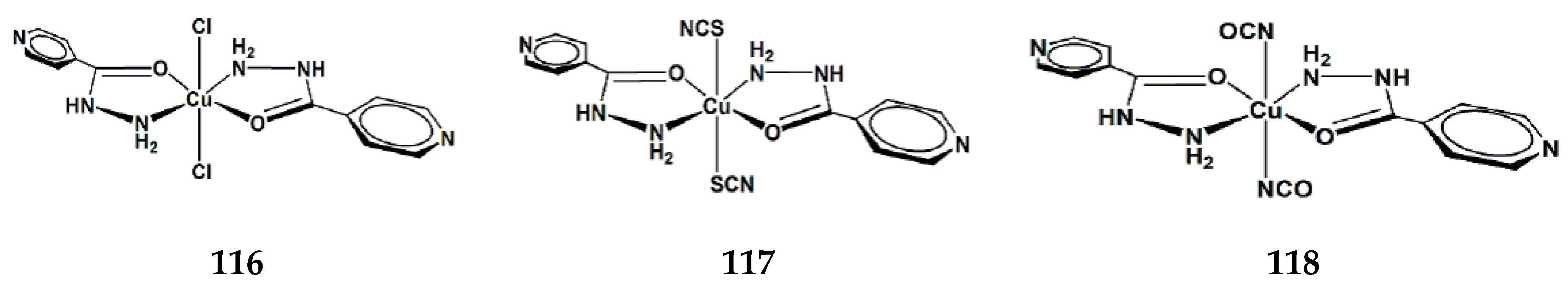
2.1. N- and O-Donor Ligands
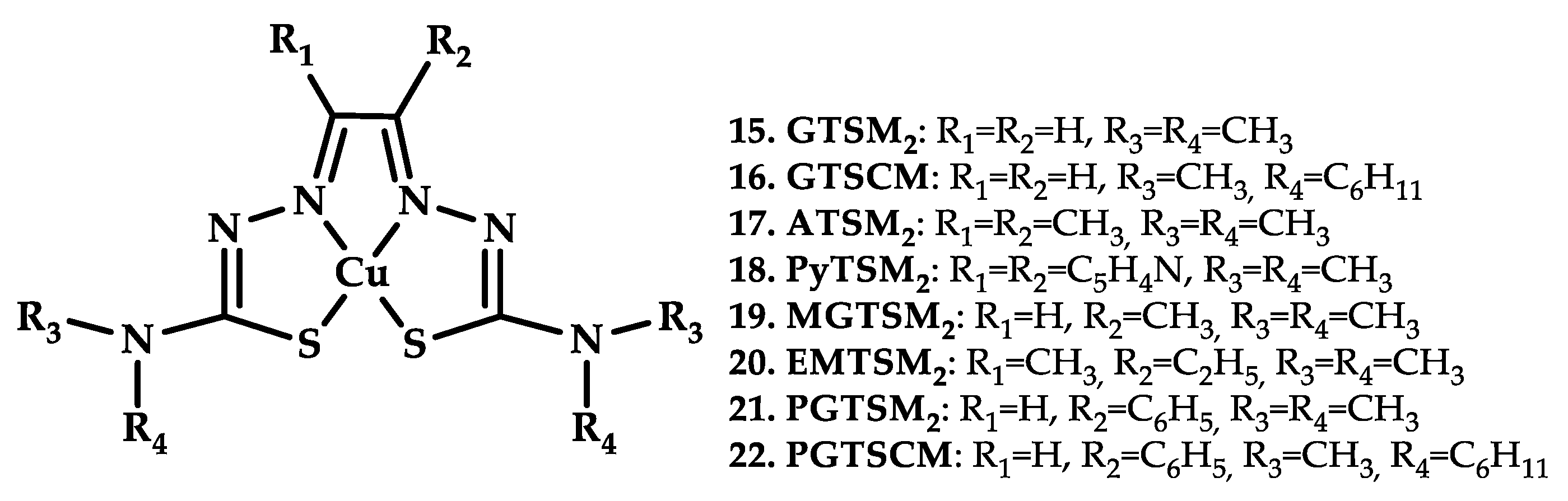
2.2. N- and S-Donor Ligands
2.3. N/N-Donor Ligands
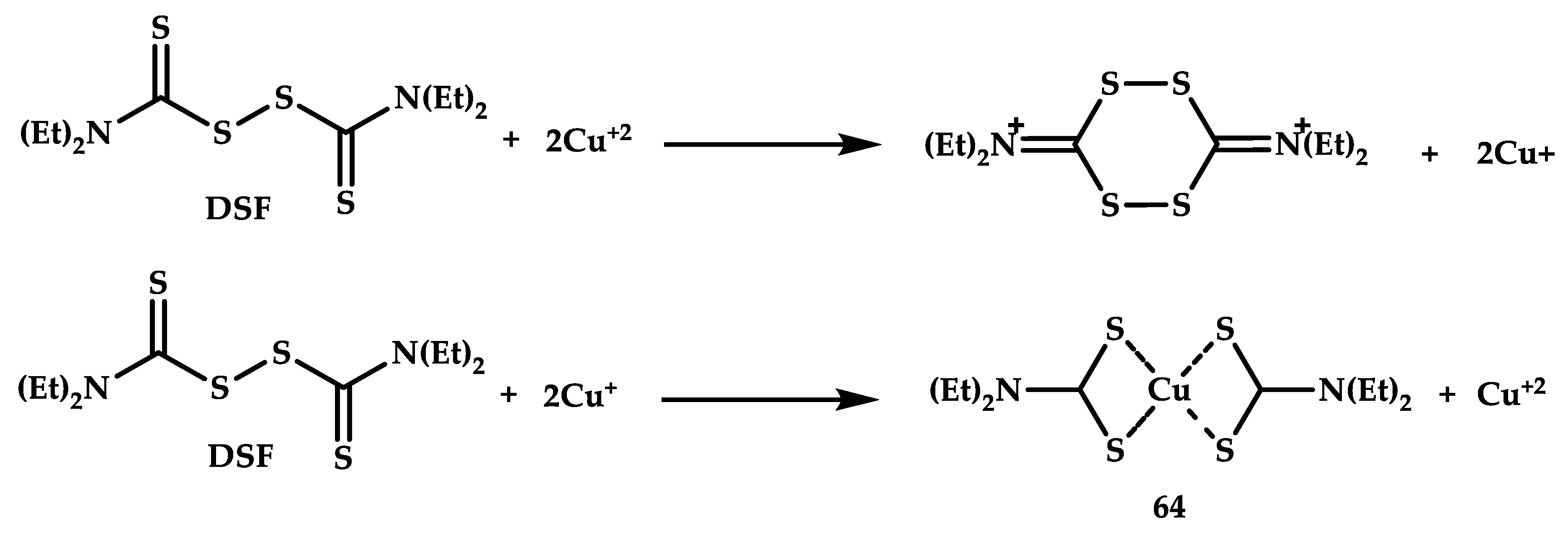
2.4. S/S-Donor Ligands
2.5. N-, O-, and S-Donor Ligands
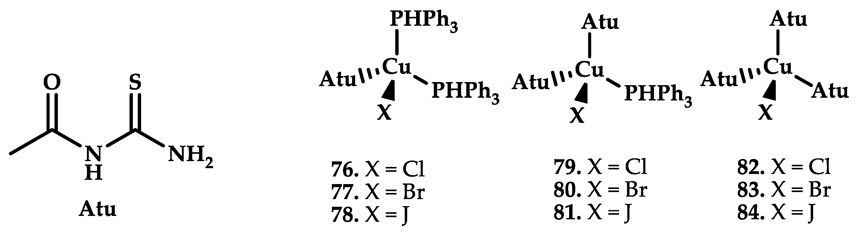
2.6. Phosphine-Donor Ligands
3. Drug-Based Copper Coordination Compounds
4. Natural Product-Based Copper Coordination Compounds
5. Conclusions
Funding
Acknowledgments
Conflicts of Interest
References
- Dömötör, O.; de Almeida, R.F.M.; Côrte-Real, L.; Matos, C.P.; Marques, F.; Matos, A.; Real, C.; Kiss, T.; Enyedy, É.A.; Garcia, M.H.; et al. Studies on the mechanism of action of antitumor bis(aminophenolate) ruthenium (III) complexes. J. Inorg. Biochem. 2017, 168, 27–37. [Google Scholar] [CrossRef] [PubMed] [Green Version]
- Dabrowiak, J.C. Metals in Medicine; Wiley: Hoboken, NJ, USA, 2009; pp. 49–249. [Google Scholar]
- Volarevic, V.; Djokovic, B.; Jankovic, M.G.; Harrell, C.R.; Fellabaum, C.; Djonov, V.; Arsenijevic, N. Molecular mechanisms of cisplatin-induced nephrotoxicity: A balance on the knife edge between renoprotection and tumor toxicity. J. Biomed. Sci. 2019, 26, 1–14. [Google Scholar] [CrossRef] [PubMed] [Green Version]
- McWhinney, S.R.; Goldberg, R.M.; McLeod, H.L. Platinum Neurotoxicity Pharmacogenetics. Mol. Cancer Ther. 2009, 8, 10–16. [Google Scholar] [CrossRef] [PubMed] [Green Version]
- Wheate, N.J.; Walker, S.; Craig, G.E.; Oun, R. The Status of Platinum Anticancer Drugs in the Clinic and in Clinical Trials. Dalton Trans. 2010, 39, 8113–8127. [Google Scholar] [CrossRef] [Green Version]
- Gaál, A.; Orgován, G.; Mihucz, V.G.; Pape, I.; Ingerle, D.; Streli, C.; Szoboszlai, N.J. Metal Transport Capabilities of Anticancer Copper Chelators. Trace Elem. Med. Biol. 2018, 47, 79–88. [Google Scholar]
- Xin, C.; Xiao, Z.; Jinghong, C.; Qianqi, Y.; Li, Y. Hinokitiol copper complex inhibits proteasomal deubiquitination and induces paraptosis-like cell death in human cancer cells. Eur. J. Pharmacol. 2017, 815, 147–155. [Google Scholar]
- Zeeshan, M.; Murugadas, A.; Ghaskadbi, S.; Rajendran, R.B.; Akbarsha, M.A. ROS dependent copper toxicity in Hydra-biochemical and molecular study. Comp. Biochem. Physiol. C Toxicol. Pharmacol. 2016, 185, 1–12. [Google Scholar] [CrossRef]
- Qin, Q.-P.; Meng, T.; Tan, M.-X.; Liu, Y.-C.; Luo, X.-J.; Zou, B.-Q.; Liang, H. Synthesis, crystal structure and biological evaluation of a new dasatinib copper(II) complex as telomerase inhibitor. Eur. J. Med. Chem. 2018, 143, 1597–1603. [Google Scholar] [CrossRef] [PubMed]
- Hernandes, M.S.; Britto, L.R. NADPH oxidase and neurodegeneration. Curr. Neuropharmacol. 2012, 10, 321–327. [Google Scholar] [CrossRef]
- Fatfat, M.; Merhi, R.A.; Rahal, O.; Stoyanovsky, D.A.; Zaki, A.; Haidar, H.; Kagan, V.E.; Gali-Muhtasib, H.; Machaca, K. Copper Chelation Selectively Kills Colon Cancer Cells through Redox Cycling and Generation of Reactive Oxygen Species. BMC Cancer. 2014, 14, 527. [Google Scholar] [CrossRef] [Green Version]
- Kremer, M.L. Mechanism of the Fenton reaction. Evidence for a new intermediate. Phys. Chem. Chem. Phys. 1999, 1, 3595–3605. [Google Scholar] [CrossRef]
- Sangeetha, S.; Murali, M. Non-covalent DNA binding, protein interaction, DNA cleavage and cytotoxicity of [Cu(quamol)Cl]·H2O. Int. J. Biol. Macromol. 2018, 107, 2501–2511. [Google Scholar] [CrossRef] [PubMed]
- Martinez-Bulit, P.; Garza-Ortíz, A.; Mijangos, E.; Barrón-Sosa, L.; Sánchez-Bartéz, F.; Gracia-Mora, I.; Flores-Parra, A.; Contreras, R.; Reedijk, J.; Barba-Behrens, N. 2,6-Bis(2,6-diethylphenyliminomethyl)pyridine coordination compounds with cobalt(II), nickel(II), copper(II), and zinc(II): Synthesis, spectroscopic characterization, X-ray study and in vitro cytotoxicity. J. Inorg. Biochem. 2015, 142, 1–7. [Google Scholar] [CrossRef]
- Fengyi, Z.; Weifan, W.; Wen, L.; Li, X.; Shilong, Y.; Xu-Min, C.; Mengyi, Z.; Meng, L.; Mengtao, M.; Hai-Jun, X.; et al. High anticancer potency on tumor cells of dehydroabietylamine Schiff-base derivatives and a copper(II) complex. Eur. J. Med. Chem. 2018, 146, 451–459. [Google Scholar]
- Chudal, L.; Pandey, N.K.; Phan, J.; Johnson, O.; Lin, L.; Yu, H.; Shu, Y.; Huang, Z.; Xing, M.; Liu, J.P.; et al. Copper-Cysteamine Nanoparticles as a Heterogeneous Fenton-Like Catalyst for Highly Selective Cancer Treatment. ACS Appl. Bio Mater. 2020, 3, 1804–1814. [Google Scholar] [CrossRef]
- Lebon, F.; Boggetto, N.; Ledecq, M.; Durant, F.; Benatallah, Z.; Sicsic, S.; Lapouyade, R.; Kahn, O.; Mouithys-Mickalad, A.; Deby-Dupont, G.; et al. Metal-Organic Compounds: A New Approach for Drug Discovery: N1-(4-Methyl-2-Pyridyl)-2,3,6-Trimethoxybenzamide Copper(II) Complex as an Inhibitor of Human Immunodeficiency Virus 1 Protease. Biochem. Pharmacol. 2002, 63, 1863–1873. [Google Scholar] [CrossRef]
- Roch-Arveiller, M.; Huy, D.P.; Maman, L.; Giroud, J.-P.; Sorenson, J.R.J. Non-Steroidal Anti-Inflammatory Drug-Copper Complex Modulation of Polymorphonuclear Leukocyte Migration. Biochem. Pharmacol. 1990, 39, 569–574. [Google Scholar] [CrossRef]
- Stanila, A.; Braicu, C.; Stanila, S.; Pop, R.M. Antibacterial Activity of Copper and Cobalt Amino Acids Complexes. Not. Bot. Horti Agrobot. Cluj-Na. 2011, 39, 124–129. [Google Scholar] [CrossRef] [Green Version]
- Weder, J.E.; Hambley, T.W.; Kennedy, B.J.; Lay, P.A.; MacLachlan, D.; Bramley, R.; Delfs, C.D.; Murray, K.S.; Moubaraki, B.; Warwick, B.; et al. Anti-Inflammatory Dinuclear Copper(II) Complexes with Indomethacin. Synthesis, Magnetism and EPR Spectroscopy. Crystal Structure of the N,N-Dimethylformamide Adduct. Inorg. Chem. 1999, 38, 1736–1744. [Google Scholar] [CrossRef]
- Liao, Y.; Zhao, J.; Bulek, K.; Tang, F.; Chen, X.; Cai, G.; Jia, S.; Fox, P.; Huang, E.; Pizarro, T.; et al. Inflammation mobilizes copper metabolism to promote colon tumorigenesis via an IL-17-STEAP4-XIAP axis. Nat. Commun. 2020, 11, 900. [Google Scholar] [CrossRef] [Green Version]
- Chen, K.T.J.; Anantha, M.; Leung, A.W.Y.; Kulkarni, J.A.; Militao, G.G.C.; Wehbe, M.; Sutherland, B.; Cullis, P.R.; Bally, M.B. Characterization of a liposom al copper(II)-quercetin formulation suitable for parenteral use. Drug Delivery Transl. Res. 2020, 10, 202–215. [Google Scholar] [CrossRef] [PubMed] [Green Version]
- Liu, X.; Chu, H.; Cui, N.; Wang, T.; Dong, S.; Cui, S.; Dai, Y.; Wang, D. In vitro and in vivo evaluation of biotin-mediated PEGylated nanostructured lipid as carrier of disulfiram coupled with copper ion. J. Drug Delivery Sci. Technol. 2019, 51, 651–661. [Google Scholar] [CrossRef]
- Pellei, M.; Bagnarelli, L.; Luciani, L.; Del Bello, F.; Giorgioni, G.; Piergentili, A.; Quaglia, W.; De Franco, M.; Gandin, V.; Marzano, C.; et al. Synthesis and Cytotoxic Activity Evaluation of New Cu(I) Complexes of Bis(pyrazol-1-yl) Acetate Ligands Functionalized with an NMDA Receptor Antagonist. Int. J. Mol. Sci. 2020, 21, 2616. [Google Scholar] [CrossRef] [PubMed]
- Takjoo, R.; Centore, R.; Hayatolgheibi, S. Mixed ligand complexes of cadmium(II) and copper(II) dithiocarbazate: Synthesis, spectral characterization, X-ray crystal structure. Inorg. Chim. Acta. 2018, 471, 587–594. [Google Scholar] [CrossRef]
- Khan, M.H.; Cai, M.; Deng, J.; Yu, P.; Liang, H.; Yang, F. Anticancer Function and ROS-Mediated Multi-Targeting Anticancer Mechanisms of Copper (II) 2-hydroxy-1-naphthaldehyde Complexes. Molecules 2019, 24, 2544. [Google Scholar] [CrossRef] [Green Version]
- Sabithakala, T.; Chittireddy, V.R.R. DNA Binding and in vitro anticancer activity of 2-((1H-benzimidazol-2-yl)methylamino)acetic acid and its copper(II) mixed-polypyridyl complexes: Synthesis and crystal structure. Appl. Organomet. Chem. 2018, 32, 4550. [Google Scholar] [CrossRef]
- Denoyer, D.; Clatworthy, S.A.S.; Cater, M.A. Copper Complexes in Cancer Therapy. Metallo-Drugs Dev. Action Anticancer Agents 2018, 16, 469–506. [Google Scholar]
- Wang, Z.; Tan, J.; McConville, C.; Kannappan, V.; Tawari, P.; Brown, J.; Ding, J.; Armesilla, A.; Irache, J.; Mei, Q.; et al. Poly lactic-co-glycolic acid controlled delivery of disulfiram to target liver cancer stem-like cells. Nanomed. Nanotechnol. Biol. Med. 2017, 13, 641–657. [Google Scholar]
- Banerjee, P.; Geng, T.; Mahanty, A.; Li, T.; Zong, L.; Wang, B. Integrating the drug, disulfiram into the vitamin E-TPGS-modified PEGylated nanostructured lipid carriers to synergize its repurposing for anti-cancer therapy of solid tumors. Int J. Pharm. 2019, 557, 374–389. [Google Scholar] [CrossRef] [PubMed]
- Tabti, R.; Tounsi, N.; Gaiddon, C.; Bentouhami, E.; Désaubry, L. Progress in Copper Complexes as Anticancer Agents. Med. Chem. 2017, 7, 1–55. [Google Scholar] [CrossRef]
- Wehbe, M.; Leung, A.W.Y.; Abrams, M.J.; Orvig, C.; Bally, M.B. A Perspective – can copper complexes be developed as a novel class of therapeutics? Dalton Trans. 2017, 46, 10758–10773. [Google Scholar] [CrossRef] [PubMed]
- Santini, C.; Pellei, M.; Gandin, V.; Porchia, M.; Tisato, F.; Marzano, C. Advances in Copper Complexes as Anticancer Agents. Chem. Rev. 2014, 114, 815–862. [Google Scholar] [CrossRef] [PubMed]
- Ching Ong, Y.; Roy, S.; Andrews, P.; Gasser, G. Metal Compounds against Neglected Tropical Diseases. Chem. Rev. 2019, 119, 730–796. [Google Scholar] [CrossRef] [PubMed]
- Ruiz-Azuara, L.; Bastian, G.; Bravo-Gómez, M.E.; Cañas, R.C.; Flores-Alamo, M.; Fuentes, I.; Mejia, C.; García-Ramos, J.; Serrano, A. Abstract CT408: Phase I study of one mixed chelates copper(II) compound, Casiopeína CasIIIia with antitumor activity and its mechanism of action. AACR. Cancer Res. 2014, 74 (Suppl. 19). [Google Scholar] [CrossRef]
- Galindo-Murillo, R.; Garcia-Ramos, J.C.; Ruiz-Azuara, L.; Cheatham, T.E.; Cortes-Guzman, F. Intercalation processes of copper complexes in DNA. Nucleic Acids Res. 2015, 43, 5364–5376. [Google Scholar] [CrossRef] [Green Version]
- Rufino-González, Y.; Ponce-Macotela, M.; García-Ramos, J.C.; Martínez-Gordillo, M.N.; Galindo-Murillo, R.; González-Maciel, A.; Reynoso-Robles, R.; Tovar-Tovar, A.; Flores-Alamo, M.; Toledano-Magaña, Y.; et al. Antigiardiasic activity of Cu(II) coordination compounds: Redox imbalance and membrane damage after a short exposure time. J. Inorg. Biochem. 2019, 195, 83–90. [Google Scholar] [CrossRef]
- Anda-Jáuregui, G.; Espinal-Enríquez, J.; Hur, J.; Alcalá-Corona, S.A.; Ruiz-Azuara, L.; Hernández-Lemus, E. Identification of Casiopeina II-gly secondary targets through a systems pharmacology approach. Comput. Biol. Chem. 2019, 78, 127–132. [Google Scholar] [CrossRef]
- Shoair, A.F.; El-Bindary, A.A.; El-Ghamaz, N.A.; Rezk, G.N. Synthesis, characterization, DNA binding and antitumor activities of Cu(II) complexes. J. Mol. Liq. 2018, 269, 619–638. [Google Scholar] [CrossRef]
- Mo, Q.; Deng, J.; Liu, Y.; Huang, G.; Li, Z.; Yu, P.; Gou, Y.; Yang, F. Mixed-ligand Cu(II) hydrazone complexes designed to enhance anticancer activity. Eur. J. Med. Chem. 2018, 156, 368–380. [Google Scholar] [CrossRef]
- Anu, D.; Naveen, P.; Vijaya, P.B.; Frampton, C.S.; Kaveri, M.V. An unexpected mixed valence tetranuclear Copper (I/II) Complex: Synthesis, Structural Characterization, DNA/Protein Binding, Antioxidant and Anticancer Properties. Polyhedron. 2019, 167, 137–150. [Google Scholar] [CrossRef]
- Synta and GlaxoSmithKline Announce Elesclomol Granted Orphan Drug Designation by the FDA. Available online: https://ir.madrigalpharma.com/news-releases/news-release-details/synta-and-glaxosmithkline-announce-elesclomol-granted-orphan (accessed on 28 January 2008).
- Hedley, D.; Shamas-Din, A.; Chow, S.; Sanfelice, D.; Schuh, A.C.; Brandwein, J.M.; Seftel, M.D.; Gupta, V.; Yee, K.W.L.; Schimmer, A.D. A phase I study of elesclomol sodium in patients with acute myeloid leukemia. Leuk. Leuk. Lymphoma. 2016, 57, 2437–2440. [Google Scholar] [CrossRef] [PubMed]
- Vincent, G.; Fayewicz, S.L.; Bateman, N.W.; Hood, B.L.; Sun, M.; Suhan, J.; Duensing, S.; Yin, Y.; Sander, C.; Kirkwood, J.M.; et al. Mitochondrial respiration-an important therapeutic target in melanoma. PLoS ONE. 2012, 7, 40690. [Google Scholar]
- Gorshkov, K.; Sima, N.; Sun, W.; Lu, B.; Huang, W.; Travers, J.; Klumpp-Thomas, C.; Michael, S.G.; Xu, T.; Huang, R. Quantitative Chemotherapeutic Profiling of Gynecologic Cancer Cell Lines Using Approved Drugs and Bioactive Compounds. Transl. Oncol. 2019, 12, 441–452. [Google Scholar] [CrossRef]
- Haynes, R.K.; Cheu, K.-W.; Chan, H.-W.; Wong, H.-N.; Li, K.-Y.; Tang, M.M.-K.; Chen, M.-J.; Guo, Z.-F.; Guo, Z.-H.; Sinniah, K. Interactions between Artemisinins and other Antimalarial Drugs in Relation to the Cofactor Model—A Unifying Proposal for Drug Action. Chem. Med. Chem. 2012, 7, 2204–2226. [Google Scholar] [CrossRef]
- Rathore, S.; Datta, G.; Kaur, I.; Malhotra, P.; Mohmmed, A. Disruption of cellular homeostasis induces organelle stress and triggers apoptosis like cell-death pathways in malaria parasite. Cell Death Dis. 2015, 6, 1803. [Google Scholar] [CrossRef] [Green Version]
- Ngwane, A.H.; Petersen, R.D.; Baker, B.; Wiid, I.; Wong, H.N.; Haynes, R.K. The evaluation of the anti-cancer drug elesclomol that forms a redox-active copper chelate as a potential anti-tubercular drug. IUBMB Life. 2019, 71, 532–538. [Google Scholar] [CrossRef]
- Paul, D.B.; Simon, R.B.; Mark, B.M.; Helen, M.B.; Jason, S.L.; Jonathan, R.D. Nitroimidazole conjugates of bis (thiosemicarbazonato) 64Cu (II) – Potential combination agents for the PET imaging of hypoxia. J. Inorg. Biochem. 2010, 104, 126–135. [Google Scholar]
- Maurer, R.I.; Blower, P.J.; Dilworth, J.R.; Reynolds, C.A.; Zheng, Y.; Mullen, G.E.D. Studies on the Mechanism of Hypoxic Selectivity in Copper Bis(Thiosemicarbazone) Radiopharmaceuticals. J. Med. Chem. 2002, 45, 1420–1431. [Google Scholar] [CrossRef]
- Basu, S.; Zhuang, H.; Torigian, D.A.; Rosenbaum, J.; Chen, W.; Alavi, A. Functional imaging of inflammatory diseases using nuclear medicine techniques. Semin. Nucl. Med. 2009, 39, 124–145. [Google Scholar] [CrossRef]
- Colombié, M.; Gouard, S.; Frindel, M.; Vidal, A.; Chérel, M.; Kraeber-Bodéré, F.; Rousseau, C.; Bourgeois, M. Focus on the Controversial Aspects of (64) Cu-ATSM in Tumoral Hypoxia Mapping by PET Imaging. Front. Med. 2015, 2, 58. [Google Scholar] [CrossRef] [Green Version]
- Clinicaltrials.gov. Phase 1 Dose Escalation and PK Study of Cu(II)ATSM in ALS/MND. NCT02870634.
- Anjum, R.; Palanimuthu, D.; Kalinowski, D.S.; Lewis, W.; Park, K.C.; Kovacevic, Z.; Khan, I.U.; Richardson, D.R. Synthesis, Characterization, and in Vitro Anticancer Activity of Copper and Zinc Bis(Thiosemicarbazone) complexes. Inorg. Chem. 2019, 58, 13709–13723. [Google Scholar] [CrossRef] [PubMed]
- Hardy, J.A.; Higgins, G.A. Alzheimer’s disease: The amyloid cascade hypothesis. Science. 1992, 256, 184–185. [Google Scholar] [CrossRef] [PubMed]
- Kung, H.F.; Choi, S.R.; Qu, W.; Zhang, W.; Skovronsky, D. 18F Stilbenes and Styrylpyridines for PET Imaging of Aβ Plaques in Alzheimer’s Disease: A Miniperspective. J. Med. Chem. 2010, 53, 933–941. [Google Scholar] [CrossRef] [Green Version]
- Clark, C.M.; Pontecorvo, M.J.; Beach, T.G.; Bedell, B.J.; Coleman, R.E.; Doraiswamy, P.M.; Fleisher, A.S.; Reiman, E.M.; Sabbagh, M.N.; Sadowsky, C.H.; et al. Cerebral PET with florbetapir compared with neuropathology at autopsy for detection of neuritic amyloid-β plaques: A prospective cohort study. Lancet Neurol. 2012, 11, 669–678. [Google Scholar] [CrossRef]
- Bagheri, S.; Squitti, R.; Haertlé, T.; Siotto, M.; Saboury, A.A. Role of Copper in the Onset of Alzheimer’s Disease Compared to Other Metals. Front. Aging. Neurosci. 2018, 9, 446. [Google Scholar] [CrossRef] [PubMed]
- Kim, A.C.; Lim, S.; Kim, Y.K. Metal Ion Effects on Aβ and Tau Aggregation. Int. J. Mol. Sci. 2018, 19, 128. [Google Scholar] [CrossRef] [Green Version]
- Lim, S.C.; Paterson, B.M.; Fodero-Tavoletti, M.T.; O’Keefe, G.J.; Cappai, R.; Barnham, K.J.; Villemagne, V.L.; Donnelly, P.S. A copperradiopharmaceutical for diagnostic imaging of Alzheimer’s disease: A bis(thiosemicarbazonato)copper(ii) complex that binds to amyloid-β plaques. Chem. Commun. 2010, 46, 5437–5439. [Google Scholar] [CrossRef]
- Hickey, J.L.; Lim, S.C.; Hayne, D.J.; Paterson, B.M.; White, J.M.; Villemagne, V.L.; Roselt, P.; Binns, D.; Cullinane, C.; Jeffery, C.M.; et al. Diagnostic Imaging Agents for Alzheimer’s Disease: Copper Radiopharmaceuticals that Target Aβ Plaques. J. Am. Chem. Soc. 2013, 135, 16120–16132. [Google Scholar] [CrossRef]
- McInnes, L.E.; Noor, A.; Kysenius, K.; Cullinane, C.; Roselt, P.; McLean, C.A.; Chiu, F.C.K.; Powell, A.K.; Crouch, P.J.; White, J.M.; et al. Potential Diagnostic Imaging of Alzheimer’s Disease with Copper-64 Complexes That Bind to Amyloid-β Plaques. Inorg. Chem. 2019, 5, 3382–3395. [Google Scholar] [CrossRef]
- Gokhale, N.H.; Padhye, S.B.; Billington, D.C.; Rathbone, D.L.; Croft, S.L.; Kendrick, H.D.; Anson, C.E.; Powell, A.K. Synthesis and characterization of copper(II) complexes of pyridine-2-carboxamidrazones as potent antimalarial agents. Inorg. Chim. Acta. 2003, 349, 23–29. [Google Scholar] [CrossRef]
- Beeton, M.L.; Aldrich-Wright, J.R.; Bolhuis, A. The antimicrobial and antibiofilm activities of copper (II) complexes. J. Inorg. Biochem. 2014, 140, 167–172. [Google Scholar] [CrossRef] [PubMed]
- Newton, G.L.; Rawat, M.; La Clair, J.J.; Jothivasan, V.K.; Budiarto, T.; Hamilton, C.J.; Claiborne, A.; Helmann, J.D.; Fahey, R.C. Bacillithiol is an antioxidant thiol produced in Bacilli. Nat. Chem. Biol. 2009, 5, 625–627. [Google Scholar] [CrossRef] [PubMed]
- Brandão, P.; Guieu, S.; Correia-Branco, A.; Silva, C.; Martel, F. Development of novel Cu(I) compounds with vitamin B1 derivative and their potential application as anticancer drugs. Inorg. Chim. Acta. 2019, 487, 287–294. [Google Scholar] [CrossRef]
- Krasnovskaya, O.O.; Fedorov, Y.V.; Gerasimov, V.M.; Skvortsov, D.A.; Moiseeva, A.A.; Mironov, A.V.; Beloglazkina, E.K.; Zyk, N.V.; Majouga, A.G. Novel 2-aminoimidazole-4-one complexes of copper(ii) and cobalt(ii): Synthesis, structural characterization and cytotoxicity. Arab. J. Chem. 2019, 12, 835–846. [Google Scholar] [CrossRef] [Green Version]
- Hussain, A.; AlAjmi, M.F.; Rehman, M.T.; Amir, S.; Husain, F.M.; Alsalme, A.; Siddiqui, M.A.; AlKhedhairy, A.A.; Khan, R.A. Copper(II) complexes as potential anticancer and Nonsteroidal anti-inflammatory agents: In vitro and in vivo studies. Sci. Rep. 2019, 9, 5237. [Google Scholar] [CrossRef] [Green Version]
- Śliwa, E.I.; Śliwińska-Hill, U.; Bażanów, B.; Siczek, M.; Kłak, J.; Smoleński, P. Synthesis, Structural, and Cytotoxic Properties of New Water-Soluble Copper(II) Complexes Based on 2,9-Dimethyl-1,10-Phenanthroline and Their One Derivative Containing 1,3,5-Triaza-7-Phosphaadamantane-7-Oxide. Molecules 2020, 25, 741. [Google Scholar]
- Milani, N.C.; Maghsoud, Y.; Hosseini, M.; Babaei, A.; Rahmani, H.; Roe, S.M.; Gholivand, K. A new class of copper(I) complexes with imine-containing chelators which show potent anticancer activity. Appl. Organometal. Chem. 2020, 34, 5526. [Google Scholar]
- Kacar, S.; Unver, H.; Sahinturk, V. A mononuclear copper(II) complex containing benzimidazole and pyridyl ligands: Synthesis, characterization, and antiproliferative activity against human cancer cells. Arabian J. Chem. 2020, 13, 4310–4323. [Google Scholar] [CrossRef]
- Majouga, A.G.; Zvereva, M.I.; Rubtsova, M.P.; Skvortsov, D.A.; Mironov, A.V.; Azhibek, D.M.; Krasnovskaya, O.O. Mixed Valence Copper(I,II) Binuclear Complexes with Unexpected Structure: Synthesis, Biological Properties and Anticancer Activity. J. Med. Chem. 2014, 14, 6252–6258. [Google Scholar] [CrossRef]
- Hald, J.; Jacobsen, E. A Drug Sensitizing the Organism to Ethyl Alcohol. Lancet. 1948, 252, 1001–1004. [Google Scholar] [CrossRef]
- Tawari, P.E.; Wang, Z.; Najlah, M.; Tsang, C.W.; Kannappan, V.; Liu, P.; McConville, C.; He, B.; Armesilla, A.L.; Wang, W. The Cytotoxic Mechanisms of Disulfiram and Copper(II) in Cancer Cells. Toxicol. Res. 2015, 4, 1439–1442. [Google Scholar] [CrossRef] [Green Version]
- Fasehee, H.; Dinarvand, R.; Ghavamzadeh, A.; Esfandyari-Manesh, M.; Moradian, H.; Faghihi, S.; Ghaffari, S.H. Delivery of Disulfiram into Breast Cancer Cells Using Folate-Receptor-Targeted Plga-Peg Nanoparticles: In Vitro and in Vivo Investigations. J. Nanobiotechnol. 2016, 14, 32. [Google Scholar] [CrossRef] [PubMed] [Green Version]
- Sedlacek, J.; Martins, L.M.D.R.S.; Danek, P.; Pombeiro, A.J.L.; Cvek, B. Diethyldithiocarbamate Complexes with Metals Used as Food Supplements Show Different Effects in Cancer Cells. J. Appl. Biomed. 2014, 12, 301–308. [Google Scholar] [CrossRef]
- Wu, X.; Xue, X.; Wang, L.; Wang, W.; Han, J.; Sun, X.; Zhang, H.; Liu, Y.; Che, X.; Yang, J.; et al. Suppressing autophagy enhances disulfiram/copper-induced apoptosis in non-small cell. Eur. J. Pharmacol. 2018, 827, 1–12. [Google Scholar] [CrossRef] [PubMed]
- Lewis, D.J.; Deshmukh, P.; Tedstone, A.A.; Tuna, F.; O’Brien, P. On the Interaction of Copper(II) with Disulfiram. Chem. Commun. 2014, 50, 13334–13337. [Google Scholar] [CrossRef] [PubMed] [Green Version]
- Wang, F.; Jiao, P.; Qi, M.; Frezza, M.; Dou, Q.P.; Yan, B. Turning Tumor-Promoting Copper into an Anti-Cancer Weapon Via High-Throughput Chemistry. Curr. Med. Chem. 2010, 17, 2685–2698. [Google Scholar] [CrossRef] [Green Version]
- Phase II Trial of Disulfiram With Copper in Metastatic Breast Cancer (DISC). The Institute of Molecular and Translational Medicine, Czech Republic. Clinicaltrials.gov Identifier: NCT03323346.
- Liu, P.; Brown, S.; Goktug, T.; Channathodiyil, P.; Kannappan, V.; Hugnot, J.-P.; Guichet, P.-O.; Bian, X.; Armesilla, A.L.; Darling, J.L.; et al. Cytotoxic effect of disulfiram/copper on human glioblastoma cell lines and ALDH-positive cancer-stem-like cells. Br. J. Cancer 2012, 107, 1488–1497. [Google Scholar] [CrossRef] [Green Version]
- Cvek, B. Comment on ‘Cytotoxic effect of disulfiram/copper on human glioblastoma cell lines and ALDH-positive cancer-stem-like cells’. Br. J. Cancer. 2013, 108, 993. [Google Scholar] [CrossRef]
- Dufour, P.; Lang, J.M.; Giron, C.; Duclos, B.; Haehnel, P.; Jaeck, D.; Jung, J.M.; Oberling, F. Sodium ditiocarb as adjuvant immunotherapy for high risk breast cancer: A randomized study. Biotherapy 1993, 6, 9–12. [Google Scholar] [CrossRef]
- Tao, X.; Gou, J.; Zhang, Q.; Tan, X.; Ren, T.; Yao, Q.; Tian, B.; Kou, L.; Zhang, L.; Tang, X. Synergistic breast tumor cell killing achieved by intracellular co-delivery of doxorubicin and disulfiram via core-shell-corona nanoparticles. Biomater. Sci. 2018, 6, 1869–1881. [Google Scholar] [CrossRef]
- Wu, W.; Yu, L.; Jiang, Q.; Huo, M.; Lin, H.; Wang, L.; Chen, Y.; Shi, J. Enhanced Tumor-Specific Disulfiram Chemotherapy by In Situ Cu2+ Chelation-Initiated Nontoxicity-to-Toxicity Transition. J. Am. Chem. Soc. 2019, 29, 11531–11539. [Google Scholar] [CrossRef] [PubMed]
- Xu, X.; Xu, J.; Zhao, C.; Hou, X.; Li, M.; Wang, L.; Chen, L.; Chen, Y.; Zhu, L.; Yang, H. Antitumor effects of disulfiram/copper complex in the poorly-differentiated nasopharyngeal carcinoma cells via activating ClC-3 chloride channel. Biomed. Pharmacother. 2019, 120, 109529. [Google Scholar] [CrossRef] [PubMed]
- Yaping, Y.; Mengjia, L.; Xiaoxue, S.; Congran, Z.; Yawei, W.; Liwei, W.; Lixin, C.; Zhihong, L.; Linyan, Z.; Haifeng, Y. The selective cytotoxicity of DSF-Cu attributes to the biomechanical properties and cytoskeleton rearrangements in the normal and cancerous nasopharyngeal epithelial cells. Int. J. Biochem. Cell Biol. 2017, 84, 96–108. [Google Scholar]
- Li, Y.; Chen, F.; Chen, J.; Chan, S.; He, Y.; Liu, W.; Zhang, G. Disulfiram/Copper Induces Antitumor Activity against Both Nasopharyngeal Cancer Cells and Cancer-Associated Fibroblasts through ROS/MAPK and Ferroptosis Pathways. Cancers 2020, 12, 138. [Google Scholar] [CrossRef] [Green Version]
- McMahon, A.; Chen, W.; Li, F. Old wine in new bottles: Advanced drug delivery systems for disulfiram-based cancer therapy. J. Control Release 2020, 319, 352–359. [Google Scholar] [CrossRef]
- Phase II Study of Disulfiram and Cisplatin in Refractory TGCTs. National Cancer Institute, Slovakia. ClinicalTrials.gov Identifier: NCT03950830.
- Phase II Study of Vinorelbine, Cisplatin, Disulfiram and Copper in CTC_EMT Positive Refractory Metastatic Breast Cancer. National Cancer Institute, Slovakia. ClinicalTrials.gov Identifier: NCT04265274.
- Blanchard, C.; Brooks, L.; Ebsworth-Mojica, K.; Didione, L.; Wucher, B.; Dewhurst, S.; Krysan, D.; Dunman, P.M.; Wozniak, R.A.F. Zinc Pyrithione Improves the Antibacterial Activity of Silver Sulfadiazine Ointment. mSphere 2016, 5, 00194-16. [Google Scholar] [CrossRef] [Green Version]
- Liu, N.N.; Liu, C.J.; Li, X.F.; Liao, S.Y.; Song, W.B.; Yang, C.S.; Zhao, C.; Huang, H.B.; Guan, L.X.; Zhang, P.Q.; et al. A Novel Proteasome Inhibitor Suppresses Tumor Growth Via Targeting Both 19s Proteasome Deubiquitinases and 20s Proteolytic Peptidases. Sci. Rep. 2014, 4, 5240. [Google Scholar] [CrossRef] [PubMed] [Green Version]
- Zhang, H.; Thomas, R.; Oupicky, D.; Peng, F. Synthesis and characterization of new copper thiosemicarbazone complexes with an ONNS quadridentate system: Cell growth inhibition, S-phase cell cycle arrest and proapoptotic activities on cisplatin-resistant neuroblastoma cells. J. Biol. Inorg. Chem. 2008, 13, 47–55. [Google Scholar] [CrossRef]
- Hancock, C.N.; Stockwin, L.H.; Han, B.; Divelbiss, R.D. A copper chelate of thiosemicarbazone NSC 689534 induces oxidative/ER stress and inhibits tumor growth in vitro and in vivo. Free Radical Biol. Med. 2011, 50, 110–121. [Google Scholar] [CrossRef] [Green Version]
- Carcelli, M.; Tegoni, M.; Bartoli, J.; Marzano, C.; Pelosi, G.; Salvalaio, M.; Rogolino, D.; Gandin, V. In vitro and in vivo anticancer activity of tridentate thiosemicarbazone copper complexes: Unravelling an unexplored pharmacological target. Eur. J. Med. Chem. 2020, 194, 112266. [Google Scholar] [CrossRef]
- Kongot, M.; Reddy, D.; Singh, V.; Patel, R.; Singhal, N.K.; Kumar, A. Potent drug candidature of an ONS donor tethered copper (II) complex: Anticancer activity, cytotoxicity and spectroscopically approached BSA binding studies. Spectrochim. Acta Part A 2019, 212, 330–342. [Google Scholar] [CrossRef] [PubMed]
- Balakrishna, M.S. Copper(I) Chemistry of Phosph ines, Functionalized Phosphines and Phosphorus Heterocycles; Elsevier: Amsterdam, The Netherlands, 2019; pp. 1–442. [Google Scholar]
- Khan, S.I.; Ahmad, S.; Altaf, A.A.; Rauf, M.K.; Badshah, A.; Azam, S.S.; Tahir, M.N. Heteroleptic copper(I) halides with triphenylphosphine and acetylthiourea: Synthesis, characterization and biological studies (experimental and molecular docking). New J. Chem. 2019, 43, 19318–19330. [Google Scholar] [CrossRef]
- Tapanelli, S.; Habluetzel, A.; Pellei, M.; Marchiò, L.; Tombesi, A.; Capparè, A.; Santini, C. Novel metalloantimalarials: Transmission blocking effects of water soluble Cu(I), Ag(I) and Au(I) phosphane complexes on the murine malaria parasite Plasmodium berghei. J. Inorg. Biochem. 2017, 166, 1–4. [Google Scholar] [CrossRef] [PubMed]
- Komarnicka, U.K.; Starosta, R.; Kyziołb, A.; Jeżowska-Bojczuk, M. Copper(I) complexes with phosphine derived from sparfloxacin. Part I – structures, spectroscopic properties and cytotoxicity. Dalton Trans. 2015, 44, 12688–12699. [Google Scholar] [CrossRef] [PubMed]
- Mashat, K.H.; Babgi, B.A.; Hussien, M.A.; Arshad, M.N.; Abdellattif, M.H. Synthesis, structures, DNA-binding and anticancer activities of some copper(I)-phosphine complexes. Polyhedron 2019, 158, 164–172. [Google Scholar] [CrossRef]
- Wallis, S.C.; Gahan, L.R.; Charles, B.G.; Hambley, T.W.; Duckworth, P.A. Copper(II) Complexes of the Fluoroquinolone Antimicrobial Ciprofloxacin. Synthesis, X-Ray Structural Characterization, and Potentiometric Study. J. Inorg. Biochem. 1996, 62, 1–16. [Google Scholar] [CrossRef]
- Hanson, J.C.; Camerman, N.; Camerman, A. Structure of a Copper-Isoniazid Complex. J. Med. Chem. 1981, 24, 1369–1371. [Google Scholar] [CrossRef]
- Bottari, B.; Maccari, R.; Monforte, F.; Ottana, R.; Rotondo, E.; Vigorita, M.G. Isoniazid Related Copper(II) and Nickel(II) Complexes with Antimycobacterial in Vitro Activity. Bioorg. Med. Chem. Lett. 2000, 10, 657–660. [Google Scholar] [CrossRef]
- Sukul, A.; Poddar, S.K.; Haque, S.; Kumar, S.; Das, S.; Mahmud, Z.; Rahman, A. Synthesis, Characterization and Comparison of Local Analgesic, Anti-Inflammatory, Anti-Ulcerogenic Activity of Copper and Zinc Complexes of Indomethacin. Anti-Inflamm. Anti-Allergy Agents Med. Chem. 2017, 15, 221–233. [Google Scholar] [CrossRef]
- Di Vaira, M.; Bazzicalupi, C.; Orioli, P.; Messori, L.; Bruni, B.; Zatta, P. Clioquinol, a Drug for Alzheimer’s Disease Specifically Interfering with Brain Metal Metabolism: Structural Characterization of Its Zinc(II) and Copper(II) Complexes. Inorg. Chem. 2004, 43, 3795–3797. [Google Scholar] [CrossRef]
- Yassin, N.Z.; El-Shenawy, S.M.; Abdel-Rahman, R.F.; Yakoot, M.; Hassan, M.; Helmy, S. Effect of a Topical Copper Indomethacin Gel on Inflammatory Parameters in a Rat Model of Osteoarthritis. Drug Des. Devel. Ther. 2015, 9, 1491–1498. [Google Scholar] [PubMed] [Green Version]
- Okuyama, S.; Hashimoto, S.; Aihara, H.; Willingham, W.M.; Sorenson, J.R. Copper Complexes of Non-Steroidal Antiinflammatory Agents: Analgesic Activity and Possible Opioid Receptor Activation. Agents Actions. 1987, 21, 130–144. [Google Scholar] [CrossRef] [PubMed]
- Sayen, S.; Carlier, A.; Tarpin, M.; Guillon, E. A Novel Copper(II) Mononuclear Complex with the Non-Steroidal Anti-Inflammatory Drug Diclofenac: Structural Characterization and Biological Activity. J. Inorg. Biochem. 2013, 120, 39–43. [Google Scholar] [CrossRef] [PubMed]
- Gumilar, F.; Agotegaray, M.; Bras, C.; Gandini, N.A.; Minetti, A.; Quinzani, O. Anti Nociceptive Activity and Toxicity Evaluation of Cu(II)-Fenoprofenate Complexes in Mice. Eur. J. Pharmacol. 2012, 675, 32–39. [Google Scholar] [CrossRef]
- Kovala-Demertzi, D.; Hadjipavlou-Litina, D.; Staninska, M.; Primikiri, A.; Kotoglou, C.; Demertzis, M.A. Anti-oxidant, in vitro, in vivo anti-inflammatory activity and antiproliferative activity of mefenamic acid and its metal complexes with manganese(II), cobalt(II), nickel(II), copper(II) and zinc(II). J. Enzyme Inhib. Med. Chem. 2009, 24, 742–752. [Google Scholar] [CrossRef]
- Shi, X.; Fang, H.; Guo, Y.; Yuan, H.; Guo, Z.; Wang, X. Anticancer copper complex with nucleus, mitochondrion and cyclooxygenase-2 as multiple targets. J. Inorg. Biochem. 2019, 190, 38–44. [Google Scholar] [CrossRef]
- Olender, D.; Żwawiak, J.; Lukianchuk, V.; Lesyk, R.; Kropacz, A.; Fojutowski, A.; Zaprutko, L. Synthesis of some N-substituted nitroimidazole derivatives as potential antioxidant and antifungal agents. Eur. J. Med. Chem. 2009, 44, 645–652. [Google Scholar] [CrossRef]
- Varshney, V.; Mishra, N.N.; Shukla, P.K.; Sahu, D.P. Synthesis of nitroimidazole derived oxazolidinones as antibacterial agents. Eur. J. Med. Chem. 2010, 45, 661–666. [Google Scholar] [CrossRef]
- Mushtaque, M.; Avecilla, F.; Haque, A.; Yab, Z.; Rizvi, M.M.A.; Khan, M.S. Synthesis, structural and biological activity of N-substituted 2-methyl-4-/5-nitroimidazole derivatives. J. Mol. Struct. 2019, 1185, 440–449. [Google Scholar] [CrossRef]
- Ding, W.Q.; Liu, B.L.; Vaught, J.L.; Yamauchi, H.; Lind, S.E. Anticancer Activity of the Antibiotic Clioquinol. Cancer Res. 2005, 65, 3389–3395. [Google Scholar] [CrossRef] [Green Version]
- Pellei, M.; Gandin, V.; Cimarelli, C.; Quaglia, W.; Mosca, N.; Bagnarelli, L.; Marzano, C.; Santini, C. Syntheses and biological studies of nitroimidazole conjugated heteroscorpionate ligands and related Cu(I) and Cu(II) complexes. J. Inorg. Biochem. 2018, 187, 33–40. [Google Scholar] [CrossRef] [PubMed]
- Tardito, S.; Barilli, A.; Bassanetti, I.; Tegoni, M.; Bussolati, O.; Franchi-Gazzola, R.; Mucchino, C.; Marchiò, L. Copper-Dependent Cytotoxicity of 8-Hydroxyquinoline Derivatives Correlates with Their Hydrophobicity and Does Not Require Caspase Activation. J. Med. Chem. 2012, 55, 10448–10459. [Google Scholar] [CrossRef] [PubMed]
- Harrison, M.D.; Jones, C.E.; Solioz, M.; Dameron, C.T. Intracellular Copper Routing: The Role of Copper Chaperones. Trends Biochem. Sci. 2000, 25, 29–32. [Google Scholar] [CrossRef]
- Shah, S.; Dalecki, A.G.; Malalasekera, A.P.; Crawford, C.L.; Michalek, S.M.; Kutsch, O.; Sun, J.; Bossmann, S.H.; Wolschendorf, F. 8-Hydroxyquinolines Are Boosting Agents of Copper-Related Toxicity in Mycobacterium Tuberculosis. Antimicrob. Agents Chemother. 2016, 60, 5765–5776. [Google Scholar] [CrossRef] [PubMed] [Green Version]
- Silva, P.B.; Souza, P.C.; Calixto, G.M.F.; Lopes, E.D.O.; Frem, R.C.G.; Netto, A.V.G.; Mauro, A.E.; Pavan, F.R.; Chorilli, M. In Vitro Activity of Copper(II) Complexes, Loaded or Unloaded into a Nanostructured Lipid System, against Mycobacterium tuberculosis. Int. J. Mol. Sci. 2016, 17, 745. [Google Scholar] [CrossRef] [Green Version]
- Fregonezi, N.F.; de Souza, F.A.; Aleixo, N.A.; da Silva Gomes, P.S.; Silvestre, R.B.; De Grandis, A.; da Silva, P.B.; Pavan, F.R.; Chorilli, M.; Resende, F.A. Cyto-genotoxic evaluation of novel anti-tubercular copper (II) complexes containing isoniazid-based ligands. Regul. Toxicol. Pharmacol. 2020, 113, 104653. [Google Scholar] [CrossRef]
- Heras, B.L.; Amesty, Á.; Estévez-Braun, A.; Hortelano, S. Metal Complexes of Natural Product Like-compounds with Antitumor Activity. Anti-Cancer Agents Med. Chem. 2019, 19, 48–65. [Google Scholar] [CrossRef]
- Fei, B.-L.; Tu, S.; Wei, Z.; Wang, P.; Long, J.-Y.; Qiao, C.; Chen, Z.-F. Biological evaluation of optically pure chiral binuclear copper(II) complexes based on a rosin derivative as highly potential anticancer agents. Dalton Trans. 2019, 48, 15646–15656. [Google Scholar] [CrossRef]


| IC50, µM ± S.D. | |||||
|---|---|---|---|---|---|
| Compound | HepG-2 | MCF-7 | Compound | HepG-2 | MCF-7 |
| L3 | 6.88 ± 0.5 | 27.19 ± 2.3 | 3 | 41.77 ± 2.7 | 26.57 ± 1.9 |
| L4 | 7.60 ± 0.9 | 14.65 ± 1.5 | 4 | 11.80 ± 1.3 | 9.38 ± 1.0 |
| L5 | 58.10 ± 3.4 | 63.13 ± 3.6 | 5 | 67.66 ± 3.8 | 46.75 ± 3.1 |
| E. Coli | S. Aureus | C. Albicans | ||||
|---|---|---|---|---|---|---|
| Compound | Diameter of Inhibition Zone (Mm) | % Activity Index | Diameter of Inhibition Zone (Mm) | % Activity Index | Diameter of Inhibition Zone (Mm) | % Activity Index |
| L3 | 13 | 52.0 | 18 | 78.3 | 21 | 80.8 |
| L4 | 8 | 32.0 | 11 | 47.8 | 16 | 61.5 |
| L5 | 6 | 24.0 | 5 | 21.7 | 8 | 30.8 |
| 3 | 3 | 12.0 | 8 | 34.8 | 14 | 53.8 |
| 4 | 9 | 36.0 | 16 | 69.6 | 19 | 73.1 |
| 5 | NA | ---- | 2 | 8.7 | 10 | 38.5 |
| Ampicillin | 25 | 100 | 23 | 100 | NA | ---- |
| Cloitrimazole | NA | ---- | NA | ---- | 26 | 100 |
| Medium Used | MIC (mg/L) |
|---|---|
| Middlebrook 7H9 * | 4 |
| Middlebrooks 7H12 * | 4 |
| HdB without CuSO4 | >32 |
| HdB (CuSO4 at 2 mg/L) | 0.5 |
| Compound | S. Aureus | S. Aureus | E. Jaecalis | E. Coli | P. Aeruginosa | % Lysis Rbcs |
|---|---|---|---|---|---|---|
| MRSA252 | MSSA209 | NCTC775 | NCTC86 | ATCC27853 | +/− (SD) | |
| 32 | 32 | 32 | 32 | 64 | >128 | 2.0 (0.4) |
| 33 | 32 | 32 | 8 | 64 | >128 | 2.1 (0.1) |
| 34 | 88 | 16 | 4 | 32 | >128 | 2.6 (0.3) |
| 35 | 8 | 4 | 2 | 32 | >128 | 2.2 (0.7) |
| 36 | 4 | 4 | 4 | 16 | >128 | 2.5 (0.3) |
| 37 | 4 | 4 | 4 | 16 | >128 | 2.0 (0.3) |
| 38 | 2 | 2 | 2 | 16 | >128 | 3.1 (0.2) |
| 39 | 2 | 2 | 2 | 16 | >128 | ND |
| 40 | 128 | 32 | 4 | 16 | >128 | ND |
| 41 | 64 | 64 | 16 | 32 | >128 | ND |
| Vancomycin | 0.25 | 0.5 | 0.5 | ND | ND | 2.6(0.2) |
| Chloramphenicol | IG | 16 | 4 | 2 | 128 | ND |
| CuCl2*2H2O | >128 | >128 | >128 | >128 | >128 | 2.0 (0.3) |
| IC50, µM ± S.D. | |||||||
|---|---|---|---|---|---|---|---|
| Cell Line/Compound | 48 | 49 | 50 | Cu(NO3)2 | PTA = O | Dmphen | CDDP |
| NHDF | 0.57 ± 0.08 | 0.23 ± 0.03 | 1.72 ± 0.25 | 310 ± 47 | Nd | nd | 16.6 ± 2.1 |
| A549 | 0.29 ± 0.01 | 0.28 ± 0.04 | 0.43 ± 0.06 | 155 ± 23 | Nd | nd | 33.3 ± 4.2 |
| HeLa | 1.12 ± 0.16 | 1.13 ± 0.17 | 0.43 ± 0.06 | 19.1 ± 2.9 | Nd | 720 ± 108 | 16.6 ± 3.1 |
| MCF-7 | 0.57 ± 0.08 | 0.57 ± 0.08 | 3.45 ± 0.51 | 155 ± 23 | Nd | nd | 33.3 ± 4.2 |
| LoVo | 0.57 ± 0.08 | 1.13 ± 0.17 | 1.72 ± 0.25 | 38.8 ± 5.8 | Nd | 360 ± 54 | 9.12 ± 0.005 |
| IC50, µM ± S.D. | |||||||
|---|---|---|---|---|---|---|---|
| Compound | 51 | 52 | 53 | 54 | 55 | 56 | 57 |
| 32.7 ± 0.6 | 31.3 ± 1.7 | 20 ± 1.5 | 46.7 ± 0.4 | 37.6 ± 1.1 | 34.5 ± 0.9 | 25.4 ± 0.5 | |
| IC50, µM ± S.D. | |||
|---|---|---|---|
| Compound | MCF-7 | SiHa | HEK293 |
| 59 | 3.7 ± 1.6 | 3.0 ± 0.2 | 2.5 ± 0.4 |
| 60 | 2.1 ± 0.8 | 2.2 ± 0.7 | 2.3 ± 0.9 |
| 61 | 7.4 ± 1.4 | 3.9 ± 2.3 | 25.3 ± 1.2 |
| 62 | 13.4 ± 3.8 | 8.5 ± 0.4 | 12.7 ± 3.7 |
| Dox | 2.1 ± 0.8 | 2.0 ± 0.8 | 1.1 ± 0.1 |
| CDDP | 64.1 ± 3.9 | - | 12.4 ± 3.9 |
| IC50, µM ± S.D. | ||
|---|---|---|
| Compound | LoVo | LoVo-OXP |
| 69 | 0.031 ± 0.001 | 0.004 ± 0.001 |
| 70 | 0.029 ± 0.008 | 0.030 ± 0.010 |
| 71 | 0.036 ± 0.009 | 0.008 ± 0.002 |
| 72 | 0.020 ± 0.001 | 0.020 ± 0.001 |
| 73 | 0.21 ± 0.08 | 0.09 ± 0.01 |
| 74 | 0.030 ± 0.001 | 0.02 ± 0.01 |
| Oxaliplatin | 2.17 ± 1.37 | 13.92 ± 1.68 |
| IC50, µM ± S.D. | ||
|---|---|---|
| Compound | HCT-116 | PSN-1 |
| 69 | 1.08 ± 0.38 | 0.90 ± 0.02 |
| 70 | 3.56 ± 1.67 | 1.17 ± 0.11 |
| 71 | 1.25 ± 0.98 | 0.90 ± 0.30 |
| 72 | 1.17 ± 0.62 | 0.94 ± 0.27 |
| 73 | 1.69 ± 0.45 | 1.18 ± 0.23 |
| 74 | 1.28 ± 0.62 | 0.91 ± 0.01 |
| CDDP | 68.20 ± 4.57 | 52.60 ± 3.78 |
| Daily Dose (Mg/Kg) | Average Tumor Weight (Mean ± S.D., G) | Inhibition of Tumor Growth (%) | |
|---|---|---|---|
| Control | - | 0.459 ± 0.130 | - |
| 69 | 3 | 0.239 ± 0.080 | 48.0 |
| 69 | 6 | 0.118 ± 0.090 | 74.3 |
| CDDP | 1.5 | 0.114 ± 0.080 | 75.2 |
| IC50, µM ± S.D. | |||
|---|---|---|---|
| Compound | SKOV-3 | HeLa | HK-2 |
| 102 (with aspirin) | 1.1 ± 0.6 | 1.5 ± 0.5 | 4.4 ± 0.5 |
| 103 (without aspirin) | 1.5 ± 0.4 | 1.8 ± 0.5 | 4.6 ± 0.8 |
| L102 | 5.4 ± 1.2 | 6.8 ± 1.2 | 12.3 ± 1.6 |
| IC50, µM ± S.D. | ||
|---|---|---|
| Compound | LoVo | LoVo-OXP |
| 104 Cu(II) | 5.9 ± 0.6 | 5.1 ± 0.5 |
| 105 Cu(II) | 4.3 ± 0.5 | 4.6 ± 1.0 |
| 106 Cu(I) | 2.1 ± 1.1 | 1.9 ± 0.9 |
| 107 Cu(I) | 4.9 ± 1.0 | 4.6 ± 0.8 |
| IC50, µM ± S.D. | ||||
|---|---|---|---|---|
| Compound | Chemical Structure | LogP (Ligand) | HeLa | PC3 |
| Cu(5-SO3-8-HQ) 108 |  | −0.21 | n.d. | n.d. |
| Cu(5-SO3-7-I-8-HQ) 109 |  | 0.70 | n.d. | n.d. |
| Cu(8-HQ) 110 |  | 1.84 | 1.9 | 1.3 |
| Cu(5-Cl-8-HQ) 111 |  | 2.58 | 3.1 | 2.3 |
| Cu(5,7-Me-8-HQ) 112 | 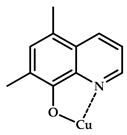 | 2.66 | 2.7 | 1.9 |
| Cu(5,7-Cl-8-HQ) 113 | 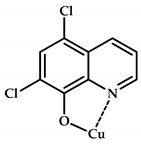 | 3.22 | 5.3 | 4.7 |
| Cu(CQ) 114 |  | 3.50 | 8.9 | 9.0 |
| Cu(5,7-I-8-HQ) 115 |  | 3.75 | 12.8 | 16.2 |
© 2020 by the authors. Licensee MDPI, Basel, Switzerland. This article is an open access article distributed under the terms and conditions of the Creative Commons Attribution (CC BY) license (http://creativecommons.org/licenses/by/4.0/).
Share and Cite
Krasnovskaya, O.; Naumov, A.; Guk, D.; Gorelkin, P.; Erofeev, A.; Beloglazkina, E.; Majouga, A. Copper Coordination Compounds as Biologically Active Agents. Int. J. Mol. Sci. 2020, 21, 3965. https://doi.org/10.3390/ijms21113965
Krasnovskaya O, Naumov A, Guk D, Gorelkin P, Erofeev A, Beloglazkina E, Majouga A. Copper Coordination Compounds as Biologically Active Agents. International Journal of Molecular Sciences. 2020; 21(11):3965. https://doi.org/10.3390/ijms21113965
Chicago/Turabian StyleKrasnovskaya, Olga, Alexey Naumov, Dmitry Guk, Peter Gorelkin, Alexander Erofeev, Elena Beloglazkina, and Alexander Majouga. 2020. "Copper Coordination Compounds as Biologically Active Agents" International Journal of Molecular Sciences 21, no. 11: 3965. https://doi.org/10.3390/ijms21113965