S100A6 and Its Brain Ligands in Neurodegenerative Disorders


Abstract
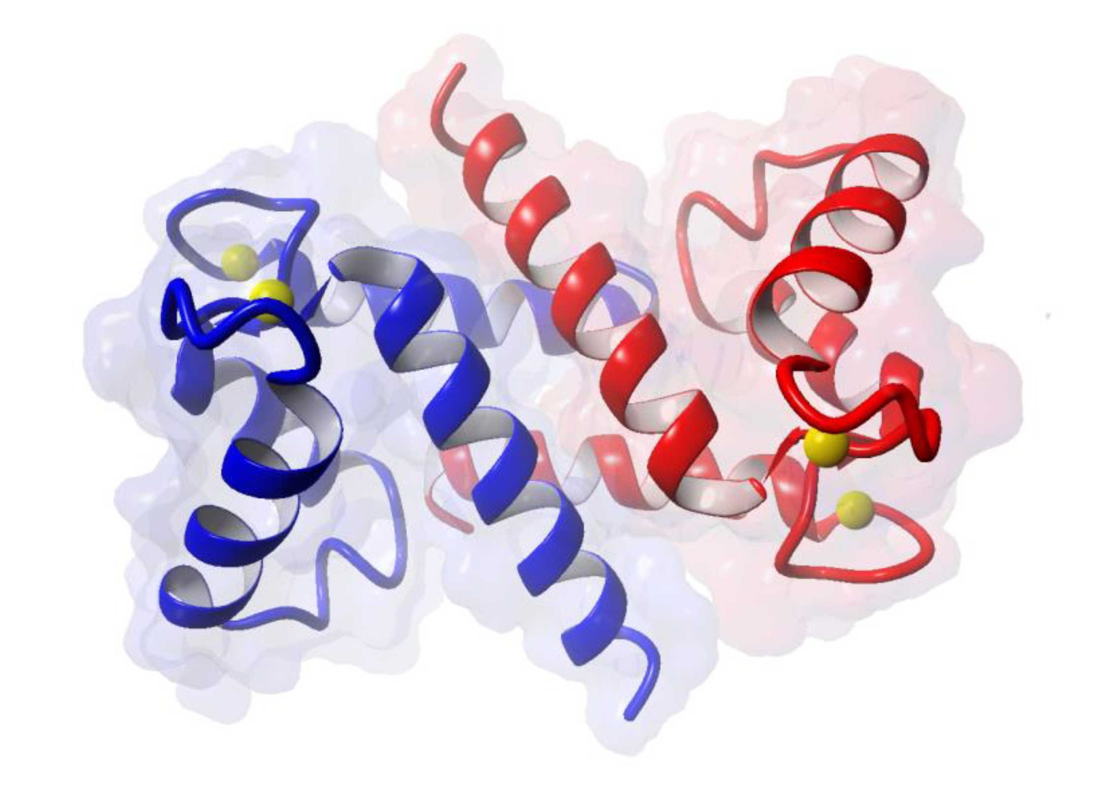
:1. Introduction
2. Expression of S100A6 in the Brain
3. Changes in S100A6 Expression in Neurodegenerative Diseases
3.1. S100A6 and Alzheimer‘s Disease (AD)
3.2. S100A6 and Other Neurodegenerative Diseases
4. Expression of CacyBP/SIP in Normal Brain and in Neurodegeneration
5. Expression of Sgt1 in Normal Brain and in Neurodegeneration
6. Summary and Conclusions
Author Contributions
Funding
Acknowledgments
Conflicts of Interest
Abbreviations
| AD | Alzheimer’s disease |
| ALS | Amyotrophic lateral sclerosis |
| C.A. | Suppressor of G2 corpora amylacea |
| CacyBP/SIP | Calcyclin (S100A6) binding protein/Siah-1 interacting protein |
| DS | Down syndrome |
| HD | Huntington’s disease |
| PD | Parkinson’s disease |
| Sgt1 | Suppressor of G2 allele of Skp1 |
| TBI | Traumatic brain injury. |
References
- Filipek, A.; Leśniak, W. Current view on cellular function of S100A6 and its ligands, CacyBP/SIP and Sgt1. Post. Biochem. 2018, 64, 242–252. [Google Scholar] [CrossRef]
- Gonzalez, L.L.; Garrie, K.; Turner, M.D. Role of S100 proteins in health and disease. Biochim. Biophys. Acta 2020, 1867, 118677. [Google Scholar] [CrossRef]
- Otterbein, L.R.; Kordowska, J.; Witte-Hoffmann, C.; Wang, C.L.; Dominguez, R. Crystal structures of S100A6 in the Ca(2+)-free and Ca(2+)-bound states: The calcium sensor mechanism of S100 proteins revealed at atomic resolution. Structure 2002, 10, 557–567. [Google Scholar] [CrossRef] [Green Version]
- Moroz, O.V.; Wilson, K.S.; Bronstein, I.B. The role of zinc in the S100 proteins: Insights from the X-ray structures. Amino Acids 2011, 41, 761–772. [Google Scholar] [CrossRef]
- Kuźnicki, J.; Filipek, A.; Heimann, P.; Kaczmarek, L.; Kamińska, B. Tissue specific distribution of calcyclin--10.5 kDa Ca2+-binding protein. FEBS Lett. 1989, 254, 141–144. [Google Scholar] [CrossRef] [Green Version]
- Filipek, A.; Puzianowska, M.; Cieslak, B.; Kuznicki, J. Calcyclin–Ca(2+)-binding protein homologous to glial S-100 beta is present in neurones. NeuroReport 1993, 4, 383–386. [Google Scholar] [CrossRef] [PubMed]
- Yamashita, N.; Ilg, E.C.; Schafer, B.W.; Heizmann, C.W.; Kosaka, T. Distribution of a specific calcium-binding protein of the S100 protein family, S100A6 (calcyclin), in subpopulations of neurons and glial cells of the adult rat nervous system. J. Comp. Neurol. 1999, 404, 235–257. [Google Scholar] [CrossRef]
- Leśniak, W.; Wilanowski, T.; Filipek, A. S100A6 - focus on recent developments. Biol. Chem. 2017, 398, 1087–1094. [Google Scholar] [CrossRef]
- Jurewicz, E.; Kasacka, I.; Bankowski, E.; Filipek, A. S100A6 and its extracellular ligands in Wharton’s jelly of healthy and preeclamptic patients. Placenta 2014, 35, 386–391. [Google Scholar] [CrossRef]
- Leclerc, E.; Fritz, G.; Weibel, M.; Heizmann, C.W.; Galichet, A. S100B and S100A6 differentially modulate cell survival by interacting with distinct RAGE (receptor for advanced glycation end products) immunoglobulin domains. J. Biol. Chem. 2007, 282, 31317–31331. [Google Scholar] [CrossRef] [Green Version]
- Jurewicz, E.; Góral, A.; Filipek, A. S100A6 is secreted from Wharton’s jelly mesenchymal stem cells and interacts with integrin β1. Int. J. Biochem. Cell Biol. 2014, 55, 298–303. [Google Scholar] [CrossRef] [PubMed]
- Jurewicz, E.; Wyroba, E.; Filipek, A. Tubulin-dependent secretion of S100A6 and cellular signaling pathways activated by S100A6-integrin β1 interaction. Cell. Signal. 2018, 42, 21–29. [Google Scholar] [CrossRef] [PubMed]
- Sakane, K.; Nishiguchi, M.; Denda, M.; Yamagchi, F.; Magari, M.; Kanayama, N.; Morishita, R.; Tokumitsu, H. Identification and characterization of a centrosomal protein, FOR20 as a novel S100A6 target. Biochem. Biophys. Res. Commun. 2017, 491, 980–985. [Google Scholar] [CrossRef] [PubMed]
- van Dieck, J.; Lum, J.K.; Teufel, D.P.; Fersht, A.R. S100 proteins interact with the N-terminal domain of MDM2. FEBS Lett. 2010, 584, 3269–3274. [Google Scholar] [CrossRef] [Green Version]
- Graczyk, A.; Słomnicki, L.P.; Leśniak, W. S100A6 competes with the TAZ2 domain of p300 for binding to p53 and attenuates p53 acetylation. J. Mol. Biol. 2013, 425, 3488–3494. [Google Scholar] [CrossRef]
- Kilanczyk, E.; Graczyk, A.; Ostrowska, H.; Kasacka, I.; Leśniak, W.; Filipek, A. S100A6 is transcriptionally regulated by β-catenin and interacts with a novel ligand, lamin A/C, in colorectal cancer cells. Cell Calcium 2012, 51, 470–477. [Google Scholar] [CrossRef]
- Takata, M.; Shimamoto, S.; Yamaguchi, F.; Tokuda, M.; Tokumitsu, H.; Kobayashi, R. Regulation of nuclear localization signal-importin α interaction by Ca2+/S100A6. FEBS Lett. 2010, 584, 4517–4523. [Google Scholar] [CrossRef] [Green Version]
- Bartkowska, K.; Swiatek, I.; Aniszewska, A.; Jurewicz, E.; Turlejski, K.; Filipek, A.; Djavadian, R.L. Stress-Dependent Changes in the CacyBP/SIP Interacting Protein S100A6 in the Mouse Brain. PLoS ONE 2017, 12, e0169760. [Google Scholar] [CrossRef] [Green Version]
- Hagmeyer, S.; Romão, M.A.; Cristóvão, J.S.; Vilella, A.; Zoli, M.; Gomes, C.M.; Grabrucker, A.M. Distribution and Relative Abundance of S100 Proteins in the Brain of the APP23 Alzheimer’s Disease Model Mice. Front. Neurosci. 2019, 13, 640. [Google Scholar] [CrossRef]
- Yamada, J.; Jinno, S. Upregulation of calcium binding protein, S100A6, in activated astrocytes is linked to glutamate toxicity. Neuroscience 2012, 226, 119–129. [Google Scholar] [CrossRef]
- Tiu, S.C.; Chan, W.Y.; Heizmann, C.W.; Schäfer, B.W.; Shu, S.Y.; Yew, D.T. Differential expression of S100B and S100A6(1) in the human fetal and aged cerebral cortex. Brain Res. Dev. Brain Res. 2000, 119, 159–168. [Google Scholar] [CrossRef]
- Yamada, J.; Jinno, S. Age-related differences in oligodendrogenesis across the dorsal-ventral axis of the mouse hippocampus. Hippocampus 2014, 24, 1017–1029. [Google Scholar] [CrossRef]
- Kjell, J.; Fischer-Sternjak, J.; Thompson, A.J.; Friess, C.; Sticco, M.J.; Salinas, F.; Cox, J.; Martinelli, D.C.; Ninkovic, J.; Franze, K.; et al. Defining the Adult Neural Stem Cell Niche Proteome Identifies Key Regulators of Adult Neurogenesis. Cell Stem Cell. 2020, 26, 277–293. [Google Scholar] [CrossRef] [Green Version]
- Kalamakis, G.; Brüne, D.; Ravichandran, S.; Bolz, J.; Fan, W.; Ziebell, F.; Stiehl, T.; Catalá-Martinez, F.; Kupke, J.; Zhao, S.; et al. Quiescence Modulates Stem Cell Maintenance and Regenerative Capacity in the Aging Brain. Cell 2019, 176, 1407–1419. [Google Scholar] [CrossRef] [Green Version]
- Sharma, P.; Sharma, A.; Fayaz, F.; Wakode, S.; Pottoo, F.H. Biological signatures of Alzheimer Disease. Curr. Top. Med. Chem. 2020, 9, 770–781. [Google Scholar] [CrossRef]
- Mietelska-Porowska, A.; Wasik, U.; Goras, M.; Filipek, A.; Niewiadomska, G. Tau protein modifications and interactions: Their role in function and dysfunction. Int. J. Mol. Sci. 2014, 15, 4671–4713. [Google Scholar] [CrossRef] [Green Version]
- Popugaeva, E.; Pchitskaya, E.; Bezprozvanny, I. Dysregulation of neuronal calcium homeostasis in Alzheimer’s disease—A therapeutic opportunity? Biochem. Biophys. Res. Commun. 2017, 483, 998–1004. [Google Scholar] [CrossRef] [Green Version]
- Bojarski, L.; Herms, J.; Kuznicki, J. Calcium dysregulation in Alzheimer’s disease. Neurochem. Int. 2008, 52, 621–633. [Google Scholar] [CrossRef]
- Mezzaroba, L.; Alfieri, D.F.; Colado Simão, A.N.; Vissoci Reiche, E.M. The role of zinc, copper, manganese and iron in neurodegenerative diseases. Neurotoxicology 2019, 74, 230–241. [Google Scholar] [CrossRef]
- Gonzalez-Reyes, R.E.; Nava-Mesa, M.O.; Vargas-Sanchez, K.; Ariza-Salamanca, D.; Mora-Munoz, L. Involvement of astrocytes in alzheimer’s disease from a neuroinflammatory and oxidative stress perspective. Front. Mol. Neurosci. 2017, 10, 427. [Google Scholar] [CrossRef] [Green Version]
- Frost, G.R.; Li, Y.M. The role of astrocytes in amyloid production and Alzheimer’s disease. Open Biol. 2017, 12, 170228. [Google Scholar] [CrossRef] [PubMed] [Green Version]
- Webers, A.; Heneka, M.T.; Gleeson, P.A. The role of innate immune responses and neuroinflammation in amyloid accumulation and progression of Alzheimer’s disease. Immunol. Cell Biol. 2020, 98, 28–41. [Google Scholar] [CrossRef] [PubMed]
- Boom, A.; Pochet, R.; Authelet, M.; Pradier, L.; Borghgraef, P.; Van Leuven, F.; Heizmann, C.W.; Brion, J.P. Astrocytic calcium/zinc binding protein S100A6 over expression in Alzheimer’s disease and in PS1/APP transgenic mice models. Biochim. Biophys. Acta 2004, 1742, 161–168. [Google Scholar] [CrossRef]
- Tian, Z.Y.; Wang, C.Y.; Wang, T.; Li, Y.C.; Wang, Z.Y. Glial S100A6 Degrades β-amyloid Aggregation through Targeting Competition with Zinc Ions. Aging Dis. 2019, 10, 756–769. [Google Scholar] [CrossRef] [Green Version]
- Takeda, A.; Tamano, H. Insight into cognitive decline from Zn(2+) dynamics through extracellular signaling of glutamate and glucocorticoids. Arch. Biochem. Biophys. 2016, 611, 93–99. [Google Scholar] [CrossRef]
- Hagmeyer, S.; Cristóvão, J.S.; Mulvihill, J.J.E.; Boeckers, T.M.; Gomes, C.M.; Grabrucker, A.M. Zinc Binding to S100B Affords Regulation of Trace Metal Homeostasis and Excitotoxicity in the Brain. Front. Mol. Neurosci. 2018, 10, 456. [Google Scholar] [CrossRef] [Green Version]
- Haldar, B.; Hamilton, C.L.; Solodushko, V.; Abney, K.A.; Alexeyev, M.; Honakanen, R.E.; Scammell, J.G.; Coffi, D.L. S100A6 is a positive regulator of PPP5C-FKBP51-dependent regulation of endothelial calcium signaling. FASEB J. 2020, 34, 3179–3196. [Google Scholar] [CrossRef]
- Wruck, W.; Schröter, F.; Adjaye, J. Meta-Analysis of Transcriptome Data Related to Hippocampus Biopsies and iPSC-Derived Neuronal Cells from Alzheimer’s Disease Patients Reveals an Association with FOXA1 and FOXA2 Gene Regulatory Networks. J. Alzheimers Dis. 2016, 50, 1065–1082. [Google Scholar] [CrossRef]
- Hoyaux, D.; Alao, J.; Fuchs, J.; Kiss, R.; Keller, B.; Heizmann, C.W.; Pochet, R.; Frermann, D. S100A6, a calcium- and zinc-binding protein, is overexpressed in SOD1 mutant mice, a model for amyotrophic lateral sclerosis. Biochim. Biophys. Acta 2000, 1498, 264–272. [Google Scholar] [CrossRef] [Green Version]
- Hoyaux, D.; Boom, A.; Van Den Bosch, L.; Belot, N.; Martin, J.-J.; Heizmann, C.W.; Kiss, R.; Pochet, R. S100A6 Overexpression within Astrocytes Associated with Impaired Axons from Both ALS Mouse Model and Human Patients. J. Neuropath. Exp. Neurol. 2002, 61, 736–744. [Google Scholar] [CrossRef] [Green Version]
- Iridoy, M.O.; Zubiri, I.; Zelaya, M.V.; Martinez, L.; Ausín, K.; Lachen-Montes, M.; Santamaría, E.; Fernandez-Irigoyen, J.; Jericó, I. Neuroanatomical Quantitative Proteomics Reveals Common Pathogenic Biological Routes between Amyotrophic Lateral Sclerosis (ALS) and Frontotemporal Dementia (FTD). Int. J. Mol. Sci. 2019, 20, 4. [Google Scholar] [CrossRef] [Green Version]
- Ragagnin, A.M.G.; Shadfar, S.; Vidal, M.; Jamali, M.S.; Atkin, J.D. Motor Neuron Susceptibility in ALS/FTD. Front. Neurosci. 2019, 13, 532. [Google Scholar] [CrossRef] [Green Version]
- Schiffer, D.; Cordera, S.; Cavalla, P.; Migheli, A. Reactive astrogliosis of the spinal cord in amyotrophic lateral sclerosis. J. Neurol. Sci. 1996, 139, 27–33. [Google Scholar] [CrossRef]
- Botelho, H.M.; Leal, S.S.; Cardoso, I.; Yanamandra, K.; Morozova-Roche, L.A.; Fritz, G.; Gomes, C.M. S100A6 amyloid fibril formation is calcium-modulated and enhances superoxide dismutase-1 (SOD1) aggregation. J. Biol. Chem. 2012, 287, 42233–42242. [Google Scholar] [CrossRef] [Green Version]
- Lee, T.; Mane, S.; Eid, T.; Zhao, H.; Lin, A.; Guan, Z.; Kim, J.H.; Schweitzer, J.; King-Stevens, D.; Weber, P.; et al. Gene expression in temporal lobe epilepsy is consistent with increased release of glutamate by astrocytes. Mol. Med. 2007, 13, 1–13. [Google Scholar] [CrossRef]
- Cacheaux, L.P.; Ivens, S.; David, Y.; Lakhter, A.J.; Bar-Klein, G.; Shapira, M.; Heinemann, U.; Friedman, A.; Kaufer, D. Transcriptome Profiling Reveals TGF- Signaling Involvement in Epileptogenesis. J. Neurosci. 2009, 29, 8927–8935. [Google Scholar] [CrossRef]
- Kobori, N.; Clifton, G.L.; Dash, P. Altered expression of novel genes in the cerebral cortex following experimental brain injury. Mol. Brain Res. 2002, 104, 148–158. [Google Scholar] [CrossRef]
- Jurewicz, E.; Bednarczyk, J.; Bot, A.; Lukasiuk, K.; Filipek, A. Status epilepticus induces long lasting increase in S100A6 expression in astrocytes. Neurochem. Res. 2013, 38, 1941–1948. [Google Scholar] [CrossRef]
- Zhang, L.; Wang, H.; Zhou, X.; Mao, L.; Ding, K.; Hu, Z. Role of mitochondrial calcium uniporter-mediated Ca(2+) and iron accumulation in traumatic brain injury. J. Cell Mol. Med. 2019, 23, 2995–3009. [Google Scholar] [CrossRef] [Green Version]
- Fang, B.; Liang, M.; Yang, G.; Ye, Y.; Xu, H.; He, X.; Huang, J.H. Expression of S100A6 in rat hippocampus after traumatic brain injury due to lateral head acceleration. Int. J. Mol. Sci. 2014, 15, 6378–6390. [Google Scholar] [CrossRef] [Green Version]
- Ding, H.; Yu, J.; Chang, W.; Liu, F.; He, Z. Searching for differentially expressed proteins in spinal cordinjury based on proteomic analysis. Life Sci. 2019, 242, 117235. [Google Scholar] [CrossRef] [PubMed]
- Hoyaux, D.; Decaestecker, C.; Heizmann, C.W.; Vogl, T.; Schäfer, B.W.; Salmon, I.; Kiss, R.; Pochet, R. S100 proteins in Corpora amylacea from normal human brain. Brain Res. 2000, 867, 280–288. [Google Scholar] [CrossRef]
- Filipek, A.; Wojda, U. p30, a novel protein target of mouse calcyclin (S100A6). Biochem. J. 1996, 320, 585–587. [Google Scholar] [CrossRef] [PubMed] [Green Version]
- Filipek, A.; Kuźnicki, J. Molecular cloning and expression of a mouse brain cDNA encoding a novel protein target of calcyclin. J. Neurochem. 1998, 70, 1793–1798. [Google Scholar] [CrossRef]
- Matsuzawa, S.I.; Reed, J.C. Siah-1, SIP, and Ebi collaborate in a novel pathway for beta-catenin degradation linked to p53 responses. Mol. Cell 2001, 7, 915–926. [Google Scholar] [CrossRef]
- Jastrzebska, B.; Filipek, A.; Nowicka, D.; Kaczmarek, L.; Kuznicki, J. Calcyclin (S100A6) binding protein (CacyBP) is highly expressed in brain neurons. J. Histochem. Cytochem. 2000, 48, 1195–1202. [Google Scholar] [CrossRef] [Green Version]
- Jurewicz, E.; Miazga, K.; Fabczak, H.; Sławińska, U.; Filipek, A. CacyBP/SIP in the rat spinal cord in norm and after transection—Influence on the phosphorylation state of ERK1/2 and p38 kinases. Neurochem. Int. 2020. accepted. [Google Scholar] [CrossRef]
- Topolska-Woś, A.M.; Chazin, W.J.; Filipek, A. CacyBP/SIP--Structure and variety of functions. Biochim. Biophys. Acta 2016, 1860, 79–85. [Google Scholar] [CrossRef]
- Topolska-Woś, A.M.; Shell, S.M.; Kilańczyk, E.; Szczepanowski, R.H.; Chazin, W.J.; Filipek, A. Dimerization and phosphatase activity of calcyclin-binding protein/Siah-1 interacting protein: The influence of oxidative stress. FASEB J. 2015, 29, 1711–1724. [Google Scholar] [CrossRef] [Green Version]
- Góral, A.; Bieganowski, P.; Prus, W.; Krzemień-Ojak, Ł.; Kądziołka, B.; Fabczak, H.; Filipek, A. Calcyclin Binding Protein/Siah-1 Interacting Protein Is A Hsp90 Binding Chaperone. PLoS ONE 2016, 11, e0156507. [Google Scholar] [CrossRef]
- Filipek, A.; Schneider, G.; Mietelska, A.; Figiel, I.; Niewiadomska, G. Age-dependent changes in neuronal distribution of CacyBP/SIP: Comparison to tubulin and the tau protein. J. Neural. Transm. 2008, 115, 1257–1264. [Google Scholar] [CrossRef]
- Wasik, U.; Schneider, G.; Mietelska-Porowska, A.; Mazurkiewicz, M.; Fabczak, H.; Weis, S.; Zabke, C.; Harrington, C.R.; Filipek, A.; Niewiadomska, G. Calcyclin binding protein and Siah-1 interacting protein in Alzheimer’s disease pathology: Neuronal localization and possible function. Neurobiol. Aging 2013, 5, 1380–1388. [Google Scholar] [CrossRef]
- Schumacher, J.; Peraza, L.R.; Firbank, M.; Thomas, A.J.; Kaiser, M.; Gallagher, P.; O’Brien, J.T.; Blamire, A.M.; Taylor, J.P. Dysfunctional brain dynamics and their origin in Lewy body dementia. Brain 2019, 142, 1767–1782. [Google Scholar] [CrossRef] [Green Version]
- Lachén-Montes, M.; González-Morales, A.; Iloro, I.; Elortza, F.; Ferrer, I.; Gveric, D.; Fernández-Irigoyen, J.; Santamaría, E. Unveiling the olfactory proteostatic disarrangement in Parkinson’s disease by proteome-wide profiling. Neurobiol. Aging 2019, 73, 123–134. [Google Scholar] [CrossRef]
- Schulte, J.; Littleton, J.T. The biological function of the Huntingtin protein and its relevance to Huntington’s Disease pathology. Curr. Trends. Neurol. 2011, 5, 65–78. [Google Scholar]
- Czeredys, M.; Gruszczynska-Biegala, J.; Schach, T.; Methner, A.; Kuznicki, J. Expression of genes encoding the calcium signalosome in cellular and transgenic models of Huntington’s disease. Front. Mol. Neurosci. 2013, 6, 42. [Google Scholar] [CrossRef] [Green Version]
- Góral, A.; Bartkowska, K.; Djavadian, R.L.; Filipek, A. CacyBP/SIP, a Hsp90 binding chaperone, in cellular stress response. Int. J. Biochem. Cell Biol. 2018, 99, 178–185. [Google Scholar] [CrossRef]
- Ishihara, K.; Kanai, S.; Sago, H.; Yamakawa, K.; Akiba, S. Comparative proteomic profiling reveals aberrant cell proliferation in the brain of embryonic Ts1Cje, a mouse model of Down syndrome. Neuroscience 2014, 281, 1–15. [Google Scholar] [CrossRef]
- Antonarakis, S.E.; Skotko, B.G.; Rafii, M.S.; Strydom, A.; Pap, S.E.; Bianchi, D.W.; Sherman, S.L.; Reeves, R.H. Down syndrome. Nat. Rev. Dis. Primers. 2020, 6, 9. [Google Scholar] [CrossRef]
- Matigian, N.; Windus, L.; Smith, H.; Filippich, C.; Pantelis, C.; McGrath, J.; Mowry, B.; Hayward, N.H. Expression profiling in monozygotic twins discordant for bipolar disorder reveals dysregulation of the WNT signalling pathway. Mol. Psychiatry 2007, 2, 815–825. [Google Scholar] [CrossRef]
- McIntyre, R.S.; Calabrese, J.R. Bipolar depression: The clinical characteristics and unmet needs of a complex disorder. Curr. Med. Res. Opin. 2019, 35, 1993–2005. [Google Scholar] [CrossRef] [PubMed] [Green Version]
- Kitagawa, K.; Skowyra, D.; Elledge, S.J.; Harper, J.W.; Hieter, P. SGT1 encodes an essential component of the yeast kinetochore assembly pathway and a novel subunit of the SCF ubiquitin ligase complex. Mol. Cell 1999, 4, 21–33. [Google Scholar] [CrossRef]
- Niikura, Y.; Kitagawa, K. Functions of SGT1, a Co-chaperone. In Heat Shock Protein 90 in Human Diseases and Disorders; Asea, A.A., Kaur, P., Eds.; Springer: Basel, Switzerland, 2019; pp. 317–370. [Google Scholar]
- Spiechowicz, M.; Bernstein, H.G.; Dobrowolny, H.; Leśniak, W.; Mawrin, C.; Bogerts, B.; Kuźnicki, J.; Filipek, A. Density of Sgt1-immunopositive neurons is decreased in the cerebral cortex of Alzheimer’s disease brain. Neurochem. Int. 2006, 49, 487–493. [Google Scholar] [CrossRef]
- Bohush, A.; Niewiadomska, G.; Weis, S.; Filipek, A. HSP90 and Its Novel Co-Chaperones, SGT1 and CHP-1, in Brain of Patients with Parkinson’s Disease and Dementia with Lewy Bodies. J. Parkinson’s Dis. 2019, 9, 97–107. [Google Scholar] [CrossRef] [Green Version]

| Protein | Disease | Examined Region | Expression | Reference |
|---|---|---|---|---|
| S100A6 | Alzheimer disease (AD) | - neocortex (white and gray matter) - prefrontal cortex - hippocampus | - immunoreactivity in astrocytes↑ - immunoreactivity in center and border zone of β amyloid plaques↑ - mRNA↑ | [33,38] |
| Amyotrophic Lateral Sclerosis (ALS) | - brainstem - spinal cord (dorsal root) - spinal cord (pyramidal tract) | - mRNA↑ - immunoreactivity in astrocytes↑ - protein ↑ | [39,40,41] | |
| Epileptogenesis | - cortex | - mRNA ↑ | [20,46,48] | |
| - hippocampus (CA1 and CA3) | - immunoreactivity in astrocytes↑ | |||
| Traumatic brain injury (TBI) | - cerebral cortex | - mRNA↑ | [47] | |
| - hippocampus | - mRNA↓ | [50] | ||
| - protein↓ | ||||
| - immunoreactivity | ||||
| in pyramidal neurons↓ | ||||
| Stress | - cortex - brainstem - hippocampus - hypothalamus | - protein↓ - immunoreactivity in neurons and tanycytes↓ | [18] | |
| CacyBP/SIP | Alzheimer disease (AD) | - hippocampus | - protein – not changed | [62] |
| - parieto-temporal cortex | ||||
| Parkinson disease (PD) | - olfactory bulbs | - protein↓ | [64] | |
| Huntington disease (HD) | - striatum | - mRNA↑ | [66] | |
| - protein↑ | ||||
| Amyotrophic Lateral Sclerosis (ALS) | - non-motor cortex | - protein↑ | [41] | |
| - spinal cord (pyramidal tract) | - protein↓ | |||
| Stress | - thalamus/ | - protein↑ | [67] | |
| hypothalamus | ||||
| - hippocampus | ||||
| - brainstem | ||||
| Down syndrome (DS) | - mouse embryo | - protein ↑ | [68] | |
| Bipolar disorder (BD) | - lymphoblastoid cells | - mRNA ↑ | [70] | |
| Sgt1 | Alzheimer disease (AD) | - cortex (temporal, angular, posterior cingulate) | - number of Sgt1 positive cells↓ | [74] |
| Parkinson disease (PD) | - temporal cortex | - mRNA↑ | [75] | |
| Dementia with Lewy bodies (DLB) | - substantia nigra | - mRNA ↑ | [75] |
© 2020 by the authors. Licensee MDPI, Basel, Switzerland. This article is an open access article distributed under the terms and conditions of the Creative Commons Attribution (CC BY) license (http://creativecommons.org/licenses/by/4.0/).
Share and Cite
Filipek, A.; Leśniak, W. S100A6 and Its Brain Ligands in Neurodegenerative Disorders. Int. J. Mol. Sci. 2020, 21, 3979. https://doi.org/10.3390/ijms21113979
Filipek A, Leśniak W. S100A6 and Its Brain Ligands in Neurodegenerative Disorders. International Journal of Molecular Sciences. 2020; 21(11):3979. https://doi.org/10.3390/ijms21113979
Chicago/Turabian StyleFilipek, Anna, and Wiesława Leśniak. 2020. "S100A6 and Its Brain Ligands in Neurodegenerative Disorders" International Journal of Molecular Sciences 21, no. 11: 3979. https://doi.org/10.3390/ijms21113979