Article

Nusbiarylins Inhibit Transcription and Target Virulence Factors in Bacterial Pathogen Staphylococcus aureus

1
Department of Microbiology, Faculty of Medicine, The Chinese University of Hong Kong, Prince of Wales Hospital, Shatin, Hong Kong
2
State Key Laboratory of Chemical Biology and Drug Discovery, Department of Applied Biology and Chemical Technology, The Hong Kong Polytechnic University, Hung Hom, Hong Kong
*
Authors to whom correspondence should be addressed.
Int. J. Mol. Sci. 2020, 21(16), 5772; https://doi.org/10.3390/ijms21165772
Submission received: 30 June 2020 / Revised: 2 August 2020 / Accepted: 7 August 2020 / Published: 11 August 2020

Abstract

:
The emergence of multidrug resistance in the clinically significant pathogen Staphylococcus aureus is a global health burden, compounded by a diminishing drug development pipeline, and a lack of approved novel antimicrobials. Our previously reported first-in-class bacterial transcription inhibitors “nusbiarylins” presented a promising prospect towards the discovery of novel antimicrobial agents with a novel mechanism. Here we investigated and characterised the lead nusbiarylin compound, MC4, and several of its chemical derivatives in both methicillin-resistant S. aureus (MRSA) and the S. aureus type strains, demonstrating their capacity for the arrest of growth and cellular respiration, impairment of RNA and intracellular protein levels at subinhibitory concentrations. In some instances, derivatives of MC4 were also shown to attenuate the production of staphylococcal virulence factors in vitro, such as the exoproteins α-toxin and Panton–Valentine Leukocidin (PVL). Trends observed from quantitative PCR assays suggested that nusbiarylins elicited these effects possibly by acting via but not limited to the modulation of global regulatory pathways, such as the agr regulon, which coordinates the expression of S. aureus genes associated with virulence. Our findings encourage the continued development of more potent compounds within this novel family of bacterial transcription inhibitors.

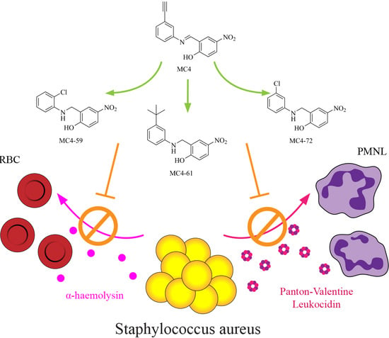
Graphical Abstract

1. Introduction

Staphylococcus aureus is an opportunistic Gram-positive pathogen of high clinical significance, responsible for skin, soft tissue, respiratory and blood infections with a wide spectrum of severity [1]. The widespread prevalence of multidrug-resistant S. aureus, be it hospital-acquired (HA-MRSA) or community-associated (CA-MRSA), presents a serious global challenge to public health as infections increasingly fail to respond to existing antibiotic treatments [2]. The continued discovery of antimicrobial agents with novel mechanisms of action is therefore vital to the provision of alternatives in the clinical management of infectious diseases caused by resistant organisms.

1.1. The Interaction between Bacterial Transcription Factors NusB-NusE as a Drug Target

A crucial step in the central dogma of molecular biology, bacterial transcription, serves as a target for developing novel antimicrobials, where two drugs—the broad-spectrum rifamycin and the narrow-spectrum anticlostridial fidaxomicin are currently in use [3]. Transcription is driven by the multi-subunit RNA polymerase (RNAP), a process regulated through the interaction with several transcription factors. The N-utilisation substances (Nus) factors belong to a key family of transcription factors comprised of NusA, NusB, NusE (ribosomal protein S10), NusG, ribosomal protein S4 and more recently discovered SuhB, all of which are involved in the transcription of bacterial ribosomal RNA (rRNA) operons, folding, ribosome biogenesis, as well as coupling cellular transcription and translation processes [4,5,6,7,8].
The formation of the NusB-NusE heterodimer is a crucial step in the bacterial rRNA operon transcription complex, which binds to boxA sequence elements of rRNA operons upstream of 16S and 23S rRNA, presenting a potential druggable target for the development of novel antimicrobials [9,10]. In our earlier attempts, we discovered a novel class of antimicrobials targeting this specific protein-protein interaction (PPI) [11,12]. The hit compound MC4 represents the first-in-class inhibitor of bacterial rRNA transcription [13]. Subsequently, chemical derivatives of MC4 have been synthesised and biologically evaluated, showing good antimicrobial activity with minimum inhibitory concentrations (MICs) of 1–2 μg/mL against pathogens of clinical significance [13]. Based on the target protein NusB and the biaryl chemical structure, MC4 and its derivatives were collectively named as nusbiarylins [11,12,13]. Notably, our selection of nusbiarylins also showed moderate to good antimicrobial activity against Staphylococcus aureus type strains, as well as both representative hospital-acquired (HA-) and community-associated (CA-) Methicillin-resistant S. aureus (MRSA) strains. To further characterise the antistaphylococcal profile of our select nusbiarylins—MC4 and its derivatives MC4-59, MC4-61 and MC4-72 (Figure 1)—so chosen for their good range of MICs (2–8 μg/mL), virulence-associated parameters were chosen and investigated to establish a more comprehensive picture.

1.2. Toxins as Virulence Factors in S. aureus

The ability to monitor environmental cues and instigate specific response patterns in metabolism and gene expression is a key prerequisite that underlies the fitness of S. aureus spp. [14]. One such mechanism in place is the agr (accessory gene regulator) quorum-sensing architecture, that drives cellular fitness and consequentially the capacity of staphylococci for opportunistic pathogenesis [14]. The ubiquitous staphylococcal agr locus is required for optimal post-log phase expression of the secretory proteins [15]. Staphylococcal alpha haemolysin (or Hla, alpha-toxin, α-toxin) is one such exoprotein key to S. aureus disease, where expression of the hla gene is induced at 37 °C at the mid-to late exponential phase [16]. Once induced, α-toxin is rapidly released into the extracellular environment, accounting for up to 33% of total protein in culture with only ≤1% α-toxin remaining intracellular [17,18]. A major cause of cellular injury, α-toxin is cytolytic to a wide range of human cell types and is a dominant virulence factor in CA-MRSA [19,20].
Panton–Valentine Leukocidin (PVL), on the other hand, is a virulence factor thought to have originated from methicillin-susceptible S. aureus (MSSA) that forms pores in the membranes of polymorphonuclear leukocytes (neutrophils) through the synergistic action of subunits LukS-PV (encoded by lukS-PV) and LukF-PV (encoded by lukF-PV), and is indicated in cases with severe inflammatory lesions, skin and soft tissue necrosis and serious systemic infections [21]. PVL is consistently associated with pathogenesis over colonisation, and is more prevalent in dermatopathological incidences than invasive staphylococcal disease [22]. Whilst PVL carriage accounts for 0.9–11.6% of S. aureus infections in high-income countries such as the UK, France, Korea and Singapore, it has been reported that PVL-producing staphylococci (both MSSA and MRSA) are predominantly community-acquired [23].

1.3. Global Regulatory Picture of agr and Associated Pathways in S. aureus

As aforementioned, the expression of α-toxin and PVL in S. aureus is regulated by the agr locus, which is the best-studied quorum-sensing system tightly-linked to toxin expression [24,25]. The agr operon both upregulates and downregulates transcription of a plethora of toxins, exoproteins and virulence determinants, and is comprised of two divergent promoters P2 and P3, the latter of which is for the primary transcript and effector molecule RNAIII (which also contains a coding sequence for hld—δ-toxin or delta-haemolysin) [26]. P2, on the other hand, oversees the expression of proteins AgrB, -D, -C and -A (agrBDCA), respectively, which are collectively known as the primary transcript RNAII and have autoinducing (AgrD and AgrB—a cyclic autoinducing peptide and its exporter), signal-transducing (AgrC, a membrane histidine kinase), autoregulatory (AgrA) and ultimately quorum-sensing functions [27,28]. Agr can therefore regulate the expression of cytolytic exoproteins, such as phenol-soluble modulins (PSMs), PVL, α- and δ-toxins, toxic shock syndrome toxins (TSST), serine protease (sspA) and staphylococcal protein A (spa), through both direct binding of downstream genes or indirectly through regulating its own effectors or via other regulators [29].
Albeit a global regulator, the activity of agr is itself also under the mediation of other regulators, such as SarA/SarR and CodY [30,31]. The staphylococcal accessory regulator (sar) locus encompasses the transcripts sarA, sarB and sarC, where SarA and its repressor homologue SarR are essential for modulating toxin production through their control over the agr P2 and P3 promoters [32,33]. Singular target virulence genes being under the dynamic influence of multiple “cross-talking” regulators has been described as an energetically-favourable mechanism [34]. The above network of regulons presents viable targets for chemotherapeutic agents to achieve virulence attenuation (Figure 2) [28,35].

1.4. Aim

The characterisation of α-toxin and PVL expression, as well as that of their associated regulatory pathways in S. aureus treated with chemotherapeutic compounds, are therefore presented as appropriate parameters and serve as objective means to assess the antimicrobial profile of our panel of nusbiarylins (listed in Section 1.1), which in turn may also shed light into their mechanism of action in molecular studies. In this study, we examined the performance of MC4, MC4-59, MC4-61 and MC4-72, previously reported for good antimicrobial activities against various S. aureus spp., with the use of both cellular and molecular techniques to provide mechanistic insights. Experiments were devised to ascertain the antimicrobial profile of the selected nusbiarylins as indicated in our previous reports by assessing their capacity to attenuate staphylococcal virulence through toxin expression, and their resulting genotypic and phenotypic responses compared to representative drugs already in clinical use.

2. Results

2.1. Minimum Inhibitory Concentrations (MICs)

Our selection of nusbiarylins was comprised of the previously published lead compound MC4 and its derivatives MC4-59, MC4-61 and MC4-72 (Figure 1; MC4 = Compound 14 in [11] and Compound 1 in [12]; MC4-59 = Compound 27 in [12]; MC4-61 = Compound 23 in [11,12]; MC4-72 = Compound 28 in [12]) were tested against S. aureus type strains ATCC® 25923, ATCC® 29213, as well as MRSA lineages USA300 (community-acquired) and ST22 (hospital-acquired) to determine their MICs as stipulated by the Clinical & Laboratory Standards Institute (CLSI) [6,11,12,13,37]. In this study we revisited the MIC screening to verify the values for the same strains when cultured in the more staphylococci-optimised tryptic soy broth (TSB) in preparation for the downstream characterisation assays, rather than the standard Mueller-Hinton Broth (MHB) used for MIC tests in the previous studies (MIC data for MC4, MC4-59, MC4-61 and MC4-72 in MHB-cultured 25923 and 29213 had previously been published as Compounds 1, 27, 23 and 28, respectively, in Table 3 in [12]; MIC data for MC4-59, MC4-61 and MC4-72 in MHB-cultured USA300 and ST22 had previously been published as Compounds 27, 23 and 28, respectively, in Table 5 in [12]; MIC data for MC4 and MC4-61 in MHB-cultured 25923, 29213, USA300 and ST22 had previously been published as Compounds 14 and 23, respectively, in Figure 2B in [11]). MIC values of MC4, MC4-59, MC4-61 and MC4-72 against TSB-cultured type strains and MRSA strains were within ±2–4 folds of MHB-cultured set-ups. Overall, the MC4 compounds showed good antimicrobial potency against the staphylococcal strains tested, achieving those of the cell wall synthesis inhibitor vancomycin and oxacillin, which are clinically available (Table 1).

2.2. Time–Kill Kinetics and Central Metabolism

MC4 was added to S. aureus ATCC® 25923 and USA300 at concentrations relative to their respective MICs, where the time- and dose-dependent relationship between nusbiarylins and bacterial growth and viability were established through time–kill kinetics and ATP production assays. Overall, MC4 was bacteriostatic against both S. aureus ATCC® 25923 and USA300 over the course of 6 h, with only slight decreases in CFU counts at the highest concentrations towards the end of the assay (Figure 3). Time–kill kinetics of MC4 against S. aureus ATCC® 25923 cultured in MHB had previously been published (as Compound 1, Figure 6 in [12]), which was consistently bacteriostatic with the TSB-cultured set-up reported in this study.
The arrest of cellular respiration is a hallmark characteristic of effective antibiotics [38]. The rate of ATP production of both S. aureus strains was monitored simultaneously as the time–kill kinetics assay proceeded using the same experimental setup, where samples were taken at identical time points for readings. Significant decrease in ATP production rate was observed beginning at ¼ × MIC of MC4 compared to untreated control, with further suppression as concentrations increased. Taking into account of the role of MC4 as a transcription inhibitor, the results were in concordance with previously reported trends displayed by the class drug rifampicin, where bacterial respiration rate is inversely correlated to the drug efficacy of antimicrobials [39,40].

2.3. Exotoxin Release

The effects of our panel of nusbiarylins on the levels of α-toxin and PVL production in S. aureus ATCC® 25923 and USA300 were assessed by immunoblotting exoproteins secreted into supernatants harvested overnight cultures grown in the presence of sub-MIC compounds and control drugs (vancomycin and rifampicin). Proteins separated into bands by gel electrophoresis were blotted onto membranes and incubated with corresponding antibodies to both toxins. Signals from bands were imaged, their relative luminescence quantified and both data presented side-by-side (Figure 4). The results showed that in S. aureus ATCC® 25923, MC4 and especially its derivatives significantly suppressed the release of α-toxin into the extracellular milieu compared to vancomycin and, to a certain extent, rifampicin (Figure 4A). Although MC4 and rifampicin also saw some response in decreasing PVL expression in the type strain, MC4 derivatives, however, did not appear to have meaningfully attenuated PVL levels in harvested supernatant (Figure 4B). In USA300, MC4-59 appeared to have elicited a significant decrease in both α-toxin and PVL expression, but a different trend for MC4 and other derivatives. α-toxin levels did not appear to have been substantially reduced by MC4-61 and MC4-72 compared to rifampicin and even, in the case of MC4, vancomycin (Figure 4C). This trend was also observed in PVL expression, but the level of toxin attenuation was more pronounced for MC4-61 and MC4-72 compared to that of vancomycin (Figure 4D).
Overall, the observed trend where vancomycin had largely little to no effect on the production of PVL in stationary phase S. aureus was consistent with previously reported findings [41,42]. The similar lack of any significant inhibitory effect was also found in USA300 for both α-toxin and PVL, and was consistent with observations previously reported in MRSA strains [43]. Nusbiarylins, in contrast, appeared to be at a similar level to the performance of the class drug rifampicin and, for some of the MC4 derivatives, even appeared to have more markedly attenuated toxin expression in both strains.
A plausible explanation for the occasionally observed nonspecific binding of α-LukS-PV primary antibodies to the membrane regions at immediately higher molecular weights than LukS-PV throughout the Western blots is the presence of staphylococcal protein A (Spa), which may be alleviated through DEPC treatment [44]. In this case, the interference was mitigated through horizontal trimming of the membrane to ensure only our target band size region is exposed to primary antibody incubation.

2.4. Rabbit Erythrocyte Haemolysis

The effects of select nusbiarylins on the in vitro production of α-toxin in S. aureus strains were assessed by the spectrophotometric analysis of haemoglobin release following exposure of the rabbit red blood cells (RBC) to the haemolytic agents. Culture supernatants containing secretory exotoxins harvested for the Western blot analysis in Section 2.3 were also used for the rabbit RBC lysis assay. Following preparation of packed and PBS-washed RBC suspension, erythrocytes were incubated with serially diluted supernatant samples and the absorbance measured at the end point.
The results showed that MC4-72 performed best in protecting RBCs from lysis by S. aureus ATCC® 25923-derived α-toxin, whereas MC-59 performed best against USA300-derived α-toxin (Figure 5). In the S. aureus type strain, MC4-72, MC4-59 and MC4-61 were able to confer decreasing degree of toxin attenuation (Figure 5A). From there onwards, there was a significant performance gap in the mitigation of RBC lysis between MC4 and rifampicin, which remained completely haemolytic even when the supernatant was diluted by four-fold. Vancomycin had no apparent effect on α-toxin production compared to untreated controls in both S. aureus ATCC® 25923 type strain and USA300.
In USA300, all supernatants used required 64- up to 1024-fold of serial dilution before attaining total absence of haemolysis, which is in line with its reputation as a hypervirulent strain. MC4-59 appeared to markedly attenuate α-toxin levels in USA300, resulting in observably less RBC lysis in comparison with other nusbiarylins, vancomycin, rifampicin as well as the untreated control (Figure 5B). All observations were in concordance with those in the Western blot analysis (Section 2.3), and greatly complemented the findings—particularly for S. aureus ATCC® 25923—by offering enhanced resolution on the differences in performance between the MC4 derivatives.

2.5. Real-Time qPCR of Virulence-Associated Gene Expression

Cells of S. aureus ATCC® 25923 and MRSA strain USA300 were treated with MC4 and the class drug rifampicin at subinhibitory concentrations, together with the untreated control. The levels of total DNA across samples set-up for qPCR analysis were first quantified to ensure conformity among harvests. Purified RNA samples were then transcribed into complementary DNA (cDNA) which acted as templates for subsequent real-time quantitative PCR (qPCR) assays.

2.5.1. Effects on rRNA Complex and Implications on the Housekeeping Genes

A defining mechanism of action of nusbiarylins is the inhibition of nascent Nus-regulated essential PPIs within the wider transcription machinery and perhaps beyond rRNA, which invites the question whether the expression levels of classical housekeeping genes commonly used in qPCR and sequencing but are integral to the bacterial rRNA operon transcription complex—such as 16S rRNA, could feasibly serve as valid points of reference [45]. To address this issue, qPCR assays were first performed by probing the distribution of both the drug-treated and untreated signal levels of 16S rRNA, 23S rRNA, DNA gyrase A (gyrA), DNA gyrase B (gyrB) and guanylate kinase (gmk) in the S. aureus type strains and MRSA strain USA300, all of which had previously been indicated or used in gene expression quantitation studies [46,47]. The results suggested that both rRNA subunits showed high variation in the disposition of CT values across the treatment and replicates, indicating that their expression had been affected by the presence of subinhibitory levels of rifampicin and MC4 during growth (Figure 6A,B). In contrast, the CT values of the more metabolism- and maintenance-related genes gyrA, gyrB and gmk were more similar across the experimental replicates, suggesting they could serve as appropriate housekeeping genes due to their relative indifference towards the transcription inhibitors used (Figure 6C–E). When considering that CT values are binary logarithms since DNA strands double after each cycle, the larger the differences therein implies a higher degree of susceptibility to inhibitory effects attributable to the use of rRNA- and RNAP-targeting agents. This method offered a quick yet informative preview into the effects of nusbiarylins, thus ensuring that unsuitable housekeeping genes were avoided as potential points of reference were given consideration in qPCR-based expression studies.

2.5.2. Quantitative PCR

Given the findings from Section 2.5.1 and relevant implications from literature, real-time qPCR assays were performed using gyrB as reference gene, with each treatment group normalised against an untreated control [48]. Relative expression levels of key genes associated with virulence, namely agrA (AgrA), RNAII (encoding the AgrB/D/C/A regulon), RNAIII (toxin-mediating regulon) and sarA (SarA/R regulon), as well as the secretory exoproteins under their modulation spa (Spa), hla (α-toxin) and lukS (LukS subunit of PVL), were probed with SYBR Green system and results extrapolated using the ΔΔCT method (Figure 6).
In general, the expression of RNAII and RNAIII in USA300 were more downregulated following the addition of both rifampicin and MC4 when compared to S. aureus ATCC® 25923. The relative lack of change in USA300 SarA expression treated with rifampicin and MC4 suggested that the drugs did not significantly promote sarA and by extension the phosphorylation of AgrA, which may also explain its more repressed agrA levels (Figure 7B). Compared to untreated control, the impact of the transcription-inhibiting agents on the expression of genes downstream of RNAIII, such as the surface-associated staphylococcal protein A (Spa), as well as the secretory exoproteins in USA300 such as α-toxin (Hla) and PVL (LukS) was limited. This result complemented the Western blot observations above, which also showed no significant difference in the levels of α-toxin and PVL in USA300 culture supernatants following treatment at ¼ × MICs.
For PVL under ⅛ × MIC of MC4, however, there appeared to be sharp downregulation in expression in both the type strain S. aureus ATCC® 25923 and USA300 when compared to the equivalent concentration of rifampicin and untreated control, suggesting a role for dosage and that lower doses may elicit more profound inhibitory effects on certain types of toxins. Apart from rifampicin-treated downregulation of RNAII and its autoinducing effector agrA, no apparent trends were observed for all probed virulence-associated genes following the use of MC4 in S. aureus when compared to both the class drug and untreated control (Figure 7A). A point of intrigue appeared to be the upregulated expression of hla in the type strain following the addition of rifampicin and to a lesser extent MC4.

3. Discussion

Overall, our findings supported the notion that the nusbiarylin lead compound MC4 and its chemical derivatives MC4-59, MC4-61 and MC4-72 were able to attenuate the release of staphylococcal α-toxin and PVL into the extracellular space compared to untreated control, which in turn protects host cells such as RBCs from injury and lysis. Notably, at least one or more nusbiarylins tested performed better in these aspects than vancomycin and even rifampicin in several occasions, despite not necessarily attaining significantly lower MICs in standard screening or eliciting a marked bactericidal response instead of being bacteriostatic at sub-MICs. Observations from qPCR also indicated that nusbiarylins were able to mediate responses in virulence-associated genomic pathways under the influence or modulation of global regulators.
At the molecular level, MRSA strain USA300 appeared more sensitive towards the addition of MC4 and rifampicin compared to the type strain S. aureus, where transcripts of the agr regulon appeared to respond more definitively to the presence of inhibitory agents. While the same could not be said for the type strain S. aureus ATCC® 25923, this suggested that both transcription inhibitors may also possess the ability to act via alternate staphylococcal pathways not encompassed in our experimental design to ultimately achieve the toxin attenuation phenotype as demonstrated in the Western blots of culture supernatants. It must be noted that cultures harvested for qPCR analysis provided only a snapshot of the cellular response to MC4 and rifampicin at mid-log phase in order to accommodate for the aforementioned optimal expression window reported for “housekeeping” genes but not necessarily agr-mediated post-log phase toxin secretion, which may contribute to the limited response resolution observed compared to that of harvests from overnight stationary phase cultures in immunological-based assays.
Subinhibitory concentrations of antibiotics are considered to have strong selection pressure on target bacteria, which drives mutation rates in response to incoming antimicrobial agents [49]. Exposure to lower doses of antibiotics could provide time and opportunity for stressed bacteria to enable adaptation to the chemotherapeutic challenge, potentially even altering pathogenicity through genotypic and phenotypic variations [50]. Given that most studies into antimicrobials were based on the potency of compounds at subinhibitory levels, it is important to take note of hormesis, a concept describing the biphasic response of bacteria where low dosage could at times paradoxically induce virulence [51]. In that regard, Davies et al. proposed that, in light of previous studies suggesting how the transcription inhibitor class drug rifampicin both targets toxin secretion yet upregulates survival, it is possible that sub-MIC rifampicin could modulate bacterial genes through allosteric changes in complex macromolecular structures unique to different bound ligands, which in turn lead to differently altered affinity and sensitivity of macromolecular targets towards environmental conditions such as the availability of magnesium ions [51]. To that end, it could be suggested that differences in sensitivity and response shown in our qPCR observations between S. aureus type strain and USA300 treated by transcription-targeting nusbiarylins at sub-MICs reflected the vastness of this wider framework of thought, where its therapeutic implications remain to be further studied.
In this work, we demonstrated that our novel family of bacterial transcription inhibitor nusbiarylins, represented by the lead drug MC4 and its derivatives, was capable of virulence attenuation in S. aureus spp. through arresting metabolism, inhibiting cell proliferation, protecting host cells from lytic effects of toxins and eliciting downregulating effects on key determinants of cellular fitness and maintenance associated with staphylococcal disease. As an inhibitor of crucial transcription-related PPIs in bacteria [52], nusbiarylins act in non-lytic means which puts it in a more favourable light than the cell wall synthesis inhibitor vancomycin, a last resort drug with increasingly emergent resistance in S. aureus species. In the face of the global antibiotic crisis and to meet the urgent clinical needs for novel treatment options, our findings warrant further development and characterisation of similar antimicrobial compounds to rapidly expand the profile of this underutilised drug class.

4. Materials and Methods

4.1. Determining Minimum Inhibitory Concentrations (MICs)

The antimicrobial activities of all compounds used in this study were assessed by broth microdilution in accordance with the guidelines issued by the CLSI [37]. Test media used were cation-adjusted Mueller-Hinton broth (CA-MHB) (CM0405, Oxoid, Basingstoke, United Kingdom), or tryptic soy broth (TSB) (CM0129, Oxoid) when staphylococcal optimisation was required to determine appropriate dilutions for downstream assays. Serial dilutions in two-folds were performed for test compounds, ranging from 256 μg/mL to 0.25 μg/mL for the nusbiarylins (MC4 and its chemical derivatives), 64 μg/mL to 0.0625 μg/mL for vancomycin and oxacillin, and 2 μg/mL to 0.002 μg/mL for rifampicin. Bacterial inoculum was adjusted to ~5 × 10⁵ CFU/mL. Following 20 h of overnight incubation at 37 °C, results were recorded, and MIC was defined as the lowest concentration of antimicrobial compounds used with no visible growth in well plates. Experiments were performed in triplicates.

4.2. Time–Kill Kinetics

The dose- and time-dependent antimicrobial effects of MC4 on S. aureus strains under aerobic conditions were assessed by adapting from relevant CLSI guidelines [53]. Log phase staphylococci cells were suspended at ~1.5 × 10⁶ CFU/mL in TSB containing test compounds at predetermined concentrations (i.e., ¼×, 1×, 4× and 16 × MICs) along with untreated controls. Cultures were set-up at 37 °C with agitation at 175 rpm, and 20 μL samples were retrieved from each treatment group at 0, 2, 4 and 6 h and underwent 10-fold serial dilutions in sterile PBS. Diluted suspensions were plated onto Columbia blood agar plates and the number of viable bacterial colonies was counted and expressed as CFU/mL following overnight incubation at 37 °C. The experiment was performed in triplicates.

4.3. ATP Production Assay

Performed simultaneously as the time–kill kinetics, 20 μL samples were retrieved from the same cultures for each treatment group at the same time points (0, 2, 4 and 6 h) and added to white 96-well plates, after which equal volumes of reagents were added from the BacTiter-Glo™ Microbial Cell Viability Assay Kit (Promega, Madison, WI, USA) and quantified according to the manufacturer’s protocol. The experiment was performed in triplicates.

4.4. S. aureus Toxin Release

To investigate the effects of our panel of nusbiarylins on the levels of α-toxin and PVL production, immunoblotting of exoproteins harvested from culture supernatants was performed. S. aureus ATCC® 25923 was chosen as the type strain control as S. aureus ATCC® 29213 is negative for PVL expression [54]. USA300, the CA-MRSA clone with exceptional cellular fitness and epidemiological success, was used as the PVL-positive (pvl+) MRSA strain due to its profound clinical significance both inside and outside hospital settings [55,56]. α-toxin, LukS-PV and LukF-PV are all polypeptides of ~33 kDa in molecular weight, and LukS-PV was chosen as the probing target for the detection of PVL expression. Purified recombinant α-toxin and LukS-PV were included in each corresponding run as standards and positive controls.
Fresh colonies of both S. aureus ATCC® 25923 and MRSA strain USA300 were attained following overnight incubation in LB agar. A starter culture of 50 mL in a 250 mL autoclaved conical flask was inoculated at OD600 0.1 and allowed to grow to OD600 ~0.2 with agitation at 175 rpm, after which it was split into 6 mL aliquots. Cells were grown overnight in TSB supplemented with ¼ × MIC of MC4, MC4-59, MC4-61 and MC4-72, respectively, after which 1 mL aliquots from each 6 mL culture were centrifuged at 3000× g for 3 min and the supernatants containing the secretory exoproteins were retained. Samples not immediately used were stored at –20 °C until use in downstream assays. Supernatants were bound with loading dye, heat-denatured and loaded onto polyacrylamide gels for SDS-PAGE analysis, after which they were blotted onto 0.2 μM nitrocellulose membranes (#1620112, Bio-Rad, Hercules, CA, USA) and probed with antibodies targeting α-toxin and LukS-PV.

4.5. Western Blot

Samples were prepared by mixing 15 μL supernatant from the corresponding harvests above with 3 μL 6× loading dye, boiled for 10 min in 0.5 mL PCR capped strip tubes and loaded onto 12% polyacrylamide gels. Then, 50 ng of recombinant alpha haemolysin protein (ab233724, abcam, Cambridge, United Kingdom) with a predicted weight of 33 kDa, and 5 ng of recombinant S. aureus LukS-PV protein from E. coli (DAGB198, Creative Diagnostics, NY, USA) with a molecular weight of 32.465 kDa were added alongside each assay to serve as standards for visual reference. Samples were separated by SDS-PAGE, where bands were first stacked at 80 V for 15 min and then resolved at 150 V for 1 hr. Peptides were blotted to a nitrocellulose membrane (#1620112, Bio-Rad, Hercules, CA, USA) at 110 V for 1 hr, after which the membrane was blocked with 5% nonfat milk in TBST for 1 h and incubated overnight with primary antibodies which were either 1 μg/mL mouse monoclonal anti-alpha-haemolysin (ab190467, abcam, Cambridge, United Kingdom) or 0.5 μg/mL rabbit polyclonal anti-PVL LukS subunit (ab190473, abcam, Cambridge, United Kingdom) at 4 °C with agitation. Membranes were then incubated at room temperature with agitation with the corresponding HRP-conjugated secondary antibodies: 1:3000 goat anti-rabbit (ab97051, abcam, Cambridge, United Kingdom) or 1:5000 goat anti-mouse (ab205719, abcam, Cambridge, United Kingdom), flanked with TBST wash cycles in between. Bio-Rad Clarity™ Western ECL Substrates were used to develop the blots and visualised in a Bio-Rad ChemiDoc™ Touch system under the Chemiluminescence mode at manual exposure settings (Bio-Rad, Hercules, CA, USA). The experiment was performed in triplicates and representative data was presented.

4.6. Rabbit RBC Lysis

Supernatants harvested from the same overnight cultures set-up for the Western blot analysis of α-toxin and PVL expression were also used for the indirect assessment of exotoxin levels in treated S. aureus cells through determining haemolysis of rabbit erythrocytes. Haemoglobin released into plasma through exposure to lytic agents serves as a measurement for RBC lysis. Rabbit RBCs were packed by discarding the supernatant from thrice PBS-washed and centrifuged (200× g for 2 min) 1 volume of cell-phase rabbit blood. When a clear supernatant was achieved with no signs of haemolysis, a 3% v/v suspension of rabbit RBCs was prepared by mixing 1 volume of packed RBCs with 32 volumes of PBS. Due to the sensitivity of rabbit RBCs to concentrated levels of toxins in harvested culture supernatants, 20 μL samples were serially diluted on a 48-well plate and 180 μL of 3% rabbit RBC was added to each well. Then, 1% v/v Triton X-100 (2 μL diluted in 18 μL PBS) was included as a positive control as it can completely lyse the 3% RBC inoculum, while 200 μL PBS served as blank. The plate was incubated at 37 °C for 1 h and centrifuged at 2000× g for 3 min. The plate supernatants were transferred to a new plate in volumes of 100 μL per well, and the absorbance measured at a wavelength of 540 nm. Results were expressed as percentage haemolysis i.e., blank-normalised sample value/value from Triton-treated well × 100%. The experiment was performed in replicates.

4.7. Real-Time qPCR

Purified RNA harvested in the above cell content quantitation assay was used as templates to synthesise cDNA for qPCR assays using the High-Capacity cDNA Reverse Transcription Kit (Applied Biosystems, Foster City, CA, USA), the product of which was quantified with the Qubit™ ssDNA Assay Kit (Invitrogen, Carlsbad, CA, USA) using the Qubit 4 Fluorometer (Thermo Fisher, Waltham, MA, USA). qPCR primers used and their respective sources of design were shown in Table 2. Primers and cDNA templates were diluted appropriately according to the recommendations of the manufacturer of the 2X PowerUp™ SYBR™ Green Master Mix (Applied Biosystems). For 20 μL reactions, 10 μL of the 2X master mix was added to each well in a 0.1 mL-sized MicroAmp® Fast 96-Well Reaction Plate (Applied Biosystems), followed by 2 μL of forward primer, 2 μL of reverse primer, 2 μL of template and 4 μL of nuclease-free water (Ambion, Austin, TX, USA). For no-template control reactions (NTCs), cDNA was not added and the corresponding 2 μL volumes were allocated to plain nuclease-free water complete with primers and master mix. The loaded well plate was placed onto a StepOnePlus™ Real-Time PCR System (Applied Biosystems) and 40-cycle reactions were performed, complete with melting curves to ensure signals were a consequence of correct primer hybridisation. Results were extrapolated and analysed using the ΔΔCT method. Experiments were performed in triplicates.

Author Contributions

Conceptualization, C.M. and X.Y.; data curation, A.J.C., R.H. and L.L.; formal analysis, A.J.C., R.H., L.L., C.M. and X.Y.; funding acquisition, C.M. and X.Y.; investigation, Y.Q., R.H. and L.L.; methodology, X.Y.; project administration, X.Y.; supervision, C.M. and X.Y.; visualization, A.J.C.; Writing — original draft, A.J.C.; Writing—review & editing, C.M. and X.Y. All authors have read and agreed to the published version of the manuscript.

Funding

This research was supported by the Research Grants Council of the Hong Kong Special Administrative Region, China (PolyU 151000/19M, CUHK 141659/17M and 141079/19M), Hong Kong Polytechnic University internal grants (G-YBYY, 1-ZVPS and large equipment fund, the State Key Laboratory of Chemical Biology and Drug Discovery to C.M.), Hong Kong Food and Health Bureau HMRF (19180052 to X.Y.) and the Chinese University of Hong Kong (Faculty of Medicine Faculty Innovation Award FIA2018/A/03 to X.Y.).

Acknowledgments

The authors would like to thank Eric Chi Hang Wong (Department of Clinical Oncology, Faculty of Medicine, The Chinese University of Hong Kong) and Martin Chi-Wai Chan (formerly of the Department of Microbiology, Faculty of Medicine, The Chinese University of Hong Kong) for their kind advice and technical support on the Western blots and qPCR work, respectively.

Conflicts of Interest

The authors declare no conflicts of interest.

Abbreviations

MRSAMethicillin-resistant Staphylococcus aureus
MSSAMethicillin-susceptible Staphylococcus aureus
HA-MRSAHospital-acquired methicillin-resistant Staphylococcus aureus
CA-MRSACommunity-associated methicillin-resistant Staphylococcus aureus
PVLPanton–Valentine Leukocidin
NusN-utilisation substances
RNAPRNA polymerase
rRNARibosomal RNA
PPIProtein-protein interaction
MICMinimum inhibitory concentration
CLSIClinical & Laboratory Standards Institute
ATCC®American Type Culture Collection
MHBMueller-Hinton broth
CA-MHBCation-adjusted Mueller-Hinton broth
TSBTryptic soy broth
CFUColony-forming unit
SDS-PAGESodium dodecyl sulphate–polyacrylamide gel electrophoresis
DEPCDiethyl pyrocarbonate
RBCRed blood cell
ODXXXOptical density at XXX nm wavelength
qPCRQuantitative polymerase chain reaction
cDNAComplementary deoxyribonucleic acid
PBSPhosphate-buffered saline

References

  1. Lowy, F.D. Staphylococcus aureus Infections. N. Engl. J. Med. 1998, 339, 520–532. [Google Scholar] [CrossRef]
  2. McGuinness, W.A.; Malachowa, N.; DeLeo, F.R. Vancomycin Resistance in Staphylococcus aureus. Yale J. Biol. Med. 2017, 90, 269–281. [Google Scholar]
  3. Zhanel, G.G.; Walkty, A.J.; Karlowsky, J.A. Fidaxomicin: A novel agent for the treatment of Clostridium difficile infection. Can. J. Infect. Dis. Med. Microbiol. 2015, 26, 305–312. [Google Scholar] [CrossRef] [Green Version]
  4. Artsimovitch, I.; Knauer, S.H. Ancient Transcription Factors in the News. mBio 2019, 10, e01547-18. [Google Scholar] [CrossRef] [Green Version]
  5. Belogurov, G.A.; Artsimovitch, I. Regulation of Transcript Elongation. Annu. Rev. Microbiol. 2015, 69, 49–69. [Google Scholar] [CrossRef] [Green Version]
  6. Luo, X.; Hsiao, H.-H.; Bubunenko, M.; Weber, G.; Court, D.L.; Gottesman, M.E.; Urlaub, H.; Wahl, M.C. Structural and Functional Analysis of the E. coli NusB-S10 Transcription Antitermination Complex. Mol. Cell 2008, 32, 791–802. [Google Scholar] [CrossRef]
  7. Huang, Y.-H.; Said, N.; Loll, B.; Wahl, M.C. Structural basis for the function of SuhB as a transcription factor in ribosomal RNA synthesis. Nucleic Acids Res. 2019, 47, 6488–6503. [Google Scholar] [CrossRef]
  8. Dudenhoeffer, B.R.; Schneider, H.; Schweimer, K.; Knauer, S.H. SuhB is an integral part of the ribosomal antitermination complex and interacts with NusA. Nucleic Acids Res. 2019, 47, 6504–6518. [Google Scholar] [CrossRef] [PubMed]
  9. Greive, S.J.; Lins, A.F.; von Hippel, P.H. Assembly of an RNA-protein complex. Binding of NusB and NusE (S10) proteins to boxA RNA nucleates the formation of the antitermination complex involved in controlling rRNA transcription in Escherichia coli. J. Biol. Chem. 2005, 280, 36397–36408. [Google Scholar] [CrossRef] [PubMed] [Green Version]
  10. Nodwell, J.R.; Greenblatt, J. Recognition of boxA antiterminator RNA by the E. coli antitermination factors NusB and ribosomal protein S10. Cell 1993, 72, 261–268. [Google Scholar] [CrossRef]
  11. Qiu, Y.; Chan, S.T.; Lin, L.; Shek, T.L.; Tsang, T.F.; Zhang, Y.; Ip, M.; Chan, P.K.; Blanchard, N.; Hanquet, G.; et al. Nusbiarylins, a new class of antimicrobial agents: Rational design of bacterial transcription inhibitors targeting the interaction between the NusB and NusE proteins. Bioorg. Chem. 2019, 92, 103203. [Google Scholar] [CrossRef] [PubMed]
  12. Qiu, Y.; Chan, S.T.; Lin, L.; Shek, T.L.; Tsang, T.F.; Barua, N.; Zhang, Y.; Ip, M.; Chan, P.K.; Blanchard, N.; et al. Design, synthesis and biological evaluation of antimicrobial diarylimine and –amine compounds targeting the interaction between the bacterial NusB and NusE proteins. Eur. J. Med. Chem. 2019, 178, 214–231. [Google Scholar] [CrossRef] [PubMed]
  13. Yang, X.; Luo, M.J.; Yeung, A.C.M.; Lewis, P.J.; Chan, P.K.S.; Ip, M.; Ma, C. First-In-Class Inhibitor of Ribosomal RNA Synthesis with Antimicrobial Activity against Staphylococcus aureus. Biochemistry 2017, 56, 5049–5052. [Google Scholar] [CrossRef] [PubMed]
  14. Jenul, C.; Horswill, A.R. Regulation of Staphylococcus aureus Virulence. Microbiol. Spectr. 2019, 6. [Google Scholar] [CrossRef]
  15. Peng, H.L.; Novick, R.P.; Kreiswirth, B.; Kornblum, J.; Schlievert, P. Cloning, characterization, and sequencing of an accessory gene regulator (agr) in Staphylococcus aureus. J. Bacteriol. 1988, 170, 4365–4372. [Google Scholar] [CrossRef] [PubMed] [Green Version]
  16. Ohlsen, K.; Koller, K.P.; Hacker, J. Analysis of expression of the alpha-toxin gene (hla) of Staphylococcus aureus by using a chromosomally encoded hla::lacZ gene fusion. Infect. Immun. 1997, 65, 3606–3614. [Google Scholar] [CrossRef] [Green Version]
  17. Abbas-ali, B.; Coleman, G. The characteristics of extracellular protein secretion by Staphylococcus aureus (Wood 46) and their relationship to the regulation of alpha-toxin formation. J. Gen. Microbiol. 1977, 99, 277–282. [Google Scholar] [CrossRef] [Green Version]
  18. McNiven, A.C.; Arbuthnott, J.P. Cell-Associated Alpha-Toxin From Staphylococcus Aureus. J. Med. Microbiol. 1972, 5, 123–127. [Google Scholar] [CrossRef] [Green Version]
  19. Berube, B.J.; Bubeck Wardenburg, J. Staphylococcus aureus α-Toxin: Nearly a Century of Intrigue. Toxins 2013, 5, 1140–1166. [Google Scholar] [CrossRef] [Green Version]
  20. Bubeck Wardenburg, J.; Bae, T.; Otto, M.; DeLeo, F.R.; Schneewind, O. Poring over pores: α-hemolysin and Panton-Valentine leukocidin in Staphylococcus aureus pneumonia. Nat. Med. 2007, 13, 1405–1406. [Google Scholar] [CrossRef]
  21. Shrestha, B. Review on Panton Valentine leukocidin toxin carriage among Staphylococcus aureus. J. Nepal Health Res. Counc. 2013, 11, 305–312. [Google Scholar] [PubMed]
  22. Shallcross, L.J.; Fragaszy, E.; Johnson, A.M.; Hayward, A.C. The role of the Panton-Valentine leucocidin toxin in staphylococcal disease: A systematic review and meta-analysis. Lancet Infect. Dis. 2013, 13, 43–54. [Google Scholar] [CrossRef] [Green Version]
  23. Holmes, A.; Ganner, M.; McGuane, S.; Pitt, T.L.; Cookson, B.D.; Kearns, A.M. Staphylococcus aureus Isolates Carrying Panton-Valentine Leucocidin Genes in England and Wales: Frequency, Characterization, and Association with Clinical Disease. J. Clin. Microbiol. 2005, 43, 2384–2390. [Google Scholar] [CrossRef] [PubMed] [Green Version]
  24. Peng, Z.; Jin, D.; Kim, H.B.; Stratton, C.W.; Wu, B.; Tang, Y.-W.; Sun, X. Update on Antimicrobial Resistance in Clostridium difficile: Resistance Mechanisms and Antimicrobial Susceptibility Testing. J. Clin. Microbiol. 2017, 55, 1998–2008. [Google Scholar] [CrossRef] [Green Version]
  25. Recsei, P.; Kreiswirth, B.; O’Reilly, M.; Schlievert, P.; Gruss, A.; Novick, R.P. Regulation of exoprotein gene expression in Staphylococcus aureus by agar. Mol. Gen. Genet. MGG 1986, 202, 58–61. [Google Scholar] [CrossRef]
  26. Novick, R.P.; Ross, H.F.; Projan, S.J.; Kornblum, J.; Kreiswirth, B.; Moghazeh, S. Synthesis of staphylococcal virulence factors is controlled by a regulatory RNA molecule. EMBO J. 1993, 12, 3967–3975. [Google Scholar] [CrossRef]
  27. Novick, R.P. Autoinduction and signal transduction in the regulation of staphylococcal virulence. Mol. Microbiol. 2003, 48, 1429–1449. [Google Scholar] [CrossRef]
  28. Tan, L.; Li, S.R.; Jiang, B.; Hu, X.M.; Li, S. Therapeutic Targeting of the Staphylococcus aureus Accessory Gene Regulator (agr) System. Front. Microbiol. 2018, 9. [Google Scholar] [CrossRef]
  29. Bronner, S.; Monteil, H.; Prévost, G. Regulation of virulence determinants in Staphylococcus aureus: Complexity and applications. FEMS Microbiol. Rev. 2004, 28, 183–200. [Google Scholar] [CrossRef] [Green Version]
  30. Painter, K.L.; Krishna, A.; Wigneshweraraj, S.; Edwards, A.M. What role does the quorum-sensing accessory gene regulator system play during Staphylococcus aureus bacteremia? Trends Microbiol. 2014, 22, 676–685. [Google Scholar] [CrossRef]
  31. Reyes, D.; Andrey, D.O.; Monod, A.; Kelley, W.L.; Zhang, G.; Cheung, A.L. Coordinated Regulation by AgrA, SarA, and SarR To Control agr Expression in Staphylococcus aureus. J. Bacteriol. 2011, 193, 6020–6031. [Google Scholar] [CrossRef] [PubMed] [Green Version]
  32. Cheung, A.L.; Bayer, M.G.; Heinrichs, J.H. sar Genetic determinants necessary for transcription of RNAII and RNAIII in the agr locus of Staphylococcus aureus. J. Bacteriol. 1997, 179, 3963–3971. [Google Scholar] [CrossRef] [PubMed] [Green Version]
  33. Cheung, A.L.; Nishina, K.A.; Trotonda, M.P.; Tamber, S. The SarA protein family of Staphylococcus aureus. Int. J. Biochem. Cell Biol. 2008, 40, 355–361. [Google Scholar] [CrossRef] [PubMed] [Green Version]
  34. Bien, J.; Sokolova, O.; Bozko, P. Characterization of Virulence Factors of Staphylococcus aureus: Novel Function of Known Virulence Factors That Are Implicated in Activation of Airway Epithelial Proinflammatory Response. J. Pathog. 2011, 2011. [Google Scholar] [CrossRef] [PubMed] [Green Version]
  35. Quave, C.L.; Horswill, A.R. Flipping the switch: Tools for detecting small molecule inhibitors of staphylococcal virulence. Front. Microbiol. 2014, 5. [Google Scholar] [CrossRef] [PubMed] [Green Version]
  36. Novick, R.P.; Geisinger, E. Quorum Sensing in Staphylococci. Annu. Rev. Genet. 2008, 42, 541–564. [Google Scholar] [CrossRef]
  37. Clinical & Laboratory Standards Institute. Performance Standards for Antimicrobial Susceptibility Testing, (M100Ed29), 29th ed.; Clinical & Laboratory Standards Institute: Wayne, PA, USA, 2018. [Google Scholar]
  38. Balemans, W.; Vranckx, L.; Lounis, N.; Pop, O.; Guillemont, J.; Vergauwen, K.; Mol, S.; Gilissen, R.; Motte, M.; Lançois, D.; et al. Novel Antibiotics Targeting Respiratory ATP Synthesis in Gram-Positive Pathogenic Bacteria. Antimicrob. Agents Chemother. 2012, 56, 4131–4139. [Google Scholar] [CrossRef] [Green Version]
  39. Lobritz, M.A.; Belenky, P.; Porter, C.B.M.; Gutierrez, A.; Yang, J.H.; Schwarz, E.G.; Dwyer, D.J.; Khalil, A.S.; Collins, J.J. Antibiotic efficacy is linked to bacterial cellular respiration. Proc. Natl. Acad. Sci. USA 2015, 112, 8173–8180. [Google Scholar] [CrossRef] [Green Version]
  40. Yang, J.H.; Bening, S.C.; Collins, J.J. Antibiotic efficacy—Context matters. Curr. Opin. Microbiol. 2017, 39, 73–80. [Google Scholar] [CrossRef]
  41. Dumitrescu, O.; Boisset, S.; Badiou, C.; Bes, M.; Benito, Y.; Reverdy, M.-E.; Vandenesch, F.; Etienne, J.; Lina, G. Effect of Antibiotics on Staphylococcus aureus Producing Panton-Valentine Leukocidin. Antimicrob. Agents Chemother. 2007, 51, 1515–1519. [Google Scholar] [CrossRef] [Green Version]
  42. Stevens, D.L.; Ma, Y.; Salmi, D.B.; McIndoo, E.; Wallace, R.J.; Bryant, A.E. Impact of Antibiotics on Expression of Virulence-Associated Exotoxin Genes in Methicillin-Sensitive and Methicillin-Resistant Staphylococcus aureus. J. Infect. Dis. 2007, 195, 202–211. [Google Scholar] [CrossRef] [PubMed] [Green Version]
  43. Thangamani, S.; Mohammad, H.; Abushahba, M.F.N.; Sobreira, T.J.P.; Hedrick, V.E.; Paul, L.N.; Seleem, M.N. Antibacterial activity and mechanism of action of auranofin against multi-drug resistant bacterial pathogens. Sci. Rep. 2016, 6, 22571. [Google Scholar] [CrossRef] [PubMed] [Green Version]
  44. Nguyen, H.M.; Rocha, M.A.; Chintalacharuvu, K.R.; Beenhouwer, D.O. Detection and quantification of Panton–Valentine leukocidin in Staphylococcus aureus cultures by ELISA and Western blotting: Diethylpyrocarbonate inhibits binding of protein A to IgG. J. Immunol. Methods 2010, 356, 1–5. [Google Scholar] [CrossRef] [PubMed] [Green Version]
  45. Baniulyte, G.; Singh, N.; Benoit, C.; Johnson, R.; Ferguson, R.; Paramo, M.; Stringer, A.M.; Scott, A.; Lapierre, P.; Wade, J.T. Identification of regulatory targets for the bacterial Nus factor complex. Nat. Commun. 2017, 8, 2027. [Google Scholar] [CrossRef] [PubMed] [Green Version]
  46. Liu, Y.; Ji, Y. Multilocus Sequence Typing of Staphylococcus aureus. In Methicillin-Resistant Staphylococcus Aureus (MRSA) Protocols: Cutting-Edge Technologies and Advancements; Ji, Y., Ed.; Methods in Molecular Biology; Springer US: New York, NY, USA, 2020; pp. 95–102. ISBN 978-1-4939-9849-4. [Google Scholar]
  47. Rocha, D.J.P.; Santos, C.S.; Pacheco, L.G.C. Bacterial reference genes for gene expression studies by RT-qPCR: Survey and analysis. Antonie Van Leeuwenhoek 2015, 108, 685–693. [Google Scholar] [CrossRef] [PubMed]
  48. Rudkin, J.K.; Laabei, M.; Edwards, A.M.; Joo, H.-S.; Otto, M.; Lennon, K.L.; O’Gara, J.P.; Waterfield, N.R.; Massey, R.C. Oxacillin Alters the Toxin Expression Profile of Community-Associated Methicillin-Resistant Staphylococcus aureus. Antimicrob. Agents Chemother. 2014, 58, 1100–1107. [Google Scholar] [CrossRef] [Green Version]
  49. Laureti, L.; Matic, I.; Gutierrez, A. Bacterial Responses and Genome Instability Induced by Subinhibitory Concentrations of Antibiotics. Antibiotics 2013, 2, 100–114. [Google Scholar] [CrossRef] [Green Version]
  50. Katahira, E.J.; Davidson, S.M.; Stevens, D.L.; Bolz, D.D. Subinhibitory concentrations of tedizolid potently inhibit extracellular toxin production by methicillin-sensitive and methicillin-resistant Staphylococcus aureus. J. Med. Microbiol. 2019, 68, 255–262. [Google Scholar] [CrossRef]
  51. Davies, J.; Spiegelman, G.B.; Yim, G. The world of subinhibitory antibiotic concentrations. Curr. Opin. Microbiol. 2006, 9, 445–453. [Google Scholar] [CrossRef]
  52. Ma, C.; Yang, X.; Lewis, P.J. Bacterial transcription as a target for antibacterial drug development. Microbiol. Mol. Biol. Rev. 2016, 80, 139–160. [Google Scholar] [CrossRef] [Green Version]
  53. Clinical and Laboratory Standards Institute. Methods for Dilution Antimicrobial Susceptibility Tests for Bacteria That Grow Aerobically, (M07Ed11), 11th ed.; Clinical & Laboratory Standards Institute: Wayne, PA, USA, 2018. [Google Scholar]
  54. Fávero Bonesso, M.; Faccioli-Martins, P.Y.; Alencar Marques, S.; Ribeiro de Souza da Cunha, M.L. Molecular analysis of community-acquired methicillin-resistant Staphylococcus aureus isolated from skin and soft tissue infections, Botucatu Medical School, Brazil. In Microbes in Applied Research; World Scientific: Singapore, 2012; pp. 556–560. ISBN 978-981-4405-03-4. [Google Scholar]
  55. Moran, G.J.; Krishnadasan, A.; Gorwitz, R.J.; Fosheim, G.E.; McDougal, L.K.; Carey, R.B.; Talan, D.A. Methicillin-Resistant S. aureus Infections among Patients in the Emergency Department. N. Engl. J. Med. 2006, 355, 666–674. [Google Scholar] [CrossRef]
  56. Otto, M. Community-associated MRSA: What makes them special? Int. J. Med. Microbiol. IJMM 2013, 303, 324–330. [Google Scholar] [CrossRef] [PubMed] [Green Version]
  57. Monday, S.R.; Bohach, G.A. Use of Multiplex PCR To Detect Classical and Newly Described Pyrogenic Toxin Genes in Staphylococcal Isolates. J. Clin. Microbiol. 1999, 37, 3411–3414. [Google Scholar] [CrossRef] [Green Version]
  58. Gaibani, P.; Mariconti, M.; Bua, G.; Bonora, S.; Sassera, D.; Landini, M.P.; Mulatto, P.; Novati, S.; Bandi, C.; Sambri, V. Development of a Broad-Range 23S rDNA Real-Time PCR Assay for the Detection and Quantification of Pathogenic Bacteria in Human Whole Blood and Plasma Specimens. BioMed Res. Int. 2013, 2013. [Google Scholar] [CrossRef] [PubMed] [Green Version]
  59. Roberts, C.; Anderson, K.L.; Murphy, E.; Projan, S.J.; Mounts, W.; Hurlburt, B.; Smeltzer, M.; Overbeek, R.; Disz, T.; Dunman, P.M. Characterizing the Effect of the Staphylococcus aureus Virulence Factor Regulator, SarA, on Log-Phase mRNA Half-Lives. J. Bacteriol. 2006, 188, 2593–2603. [Google Scholar] [CrossRef] [PubMed] [Green Version]
  60. Seidl, K.; Chen, L.; Bayer, A.S.; Hady, W.A.; Kreiswirth, B.N.; Xiong, Y.Q. Relationship of agr Expression and Function with Virulence and Vancomycin Treatment Outcomes in Experimental Endocarditis Due to Meth54icillin-Resistant Staphylococcus aureus. Antimicrob. Agents Chemother. 2011, 55, 5631–5639. [Google Scholar] [CrossRef] [PubMed] [Green Version]
  61. Eleaume, H.; Jabbouri, S. Comparison of two standardisation methods in real-time quantitative RT-PCR to follow Staphylococcus aureus genes expression during in vitro growth. J. Microbiol. Methods 2004, 59, 363–370. [Google Scholar] [CrossRef]
  62. Sambanthamoorthy, K.; Smeltzer, M.S.; Elasri, M.O. Identification and characterization of msa (SA1233), a gene involved in expression of SarA and several virulence factors in Staphylococcus aureus. Microbiology 2006, 152, 2559–2572. [Google Scholar] [CrossRef] [Green Version]
Figure 1. The chemical structures of selected nusbiarylins studied in this work. MC4, the lead compound, and its derivatives MC4-59, MC4-61 and MC4-72 [11,12,13].
Ijms 21 05772 g001
Figure 2. Schematics of the agr regulon in S. aureus and its relationship with key determinants of virulence and cell maintenance. AIP: autoinducing peptide; PSMs: phenol-soluble modulins. Figure modified from [36].
Ijms 21 05772 g002
Figure 3. Effects of MC4 on the (i) time–kill kinetics and (ii) ATP production of (A) S. aureus type strain ATCC® 25923 and (B) CA-MRSA strain USA300 when challenged at ¼ ×, 1 ×, 4 × and 16 × MIC in TSB media. Experiments were repeated in triplicates. 25923: S. aureus ATCC® 25923; USA300: CA-MRSA strain USA300.
Ijms 21 05772 g003
Figure 4. Western blot analysis of the effects of selected nusbiarylins in the expression of (A) α-toxin and (B) Panton–Valentine Leukocidin (PVL) in S. aureus ATCC® 25923; (C) α-toxin and (D) PVL in MRSA USA300 at subinhibitory concentration, presented as (i) blot image and (ii) relative intensity. Each experiment was performed in triplicates and representative figures shown. 25923: S. aureus ATCC® 25923; USA300: CA-MRSA strain USA300; Van: vancomycin; Rif: rifampicin.
Ijms 21 05772 g004
Figure 5. Effects of selected nusbiarylins on α-toxin-mediated lysis of rabbit red blood cells (RBCs) in (A) S. aureus ATCC® 25923 and (B) MRSA USA300 (readings normalised with maximum as 100%) at subinhibitory concentrations, shown as (i) plate image and (ii) % lysis plot derived from absorbance changes. Supernatants from both drug-treated and untreated overnight bacterial cultures were serially diluted (× = folds diluted) and incubated with suspension of packed rabbit RBCs, with plain phosphate-buffered saline (PBS) as negative lysis control and 1% Triton X-100 as positive lysis control. Experiments were repeated in duplicates and representative data presented. Van: vancomycin; Rif: rifampicin.
Ijms 21 05772 g005
Figure 6. Distribution of CT values of some commonly used “housekeeping” genes. Relative qPCR assay signal levels of (A) 16S rRNA, (B) 23S rRNA, (C) gyrA, (D) gyrB and (E) gmk in S. aureus ATCC® 25923, 29213 and MRSA strain USA300. Data irrespective of strains or treatment groups were all plotted together as y-values and segregated by their corresponding replicate sets on the x-axis. Experiments were performed in replicates.
Ijms 21 05772 g006
Figure 7. Effects of subinhibitory levels of MC4 and rifampicin on the relative expression of virulence-associated genes in (A) S. aureus ATCC® 25923 and (B) MRSA strain USA300. Probed transcripts belong to the staphylococcal SarA/SarR (sarA) and agr regulons (agrA, RNAII, RNAIII), as well as clinically significant exotoxins Protein A (spa), α-toxin (hla) and PVL (lukS). Fold change values (2ΔΔCT for upregulation; 1/−2ΔΔCT for downregulation) were extrapolated from ΔΔCT calculations. Experiments were performed in triplicates. 25923: S. aureus ATCC® 25923; USA300: CA-MRSA strain USA300; Rif: rifampicin.
Ijms 21 05772 g007
Table 1. Minimal inhibitory concentrations (MICs) expressed in μg/mL of select MC4 family of nusbiarylins and control drugs used in this work against both methicillin-sensitive and resistant S. aureus strains cultured in TSB.
Compd.2592329213USA300ST22
MC41884
MC4-594484
MC4-618888
MC4-728488
Van2221
Oxa11>64>64
Rif0.00790.00790.00790.0039
25923: S. aureus ATCC® 25923; 29213: S. aureus ATCC® 29213; USA300: CA-MRSA strain USA300; ST22: HA-MRSA epidemic clone ST22; Van: vancomycin; Oxa: oxacillin; Rif: rifampicin.
Table 2. Primers used for qPCR assays.
PrimerSequenceReference
16S_rRNA_F1GTAGGTGGCAAGCGTTATCC[57]
16S_rRNA_R1CGCACATCAGCGTCAG[57]
PAN23S-FTCGCTCAACGGATAAAAG[58]
PAN23S-RGATGAACCGACATCGAGGTGC[58]
gyrA-FCTGAGCGTAATGGTAATGTTGTATG[59]
gyrA-RTGCATCTTCTTTTACTTTAGCAACC[59]
gyrB.MB-F2CGCAGGCGATTTTACCATTA[60]
gyrB.MB-R2GCTTTCGCTAGATCAAAGTCG[60]
gmk-1TCGTTTTATCAGGACCATCTGGAGTAGGTA[61]
gmk-2CATCTTTAATTAAAGCTTCAAACGCATCCC[61]
RNAII-11TATGAATAAATGCGCTGATGATATACCACG[61]
RNAII-12TTTTAAAGTTGATAGACCTAAACCACGACC[61]
RNA3.MB-FGCCATCCCAACTTAATAACCA[60]
RNA3.MB-RTGTTGTTTACGATAGCTTACATGC[60]
agrA (F)TGATAATCCTTATGAGGTGCTT[59]
agrA (R)CACTGTGACTCGTAACGAAAA[59]
hla (F) *GGGGACCATATGATAGAGATT[59]
hla (R) *TGTAGCGAAGTCTGGTGAAA[59]
hla-3 forward **TGGCCTTCAGCATTTAAGGT[48]
hla-3 reverse **CAATCAAACCGCCAATTTTT[48]
lukS forwardTGAGGTGGCCTTTCCAATAC[48]
lukS reverseCCTCCTGTTGATGGACCACT[48]
spa-FCAGATAACAAATTAGCTGATAAAAACAT[59]
spa-RCTAAGGCTAATGATAATCCACCAAATAC[59]
sarA_F4TCTTGTTAATGCACAACAACGTAA[62]
sarA_R4TGTTTGCTTCAGTGATTCGTTT[62]
* Exclusively used for transcripts derived from MRSA strain USA300; ** Exclusively used for transcripts derived from S. aureus ATCC® 25923.