Article

OsExo70B1 Positively Regulates Disease Resistance to Magnaporthe oryzae in Rice

1
State Key Laboratory of Ecological Control of Fujian-Taiwan Crop Pests, Key Laboratory of Ministry of Education for Genetics, Breeding and Multiple Utilization of Crops, Plant Immunity Center, Fujian Agriculture and Forestry University, Fuzhou 350002, China
2
College of Life Science, Fujian Agriculture and Forestry University, Fuzhou 350002, China
3
Rice Research Institute, Fujian Academy of Agricultural Sciences, Fuzhou 350019, China
*
Authors to whom correspondence should be addressed.
Int. J. Mol. Sci. 2020, 21(19), 7049; https://doi.org/10.3390/ijms21197049
Submission received: 9 September 2020 / Revised: 22 September 2020 / Accepted: 23 September 2020 / Published: 25 September 2020
(This article belongs to the Special Issue Plant Disease Resistance)

Abstract

:
The exocyst, an evolutionarily conserved octameric protein complex, mediates tethering of vesicles to the plasma membrane in the early stage of exocytosis. Arabidopsis Exo70, a subunit of the exocyst complex, has been found to be involved in plant immunity. Here, we characterize the function of OsExo70B1 in rice. OsExo70B1 mainly expresses in leaf and shoot and its expression is induced by pathogen-associated molecular patterns (PAMPs) and rice blast fungus Magnaporthe oryzae (M. oryzae). Knocking out OsExo70B1 results in significantly decreased resistance and defense responses to M. oryzae compared to the wild type, including more disease lesions and enhanced fungal growth, downregulated expression of pathogenesis-related (PR) genes, and decreased reactive oxygen species accumulation. In contrast, the exo70B1 mutant does not show any defects in growth and development. Furthermore, OsExo70B1 can interact with the receptor-like kinase OsCERK1, an essential component for chitin reception in rice. Taken together, our data demonstrate that OsExo70B1 functions as an important regulator in rice immunity.

1. Introduction

Plants have evolved two layers of the innate immunity system, pattern-triggered immunity (PTI) and effector-triggered immunity (ETI), to defend against pathogens [1,2,3,4]. In PTI and ETI, many defense-related proteins must be transported to the suitable sites and then exercise their function [5,6], which is partly dependent on the secretory trafficking system. In plants, the defense-related secretory trafficking is associated with three types of membrane vesicles: the trans-Golgi network/early endosome (TGN/EE), the multivesicular body (MVB), and the exocyst positive organelle (EXPO) [6].
EXPO is uniquely labeled by components of the exocyst complex, an evolutionarily conserved octameric tethering factor, which mediates post Golgi vesicle fusion with the plasma membrane (PM) and plays a major role in exocytosis [7,8,9,10,11]. The exocyst complex consists of eight subunits: Sec3, Sec5, Sec6, Sec8, Sec10, Sec15, Exo70 and Exo84 [12]. Among them, Exo70 is a key member of the exocyst complex and has been found to be widely present in yeast, mammals and plants [13,14]. In yeast and mammals, there is one Exo70 gene, while there are multiple copies of Exo70 genes in plants [15] ranging from 21 to 47 Exo70 members in potatoes, Arabidopsis, Populus trichocarpa, wheat and rice [16,17,18,19,20]. Exo70 genes have been duplicated independently in the moss, lycophyte and angiosperm lineages, and in the subsequent lineage-specific multiplications which are represented by nine subgroups (Exo70A–Exo70I) [15] and their function ranges from growth, development to biotic and abiotic stresses [21,22,23,24,25,26,27,28].
In recent years, increased evidence indicates that some exocyst complex members, especially Exo70 family genes, are involved in plant immunity. For instance, in Arabidopsis, Exo70B2 and Exo70H1 are upregulated after treatment with the elicitor elf18, and exo70B2 and exo70H1 mutants are more susceptible to Pseudomonas syringae pv. maculicola compared with the wild type [29]. PUB22-mediated ubiquitination and degradation of Exo70B2 contribute to PAMP-triggered responses [21]. Exo70B1 is implicated in autophagy-related transport to the vacuole. Loss of function of Exo70B1 causes reduction in internalized autophagic bodies inside the vacuole [30], along with ectopic hypersensitive responses [23,30] and enhanced resistance to several pathogens, including the powdery mildew Golovinomyces cichoracearum, the bacterial pathogen Pseudomonas syringae pv. tomato (Pto) DC3000, and the oomycete pathogen Hyaloperonospora arabidopsidis Noco2 [23]. Recently, it has been found that Exo70B1-enhanced disease resistance and cell death in the exo70B1 mutant are dependent on TIR-NBS2 (TN2) and calcium-dependent protein kinase 5 (CPK5) [23,31]. Moreover, OsSEC3A, an important subunit of the exocyst complex in rice, participate in rice immunity by interacting with rice SNAP025-type SNARE protein OsSNAP32 and phosphatidylinositol-3-phosphate (PI(3)P) [32]. Further studies showed that some pathogen effectors can exploit exocyst subunits as host targets to modulate defense. For instance, Phytophthora infestans RXLR Effector AVR1 binds SEC5 to disturb vesicle trafficking thereby suppressing host defense [33]. The effector AvrPtoB, an E3 ligase from Pto DC3000, ubiquitinates Exo70B1 and mediates Exo70B1 degradation [34]. In addition, the effector AVR-Pii forms a complex with two rice Exo70 proteins, OsExo70F2 and OsExo70F3. Remarkably, OsExo70F3 was found to be necessary for Pii-dependent resistance [35]. Increased evidence shows that exocyst complexes play important roles in plant immunity, but the functions and regulation of these family genes are still largely unknown.
In rice, there are three Exo70B members, OsExo70B1, OsExo70B2 and OsExo70B3 [17], but their functions are still unknown. Here, we characterize the function of OsExo70B1 in rice. OsExo70B1 expression level is induced by pathogen-associated molecular patterns (PAMPs) and rice blast fungus M. oryzae. Then, we generated rice exo70B1 mutants by CRISPER/Cas9 method and we found that the exo70B1-1 and exo70B1-2 mutants both display enhanced susceptibility to M. oryzae. Further study indicated that OsExo70B1 interacts with OsCERK1, which is essential for chitin-induced defenses in plants [36,37,38]. Our results indicated that OsExo70B1 positively regulates disease resistance in rice.

2. Results

2.1. Characterization of OsExo70B1

Previous studies indicated that the exocyst subunit Exo70B1 is involved in the immune response to different pathogens in Arabidopsis thaliana [23,31]. However, the function of Exo70B1 in rice is still unknown. In order to understand its function, we first examined the expression patterns of OsExo70B1 in different developmental stages using quantitative reverse transcription-PCR (qRT-PCR). As shown in Figure 1, OsExo70B1 expressed in all tissues tested here, including root, shoot, leaf, panicle, seed and callus, but it predominantly expressed in the leaf and shoot, then in panicle and seed.
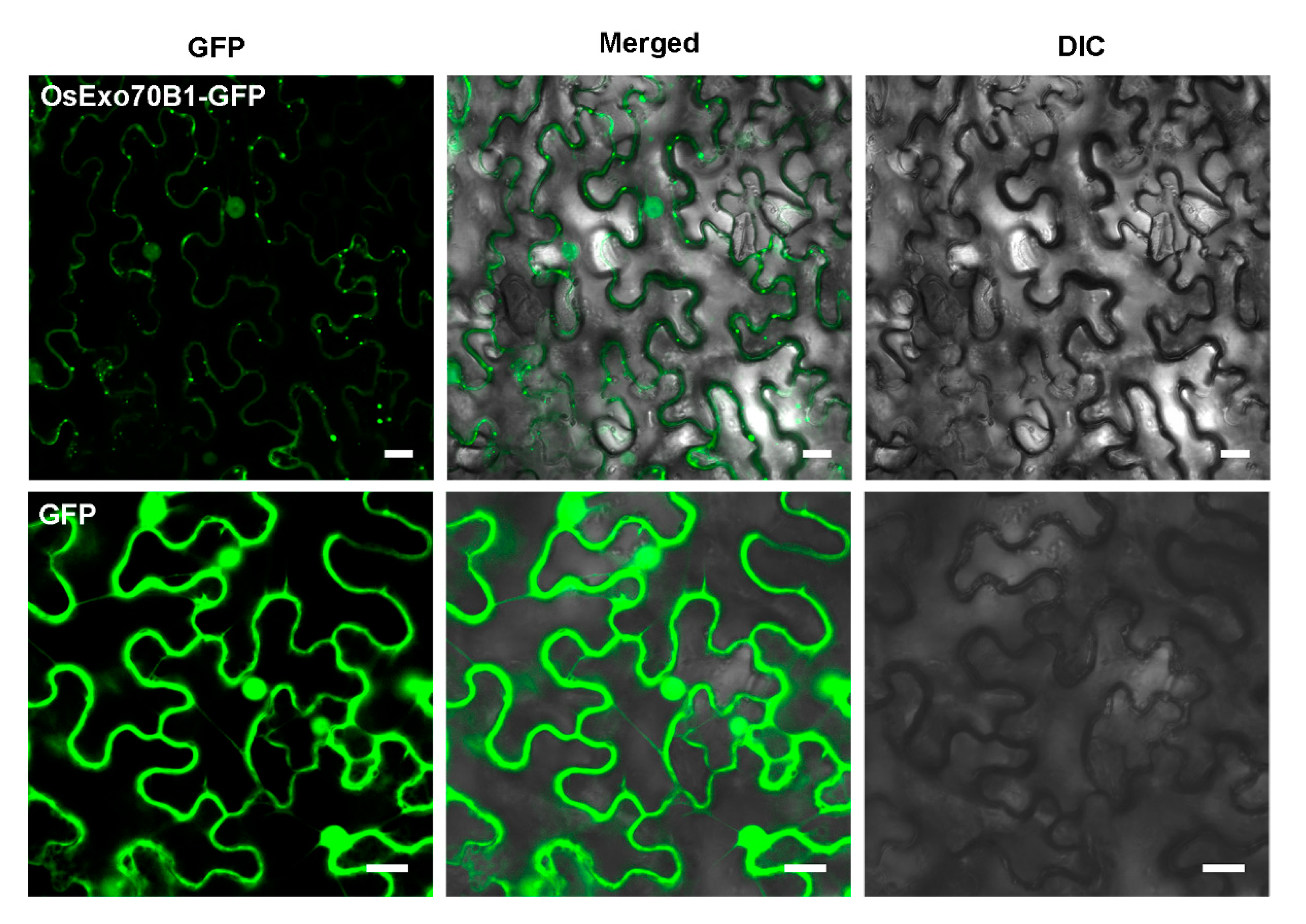
Then, in order to examine the subcellular localization of OsExo70B1, the OsExo70B1 fused with the green fluorescent protein (GFP) was expressed under the control of the 35S promoter in N. benthamiana leaves by agrobacterium-mediated genetic transformation. We observed clear fluorescence signals in the cytoplasm, nucleus, and plasma membrane (PM) (Figure 2), indicating that OsExo70B1 distributed throughout the whole cell, which is similar to some Exo70 members in Arabidopsis and H. villosa, such as AtExo70B1, Exo70D1-V and Exo70F2-V [20,39]. Interestingly, OsExo70B1 formed some strong dotted fluorescence in the cytoplasm and plasma membrane, which is similar to Exo70A1, Exo70B1 and Exo70E2 in Arabidopsis [24,30,40].
Determination of the expression patterns of OsExo70B1 via qRT-PCR. RNA samples were extracted from different tissues of ZH11, including roots, shoots and leaves of three different developmental stages, panicles of different length, germinating and mature seeds and callus. Data represent the mean and standard deviation of three biological replicates. Three technical replicates for each biological sample were used. The asterisk indicates significant differences (* p < 0.05; Student’s t-test).
Confocal microscopy observation of OsExo70B1-GFP in N. benthamiana leaves. The results showed that OsExo70B1-GFP was expressed in the nucleus, cytoplasm and plasma membrane. Bar = 20 μm.

2.2. Transcription of OsExo70B1 Is Induced by PAMPs and M. oryza

It has been reported that AtExo70B1 regulates immunity in Arabidopsis [21,34,41]. Pathogen-derived elicitors, often called pathogen-associated molecular patterns (PAMPs), are recognized by pattern-recognition receptors (PRRs) of hosts, and activate plant immune responses [1,2,3,4]. To test whether Exo70B1 is involved in the immune response regulation in rice, the expression of OsExo70B1 upon PAMPs treatment was analyzed. When the wild-type ZH11 seedings grew to four weeks, leaf segments that were 2–3 cm in length were collected and treated with flg22 or chitin, two well-characterized PAMPs. Then, the expression levels of OsExo70B1 at different time periods were detected. The results showed that the expression level of OsExo70B1 was significantly increased after flg22 and chitin treatments (Figure 3A,B). Furthermore, we also detected the transcription levels of OsExo70B1 after M. oryzae inoculation and found that OsExo70B1 was also upregulated upon M. oryzae infection (Figure 3C). The results above indicated that the expression of OsExo70B1 is induced by PAMPs and M. oryzae, revealed its potential roles in rice immunity regulation.

2.3. Knocking out OsExo70B1 Compromises Plant Resistance to M. oryzae

To further investigate the genetic role of OsExo70B1 in rice immunity, we generated the exo70B1 mutant by CRISPR-Cas9 methods in the ZH11 background. As shown in Figure S1, we obtained two OsExo70B1 allelic mutants, designated exo70B1-1, which has a T base insertion at the 245 position that resulted in a code-shift mutation from Pro84, and exo70B1-2, which has a C base insertion at the 145 position that resulted in a code-shift mutation from His49, which are likely to be two loss-of-function mutants. Moreover, we also examined the transcripts of OsExo70B1 in both exo70B1-1 and exo70B1-2 mutants. The expression level of OsExo70B1 was not changed in the exo70B1 mutants compared to the wild type (Figure S2). We then inoculated exo70B1-1, exo70B1-2 and the wild-type plants with the M. oryzae isolate Guy11. Three days post-inoculation, the exo70B1-1 and exo70B1-2 mutants displayed enhanced susceptibility and showed more and extended lesions, while the wild-type had only a few lesions (Figure 4A). Consistent with this, the fungal biomass was accumulated at a higher level in the infected exo70B1-1 and exo70B1-2 leaves than in the wild type.
Moreover, we measured the transcription levels of pathogenesis-related genes using qRT-PCR. OsPR5 and OsPR10, which are known to be involved in the salicylic acid (SA) signaling pathway [42], were both significantly down-regulated in exo70B1-1 and exo70B1-2 compared to the wild type (Figure 4C,D).
In addition, we expressed the full-length coding sequence of OsExo70B1 under the control of the native promoter in the exo70B1-1 mutant. We then used the positive T1 complementary plants, which were identified by PCR with specific primers before use (Figure S3), for blast resistance analysis. After inoculating with the M. oryzae isolate Guy11, the complementary transgenic plants of exo70B1-1 were similar to those in the wild type in the lesion area and fungal biomass analysis (Figure 4E,F), as well as the expression level of OsPR5 and OsPR10 (Figure 4G,H).
Taken together, the results above indicate that knocking out Exo70B1 decreased plant resistance to M. oryzae in rice.

2.4. Accumulation of H2O2 Is Lower and the Infection Progress Is Faster in exo70B1-1 Mutant Compared to the Wild Type

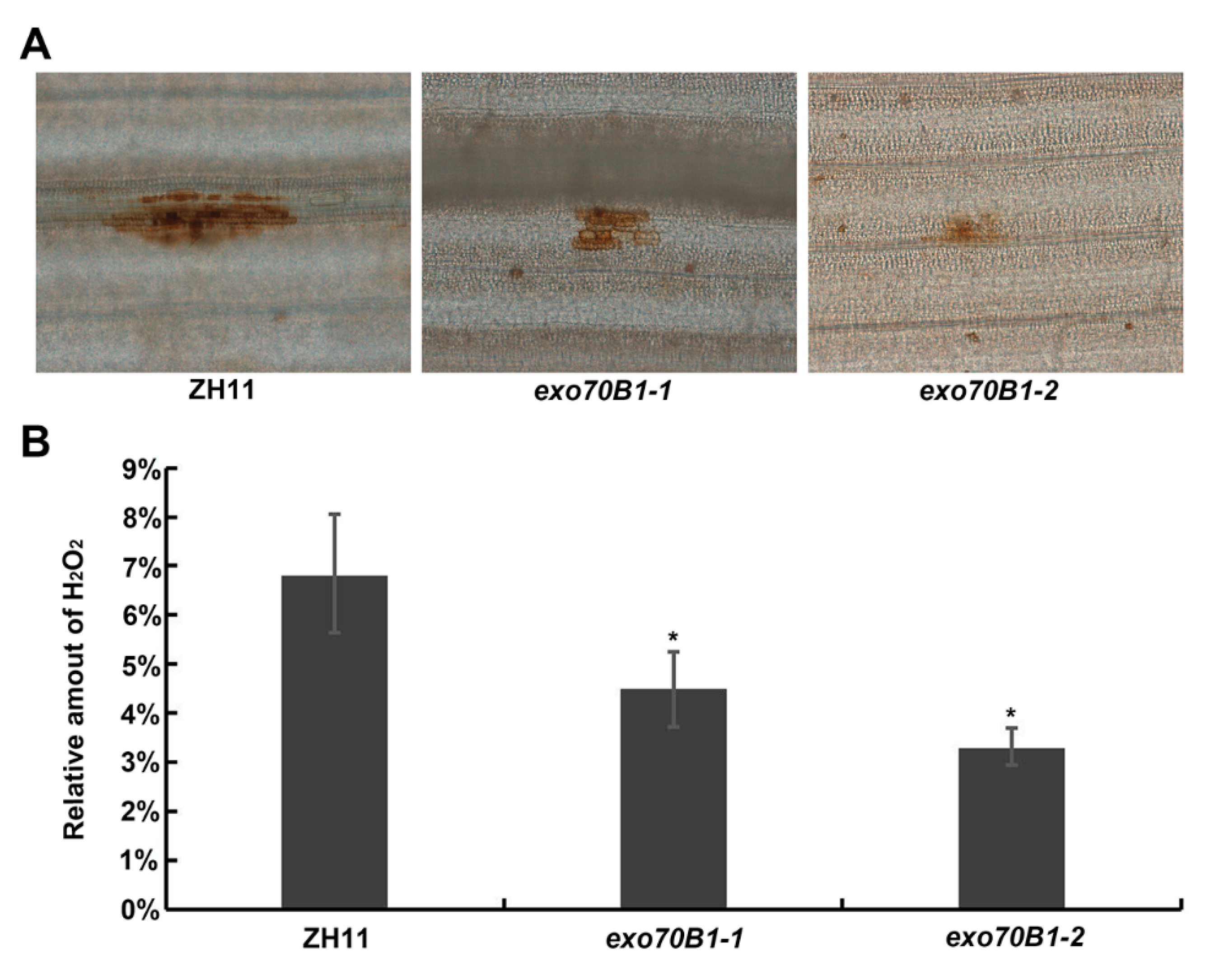
Accumulation of H2O2 is a common defense response to M. oryzae in rice [43,44,45]. To test whether H2O2 accumulation is different in exo70B1 mutants compared to the wild type, staining of the inoculated leaf cells with 3,3′-diamino-bezidine (DAB) for H2O2 revealed that the two allelic mutants exo70B1-1 and exo70B1-2 both produced much lower amounts of H2O2 in the inoculated leaf sheath cells than the wild type (Figure 5). This result suggests that decreased reactive oxygen species (ROS) burst may be part of the reason why exo70b1-1 and exo70b1-2 mutants display enhanced susceptibility to rice blast fungus.
To gain more insight into how exo70B1-1 mutants are more susceptible to blast fungi compared to the wild type, the formation and expansion of invasive hyphae in leaf sheath cells were observed and quantified. Consistent with the decreased disease phenotypes, the infection progress occurred significantly earlier in exo70B1-1 and exo70B1-2 compared with the wild type (Figure 6). After leaf sheath injection with the eGFP-tagged M. oryzae strain Zhong1, we found that in ZH11, 41.1% of the spores of M. oryzae formed appressoria and 58.9% of the spores did not form appressoria at 12 hpi, while 61.6% of the spores in exo70B1-1 and 56.3% in exo70B1-2 formed appressoria and 38.4% in exo70B1-1 and 43.7% in exo70B1-2 did not form appressoria. At 24 hpi, only 24.8% of spores formed invasive hyphae, while the remaining 75.2% of appressoria did not invade cells in ZH11; however, in exo70B1-1, 76.6% of spores formed infective hyphae, and 23.4% of them expanded into neighbor cells. Similarly, 65.0% spores formed infective hyphae in exo70B1-2 and 35.0% of them expanded into neighbor cells. Moreover, 78.3% of the infected hyphae in ZH11 had expanded to neighboring cells, while 96.1% of infective hyphae in exo70B1-1 and 97.9% in exo70B1-2 had expanded to more adjacent cells at 48 hpi. In conclusion, the infection progress is significantly faster in the exo70B1-1 mutant compared to the wild type.

2.5. Knocking out OsExo70B1 Does Not Alter Plant Architecture or Grain Yield

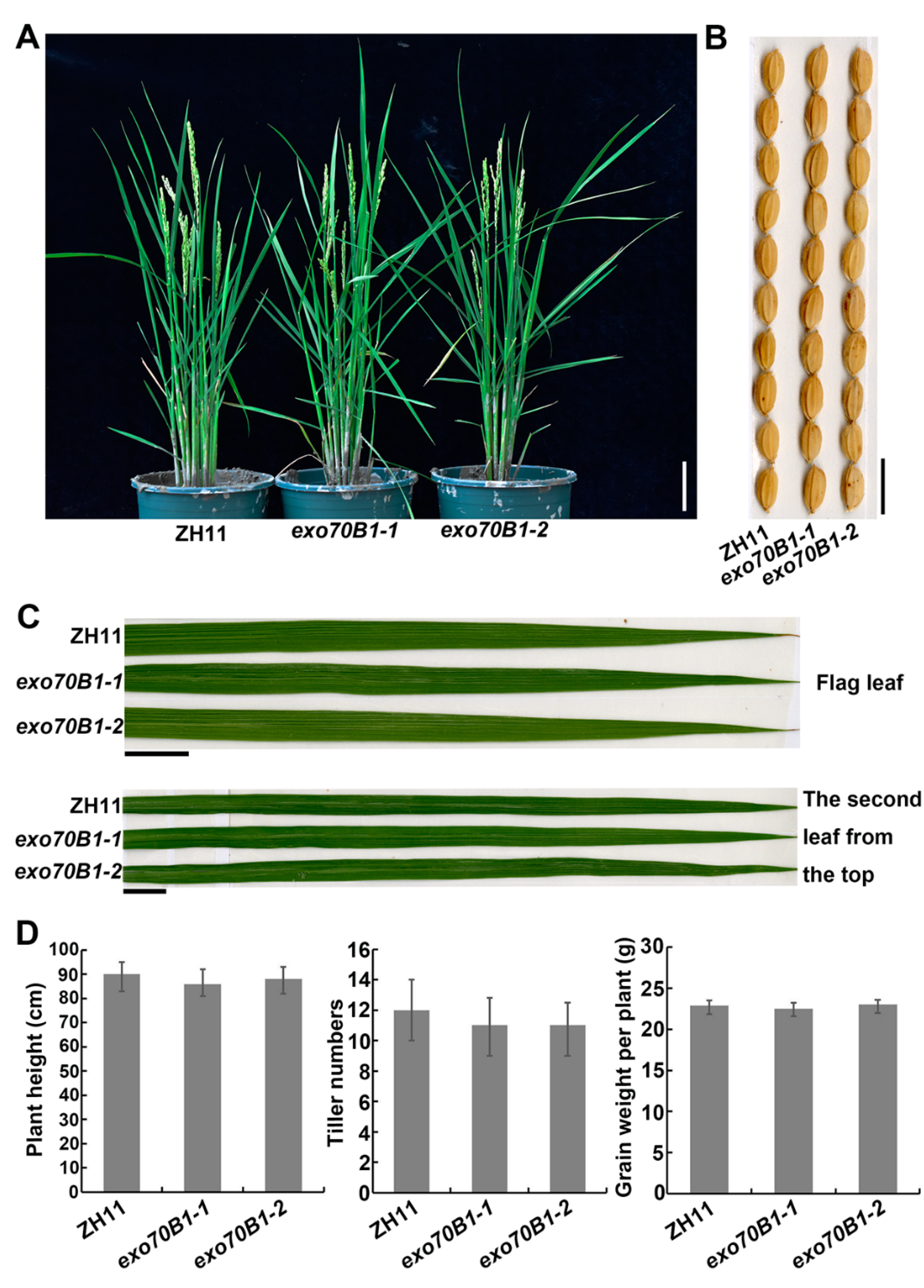
In Arabidopsis, it has been reported that exo70B1-1 grew normally up to approximately 4 weeks after germination and then became smaller than the wild type and displayed HR-like cell death lesions [23,30,41]. Therefore, we also investigated the phenotypes of the rice exo70B1 mutant in growth and development. As shown in Figure 7, exo70B1-1 and exo70B1-2 grew normally until the ripening phase (Figure 7A), and no cell death lesions appeared in exo70B1-1 and exo70B1-2 leaves (Figure 7C). Moreover, exo70B1-1 showed no significant differences in plant height, tiller number, seed size and grain weight per plant compared to the wild type (Figure 7), indicating that knocking out Exo70B1 does not affect growth and development in rice.

2.6. OsExo70B1 Associates with OsCERK1

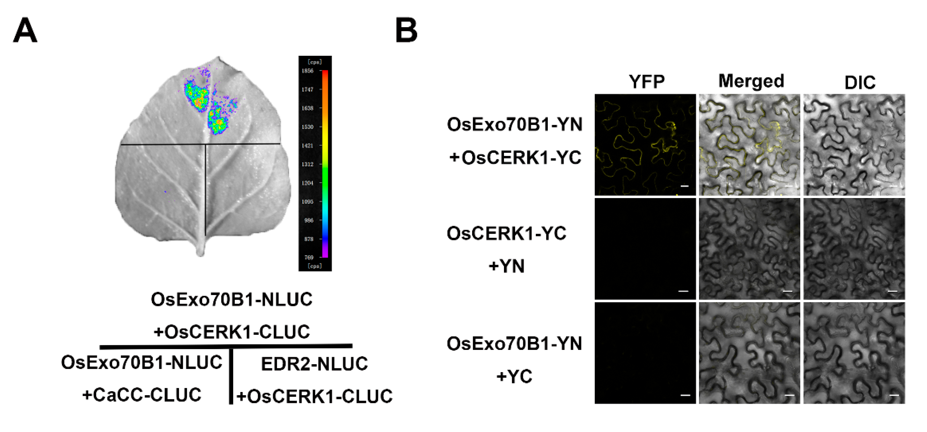
It has been shown that the exocyst complex plays important roles in vesicle trafficking, specifically tethering and spatially targeting post-Golgi vesicles to the plasma membrane prior to vesicle fusion. Recently, Wang et al. showed that Arabidopsis Exo70B1 regulates the trafficking of FLS2, the receptor of bacterial flagellin, to the PM, thus mediating the immunity responses in Arabidopsis [46]. We speculate that OsExo70B1 may also be responsible for the membrane location of some defense-related proteins in rice. As OsCERK1 is a well-characterized receptor-like kinase (RLK) and mediates the signal of chitin reception by coordinating with a lysin motif (LysM)-containing protein CEBiP [37], we first performed a split-luciferase complementation imaging (LCI) assay in N. benthamiana. As shown in Figure 8A, luminescent signals were detected when OsCERK1 was co-transformed with OsExo70B1, but not with the negative controls. Then, a bimolecular fluorescence complementation (BiFC) assay also showed that OsExo70B1 can interact with OsCERK1 in the plasma membrane predominantly (Figure 8B). Taken together, the results above showed that OsExo70B1 associated with OsCERK1 in BiFC and LCI assays.

3. Discussion

EXO70B1 is a component of the exocyst complex and belongs to the EXO70 protein subfamily. In this study, we characterized the expression pattern and function of OsExo70B1 in rice, revealing its important roles in plant immune response in rice. OsExo70B1 mainly expressed in leaves and its expression level can be induced by PAMPs and rice blast fungus (Figure 1 and Figure 3). Knocking out OsExo70B1 resulted in decreased plant resistance to M. oryzae, as well as decreased H2O2 accumulation, downregulation of PR genes and more fungi growth (Figure 4 and Figure 5). However, we did not observe any defects associated with growth and development in exo70B1 mutant (Figure 7), indicating that OsExo70B1 mainly plays important roles in rice immunity regulation.
Several studies have shown the important roles of Exo70B1 in immune regulation in Arabidopsis [21,23,30,31,41]. However, there are some differences of Exo70B1 in rice and Arabidopsis. First, under standard short-day conditions, Arabidopsis exo70B1 mutants grew normally up to 4 weeks of age. At this time point, exo70B1 plants started to become smaller compared to the wild type, and displayed hypersensitive response-like cell death after 5 weeks [23,30,41]. However, the rice exo70B1 mutant grew normally throughout its life cycle and no cell death appeared in its leaves (Figure 7). Second, Arabidopsis exo70B1 loss-of-function mutants emerged activated defense responses upon infection and enhanced resistance to fungal, oomycete and bacterial pathogens [23]. By contrast, knocking out OsExo70B1 compromises plant resistance in rice, and the rice exo70B1 mutants displayed enhanced susceptibility to M. oryzae. Further study showed that enhanced disease resistance and cell death in Arabidopsis exo70B1 mutant are dependent on TN2, a truncated TIR-NBS-LRR intracellular immune receptor. TN2 interacted with Exo70B1 directly, and TN2 transcripts accumulate at much higher levels in the exo70B1 mutant [23]. TN2 belongs to the TIR-NBS (TN) family, which has 21 members in Arabidopsis ecotype Col-0 [47], but TIR-NBS-LRR and TIR-NBS proteins are absent from cereal species, including rice [48], which may explain why no cell death occurred in rice exo70B1 mutant. Thirdly, Arabidopsis Exo70B1 is reported to be involved in autophagy-related membrane traffic to the vacuole, and the exo70B1 mutant displays ectopic hypersensitive reaction mediated by salicylic acid (SA) accumulation, as well as defects in autophagy-related phenotypes [30], but no cell death appeared in rice exo70B1 mutants, and although we did not examine the autophagy-related phenotypes of rice exo70B1 mutants, those mutants did not show growth defects, suggesting that there is no defect in autophagy-related pathways in rice exo70b1 mutants. Therefore, OsExo70B1 may not participate in autophagy-related membrane traffic in rice or some other proteins, such as OsExo70B2 and OsExo70B3, function redundantly. In addition, Exo70B1 also participates in ABA mediated stomatal closure and light-induced stomatal opening in Arabidopsis [24,49]. It would be interesting to know whether OsExo70B1 is also involved in stomatal opening and closure regulation in rice.
How Exo70B1 functions in plant immunity has been extensively studied in Arabidopsis. It is reported that RIN4 can recruit Exo70B1 to the plasma membrane and AvrRpt2 can release both RIN4 and Exo70B1 to the cytoplasm, indicating that the localization of Exo70B1 at PM may be required for plant immunity [39]. The plant U-box-type ubiquitin ligase 18 (PUB18) ubiquitinates and degrades Exo70B1 in ABA-mediated stomatal movement regulation [24]. Exo70B1 is also ubiquitinated by bacterial effector AvrPtoB after Pto DC3000 infects Arabidopsis [34]. Furthermore, the activated immune responses in the Arabidopsis exo70B1 mutant require TN2 and CPK5 [23,31]. Most recently, Wang et al. (2020) found that both Exo70B1 and Exo70B2 are involved in the regulation of FLS2 accumulation at the PM. Moreover, Exo70B1 is also responsible for the membrane accumulation of BRI1 and CERK1, but not of RLK902-GFP, indicating that the exocyst complex subunits can regulate the trafficking of some defense-related proteins to the PM, thus mediating the immunity responses [46]. In this study, we detected the interaction between OsExo70B1 and OsCERK1 in LCI and BiFC assays (Figure 8), suggested that OsExo70B1 may also be involved in rice immunity by affecting the accumulation of some important PRRs, including OsCERK1, to the plasma membrane. However, the mechanisms of how OsExo70B1 regulates immunity are still largely unknown. It would be interested to examine whether OsExo70B1 contributes to the accumulation of OsCERK1 and other receptors of PAMPs at the PM.
Interestingly, there are only two Exo70B family genes in Arabidopsis, Exo70B1 and Exo70B2, while there are three Exo70B family genes in rice [17]. In Arabidopsis, Exo70B2 also has been reported to function in immunity regulation [21,29]. Exo70B2 also contributes to the accumulation of FLS2 at the PM [46]. However, the FLS2 accumulation level at the PM in the exo70B1-3exo70B2-1 double mutant was not further reduced relative to that in exo70B1-3 and exo70B2-1 single mutants and Exo70B1 can form a heterodimer with Exo70B2, indicating that Exo70B1 and Exo70B2 cooperatively function in trafficking FLS2 to the PM [46]. In rice, the role of three Exo70B family genes is not studied, and their functions are still unknown. Here, we showed that OsExo70B1 positively regulates rice immunity. Though whether OsExo70B2 and OsExo70B3 have a similar function, and whether the three Exo70B genes function redundantly, still require further studies. In addition, whether the HR-like cell death not observed in the rice exo70B1 mutant is due to the redundancy of rice Exo70Bs remains to be determined. It would be interesting to examine the triple mutant of OsExo70B1, OsExo70B2 and OsExo70B3, and the characterization of the phenotypes of the triple mutants will help us to understand the functions of Exo70B genes in rice.
Our data collectively indicate the positive regulation roles of OsExo70B1 in rice immunity, and they may function by mediating the trafficking and enrichment of OsCERK1 on the membrane. Then OsCERK1 mediated immune responses, including ROS burst and PTI-related defense genes expression, were activated, which ultimately lead to enhanced disease resistance in rice.

4. Materials and Methods

4.1. Plant Materials and Growth Conditions

Rice and Nicotiana benthamiana (N. benthamiana) plants were used in this study. The exo70B1-1 and exo70B1-2 mutants were constructed with the CRISPR/Cas9 system in Zhonghua 11 (ZH11) [50], and the sgRNA for exo70B1-1 is 5′-GATCTCGCAGTTCGTGACGA-3′ and for exo70B1-2 is 5′-GGGACAAACTCTACGCCACG-3′. All rice plants used in this study were grown in paddy fields of the Fujian Agriculture and Forestry University in Fuzhou, Fujian province, paddy fields of the Institute of Crop Science in Sanya (China), or a greenhouse at 25–28 °C with a 16 h:8 h, light:dark photoperiod. N. benthamiana plants were growth in a greenhouse at 22 °C with 12 h:12 h, light:dark photoperiod.

4.2. Subcell Localization Analysis

The full-length cDNA sequence of OsExo70B1 was amplified and inserted between EcoRI and SmaI sites of the pCMABIA2300-35S-eGFP vector with an in-fusion PCR cloning kit, thus producing the 35S-OsExo70B1-eGFP construct. Then, the constructs were transformed into agrobacterium strain GV3101 and injected into N. benthamiana leaves. A GFP signal was observed and photographed with a Zeiss 880 confocal microscope at 3 days after injection.

4.3. Construction of Complementary Transgenic Plant of exo70B1-1

To produce the OsExo70B1pro:OsExo70B1-GFP construct, 1511 bp of the OsExo70B1 promoter sequence, together with the full length of the OsExo70B1 cDNA sequence without the stop codon, was amplified and inserted between EcoRI and KpnI, and KpnI and SpeI sites of the pCAMBIA2300 vector with in-fusion PCR cloning kit, respectively. Then, the construct was introduced into Agrobacterium tumefaciens strain EHA105 and then transferred into the exo70B1-1 mutant as described previously [51].

4.4. Gene Expression Analysis

Total RNA was isolated using TRIzol (Invitrogen) according to the manufacturer’s instructions, and then was synthetized into cDNA with an RT reagent Kit (code number is RR047A; Takara, Dalian, China) for qRT-PCR and 1st Strand cDNA Synthesis Kit for RT-PCR (code number is 6110A; Takara, Dalian, China). All qRT–PCR assays were performed with the Premix Ex TaqKit (code number is RR420A; TaKaRa, Dalian, China) in a CFX Connect Real-time PCR System (BIO-RAD, Hercules, CA, USA). RT-PCR assays were performed with the Ex Taq DNA Polymerase (code number is RR001A; TaKaRa, Dalian, China) in a T100 PCR System (BIO-RAD, Hercules, CA, USA). All of the primers used here are listed in Supplementary Table S1. qRT-PCR analysis was performed as previously described [52] with three independent biological replicates.

4.5. Chitin and flg22 Treatments

Chitin and flg22 treatment assays were performed as described previously [53]. When the wild-type ZH11 seedings grew to four weeks, the leaf segments that were 2–3 cm in length were collected and balanced in ddH2O for 12 h, and then they were treated with 1 µM flg22 (synthesized by Sangon Biotech Co., Ltd., Shanghai, China) or 20 µg/mL chitin (Sigma, St. Louis, MO, USA). The samples were collected at 6 h and 12 h after treatment for further analysis.

4.6. Rice Blast Fungus Incubation and Rice Sheath Penetration Assay

Blast fungus inoculation and a rice sheath penetration assay were performed as described previously with minor modification [54,55]. Briefly, two-week-old seedlings were sprayed with a spore suspension (1 ×10⁵ spores/mL) of the M. oryzae isolate Guy11. The inoculated plants were placed into dark and humid containers for 24 h. The plants were then transferred to a humidity growth chamber and grown under a 12 h:12 h, light:dark photoperiod. The disease lesions were examined and scored at 3 or 4 days after inoculation. The fungal biomass assay was performed as described previously [42,45,53]. Total DNA was extracted from ten diseased leaves of both ZH11 and exo70B1 mutants, respectively, and then the fungal biomass was calculated by comparing the total DNA of M. oryzae Pot2, an inverted repeat transposon and represents one of the major repetitive DNAs in M. oryzae [56], to the Ubiquitin DNA of rice. Moreover, for rice sheath inoculation assays, spores of Zhong1 tagged with GFP were diluted to a concentration of 1 × 10 5 spores/mL and injected into the inner leaf sheath of 3-week-old ZH11 plants. Then the inoculated leaf sheaths were incubated in a dark and humid container at 28 ℃. At 12 h, 24 h and 48 h after infection, the fungal growth in rice leaf sheath cells was observed under a fluorescence microscope.

4.7. DAB Staining

DAB staining was performed as described before [54]. Briefly, the leaf sheaths were immersed in 1 mg/mL DAB (pH = 3.8) at the indicated time after inoculation with M. oryzae. After vacuum infiltration for 30 min, the samples were incubated at room temperature for 12 h in the dark. When the brown spots appeared clearly, samples were bleached by boiling in ethanol:lactic acid:glycerol (1:1:1, v/v/v) for 15 min. Images were captured using a microscope with a CCD camera (Olympus BX51, Tokyo, Japan).

4.8. Split-Luciferase Complementation Assay

For the split-luciferase complementation assay, the full-length CDSs of OsExo70B1 and OsCERK1 were amplified with specific primers (Supplementary Table S1) and then cloned into both the KpnI and SalI sites of NLuc and CLuc vectors, respectively [57], producing the 35Spro-OsExo70B1-NLUC and 35Spro-OsCERK1-CLUC constructs. Pairs of Agrobacterium tumefaciens GV3101 strains containing the desired plasmid combinations were co-transformed into 4-week-old N. benthamiana leaves and incubated in the green room for 72 h. The assay was then performed as previously described [58]. The N. benthamiana leaves were sprayed with 1 mM luciferin and placed in darkness for 5 min. Subsequently, LUC images were captured with a low-light cooled CCD imaging apparatus.

4.9. Bimolecular Fluorescence Complementation (BiFC) Assay

The BiFC assay was performed as previously described [57]. Briefly, the coding sequences of OsExo70B1 and OsCERK1 were cloned into pDONR207 ENTRY vector first, and then into the pSPYNE and pSPYCE vectors respectively, in-frame with the N- and C-terminus of the yellow fluorescent protein (YFP) with the gateway cloning system, thus producing 35Spro-OsExo70B1-YN and 35Spro-OsCERK1-YC constructs. Then, agrobacterium strain GV3101 containing the respective plasmids was injected into the leaves of 4-week-old N. benthamiana plants. YFP signal was detected with a Zeiss 880 confocal microscope on the third day after injection.

5. Conclusions

In this study, we characterized the function of OsExo70B1, the subunit of exocyst in rice, revealing its important roles in plant immune response. Knocking out OsExo70B1 resulted in enhanced susceptibility to M. oryzae, decreased H2O2 accumulation, and downregulation of PR genes. However, we did not observe any defects associated with growth and development in exo70B1 mutant, which is in contrast to the autoimmune phenotypes in Arabidopsis exo70B1 mutant. Moreover, we find OsExo70B1 interacts with OsCERK1, an essential component for chitin reception in rice. Our findings reveal the positive roles of OsExo70B1 in rice immunity regulation, as well as the functional differentiation of Exo70B1 in Arabidopsis and rice.

Supplementary Materials

Supplementary materials can be found at https://www.mdpi.com/1422-0067/21/19/7049/s1

Author Contributions

D.T. and S.L. conceived and designed the research. H.H., J.F., J.L. and Z.D. carried out most of the experiments. S.L., H.H., W.W., D.Y. and D.T. wrote the manuscript. H.H., S.L. and D.T. analyzed the data. All authors have read and agreed to the published version of the manuscript.

Funding

This work was supported by Natural Science Foundation of Fujian Province (2019J01424) and the National Natural Science Foundation of China (31761133017).

Acknowledgments

We thank Xuewei Chen and Wenming Wang, who provided M. oryzae isolates Guy11 and Zhong1 for us.

Conflicts of Interest

The authors declare no conflict of interest.

Gene Accession Numbers

Sequence data from this article can be found in Rice Genome Annotation Project databases under the following accession numbers:
OsExo70B1(LOC_Os01g61180)
OsCERK1(LOC_Os08g425880)
Ubiquitin(LOC_Os03g13170)
Actin(LOC_Os03g50885)
PR5(LOC_Os03g46070)
PR10(LOC_Os12g36880)
Pot2(EMBL accession Z33638)

References

  1. Jones, J.D.; Dangl, J.L. The plant immune system. Nature 2006, 444, 323–329. [Google Scholar] [CrossRef] [PubMed] [Green Version]
  2. Dodds, P.N.; Rathjen, J.P. Plant immunity: Towards an integrated view of plant-pathogen interactions. Nat. Rev. Genet. 2010, 11, 539–548. [Google Scholar] [CrossRef] [PubMed]
  3. Dangl, J.L.; Horvath, D.M.; Staskawicz, B.J. Pivoting the plant immune system from dissection to deployment. Science 2013, 341, 746–751. [Google Scholar] [CrossRef] [PubMed] [Green Version]
  4. Wang, W.; Feng, B.; Zhou, J.M.; Tang, D. Plant immune signaling: Advancing on two frontiers. J. Integr. Plant Biol. 2020, 62, 2–24. [Google Scholar] [CrossRef] [PubMed] [Green Version]
  5. Wang, W.M.; Liu, P.Q.; Xu, Y.J.; Xiao, S. Protein trafficking during plant innate immunity. J. Integr. Plant Biol. 2016, 58, 284–298. [Google Scholar] [CrossRef]
  6. Gu, Y.; Zavaliev, R.; Dong, X. Membrane Trafficking in Plant Immunity. Mol. Plant 2017, 10, 1026–1034. [Google Scholar] [CrossRef]
  7. Zarsky, V.; Cvrckova, F.; Potocky, M.; Hala, M. Exocytosis and cell polarity in plants—Exocyst and recycling domains. New Phytol. 2009, 183, 255–272. [Google Scholar] [CrossRef]
  8. Zarsky, V.; Kulich, I.; Fendrych, M.; Pecenkova, T. Exocyst complexes multiple functions in plant cells secretory pathways. Curr. Opin. Plant Biol. 2013, 16, 726–733. [Google Scholar] [CrossRef]
  9. Pecenkova, T.; Sabol, P.; Kulich, I.; Ortmannova, J.; Zarsky, V. Constitutive Negative Regulation of R Proteins in Arabidopsis also via Autophagy Related Pathway? Front. Plant Sci. 2016, 7, 260. [Google Scholar] [CrossRef] [Green Version]
  10. Pecenkova, T.; Markovic, V.; Sabol, P.; Kulich, I.; Zarsky, V. Exocyst and autophagy-related membrane trafficking in plants. J. Exp. Bot. 2017, 69, 47–57. [Google Scholar] [CrossRef] [Green Version]
  11. Saeed, B.; Brillada, C.; Trujillo, M. Dissecting the plant exocyst. Curr. Opin. Plant Biol. 2019, 52, 69–76. [Google Scholar] [CrossRef] [PubMed]
  12. Mei, K.; Guo, W. The exocyst complex. Curr. Biol. 2018, 28, R922–R925. [Google Scholar] [CrossRef] [PubMed] [Green Version]
  13. Ma, W.; Wang, Y.; Yao, X.; Xu, Z.; An, L.; Yin, M. The role of Exo70 in vascular smooth muscle cell migration. Cell Mol. Biol. Lett. 2016, 21, 20. [Google Scholar] [CrossRef] [Green Version]
  14. Cvrckova, F.; Zarsky, V. Old AIMs of the exocyst: Evidence for an ancestral association of exocyst subunits with autophagy-associated Atg8 proteins. Plant Signal. Behav. 2013, 8, e27099. [Google Scholar] [CrossRef] [Green Version]
  15. Elias, M.; Drdova, E.; Ziak, D.; Bavlnka, B.; Hala, M.; Cvrckova, F.; Soukupova, H.; Zarsky, V. The exocyst complex in plants. Cell Biol. Int. 2003, 27, 199–201. [Google Scholar] [CrossRef]
  16. Chong, Y.T.; Gidda, S.K.; Sanford, C.; Parkinson, J.; Mullen, R.T.; Goring, D.R. Characterization of the Arabidopsis thaliana exocyst complex gene families by phylogenetic, expression profiling, and subcellular localization studies. New Phytol. 2010, 185, 401–419. [Google Scholar] [CrossRef]
  17. Cvrckova, F.; Grunt, M.; Bezvoda, R.; Hala, M.; Kulich, I.; Rawat, A.; Zarsky, V. Evolution of the land plant exocyst complexes. Front. Plant Sci. 2012, 3, 159. [Google Scholar] [CrossRef] [Green Version]
  18. Tu, B.; Hu, L.; Chen, W.; Li, T.; Hu, B.; Zheng, L.; Lv, Z.; You, S.; Wang, Y.; Ma, B.; et al. Disruption of OsEXO70A1 Causes Irregular Vascular Bundles and Perturbs Mineral Nutrient Assimilation in Rice. Sci. Rep. 2015, 5, 18609. [Google Scholar] [CrossRef]
  19. Du, Y.; Overdijk, E.J.R.; Berg, J.A.; Govers, F.; Bouwmeester, K. Solanaceous exocyst subunits are involved in immunity to diverse plant pathogens. J. Exp. Bot. 2018, 69, 655–666. [Google Scholar] [CrossRef] [Green Version]
  20. Zhao, J.; Zhang, X.; Wan, W.; Zhang, H.; Liu, J.; Li, M.; Wang, H.; Xiao, J.; Wang, X. Identification and Characterization of the EXO70 Gene Family in Polyploid Wheat and Related Species. Int. J. Mol. Sci. 2018, 20, 60. [Google Scholar] [CrossRef] [PubMed] [Green Version]
  21. Stegmann, M.; Anderson, R.G.; Ichimura, K.; Pecenkova, T.; Reuter, P.; Zarsky, V.; McDowell, J.M.; Shirasu, K.; Trujillo, M. The ubiquitin ligase PUB22 targets a subunit of the exocyst complex required for PAMP-triggered responses in Arabidopsis. Plant Cell 2012, 24, 4703–4716. [Google Scholar] [CrossRef] [Green Version]
  22. Drdova, E.J.; Synek, L.; Pecenkova, T.; Hala, M.; Kulich, I.; Fowler, J.E.; Murphy, A.S.; Zarsky, V. The exocyst complex contributes to PIN auxin efflux carrier recycling and polar auxin transport in Arabidopsis. Plant J. 2013, 73, 709–719. [Google Scholar] [CrossRef]
  23. Zhao, T.; Rui, L.; Li, J.; Nishimura, M.T.; Vogel, J.P.; Liu, N.; Liu, S.; Zhao, Y.; Dangl, J.L.; Tang, D. A truncated NLR protein, TIR-NBS2, is required for activated defense responses in the exo70B1 mutant. PLoS Genet. 2015, 11, e1004945. [Google Scholar] [CrossRef]
  24. Seo, D.H.; Ahn, M.Y.; Park, K.Y.; Kim, E.Y.; Kim, W.T. The N-Terminal UND Motif of the Arabidopsis U-Box E3 Ligase PUB18 Is Critical for the Negative Regulation of ABA-Mediated Stomatal Movement and Determines Its Ubiquitination Specificity for Exocyst Subunit Exo70B1. Plant Cell 2016, 28, 2952–2973. [Google Scholar] [CrossRef] [Green Version]
  25. Synek, L.; Vukasinovic, N.; Kulich, I.; Hala, M.; Aldorfova, K.; Fendrych, M.; Zarsky, V. EXO70C2 Is a Key Regulatory Factor for Optimal Tip Growth of Pollen. Plant Physiol. 2017, 174, 223–240. [Google Scholar] [CrossRef] [Green Version]
  26. Vukasinovic, N.; Oda, Y.; Pejchar, P.; Synek, L.; Pecenkova, T.; Rawat, A.; Sekeres, J.; Potocky, M.; Zarsky, V. Microtubule-dependent targeting of the exocyst complex is necessary for xylem development in Arabidopsis. New Phytol. 2017, 213, 1052–1067. [Google Scholar] [CrossRef]
  27. Kulich, I.; Vojtikova, Z.; Sabol, P.; Ortmannova, J.; Nedela, V.; Tihlarikova, E.; Zarsky, V. Exocyst Subunit EXO70H4 Has a Specific Role in Callose Synthase Secretion and Silica Accumulation. Plant Physiol. 2018, 176, 2040–2051. [Google Scholar] [CrossRef] [Green Version]
  28. Kulich, I.; Vojtikova, Z.; Glanc, M.; Ortmannova, J.; Rasmann, S.; Zarsky, V. Cell wall maturation of Arabidopsis trichomes is dependent on exocyst subunit EXO70H4 and involves callose deposition. Plant Physiol. 2015, 168, 120–131. [Google Scholar] [CrossRef] [Green Version]
  29. Pecenkova, T.; Hala, M.; Kulich, I.; Kocourkova, D.; Drdova, E.; Fendrych, M.; Toupalova, H.; Zarsky, V. The role for the exocyst complex subunits Exo70B2 and Exo70H1 in the plant-pathogen interaction. J. Exp. Bot. 2011, 62, 2107–2116. [Google Scholar] [CrossRef] [Green Version]
  30. Kulich, I.; Pecenkova, T.; Sekeres, J.; Smetana, O.; Fendrych, M.; Foissner, I.; Hoftberger, M.; Zarsky, V. Arabidopsis exocyst subcomplex containing subunit EXO70B1 is involved in autophagy-related transport to the vacuole. Traffic 2013, 14, 1155–1165. [Google Scholar] [CrossRef] [Green Version]
  31. Liu, N.; Hake, K.; Wang, W.; Zhao, T.; Romeis, T.; Tang, D. CALCIUM-DEPENDENT PROTEIN KINASE5 Associates with the Truncated NLR Protein TIR-NBS2 to Contribute to exo70B1-Mediated Immunity. Plant Cell 2017, 29, 746–759. [Google Scholar] [CrossRef] [PubMed] [Green Version]
  32. Ma, J.; Chen, J.; Wang, M.; Ren, Y.; Wang, S.; Lei, C.; Cheng, Z.; Sodmergen. Disruption of OsSEC3A increases the content of salicylic acid and induces plant defense responses in rice. J. Exp. Bot. 2018, 69, 1051–1064. [Google Scholar] [CrossRef] [PubMed] [Green Version]
  33. Du, Y.; Mpina, M.H.; Birch, P.R.; Bouwmeester, K.; Govers, F. Phytophthora infestans RXLR Effector AVR1 Interacts with Exocyst Component Sec5 to Manipulate Plant Immunity. Plant Physiol. 2015, 169, 1975–1990. [Google Scholar] [CrossRef] [PubMed] [Green Version]
  34. Wang, W.; Liu, N.; Gao, C.; Rui, L.; Tang, D. The Pseudomonas Syringae Effector AvrPtoB Associates With and Ubiquitinates Arabidopsis Exocyst Subunit EXO70B1. Front. Plant Sci. 2019, 10, 1027. [Google Scholar] [CrossRef] [Green Version]
  35. Fujisaki, K.; Abe, Y.; Ito, A.; Saitoh, H.; Yoshida, K.; Kanzaki, H.; Kanzaki, E.; Utsushi, H.; Yamashita, T.; Kamoun, S.; et al. Rice Exo70 interacts with a fungal effector, AVR-Pii, and is required for AVR-Pii-triggered immunity. Plant J. 2015, 83, 875–887. [Google Scholar] [CrossRef] [Green Version]
  36. Kaku, H.; Nishizawa, Y.; Ishii-Minami, N.; Akimoto-Tomiyama, C.; Dohmae, N.; Takio, K.; Minami, E.; Shibuya, N. Plant cells recognize chitin fragments for defense signaling through a plasma membrane receptor. Proc. Natl. Acad. Sci. USA 2006, 103, 11086–11091. [Google Scholar] [CrossRef] [Green Version]
  37. Shimizu, T.; Nakano, T.; Takamizawa, D.; Desaki, Y.; Ishii-Minami, N.; Nishizawa, Y.; Minami, E.; Okada, K.; Yamane, H.; Kaku, H.; et al. Two LysM receptor molecules, CEBiP and OsCERK1, cooperatively regulate chitin elicitor signaling in rice. Plant J. 2010, 64, 204–214. [Google Scholar] [CrossRef] [Green Version]
  38. Carotenuto, G.; Chabaud, M.; Miyata, K.; Capozzi, M.; Takeda, N.; Kaku, H.; Shibuya, N.; Nakagawa, T.; Barker, D.G.; Genre, A. The rice LysM receptor-like kinase OsCERK1 is required for the perception of short-chain chitin oligomers in arbuscular mycorrhizal signaling. New Phytol. 2017, 214, 1440–1446. [Google Scholar] [CrossRef] [Green Version]
  39. Sabol, P.; Kulich, I.; Zarsky, V. RIN4 recruits the exocyst subunit EXO70B1 to the plasma membrane. J. Exp. Bot. 2017, 68, 3253–3265. [Google Scholar] [CrossRef]
  40. Wang, J.; Ding, Y.; Wang, J.; Hillmer, S.; Miao, Y.; Lo, S.W.; Wang, X.; Robinson, D.G.; Jiang, L. EXPO, an exocyst-positive organelle distinct from multivesicular endosomes and autophagosomes, mediates cytosol to cell wall exocytosis in Arabidopsis and tobacco cells. Plant Cell 2010, 22, 4009–4030. [Google Scholar] [CrossRef] [Green Version]
  41. Stegmann, M.; Anderson, R.G.; Westphal, L.; Rosahl, S.; McDowell, J.M.; Trujillo, M. The exocyst subunit Exo70B1 is involved in the immune response of Arabidopsis thaliana to different pathogens and cell death. Plant Signal Behav. 2013, 8, e27421. [Google Scholar] [CrossRef] [PubMed] [Green Version]
  42. Park, C.H.; Chen, S.; Shirsekar, G.; Zhou, B.; Khang, C.H.; Songkumarn, P.; Afzal, A.J.; Ning, Y.; Wang, R.; Bellizzi, M.; et al. The Magnaporthe oryzae effector AvrPiz-t targets the RING E3 ubiquitin ligase APIP6 to suppress pathogen-associated molecular pattern-triggered immunity in rice. Plant Cell 2012, 24, 4748–4762. [Google Scholar] [CrossRef] [PubMed] [Green Version]
  43. Shimono, M.; Koga, H.; Akagi, A.; Hayashi, N.; Goto, S.; Sawada, M.; Kurihara, T.; Matsushita, A.; Sugano, S.; Jiang, C.J.; et al. Rice WRKY45 plays important roles in fungal and bacterial disease resistance. Mol. Plant Pathol. 2012, 13, 83–94. [Google Scholar] [CrossRef] [PubMed]
  44. Wang, Y.; Wu, J.; Kim, S.G.; Tsuda, K.; Gupta, R.; Park, S.Y.; Kim, S.T.; Kang, K.Y. Magnaporthe oryzae-Secreted Protein MSP1 Induces Cell Death and Elicits Defense Responses in Rice. Mol. Plant Microbe. Interact. 2016, 29, 299–312. [Google Scholar] [CrossRef] [Green Version]
  45. Li, Y.; Cao, X.L.; Zhu, Y.; Yang, X.M.; Zhang, K.N.; Xiao, Z.Y.; Wang, H.; Zhao, J.H.; Zhang, L.L.; Li, G.B.; et al. Osa-miR398b boosts H2O2 production and rice blast disease-resistance via multiple superoxide dismutases. New Phytol. 2019, 222, 1507–1522. [Google Scholar] [CrossRef] [Green Version]
  46. Wang, W.; Liu, N.; Gao, C.; Cai, H.; Romeis, T.; Tang, D. The Arabidopsis exocyst subunits EXO70B1 and EXO70B2 regulate FLS2 homeostasis at the plasma membrane. New Phytol. 2020, 227, 529–544. [Google Scholar] [CrossRef]
  47. Nandety, R.S.; Caplan, J.L.; Cavanaugh, K.; Perroud, B.; Wroblewski, T.; Michelmore, R.W.; Meyers, B.C. The role of TIR-NBS and TIR-X proteins in plant basal defense responses. Plant Physiol. 2013, 162, 1459–1472. [Google Scholar] [CrossRef] [Green Version]
  48. McHale, L.; Tan, X.; Koehl, P.; Michelmore, R.W. Plant NBS-LRR proteins: Adaptable guards. Genome Biol. 2006, 7, 212. [Google Scholar] [CrossRef] [Green Version]
  49. Hong, D.; Jeon, B.W.; Kim, S.Y.; Hwang, J.U.; Lee, Y. The ROP2-RIC7 pathway negatively regulates light-induced stomatal opening by inhibiting exocyst subunit Exo70B1 in Arabidopsis. New Phytol. 2016, 209, 624–635. [Google Scholar] [CrossRef] [Green Version]
  50. Lu, Y.; Ye, X.; Guo, R.; Huang, J.; Wang, W.; Tang, J.; Tan, L.; Zhu, J.K.; Chu, C.; Qian, Y. Genome-wide Targeted Mutagenesis in Rice Using the CRISPR/Cas9 System. Mol. Plant 2017, 10, 1242–1245. [Google Scholar] [CrossRef] [Green Version]
  51. Hiei, Y.; Ohta, S.; Komari, T.; Kumashiro, T. Efficient transformation of rice (Oryza sativa L.) mediated by Agrobacterium and sequence analysis of the boundaries of the T-DNA. Plant J. 1994, 6, 271–282. [Google Scholar] [CrossRef] [Green Version]
  52. Bustin, S.A.; Benes, V.; Garson, J.A.; Hellemans, J.; Huggett, J.; Kubista, M.; Mueller, R.; Nolan, T.; Pfaffl, M.W.; Shipley, G.L.; et al. The MIQE guidelines: Minimum information for publication of quantitative real-time PCR experiments. Clin. Chem. 2009, 55, 611–622. [Google Scholar] [CrossRef] [Green Version]
  53. Wang, J.; Shi, H.; Zhou, L.; Peng, C.; Liu, D.; Zhou, X.; Wu, W.; Yin, J.; Qin, H.; Ma, W.; et al. OsBSK1-2, an Orthologous of AtBSK1, Is Involved in Rice Immunity. Front. Plant Sci. 2017, 8, 908. [Google Scholar] [CrossRef] [Green Version]
  54. Yang, C.; Li, W.; Cao, J.; Meng, F.; Yu, Y.; Huang, J.; Jiang, L.; Liu, M.; Zhang, Z.; Chen, X.; et al. Activation of ethylene signaling pathways enhances disease resistance by regulating ROS and phytoalexin production in rice. Plant J. 2017, 89, 338–353. [Google Scholar] [CrossRef] [Green Version]
  55. Li, Y.B.; Xu, R.; Liu, C.; Shen, N.; Han, L.B.; Tang, D. Magnaporthe oryzae fimbrin organizes actin networks in the hyphal tip during polar growth and pathogenesis. PLoS Pathog. 2020, 16, e1008437. [Google Scholar] [CrossRef] [Green Version]
  56. Kachroo, P.; Leong, S.A.; Chattoo, B.B. Pot2, an inverted repeat transposon from the rice blast fungus Magnaporthe grisea. Mol. Gen. Genet. 1994, 245, 339–348. [Google Scholar] [CrossRef]
  57. Chen, H.; Zou, Y.; Shang, Y.; Lin, H.; Wang, Y.; Cai, R.; Tang, X.; Zhou, J.M. Firefly luciferase complementation imaging assay for protein-protein interactions in plants. Plant Physiol. 2008, 146, 368–376. [Google Scholar] [CrossRef] [Green Version]
  58. Zhao, Y.; Wu, G.; Shi, H.; Tang, D. Receptor-like kinase 902 associates with and phosphorylates brassinosteroid-signaling kinase1 to regulate plant immunity. Mol. Plant 2019, 12, 59–70. [Google Scholar] [CrossRef] [Green Version]
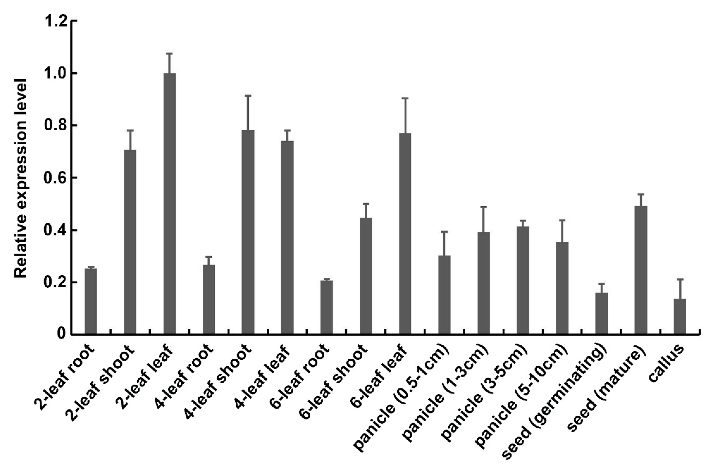
Figure 1. Expression patterns of OsExo70B1.
Ijms 21 07049 g001
Figure 2. Subcellular localizations of OsExo70B1.
Ijms 21 07049 g002
Figure 3. Transcripts of OsExo70B1 are induced by pathogen-associated molecular patterns (PAMPs) and M. oryzae. (A) Expression analysis of OsExo70B1 after chitin treatment, with H2O set as a control. (B) Expression analysis of OsExo70B1 after flg22 treatment, with H2O set as a control. (C) Expression analysis of OsExo70B1 after M. oryzae inoculation, with H2O set as a control. Data represent the mean and standard deviation of three biological replicates. Three technical replicates for each biological sample were used. The asterisk indicates significant differences (* p < 0.05; Student’s t-test).
Ijms 21 07049 g003
Figure 4. Knocking out Exo70B1 decreased plant resistance to M. oryzae. (A) exo70b1-1 and exo70B1-2 produced more diseased lesions compared to the wild type after inoculation with Guy11 using the spraying method. Bar = 1 cm. (B) Relative fungal amounts as determined by qPCR of the M. oryzae Pot2 gene compared to the Ubiquitin gene and compared to ZH11. Data represent the mean and standard deviation of three biological replicates. Three technical replicates for each biological sample were used. Asterisks represent significant differences (* p < 0.05, Student’s t-test). (C,D) Transcripts of OsPR5 and OsPR10 were both significantly downregulated in exo70b1-1 and exo70B1-2 compared to the wild type. Data represent the mean and standard deviation of three biological replicates. Three technical replicates for each biological sample were used. Asterisks represent significant differences relative to wild-type ZH11 plants (* p < 0.05, ** p < 0.01, Student’s t-test). (E) Rice disease lesions caused by M. oryzae locally inoculated (105 spores/mL) on leaves of ZH11, exo70B1-1 and exo70B1-1 complementary plants (CP). Bar = 1 cm. (F) Relative fungal amounts of ZH11, exo70B1-1 and exo70B1-1 complementary plants (CP). Data represent the mean and standard deviation of three biological replicates. Three technical replicates for each biological sample were used. Asterisks represent significant differences (* p < 0.05, Student’s t-test). (G,H) Transcripts of OsPR5 and OsPR10 were examined in the wild type, exo70B1-1 and the exo70B1-1 complementary plants (CP). Data represent the mean and standard deviation of three biological replicates. Three technical replicates for each biological sample were used. Asterisks represent significant differences relative to wild-type ZH11 plants (* p < 0.05, Student’s t-test).
Ijms 21 07049 g004
Figure 5. Accumulation of H2O2 is lower in exo70B1-1 and exo70B1-2 mutants compared to the wild type. (A) DAB (3,3′-diamino-bezidine) staining at infection sites of exo70b1-1, exo70B1-2 and ZH11 plants 2 days post-inoculation (dpi). Darker staining indicates accumulation of H2O2. (B) Relative amount of H2O2 was calculated based on pixels taken with ImageJ using the following formula: H2O2 area per rectangle = pixel of H2O2 area per leaf/pixel of rectangle. Data are represented as the mean ± standard error of mean (SEM). Asterisks represent significant differences (* p < 0.05, Student’s t-test). Bar = 10 μm.
Ijms 21 07049 g005
Figure 6. The infection progress of the enhanced green fluorescent protein (eGFP)-tagged M. oryzae isolate Zhong1 in exo70B1-1, exo70B1-2 and ZH11 leaf sheath cells at different time periods after inoculation. (A) Representative laser scanning microscopy images of ZH11, exo70B1-1 and exo70B1-2 leaf sheath cells infected by the eGFP-tagged M. oryzae isolate Zhong1. Bar = 10 μm. (B) Distribution of fungal infection progression at 12, 24, and 48 hpi. At least 30 single-cell interaction sites were examined per replication. Each bar represents the mean of three replications. Asterisks represent significant differences (* p < 0.05, Student’s t-test).
Ijms 21 07049 g006
Figure 7. Growth and development phenotypes of the exo70B1-1 and exo70B1-2 mutants. (A) The plant architecture of ZH11, exo70B1-1 and exo70B1-2 at the heading stage. Bar = 15 cm. (B) Photograph of the seeds of ZH11, exo70B1-1 and exo70B1-2. Bar = 1 cm. (C) Photograph of the flag leaf and top second leaf of the 4-week-old ZH11, exo70B1-1 and exo70B1-2. Bar = 2 cm. (D) The plant height, tiller number and grain weight per plant of ZH11, exo70B1-1 and exo70B1-2. Each bar represents the mean of twenty plants.
Ijms 21 07049 g007
Figure 8. OsExo70B1 associates with OsCERK1. (A) The interaction between OsExo70B1 and OsCERK1 was examined with the split-luciferase complementation assay in N. benthamiana. Co-infiltrated of OsExo70B1 fused with N-terminus of the Renilla luciferase fragment and OsCERK1 fused with the C-terminus of the Renilla luciferase fragment in N. benthamiana leaves by agrobacterium tumefaciens strain GV3101 mediated transformation. At the second day after injection, the infiltrated leaves were sprayed with 1 mM luciferin and the fluorescence signal was captured by a cooled charge coupled device (CCD) camera. The combinations of OsExo70B1-CaCC and OsCERK1-EDR2 were used as the controls. (B) The interaction between OsExo70B1 and OsCERK1 was examined with the BiFC assay in N. benthamiana. OsExo70B1 and OsCERK1 were fused to the N-terminus of the yellow fluorescent protein (YFP) fragment (YN) or the C-terminus of the YFP fragment (YC), respectively, and agrobacterium tumefaciens strain GV3101 containing the indicated construct pairs were co-infiltrated into N. benthamiana leaves. YFP fluorescence was detected by confocal microscopy. Bar = 20 μm.
Ijms 21 07049 g008